Article

Antibacterial Capability of Air Filter Fiber Materials Treated with Triclosan against Indoor Environmental Microbes

School of Energy and Safety Engineering, Tianjin Chengjian University, Tianjin 300384, China
*
Author to whom correspondence should be addressed.
Atmosphere 2022, 13(7), 1104; https://doi.org/10.3390/atmos13071104
Submission received: 18 June 2022 / Revised: 10 July 2022 / Accepted: 12 July 2022 / Published: 13 July 2022

Abstract

:
Antibacterial filtration materials have been used effectively to control biological pollutants and purify indoor air. This study aimed to assess the antibacterial capability of three fiber filter materials treated with triclosan: glass fiber (GF), non-woven fabric (NF) and chemical fiber (CF). Triclosan was loaded onto the filtration materials by the impregnation method. The triclosan-treated filter materials exhibited antibacterial zones obviously: the average antibacterial bands against E. coli were 11.8 mm (GF), 13.3 mm (NF) and 10.5 mm (CF); against S. albus, they were 25.5 mm (GF), 21.0 mm (NF) and 23.5 mm (CF). The percent reductions of bacteria for the antibacterial air fiber materials treated with triclosan against E. coli were 71.4% (CF) and 62.6% (GF), while the percent reductions against S. albus were 61.3% (NF) and 84.6% (CF). These findings could help to reduce the transmission and threat of epidemic and purify the environment through the use of environmentally friendly antibacterial filter fibers.

1. Introduction

Airborne microbes, as the major biological pollutant in the air, have a possibility of affecting human health negatively and causing or enhancing infectious diseases, such as influenza, pneumonia, coronavirus (COVID-19) and so on [1]. Therefore, an efficient and environmentally friendly technique is required for the control of bioaerosols, in order to reduce the transmission and harm of infectious diseases and decontaminate indoor air. According to the field literature, much effort has been devoted to developing active control technologies for airborne bacteria, including ultraviolet irradiation [2], static electricity, microwave irradiation [3], plasma [4], ozone, gas fumigation, lysozymes, photocatalysis [5] and air bactericides. These active sterilization technologies have an obvious sterilization effect and control effect on indoor microorganisms. However, a number of problems remain, such as the instability of the sterilization effect (light blocking, electric field characteristics), secondary pollution (residual ozone, heat or formaldehyde), and clear toxicity (chemical bactericides). Air filter fiber containing antibacterial agents is efficient and commonly used in removing airborne biological pollutant.
In recent years, metal nanoparticles (silver [6,7] and copper [8,9]) and natural plant essential oils, such as thyme essential oil [10] and Picea abies essential oil [11], have been widely used in antibacterial filter materials due to their sterilization efficiency, but the attenuation of the germicidal efficacy caused by self-oxidation and difficult extraction have limited their use as antimicrobial agents in some conditions. Additionally, metal ions have a potential toxicity to human health [12]. Triclosan is well known to be a broad-spectrum and environmentally friendly antimicrobial agent and has been commonly added in soap [13], cosmetics [14] and the adhesive resin employed in medical application [15,16,17].
Several studies [18,19,20,21,22] on the application of triclosan in the field of stomatology found that triclosan has a good disinfection effect on bacteria. Lee et al. [23] reported that the antibacterial rate of triclosan was >97% against S. aureus. Moreover, the antibacterial properties have been affected by the triclosan concentration and the coating process of treated cotton [24]. Furthermore, when the triclosan is more concentrated, the antibacterial effect is greater. The antibacterial properties of triclosan were reflected by its inhibition of microbial growth and reduction of adherence to polymers. Triclosan also exhibits a strong antimicrobial result when used as an antimicrobial agent for textile materials [25,26,27,28]. Pelia et al. [29] had obtained a good antimicrobial result of cotton textile treated with triclosan. Field studies [30,31,32] resulted in a significant and persistent antimicrobial performance of triclosan against bacteria (S. aureus and E. coli). Air filter non-wovens with triclosan showed good and stable antimicrobial properties for 12 months when used in an air conditioner [26,33]. At present, because of the huge demand for antibacterial filtration materials to reduce the microbe pollutant in the air, there is still not sufficient detailed information on the use and efficacy of triclosan as an antibacterial agent when applied to ventilation filter materials.
Therefore, the aims of this study were (1) to prepare three kinds of triclosan-loaded antibacterial filter materials and (2) to assess the antibacterial capability of triclosan-treated fiber filter materials. Chemical fiber (CF, polypropylene), glass fiber (GF) and non-woven fabrics (NF) were selected as the experimental filter materials, because these materials are widely used in ventilation filters. Triclosan was loaded onto the filter materials by a soaking method. In this study, Escherichia coli (E. coli, Gram-negative, the indicator bacteria in water) and Staphylococcus albus (S. albus, Gram-positive, the representative bacteria of airborne microorganisms) were chosen as the biological agents for testing the antibacterial capability of triclosan-treated filter materials.

2. Materials and Methods

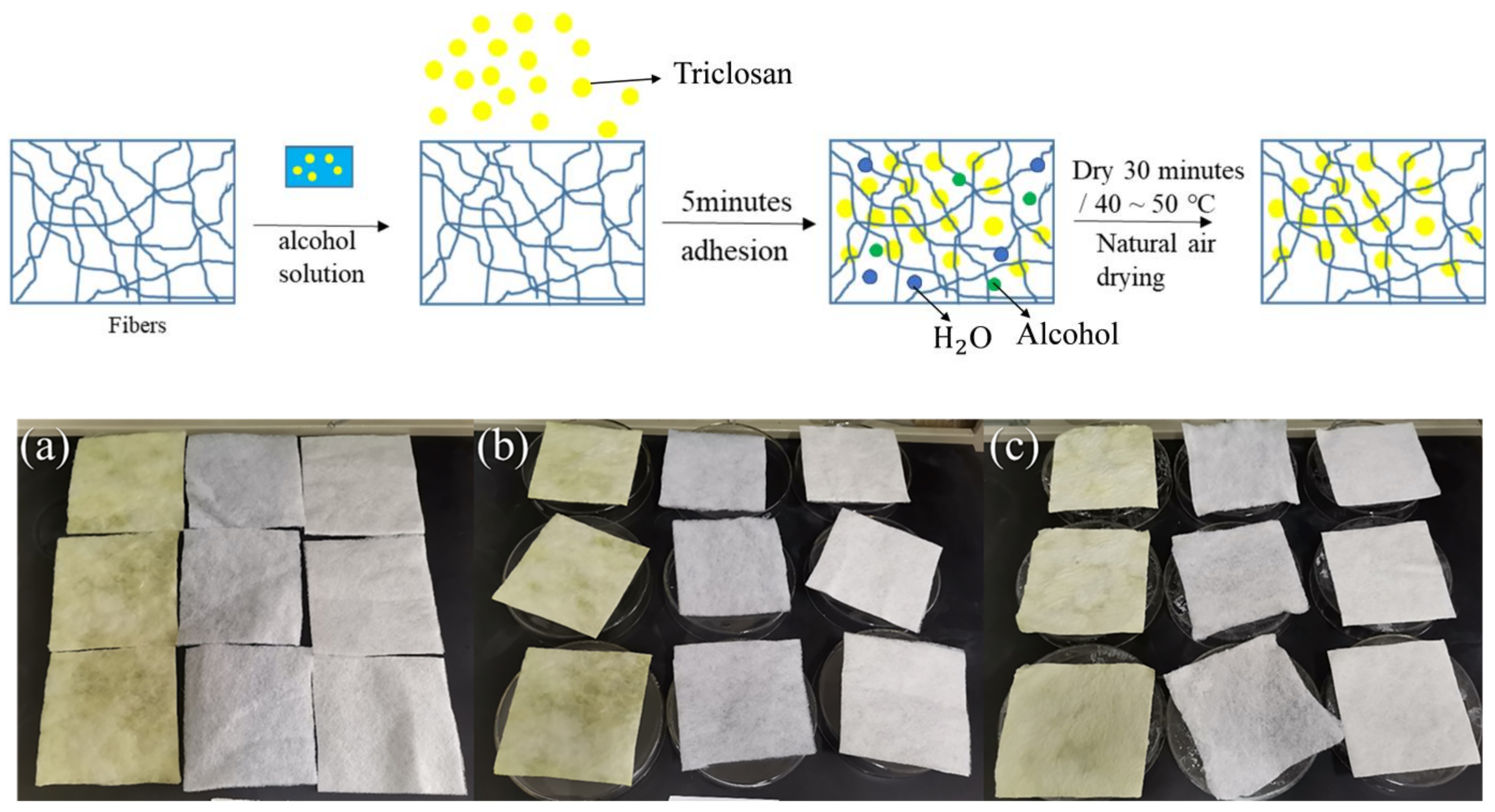
2.1. Preparation of Antibacterial Fibers
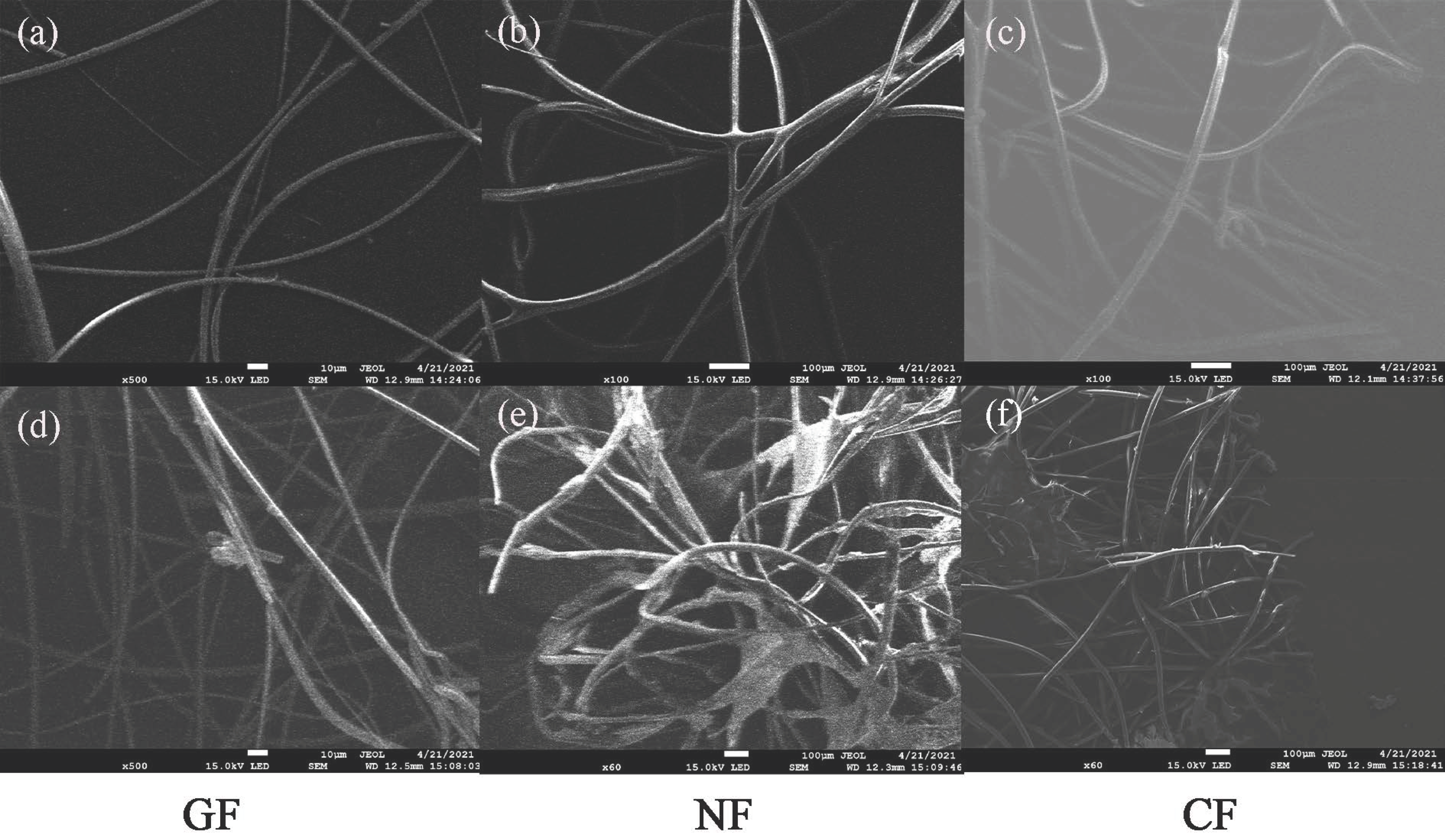
The three kinds of filter materials used in this paper (glass fiber (GF) was a fiber filter material made of glass, non-woven fabrics (NF) were composed of directional or random fibers, and chemical fiber (CF) was a fiber filter material made of polypropylene) were evaluated as square-shaped coupons with dimensions of 12 cm × 12 cm. The coupons were treated by immersion in an alcoholic-triclosan solution.
Figure 1 shows the preparation of the triclosan-treated fiber coupons (triclosan-treated (TT)) and the treated filter materials. The temperature ranged from 18–26 °C during the treating process. The relative humidity of air during the treating process was in the range of 40–60%. First, the triclosan was added to a beaker containing an alcoholic solution, until a white crystal appeared. After ten minutes, the supernatant of the saturated solution of triclosan was poured into a glassware, and the three filter materials were soaked in the solution for five minutes. Next, the treated filter materials were put in a petri dish (90 mm) that was placed in an air-blast drier (101-1AB) at 50 °C for 30 min, and then dried naturally in air for 24 h. Subsequently, a secondary impregnation for the filter materials was performed in the supernatant, after which the filter materials were dried in the air-blast drier at 50 °C for 30 min and placed in air for 24 h. The add-on of triclosan was gained by the difference of weight between the treated and untreated material. The preparation of the alcohol-treated fiber coupons (AT) was impregnated in alcohol solation, while the no-treatment fiber coupons (NT) were soaked in distilled water. The filtration efficiency of the filter materials was tested with a filtration velocity of 5.3 cm/s and air flow rate of 32 L/min based on the standard of EN 1822 [34]. The particle size ranged from 0.04 μm to 1.0 μm in the efficiency testing.

2.2. Test Materials

E. coli and S. albus were chosen as test bacteria in antibacterial assays. The bacterial suspensions were prepared by adding the E. coli or S. albus into the sterilized nutrient broth (NB), and diluted to the concentrations of about 108 CFU/mL (the qualitative assays) and 106 CFU/mL (the quantitative assays) by PBS buffer (0.3 mol/L). In all antibacterial assays, the bacteria were cultured in the plate containing 15 mL of sterilized agar medium (AM).
For all antibacterial assays, a single active colony was picked up from the cultured plate with active colony. The selected colony was then inoculated in NB and propagated in an oscillating incubator at 37 °C with 130 r/min for 24 h. Next, the concentration of the bacterial suspension was determined from the absorbance value measured with a UV spectrophotometer at 660 nm. Equation (1) was used to calculate the concentration:
C sus = OD 660 mm × 10 9
where C sus is the concentration of bacterial suspension, CFU/mL, and OD 660 mm is the absorbance value.

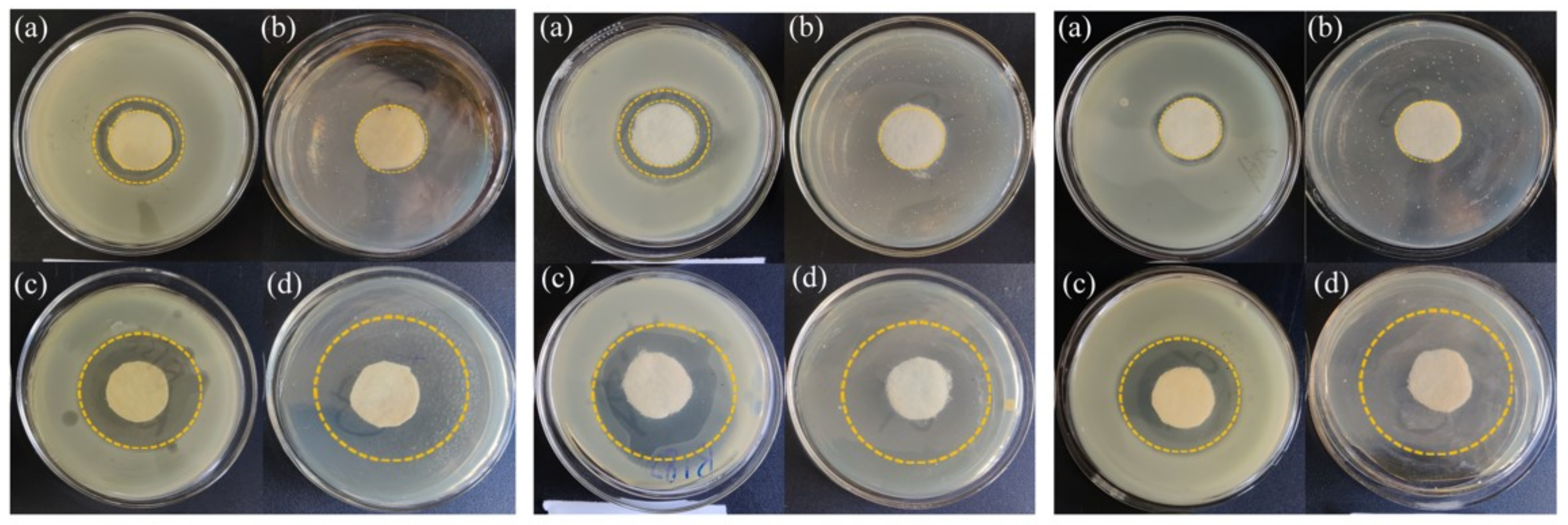
2.3. Qualitative Antibacterial Efficiency Assays

The preparation of the coupons involved cutting several circles (diameter 24 mm) out of each coupon [35] and exposing them to UV for 30 min.
The experiments were conducted as follows. The 10 mL AM was poured into the petri dish (90 mm) to solidify it as the underlying medium. Next, 1 mL of E. coli (or S. albus) suspension was taken by a pipette, put into 400 mL AM at 50 °C and shaken evenly; 5 mL of the mixture was poured into the dish containing 10 mL AM. The coupons were positioned in the middle region of the plates with sterilized tweezers, and pressed to have enough contact with the AM. Then, the coupons in the dishes were placed and incubated at 37 °C for 24 h. Bacteriostatic zones around the coupons were then detected. In addition, the presence or absence of bacterial growth at the bottom of the filter material was observed. Since S. albus grew slowly in the experiments, the cultivation time was longer (72 h), which led to the AM cracking and stripping from the bottom when only 10 mL of AM was added. Therefore, it was appropriate that the amount of AM of each disk was improved to 20 mL in the preparation of culture plates.

2.4. Quantitative Antibacterial Efficiency Assays

The antibacterial fiber was cut into the square-shaped coupons (5 mm × 5 mm), which were weighed to 0.7 g each [36,37], and then irradiated under UV light for 30 min for sterilization.
Nine 250-mL flasks, each containing 5 mL bacterial suspension and 70 mL PBS buffer, were divided into three equal groups. In Figure 2, distilled water was added to one group of flasks (the blank group) and alcohol was added to another group (the control group); in the experimental group, distilled water and alcohol were added to treat the filter material. The two groups were then placed in the incubator at 37 °C with 250 r/min, and shaken for 1 min (referred to as “0” contact time). Next, 0.6 mL of the bacterial suspension from each of the flasks was transferred into a 10-mL test tube containing 5.4 mL of 0.03% PBS buffer solution, and four gradients were diluted. One milliliter of bacterial diluent from each tube was taken to duplicate two plates, which were incubated for 24–48 h at 37 °C. The number of bacterial colonies on each plate was recorded. The final group of flasks contained filter material treated with triclosan as the experimental group. These flasks were placed in the incubator at 37 °C with a rotation speed of 150 r/min, and the oscillation time was 18 h (referred to as “18 h” contact time) (Figure 2). The bacterial suspension in each flask was then diluted. Two parallel templates were prepared for each dilution gradient, and all the plates were placed in the incubator and cultured at 37 °C for 24–48 h. The number of colonies was counted for each dilution gradient.

2.5. Qualitative Evaluation on Antimicrobial Capability

Qualitative antibacterial properties were evaluated according to ISO 20645 [35]. Bacteriostatic zone was sized, the inhibition zone was defined by Equation (2):
H = D d 2
where H is the inhibition zone, mm, D is the diameter of the antibacterial area, mm, and d is the diameter of coupons, mm.
After measuring the bacteriostatic zone, the coupons were removed from the AM. Additionally, the active bacterial grown was observed in the contact area under the coupons. The antibacterial performance of triclosan-treated coupons was evaluated by ISO 20645 [35].

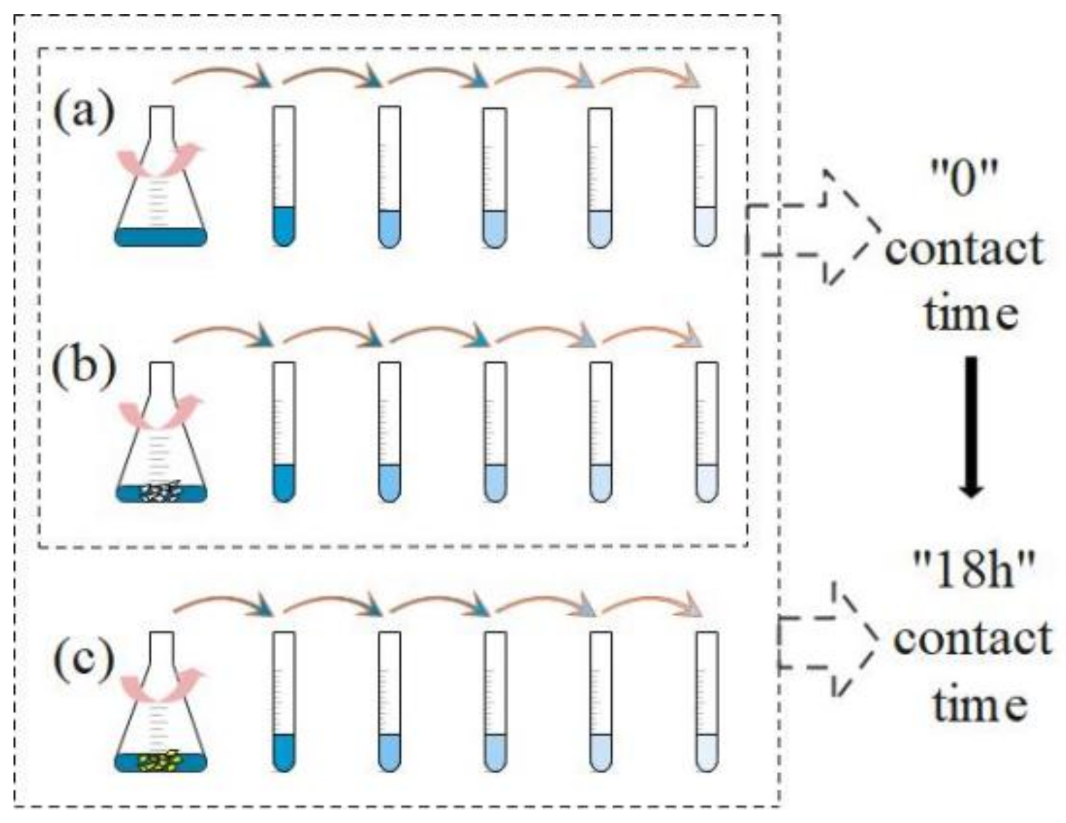
2.6. Quantitative Evaluation on Antibacterial Capability

The quantitative antibacterial activity of triclosan-treated coupons against E. coli and S. albus was evaluated, according to the quantitative assessment of the antibacterial textile materials [37]. After oscillating, “18 h” contact and being serially diluted by ten-fold, the bacterial suspension containing the triclosan-treated coupons was sucked and painted on the surface of plate with10 mL AM, then incubated for 24–48 h at 37 °C. The colonies, grown on the cultured plates, were counted by eyes. The bacteriostatic rate of TT filter fiber was defined as Equation (3) [37,38]:
BR = CC N EC N CC N × 100 %
where BR is the bacteriostatic rate, %, CC N is the viable bacteria in control coupons after “18 h” oscillating, CFU, and EC N is the viable bacteria in experimental coupons after “18 h” of oscillating, CFU.

3. Results and Discussion

3.1. Antibacterial Filter Material Performance

The weights and thicknesses of the antimicrobial filter materials were measured. Compared with the untreated materials, the average weights of the GF, NF and CF treated with triclosan increased by 2.77 g, 2.25 g and 1.65 g, respectively. The weight of the filter material treated with alcohol alone did not increase much. In addition, as calculated by Equation (4), the thicknesses of GF and NF with triclosan increased by 18.50% and 14.21%, respectively, while that of CF decreased by 5.23%, compared with untreated filter materials. Because the layered structure of CF was looser, the smaller space in the structure of CF resulted in more compact when treated with triclosan by soaking. The explanation for this phenomenon is that the addition of triclosan may have led to an increase in weight with the reduced thickness.
Z = Z f Z i Z i × 100 %
where Z is the change rate of the thickness, %, Z i is the thickness of the filter material sample without any treatment, g, and Z f is the thickness of the filter material sample after the addition of triclosan, g.
Fiber images obtained with a scanning electron microscope (SEM) are shown in Figure 3. Compared with the non-treated fiber, the fiber materials to which triclosan was attached had a cloudy condition (Figure 3). This phenomenon also explains the increase in weight that resulted from the addition of triclosan.
Compared with the non-treated filter materials, the GF and NF treated with triclosan exhibited filtration efficiencies that were 4.68% and 7.14% higher, respectively, while the filtration efficiency of CF was 12.27% lower. This finding indicates that the alcohol in the impregnation solution weakened the static electricity in the CF, leading to a decrease in filtration efficiency, while the increase in the filtration efficiency of the other two filter materials resulted from smaller space between fibers and reducing the porosity due to the add-on of triclosan (Figure 3). Meanwhile, the resistance of GF and CF increased by 4.3 Pa and 4.0 Pa, respectively, and that of NF decreased by 1.2 Pa, compared with untreated filter material. This study speculated that the increase in resistance was caused by the adhesion of triclosan to the fibers, which reduced the porosity, while the decrease was due to the change in NF fiber structure by the alcohol.

3.2. Antibacterial Capability of the Qualitative Assays

The bacteriostatic bands of GF, NF and CF for E. coli and S. albus are displayed in Table 1. It can be seen that all bacteriostatic bands were more than 1 mm, and there were no viable bacteria in the contact area between the coupons and AM except for NF (AT). The average antibacterial bands against E. coli were 11.8 mm (GF), 13.3 mm (NF) and 10.5 mm (CF); against S. albus, they were 25.5 mm (GF), 21.0 mm (NF) and 23.5 mm (CF) (Table 1). Celebioglu et al. [32] also observed clear antibacterial bands (>5.0 mm) against E. coli. It was demonstrated that triclosan-treated GF, NF and CF exhibited a strong antibacterial action on the tested bacteria under ISO 20645 [35]. Moreover, no bacterial reproduction was observed on the bottoms of the coupons, and S. albus was found to be growing at the bottom of the NF treated with AT (Table 1). Furthermore, no bacterial reproduction occurred in the area of contact between the coupons and AM. The different size of inhibition zones has been reported compared with that in field study [26] due to the different treatment methods.
The typical bacteriostatic bands of GF, NF and CF treated with triclosan are shown in Figure 4. The bacteriostatic bands of the triclosan-treated GF, NF and CF were obvious and were wider than the zones of the alcohol-treated materials (Figure 4), against the E. coli and S. albus. The results indicated that there was no viable bacteria growth on the bottom of the triclosan-treated material against the E. coli. The triclosan-treated air filter material have a strong antibiotic action, resulting from the appearance of bacteriostatic bands. Field studies have all demonstrated the reliability of triclosan’s antimicrobial ability, and the findings of those studies agree with the qualitative experimental results in the present study. Cui et al.’s [25] study also obtained bacteriostatic bands by a disk-diffusion assay. Furthermore, Karaszewska et al. [26] also found that polylactide microparticles with triclosan had a clear bacteriostatic band. Additionally, Figure 4 reveals that CF, NF and GF all had zones of E. coli inhibition, even when there was no triclosan. This phenomenon may have been due to the static electricity [39] loaded in the filter media.
Meanwhile, the colony self-activity was a significant factor effect on the antibacterial assays. Field studies [10] have reported that bacteria exhibit a greater logarithmic change in the exponential phase than the stable phase. Moreover, the bacteria in the present study are facultative and slow-growth anaerobes, and the oxygen obtained by colonies was lower after they had been cultured into the AM. Due to the small growth form of S. albus and the similarity of the colony’s color to that of the culture medium, the bacteriostatic bands could not be clearly identified in the photographs in Figure 4.

3.3. Antibacterial Capability of the Quantitative Assays

In this study, 6 assays were conducted on the quantitative evaluation, and 153 samples were collected in each quantitative experiment. The bacteriostatic rates in the quantitative experiments are shown in Table 2. The bacteriostatic rates of GF (TT) and CF (TT) against E. coli were 62.7% and 71.4%, respectively, while the bacteriostatic rates of NF (TT) and CF (TT) against S. albus were 61.3% and 84.6%, respectively. Analysis of the results showed that the antibacterial activity of CF treated with triclosan against S. albus was higher than that against E. coli, which was agreed with the field study [30,32]. This finding was resulted from that Gram-positive bacteria are more active than Gram-negative bacteria [30,40]. The bacteriostatic rates in this study were lower than the results reported by Guo et al. [20] (more than 99%) and Kamalipour et al. [41] (more than 95% in a three-day period). Paula et al. [19] have reported that adding triclosan to resin composites can reduce the formation time of bacterial biofilms by more than 10 days, and thus, reduce bacterial adhesion. Therefore, the concentration of triclosan and duration of action, as well as the way in which the triclosan is added, can help to explain the different bactericidal effects mentioned above.

4. Conclusions

In this study, three kinds of triclosan-loaded antibacterial filter materials (chemical fiber, glass fiber and non-woven fabrics) were prepared, and the antibacterial capability of triclosan-treated fiber filter materials was assessed for selected bacterial agents. The filtration materials with triclosan adhesion had a strong antimicrobial effect, as shown by the obvious bacteriostatic bands in qualitative evaluation experiments. The results showed that three triclosan-loaded antibacterial filter materials obviously reduced the selected bacteria (bacteriostatic rates > 60%). In the future, more work would be focused on the relationship with particle size of the triclosan and porosity result for the use of antibacterial filter materials in air conditioning systems. The present study has provided an effective antibacterial fiber filter material loaded with triclosan that can help to reduce the transmission and threat of infectious diseases and improve the indoor environment.

Author Contributions

Conceptualization, Y.L.; methodology, Y.L. and Q.M.; validation, Y.L., Q.M. and X.W.; formal analysis, Y.L. and Q.M.; data curation, Y.L., Q.M. and X.W.; writing—original draft preparation, Y.L. and Q.M.; writing—review and editing, Y.L., Q.M. and X.W. All authors have read and agreed to the published version of the manuscript.

Funding

This research was funded by The Special Fund Project for Technology Innovation of Tianjin, grant number 21YDTPJC00560.

Institutional Review Board Statement

Not applicable.

Informed Consent Statement

Not applicable.

Data Availability Statement

Data are available on request due to their robustness and restrictions on public sharing.

Acknowledgments

We acknowledge Liu Junjie and Liu Mingxin of Tianjin University for their help in this present study.

Conflicts of Interest

The authors declare no conflict of interest.

References

  1. World Health Organization (WHO). Available online: https://www.who.int (accessed on 17 June 2022).
  2. Yang, Y.; Zhang, H.; Nunayon, S.S.; Chan, V.; Lai, A.C.K. Disinfection efficacy of ultraviolet germicidal irradiation on airborne bacteria in ventilation ducts. Indoor Air 2018, 28, 806–817. [Google Scholar] [CrossRef] [PubMed]
  3. Wu, Y.; Yao, M. Inactivation of bacteria and fungus aerosols using microwave irradiation. J. Aerosol Sci. 2010, 41, 682–693. [Google Scholar] [CrossRef]
  4. Bahri, M.; Haghighat, F. Plasma-based indoor air cleaning technologies: The state of the art-review. CLEAN—Soil Air Water 2014, 42, 1667–1680. [Google Scholar] [CrossRef]
  5. Lee, S.H. Development of Photocatalyst Plasma Air Cleaning Filter Used in Air Conditioner. J. Adv. Oxid. Technol. 2016, 6, 13–16. [Google Scholar] [CrossRef]
  6. Abdulla, N.K.; Siddiqui, S.I.; Fatima, B.; Sultana, R.; Tara, N.; Hashmi, A.A.; Ahmad, R.; Mohsin, M.; Nirala, K.R.; Linh, T.N.; et al. Silver based hybrid nanocomposite: A novel antibacterial material for water cleansing. J. Clean. Prod. 2021, 284, 124746. [Google Scholar] [CrossRef]
  7. Balagna, C.; Perero, S.; Bosco, F.; Mollea, C.; Irfan, M.; Ferraris, M. Antipathogen nanostructured coating for air filters. Appl. Surf. Sci. 2020, 508, 145283. [Google Scholar] [CrossRef]
  8. Chowdhury, M.N.K.; Beg, M.D.H.; Khan, M.R.; Mina, M.F. Synthesis of copper nanoparticles and their antimicrobial performances in natural fibres. Mater. Lett. 2013, 98, 26–29. [Google Scholar] [CrossRef] [Green Version]
  9. Cao, Y.; Xin, B.; Wu, X.; Du, W. Research on progress and possibility of electrospining of native cellulose and preparation of copper-based antimicrobial fiber. Adv. Mater. Res. 2014, 850–851, 53–56. [Google Scholar] [CrossRef]
  10. Salussoglia, A.I.P.; de Souza, C.W.O.; Tanabe, E.H.; Lopes Aguiar, M. Evaluation of filter media covered with spun fibres and containing thyme essential oil with antimicrobial properties. Environ. Technol. 2020, 43, 301–310. [Google Scholar] [CrossRef]
  11. Asanović, K.; Mihailović, T.; Škundrić, P.; Simović, L. Some Properties of Antimicrobial Coated Knitted Textile Material Evaluation. Text. Res. J. 2010, 80, 1665–1674. [Google Scholar] [CrossRef]
  12. Sánchez-López, E.; Gomes, D.; Esteruelas, G.; Bonilla, L.; Lopez-Machado, A.L.; Galindo, R.; Cano, A.; Espina, M.; Ettcheto, M.; Camins, A.; et al. Metal-based nanoparticles as antimicrobial agents: An overview. Nanomaterials 2020, 10, 292. [Google Scholar] [CrossRef] [PubMed] [Green Version]
  13. Kim, S.A.; Moon, H.; Lee, K.; Rhee, M.S. Bactericidal effects of triclosan in soap both in vitro and in vivo. J. Antimicrob. Chemother. 2015, 70, 3345–3352. [Google Scholar] [CrossRef] [PubMed] [Green Version]
  14. Halla, N.; Fernandes, I.P.; Heleno, S.A.; Costa, P.; Boucherit-Otmani, Z.; Boucherit, K.; Rodrigues, A.E.; Ferreira, L.C.F.R.; Barreiro, M.F. Cosmetics preservation: A review on present strategies. Molecules 2018, 23, 1571. [Google Scholar] [CrossRef] [PubMed] [Green Version]
  15. Machado, A.H.S.; Garcia, I.M.; Motta, A.D.S.D.; Leitune, V.C.B.; Collares, F.M. Triclosan-loaded chitosan as antibacterial agent for adhesive resin. J. Dent. 2019, 83, 33–39. [Google Scholar] [CrossRef]
  16. Arslan, N.C.; Atasoy, G.; Altintas, T.; Terzi, C. Effect of triclosan-coated sutures on surgical site infections in pilonidal disease: Prospective randomized study. Int. J. Colorectal Dis. 2018, 33, 1445–1452. [Google Scholar] [CrossRef]
  17. Wang, Z.X.; Jiang, C.P.; Cao, Y.; Ding, Y.T. Systematic review and meta-analysis of triclosan-coated sutures for the prevention of surgical-site infection. Br. J. Surg. 2013, 100, 465–473. [Google Scholar] [CrossRef]
  18. Aminu, N.; Chan, S.Y.; Yam, M.F.; Toh, S.M. A dual-action chitosan-based nanogel system of triclosan and flurbiprofen for localised treatment of periodontitis. Int. J. Pharm. 2019, 570, 118659. [Google Scholar] [CrossRef]
  19. Paula, A.B.; Alonso, R.C.B.; Taparelli, J.R.; Camassari, J.R.; Innocentini-Mei, L.H.; Correr-Sobrinho, L.; Puppin-Rontani, R.M. Influence of the incorporation of triclosan methacrylate on the physical properties and antibacterial activity of resin composite. J. Appl. Oral Sci. 2019, 27, 1–8. [Google Scholar] [CrossRef]
  20. Guo, X.; Cheng, Q.; Yu, G.; Wang, H.; Tian, Z.; Shi, Z.; Cui, Z.; Zhu, S. The functions of hydrophobic elastic polyurethane combined with an antibacterial triclosan derivative in the dentin restoration interface. J. Mech. Behav. Biomed. Mater. 2020, 102, 103471. [Google Scholar] [CrossRef] [PubMed]
  21. Petersen, R.C. Computational conformational antimicrobial analysis developing mechanomolecular theory for polymer biomaterials in materials science and engineering. Int. J. Comput. Mater. Sci. Eng. 2014, 3, 48. [Google Scholar] [CrossRef]
  22. Kayaci, F.; Umu, O.C.O.; Tekinay, T.; Uyar, T. Antibacterial electrospun poly(lactic acid) (PLA) nanofibrous webs incorporating triclosan/cyclodextrin inclusion complexes. Agric. Food Chem. 2013, 16, 3901–3908. [Google Scholar] [CrossRef]
  23. Lee, J.H.; Park, S.H.; Kim, S.H. Fabrication of bio-based polyurethane nanofibers incorporated with a triclosan/cyclodextrin complex for antibacterial applications. RSC Adv. 2020, 10, 3450–3458. [Google Scholar] [CrossRef] [PubMed]
  24. Novikov, M.; Thong, K.L.; Zazali, N.I.M.; Hamid, S.B.A. Treatment of Cotton by β-Cyclodextrin/Triclosan Inclusion Complex and Factors Affecting Antimicrobial Properties. Fibers Polym. 2018, 19, 548–560. [Google Scholar] [CrossRef]
  25. Cui, H.; Wu, X.; Zhang, D.; Zhang, J.; Xiao, H.; Chen, Y. Thermotolerance and antibacterial properties of MgO-triclosan nanocomposites. Procedia Eng. 2015, 102, 410–416. [Google Scholar] [CrossRef] [Green Version]
  26. Karaszewska, A.; Kamińska, I.; Kiwała, M.; Gadzinowski, M.; Gosecki, M.; Slomkowski, S. Preparation and properties of textile materials modified with triclosan-loaded polylactide microparticles. Polym. Adv. Technol. 2017, 28, 1185–1193. [Google Scholar] [CrossRef]
  27. Peila, R.; Vineis, C.; Varesano, A.; Ferri, A. Different methods for β-cyclodextrin/triclosan complexation as antibacterial treatment of cellulose substrates. Cellulose 2013, 20, 2115–2123. [Google Scholar] [CrossRef]
  28. Escalada, M.G.; Russell, A.D.; Maillard, J.Y.; Ochs, D. Triclosan bacteria interactions: Single or multiple target sites. Lett. Appl. Microbiol. 2005, 41, 476–481. [Google Scholar] [CrossRef]
  29. Peila, R.; Migliavacca, G.; Aimone, F.; Ferri, A.; Sicardi, S. A comparison of analytical methods for the quantification of a reactive β-cyclodextrin fixed onto cotton yarns. Cellulose 2012, 19, 1097–1105. [Google Scholar] [CrossRef]
  30. Orhan, M. Triclosan applications for biocidal functionalization of polyester and cotton surfaces. J. Eng. Fibers Fabr. 2020, 15, 1558925020940104. [Google Scholar] [CrossRef]
  31. Orhan, M.; Kut, D.; Güneşoğlu, C. Improving the antibacterial activity of cotton fabrics finished with triclosan by the use of 1, 2, 3, 4-butanetetracarboxylic acid and citric acid. Appl. Polym. Sci. 2009, 111, 1344–1352. [Google Scholar] [CrossRef]
  32. Celebioglu, A.; Umu, O.C.O.; Tekinay, T.; Uyar, T. Antibacterial electrospun nanofibers from triclosan/cyclodextrin inclusion complexes. Colloids Surf. B Biointerfaces 2014, 116, 612–619. [Google Scholar] [CrossRef] [PubMed]
  33. Goetzendorf-Grabowska, B.; Polus, Z.; Kiwała, M.; Karaszewska, A.; Kamińska, I.; Mączka, I. Antibacterial air filter nonwovens modifed by poly(lactide) microspheres containing triclosan. Fibres Text. 2015, 23, 114–119. [Google Scholar]
  34. EN 1822-3-2009; High Efficiency Air Filters-Part 3: Testing Flat Sheet Filter Media. European Committee for Standardization: Brussels, Belgium, 2009.
  35. ISO20645-2004; Textile Fabrics—Determination of Antibacterial Activity—Agar Diffusion Plate Test. International Organization for Standardization (ISO): Geneva, Switzerland, 2004.
  36. ASTME2149-2020; Test Method for Determining the Antibacterial Activity of Antimicrobial Agents Under Dynamic Contact Conditions. American Society for Testing and Materials (ASTM): West Conshohocken, PA, USA, 2020.
  37. GB/T20944.3-2008; Textiles-Evaluation for Antibacterial Activity-Part 3: Shake Flask Method. Standardization Administration of China (SAC): Beijing, China, 2008. (In Chinese)
  38. Yang, J.; Wang, Z.; Wu, J.; Hao, L.; Tian, T.; Lin, S. Study on an air filter material immobilized with bio-antimicrobials. Adv. Mater. Res. 2011, 152–153, 1519–1524. [Google Scholar] [CrossRef]
  39. Lee, H.K. Electrical sterilization of Escherichia coli by electrostatic atomization. J. Electrost. 2001, 51–52, 71–75. [Google Scholar] [CrossRef]
  40. Sanbhal, N.; Mao, Y.; Sun, G.; Li, Y.; Peerzada, M.; Wang, L. Preparation and characterization of antibacterial polypropylene meshes with covalently incorporated β-cyclodextrins and captured antimicrobial agent for hernia repair. Polymers 2018, 10, 58. [Google Scholar] [CrossRef] [Green Version]
  41. Kamalipour, J.; Masoomi, M.; Khonakdar, H.A.; Razavi, S.M.R. Preparation and release study of Triclosan in polyethylene/Triclosan anti-bacterial blend. Colloids Surf. B Biointerfaces 2016, 145, 891–898. [Google Scholar] [CrossRef]
Figure 1. Preparation process for the filter materials treated with triclosan and the treated filter material ((a) no treatment (NT), (b) alcohol-treated (AT), and (c) triclosan-treated (TT); in each set of images, the left is GF, the middle is NF and the right is CF).
Atmosphere 13 01104 g001
Figure 2. Steps of oscillation and dilution during the quantitative experiments. ((a) blank group, (b) control group and (c) experimental group; each group consisted of three identical flasks, and the operation procedure was exactly the same). ”0” contact time: oscillated (a,b) for 1 min at 24 °C, 250 r/min; “18 h” contact time: oscillated (ac) for 18 h at 24 °C, 150 r/min; the bacterial suspensions taken from each flask were successively diluted to five gradients (101, 102, 103, 104, 105).
Atmosphere 13 01104 g002
Figure 3. Photographs of non-treated (NT, upper) and triclosan-treated (TT, lower) GF, NF and CF filter fibers by SEM. ((a)-GF (NT); (b)-NF (NT); (c)-CF (NT); (d)-GF (TT); (e)-NF (TT); (f)-CF (TT)).
Atmosphere 13 01104 g003
Figure 4. Typical bacteriostatic bands of GF, NF and CF treated with triclosan (Left: (a)-GF (AT) against E. coli; (b)-GF (AT) against S. albus; (c)-GF (TT) against E. coli; (d)-GF (TT) against S. albus; Middle: (a)-NF (AT) against E. coli; (b)-NF (AT) against S. albus; (c)-NF (TT) against E. coli; (d)-NF (TT) against S. albus; Right: (a)-CF (AT) against E. coli; (b)-CF (AT) against S. albus; (c)-CF (TT) against E. coli; (d)-CF (TT) against S. albus).
Atmosphere 13 01104 g004
Table 1. Bacteriostatic bands of GF, NF and CF to E. coli and S. albus (units: mm).
CouponsE. coliS. albus
GFTT 11.8   ±   0.4 - 25.5   ±   3.5 -
AT 4.5   ±   2.1 - 0.1   ±   0.1 -
NFTT 13.3   ±   3.9 - 21.0   ±   4.2 -
AT 4.8   ±   1.1 - 0.1   ±   0.1 +
CFTT 10.5   ±   0.7 - 23.5   ±   2.1 -
AT 1.6   ±   0.3 - 0.1   ±   0.1 -
“-” no bacterial growth in contract area. “+” viable bacteria in contract area.
Table 2. Bacteriostatic rates of tested filter materials treated with triclosan (a: E. coli, b: S. albus).
CouponsE. coliS. albus
GF (TT) 62.6   ±   27.7 % --
NF (TT)-- 61.3   ±   22.3 %
CF (TT) 71.4   ±   20.2 % 84.6   ±   7.3 %
“--” No petri dishes meeting the standard range of colony data were detected (30–300 CFU).
Publisher’s Note: MDPI stays neutral with regard to jurisdictional claims in published maps and institutional affiliations.

