Review

Single-Particle Analysis of Atmospheric Aerosols: Applications of Raman Spectroscopy

by
Vishnu S. Moorchilot
1,
Usha K. Aravind
2,
Sunil Paul M. Menacherry
3 and
Charuvila T. Aravindakumar
1,4,5,*
1
School of Environmental Sciences, Mahatma Gandhi University, Kottayam 686560, India
2
School of Environmental Studies, Cochin University of Science and Technology, Kochi 682022, India
3
Department of Soil Science and Soil Protection, Faculty of Agrobiology, Food and Natural Resources, Czech University of Life Sciences Prague, Kamýcká 129, 16500 Prague, Czech Republic
4
Inter-University Instrumentation Centre, Mahatma Gandhi University, Kottayam 686560, India
5
International Centre for Polar Studies, Mahatma Gandhi University, Kottayam 686560, India
*
Author to whom correspondence should be addressed.
Atmosphere 2022, 13(11), 1779; https://doi.org/10.3390/atmos13111779
Submission received: 24 September 2022 / Revised: 22 October 2022 / Accepted: 25 October 2022 / Published: 28 October 2022
(This article belongs to the Special Issue Air Pollution from Wastewater Management)

Abstract

:
Atmospheric aerosols, produced as a consequence of different anthropogenic and natural processes, impart significant control over the global energy budget, climate, and human–environmental health. Their size varies across the nano–micrometer scale. Based on their origin, they may be classified into primary or secondary aerosols. Biomass burning, incomplete combustion of fossil fuels, volcanic eruptions, and traffic-related and wind-driven suspensions contribute to primary aerosol emissions. In contrast, gas-to-particle conversion within the atmosphere leads to secondary particle production. The study of atmospheric aerosols is vital to the field of atmospheric research. The dynamic nature (highly variable concentration composition and size with space and time) of aerosols makes them difficult to investigate. Today, aerosol research involves the application of various spectrometric and spectroscopic techniques. The single-particle analysis of aerosols is yet a challenge. In this review, the merits and demerits of various offline and online techniques used for aerosol research are discussed in a nutshell. Mass spectrometric techniques fail in distinguishing certain species. However, Raman spectroscopy’s emergence for the compositional analysis of aerosols resolves most of the present characterization challenges. This review focuses on Raman spectroscopy applications, the merits of this technique, and its immense scope for the measurement of various types of aerosols and their properties. Surface-enhanced Raman spectroscopy (SERS) has an advantage over conventional micro-Raman spectroscopy (MRS). The review depicts the dominance of SERS, specifically in the context of the measurement of ambient atmospheric aerosols. This review discusses two important components, namely laboratory simulation and ambient aerosol studies.

1. Introduction

Atmospheric aerosols are suspensions of liquid/solid particles in the atmosphere. Their size (diameter) varies from a few nanometers to tens of micrometers and they play a significant role in controlling the hydrological cycle and energy balance of the Earth. They can manipulate the global climate and local air quality [1]. Both anthropogenic and natural activities contribute to aerosol emissions. Aerosols are broadly classified into primary and secondary aerosols. Primary aerosols are directly emitted into the atmosphere from the source. In contrast, secondary aerosols are formed by the oxidation of gaseous precursors that nucleate to form particles or condense onto an existing particle [1]. The residence time of aerosols in the atmosphere varies from a few seconds to a month. The lifetime of aerosols in the atmosphere is determined by their particle size, chemical composition, and local meteorology [2]. Meanwhile, the bulk particle composition is determined by their source and different aging processes in the atmosphere [2,3]. The atmospheric transport can alter their size, phase, structure, and chemical composition. The factors that govern this modification involve coagulation, gas–particle partitioning of semi-volatile species, heterogeneous reactions of particles with trace gases, and aqueous-phase processing [1]. The dynamic nature of the physical and chemical properties of the aerosols makes their study challenging.
A single aerosol may host a staggering number of molecules. The number of aerosols per unit volume (concentration number) and the total mass of the aerosols per unit volume (mass concentrations) vary spatially and temporally. It is reported that the particle concentration may range from 15 particles/cm3 at remote locations to 105 particles/cm3 or greater in urban areas. The mass concentrations of PM2.5 particles range from 1–50 mg/m3 in rural areas to ~200 mg/m3 in heavily polluted urban areas [4]. PM2.5 particles are fine, inhalable particles having an aerodynamic diameter of ≤2.5 µm. They can scatter visible light efficiently; thus, high PM2.5 levels in the atmosphere often lead to reduced visibility/haze events. The available literature on atmospheric aerosols mainly focuses on characterizing their bulk properties. This can be attributed to the various challenges faced during characterization at the single-particle scale.
A wide range of instruments are available for both the offline and online analysis of aerosols. Offline techniques involve the collection and transportation of samples from the field to the laboratory. In the case of online analysis, the samples are measured in real time. Hence, offline analysis is often time-consuming when compared with online analysis. However, a single practical technique is still not available for obtaining all the required information. For example, if we are interested in molecular and structural analysis, offline techniques are superior to online techniques. Likewise, online techniques are better suited for monitoring the modification in chemical composition on short time scales [5]. Some offline and online techniques for carrying out the single-particle analysis of aerosols are listed in Table 1 [6].
The chemical composition of an aerosol is highly sensitive to the surrounding environment. Thus, during sample processing, preparation, or analysis, there is a possibility of the contamination/alteration of the chemical species in the aerosols. SEM-EDX and HRTEM are widely used for the analysis of fine atmospheric particulates, but the application of a vacuum at the analysis stage results in the loss of volatile compounds [7,8]. The spectrometric techniques, such as Nano SIMS, TOF-SIMS SP-ICPMS, LMMS, ATOFMS, and AMS, provide high-resolution information on the distribution of chemical species over the aerosols. However, these techniques are destructive in nature. There are non-destructive spectroscopic techniques, such as XPS, PIXE, and EELS, which provide the scope for monitoring ultrafine particles [6,9,10]. However, certain limitations exist with these techniques; for example, in the case of XPS and EELS, it is extremely difficult to interpret the spectra generated from the analysis, while the PIXE technique falls short when it comes to the quantification of low-Z elements [6,11]. In the case of XANES and XAFS, even though they offer high detection sensitivity, their limitation lies in the requirement of a sophisticated synchrotron X-ray source [12]. LIBS is an effective tool for monitoring aerosols at single-particle scale. It does not require sample preparation and allows real-time measurement of the particle mass and composition. However, a drawback associated with this technique is its susceptibility to large interference effects [6]. In this context, MRS appears to be a potential candidate over other techniques for the characterization of atmospheric aerosols at single-particle scale. It does not require tedious sample processing steps, and also provides rapid information on the chemical composition, phase, and crystal orientation of particles. MRS can also determine the different oxidation states of chemical species distributed over the aerosols. However, the limitation of the MRS technique results from its inability to monitor particulates in the nanoscale range [6,13].
The mixing states of aerosols are classified into external or internal mixtures. The aerosols in internal mixtures contain various chemical species with the same mixture composition throughout a single particle. External mixtures are composed of aerosols that only host a single pure species within a particle [14]. These mixing states can be effectively monitored using spectroscopy. Further, due to their non-destructive nature, the samples could be used for other analyses involving different instrumentation techniques [15]. Molecular specificity, the ability to neutralize the spectral interference from water, and accessibility to the visible spectrum’s low-frequency region make Raman spectroscopy an ideal tool for aerosol characterization [16].
Techniques such as attenuated total reflectance Fourier transform infrared spectroscopy (ATR FT-IR), diffuse reflectance FT-IR, or optical microscopy coupled with FT-IR (micro-FT-IR) can also be used for the analysis of the chemical composition of aerosols. However, their application is not recommended in the presence of water due to the strong absorbance interference. Thus, they are limited to bulk particle analysis [17]. In this context, micro-Raman spectroscopy (MRS) offers immense scope for analysis at a single-particle scale. The advantages of MRS include a non-intrusive investigation at ambient temperature and pressure, and minimal sample preparation convenience. Lately, the MRS technique has become a highly preferred one for studies concerning the composition and mixing state of individual aerosols [18]. Raman imaging provides spatial distribution data about the functional groups in an aerosol, rather than only producing vibrational frequencies of functional groups. MRS is also very effective in measuring the acidity of aerosols at a single-particle scale [17,19]. Of late, surface-enhanced Raman spectroscopy (SERS) has gained high interest in the single-particle analysis of atmospheric aerosols. This technique provides strong enhancements in the Raman signal owing to the localized surface plasmon resonances (LSPRs) activated over small (nano-scale) spatial regions of the substrate [20]. Herein, we present a detailed overview of Raman spectroscopy’s application as a tool for the characterization of atmospheric aerosols.

2. Background

Raman Spectroscopy

The possibility of a new form of light scattering other than Rayleigh was theoretically postulated in 1923 by Smeckel. The experimental proof for this new type of scattering (Raman effect) came from the famous light scattering experiment conducted by C. V. Raman and K. S. Krishnan in 1928 [21,22]. For this discovery, C. V. Raman was awarded the Nobel Prize for Physics in 1930.
Two types of light scattering (elastic and inelastic) are produced on the illumination of a sample. For elastic scattering, the light scattered has the same wavelength and frequency as the incident photon; thus, the energy loss is negligible. With inelastic scattering, the photons scatter inelastically due to a shift in frequency and hence energy. Raman spectroscopy is focused on measuring the inelastic (Raman) scattering of light. There are two types of Raman scattering: anti-stokes and stokes. Anti-stokes scattering occurs when a molecule from an excited vibrational state moves to the ground state by transferring its energy to the scattered photon. Stokes scattering is produced when a molecule in a ground state absorbs the energy from an incident photon and is transported to the excited vibrational state. The stokes-scattered photon has lower energy compared to that of the incident photon. In contrast, the anti-stokes-scattered photon has higher energy than an incident photon [23,24]. At room temperature, most of the molecules exist in the ground state. Thus, stokes scattering is expected to dominate over anti-stokes scattering.
The application of Raman spectroscopy did not receive significant interest in the initial years after its discovery. This could be attributed to the weak nature of the scattered signals and the lack of sophisticated instrumental setups. The introduction of lasers in the 1960s, charge-coupled devices (CCDs), the development of SERS in the 1970s, and, later on, the coupling of a microscope with a spectrometer (micro-Raman spectroscopy) in the 1980s created a paradigm shift in the application of Raman spectroscopy [23]. A schematic representation of a micro-Raman spectroscope is presented in Figure 1. The microscopic image and the Raman spectrum of a dust particulate included in this figure were recorded in our laboratory. Today, this spectroscopic technique finds wide application in almost all fields of science.
In aerosol research, MRS offers immense scope and provides a platform to perform long-duration gas–particle interaction studies at ambient conditions. It can provide valid information concerning the distribution/mixing states of chemical species over an aerosol system at 2-D and 3-D scales. Of late, various advancements in instrumentation systems have considerably improved the spatial resolution (µm to nm range) and have reduced the scanning time from hours to a few seconds [25]. With little to no sample preparation, one may acquire the Raman spectra of atmospheric samples. Additionally, analysis can be performed through a variety of containers, including plastic containers, glass bottles, and blister packs. However, certain limitations are also associated with this technique. As this technique is based on the identification of Raman active functional groups, it cannot provide information on exact molecular structures. Furthermore, fluorescence interference from light-absorbing compounds (e.g., humic-like substances (HULIS)) often overlaps with Raman bands of interest, thus making spectral interpretation difficult [26].

3. Laboratory Studies

3.1. Hygroscopicity and Ice Nucleation Activity

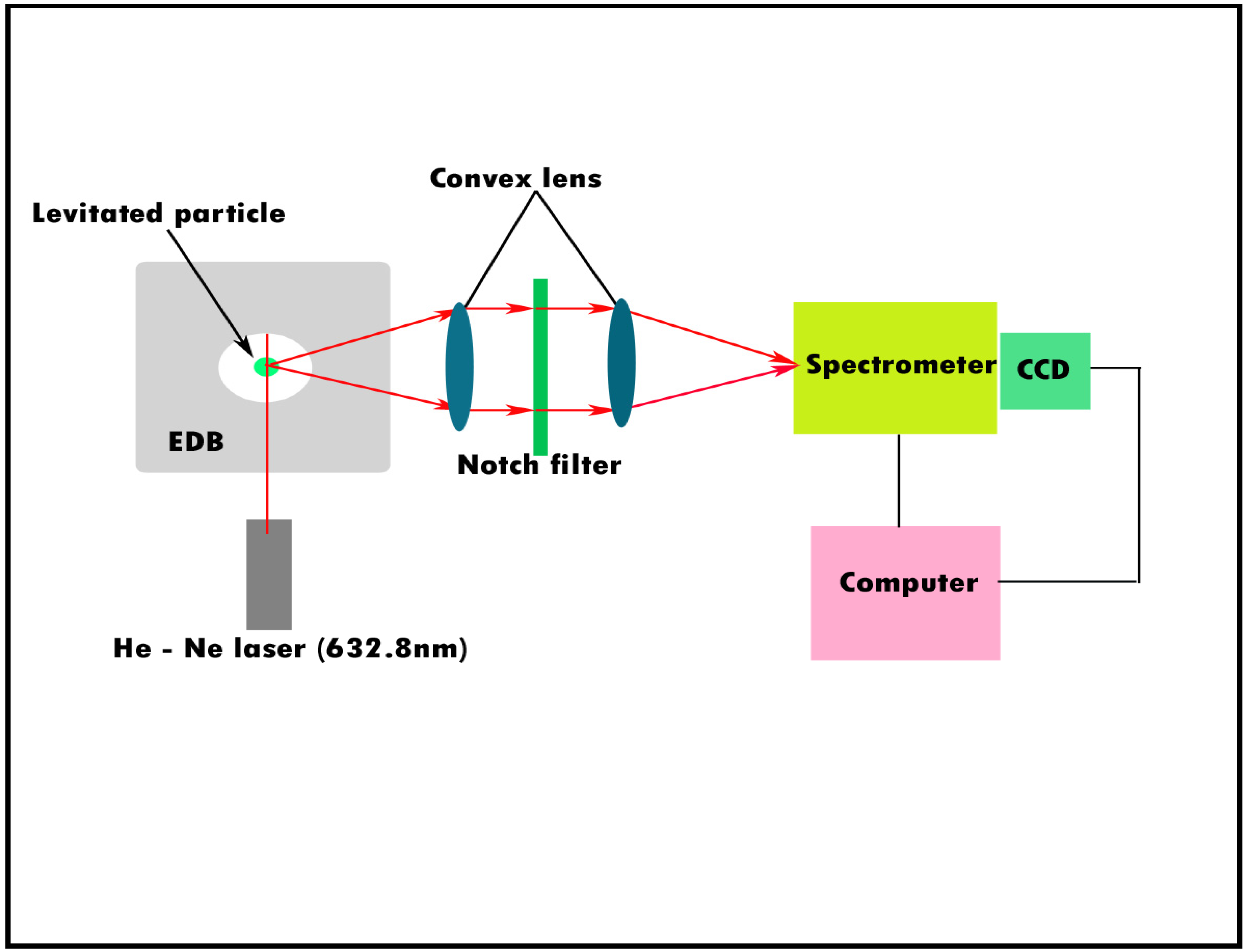
The hygroscopicity of an aerosol is its capacity to absorb water. Of late, this property of aerosols has been investigated with much interest, specifically in climate research. The uptake of water by aerosols can alter the cloud condensation activity and structural and optical properties [27,28,29]. This water uptake is primarily influenced by the relative humidity (RH). Previous studies have indicated the sensitivity of the structural properties of aerosols to varying RH [30,31]. However, monitoring these subtle changes in the ambient atmosphere is a laborious task. Thus, studies are often carried out in laboratory environments. The electrodynamic balance (EDB)/micro-Raman spectroscopy (MRS) system (Figure 2) is a valuable technique to monitor structural alterations to an aerosol system [32]. In an EDB/Raman system, particle levitation is achieved by EDB, while the fluxes in the properties of the suspended particle in response to varying experimental conditions are detected by the Raman spectrometer (Figure 2). Zhang and Chan applied this technique and highlighted the use of the Raman signal intensity ratio of aerosol systems (I995/I983 for MgSO4, I987/I981 for ZnSO4, and I1002/I982for 1:1 Na2SO4/MgSO4 droplets) to effectively monitor the structural modifications under different RH conditions.
The influence of inorganic components on aerosol hygroscopicity is well documented. However, only limited information is available concerning the role of organic components [33]. In an inorganic/organic aerosol system, the organic components can manipulate (decrease, increase, or cause no effect on) the water uptake characteristics of inorganic species. The full width at half height (fwhh) of distinct Raman bands produced by inorganic species in a mixed-particle (inorganic/organic) aerosol system can be utilized to monitor its deliquescence characteristics in response to varying RH conditions [34,35]. Deliquescence is a process by which a crystalline substance, when exposed to the atmosphere at ambient temperature, continuously absorbs moisture and ultimately dissolves in water to form unsaturated solutions. These fwhh of Raman peaks can be sensitive to the mass fraction of the solute. Since the mass fraction of the solute is altered by fluctuations in RH conditions [34], the use of the fwhh of characteristic Raman bands becomes extremely useful for studies concerning aerosol hygroscopicity.
Oxalic acid and oxalates are predominantly detected in aerosols. However, their influence on the hygroscopicity of aerosols is not well explored. In this respect, Ma et al. demonstrated that a UV resonance Raman microscope could effectively reveal the hygroscopic nature of oxalic acid and oxalate salts subjected to ambient RH conditions [36]. The O–H stretching mode occurring between 3000 and 3500 cm−1 was used to indicate the existence of hydrated species. For example, peaks at 3845 cm−1 and 3445 cm−1 were assigned to H2C2O4.2H2O, and a peak at 3334 cm−1 was assigned to FeC2O4.2H2O.
Ice-nucleating particles (INPs) have an important influence on the global climate. Thus, INPs are investigated with great interest in atmospheric chemistry and climate research. The terrestrial sources of INPs, such as mineral dust, are well studied. Although oceans cover a significant part of the Earth’s surface, there is only limited knowledge regarding their contribution to INP loading and their effect on the global climate [37]. Sea salts are marine aerosols found ubiquitously across the globe. They are mainly composed of NaCl and other inorganic species related to seawater. Recently, studies have also shown that sea salt contains traces of organic compounds [38,39]. In laboratory-scale studies, NaCl is often used as a proxy for INPs. The Raman vibration mode for water ice occurs at 3132 cm−1 [40]. Wise et al. used this vibrational mode at 3132 cm−1 to confirm the formation of ice on hydrated and dehydrated NaCl particles [41]. The phase transition and lattice disorder of particles can influence their ice nucleation potential [42,43]. In this respect, Raman spectroscopy provides an advantage as it is sensitive to the crystal and molecular structure of materials [44,45]. The Raman G (graphitic) band (~1580 cm−1) can reveal the nature of the lattice order of soot proxies (graphene and graphene oxides) and their potential role in ice nucleation activity [46]. The samples showing a highly ordered (G band intensity > 50%) graphene lattice have better nucleation potential compared to those having a low ordered (G band intensity < 20%) graphene lattice.

3.2. Organic Aerosols

The influence of organic aerosols on the Earth’s climate and energy budget is not well established [47]. The dynamic nature and diversity of organic aerosols make their study challenging. Most of the literature is focused on secondary organic aerosols (SOAs) because of their varied effects on the climate and human health.
The photochemical reaction between volatile organic compounds (VOCs) and atmospheric oxidants leads to the production of SOAs. Isoprene is an important precursor of SOA. It is the most abundant biogenic volatile organic carbon (BVOC) in the atmosphere [48,49,50,51]. Two strong characteristic organosulfate vibrational modes for isoprene-derived organosulfate compounds occur at 846 ± 4 cm–1 v (RO-SO3) and 1065 ± 2 cm–1 v (SO3) [52]. These vibrational modes can be used as a reference for the detection of isoprene-derived organosulfate components in ambient aerosols.
Optical tweezers are focused laser beams that are extremely useful for aerosol research. They provide the scope for both the levitation and manipulation of microscopic particles [53]. When coupled with an MRS system, these tweezers become a powerful tool for the real-time investigation of heterogeneous chemical reactions in aerosols. A schematic representation of this technique is presented in Figure 3. Here, we have included a representative Raman spectrum of a microplastic (polystyrene (PS)—characteristic peaks: ~1000, 2855, 2907, and 3058 cm−1), recorded in our laboratory, isolated from dust particles. An optical tweezer/Raman system was employed to study the degradation mechanism of some unsaturated organic compounds (benzoate ions, fumarate ions, and α-pinene) exposed to gaseous ozone [53]. Here, Raman spectra were effectively used to identify the degradation products from the heterogeneous reactions. For example, the occurrence of a carbonate anion, a degradation product resulting from the oxidation of a fumarate ion, was identified from the occurrence of the characteristic Raman signal at 1065 cm−1.
Chemical warfare involves the use of hazardous organic compounds [54]. These compounds are fatal and highly bioavailable in their aerosolized forms. The molecular characterization of such aerosols is necessary for the field of defense research. The investigation of the use of these chemicals requires sound evidence; therefore, highly sensitive and sophisticated equipment is required. The MRS technique was successfully used to detect diethyl phthalate (DEPh) microdroplets (21 µm). It is a safe chemical stimulant of a lethal VX nerve agent that can be used in chemical warfare [55]. This study further highlighted the potential of MRS for the investigation of such hazardous chemicals in ambient aerosols.
The incidence of organic acids in atmospheric aerosols is well recognized. Carboxylic acids are some of the dominant organic acids in ambient aerosols. The highly volatile nature of the lower-molecular-weight carboxylic acids (LMWCA) makes them ubiquitous in the atmosphere [56,57]. Both biogenic and anthropogenic activities result in carboxylic acid emissions. Forests with thick vegetation cover are substantial emitters of carboxylic acids. It is also documented that forest soils emit a large amount of carboxylic acids [58,59,60,61]. Photochemical reactions in the atmosphere are an essential pathway for the formation of LMWCA. In this context, Kuo et al. demonstrated the use of MRS to identify the products of the photochemical degradation of a dicarboxylic acid (maleic acid) subjected to a sulfate-containing environment [56]. The Raman bands revealed the degradation of maleic acid (1623 cm−1) to tartaric acid (522 cm−1), which undergoes further degradation to formic acid (1395 cm−1). Thus, the MRS technique provides the scope to predict such possible degradation pathways for atmospherically relevant aerosol components.

3.3. Mixed Salts/Mineral Dust

Soil contains a vast array of minerals. The winds blowing over exposed soils can transport these mineral particulates into the atmosphere [62,63]. These minerals occupy a substantial portion of airborne particulate matter. However, their concentration varies both spatially and temporally [64]. Thus, there is a high probability of interaction between organic acids and mineral dust in the atmosphere [65,66,67]. MRS is useful for the investigation of such complex systems at the microscopic scale. Laskina et al. projected the capability of this technique in monitoring the interactions between specific components (calcite, quartz, and kaolinite) of mineral dust and organic acids (oxalic acid, acetic acid, and humic acid) over a small sample region (0.6 µm) [68]. For example, the formation of calcium oxalate from the reaction between calcite and oxalic acid was identified from the stretching vibrations of C = O, C–O, and C–C at 1491 cm−1, 1464 cm−1, and 897 cm−1, respectively.
Deserts are significant sources of mineral dust in the atmosphere. Through atmospheric circulation processes, the suspended dust particles are further transported to different parts of the world [69]. The large surface area offered by these submicron particles becomes the perfect site for heterogeneous reactions [70]. A dual-beam optical trap/Raman system (Figure 3) is useful for the study of heterogeneous reactions on these particles. In this technique, the particle is elevated and optically trapped with the help of a dual-beam laser. The modifications to the particle from varying experimental conditions are recorded by a Raman spectrometer. A study established the practicality of this technique to monitor the heterogeneous interaction between silica particles (2 µm) and sulfuric acid mist (droplet size ranged from 1 to 7 µm) [71]. The Raman spectra indicated the speciation of sulfuric acid into sulfate (979 cm−1) and bisulfate (1042 cm−1) ions on the silica particles.
The interaction between SiO2 and N2O5 plays a vital role in atmospheric chemistry. It is a significant pathway for the removal of reactive nitrogen species from the atmosphere. In this respect, Tang et al. carried out a study based on the interaction of optically elevated SiO2 particles with different N2O5 concentrations and RH conditions [72]. Here, the Raman spectra indicated a rise in the strength of the NO3 peak (1048 cm−1) over SiO2 concerning an elevation in RH. This study revealed a deviation from the existing knowledge about the removal mechanism of reactive nitrogen species (N205) in the atmosphere. It was found that the interaction between SiO2 and N2O5 under humid conditions leads to the formation of another nitrogen reservoir (HNO3) on mineral particles. Thus, the above studies indicate that the MRS technique is useful for aerosol research concerning the heterogeneous interaction of mineral salt/mineral dust particulates.

3.4. Liquid–Liquid Phase Separation (LLPS)

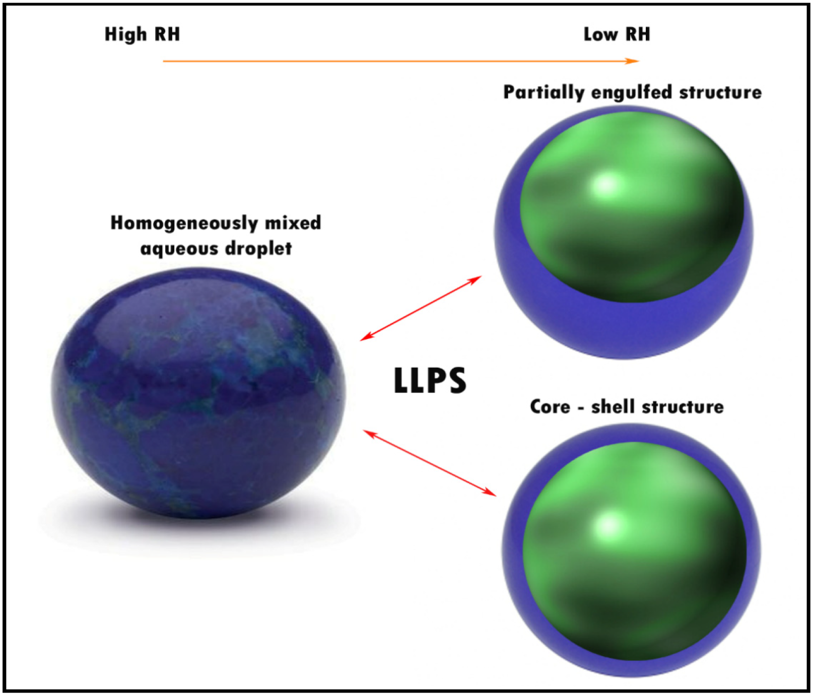
The interactions of polar and non-polar components within an aerosol system can pave the way for liquid–liquid phase separation (LLPS) [73]. A schematic representation of LLPS in a homogeneously mixed inorganic/organic aerosol system under varying RH conditions is presented in Figure 4. LLPS leads to the alteration of the physical properties of an aerosol [74], changing its radiative properties.
The characteristic Raman peak ratio of components can be used to monitor their distribution over an aerosol system. It can provide insights into the redistribution of components in an aerosol as a consequence of LLPS. Wu et al. demonstrated the use of this technique to monitor LLPS in a magnesium sulfate/glutaric acid (MgSO4/GA) system exposed to varying RH [75]. MgSO4 was chosen as the inorganic phase because of its dominance in sea salt and crustal aerosols, whereas GA was chosen as the organic phase due to its abundance among organic aerosols. The area of the ratio of intensities of distinct Raman bands (I2949+2924/I981) for GA and MgSO4 was effectively used to monitor the distribution of these components over the aerosol system. Another study used micro-Raman imaging to monitor the LLPS of a black carbon (BC)-containing organic/inorganic aerosol system [76]. Here, an interesting method was adopted to monitor the redistribution of BC in a mixed-particle aerosol system. The fluorescence observed in the region between 1750 and 2250 cm−1 in the Raman spectrum was used to indicate the existence of BC. Recently, transmission electron microscopy (TEM) revealed the phenomenon of the redistribution of BC within a single-particle system in response to fluxes in RH [77], thus supporting the findings of micro-Raman imaging studies. Of late, MRS studies concerning liquid–liquid and solid–liquid phase separation in aerosols have employed the optical trapping of aerosols [78,79]. This appears to be an excellent technique as it helps to attenuate the distortions of the aerosol particle’s shape as a consequence of its deposition on a substrate.

4. Ambient Aerosol Studies

A large number of studies are available based on the characteristics of ambient aerosols. However, most of them are limited to bulk analysis. To obtain insights into the effect of aerosols on climate, human, and environmental health, it is recommended to carry out a single-particle analysis of ambient aerosols [6]. Due to the dynamic nature of ambient particulates, it is always a challenge to decipher their physical and chemical characteristics. Micro-Raman spectroscopy (MRS) provides the opportunity to rapidly analyze aerosol particles at the microscopic scale. The non-destructive nature of this technique [80] makes it a potential tool for studying ambient aerosols.
Aerosols may contain noxious components that are detrimental to human health [81]. The capability of an airborne particle to cause health issues is directly correlated with its size. Particles that have a size of <10 µm are found to cause the most health problems. This is due to their ability to penetrate deeply into the lungs and even into the bloodstream. Thus, size-segregated particle analysis is necessary to identify the role of particulates in causing respiratory and cardiovascular dysfunction.
Earlier studies have established a potential link between soot exposure and respiratory and cardiovascular diseases. MRS is a powerful technique for the size-segregated characterization of carbonaceous particulates. A study employed this technique to effectively characterize the different aerosol fractions obtained from an urban atmosphere [82]. The prevalence of soot particles (1323 cm−1 and 1582 cm−1) in finer aerosol fractions, such as PM < 1 and 1–2.5 µm, was noted.
Oceans contribute significantly to the total aerosol concentration in the atmosphere [83]. However, limited information exists on the chemical composition of the emitted aerosols. The MRS analysis of the aerosols collected during a long (11,000 km) cruise between Chile and the USA indicated the dominance of long-chained organic compounds [84]. These compounds were identified by their intense doublet peaks occurring at ~2880 cm−1 and ~2850 cm−1 from C–H stretching.
Source apportionment studies focused on the long-distance transport of aerosols have effectively used MRS. A study over Kozani, Northern Greece [85] showed the existence of various mineral phases in the collected aerosols, such as calcite (1084, 710, 274, and 149 cm−1), gypsum (1134, 1006, and 413 cm−1), titanium oxide (627, 390, and 148 cm−1), feldspar (510, 474,285, 265, 150, and 109 cm−1), lepidolite (1573, 1347, 728, 382, and 288 cm−1), and smectite (1379, 731, 385, 288, and 141 cm−1). The presence of lepidolite (mica) and smectite (clay) was associated with the long-distance transport of Saharan dust.
Acid precipitation is mainly influenced by the availability of acidic components (mainly sulfate and nitrates) in fine atmospheric aerosols. It can occur over less polluted areas due to the long-distance transport of aerosols from industrialized regions. In this respect, MRS was effective in characterizing PM1 samples over a mountain environment subjected to acid precipitation because of the transport of aerosols from industrialized regions [86]. The Raman spectrum could successfully reveal the dominance of sulfate ion (~1000 cm−1) species in the aerosols. Another study used this technique to monitor aerosol samples collected from an urban location [87]. The Raman spectrum indicated the dominance of particles with metal oxides (Fe2O3) coated with carbonaceous species. This study further showed that altering the laser beam intensity can unveil the species distribution in a particle. For example, at a low beam intensity (5%), only G (1361.5 cm−1) and D (1576.5 cm−1) bands of carbonaceous species were noted, whereas at a high beam intensity (50%), the strength of bands (G and D) of carbonaceous species decreased, with a corresponding enhancement in the peaks of Fe2O3.
A large volume of studies worldwide have found that various ecosystems face a significant threat from microplastic pollution [88]. Most of these studies are mainly concentrated on marine and freshwater environments. Only a few studies are available indicating the contamination of air with microplastics [89]. The available literature has highlighted the use of the MRS technique to detect microplastics [90,91,92]. Recently, MRS was applied for the analysis of microplastics in air samples collected from a metropolitan area in Hamburg, Germany [93]. The spectra of collected microplastic fragments and fibers revealed polyethylene terephthalate (PET) and polyethylene (PE). Different microplastics can be identified from a pure sample or a mixture from their characteristic peak. The peaks at 1000 cm−1, 1720 cm−1, 1059 cm−1, 695 cm−1, and 402 cm−1 correspond to PS, PET, PE, polyvinyl chloride (PVC), and polypropylene (PP), respectively [94].

5. Surface-Enhanced Raman Spectroscopy (SERS)

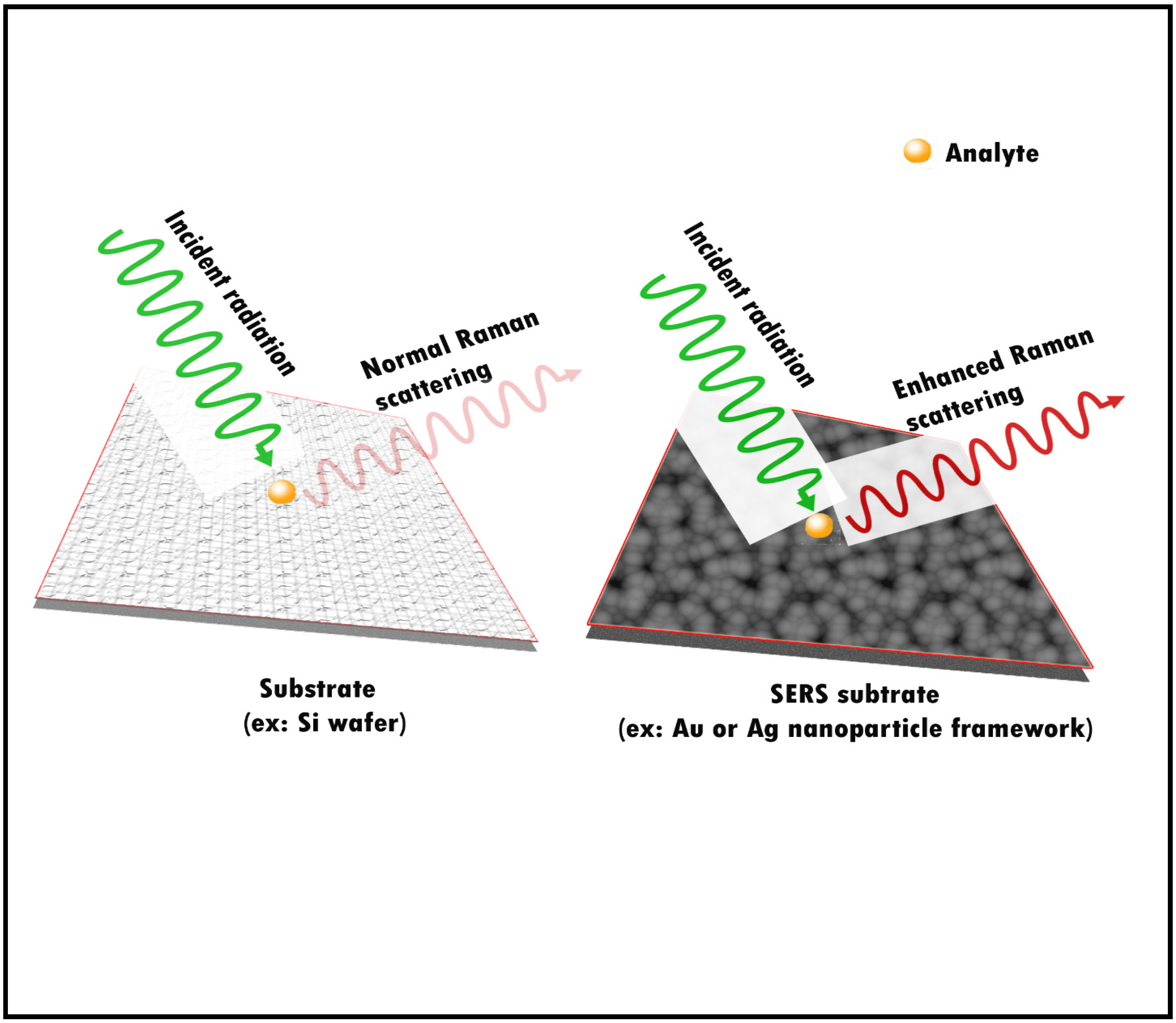
Traditionally, the most preferred methods for the chemical characterization of aerosols involve the use of mass spectrometric techniques. This is due to their capability in detecting species present at trace (ng/L) concentrations [95]. These techniques are, however, costly and time-consuming. Because of the dynamic nature of the aerosols, it is always better to analyze samples rapidly after collection. The mass spectrometric technique often falls short for the rapid analysis of samples, as it requires pre-sample processing [95]. These steps may alter the properties of the aerosols. This highlights the necessity of a sophisticated technique that is fast and does not require pre-sample processing. With surface-enhanced Raman spectroscopy (SERS), it is now possible to detect species at the atto-gram to femtogram level in particles [18]. In the SERS technique, the signals emanating from the analytes are amplified to a great extent by the localized surface plasmon resonance (LSPR) produced by the nanoparticle framework in the substrate used for the analysis (Figure 5) [95].
Silver (Ag) or gold (Au) nanoparticle-coated quartz filters (SERS substrates) are often used for aerosol studies. A Au-coated quartz filter could detect the intraparticle distribution of functional groups νw (CH2)–1022 cm–1, δ (C–C)–1480 cm–1, and νas (NO3)–1370 cm–1) over an SOA particle with 0.5 µm spatial resolution [18].
The amplification of the scattered signals is primarily attributed to the distribution of active sites (localized regions with enhanced electromagnetic fields) over the SERS substrate. The distribution of these over Ag- or Au-coated quartz filters is reported to be non-uniform. Hence, it can sometimes result in the weak enhancement of scattered signals, besides low measurement reproducibility [96]. Thus, better enhancement is only given by substrates hosting a uniform distribution of active sites over their surface. In this context, the commercially available Klarite substrate is recommended [96].
Some filters used to collect ambient aerosols include quartz, Teflon, polycarbonate, polyester, nylon, silver (Ag) foil, and aluminum (Al) foil [6,97,98,99]. A study showed that Ag foil can act as a SERS substrate by producing intense Raman peaks for species such as soot (1350 cm−1 and 1589 cm−1), nitrates (1045 cm−1), and sulfates (978 cm−1) in samples [100].
Haze events often result from high concentrations of sulfate–nitrate–ammonium (SNA) components in the fine aerosol fractions (<2.5 µm). However, understanding their mixing at a single-particle scale is a challenge. Therefore, a highly sophisticated and non-destructive technique is necessary to obtain insights into their distribution over an aerosol. The SERS technique was employed successfully to delineate the distribution of SNA species over aerosol particles [101]. The identified salts included 3(NH4NO3)·(NH4)2SO4, 2(NH4NO3)·(NH4)2SO4, and NH4NO3. Of late, Ag-SiO2 nanocomposites have been found to be a highly sensitive SERS substrate for monitoring anthropogenic VOC mixtures (toluene, benzene, chloroform, and acetone) at trace levels under room temperature [102]. Although the SERS technique is slowly gaining interest in atmospheric research, this method seems to be largely underutilized when considering its huge potential.

6. Stimulated Raman Scattering (SRS) Microscopy

One of the most commonly applied Raman microscopy methods is spontaneous Raman microscopy. In this technique, a single laser beam is used to illuminate the particle, and the information from the Raman-scattered light is used for the chemical profiling. Since this type of scattering is an innately weak process, a fundamental issue in such spontaneous microscopy is the long accumulation period and limited chemical sensitivity [25,103]. The SERS technique has led to a significant amplification in weak Raman signals, thus enhancing the sensitivity of Raman microscopy [104]. However, some major limitations associated with this technique are related to the substrate. The substrates are expensive and can have compatibility issues with certain analytes. Besides this, the SERS technique is also not appropriate for carrying out volumetric imaging [105]. In this context, SRS microscopy has superiority over existing techniques. It can provide 3-D spatial information regarding the distribution of species within a single-particle system in a short time [25]. In SRS, two laser beams collide on a sample. When the photon energy difference between the beams matches the vibrational state of the molecules in the focus volume, the signal is created. The detection is achieved by amplitude-modulating one beam before the sample and detecting the modulated signal from the other beam. The signal intensity depends primarily on the chemical concentration, offering the potential for the quantitative analysis of mixed species in a system [106,107]. Since the signal acquisition time per pixel can be shortened by many orders of magnitude when compared to spontaneous Raman, SRS is frequently used for label-free imaging [105]. Although there is huge potential for this technique in aerosol research, it remains fairly underutilized. Of late, Ao et al. demonstrated the effectiveness of this technique for single-particle analysis of both laboratory-generated and ambient aerosols [25]. They could achieve the chemical imaging of particles four times faster with a superior spatial resolution compared to traditional MRS. Furthermore, the 3-D chemical distribution profile of diverse chemical components were effectively disclosed via SRS imaging of individual aerosols. Thus, this non-intrusive technique is a powerful tool for monitoring the formation and aging mechanisms of atmospheric aerosols.

7. Summary

Raman spectroscopy is, undoubtedly, an efficient technique for atmospheric research. There is a wide range of applications for this technique in atmospheric chemistry, especially in the study of atmospheric aerosols. Micro-Raman spectroscopy offers scope for the single-particle analysis of particulate matter, which is an emerging field of investigation. One of the major advantages of this technique is that it offers non-destructive sample analysis. This technique could be used for real-time chemical composition and mixing state studies within a single atmospheric particle. Many existing sophisticated equipment setups fall short in this capability. Molecular specificity and immunity to spectral interferences from water make this a superior tool compared to other spectroscopic techniques. Today, Raman spectroscopy is utilized for the study of both laboratory-generated and ambient aerosols. However, the technique is underutilized when considering its immense potential. Lately, among Raman spectroscopic techniques, the SERS technique has gained much interest in aerosol research. This may be attributed to the possibility of a massive enhancement in the intensity of Raman signals available with this technique, making better characterization possible. There has been a surge in Raman spectroscopy’s usage for aerosol studies in the past decade, as demonstrated in this report. It is, thus, predicted that further development of the technology and greater sophistication in the instrumentation will pave the way towards a better understanding of the complexity of aerosol measurements.

Author Contributions

Conceptualization, V.S.M.; writing—original draft preparation, V.S.M.; writing—review and editing, S.P.M.M.; supervision, U.K.A.; supervision, C.T.A. All authors have read and agreed to the published version of the manuscript.

Funding

This research received no external funding.

Institutional Review Board Statement

Not applicable.

Informed Consent Statement

Not applicable.

Data Availability Statement

Not applicable.

Acknowledgments

MVS is thankful to UGC for the (UGC-JRF) research fellowship.

Conflicts of Interest

The authors declare no competing interest.

Abbreviations

AMSAerosol mass spectrometry
ATOFMSAerosol time-of-flight mass spectrometry
ATR–FTIRAttenuated total reflectance Fourier transform infrared
BCBlack carbon
BVOCBiogenic volatile organic carbon
CCDsCharge-coupled devices
DEPhDiethyl phthalate
EDBElectrodynamic balance
EELSElectron energy loss spectrometry
fwhhFull width at half height
TEMTransmission electron microscopy
HRTEMHigh-resolution transmission electron microscopy
INPsIce-nucleating particles
LIBSLaser-induced breakdown spectroscopy
LMMSLaser microprobe mass spectrometry
LMWCALower-molecular-weight carboxylic acids
LSPRsLocalized surface plasmon resonances
MRSMicro-Raman spectroscopy
PEPolyethylene
PETPolyethylene terephthalate
PIXEProton-induced X-ray emission
PMParticulate matter
PPPolypropylene
PSPolystyrene
PVCPolyvinyl chloride
RHRelative humidity
SEM-EDXScanning electron microscopy–energy-dispersive X-ray
SERSSurface-enhanced Raman spectroscopy
SNASulfate–nitrate–ammonium
SOAsSecondary organic aerosols
SIMSSecondary ion mass spectrometry
SP-ICPMSSingle-particle inductively coupled mass spectrometry
TOF-SIMSTime-of-flight–secondary ion mass spectrometry
VOCsVolatile organic compounds
XAFSX-ray absorption fine structure
XANESX-ray absorption near-edge structure
XPSX-ray photoelectron spectroscopy

References

  1. Pöschl, U. Atmospheric Aerosols: Composition, Transformation, Climate and Health Effects. Angew. Chem. Int. Ed. 2005, 44, 7520–7540. [Google Scholar] [CrossRef] [PubMed]
  2. Williams, J.; de Reus, M.; Krejci, R.; Fischer, H.; Ström, J. Application of the variability-size relationship to atmospheric aerosol studies: Estimating aerosol lifetimes and ages. Atmos. Chem. Phys. 2002, 2, 133–145. [Google Scholar] [CrossRef] [Green Version]
  3. McMurry, P.H. A review of atmospheric aerosol measurements. Atmos. Environ. 2000, 34, 1959–1999. [Google Scholar] [CrossRef]
  4. Finlayson-Pitts, B.; Pitts, J. Chemistry of Upper and Lower Atmosphere; Academic Press: Cambridge, MA, USA, 2000. [Google Scholar]
  5. Pratt, K.A.; Prather, K.A. Mass spectrometry of atmospheric aerosols—Recent developments and applications. Part II: On-line mass spectrometry techniques. Mass Spectrom. Rev. 2012, 31, 17–48. [Google Scholar] [CrossRef]
  6. Elmes, M.; Gasparon, M. Sampling and single particle analysis for the chemical characterisation of fine atmospheric particulates: A review. J. Environ. Manage. 2017, 202, 137–150. [Google Scholar] [CrossRef]
  7. Laskin, A.; Cowin, J.P. Automated Single-Particle SEM/EDX Analysis of Submicrometer Particles down to 0.1 μm. Anal. Chem. 2001, 73, 1023–1029. [Google Scholar] [CrossRef]
  8. Mäkelä, J.M.; Hoffmann, T.; Holzke, C.; Väkevä, M.; Suni, T.; Mattila, T.; Aalto, P.P.; Tapper, U.; Kauppinen, E.I.; O’Dowd, C.D. Biogenic iodine emissions and identification of end-products in coastal ultrafine particles during nucleation bursts. J. Geophys. Res. Atmos. 2002, 107, PAR 14-11–PAR 14-14. [Google Scholar] [CrossRef] [Green Version]
  9. Ma, C.-J.; Kasahara, M.; Höller, R.; Kamiya, T. Characteristics of single particles sampled in Japan during the Asian dust–storm period. Atmos. Environ. 2001, 35, 2707–2714. [Google Scholar] [CrossRef]
  10. Angyal, A.; Kertész, Z.; Szikszai, Z.; Szoboszlai, Z. Study of Cl-containing urban aerosol particles by ion beam analytical methods. Nucl. Instrum. Methods Phys. Res. B 2010, 268, 2211–2215. [Google Scholar] [CrossRef]
  11. Cheng, W.; Weng, L.-T.; Li, Y.; Lau, A.; Chan, C.K.; Chan, C.-M. Surface Chemical Composition of Size-Fractionated Urban Walkway Aerosols Determined by X-Ray Photoelectron Spectroscopy. Aerosol Sci. Technol. 2013, 47, 1118–1124. [Google Scholar] [CrossRef]
  12. Davidson, R.A.; Anderson, D.S.; Van Winkle, L.S.; Pinkerton, K.E.; Guo, T. Evolution of Silver Nanoparticles in the Rat Lung Investigated by X-ray Absorption Spectroscopy. J. Phys. Chem. A 2015, 119, 281–289. [Google Scholar] [CrossRef] [PubMed] [Green Version]
  13. Sobanska, S.; Falgayrac, G.; Rimetz-Planchon, J.; Perdrix, E.; Brémard, C.; Barbillat, J. Resolving the internal structure of individual atmospheric aerosol particle by the combination of Atomic Force Microscopy, ESEM–EDX, Raman and ToF–SIMS imaging. Microchem. J. 2014, 114, 89–98. [Google Scholar] [CrossRef]
  14. Riemer, N.; West, M. Quantifying aerosol mixing state with entropy and diversity measures. Atmos. Chem. Phys. 2013, 13, 11423–11439. [Google Scholar] [CrossRef] [Green Version]
  15. Lee, S.-H.; Allen, H.C. Analytical Measurements of Atmospheric Urban Aerosol. Anal. Chem. 2012, 84, 1196–1201. [Google Scholar] [CrossRef]
  16. Zangmeister, C.D.; Pemberton, J.E. Raman Spectroscopy and Atomic Force Microscopy of the Reaction of Sulfuric Acid with Sodium Chloride. J. Am. Chem. Soc. 2000, 122, 12289–12296. [Google Scholar] [CrossRef]
  17. Craig, R.L.; Bondy, A.L.; Ault, A.P. Computer-controlled Raman microspectroscopy (CC-Raman): A method for the rapid characterization of individual atmospheric aerosol particles. Aerosol Sci. Technol. 2017, 51, 1099–1112. [Google Scholar] [CrossRef]
  18. Craig, R.; Bondy, A.; Ault, A. Surface Enhanced Raman Spectroscopy Enables Observations of Previously Undetectable Secondary Organic Aerosol Components at the Individual Particle Level. Anal. Chem. 2015, 87, 7510–7514. [Google Scholar] [CrossRef] [PubMed]
  19. Rindelaub, J.D.; Craig, R.L.; Nandy, L.; Bondy, A.L.; Dutcher, C.S.; Shepson, P.B.; Ault, A.P. Direct Measurement of pH in Individual Particles via Raman Microspectroscopy and Variation in Acidity with Relative Humidity. J. Phys. Chem. A 2016, 120, 911–917. [Google Scholar] [CrossRef]
  20. Tirella, P.N.; Craig, R.L.; Tubbs, D.B.; Olson, N.E.; Lei, Z.; Ault, A.P. Extending surface enhanced Raman spectroscopy (SERS) of atmospheric aerosol particles to the accumulation mode (150–800 nm). Environ. Sci. Process Impacts 2018, 20, 1570–1580. [Google Scholar] [CrossRef]
  21. Raman, C.V.; Krishnan, K.S. A New Type of Secondary Radiation. Nature 1928, 121, 501–502. [Google Scholar] [CrossRef]
  22. Gardiner, D.J. Introduction to Raman scattering. In Practical Raman Spectroscopy; Springer: Berlin/Heidelberg, Germany, 1989; pp. 1–12. [Google Scholar]
  23. Alula, M.T.; Mengesha, Z.T.; Mwenesongole, E. Advances in surface-enhanced Raman spectroscopy for analysis of pharmaceuticals: A review. Vib. Spectrosc. 2018, 98, 50–63. [Google Scholar] [CrossRef]
  24. Smith, E.; Dent, G. Modern Raman spectroscopy: A practical approach. John Wiley & Sons Ltd.: Hoboken, NJ, USA, 2005. [Google Scholar]
  25. Ao, J.; Feng, Y.; Wu, S.; Wang, T.; Ling, J.; Zhang, L.; Ji, M. Rapid, 3D Chemical Profiling of Individual Atmospheric Aerosols with Stimulated Raman Scattering Microscopy. Small Methods 2020, 4, 1900600. [Google Scholar] [CrossRef] [Green Version]
  26. Ivleva, N.P.; McKeon, U.; Niessner, R.; Pöschl, U. Raman Microspectroscopic Analysis of Size-Resolved Atmospheric Aerosol Particle Samples Collected with an ELPI: Soot, Humic-Like Substances, and Inorganic Compounds. Aerosol Sci. Technol. 2007, 41, 655–671. [Google Scholar] [CrossRef] [Green Version]
  27. Ye, X.; Ma, Z.; Hu, D.; Yang, X.; Chen, J. Size-resolved hygroscopicity of submicrometer urban aerosols in Shanghai during wintertime. Atmos. Res. 2011, 99, 353–364. [Google Scholar] [CrossRef]
  28. Pringle, K.; Tost, H.; Pozzer, A.; Pöschl, U.; Lelieveld, J. Global distribution of the effective aerosol hygroscopicity parameter for CCN activation. Atmos. Chem. Phys 2010, 10, 5241–5255. [Google Scholar] [CrossRef] [Green Version]
  29. Randles, C.; Russell, L.; Ramaswamy, V. Hygroscopic and optical properties of organic sea salt aerosol and consequences for climate forcing. Geophys. Res. Lett. 2004, 31, L16108. [Google Scholar] [CrossRef]
  30. Weingartner, E.; Baltensperger, U.; Burtscher, H. Growth and structural change of combustion aerosols at high relative humidity. Environ. Sci. Technol. 1995, 29, 2982–2986. [Google Scholar] [CrossRef]
  31. Montgomery, J.F.; Rogak, S.N.; Green, S.I.; You, Y.; Bertram, A.K. Structural change of aerosol particle aggregates with exposure to elevated relative humidity. Environ. Sci. Technol. 2015, 49, 12054–12061. [Google Scholar] [CrossRef]
  32. Zhang, Y.-H.; Chan, C.K. Understanding the Hygroscopic Properties of Supersaturated Droplets of Metal and Ammonium Sulfate Solutions Using Raman Spectroscopy. J. Phys. Chem. A 2002, 106, 285–292. [Google Scholar] [CrossRef]
  33. Wu, Z.; Nowak, A.; Poulain, L.; Herrmann, H.; Wiedensohler, A. Hygroscopic behavior of atmospherically relevant water-soluble carboxylic salts and their influence on the water uptake of ammonium sulfate. Atmos. Chem. Phys. 2011, 11, 12617–12626. [Google Scholar] [CrossRef]
  34. Yeung, M.C.; Lee, A.K.Y.; Chan, C.K. Phase Transition and Hygroscopic Properties of Internally Mixed Ammonium Sulfate and Adipic Acid (AS-AA) Particles by Optical Microscopic Imaging and Raman Spectroscopy. Aerosol Sci. Technol. 2009, 43, 387–399. [Google Scholar] [CrossRef] [Green Version]
  35. Choi, M.Y.; Chan, C.K. The effects of organic species on the hygroscopic behaviors of inorganic aerosols. Environ. Sci. Technol. 2002, 36, 2422–2428. [Google Scholar] [CrossRef] [PubMed]
  36. Ma, Q.; He, H.; Liu, C. Hygroscopic properties of oxalic acid and atmospherically relevant oxalates. Atmos. Environ. 2013, 69, 281–288. [Google Scholar] [CrossRef]
  37. DeMott, P.J.; Hill, T.C.J.; McCluskey, C.S.; Prather, K.A.; Collins, D.B.; Sullivan, R.C.; Ruppel, M.J.; Mason, R.H.; Irish, V.E.; Lee, T.; et al. Sea spray aerosol as a unique source of ice nucleating particles. Proc. Natl. Acad. Sci. USA 2016, 113, 5797–5803. [Google Scholar] [CrossRef] [Green Version]
  38. Wolf, M.J.; Zhang, Y.; Zhou, J.; Surratt, J.D.; Turpin, B.J.; Cziczo, D.J. Enhanced Ice Nucleation of Simulated Sea Salt Particles with the Addition of Anthropogenic Per- and Polyfluoroalkyl Substances. ACS Earth Space Chem. 2021, 5, 2074–2085. [Google Scholar] [CrossRef]
  39. Burrows, S.M.; Easter, R.C.; Liu, X.; Ma, P.L.; Wang, H.; Elliott, S.M.; Singh, B.; Zhang, K.; Rasch, P.J. OCEANFILMS (Organic Compounds from Ecosystems to Aerosols: Natural Films and Interfaces via Langmuir Molecular Surfactants) sea spray organic aerosol emissions—implementation in a global climate model and impacts on clouds. Atmos. Chem. Phys. 2022, 22, 5223–5251. [Google Scholar] [CrossRef]
  40. Baustian, K.J.; Wise, M.E.; Tolbert, M.A. Depositional ice nucleation on solid ammonium sulfate and glutaric acid particles. Atmos. Chem. Phys. 2010, 10, 2307–2317. [Google Scholar] [CrossRef] [Green Version]
  41. Wise, M.E.; Baustian, K.J.; Koop, T.; Freedman, M.A.; Jensen, E.J.; Tolbert, M.A. Depositional ice nucleation onto crystalline hydrated NaCl particles: A new mechanism for ice formation in the troposphere. Atmos. Chem. Phys. 2012, 12, 1121–1134. [Google Scholar] [CrossRef] [Green Version]
  42. Lupi, L.; Hudait, A.; Peters, B.; Grünwald, M.; Mullen, R.G.; Nguyen, A.H.; Molinero, V. Role of stacking disorder in ice nucleation. Nature 2017, 551, 218–222. [Google Scholar] [CrossRef]
  43. Llombart, P.; Noya, E.G.; MacDowell, L.G. Surface phase transitions and crystal habits of ice in the atmosphere. Sci. Adv. 2020, 6, eaay9322. [Google Scholar] [CrossRef]
  44. Beyssac, O.; Goffé, B.; Petitet, J.-P.; Froigneux, E.; Moreau, M.; Rouzaud, J.-N. On the characterization of disordered and heterogeneous carbonaceous materials by Raman spectroscopy. Spectrochim. Acta A 2003, 59, 2267–2276. [Google Scholar] [CrossRef]
  45. Andò, S.; Garzanti, E. Raman spectroscopy in heavy-mineral studies. Geol. Soc. Lond. Spec. Publ. 2014, 386, 395–412. [Google Scholar] [CrossRef]
  46. Hausler, T.; Gebhardt, P.; Iglesias, D.; Rameshan, C.; Marchesan, S.; Eder, D.; Grothe, H. Ice Nucleation Activity of Graphene and Graphene Oxides. J. Phys. Chem. C 2018, 122, 8182–8190. [Google Scholar] [CrossRef] [PubMed] [Green Version]
  47. Huber, M.; Knutti, R. Anthropogenic and natural warming inferred from changes in Earth’s energy balance. Nat. Geosci. 2012, 5, 31–36. [Google Scholar] [CrossRef]
  48. Pandis, S.N.; Harley, R.A.; Cass, G.R.; Seinfeld, J.H. Secondary organic aerosol formation and transport. Atmos. Environ. 1992, 26, 2269–2282. [Google Scholar] [CrossRef]
  49. Huang, C.; Wang, H.; Li, L.; Wang, Q.; Lu, Q.; De Gouw, J.; Zhou, M.; Jing, S.; Lu, J.; Chen, C. VOC species and emission inventory from vehicles and their SOA formation potentials estimation in Shanghai, China. Atmos. Chem. Phys. 2015, 15, 11081–11096. [Google Scholar] [CrossRef] [Green Version]
  50. Lim, H.-J.; Carlton, A.G.; Turpin, B.J. Isoprene forms secondary organic aerosol through cloud processing: Model simulations. Environ. Sci. Technol. 2005, 39, 4441–4446. [Google Scholar] [CrossRef]
  51. Carlton, A.; Wiedinmyer, C.; Kroll, J. A review of Secondary Organic Aerosol (SOA) formation from isoprene. Atmos. Chem. Phys. 2009, 9, 4987–5005. [Google Scholar] [CrossRef] [Green Version]
  52. Bondy, A.L.; Craig, R.L.; Zhang, Z.; Gold, A.; Surratt, J.D.; Ault, A.P. Isoprene-Derived Organosulfates: Vibrational Mode Analysis by Raman Spectroscopy, Acidity-Dependent Spectral Modes, and Observation in Individual Atmospheric Particles. J. Phys. Chem. A 2018, 122, 303–315. [Google Scholar] [CrossRef]
  53. King, M.D.; Thompson, K.C.; Ward, A.D.; Pfrang, C.; Hughes, B.R. Oxidation of biogenic and water-soluble compounds in aqueous and organic aerosol droplets by ozone: A kinetic and product analysis approach using laser Raman tweezers. Faraday Discuss. 2008, 137, 173–192. [Google Scholar] [CrossRef]
  54. Ganesan, K.; Raza, S.; Vijayaraghavan, R. Chemical warfare agents. J. Pharm. Bioallied Sci. 2010, 2, 166. [Google Scholar] [CrossRef] [PubMed]
  55. Kalume, A.; Beresnev, L.A.; Santarpia, J.; Pan, Y.L. Detection and characterization of chemical aerosol using laser-trapping single-particle Raman spectroscopy. Appl. Opt. 2017, 56, 6577–6582. [Google Scholar] [CrossRef] [PubMed]
  56. Kuo, S.-C.; Tsai, Y.I.; Tsai, C.-H.; Hsieh, L.-Y. Carboxylic acids in PM2.5 over Pinus morrisonicola forest and related photoreaction mechanisms identified via Raman spectroscopy. Atmos. Environ. 2011, 45, 6741–6750. [Google Scholar] [CrossRef]
  57. Limbeck, A.; Puxbaum, H. Organic acids in continental background aerosols. Atmos. Environ. 1999, 33, 1847–1852. [Google Scholar] [CrossRef]
  58. Enders, G.; Dlugi, R.; Steinbrecher, R.; Clement, B.; Daiber, R.; Eijk, J.; Gäb, S.; Haziza, M.; Helas, G.; Herrmann, U. Biosphere/atmosphere interactions: Integrated research in a European coniferous forest ecosystem. Atmos. Environ. 1992, 26, 171–189. [Google Scholar] [CrossRef]
  59. Herckes, P.; Engling, G.; Kreidenweis, S.M.; Collett, J.L. Particle size distributions of organic aerosol constituents during the 2002 Yosemite aerosol characterization study. Environ. Sci. Technol. 2006, 40, 4554–4562. [Google Scholar] [CrossRef]
  60. Alves, C.A.; Pio, C.A.; Duarte, A.C. Particulate size distributed organic compounds in a forest atmosphere. Environ. Sci. Technol. 2000, 34, 4287–4293. [Google Scholar] [CrossRef]
  61. Fu, P.; Kawamura, K.; Kanaya, Y.; Wang, Z. Contributions of biogenic volatile organic compounds to the formation of secondary organic aerosols over Mt. Tai, Central East China. Atmos. Environ. 2010, 44, 4817–4826. [Google Scholar] [CrossRef] [Green Version]
  62. Free, E.E. The Movement of Soil Material by the Wind; US Government Printing Office: Washington, DC, USA, 1911. [Google Scholar]
  63. Dixon, J.B.; Weed, S.B. Minerals in Soil Environments; Soil Science Society of America Inc. (SSSA): Madison, WI, USA, 1989. [Google Scholar]
  64. Hand, J.L.; Gill, T.; Schichtel, B. Spatial and seasonal variability in fine mineral dust and coarse aerosol mass at remote sites across the United States. J. Geophys. Res. Atmos. 2017, 122, 3080–3097. [Google Scholar] [CrossRef]
  65. Falkovich, A.H.; Schkolnik, G.; Ganor, E.; Rudich, Y. Adsorption of organic compounds pertinent to urban environments onto mineral dust particles. J. Geophys. Res. Atmos. 2004, 109, D02208. [Google Scholar] [CrossRef]
  66. Ponczek, M.; Hayeck, N.; Emmelin, C.; George, C. Heterogeneous photochemistry of dicarboxylic acids on mineral dust. Atmos. Environ. 2019, 212, 262–271. [Google Scholar] [CrossRef]
  67. Sullivan, R.; Guazzotti, S.; Sodeman, D.; Prather, K. Direct observations of the atmospheric processing of Asian mineral dust. Direct observations of the atmospheric processing of Asian mineral dust. Atmos. Chem. Phys. 2007, 7, 1213–1236. [Google Scholar] [CrossRef] [Green Version]
  68. Laskina, O.; Young, M.A.; Kleiber, P.D.; Grassian, V.H. Infrared extinction spectroscopy and micro-Raman spectroscopy of select components of mineral dust mixed with organic compounds. J. Geophys. Res. Atmos. 2013, 118, 6593–6606. [Google Scholar] [CrossRef]
  69. Tegen, I.; Schepanski, K. The global distribution of mineral dust. In Proceedings of the IOP Conference Series: Earth and Environmental Science, Barcelona, Spain, 7–9 November 2007; p. 012001. [Google Scholar]
  70. Yu, Z.; Jang, M. Simulation of heterogeneous photooxidation of SO 2 and NO x in the presence of Gobi Desert dust particles under ambient sunlight. Atmos. Chem. Phys. 2018, 18, 14609–14622. [Google Scholar]
  71. Rkiouak, L.; Tang, M.J.; Camp, J.C.J.; McGregor, J.; Watson, I.M.; Cox, R.A.; Kalberer, M.; Ward, A.D.; Pope, F.D. Optical trapping and Raman spectroscopy of solid particles. Phys. Chem. Chem. Phys. 2014, 16, 11426–11434. [Google Scholar] [CrossRef]
  72. Tang, M.J.; Camp, J.C.; Rkiouak, L.; McGregor, J.; Watson, I.M.; Cox, R.A.; Kalberer, M.; Ward, A.D.; Pope, F.D. Heterogeneous interaction of SiO2 with N2O5: Aerosol flow tube and single particle optical levitation-Raman spectroscopy studies. J. Phys. Chem. A 2014, 118, 8817–8827. [Google Scholar] [CrossRef]
  73. Song, M.; Marcolli, C.; Krieger, U.; Zuend, A.; Peter, T. Liquid-liquid phase separation in aerosol particles: Dependence on O: C, organic functionalities, and compositional complexity. Geophys. Res. Lett. 2012, 39, L19801. [Google Scholar] [CrossRef] [Green Version]
  74. Marcolli, C.; Krieger, U.K. Relevance of Particle Morphology for Atmospheric Aerosol Processing. Trends Chem. 2020, 2, 1–3. [Google Scholar] [CrossRef] [Green Version]
  75. Wu, F.-M.; Wang, X.-W.; Jing, B.; Zhang, Y.-H.; Ge, M.-F. Liquid-liquid phase separation in internally mixed magnesium sulfate/glutaric acid particles. Atmos. Environ. 2018, 178, 286–292. [Google Scholar] [CrossRef]
  76. Brunamonti, S.; Krieger, U.K.; Marcolli, C.; Peter, T. Redistribution of black carbon in aerosol particles undergoing liquid-liquid phase separation. Geophys. Res. Lett. 2015, 42, 2532–2539. [Google Scholar] [CrossRef]
  77. Zhang, J.; Wang, Y.; Teng, X.; Liu, L.; Xu, Y.; Ren, L.; Shi, Z.; Zhang, Y.; Jiang, J.; Liu, D.; et al. Liquid-liquid phase separation reduces radiative absorption by aged black carbon aerosols. Commun. Earth Environ. 2022, 3, 128. [Google Scholar] [CrossRef]
  78. Sullivan, R.C.; Boyer-Chelmo, H.; Gorkowski, K.; Beydoun, H. Aerosol Optical Tweezers Elucidate the Chemistry, Acidity, Phase Separations, and Morphology of Atmospheric Microdroplets. Acc. Chem. Res. 2020, 53, 2498–2509. [Google Scholar] [CrossRef] [PubMed]
  79. Tong, Y.-K.; Meng, X.; Zhou, B.; Sun, R.; Wu, Z.; Hu, M.; Ye, A. Detecting the pH-dependent liquid-liquid phase separation of single levitated aerosol microdroplets via laser tweezers-Raman spectroscopy. Front. Phys. 2022, 10, 717. [Google Scholar] [CrossRef]
  80. Darimont, G.; Gilbert, B.; Cloots, R. Non-destructive evaluation of crystallinity and chemical composition by Raman spectroscopy in hydroxyapatite-coated implants. Mater. Lett. 2004, 58, 71–73. [Google Scholar] [CrossRef]
  81. Heidam, N.Z.; Christensen, J.; Wåhlin, P.; Skov, H. Arctic atmospheric contaminants in NE Greenland: Levels, variations, origins, transport, transformations and trends 1990–2001. Sci. Total Environ. 2004, 331, 5–28. [Google Scholar] [CrossRef]
  82. Batonneau, Y.; Sobanska, S.; Laureyns, J.; Bremard, C. Confocal Microprobe Raman Imaging of Urban Tropospheric Aerosol Particles. Environ. Sci. Technol. 2006, 40, 1300–1306. [Google Scholar] [CrossRef]
  83. Prospero, J. Mineral and sea salt aerosol concentrations in various ocean regions. J. Geophys. Res. Oceans 1979, 84, 725–731. [Google Scholar] [CrossRef]
  84. Deng, C.; Brooks, S.D.; Vidaurre, G.; Thornton, D.C.O. Using Raman Microspectroscopy to Determine Chemical Composition and Mixing State of Airborne Marine Aerosols over the Pacific Ocean. Aerosol Sci. Technol. 2013, 48, 193–206. [Google Scholar] [CrossRef]
  85. Iordanidis, A.; Garcia-Guinea, J.; Garas, S.; Asvesta, A.; Triantafyllou, A.G. Application of μRaman Microscopy to the Identification of Individual Airborne Particles: Preliminary Results from Kozani’s Area, Northern Greece. Part. Sci. Technol. 2014, 32, 355–359. [Google Scholar] [CrossRef]
  86. Vineyard, M.F.; LaBrake, S.M.; Ali, S.F.; Nadareski, B.J.; Safiq, A.D.; Smith, J.W.; Yoskowitz, J.T. Characterization of atmospheric aerosols in the Adirondack Mountains using PIXE, SEM/EDX, and Micro-Raman spectroscopies. Nucl. Instrum. Methods Phys. Res. B 2015, 350, 77–80. [Google Scholar] [CrossRef]
  87. Gonzalez, L.T.; Longoria-Rodriguez, F.E.; Sanchez-Dominguez, M.; Leyva-Porras, C.; Acuna-Askar, K.; Kharissov, B.I.; Arizpe-Zapata, A.; Alfaro-Barbosa, J.M. Seasonal variation and chemical composition of particulate matter: A study by XPS, ICP-AES and sequential microanalysis using Raman with SEM/EDS. J. Environ. Sci. 2018, 74, 32–49. [Google Scholar] [CrossRef] [PubMed]
  88. Zhang, S.; Wang, J.; Liu, X.; Qu, F.; Wang, X.; Wang, X.; Li, Y.; Sun, Y. Microplastics in the environment: A review of analytical methods, distribution, and biological effects. Trends Anal. Chem. 2019, 111, 62–72. [Google Scholar] [CrossRef]
  89. Enyoh, C.E.; Verla, A.W.; Verla, E.N.; Ibe, F.C.; Amaobi, C.E. Airborne microplastics: A review study on method for analysis, occurrence, movement and risks. Environ. Monit. Assess. 2019, 191, 668. [Google Scholar] [CrossRef] [PubMed]
  90. Araujo, C.F.; Nolasco, M.M.; Ribeiro, A.M.; Ribeiro-Claro, P.J. Identification of microplastics using Raman spectroscopy: Latest developments and future prospects. Water Res. 2018, 142, 426–440. [Google Scholar] [CrossRef] [PubMed]
  91. Schymanski, D.; Goldbeck, C.; Humpf, H.-U.; Fürst, P. Analysis of microplastics in water by micro-Raman spectroscopy: Release of plastic particles from different packaging into mineral water. Water Res. 2018, 129, 154–162. [Google Scholar] [CrossRef]
  92. Kniggendorf, A.-K.; Wetzel, C.; Roth, B. Microplastics detection in streaming tap water with Raman spectroscopy. Sensors 2019, 19, 1839. [Google Scholar] [CrossRef] [Green Version]
  93. Klein, M.; Fischer, E.K. Microplastic abundance in atmospheric deposition within the Metropolitan area of Hamburg, Germany. Sci. Total Environ. 2019, 685, 96–103. [Google Scholar] [CrossRef]
  94. Sobhani, Z.; Al Amin, M.; Naidu, R.; Megharaj, M.; Fang, C. Identification and visualisation of microplastics by Raman mapping. Anal. Chim. Acta 2019, 1077, 191–199. [Google Scholar] [CrossRef]
  95. Ong, T.T.X.; Blanch, E.W.; Jones, O.A.H. Surface Enhanced Raman Spectroscopy in environmental analysis, monitoring and assessment. Sci. Total Environ. 2020, 720, 137601. [Google Scholar] [CrossRef]
  96. Fu, Y.; Kuppe, C.; Valev, V.K.; Fu, H.; Zhang, L.; Chen, J. Surface-Enhanced Raman Spectroscopy: A Facile and Rapid Method for the Chemical Component Study of Individual Atmospheric Aerosol. Environ. Sci. Technol. 2017, 51, 6260–6267. [Google Scholar] [CrossRef]
  97. Hu, J.; Duan, F.; He, K.; Ma, Y.; Dong, S.; Liu, X. Characteristics and mixing state of S-rich particles in haze episodes in Beijing. Front Environ. Sci. Eng. 2016, 10, 12. [Google Scholar] [CrossRef]
  98. Eom, H.-J.; Gupta, D.; Li, X.; Jung, H.-J.; Kim, H.; Ro, C.-U. Influence of collecting substrates on the characterization of hygroscopic properties of inorganic aerosol particles. Anal. Chem. 2014, 86, 2648–2656. [Google Scholar] [CrossRef] [PubMed]
  99. He, K.; Yang, F.; Ma, Y.; Zhang, Q.; Yao, X.; Chan, C.K.; Cadle, S.; Chan, T.; Mulawa, P. The characteristics of PM2.5 in Beijing, China. Atmos. Environ. 2001, 35, 4959–4970. [Google Scholar] [CrossRef]
  100. Sun, Z.; Duan, F.; He, K.; Du, J.; Yang, L.; Li, H.; Ma, T.; Yang, S. Physicochemical analysis of individual atmospheric fine particles based on effective surface-enhanced Raman spectroscopy. J. Environ. Sci. 2019, 75, 388–395. [Google Scholar] [CrossRef]
  101. Liu, C.; Li, J.; Zhang, Y.; Wang, L.; Deng, J.; Gao, Y.; Yu, L.; Zhang, J.; Sun, H. Widespread distribution of PET and PC microplastics in dust in urban China and their estimated human exposure. Environ. Int. 2019, 128, 116–124. [Google Scholar] [CrossRef]
  102. Zhang, M.; Liu, Y.; Jia, P.; Feng, Y.; Fu, S.; Yang, J.; Xiong, L.; Su, F.; Wu, Y.; Huang, Y. Ag Nanoparticle-Decorated Mesoporous Silica as a Dual-Mode Raman Sensing Platform for Detection of Volatile Organic Compounds. ACS Appl. Nano Mater. 2021, 4, 1019–1028. [Google Scholar] [CrossRef]
  103. Freudiger, C.W.; Min, W.; Saar, B.G.; Lu, S.; Holtom, G.R.; He, C.; Tsai, J.C.; Kang, J.X.; Xie, X.S. Label-free biomedical imaging with high sensitivity by stimulated Raman scattering microscopy. Science 2008, 322, 1857–1861. [Google Scholar] [CrossRef] [Green Version]
  104. Nie, S.; Emory Steven, R. Probing Single Molecules and Single Nanoparticles by Surface-Enhanced Raman Scattering. Science 1997, 275, 1102–1106. [Google Scholar] [CrossRef] [PubMed]
  105. Cheng, Q.; Miao, Y.; Wild, J.; Min, W.; Yang, Y. Emerging applications of stimulated Raman scattering microscopy in materials science. Matter 2021, 4, 1460–1483. [Google Scholar] [CrossRef]
  106. Houle, M.-A.; Burruss, R.C.; Ridsdale, A.; Moffatt, D.J.; Légaré, F.; Stolow, A. Rapid 3D chemical-specific imaging of minerals using stimulated Raman scattering microscopy. J. Raman Spectrosc. 2017, 48, 726–735. [Google Scholar] [CrossRef]
  107. Zada, L.; Leslie, H.A.; Vethaak, A.D.; Tinnevelt, G.H.; Jansen, J.J.; de Boer, J.F.; Ariese, F. Fast microplastics identification with stimulated Raman scattering microscopy. J. Raman Spectrosc. 2018, 49, 1136–1144. [Google Scholar] [CrossRef]
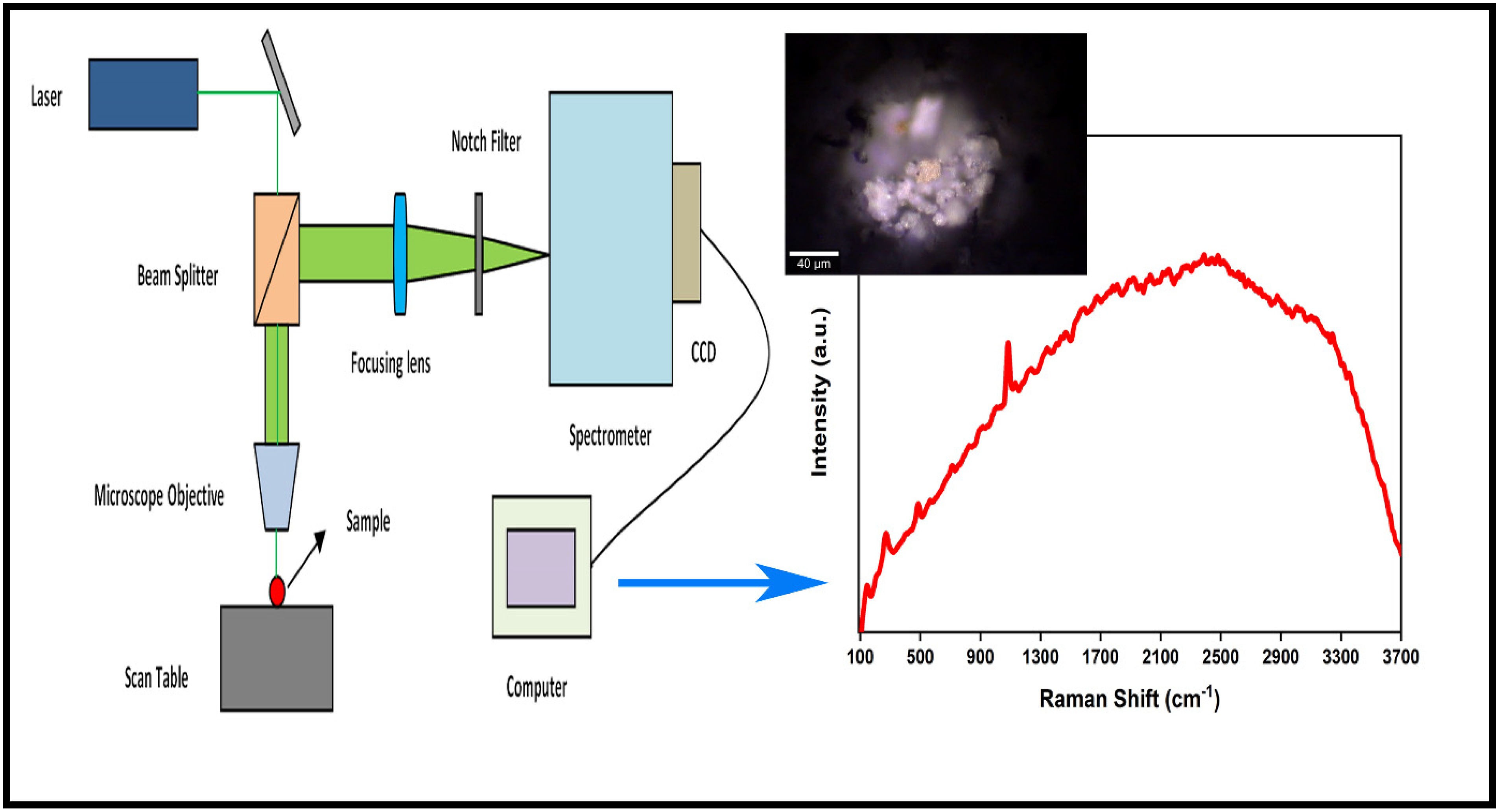
Figure 1. Schematic representation of micro-Raman spectroscope. The microscopic image (20×) and the Raman spectrum of a dust particle (calcite mineral-characteristic vibration: ~1085 cm−1).
Atmosphere 13 01779 g001
Figure 2. Schematic representation of electrodynamic balance (EDB)–Raman system.
Atmosphere 13 01779 g002
Figure 3. Schematic representation of optical tweezer/Raman system.
Atmosphere 13 01779 g003
Figure 4. Schematic representation of a homogenously mixed inorganic/organic aerosol droplet undergoing liquid–liquid phase separation (LLPS) at varying relative humidity (RH) conditions.
Atmosphere 13 01779 g004
Figure 5. Schematic representation of surface-enhanced Raman spectroscopy (SERS).
Atmosphere 13 01779 g005
Table 1. Some offline and online techniques used for the single-particle-level characterization of aerosols.
Offline TechniquesOnline Techniques
Scanning electron microscopy–energy-dispersive X-ray (SEM-EDX)
Micro-Raman spectroscopy (MRS)
High-resolution transmission electron microscopy (HRTEM)
X-ray photoelectron spectroscopy (XPS)
Nano-scale secondary ion mass spectrometry (Nano SIMS)
Time-of-flight SIMS (TOF-SIMS)
X-ray absorption fine structure spectroscopy (XAFS)
X-ray absorption near-edge structure (XANES) spectroscopy
Electron energy loss spectrometry (EELS)
Proton-induced X-ray emission (PIXE)
Single-particle inductively coupled mass spectrometry (SP-ICPMS)
Laser microprobe mass spectrometry (LMMS)
Aerosol time-of-flight mass spectrometer (ATOFMS)
Laser-induced breakdown spectroscopy (LIBS)
Aerosol mass spectrometry (AMS)
Publisher’s Note: MDPI stays neutral with regard to jurisdictional claims in published maps and institutional affiliations.