Article

Primary Ciliary Dyskinesia from Embryogenesis to Adulthood: Micro-CT Analysis of Stage-Dependent Upper Airway Abnormalities in Odad3 Loss-of-Function Mouse Models


1
Institute of Biochemistry and Cell Biology (IBBC), National Research Council of Italy (CNR), Adriano Buzzati-Traverso Campus, Via Ramarini, 32, 00015 Monterotondo, Italy
2
European Mouse Mutant Archive (EMMA), INFRAFRONTIER, Monterotondo Mouse Clinic, National Research Council of Italy (CNR), Adriano Buzzati-Traverso Campus, Via Ramarini, 32, 00015 Monterotondo, Italy
*
Author to whom correspondence should be addressed.
These authors contributed equally to this work.
Genes 2026, 17(9), 1012; https://doi.org/10.3390/genes17091012
Submission received: 28 July 2026 / Revised: 22 August 2026 / Accepted: 24 August 2026 / Published: 27 August 2026
(This article belongs to the Special Issue Utilizing Animal Disease Models to Understand Human Genetics)

Abstract

Background/Objectives: Primary ciliary dyskinesia (PCD) is a genetically heterogeneous disorder characterized by impaired ciliary function, leading to chronic airway disease. Loss-of-function mutations in ODAD3 (CCDC151) represent an established cause in patients, and Odad3-deficient mice recapitulate key disease traits. However, the impact of Odad3 ablation on upper airway development and function remains unexplored. This study investigated the consequences of Odad3 disruption on upper airway structures during development and in adult animals. Methods: Constitutive (Odad3/−) and inducible conditional (Odad3icKO) mouse models were analyzed alongside heterozygous and wild-type littermates during embryonic, postnatal, and adult stages. Optimized high-resolution 3D micro-computed tomography (micro-CT or µCT) combined with histology was utilized to conduct systematic genotype-phenotype evaluations, map upper airway anatomical architecture, and assess PCD disease onset. Results: Genetic dissection revealed a marked dependence of the phenotype on whether Odad3 loss occurred during development or in adulthood. Constitutive Odad3 deletion resulted in pervasive craniofacial remodeling and turbinate hypoplasia during embryonic stages and early postnatal development. Conversely, adult-induced conditional ablation produced localized caudal atrophy of the nasal turbinates accompanied by massive mucus accumulation, consistent with impaired mucociliary clearance and providing a 3D structural and morphological characterization of chronic rhinosinusitis-like pathology in PCD mouse models. Heterozygous Odad3icKO/+ and Odad3+/− mice were phenotypically indistinguishable from wild-type controls, indicating that single-allele loss does not disrupt upper airway morphology. Conclusions: This study characterizes the structural timeline of upper airway pathology in PCD and validates the Odad3icKO model as a robust 3D structural phenotyping platform for investigating airway disease in ciliopathies. Combining targeted genetic disruption with 3D µCT virtual histology offers a powerful framework for comprehensive studies of human genetic variants and gene knockouts in mouse models of PCD.

1. Introduction

Primary ciliary dyskinesia (PCD; OMIM Phenotypic Series: PS244400) is a genetically heterogeneous disorder characterized by defects in the assembly, structure, or coordinated beating of motile cilia. These microscopic, microtubule-based organelles are essential for fluid homeostasis and are found on the apical surface of specialized multiciliated epithelial cells lining the respiratory tract, brain ventricles, and reproductive ducts. Additionally, motile cilia are present as solitary structures at the embryonic node, where they generate leftward fluid flow essential for establishing left-right body asymmetry and form the axonemal core of the sperm flagellum [1,2]. In the airways, ciliary activity is a central component of the mucosal defense system, as it drives mucociliary clearance (MCC). In healthy individuals, cilia propel mucus and trapped pathogens toward the pharynx through rhythmic, wave-like beats [3,4,5]. Consequently, patients with PCD exhibit severe impairment of MCC, leading to chronic sinopulmonary infections, progressive bronchiectasis, and significant upper airway disease beginning in early childhood [6,7].
At the molecular level, the rhythmic beating of the ciliary axoneme, typically organized in a (9 × 2 + 2) microtubule arrangement, is powered by specialized motor complexes known as inner and outer dynein arms (IDAs and ODAs) [8,9]. The precise assembly and attachment of these motors are regulated by the outer dynein arm docking complex (ODA-DC), where mutations in subunit-encoding genes frequently cause PCD, with ODAD3 (formerly CCDC151) representing a component. In humans, nonsense and loss-of-function mutations in ODAD3 result in the absence of ODAs and subsequent profound impairment in ciliary motility, which clinically translates into severe respiratory distress, chronic sinusitis, and laterality defects such as situs inversus totalis [3,8,9].
We previously characterized a constitutive Odad3 knockout mouse model [10]. Phenotypic analysis confirmed that this model faithfully recapitulates the cardinal features of human primary ciliary dyskinesia (PCD) because the loss of Odad3 function results in situs abnormalities and hydrocephalus. However, Odad3−/− animals succumb early in postnatal life, most likely due to the severity of the hydrocephalus. To circumvent this early lethality and bypass the early embryonic window of left-right axis determination, we generated a mouse model harboring a conditional Odad3 allele that enables Cre-mediated deletion upon tamoxifen administration using the Rosa26ERT2-Cre driver. This strategy effectively bypasses the lethal hydrocephalic phenotype and preserves normal organ laterality, enabling late postnatal phenotypic analysis [10,11]. Although the conditional deletion of Odad3 in adult mice has been shown to impair male fertility, recapitulating another phenotype commonly observed in patients with PCD, upper airway abnormalities have not yet been investigated in this model. While the molecular basis of Odad3-related PCD is well documented, the impact of ciliary dysfunction on long-term structural development and remodeling of the upper respiratory tract remains poorly understood. Building upon the previously established systemic Cre-mediated recombination and target deletion of the inducible Odad3icKO mouse model [10,11], the present study extends this characterization by providing a non-destructive, three-dimensional (3D) tissue- and organ-level morphometric assessment of upper respiratory tract development and remodeling across developmental stages.
Defective mucociliary clearance is a central feature of PCD and is associated with high rates of chronic sinonasal and middle ear symptoms. Interestingly, in cystic fibrosis (CF), despite a distinct genetic etiology driven by ion transport defects rather than by impaired primary ciliary motility, patients also develop chronic rhinosinusitis, with computed tomography (CT) scans often revealing agenesis of the paranasal sinuses. While this sinus hypoplasia was initially attributed to chronic inflammation, recent studies in CF animal models have demonstrated that structural sinus defects precede inflammation and may already be present at birth [12,13,14]. Similarly, several cohort studies of patients with PCD have reported a high prevalence of hypoplastic or aplastic paranasal sinuses [14,15]. Therefore, it remains unclear whether sinus abnormalities in PCD have a primary developmental origin or occur as a secondary consequence of chronic, recurrent infections.
The nasal cavity has a highly intricate skeletal organization. It is divided by the nasal septum and internally structured by scroll-like bony formations known as nasal turbinates [16]. These structures, lined by specialized respiratory and olfactory epithelia, perform several essential physiological functions. By markedly increasing the internal surface area of the nasal cavity, they contribute to warming, humidifying, and filtering inspired air while also providing a structural scaffold for the olfactory neuroepithelium [17,18]. Anatomical complexity varies significantly across species. Whereas humans possess simplified turbinates, macrosmatic rodents have a highly elaborate nasal labyrinth optimized to maximize olfactory and respiratory efficiency [19,20]. Despite their physiological importance, the dense and fragile nature of the nasal skeleton poses a significant challenge for traditional diagnostic and research methods. This technical difficulty may partly explain why upper airway defects in animal models of PCD have historically been understudied. Standard two-dimensional (2D) histological sectioning may fail to preserve or reconstruct the spatial continuity of the turbinate system, leading to fragmented interpretations of pathological remodeling [21,22].
Micro-computed tomography (µCT) has emerged as a powerful tool in developmental biology. By providing high-resolution, non-destructive three-dimensional imaging, µCT allows for the precise phenotyping of skeletal and soft tissue defects across multiple developmental stages and disease conditions [23,24]. In this study, we applied an optimized µCT-mediated virtual histology protocol to enhance X-ray contrast in soft tissues while preserving the integrity of the delicate nasal architecture. To determine whether PCD-related upper airway pathology has a primary developmental origin or arises postnatally, we systematically analyzed an allelic series of constitutive (Odad3−/−) and inducible conditional (Odad3icKO) mouse models. Specifically, we tracked the structural and morphological progression of the nasal cavity across three major temporal windows: (1) embryonic development; (2) early postnatal life; and (3) full adulthood. Embryonic analysis revealed pervasive craniofacial remodeling and turbinate hypoplasia. Analysis of the animals postnatally demonstrated structural defects characterized by ethmoturbinate mucosal degeneration and nasal passage obstruction by mucus. Adult Odad3 ablation yielded a phenotype comparable to the postnatal phenotype, characterized by ethmoturbinate mucosal degeneration and caudal mucus accumulation. Overall, our findings indicate that impaired mucociliary clearance is a primary driver of PCD-like chronic rhinosinusitis. Ultimately, this work establishes Odad3 loss-of-function mice as valuable experimental models and provides a powerful platform for interpreting the clinical spectrum of human genetic variants and evaluating the efficacy of emerging targeted therapies.

2. Materials and Methods

2.1. Ethics Statement

This study adhered to all relevant ethical and regulatory frameworks for animal research. All animal procedures were conducted in accordance with protocols approved by the Ethical and Scientific Commission of the Veterinary Health and Welfare Department of the Italian Ministry of Health (protocol approval reference: N:0000688 21 March 2008). The study also complied with established guidelines for animal welfare set forth by Italian laws and regulations, which are aligned with the relevant European Union directives (86/609/EEC and 2010/63/EU).

2.2. Animal Model

All mice utilized in this study were of the C57BL/6N background and were bred and maintained at the European Mouse Mutant Archive (EMMA) facility in Monterotondo, Italy, under specific pathogen-free (SPF) conditions. Animals were housed under strictly controlled environmental conditions, including a temperature of 21 °C, a 12 h light/dark cycle (lights on 7 a.m. to 7 p.m.), and ad libitum access to food and water, with mice grouped 3–5 per cage by litter and sex. The Odad3tm1b/+ line (previously Ccdc151tm1b/+) was originally generated by the IKMC consortium at Monterotondo (details in [10,11]). Genotyping of the Odad3tm1b allele was performed using specific primers (Odad3-F: AGAGCCCTGGATCTTAACTGCTGA; Odad3-R: TCCAAGTCATGCAGAGCTGGGATT; Frt-Rev: CCTTCCTCCTACATAGTTGGCAGT), yielding a PCR product of 307 bp for the wild-type (WT) allele and 270 bp for the Odad3tm1b allele. Furthermore, the generation of mice harboring the conditional knockout allele (Odad3icKO) is detailed in the aforementioned publications, and this allele was genotyped using the same set of primers, resulting in WT and conditional knockout bands of 305 bp and 481 bp, respectively. The cohorts used for Cre-mediated induction were generated such that the experimental group carried homozygous Odad3 conditional alleles, while the control group carried heterozygous Odad3 conditional alleles. Both groups were heterozygous for the ROSA26ERT2-Cre allele (MGI:3764519). Cre-recombinase expression was induced in 4-month-old mice via daily intraperitoneal (i.p.) injections of tamoxifen (1 mg/day; Sigma-Aldrich, St. Louis, MO, USA, T5648), dissolved in corn oil (Sigma-Aldrich, St. Louis, MO, USA, C8267) at a concentration of 10 mg/mL for five consecutive days. Nasal structure analysis was performed 4 months after tamoxifen injections.

2.3. Optimized Tissue Processing Protocol: Enhancing Soft Tissue Contrast for Micro-CT and Histology

To enable complementary high-resolution µCT analysis and subsequent histological evaluation, we optimized the tissue processing protocol. This method was specifically designed to enhance the inherent X-ray contrast of soft, connective, and epithelial tissues while eliminating the high X-ray attenuation of calcified bone (via decalcification), ensuring the tissue is suitable for both imaging and sectioning.

2.4. Processing of Calcified Skulls (Adult/Postnatal Samples)

All skulls were initially fixed in 4% paraformaldehyde (PFA) overnight (O.N.) at 4 °C to preserve tissue architecture prior to chemical decalcification. Decalcification was performed by immersion in an 8% EDTA/50 mM Tris solution (pH 8.5) at room temperature with agitation for approximately two weeks. This extensive aldehyde fixation and prolonged chelation treatment were essential to remove the highly attenuating bone matrix for 3D structural µCT and histological sectioning, although such processing precludes quantitative local tissue protein/RNA extractions (e.g., Western blot, qPCR) or epitope-sensitive immunohistochemistry on these specific imaging cohorts. Following decalcification, the samples were extensively washed with phosphate-buffered saline (1× PBS) to remove debris and post-fixed in 4% PFA O.N. to ensure optimal tissue preservation. The subsequent steps of dehydration, clearing, and paraffin infiltration followed the established protocol described by Ermakova et al. (2018) [25]: samples were dehydrated through a graded ethanol series (50% and 70% overnight, 95% for 30 min, and 100% for 1 h), cleared in xylene (45 min), and sequentially infiltrated with a 1:1 xylene/paraffin solution (45 min at 56 °C) and pure paraffin wax (overnight at 56 °C). Final embedding was performed in molds for solidification (1 h). Crucially, µCT imaging was performed on the resulting paraffin blocks prior to sectioning, ensuring the preservation of the native 3D spatial orientation.

2.5. Processing of Embryos (E12.5 Samples)

E12.5 embryos were carefully dissected and staged according to standard morphological criteria prior to processing [24,25]. These samples, which exhibit minimal bone mineralization, were fixed in 4% PFA overnight at 4 °C immediately following washing in 1× PBS. As significant calcified structures were absent at this developmental stage, the decalcification step was logically omitted to preserve soft tissue integrity. The subsequent dehydration, clearing, and paraffin infiltration procedures were performed identically to the skull processing protocol.

2.6. High-Resolution µ-CT Analysis

Three-dimensional micro-computed tomography (micro-CT or µCT) images were acquired using a high-resolution imaging system (SkyScan 1172G, Bruker, Kontich, Belgium) coupled with an L7901-20 Microfocus X-ray Source (Hamamatsu Photonics, Hamamatsu, Japan). Volume acquisition for the skulls was performed using a camera pixel size of 9 µm, a camera binning of 2 × 2, a tube voltage of 39 kV, a tube current of 240 µA, and an exposure time of 650 ms, resulting in a final isotropic voxel size of 13.56 µm. Conversely, for embryos, volume acquisition utilized a camera pixel size of 9 µm, a camera binning of 2 × 2, a tube voltage of 39 kV, a tube current of 240 µA, and an exposure time of 400 ms, resulting in a 4.88 µm isotropic voxel size. Subsequently, tomographic datasets were reconstructed using the built-in NRecon SkyScan Software (version: 1.6.6.0; Bruker). For visualization and qualitative assessment, the 3D volumes were analyzed using CTvox v. 2.5 (Bruker), while morphometric quantification of ethmoturbinate thickness was performed using Bruker micro-CT Analyser Version 1.13 software after datasets were accurately reoriented to identify the optimal cutting plane for ethmoturbinate visualization. Finally, for statistical analysis, differences between the experimental groups were analyzed using unpaired Student’s t-tests (with data expressed as mean ± SEM), and all data analysis was conducted using GraphPad Prism 6 software, with statistical significance set at p < 0.05. Morphometric measurements were focused on adult mice, as they provided a stabilized pathological framework for quantification of turbinate atrophy and mucosal thickness. Embryonic (E12.5) and postnatal (P11) stages were primarily utilized for a 3D qualitative assessment to identify the ontogenetic onset of the phenotype.

2.7. Histological Validation

Following µCT acquisition, the same paraffin blocks were sectioned serially at 8 µm thickness using a microtome. The resulting cross-sections were prepared for standard Hematoxylin and Eosin (H&E) staining [26] by overnight heating at 45 °C, followed by dewaxing with xylene and rehydration through a graded ethanol series. The staining procedure involved two minutes in hematoxylin, differentiation with 1% hydrochloric ethanol (30 s), bluing with 1% ammonia (30 s), and two minutes of eosin counterstaining. Sections were then dehydrated, cleared twice with xylene (5 min each), and mounted using neutral mounting medium. Bright-field images of the complete cross-sections were acquired using a motorized scope (LMD 7000, Leica Microsystems) equipped with 20× and 40× dry objectives (HC PL FLUOTAR 20× NA: 0.4, Leica Microsystems, Wetzlar, Germany). Images were subsequently imported into ImageJ version 1.54g (National Institutes of Health, Bethesda, MA, USA) for high-resolution correlation with the corresponding µCT virtual sections.

3. Results

3.1. A µCT-Based Virtual Histology Workflow Enables Standardized Mapping of the Adult Mouse Nasal Cavity

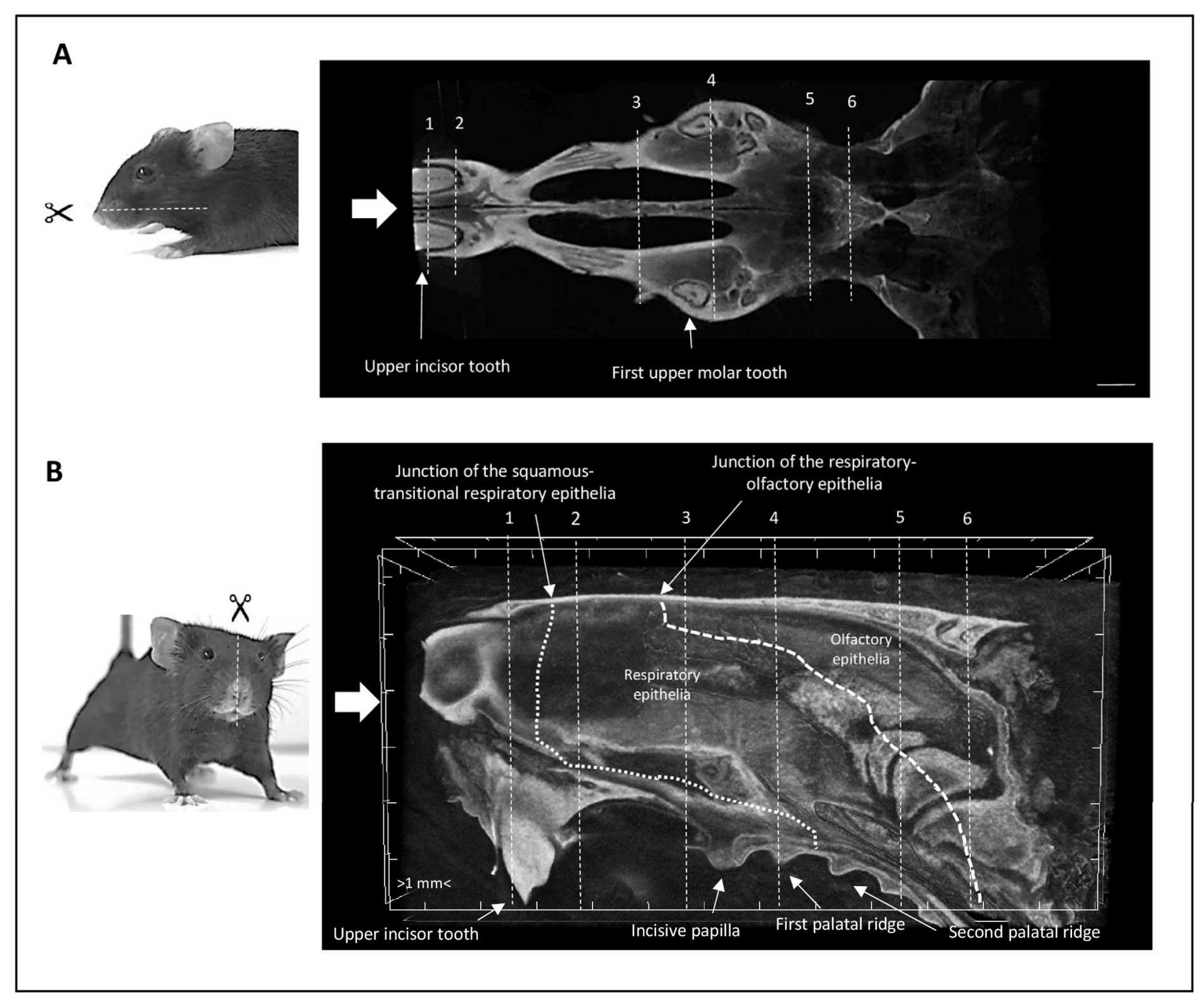
To investigate the structural effects of Odad3 gene ablation on the upper respiratory tract, we applied a tailored three-dimensional (3D) micro-computed tomography (µCT) imaging protocol. This approach enables the simultaneous visualization of both bony structures and soft tissues by exploiting differences in X-ray attenuation following specialized sample processing. Specifically, the methodology combines bone decalcification, paraffin embedding, and high-resolution volumetric µCT imaging to enhance contrast among the soft, connective, and epithelial tissues associated with the nasal cavity. We first established this virtual histology approach by constructing a standardized 3D virtual atlas of the murine upper airways and defining six principal coronal planes suitable for the analysis of PCD mouse models (Figure 1 and Figure 2).
This mapping used established anatomical landmarks within the palatal cavity as reference points, following the standard methodology described by Mery et al. (1994) [27]. Anatomical landmarks within the wild-type (WT) adult palatal cavity were used to anchor the six reference coronal planes (Figure 1). Specifically, the transverse tomographic section (Figure 1A) shows the positions of the incisors and anterior molars, which define the locations of planes 1, 2, and 4; planes 1 and 2 are situated anterior and posterior to the incisors, respectively, whereas plane 4 lies immediately caudal to the anterior molars. The central sagittal section (Figure 1B) illustrates how the incisive papilla and palatal ridges were used to define the remaining planes (3, 5, and 6). All corresponding coronal views are sequentially displayed in Figure 2.
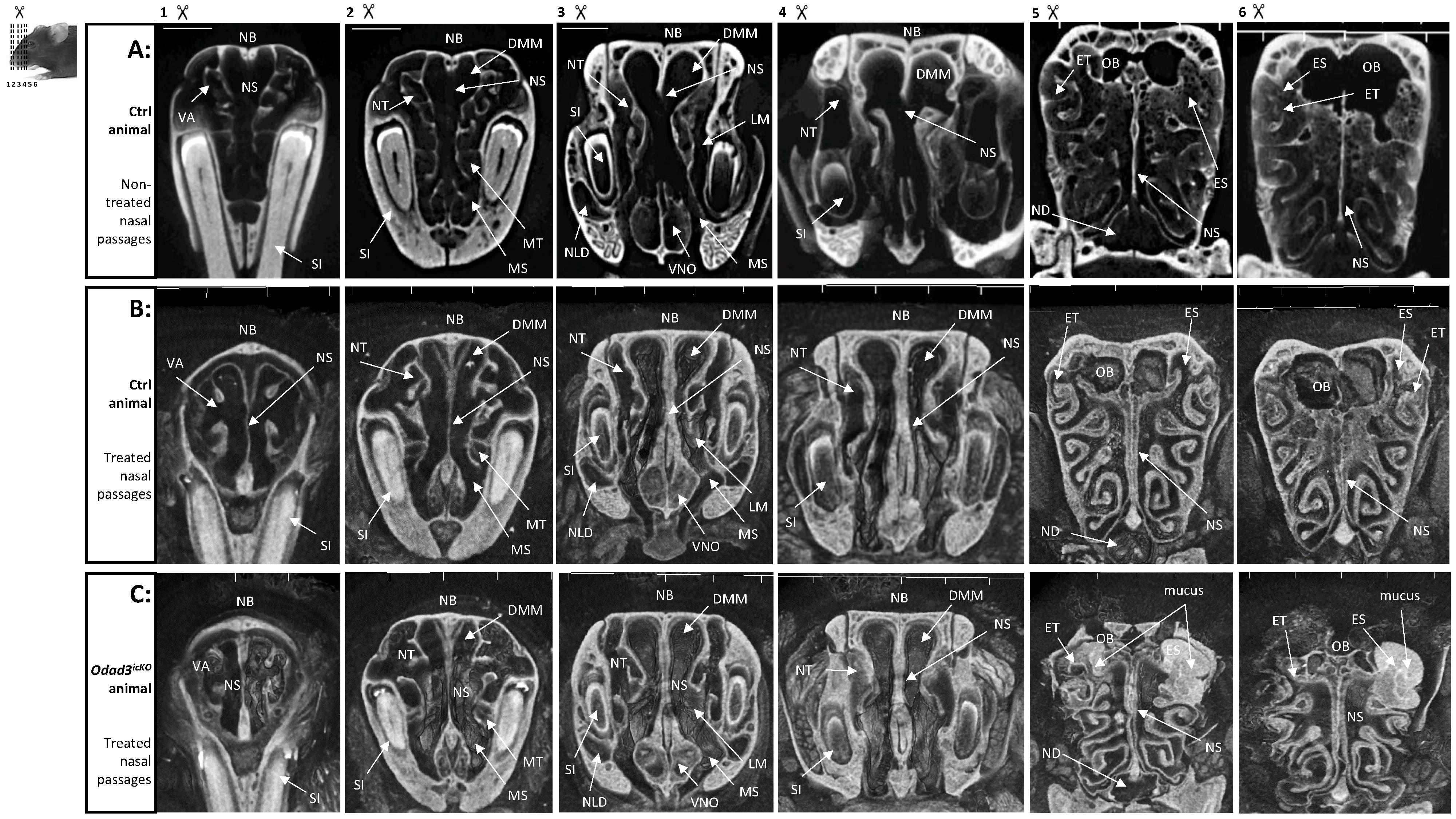
These six standard coronal planes provide an anatomical reference for the principal nasal passages in adult murine models, detailing their underlying bony and cartilaginous frameworks. To optimize soft-tissue contrast and enable subsequent histological correlation on the same specimens, we implemented a tissue processing protocol based on EDTA decalcification followed by paraffin embedding prior to µCT scanning. To assess the effect of this sample preparation procedure, we compared µCT images obtained from standard, non-decalcified control skulls (Figure 2A) with those obtained after decalcification and paraffin embedding (Figure 2B). Imaging of non-decalcified samples clearly delineated highly attenuating structures, such as the nasal bone (NB), nasal septum (NS), and the bony cores of the turbinates (ethmoturbinate—ET, maxilloturbinate—MT, nasoturbinate—NT, atrioturbinate—VA) but provided limited contrast of the overlying soft tissues. In contrast, tomographic volumes obtained after decalcification and paraffin embedding permitted the simultaneous visualization of the skeletal framework and the spatial organization of the respiratory and olfactory epithelia across all six coronal planes (Figure 2B). The improved soft-tissue contrast was particularly evident in the complex ethmoturbinate region represented by planes 5 and 6. It also enabled the identification of adjacent anatomical structures, including the maxillary sinus (MS), ethmoid sinus (ES), nasolacrimal duct (NLD), nasopharyngeal duct (ND), and vomeronasal organ (VNO).

3.2. Adult-Induced Odad3 Deletion Causes Caudal Upper-Airway Remodeling and Luminal Obstruction

To ensure appropriate controls in the inducible models, tamoxifen-induced heterozygous mice (Odad3icKO/+) were utilized as the primary control group (Figure 2A,B, Supplementary Video S4). Preliminary morphometric assessment indicated that these induced heterozygous controls were phenotypically and structurally indistinguishable from untreated WT littermates and constitutive heterozygous (Odad3+/−) mice (Supplementary Videos S1 and S3). We next used high-resolution µCT to characterize the structural consequences of adult Odad3 deletion while circumventing the early postnatal lethality observed in constitutive Odad3−/− mutants. Conditional deletion of the Odad3 gene was induced in adult mice via tamoxifen injections, which activate the expression of the ERT2-Cre recombinase placed under the control of the ubiquitous Rosa26 promoter (Odad3icKO). Following a 4-month post-induction period, the skulls were processed using our sample preparation protocol and imaged by µCT. We observed pronounced alterations in the upper airway architecture of the Odad3icKO but not in the Odad3icKO/+ (Figure 2C, Supplementary Video S2). A detailed rostral-to-caudal comparison between the control passages (Figure 2B) and their Odad3icKO counterparts (Figure 2C) under identical imaging parameters revealed a spatially distinct pattern of pathology. No overt structural abnormalities were observed in the anterior regions corresponding to planes 1–4 (Figure 2B versus Figure 2C). In contrast, severe tissue remodeling was detected in the caudal regions represented by planes 5 and 6. Conditional Odad3 ablation was associated with pronounced thinning and atrophy of the ethmoturbinates, extensive mucosal degeneration, and narrowing of the corresponding nasal passages. Furthermore, the nasal cavities and meati contained abundant luminal material consistent with mucus accumulation, resulting in extensive obstruction of the caudal nasal passages. (Figure 2C, planes 5 and 6). Together, these morphological changes were consistent with severe chronic rhinosinusitis-like upper-airway pathology developing within four months of adult Odad3 deletion. Consistent with successful Cre-mediated induction, this phenotype was observed in all examined homozygous Odad3icKO mice, whereas tamoxifen-treated heterozygous controls (Odad3icKO/+) retained completely normal upper-airway architecture.

3.3. Correlative Histology and µCT Morphometry Confirm Ethmoturbinate Atrophy and Mucosal Degeneration in Odad3icKO Mice

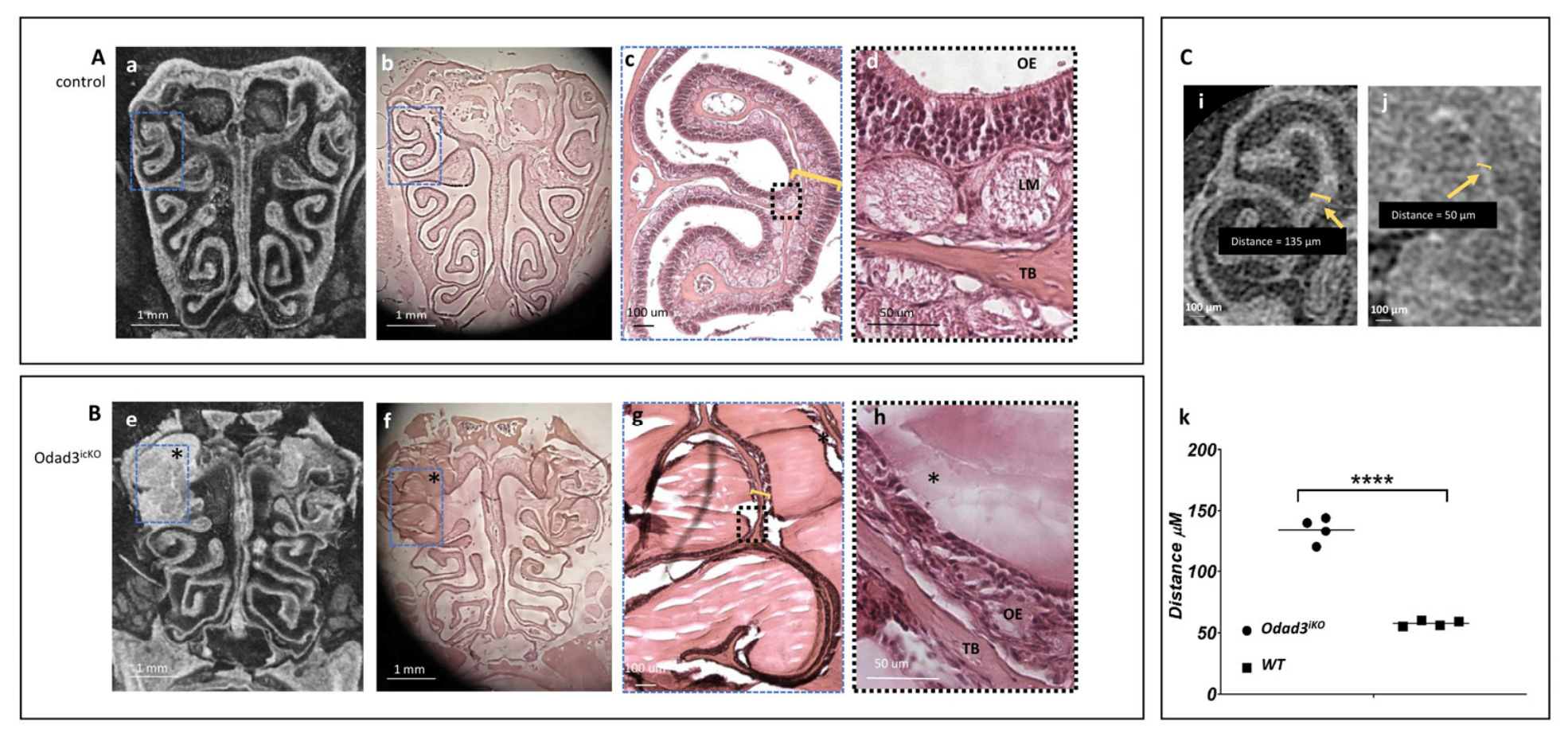
To validate the structural and obstructive alterations detected via 3D volumetric imaging, a correlative histopathological analysis was conducted at the precise anatomical levels identified via µCT. We focused our assessment on coronal plane 5, corresponding to the caudal regions shown in Figure 2B for controls and Figure 2C for Odad3icKO mice, where the three-dimensional rendering revealed the most pronounced tissue remodeling and airway occlusion (Figure 3A versus Figure 3B). Multi-planar reformatting allowed the µCT volumes to be reoriented along different spatial axes and aligned with the physical sectioning plane of the corresponding paraffin blocks. By aligning the virtual datasets with the palatal landmarks defined in Figure 1, histological sections from plane 5 in adult control nasal cavities (Figure 3b–d) showed close anatomical correspondence with the virtual µCT sections (Figure 3a), reproducing the organization of the skeletal framework, epithelial linings, and patent airspaces.
Histological sections from Odad3icKO mice corroborated the abnormalities detected by µCT, confirming severe mucosal degeneration and extensive obstruction of the respiratory passages (Figure 3f–h). At higher magnification, mutant ethmoturbinates displayed marked disruption and thinning of the olfactory epithelium (Figure 3b–d versus Figure 3f–h). The lamina propria (LM) also exhibited structural disorganization, thinning, and cellular degeneration (Figure 3g,h). Furthermore, the ethmoturbinate region was surrounded by abundant mucoid luminal material (Figure 3e,f,h), consistent with stagnant mucus obstructing the nasal meati.
To quantify the extent of ethmoturbinate remodeling, we used multi-planar reformatting to perform morphometric measurements along consistent anatomical axes. This approach enabled reproducible evaluation of the convoluted ethmoturbinate structures while reducing variability associated with differences in physical sectioning orientation.
Total ethmoturbinate thickness was defined as the combined cross-sectional width of the olfactory epithelium (OE), lamina propria (LM), and central turbinate bone (TB) core. Morphometric analysis revealed marked thinning of the ethmoturbinates in adult Odad3icKO mice compared with controls (Figure 3i,j). Quantification confirmed a highly significant reduction in total ethmoturbinate thickness (Figure 3i,j). These findings support the presence of severe ethmoturbinate atrophy following adult Odad3 deletion (Figure 3k).

3.4. Constitutive Odad3 Loss Causes Widespread Upper-Airway Abnormalities at the Postnatal Stage (P11)

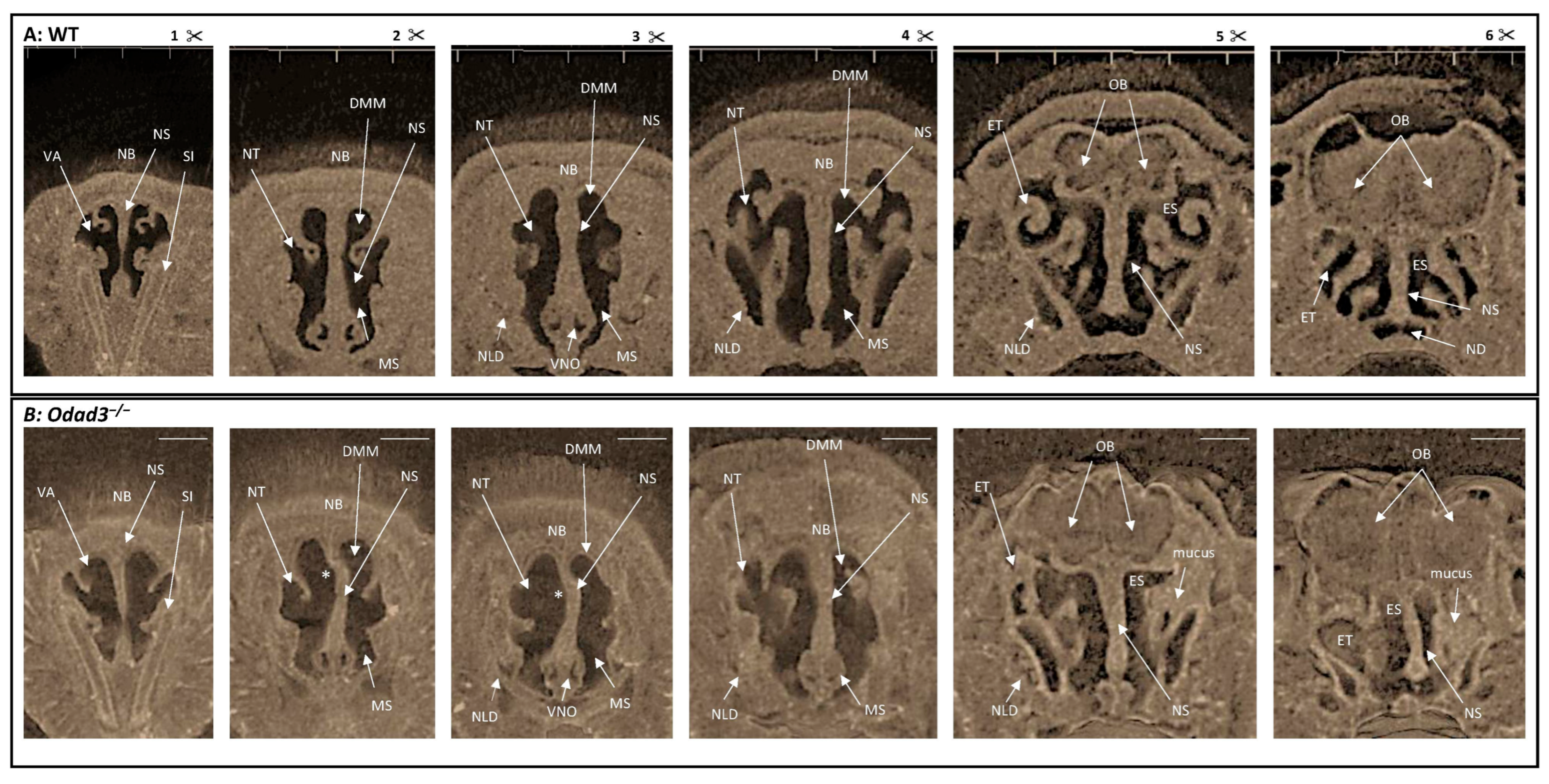
To investigate the upper-airway phenotype during early postnatal development, we performed µCT analysis on constitutive knockout (Odad3−/−) mice at postnatal day 11 (P11; Figure 4), immediately before the period of early lethality previously reported for this model [10]. P11 skulls were processed using the same contrast-enhancing protocol applied to adult samples. The reconstructed datasets were then spatially reoriented according to the anatomical alignment strategy established for adult specimens (Figure 1 and Figure 2), allowing the six reference coronal planes to be reproduced across the postnatal nasal cavities (Figure 4).
Comparison of WT (Figure 4A, Supplementary Video S5) and Odad3−/− pups (Figure 4B, Supplementary Video S6) revealed several abnormalities resembling those observed in adult conditional mutants. These included ethmoturbinate mucosal degeneration, disorganization of the olfactory epithelium, and obstruction of the nasal passages by accumulated luminal material consistent with mucus. However, the phenotype in constitutive Odad3−/− mice was more widely distributed than that observed following adult-induced deletion. Whereas adult Odad3icKO mice showed predominantly caudal abnormalities involving planes 5 and 6, P11 Odad3−/− pups displayed structural alterations across all six analyzed coronal planes. Constitutive mutants also exhibited marked deviation of the nasal septum (Figure 4B, NS*) and pronounced hypoplasia of the supporting structures of the nasal cavity, which may reflect, at least in part, mechanical distortion secondary to the severe hydrocephalus present at this developmental stage. These abnormalities were already evident in the anterior regions represented by planes 1–4. Thus, constitutive loss of Odad3 was associated with widespread structural abnormalities throughout the nasal cavity by early postnatal life.

3.5. Nasal Cavity Abnormalities Are Detectable in Odad3−/− Embryos at E12.5

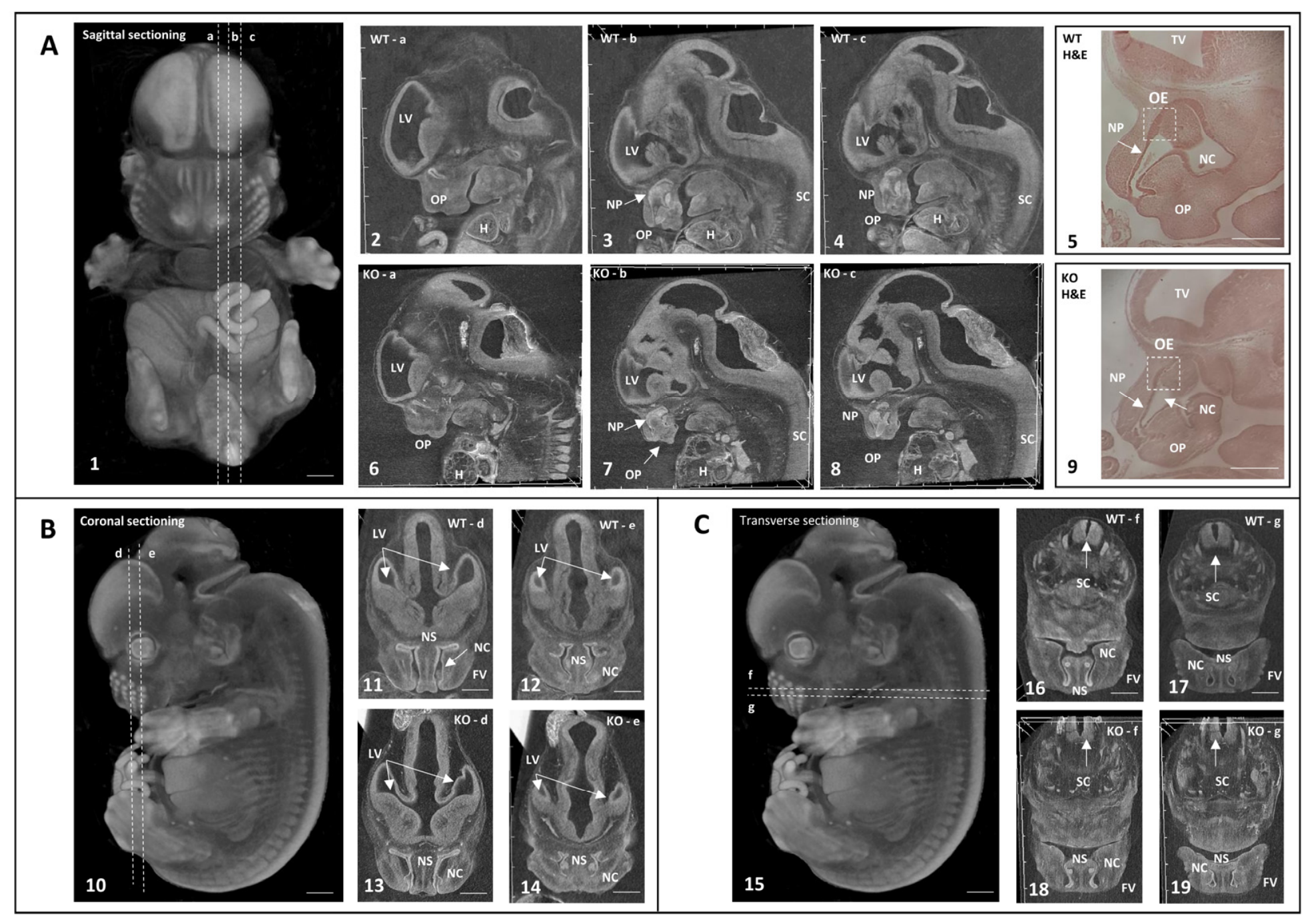
To determine whether nasal abnormalities were already present during embryonic development, we extended the analysis to E12.5 embryos (Figure 5). This stage coincides with the emergence of recognizable nasal cavity primordia, including the nasal pits, olfactory placode, turbinate primordia, and cartilaginous nasal septum [21]. The nascent epithelia are also clearly discernible, particularly the olfactory epithelium lining the posterior regions of the embryonic nasal placode. Notably, because the embryonic craniofacial skeleton at E12.5 consists entirely of unmineralized cartilage primordia rather than mature bone, our contrast-enhancing tissue processing workflow facilitated high-resolution µCT imaging directly within paraffin-embedded samples without requiring a decalcification step.
Leveraging the multi-planar versatility of 3D volumetric analysis, we performed virtual histology of the embryonic nasal cavities along the sagittal (Figure 5A, panels 1–9), coronal (Figure 5B, panels 10–14), and transverse (Figure 5C, panels 15–19) axes. To ensure thorough anatomical sampling, multiple progressive depths were systematically analyzed for each orientation, spanning three cutting levels for the sagittal axis (a–c) and two levels each for the coronal (d,e) and transverse planes (f,g). Crucially, this non-destructive imaging approach preserved sample integrity for subsequent conventional histological validation via hematoxylin and eosin (H&E) staining (Figure 5A, panels 5 vs. 9, WT vs. Odad3−/−, respectively). A systematic comparative analysis between WT (Supplementary Video S7) and Odad3−/− (Supplementary Video S8) embryos revealed structural differences during early nasal cavity development. Mutant embryos showed reduced development of the olfactory nasal placode and hypoplasia of the ethmoturbinate, maxilloturbinate, and nasoturbinate primordia. These differences were visible across sagittal, coronal, and transverse orientations (compare mutant panels 6–8, 13–14, and 18–19 with their respective WT controls, panels 2–4, 11–12, and 16–17). Histological analysis supported the µCT observations and showed a thinner olfactory epithelium lining the primitive dorsal region of the nasal cavity in Odad3−/− embryos (Figure 5A, panels 5 and 9). These observations indicate that structural abnormalities are detectable in constitutive Odad3−/− embryos by E12.5, although further quantitative analysis is required to determine whether this represents a localized nasal defect or to rule out a generalized developmental delay. Importantly, in contrast to the postnatal and adult phenotypes, no luminal material accumulation or mechanical obstruction was detected at E12.5, indicating that mucus accumulation and airway blockage emerge strictly after birth.
Taken together, these cross-sectional analyses identify distinct stage-dependent upper-airway consequences of Odad3 loss. Constitutive disruption was associated with early nasal developmental abnormalities detectable at E12.5 and widespread structural defects at P11. By contrast, deletion induced after completion of development produced a predominantly caudal phenotype characterized by ethmoturbinate atrophy, mucosal degeneration, and luminal obstruction. These findings distinguish an early developmental component associated with constitutive Odad3 loss from an acquired upper-airway phenotype following adult gene ablation.

4. Discussion

The integration of high-resolution µCT-based virtual histology with conventional histological validation provides a robust approach for the three-dimensional assessment of craniofacial and upper-airway anatomy [28]. By leveraging this non-destructive 3D imaging approach, we ensured high-fidelity anatomical registration and precise orientation across different genotypes and developmental stages, overcoming the inherent limitations of traditional two-dimensional (2D) sectioning, which often suffers from spatial data loss. We applied µCT imaging to study the normal anatomy of the nasal cavities and pathological remodeling in an allelic series of the Odad3 loss-of-function mouse models of PCD.
µCT imaging studies have previously been used to evaluate mucociliary clearance (MCC) efficiency and mucus accumulation in the upper airways of a PCD mouse model carrying a conditional deletion of the dynein axonemal intermediate chain 1 protein (Dnaic1) gene. In those studies, the µCT X-ray imaging was performed on untreated animal heads [23], and mucus accumulation in the nasal cavities was inferred indirectly from the absence of airspace, as the air in affected regions, particularly the ethmoturbinate region and maxillary sinuses, was replaced by mucus. In our approach, we implemented a decalcification protocol combined with paraffin embedding prior to X-ray imaging. This procedure significantly enhances contrast in soft tissues within the nasal cavities, enabling enhanced contrast-based visualization of luminal material accumulation as well as the direct measurement of ethmoturbinate thickness. Consequently, this protocol allows for a quantitative assessment of mucosal epithelial degeneration at sufficient resolution for tissue-level morphometry and histological co-registration. Together, our results highlight the utility of this µCT-based approach for the quantitative evaluation of upper airway defects in PCD mouse models and pathology progression directly in 3D.
Here, we report that adult-induced deletion of the Odad3 gene leads to a severe chronic rhinosinusitis (CRS)-like pathology after a 4-month post-ablation period. This is in agreement with the studies performed by Ostrowski et al. (2010) [29], who demonstrated that the conditional deletion of the Dnaic1 gene, a crucial component of the outer dynein arm, using a similar induction protocol (Rosa26ERT2-Cre driver activated by tamoxifen), leads to a reduction in MCC at 1 month post-induction and a complete loss of MCC after 3 months. Animals lacking MCC developed full-blown CRS with mucus accumulation, a loss of normally differentiated epithelial cells at the sites of mucus stagnation, and a thinning of the lamina propria (LM), confirming these features as the most common manifestations of an MCC defect. Overall, we have demonstrated that the Odad3icKO model recapitulates similar histopathological features reported following inducible Dnaic1 ablation, including luminal material accumulation, epithelial degeneration, and ethmoturbinate atrophy.
Clinical symptoms of PCD frequently manifest immediately after birth as respiratory distress in newborns via mechanisms that remain poorly understood. In addition, common findings in infants and children with PCD include daily nasal congestion (rhinitis) and a year-round wet cough occurring soon after birth [30]. We therefore asked whether early postnatal constitutive Odad3 knockout animals already exhibit upper-airway abnormalities. Indeed, luminal material accumulation was widespread, coinciding with severe hypoplasia of the olfactory epithelium detected at postnatal day 11 (P11). These data are in agreement with another murine PCD model carrying a knockout of the radial spoke head 1 homolog (Rsph1) protein, where mucus accumulation was detected as early as postnatal day 3 [31]. Thus, Odad3−/− animals recapitulate early upper-airway manifestations associated with PCD early in life.
Regarding the widespread phenotype observed in Odad3−/− neonates, the marked septal deviation and upper-airway distortion at P11 warrant careful consideration. Given the early onset and severity of hydrocephalus in constitutive mutants, increased intracranial pressure and cranial vault remodeling may exert physical forces that mechanically deform the developing nasal septum and surrounding facial structures. Consequently, while adult-induced deletion isolates the direct effects of Odad3 loss in the upper respiratory tract, the early postnatal phenotype likely reflects a combination of primary structural alterations and secondary mechanical strain driven by hydrocephalus.
Our findings suggest that constitutive Odad3 loss is associated with an early developmental alteration of nasal cavity morphogenesis. Importantly, multiciliated epithelial differentiation and functional mucociliary clearance in the respiratory tract are not yet established at E12.5; therefore, the observed turbinate hypoplasia cannot be attributed to secondary mechanical or obstructive processes (such as mucus accumulation or physical blockage). This early embryonic manifestation raises an intriguing mechanistic question. Specifically, the observation of early nasal hypoplasia and stunted turbinate growth as early as E12.5 raises the possibility that early nodal ciliary dysfunction may impact craniofacial development. We hypothesize that this early developmental defect stems from the crucial role of Odad3 in the motile monocilia of the embryonic node during gastrulation (embryonic days E7.5–E8.0). Disruption of nodal ciliary motility is known to perturb left–right patterning through altered Nodal–Lefty signaling, accounting for the situs inversus observed in constitutive mutants. In contrast, temporal conditional ablation bypasses this early gastrulation window, preserving normal organ laterality (situs solitus) while selectively enabling the isolation of post-developmental respiratory pathology. Whether early nodal ciliary function requirements and associated laterality defects indirectly influence craniofacial or nasal morphogenesis in Odad3-deficient embryos remains to be established [32,33]. One possible interpretation is that the E12.5 nasal phenotype represents an indirect consequence of altered early embryonic patterning. However, alternative explanations, including generalized developmental delay or previously unrecognized local effects of Odad3 loss, cannot currently be excluded. In addition, because the E12.5 analysis was primarily qualitative, further morphometric assessment and rigorous developmental staging will be required to determine whether this represents a localized nasal defect or to rule out a generalized developmental delay.
The broader spatial distribution of the constitutive Odad3−/− phenotype compared to the adult-induced Odad3icKO model suggests that constitutive and adult-induced loss of Odad3 have spatially and anatomically distinct consequences: while the conditional model primarily exhibits localized caudal defects, the constitutive absence of the gene disrupts the fundamental skeletal and mucosal scaffolding across the entire upper respiratory tract. The absence of detectable structural abnormalities in heterozygous (Odad3+/−) mice is consistent with haplosufficiency for the gross upper-airway phenotype under the conditions examined. However, molecular and functional analyses will be required to determine whether more subtle gene-dosage effects on ciliary motility or mucociliary clearance are present. Together with the previously reported reproductive phenotype during spermiogenesis [11], these observations raise the possibility of tissue-specific sensitivity to Odad3 dosage.
The absence of detectable luminal material at E12.5 indicates that mucus accumulation emerges postnatally, after the developmental onset of the structural phenotype. Crucially, because the embryo develops in an environment sheltered from external airflow and pathogens, luminal secretions do not accumulate prior to birth. Following birth, impaired mucociliary clearance may promote secretion retention, which could in turn contribute to epithelial injury, inflammation, and progressive tissue remodeling. In constitutive mutants, these findings are consistent with a two-stage pathological process comprising an early developmental abnormality followed by postnatal mucus accumulation and tissue degeneration. In contrast, the adult-induced model demonstrates that post-developmental loss of Odad3 is independently sufficient to produce an acquired caudal upper-airway phenotype.
Several limitations should be considered when interpreting these findings. First, the developmental stages were examined cross-sectionally using distinct genetic strategies; therefore, the constitutive and adult-induced models do not represent a single longitudinal disease trajectory. Second, the embryonic and early postnatal analyses were primarily qualitative, and contributions from generalized developmental delay or hydrocephalus-associated alterations in cranial growth cannot be excluded. Third, while systemic Cre-mediated recombination and target deletion in this inducible model were previously established [10,11], local recombination efficiency, protein loss, and functional ciliary motility (such as ciliary beat frequency) were not directly quantified in the nasal tissues of these archived imaging cohorts due to the technical requirements of PFA fixation and long-term EDTA decalcification optimized for 3D structural µCT mapping; therefore, the functional link between local Odad3 loss, clearance failure, and CRS-like pathology remains inferential from previously published studies of airway defects in PCD mouse models [10,11]. Finally, although µCT and H&E analysis identified luminal material and extensive tissue remodeling, mucin-specific staining and inflammatory profiling will be required to define the composition of the accumulated material and confirm chronic inflammatory disease.
Looking forward, several avenues for future investigation emerge from this work. First, it will be imperative to decipher the specific molecular interactome of Odad3 during the critical E12.5–E14.5 developmental window, exploring its potential involvement in key signaling pathways such as Hedgehog or Notch, which are known to govern epithelial patterning and turbinate outgrowth [34,35]. Second, to build directly upon the 3D structural baseline established here, future studies will incorporate tissue-specific molecular profiling (qPCR/IHC) and live ex vivo functional assays, such as high-speed video microscopy (HSVM) on fresh mucosal explants, to directly quantify local recombination, ciliary beat frequency (CBF), and beating kinematics following Odad3 deletion. Additionally, ultrastructural transmission electron microscopy (TEM) on dedicated glutaraldehyde-fixed samples will serve to map axonemal dynein arm disruption at nanometer resolution. Third, the absence of an overt heterozygous structural phenotype motivates future dose–response and rescue studies to determine the minimum level of Odad3 expression required to preserve upper-airway function [36]. Finally, combined with an independent gene-replacement or inducible rescue strategy, the Odad3icKO model could be used to test whether restoration of ODAD3 function prevents or reverses established upper-airway pathology.
More broadly, recent comprehensive cryo-EM catalogs of the mammalian ciliary proteome have expanded the list of structural candidates well beyond the ~60 genes currently linked to PCD [37]. By cross-referencing these candidate genes with knockout lines available through the International Mouse Phenotyping Consortium (IMPC; https://www.mousephenotype.org/) [38], our decalcification-based µCT pipeline provides a high-content secondary screening framework to prioritize uncharacterized genes for functional validation. However, as illustrated by our findings in the Odad3 model, constitutive ablation of essential axonemal proteins frequently leads to embryonic or perinatal lethality, often driven by severe hydrocephalus or laterality defects. Future screening applications should therefore favor conditional knockouts or adult-viable hypomorphic alleles to avoid systematically excluding essential structural genes. With these considerations, targeted µCT screening of IMPC ciliary mutants offers a tractable, orthogonal approach to connect structural proteomics with clinical genotype–phenotype correlations.
Collectively, our findings reveal distinct temporal consequences of Odad3 loss in the murine upper airway. Constitutive disruption was associated with early abnormalities of nasal cavity morphogenesis that were detectable at E12.5 and accompanied by widespread structural alterations and luminal material accumulation after birth. By contrast, adult-induced deletion produced a predominantly caudal mucus-obstructive and degenerative phenotype, demonstrating that post-developmental loss of Odad3 is sufficient to disrupt upper-airway homeostasis and cause PCD-like pathology.
These observations support the existence of both developmental and acquired components of Odad3-associated upper-airway disease. The combination of inducible genetic modeling, µCT-based virtual histology, and anatomically matched conventional histology provides a useful experimental framework for addressing these questions and for evaluating future therapeutic interventions.

Supplementary Materials

The following supporting information can be downloaded at https://zenodo.org/records/21475754 (accessed on 23 August 2026). Video S1: 3D virtual sectioning of nasal structure in adult WT mice; Video S2: 3D virtual sectioning of nasal structure in adult KO mice; Video S3: 3D virtual sectioning of nasal structure in constitutive heterozygous adult mice; Video S4: 3D virtual sectioning of nasal structure in tamoxifen induced heterozygous adult mice; Video S5: 3D virtual sectioning of nasal structure in early postnatal stage (11 days) WT mice; Video S6: 3D virtual sectioning of nasal structure in early postnatal stage (11 days) KO mice; Video S7: 3D virtual sectioning of nasal structure in WT mice embryos (E12.5); Video S8: 3D virtual sectioning of nasal structure in KO mice embryos (E12.5).

Author Contributions

Conceptualization, T.O., S.P. and O.E.; methodology, T.O., S.P. and O.E.; software, T.O.; validation, T.O., S.P. and O.E.; formal analysis, T.O. and O.E.; investigation, T.O., S.P. and O.E.; resources, T.O., S.P., O.E., F.C., A.G. and M.P.; data curation, T.O.; writing—original draft preparation, T.O.; writing—review and editing, T.O., O.E. and S.P.; visualization, T.O., S.P., O.E., F.C., A.G. and M.P.; supervision, T.O. and O.E.; project administration, T.O. and O.E.; funding acquisition, O.E. All authors have read and agreed to the published version of the manuscript.

Funding

This work was supported in part by Consiglio Nazionale delle Ricerche Progetto d’Interesse Strategico Invecchiamento 2012–2016 (DSB.AD009.001) and European Union Framework Programmes 6–7: EUCOMM and EUCOMMTools (ID: 261492), PHENOSCALE (ID: 223263) grants; EMMA—Sviluppo Internazionale Campus Monterotondo, Consiglio Nazionale delle Ricerche (ME.P05.001.003).

Institutional Review Board Statement

The animal study protocol was approved by the Ethical and Scientific Commission of the Veterinary Health and Welfare Department of the Italian Ministry of Health (protocol approval reference: N:0000688 21 March 2008). The ethical and safety rules and guidelines for the use of animals in biomedical research are provided by the Italian laws and regulations, in the application of the relevant European Union’s directives (n. 86/609/EEC and 2010/63/EU).

Informed Consent Statement

Not applicable.

Data Availability Statement

The original contributions presented in the study are included in the article/Supplementary Materials; further inquiries can be directed to the corresponding authors. Supplementary Materials and dataset are deposited on Zenodo at https://doi.org/10.5281/zenodo.21475754.

Acknowledgments

We are grateful to M. Raspa and F. Scavizzi for animal care organization and control. Special thanks to M. Parmigiani, A. Braconcini and A. Grop from European Mouse Model Archives (EMMA), Monterotondo, Italy, for technical support in animal handling, sample preparation and data analysis.

Conflicts of Interest

The authors declare no conflicts of interest.

Abbreviations

The following abbreviations are used in this manuscript:
PCDPrimary Ciliary Dyskinesia
Micro-CTMicro-Computed Tomography
µCTMicro-Computed Tomography
MCCMucociliary Clearance
ODA-DCOuter Dynein Arm Docking Complex
IDAsInner Dynein Arms
ODAsOuter Dynein Arms
CFCystic Fibrosis
2DTwo-Dimensional
3DThree-Dimensional
WTWild-Type
NBNasal Bone
NSNasal Septum
ETEthmoturbinate
MTMaxilloturbinate
NTNasoturbinate
VAAtrioturbinate
MSMaxillary Sinus
ESEthmoid Sinus
NLDNasolacrimal Duct
NDNasopharyngeal Duct
VNOVomeronasal Organ
H&EHematoxylin and Eosin staining
P11Postnatal Stage 11 days
CRSChronic Rhinosinusitis
SPFSpecific Pathogen-Free
i.p.Intraperitoneal
PFAParaformaldehyde
O.N.Overnight
PBSPhosphate-Buffered Saline
E12.5Embryonic stage 12.5 days
OBOlfactory Bulb
DMMDorsal Medial Meatus
SISuperior Incisor
OEOlfactory Epithelium
LMLamina Propria
TBTurbinate Bone
NCPrimitive Nasal Cavity
NSCartilage primordium of the Nasal Septum
LVLateral Ventricle
FVFollicles of Vibrissae
NPNasal Pit
OPOlfactory nasal Placode
HHeart
SCSpinal Cord

References

  1. Fliegauf, M.; Benzing, T.; Omran, H. When cilia go bad: Cilia defects and ciliopathies. Nat. Rev. Mol. Cell Biol. 2007, 8, 880–893. [Google Scholar] [CrossRef] [Scilit]
  2. Satir, P.; Christensen, S.T. Overview of Structure and Function of Mammalian Cilia. Annu. Rev. Physiol. 2007, 69, 377–400. [Google Scholar] [CrossRef] [Scilit]
  3. Horani, A.; Wee, W.; Omran, H.; Ferkol, T. Primary ciliary dyskinesia phenotypes and correlation with genotype. Curr. Opin. Pulm. Med. 2025, 31, 628–634. [Google Scholar] [CrossRef] [Scilit]
  4. Ferkol, T. Understanding primary ciliary dyskinesia. Pediatr. Pulmonol. 2025, 60, S86–S87. [Google Scholar] [CrossRef] [Scilit]
  5. Meeks, M.; Bush, A. Primary ciliary dyskinesia (PCD). Pediatr. Pulmonol. 2000, 29, 307–316. [Google Scholar] [CrossRef] [Scilit]
  6. Lucas, J.S.; Behan, L.; Galvin, A.D.; Alpern, A.; Morris, A.M.; Carroll, M.P.; Knowles, M.R.; Leigh, M.W.; Quittner, A.L. A quality-of-life measure for adults with primary ciliary dyskinesia: QOL–PCD. Eur. Respir. J. 2015, 46, 375–383. [Google Scholar] [CrossRef] [Scilit]
  7. Shapiro, A.J.; Zariwala, M.A.; Ferkol, T.; Davis, S.D.; Sagel, S.D.; Dell, S.D.; Rosenfeld, M.; Olivier, K.N.; Milla, C.; Daniel, S.J.; et al. Diagnosis, monitoring, and treatment of primary ciliary dyskinesia: PCD foundation consensus recommendations based on state of the art review. Pediatr. Pulmonol. 2016, 51, 115–132. [Google Scholar] [CrossRef] [Scilit]
  8. Hjeij, R.; Onoufriadis, A.; Watson, C.M.; Slagle, C.E.; Klena, N.T.; Dougherty, G.W.; Kurkowiak, M.; Loges, N.T.; Diggle, C.P.; Morante, N.F.C.; et al. CCDC151 Mutations Cause Primary Ciliary Dyskinesia by Disruption of the Outer Dynein Arm Docking Complex Formation. Am. J. Hum. Genet. 2014, 95, 257–274. [Google Scholar] [CrossRef] [Scilit]
  9. Shapiro, A.J.; Davis, S.D.; Ferkol, T.; Dell, S.D.; Rosenfeld, M.; Olivier, K.N.; Sagel, S.D.; Milla, C.; Zariwala, M.A.; Wolf, W.; et al. Laterality Defects Other Than Situs Inversus Totalis in Primary Ciliary Dyskinesia. Chest 2014, 146, 1176–1186. [Google Scholar] [CrossRef] [Scilit]
  10. Chiani, F.; Orsini, T.; Gambadoro, A.; Pasquini, M.; Putti, S.; Cirilli, M.; Ermakova, O.; Tocchini-Valentini, G.P. Functional loss of Ccdc1 51 leads to hydrocephalus in a mouse model of primary ciliary dyskinesia. Dis. Model. Mech. 2019, 12, dmm038489. [Google Scholar] [CrossRef] [Scilit]
  11. Pasquini, M.; Chiani, F.; Gambadoro, A.; Di Pietro, C.; Paoletti, R.; Orsini, T.; Putti, S.; Scavizzi, F.; La Sala, G.; Ermakova, O. The Odad3 Gene Is Necessary for Spermatozoa Development and Male Fertility in Mice. Cells 2024, 13, 1053. [Google Scholar] [CrossRef] [Scilit]
  12. Chang, E.H.; Pezzulo, A.A.; Meyerholz, D.K.; Potash, A.E.; Wallen, T.J.; Reznikov, L.R.; Sieren, J.C.; Karp, P.H.; Ernst, S.; Moninger, T.O.; et al. Sinus hypoplasia precedes sinus infection in a porcine model of cystic fibrosis. Laryngoscope 2012, 122, 1898–1905. [Google Scholar] [CrossRef] [Scilit]
  13. Grayson, J.; Tipirneni, K.E.; Skinner, D.F.; Fort, M.; Cho, D.; Zhang, S.; Prince, A.C.; Lim, D.; Mackey, C.; Woodworth, B.A. Sinus hypoplasia in the cystic fibrosis rat resolves in the absence of chronic infection. Int. Forum Allergy Rhinol. 2017, 7, 904–909. [Google Scholar] [CrossRef] [Scilit]
  14. Pifferi, M.; Bush, A.; Caramella, D.; Di Cicco, M.; Zangani, M.; Chinellato, I.; Macchia, P.; Boner, A.L. Agenesis of paranasal sinuses and nasal nitric oxide in primary ciliary dyskinesia. Eur. Respir. J. 2011, 37, 566–571. [Google Scholar] [CrossRef] [Scilit]
  15. Pappa, A.K.; Sullivan, K.M.; Lopez, E.M.; Adams, K.N.; Zanation, A.M.; Ebert, C.S., Jr.; Thorp, B.D.; Senior, B.A.; Leigh, M.W.; Knowles, M.R.; et al. Sinus Development and Pneumatization in a Primary Ciliary Dyskinesia Cohort. Am. J. Rhinol. Allergy 2021, 35, 72–76. [Google Scholar] [CrossRef] [Scilit]
  16. Morgan, K.T. Approaches to the Identification and Recording of Nasal Lesions in Toxicology Studies. Toxicol. Pathol. 1991, 19, 337–351. [Google Scholar] [CrossRef] [Scilit]
  17. Gross, E.A.; Swenberg, J.A.; Fields, S.; Popp, J.A. Comparative morphometry of the nasal cavity in rats and mice. J. Anat. 1982, 135, 83–88. [Google Scholar]
  18. Chamanza, R.; Wright, J.A. A Review of the Comparative Anatomy, Histology, Physiology and Pathology of the Nasal Cavity of Rats, Mice, Dogs and Non-human Primates. Relevance to Inhalation Toxicology and Human Health Risk Assessment. J. Comp. Pathol. 2015, 153, 287–314. [Google Scholar] [CrossRef] [Scilit]
  19. Alvites, R.D.; Caseiro, A.R.; Pedrosa, S.S.; Branquinho, M.E.; Varejão, A.S.P.; Maurício, A.C. The Nasal Cavity of the Rat and Mouse—Source of Mesenchymal Stem Cells for Treatment of Peripheral Nerve Injury. Anat. Rec. 2018, 301, 1678–1689. [Google Scholar] [CrossRef] [Scilit]
  20. Wu, Z.; Jiang, J.; Lischka, F.W.; Zhao, K. Is the mouse nose a miniature version of a rat nose? A computational comparative study. Comput. Methods Programs Biomed. 2024, 254, 108282. [Google Scholar] [CrossRef] [Scilit]
  21. Parslow, V.R.; Elmore, S.A.; Cochran, R.Z.; Bolon, B.; Mahler, B.; Sabio, D.; Lubeck, B.A. Histology Atlas of the Developing Mouse Respiratory System from Prenatal Day 9.0 Through Postnatal Day 30. Toxicol. Pathol. 2024, 52, 153–227. [Google Scholar] [CrossRef] [Scilit] [PubMed]
  22. Smith, T.D.; Craven, B.A.; Engel, S.M.; Van Valkenburgh, B.; DeLeon, V.B. “Mucosal maps” of the canine nasal cavity: Micro-computed tomography and histology. Anat. Rec. 2021, 304, 127–138. [Google Scholar] [CrossRef] [Scilit]
  23. Yin, W.; Golliher, H.L.; Ferguson, A.J.; Kimbell, J.S.; Livraghi-Butrico, A.; Rogers, T.D.; Grubb, B.R.; Kimple, A.J.; Ostrowski, L.E. Mucolytic treatment of chronic rhinosinusitis in a murine model of primary ciliary dyskinesia. Front. Mol. Biosci. 2023, 10, 1221796. [Google Scholar] [CrossRef] [Scilit]
  24. Li-Villarreal, N.; Rasmussen, T.L.; Christiansen, A.E.; Dickinson, M.E.; Hsu, C.-W. Three-dimensional microCT imaging of mouse heart development from early post-implantation to late fetal stages. Mamm. Genome 2023, 34, 156–165. [Google Scholar] [CrossRef] [Scilit]
  25. Ermakova, O.; Orsini, T.; Gambadoro, A.; Chiani, F.; Tocchini-Valentini, G.P. Three-dimensional microCT imaging of murine embryonic development from immediate post-implantation to organogenesis: Application for phenotyping analysis of early embryonic lethality in mutant animals. Mamm. Genome 2018, 29, 245–259. [Google Scholar] [CrossRef] [Scilit]
  26. Cheng, J.; Chen, J.; Zhao, Y.; Yang, J.; Xue, K.; Wang, Z. MicroRNA-761 suppresses remodeling of nasal mucosa and epithelial–mesenchymal transition in mice with chronic rhinosinusitis through LCN2. Stem Cell Res. Ther. 2020, 11, 151. [Google Scholar] [CrossRef] [Scilit]
  27. Mery, S.; Gross, E.A.; Joyner, D.R.; Godo, M.; Morgan, K.T. Nasal Diagrams: A Tool for Recording the Distribution of Nasal Lesions in Rats and Mice. Toxicol. Pathol. 1994, 22, 353–372. [Google Scholar] [CrossRef] [Scilit]
  28. Metscher, B.D. MicroCT for comparative morphology: Simple staining methods allow high-contrast 3D imaging of diverse non-mineralized animal tissues. BMC Physiol. 2009, 9, 11. [Google Scholar] [CrossRef] [Scilit]
  29. Ostrowski, L.E.; Yin, W.; Rogers, T.D.; Busalacchi, K.B.; Chua, M.; O’Neal, W.K.; Grubb, B.R. Conditional Deletion of Dnaic1 in a Murine Model of Primary Ciliary Dyskinesia Causes Chronic Rhinosinusitis. Am. J. Respir. Cell Mol. Biol. 2010, 43, 55–63. [Google Scholar] [CrossRef] [Scilit]
  30. Knowles, M.R.; Leigh, M.W.; Ostrowski, L.E.; Huang, L.; Carson, J.L.; Hazucha, M.J.; Yin, W.; Berg, J.S.; Davis, S.D.; Dell, S.D.; et al. Exome Sequencing Identifies Mutations in CCDC114 as a Cause of Primary Ciliary Dyskinesia. Am. J. Hum. Genet. 2013, 92, 99–106. [Google Scholar] [CrossRef] [Scilit]
  31. Yin, W.; Livraghi-Butrico, A.; Sears, P.R.; Rogers, T.D.; Burns, K.A.; Grubb, B.R.; Ostrowski, L.E. Mice with a Deletion of Rsph1 Exhibit a Low Level of Mucociliary Clearance and Develop a Primary Ciliary Dyskinesia Phenotype. Am. J. Respir. Cell Mol. Biol. 2019, 61, 312–321. [Google Scholar] [CrossRef] [Scilit]
  32. Brennan, J.; Norris, D.P.; Robertson, E.J. Nodal activity in the node governs left-right asymmetry. Genes Dev. 2002, 16, 2339–2344. [Google Scholar] [CrossRef] [Scilit]
  33. Shiratori, H.; Hamada, H. The left-right axis in the mouse: From origin to morphology. Development 2006, 133, 2095–2104. [Google Scholar] [CrossRef] [Scilit]
  34. Xu, J.; Iyyanar, P.P.R.; Lan, Y.; Jiang, R. Sonic hedgehog signaling in craniofacial development. Differentiation 2023, 133, 60–76. [Google Scholar] [CrossRef] [Scilit]
  35. Pakvasa, M.; Haravu, P.; Boachie-Mensah, M.; Jones, A.; Coalson, E.; Liao, J.; Zeng, Z.; Wu, D.; Qin, K.; Wu, X.; et al. Notch signaling: Its essential roles in bone and craniofacial development. Genes Dis. 2021, 8, 8–24. [Google Scholar] [CrossRef] [Scilit]
  36. McIntyre, J.C.; Davis, E.E.; Joiner, A.; Williams, C.L.; Tsai, I.-C.; Jenkins, P.M.; McEwen, D.P.; Zhang, L.; Escobado, J.; Thomas, S.; et al. Gene therapy rescues cilia defects and restores olfactory function in a mammalian ciliopathy model. Nat. Med. 2012, 18, 1423–1428. [Google Scholar] [CrossRef] [Scilit]
  37. Gui, M.; Farley, H.; Anujan, P.; Anderson, J.R.; Maxwell, D.W.; Whitchurch, J.B.; Botsch, J.J.; Qiu, T.; Meleppattu, S.; Singh, S.K.; et al. De novo identification of mammalian ciliary motility proteins using cryo-EM. Cell 2021, 184, 5791–5806.e19. [Google Scholar] [CrossRef] [Scilit]
  38. Groza, T.; Gomez, F.L.; Mashhadi, H.H.; Muñoz-Fuentes, V.; Gunes, O.; Wilson, R.; Cacheiro, P.; Frost, A.; Keskivali-Bond, P.; Vardal, B.; et al. The International Mouse Phenotyping Consortium: Comprehensive knockout phenotyping underpinning the study of human disease. Nucleic Acids Res. 2023, 51, D1038–D1045. [Google Scholar] [CrossRef] [Scilit]
Figure 1. Transverse and sagittal views of the dorsal buccal cavity and nasal passages in a non-treated WT mouse. (A) Tomographic transverse section of the dorsal buccal cavity in WT mice, with numbering of the six main cutting planes for the identification of the corresponding principal nasal passages, as shown in Figure 2 (coronal view). (B) Tomographic sagittal section of the dorsal buccal cavity and nasal passages in WT mice, which shows the corresponding numbering of the cutting planes indicated in (A) and represented in Figure 2; scale bar 1 mm. Scissors and arrow indicate the sectioning plane.
Genes 17 01012 g001
Figure 2. Six principal coronal section levels of the adult mouse nasal cavity. (A) Tomographic coronal sections of a non-decalcified nasal cavity from a control mouse (tamoxifen-induced heterozygous Odad3icKO/+ littermate, indistinguishable from WT); scale bar 1 mm. (B) Tomographic coronal sections of the same nasal cavity following tissue processing (decalcification and paraffin embedding); scale bar 1 mm. (C) Tomographic coronal sections of a processed (decalcified and paraffin-embedded) nasal cavity from a homozygous mutant Odad3icKO mouse; scale bar 1 mm (N = 4 per genotype). Nasal bone (NB); olfactory bulb (OB); ethmoturbinate (ET); ethmoid sinus (ES); nasal septum (NS); maxilloturbinate (MT); maxillary sinus (MS); dorsal medial meatus (DMM); nasoturbinate (NT); atrioturbinate (VA); lateral meatus (LM); nasolacrimal duct (NLD); nasopharyngeal duct (ND); vomeronasal organ (VNO); superior incisor (SI). Scissors icons, dashed lines, and numbers indicate the location, orientation, and sequence of the sectioning planes.
Genes 17 01012 g002
Figure 3. Structural analysis of ethmoturbinates in control and Odad3icKO mice. (A) Representative coronal µCT virtual histology (a) and corresponding hematoxylin and eosin (H&E) histological sections (bd) of the olfactory turbinates in Odad3icKO/+ control mice. The blue squares in (a,b,e,f) indicate the areas enlarged in (c,g). The dashed black squares in (c,g) mark the regions shown at higher magnification in (d,h). Scale bars: 1 mm (a,b); 100 µm (c); 50 µm (d). (B) Coronal µCT virtual histology (e) and H&E tracking (fh) of degenerate olfactory turbinates in Odad3icKO mice. Scale bars: 1 mm (e); 100 µm (f,g); 50 µm (h). (C) Morphometric µCT evaluation of ethmoturbinate structure thickness of control (i) and Odad3icKO (j); yellow square brackets and arrows indicate the measured thickness of the ethmoturbinate framework. Scale bars: 100 µm (i,j). Asterisks (*) indicate pathological mucus accumulation. Graphical quantification (k) demonstrates a severe reduction in ethmoturbinate thickness in Odad3icKO animals compared to controls. (horizontal lines represent group means). Data are presented as mean ± SEM (N = 4 per genotype); **** p ≤ 0.0001 by an unpaired two-tailed Student’s t-test. Abbreviations: olfactory epithelium (OE); lamina propria (LM); turbinate bone (TB).
Genes 17 01012 g003
Figure 4. Postnatal development of nasal structures in constitutive knockout (Odad3−/−) mice at postnatal day 11 (P11). (A) Representative coronal µCT virtual histology sections of nasal passages across planes 1–6 in wild-type (WT) pups. Scale bar: 1 mm. (B) Representative coronal µCT virtual histology sections of nasal passages across planes 1–6 in Odad3−/− pups. Scale bar: 1 mm. The asterisk (*) indicates pronounced septal deviation. N = 4 per genotype. Abbreviations: nasal bone (NB); olfactory bulb (OB); ethmoturbinate (ET); ethmoid sinus (ES); nasal septum (NS); maxillary sinus (MS); dorsal medial meatus (DMM); nasoturbinate (NT); atrioturbinate (VA); nasolacrimal duct (NLD); nasopharyngeal duct (ND); vomeronasal organ (VNO); superior incisor (SI). Scissors icons and numbers indicate the location and sequence of the sectioning planes.
Genes 17 01012 g004
Figure 5. Micro-CT imaging and multi-planar virtual histology of wild-type and Odad3−/− embryos at stage E12.5. (A) Sagittal tomographic workflow showing 3D volume orientation (panel 1) across three progressive depths (a–c) for wild-type (WT; panels 2–4) and Odad3−/− embryos (KO; panels 6–8), alongside corresponding H&E histological validation for WT (panel 5) and Odad3−/− (panel 9). Dashed lines labeled with letters indicate the sectioning depths across the respective anatomical planes (a–c for sagittal, d,e for coronal, and f,g for transverse). White dotted squares highlight structural details of the olfactory epithelium. Scale bar: 500 µm. (B) Coronal tomographic orientation (panel 10) across two reference depths (d,e) comparing WT (panels 11–12) with Odad3−/− embryos (panels 13–14). Scale bar: 500 µm. (C) Transverse tomographic orientation (panel 15) across two reference depths (f,g) comparing WT (panels 16–17) with Odad3−/− embryos (panels 18–19). Scale bar: 500 µm; N = 4 per genotype. Abbreviations: olfactory epithelium (OE); primitive nasal cavity (NC); cartilage primordium of the nasal septum (NS); lateral ventricle (LV); follicles of vibrissae (FV); nasal pit (NP); olfactory nasal placode (OP); heart (H); spinal cord (SP).
Genes 17 01012 g005
Disclaimer/Publisher’s Note: The statements, opinions and data contained in all publications are solely those of the individual author(s) and contributor(s) and not of MDPI and/or the editor(s). MDPI and/or the editor(s) disclaim responsibility for any injury to people or property resulting from any ideas, methods, instructions or products referred to in the content.