Article

Genetic Characterization of Primordial Germ Cells in Spotted Sea Bass (Lateolabrax maculatus)

State Key Laboratory of Mariculture Biobreeding and Sustainable Goods (BRESG), Guangdong Provincial Key Laboratory of Fishery Ecology and Environment, Key Laboratory of South China Sea Fishery Resources Exploitation & Utilization, Ministry of Agriculture and Rural Affairs, South China Sea Fisheries Research Institute, Chinese Academy of Fishery Sciences, Guangzhou 510300, China
*
Author to whom correspondence should be addressed.
These authors contributed equally to this work.
Genes 2025, 16(9), 1012; https://doi.org/10.3390/genes16091012
Submission received: 15 July 2025 / Revised: 12 August 2025 / Accepted: 19 August 2025 / Published: 27 August 2025

Abstract

Background: Primordial germ cells (PGC) are the progenitor cells of sperm and eggs during the embryonic stage. The maternal gene vasa has been widely studied for its role in PGC origin, and other genes like dead end (dnd) have also been identified. Objectives: Spotted sea bass is an important economic marine fish, and the study of its germ cell characteristics provides important basic data for future population breeding and protection. Methods: In this study, we cloned the full-length sequences of Lmvasa (2384 bp, encoding 1905 aa) and Lmdnd (1523 bp, encoding 386 aa) using RACE. Temporal and spatial expression patterns of Lmvasa and Lmdnd in embryos and gonads were analyzed by PCR, immunohistochemistry, and in situ hybridization. We also used microinjections of chimeric RNA containing GFP and Lmvasa 3′ UTR to visualize PGCs. Results: Our results showed that Lmvasa and Lmdnd are expressed primarily in early embryonic development (pre-blastula stage) and were expressed only in the gonads. Immunohistochemistry revealed abundant expression of Lmvasa and Lmdnd proteins in spermatogonia, weak expression in spermatocytes, and no expression in spermatozoa. In ovaries, both genes were expressed throughout oogenesis. Furthermore, PGCs in spotted sea bass belonged to an early localization pattern. Microinjection experiments demonstrated that Lmvasa 3′ UTR effectively labeled PGCs in embryos of spotted sea bass, zebrafish, and medaka. Conclusions: These findings may contribute to understanding PGC development in spotted sea bass and other Percidae.

1. Introduction

As sexually reproducing organisms, fish are composed of somatic and germ cells. The pre-sexual germ cells (i.e., primordial germ cells (PGCs)) are specialized from cells that acquire germ plasm components during early embryonic development [1]. PGCs enter the primordial genital ridges through various pathways, form the primordial gonads, and differentiate into mature germ cells [2]. These cells pass on genetic information from generation to generation, which allows species to develop and evolve.
Initial studies found that the germ cells of anuran amphibians contain an electron-dense structure rich in mitochondria, which was named germ plasm [3]. Such electron-dense bodies are found in PGCs and also in germ cells at different stages of development. In teleosts, studies of removed [4,5] and transplanted [6] germ plasm revealed that it is essential for the identification of PGCs and the developmental process. Fruit flies (Drosophila melanogaste) contain a similar structure, and this germ cell is a precursor of the polar cell [7]. The pole plasm has the potential to induce germ cell formation. In addition, the maternal effector vasa is required for the normal formation of the pole cells [8]. Vasa was first discovered in Drosophila, and this polar component is involved in the regulation of germ cell translation and plays an important role in polar formation and germ cell development. Vasa belongs to the DEAD family, and its members encode ATP-dependent RNA helicases [9,10,11]. Vasa is specifically expressed in the germ cell of many organisms, and this expression pattern is highly conserved [12].
In teleosts, vasa has been used as a whole mount in situ hybridization (WISH) probe to investigate two classical modes of origin of PGCs. WISH showed that early localization is present in zebrafish (Danio rerio), and that vasa mRNAs are concentrated in the cleavage furrow in the early stage of cleavage [13]. In contrast, medaka (Oryzias. latipes) has lost the function of early localization; vasa mRNAs are distributed in various blastomeres in the early embryonic period, and they do not accumulate in certain cells until late gastrulation [14]. Germ cells in the gonads can also be identified by sectioning with vasa anti-sense probes or immunohistochemistry (IHC) of anti-vasa antibodies, especially for early germ cells such as spermatocytes and oocytes [15,16,17]. Stable germ cell labeling has also been achieved by generating transgenic animals utilizing vasa regulatory sequences, which include both the promoter and the 3′ untranslated region (UTR). For instance, transgenic zebrafish driven by the vasa promoter were able to specifically express GFP within the germ cell lineage [18]. In certain commercial fish species, such as rainbow trout (Oncorhynchus mykiss) and turbot (Scophthalmus maximus), transient labeling of PGCs with fluorescent proteins (GFP or mCherry) was accomplished in vivo through the microinjection of synthetic chimeric mRNA containing these fluorescent proteins into zygotes [19,20,21].
Dead end (Dnd) is a maternal RNA that is specific to vertebrate germ plasm, and it has been identified in zebrafish as an RNA-binding protein uniquely expressed in germ cells [22]. In zebrafish [23], Xenopus [24], and mouse [25], loss of dnd function leads to abnormal migration, apoptosis, or deletion of PGCs. Human adolescent testicular germ cell tumors are also associated with dnd variants, suggesting a conserved role for dnd in vertebrate germline development [26]. However, dnd mRNA expression exhibits sexual dimorphism in adult gonads across species. For example, it was detected only in mouse testis [27], whereas in Xenopus [28] and the catfish (Pseudopimelodus mangurus) [29], dnd mRNA expression was restricted to the ovary. Dnd has also been found in the sexual germ cells of medaka [30], zebrafish [23], spinyhead croaker (Collichthys lucidus) [31], and starry flounder (Platichthys stellatus) [32].
The spotted sea bass (L. maculatus) is an economically significant species in the mariculture industry in China, where it is widely cultivated due to its delicious taste and high nutritional value [33,34]. However, the relatively long sexual maturation cycle (approximately 3 years) coupled with the strict requirements for ovarian maturation limit the breeding efficiency and egg yield of this species. Due to habitat degradation, overfishing, and other reasons, the germ plasm quality of spotted sea bass has also degraded. Moreover, the introgression of breeding offspring and adult fish that have escaped from culture ponds or cages has negatively impacted the biodiversity of wild populations of spotted sea bass [35]. However, the development of germ cell technology has made it possible to transplant germline stem cells into fish to shorten the breeding cycle and improve breeding efficiency.
Previous studies have identified PGC through the expression of Lcvasa in adult gonadal tissue in Asian bass (Lates calcarifer) [36]. Recently, single-cell RNA sequencing has also validated Lcvasa and other germline-specific markers in adult ovaries [37]. However, studies on PGC migration pathways during early embryogenesis in Perciformes have not been reported, with a specific lack of studies on the migration pathways of Percidae. In this study, we aimed to identify suitable PGC marker genes in spotted bass and characterize PGC distribution during embryogenesis using WISH and in vivo PGC labeling techniques.

2. Materials and Methods

2.1. Fish and Sample Collection

Spotted sea bass used in this study were 2-year-olds (body length: 58 ± 2 cm, weight: 2 kg, 3 male and 3 female) sourced from ChangFeng Aquatic Sci-Tech Co., Ltd. (Zhuhai, Guangdong Province, China). Fertilized eggs were obtained by natural spawning and cultured at 18 °C ± 1 °C in fresh seawater at Hongxinrong Aquaculture Hatchery (Zhangpu, Fujian Province, China).
After anesthetizing the spotted sea bass, tissue samples (gills, liver, spleen, kidneys, brain, intestines, muscles, ovaries, and testes) were collected and stored in liquid nitrogen (n = 3). After laying eggs, the fertilized eggs were quickly and gently collected with a net. Embryo development was first observed under a stereo microscope (M165 FC, Leica, Wetzlar, Germany). The embryos of the same period were collected with a 5 mL straw and washed with phosphate-buffered saline (PBS). The liquid was aspirated, then transferred to a 2 mL cryopreservation tube and immediately put into liquid nitrogen (n = 3, embryos in each tube ≥ 20).
For IHC and WISH analyses, gonad and embryos were washed in PBS and placed in a fixation solution overnight at 4 °C. Gonad tissues and embryos were dehydrated using PBS-diluted 50% methanol solution for 2 h at room temperature, transferred to 100% methanol solution for 2 h, and finally placed in new 100% methanol solution for storage at −20 °C until used for analysis (n = 3, embryos in each tube ≥ 30).
The marina medaka (Oryzias melastigma) was purchased from Shanghai Feixi Biotechnology Co., Ltd. (Shanghai, China), and the AB zebrafish came from the Wuhan Institute of Hydrobiology (Wuhan, China). Medaka were raised in a circulation system of 30‰ seawater, and zebrafish were raised in a freshwater circulation system. They were raised at 28 °C, and the light period was 14 h in light/10 h in dark. Embryo Collection: female and male fish were reared separately the night before and mixed at a ratio of 2:1 (female:male) the next morning. The fertilized eggs were collected immediately after the female laid the eggs.

2.2. Total RNA Isolation and cDNA Synthesis

Spotted sea bass tissues and embryos total RNA were extracted with Trizol reagent. Reverse transcription of first-strand cDNA was performed using FastKing gDNA Dispelling RT SuperMix kit (Tiangen, Beijing, China). We amplified the open reading frame (ORF) of Lmvasa and Lmdnd from the expression sequence tags (ESTs) of the ovaries of spotted bass in this research group. To obtain full-length cDNA sequences for vasa and dnd, we designed fragments amplified from the expression sequence tags of the of the spotted sea bass in this topic, and these sequences were uploaded to the NCBI database. Lmvasa and Lmdnd full-length cDNA sequences were amplified and sequenced, then aligned using the SMARTer RACE 5′/3′ Kit (Clontech, San Jose, CA, USA). Table 1 lists all primers used in this study. During this period, the sequence was uploaded to the NCBI database, and the accession number was obtained: vasa (GenBank login: PV982366) and dnd (GenBank login: PV982367).

2.3. Phylogenetic Analysis

The amino acid sequences and similarity analyses of Lmvasa and Lmdnd were conducted using BLASTn and BLASTp from the NCBI databases. Jalview 2.11 and Clustal Omega 1.2.4 software were used for sequence alignment. Phylogenetic analysis was carried out using MEGA v5.2 software according to the previous method [38,39].

2.4. RT-qPCR and RT-PCR

RT-qPCR was performed on a LightCycler® 480 II PCR (Roche, Basel, Switzerland) according to the Taq Pro Universal SYBR qPCR Master Mix Kit (Vazyme, Nanjing, China). RT-PCR was performed according to the 2× Rapid Taq Master Mix (Vazyme, Nanjing, China). β-actin was used as a standardized control [16,40]. For RT-PCR, β-actin was amplified with 25 cycles, and Lmvasa and Lmdnd were amplified with 30 cycles. Relative gene-expression levels were calculated using the 2−ΔΔCt method. The primer sequences are shown in Table 1.

2.5. WISH and Histology

Sense and antisense Lmvasa and Lmdnd probes were synthesized from the pGEM-T easy vector containing a 1017 bp Lmvasa fragment and a 1155 bp Lmdnd fragment following a previously published method (Table 1) [41]. The resulting plasmids were linearized by Sac II and Nco I monodigestion, respectively. According to the digoxin RNA labeling kits (SP6/T7; Roche, Mannheim, Germany), purified Lmvasa and Lmdnd probes were obtained, dissolved in RNase-free water, and stored in aliquots at −80 °C. The probe concentration for Lmvasa and Lmdnd was 2 ng/μL.
For histology, the gonads, fixed as described in Section 2.2, were sent to Wuhan Servicebio Technology (China) for embedding, section treatment (4 μm), IHC, and hematoxylin-eosin (H&E) staining (n = 3). All sliced images were observed and acquired under a Leica (DM2500, Leica) with 20.0× and 40.0× lenses. Each slice randomly selected 10 areas for observation, and corresponding typical field of view pictures were provided. For the WISH assay, dehydrated embryos (n ≥ 20) from different periods were removed from storage at −20 °C and gradually diluted with methanol and 0.1% Tween 20 (PBST) at room temperature. Procedures such as embryo permeability treatment, fixation, probe hybridization, washing, chromogenic and embedding were performed according to the methods of Narayanan et al. [41] and Kimmel et al. [42]. Finally, photographs were taken using the stereomicroscope.

2.6. Expression of Recombinant Lmvasa Protein and Preparation of Its Antibody

Lmvasa protein was obtained by inducing the expression of the fusion protein (Lmvasa-His) following a previously established protocol [17]. Briefly, the vasa coding region was amplified and inserted into pET28a digested with EcoR I and Xho I using the In-fusion® HD Cloning Kit (Takara, Tokyo, Japan) (Table 1). Vasa-PET28a was recombinantly expressed in Escherichia coli (E. coli) cells and purified in the HyPur T Ni-TED 6FF (His-Tag) PrePacked Gravity Column Kit (Sangon, Shanghai, China), then detected by SDS-PAGE. The laboratory-purified vasa protein was sent to Hangzhou Huaan Biotechnology (Hangzhou, China) to prepare vasa rabbit polyclonal antibodies (pAbs).

2.7. Western Blotting Analysis

Ovarian tissue was incubated on ice for 20 min in protein lysate containing a final concentration of 1 mM benzsulfonyl fluoride (Beyotime, Nantong, China) and transferred to SDS-PAGE. Western blot analysis of tissue lysates was basically completed as previously described [36].
The primary antibodies used to detect vasa were rabbit pAb prepared by Hangzhou Huaan Biotechnology and β-actin (HRP-conjugated) rabbit mAb (49381) purchased from Signalway Antibody LLC (Greenbelt, MD, USA).

2.8. Preparation of Chimeric mRNAs and Microinjection

PCS2+-EGFP plasmid was used as a template for RNA synthesis in vitro. GFP-Lmvasa 3′-UTR was constructed from previous descriptions [43]. In brief, the 3′-UTR regions were amplified and inserted into PCS2+-EGFP digested with Xho I-Xba I, and the In-Fusion ® HD Cloning Kit was used (Table 1). The resultant plasmid was linearized by BamH I digestion for in vitro transcription using the mMESSAGE mMACHINE® SP6 transcription kit (Invitrogen, Waltham, MA, USA) and stored at −80 °C until use.
As previously described [18,31], mRNA (300 ng/μL) of spotted sea bass, zebrafish, or medaka embryos was prepared. The embryos were placed in a hole on the surface of the agar, and then RNA microinjection solution was microinjected at the 1–4 cell stage for PGCs visualization. In spotted sea bass and zebrafish, each experimental group ≥500 embryos were injected (n = 5). In medaka, each experimental group ≥ 150 embryos were injected (n = 5). The control group was not injected with embryos, and there were the same number of control and experimental groups.

2.9. Microscopy

Observe and photograph embryos using Leica DMi8 inverted microscope DMi8 and stereomicroscope (Leica, Wetzlar, Germany).

2.10. Statistical Analysis

Data was presented as mean ± SEM. All statistical data analyses were performed using GraphPad Prism 10.1 software. Significant differences were assessed by one-way ANOVA and Tukey’s multiple comparison test. When p < 0.05, the difference was considered significant.

3. Results

3.1. Cloning and Characterization of Lmvasa and Lmdnd

Lmvasa fragments of 1905 bp were obtained from the ovaries of spotted bass using specific primers. The Lmvasa cDNA was obtained using 3′ and 5′ RACE techniques has a total length of 2384 bp and consists of 152 bp 5′-UTR, 635 bp ORF, encoding 1905 aa, and 327 bp 3′-UTR (Figure S1).
Similarly, the Lmdnd ORF was amplified using specific primers to 1158 bp. The total length of the Lmdnd amplified by RACE technology was 1523 bp and consists of 84-bp 5′-UTR, 1158 bp ORF encoding 386 aa, and 281 bp 3′-UTR (Figure S2).
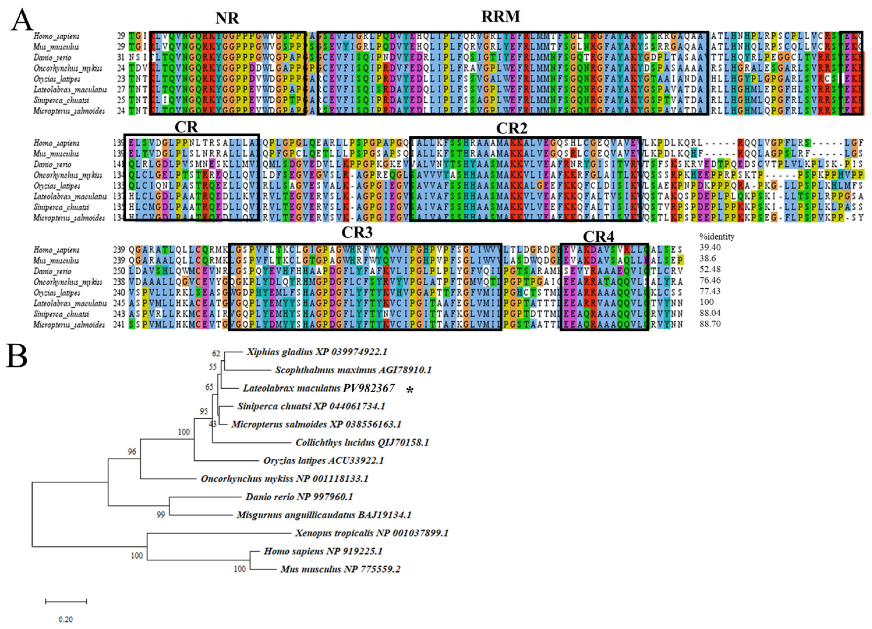
Multiple sequence alignment revealed that Lmvasa shared high identity (71.87–99.72%) with vasa sequences from other fish (Figure 1A). The deduced spotted sea bass vasa protein possessed eight consensus motifs characteristic of the DEAD-box protein family, and the N-terminal region was rich in glycine (G) and arginine (R) residues-RGG and RG motifs (Figure S1 and Figure 1A).
Multiple sequence alignment showed that the aa sequence of Lmdnd contained an RNA recognition motif (RRM) and five conserved regions, namely the N-terminal NR domain and the C-terminal CR1–4 domains (Figure 2A). Blast alignment showed that Lmdnd shared 61.46–88.70% identity with fish dnd sequences and 39.40–52.48% with other species (Figure 2A).
A phylogenetic tree was constructed based on several representative and well-studied aa sequences of teleosts and mammals Lmvasa and Lmdnd. The results showed that the vasa and dnd of teleosts were different from mammals. Further subdivision of teleosts suggests that Lmvasa was phylogenetically close to that of Japanese bass (Lateolabrax japonicus) (Figure 1B). In addition, Lmdnd was clustered with swordfish (Xiphias gladius) and turbot dnd proteins (Figure 2B).

3.2. Expression and Purification of Recombinant Lmvasa Protein

The recombinant protein His-Lmvasa was mainly expressed in inclusion bodies. The protein was purified using Ni2+-TED affinity chromatography and detected using SDS-PAGE analysis. Thick bands were observed at 75 kDa (Figure S3A,B). Anti-Lmvasa specific antibody against purified fusion protein was successfully generated in rabbits.

3.3. Lmvasa and Lmdnd Are Specifically Expressed in Germ Cells and During Embryonic Development

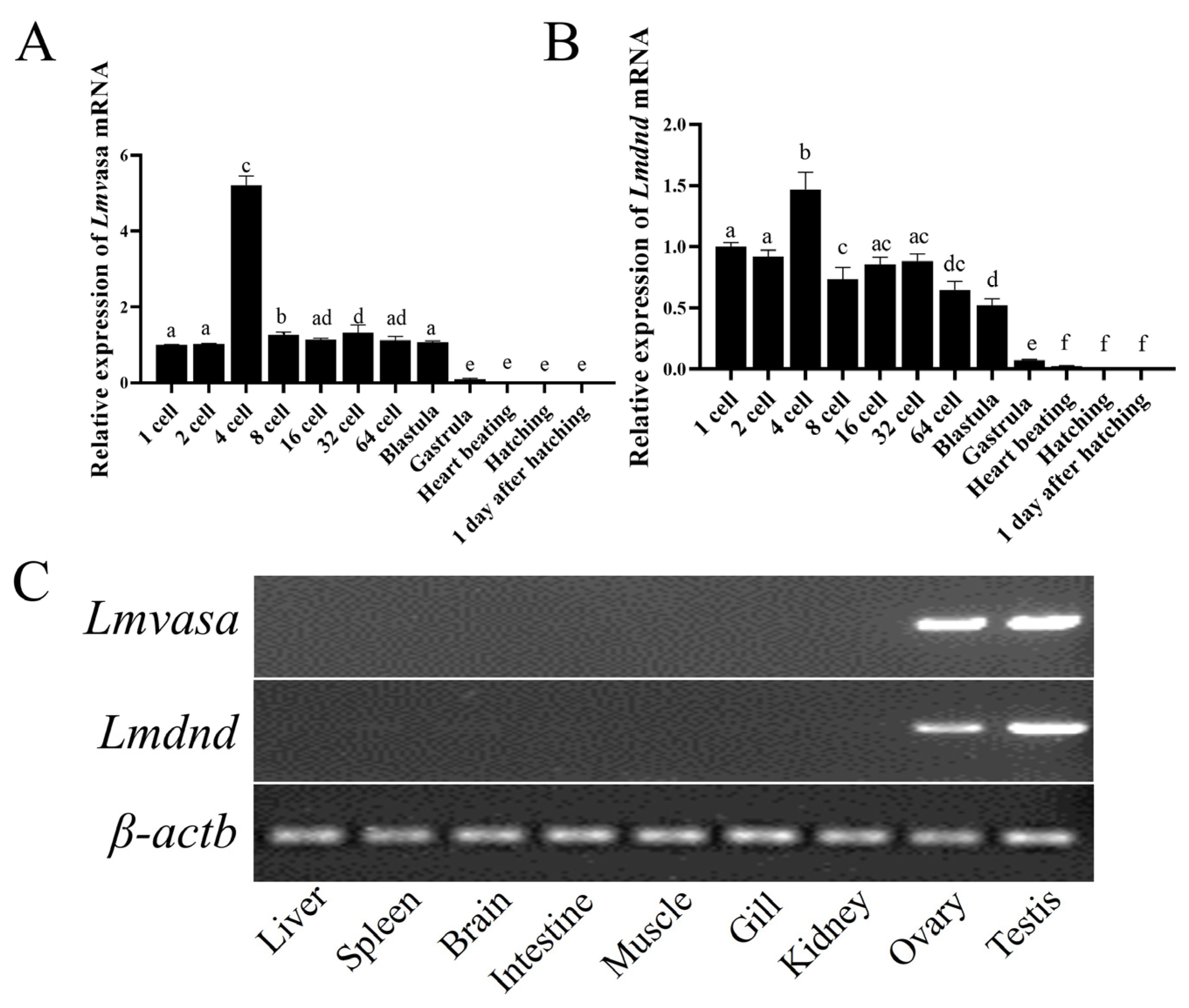
RT-qPCR and RT-PCR were conducted to explore the expression of Lmvasa and Lmdnd during embryonic development and in different tissues. RT-qPCR analysis revealed that the expression of Lmvasa and Lmdnd were detected in the early stages of development, from embryos until the blastula stage. Lmvasa mRNA expression began at the 1–2-cell stage, increased significantly at 4-cells, then decreased gradually, and remained stable from the 8-cell stage to the blastocyst stage. After the gastrula stage, the expression of Lmvasa decreased significantly, and eventually became undetectable (Figure 3A). Similarly, Lmdnd mRNA reached its highest expression level at the 4-cell stage, followed by a gradual decrease in expression, and a significant decrease after the gastrula stage until it became undetectable (Figure 3B). Analysis by RT-PCR in different tissues showed that Lmvasa and Lmdnd were detected almost exclusively in the ovary and testis, as almost no expression was detected in other tissues (Figure 3C).
IHC was carried out to investigate the localization of Lmvasa and Lmdnd proteins in the gonads. In the finger-like seminiferous lobules of the testis, different types of germ cell sacs and single spermatogonia were distributed in the germinal epithelium (Figure 4A). The signals for Lmvasa and Lmdnd proteins were most intense in spermatogonia, faint in spermatocytes, and absent in spermatids (Figure 4B,C). In the mature female ovary, Lmvasa and Lmdnd proteins were expressed at all stages of ovarian development, but expression was mainly concentrated in late vitellogenic oocytes (LVO, IV) and postvitellogenic oocytes (V). Expression was weak in primary growth oocytes (II) and perinucleolar (PNO) and previtellogenic oocytes (III) (Figure 4E,F). In stage II oocytes, Lmvasa and Lmdnd were expressed in granules around the nucleus, and they were weakly expressed in stage III oocytes. Lmvasa and Lmdnd were diffused weakly in the cytoplasm in stage IV oocytes. In stage V oocytes, Lmvasa and Lmdnd proteins were evenly distributed in the follicles.

3.4. Vasa and Dnd RNA Localization in Spotted Sea Bass Visualized by WISH

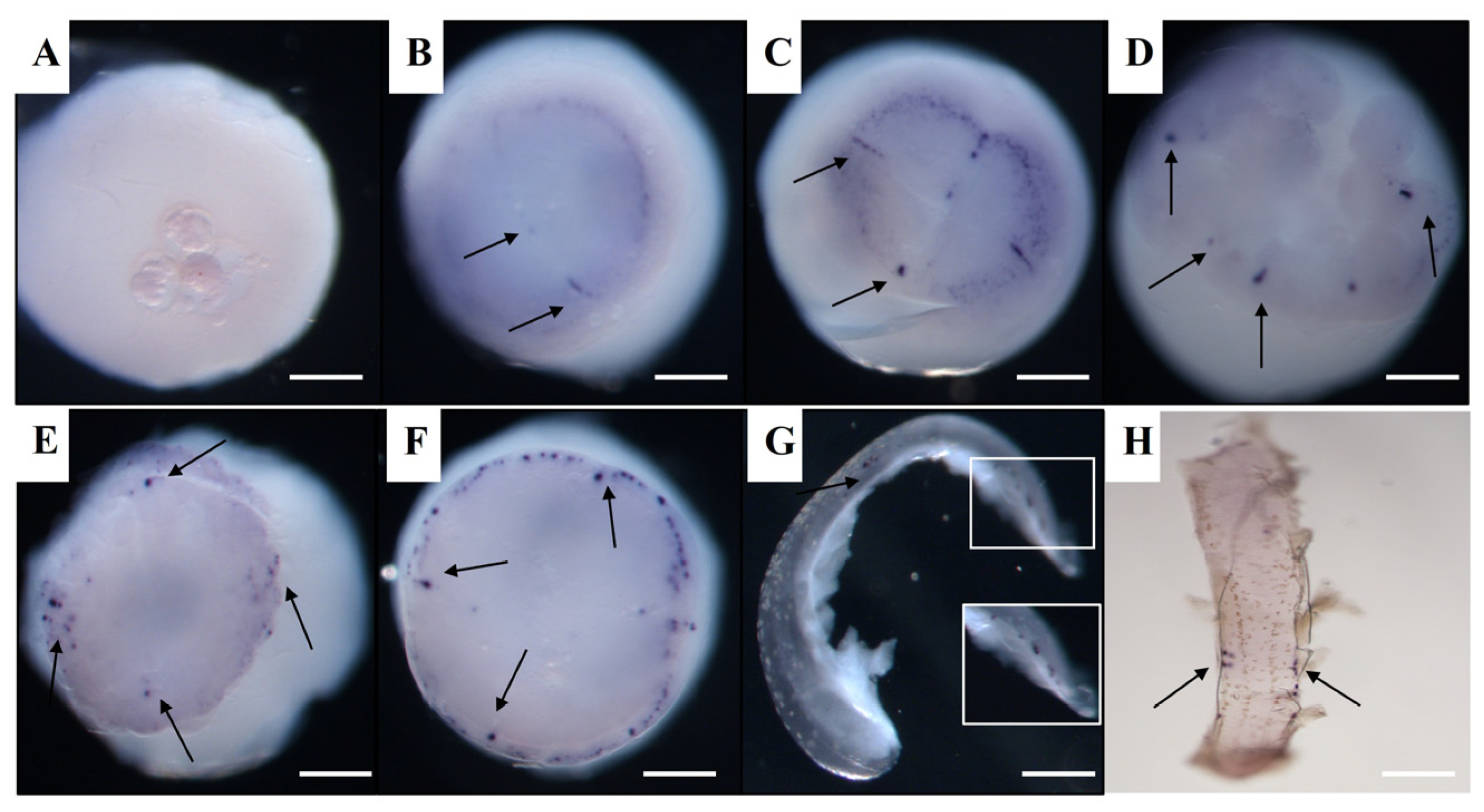
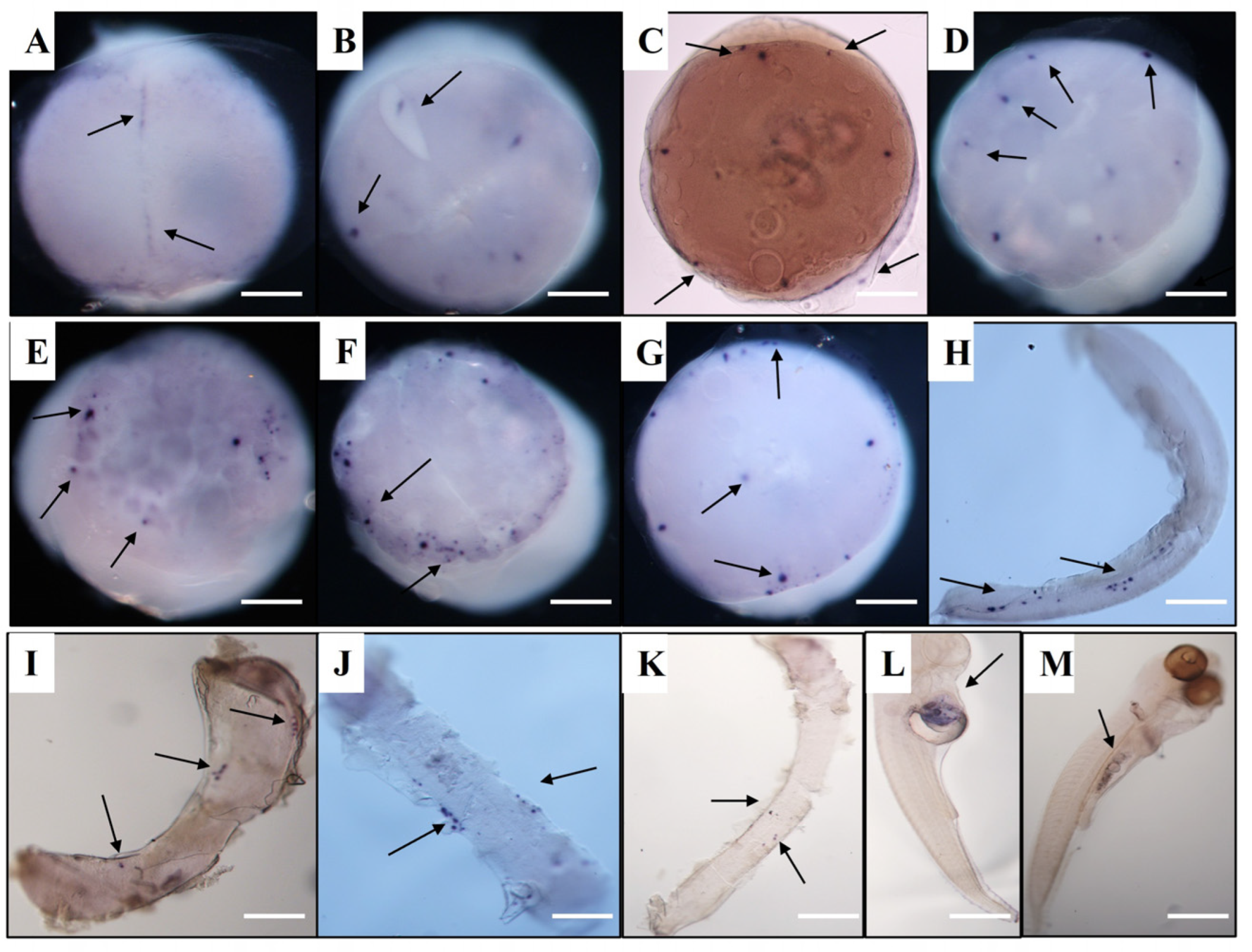
At the cleavage stages, Lmvasa and Lmdnd mRNA aggregated along the cleavage planes before being localized within several individual cells at the morula stage. At the 1-cell stage, Lmvasa and Lmdnd mRNA could not be detected using WISH (Figure 5A). At the 2-cell stage, many small signals of Lmvasa and Lmdnd mRNA were detected within the forming cleavage furrow and around the cell margin of the first cleavage furrow (Figure 5B and Figure 6A). At the 4-cell stage, four Lmvasa and Lmdnd signals were found in the first and second cleavage furrow, and each of the signals remained localized at the outer edge of the cleavage plane (Figure 5C and Figure 6B). At the 8-cell stage, four new Lmdnd signals were present along the two new cleavage furrows (Figure 6C), and at the 16–32 cell stage, eight Lmvasa and Lmdnd signals were still localized in the cleavage furrow (Figure 5D and Figure 6D).
At the morula stage, Lmdnd signals were observed within individual cells. The signals were usually in four to six large clusters within individual cells, with smaller fragments in neighboring cells (Figure 6E). At the blastula stage, more Lmvasa and Lmdnd signal-expressing cells were observed in each group, and most of the positive cells were distributed at the edge of the blastoderm to form PGCs (Figure 5E and Figure 6F). At the early gastrula stage, Lmvasa-and Lmdnd-positive cells were predominantly located at the edge of the blastoderm, but a few signals were occasionally observed at the upper or middle part of the blastoderm (Figure 5F and Figure 6G). During the late gastrula stages, Lmvasa-and Lmdnd-positive cells moved towards the posterior part of the embryo as the embryonic body was extended (Figure 5G and Figure 6H).
As the embryo developed further, Lmdnd-positive cells migrated towards the embryonic body in the early somite stage and were loosely arranged on both sides of the embryonic body from the anterior side of the embryonic axis (head region) to the caudal bud (Figure 5H and Figure 6I). At the late somite stage, the bilateral clusters formed by Lmvasa-and Lmdnd-positive cells actively migrated posteriorly (Figure 6J) and gathered into two clusters at the intended genital crest site during the heart beating stage (Figure 6K). At the hatching stage, Lmdnd-positive cells were located below the spine at the junction of the yolk bulb and yolk sac extension (Figure 6L). Finally, Lmdnd-positive cells were located at the dorsal side of the gut and the ventral side of the notochord at 3 days after hatching (dah) (Figure 6M). Figure 7 is a schematic illustration of the spatiotemporal expression pattern of Lmvasa and Lmdnd during spotted sea bass embryogenesis.

3.5. Visualization of Three Fish PGCs by GFP-Lmvasa 3′-UTR mRNA

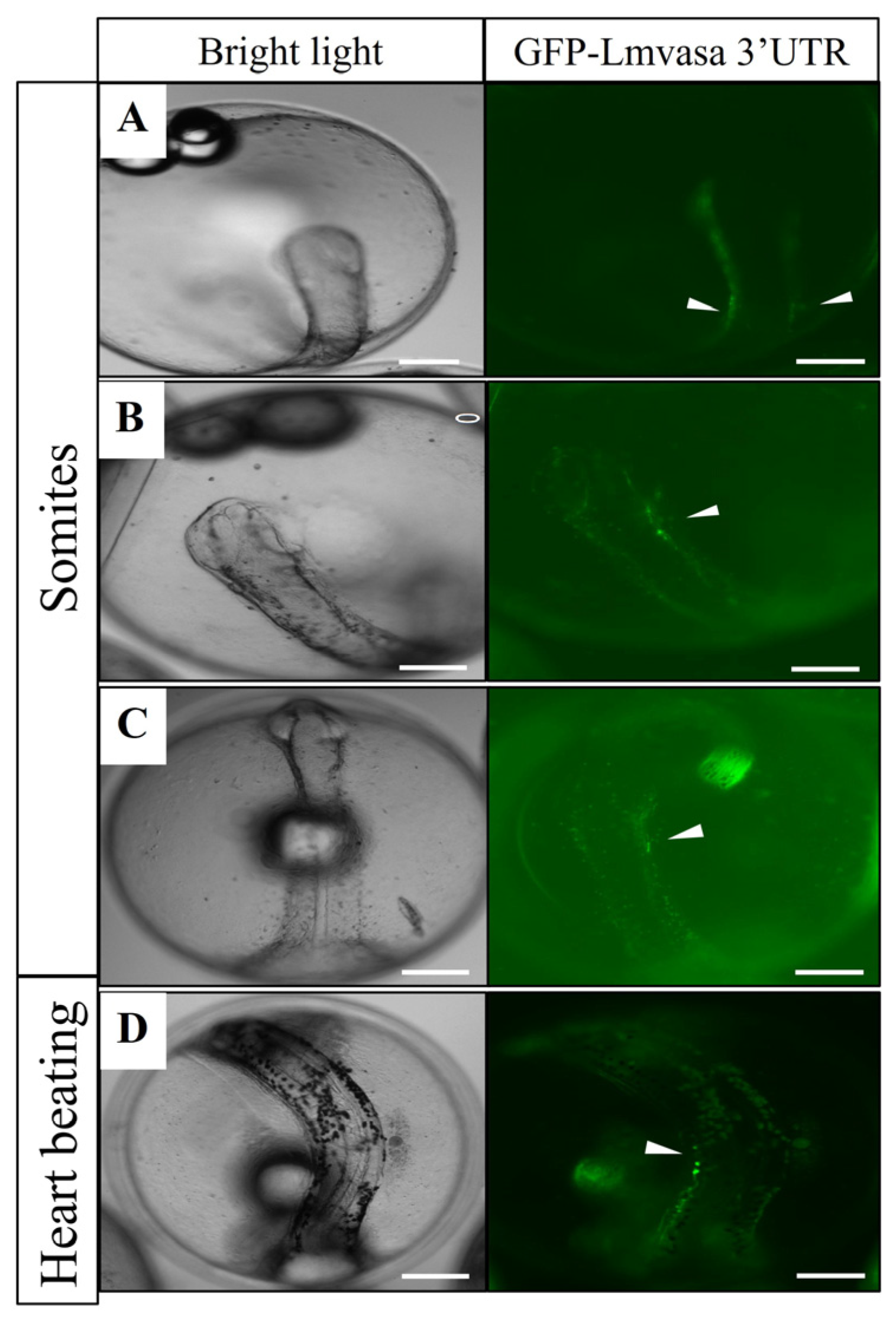
At the somite stage, weak GFP-positive cells (PGCs) were observed on one or both sides of the embryonic body (Figure 8A–C). At the heart beating stage, PGCs migrated dorsally and were closer to the gonadal ridge (Figure 8D). These results suggest that GFP-Lmvasa 3′-UTR mRNA can label the PGCs of spotted sea bass.
To verify the functional conservation of the 3′-UTR sequence in spotted seabass, GFP-Lmvasa 3′-UTR mRNA was injected into the embryos of zebrafish and medaka. At the somite stage, PGCs with green fluorescence were observed in the embryonic body of zebrafish (Figure 9A,B). As the embryo developed further, PGCs moved towards the dorsal side of the embryo (Figure 9C). At 1 dah, PGCs aligned at the dorsal side of the peritoneal cavity where the gonad would eventually form (Figure 9D). At the organogenesis stage of medaka, PGCs with green fluorescence were localized in the embryos (Figure 10A,B). These results suggest that the GFP-Lmvasa 3′-UTR mRNA can label PGCs in two other types of fish: zebrafish (early localization) and medaka (loss of early specific localization).

4. Discussion

In this study, Lmvasa and Lmdnd were identified as both sex marker genes for spotted sea bass. The vasa protein of spotted sea bass, like those of other teleosts, has a conserved domain of the typical DEAD-box protein family, which is involved in ATPase-dependent RNA binding and unwinding activity [14,45,46,47]. The results further confirmed that the isolated vasa gene was a vasa homologous gene, and that vasa was evolutionarily conserved. The dnd protein has an RNA-binding protein with a typical RRM motif in spotted sea bass, which is consistent with those of zebrafish [48] and medaka [22].
Phylogenetic analysis showed that aa sequences of Lmvasa and Lmdnd were highly homologous to those of teleosts. RT-qPCR results showed that Lmvasa and Lmdnd were expressed from early embryonic development to blastula stage. Zebrafish [23], medaka [49], and P. Mangurus [29] has a similar expression trend in the embryonic stage. This expression trait occurs in the embryonic stage, possibly after the gastrulation stage, when somatic cells proliferate faster than germ cells [50]. These results confirm the high activity of these genes in early ontogeny, as they are crucial during the PGC formation stage.
Lmvasa and Lmdnd mRNA were only expressed in gonad tissue, which was also reported for medaka [51], summer flounder (Paralichthys dentatus) [52], and spinyhead croaker (Collichthys lucidus) [31]. This indicates that Lmvasa and Lmdnd homologs can serve as specific molecular markers for spotted sea bass germ cells. Studies have shown that dnd mRNA exhibits sexual dimorphism in adult gonads across species. For example, dnd expression was detected only in mouse testis [27], whereas in Xenopus [28] and P. mangurus [29], its expression was restricted to the ovary. Our results showed that Lmdnd mRNA was expressed in both gonads in spotted bass. IHC analysis showed that in the testis, the signal of Lmvasa and Lmdnd proteins was strongest in spermatogonia, weak in spermatocytes, and absent in spermatocytes. Lmvasa and Lmdnd proteins are expressed in the ovaries at various stages of oogenesis. These phenomena have also occurred in Chinese hooknose carp (Opsariichthys bidens) [53], rainbow trout [54], and medaka [55] and turbot [16]. These results suggest that vasa and dnd play an important role in germ cells development as germ cells marker genes in spotted sea bass. In addition, a specific Lmvsas antibody against spotted bass germ cells was generated in this study. This tool will facilitate the preparation and purification of spermatogonia or eggs, providing a new solution for germ cell transplantation or cryopreservation.
WISH and mRNA microinjection techniques were used to label and track PGCs during embryonic development. The migration patterns of PGCs are not exactly the same among different fish species [56]. In this study, it was found that PGC of spotted bass belonged to the early localization pattern. In both species, the early germ is located in the cleavage furrow, and PGCs form at the blastula stage, similar to zebrafish [18,57]. The difference is that during somite formation, spotted sea bass PGCs are loosely arranged on both sides of the embryonic body, and occasionally ectopic PGCs are found near the brain, which is similar to the patterns reported for turbot [52] and goldfish (Carassius auratus) [58]. In contrast, zebrafish PGCs aggregate in clusters on the outside of the embryo. These ectopic PGCs in the brain may be due to cell mixing at the blastula stage and convergence and extension movement in the gastrula stages.
In many animals, PGCs inherit a special type of cytoplasm, the germ plasm, which distinguishes these cells from somatic cells. Vasa RNA is a component of germ plasm and can be localized by a germ plasm localization signal within the vasa 3′-UTR in different teleosts [18]. The zebrafish vasa 3′-UTR with early localization contains a rich set of positioning elements that can be used to label a wide variety of fish PGCs. For example, the PGCs of trout can be visualized using the vasa 3′-UTR from zebrafish [59], and the PGCs of half-smooth tongue sole (Cynoglossus semilaevis) can be visualized using the vasa 3′-UTR from medaka [60]. However, not all fish PGCs can be labeled with chimeric mRNA containing the vasa 3′-UTR from other fish. For example, GFP-vasa 3′-UTR mRNA from medaka and red seabream (Pagrus major) failed to label zebrafish PGCs [30]. In this study, the synthesized Lmvasa 3′-UTR mRNA was injected into embryos of multiple spotted sea bass, zebrafish, and medaka, respectively. It was found that PGCs could be labeled in all three fish, although the evolutionary relationship between spotted sea bass and zebrafish was very distant. Zhou et al. [61] reported that vasa 3′-UTR in black rockfish (Sebastes schlegelii) can label zebrafish and medaka PGCs because the black rockfish vasa 3′-UTR contains elements common to other fish, as well as the same three functional elements as zebrafish vasa. Thus, the vasa 3′-UTR of different teleosts differs in the labeling of zebrafish PGCs, which may be due to specific functional elements in the 3′-UTR. These differences also indicate that the mechanism of vasa 3′-UTR mRNA localization in teleost is complex and requires further investigation.

5. Conclusions

In our study, we validated Lmvasa and Lmdnd as key PGC-specific marker genes and identified the migration of these marker genes in early embryos of spotted bass, revealing the origin of PGC and its early distribution patterns.

Supplementary Materials

The following supporting information can be downloaded at: https://www.mdpi.com/article/10.3390/genes16091012/s1, Figure S1: Full-length cDNA and aa sequences of Lmvasa from spotted sea bass; Figure S2: Full-length cDNA and aa sequences of Lmdnd from spotted sea bass; Figure S3: Expression of recombinant Lmvasa protein.

Author Contributions

Conceptualization, J.G. and L.Y.; Data curation, J.G. and L.Y.; Formal analysis, J.G. and L.Y.; Funding acquisition, L.Y., B.Z. (Bo Zhang 1) and L.Q.; Investigation, J.G. and L.Y.; Methodology, J.G., L.Y., C.Z. and B.Z. (Bo Zhang 2); Project administration, L.Y. and L.Q.; Resources, C.Z. and B.Z. (Bo Zhang 2); Software, J.G.; Supervision, L.Y.; Validation, L.Y. and C.Z.; Visualization, B.Z. (Bo Zhang 1); Writing—original draft, J.G.; Writing—review & editing, L.Y. All authors have read and agreed to the published version of the manuscript.

Funding

This work was supported by the National Key Research and Development Program of China (No. 2022YFD2400503), Central Public-interest Scientific Institution Basal Research Fund, South China Sea Fisheries Research Institute, CAFS (No. 2024JS04), National Key Research and Development Program of China (No. 2023YFD2401701), Guangdong Province Strategic projects for rural revitalization (2024-SPY-00-008), Innovative Team Building Project of Guangdong Modern Agricultural Industrial Technology System (No. 2024CXTD27), the Central Public-interest Scientific Institution Basal Research Fund, CAFS (No. 2023TD21), the Central Public-interest Scientific Institution Basal Research Fund, CAFS (No. 2024XT02), Central Public-interest Scientific Institution Basal Research Fund, South China Sea Fisheries Research Institute, CAFS (No. 2024RC11).

Institutional Review Board Statement

The specimens utilized in this study did not involve endangered or protected species, and no specific permits were required for the experiment. In the present study, all experimental procedures performed on fish care were in strict accordance with the Management Rule of Laboratory Animals (Chinese Order No. 676 of the State Council, revised 1 March 2017).

Data Availability Statement

The original contributions presented in this study are included in the article/Supplementary Materials. Further inquiries can be directed to the corresponding author.

Conflicts of Interest

The authors declare no conflicts of interest.

Abbreviations

The following abbreviations are used in this manuscript:
PGCsPrimordial germ cells
dndDead end
WISHWhole mount in situ hybridization
ISHIn situ hybridization
3′-UTR3′ untranslated region
GFPGreen fluorescent protein
H&EHematoxylin-eosin
HRPHorseradish peroxidase
RRMRNA recognition motif
dahdays after hatching

References

  1. Raz, E. Primordial germ cell development in zebrafish. Semin. Cell Dev. Biol. 2002, 13, 489–495. [Google Scholar] [CrossRef] [PubMed]
  2. Wylie, C. Germ Cells. Cell 2000, 10, 410–413. [Google Scholar] [CrossRef]
  3. Nagao, I.; Aoki, Y.; Tanaka, M.; Kinjo, M. Analysis of the molecular dynamics of medaka nuage proteins by fluorescence correlation spectroscopy and fluorescence recovery after photobleaching. FEBS J. 2008, 275, 341–349. [Google Scholar] [CrossRef] [PubMed]
  4. Ikenishi, K.; Nakazato, S.; Okuda, T. Direct Evidence for the Presence of Germ Cell Determinant in Vegetal Pole Cytoplasm of Xenopus laevis and in a Subcellular Fraction of It: (Xenopus laevis/germ cell determinant/germ plasm/PGC induction). Dev. Growth Differ. 1986, 28, 563–568. [Google Scholar] [CrossRef] [PubMed]
  5. Wong, T.T.; Zohar, Y. Production of reproductively sterile fish by a non-transgenic gene silencing technology. Sci. Rep. 2015, 5, 15822. [Google Scholar] [CrossRef] [PubMed Central]
  6. Takeuchi, Y.; Yoshizaki, G.; Takeuchi, T. Biotechnology: Surrogate broodstock produces salmonids. Nature 2004, 430, 629–630. [Google Scholar] [CrossRef] [PubMed]
  7. Houston, D.W.; King, M.L. Germ plasm and molecular determinants of germ cell fate. Curr. Top. Dev. Biol. 2000, 50, 155–181. [Google Scholar] [PubMed]
  8. Yoshizaki, G.; Sakatani, S.; Tominaga, H.; Takeuchi, T. Cloning and characterization of a vasa-like gene in rainbow trout and its expression in the germ cell lineage. Mol. Reprod. Dev. 2000, 55, 364–371. [Google Scholar] [CrossRef] [PubMed]
  9. Schüpbach, T.; Wieschaus, E. Maternal-effect mutations altering the anterior-posterior pattern of the Drosophila embryo. Rouxs Arch. Dev. Biol. 1986, 195, 302–317. [Google Scholar] [CrossRef] [PubMed]
  10. Liang, L.; Diehl-Jones, W.; Lasko, P. Localization of vasa protein to the Drosophila pole plasm is independent of its RNA-binding and helicase activities. Development 1994, 120, 1201–1211. [Google Scholar] [CrossRef] [PubMed]
  11. Linder, P.; Lasko, P. Bent out of shape: RNA unwinding by the DEAD-box helicase Vasa. Cell 2006, 125, 219–221. [Google Scholar] [CrossRef] [PubMed]
  12. Yajima, M.; Wessel, G.M. The multiple hats of Vasa: Its functions in the germline and in cell cycle progression. Mol. Reprod. Dev. 2011, 78, 861–867. [Google Scholar] [CrossRef] [PubMed]
  13. Yoon, C.; Kawakami, K.; Hopkins, N. Zebrafish vasa homologue RNA is localized to the cleavage planes of 2- and 4-cell-stage embryos and is expressed in the primordial germ cells. Development 1997, 124, 3157–3165. [Google Scholar] [CrossRef] [PubMed]
  14. Shinomiya, A.; Tanaka, M.; Kobayashi, T.; Nagahama, Y.; Hamaguchi, S. The vasa-like gene, olvas, identifies the migration path of primordial germ cells during embryonic body formation stage in the medaka, Oryzias latipes. Dev. Growth Differ. 2000, 42, 317–326. [Google Scholar] [CrossRef] [PubMed]
  15. Xu, H.; Gui, J.; Hong, Y. Differential expression of vasa RNA and protein during spermatogenesis and oogenesis in the gibel carp (Carassius auratus gibelio), a bisexually and gynogenetically reproducing vertebrate. Dev. Dyn. 2005, 233, 872–882. [Google Scholar] [CrossRef] [PubMed]
  16. Lin, F.; Zhao, C.Y.; Xu, S.H.; Ma, D.Y.; Xiao, Z.Z.; Xiao, Y.S.; Xu, C.A.; Liu, Q.H.; Li, J. Germline-specific and sexually dimorphic expression of a dead end gene homologue in turbot (Scophthalmus maximus). Theriogenology 2013, 80, 665–672. [Google Scholar] [CrossRef] [PubMed]
  17. Chen, Y.; Fang, X.; Tian, X.Q.; Cui, Z.; Feng, H.Y.; Qiu, G.F. Germ plasm and the origin of the primordial germ cells in the oriental river prawn Macrobrachium nipponense. Cell Tissue Res. 2021, 386, 559–569. [Google Scholar] [CrossRef] [PubMed]
  18. Knaut, H.; Steinbeisser, H.; Schwarz, H.; Nüsslein-Volhard, C. An evolutionary conserved region in the vasa 3′UTR targets RNA translation to the germ cells in the zebrafish. Curr. Biol. 2002, 12, 454–466. [Google Scholar] [CrossRef] [PubMed]
  19. Takeuchi, Y.; Yoshizaki, G.; Takeuchi, T. Generation of live fry from intraperitoneally transplanted primordial germ cells in rainbow trout. Biol. Reprod. 2003, 69, 1142–1149. [Google Scholar] [CrossRef] [PubMed]
  20. Lee, S.; Katayama, N.; Yoshizaki, G. Generation of juvenile rainbow trout derived from cryopreserved whole ovaries by intraperitoneal transplantation of ovarian germ cells. Biochem. Biophys. Res. Commun. 2016, 478, 1478–1483. [Google Scholar] [CrossRef] [PubMed]
  21. Zhou, L.; Wang, X.; Liu, Q.; Yang, J.; Xu, S.; Wu, Z.; Wang, Y.; You, F.; Song, Z.; Li, J. Successful Spermatogonial Stem Cells Transplantation within Pleuronectiformes: First Breakthrough at inter-family Level in Marine Fish. Int. J. Biol. Sci. 2021, 17, 4426–4441. [Google Scholar] [CrossRef] [PubMed]
  22. Baloch, A.R.; Franěk, R.; Saito, T.; Pšenička, M. Dead-end (dnd) protein in fish-a review. Fish. Physiol. Biochem. 2021, 47, 777–784. [Google Scholar] [CrossRef] [PubMed]
  23. Weidinger, G.; Stebler, J.; Slanchev, K.; Dumstrei, K.; Wise, C.; Lovell-Badge, R.; Thisse, C.; Thisse, B.; Raz, E. dead end, a novel vertebrate germ plasm component, is required for zebrafish primordial germ cell migration and survival. Curr. Biol. 2003, 13, 1429–1434. [Google Scholar] [CrossRef] [PubMed]
  24. Horvay, K.; Claussen, M.; Katzer, M.; Landgrebe, J.; Pieler, T. Xenopus Dead end mRNA is a localized maternal determinant that serves a conserved function in germ cell development. Dev. Biol. 2006, 291, 1–11. [Google Scholar] [CrossRef] [PubMed]
  25. Youngren, K.K.; Coveney, D.; Peng, X.; Bhattacharya, C.; Schmidt, L.S.; Nickerson, M.L.; Lamb, B.T.; Deng, J.M.; Behringer, R.R.; Capel, B.; et al. The Ter mutation in the dead end gene causes germ cell loss and testicular germ cell tumours. Nature 2005, 435, 360–364. [Google Scholar] [CrossRef] [PubMed]
  26. Linger, R.; Dudakia, D.; Huddart, R.; Tucker, K.; Friedlander, M.; Phillips, K.A.; Hogg, D.; Jewett, M.A.; Lohynska, R.; Daugaard, G.; et al. Analysis of the DND1 gene in men with sporadic and familial testicular germ cell tumors. Genes Chromosomes Cancer 2008, 47, 247–252. [Google Scholar] [CrossRef] [PubMed]
  27. Bhattacharya, C.; Aggarwal, S.; Zhu, R.; Kumar, M.; Zhao, M.; Meistrich, M.L.; Matin, A. The mouse dead-end gene isoform alpha is necessary for germ cell and embryonic viability. Biochem. Biophys. Res. Commun. 2007, 355, 194–199. [Google Scholar] [CrossRef] [PubMed]
  28. Mei, W.; Jin, Z.; Lai, F.; Schwend, T.; Houston, D.W.; King, M.L.; Yang, J. Maternal Dead-End1 is required for vegetal cortical microtubule assembly during Xenopus axis specification. Development 2013, 140, 2334–2344. [Google Scholar] [CrossRef] [PubMed]
  29. Shiguemoto, G.F.; Coelho, G.C.Z.; López, L.S.; Pessoa, G.P.; Dos Santos, S.C.A.; Senhorini, J.A.; Monzani, P.S.; Yasui, G.S. Primordial germ cell identification and traceability during the initial development of the Siluriformes fish Pseudopimelodus mangurus. Fish Physiol. Biochem. 2022, 48, 1137–1153. [Google Scholar] [CrossRef] [PubMed]
  30. Zhou, L.; Xu, S.; Lin, F.; Wang, X.; Wang, Y.; Wang, Y.; Yu, D.; Liu, Q.; Li, J. Both of marine fish species Oryzias melastigma and Pagrus major all failing in early localization at embryo stage by vasa RNA. Gene 2021, 769, 145204. [Google Scholar] [CrossRef] [PubMed]
  31. Xu, C.; Li, Y.; Wen, Z.; Jawad, M.; Gui, L.; Li, M. Spinyhead Croaker Germ Cells Gene dnd Visualizes Primordial Germ Cells in Medaka. Life 2022, 12, 1226. [Google Scholar] [CrossRef] [PubMed]
  32. Yoon, J.H.; Cho, Y.S.; Lee, H.B.; Park, J.Y.; Lim, H.K. Dead-End (dnd) Gene Cloning and Gonad-Specific Expression Pattern in Starry Flounder (Platichthys stellatus). Animals 2021, 11, 2256. [Google Scholar] [CrossRef] [PubMed]
  33. Zhao, Y.; Peng, W.; Guo, H.; Chen, B.; Zhou, Z.; Xu, J.; Zhang, D.; Xu, P. Population Genomics Reveals Genetic Divergence and Adaptive Differentiation of Chinese Sea Bass (Lateolabrax maculatus). Mar. Biotechnol. 2018, 20, 45–59. [Google Scholar] [CrossRef] [PubMed]
  34. Zhang, B.; Yan, L.; Lin, C.; Liu, Y.; Zhao, C.; Wang, P.; Zhang, Y.; Qiu, L. Asymmetric evolution of ISG15 homologs and the immune adaptation to LBUSV infection in spotted seabass (Lateolabrax maculatus). Fish Shellfish Immunol. 2024, 148, 109441. [Google Scholar] [CrossRef] [PubMed]
  35. Wang, W.; Ma, C.; Ouyang, L.; Chen, W.; Zhao, M.; Zhang, F.; Fu, Y.; Jiang, K.; Liu, Z.; Zhang, H.; et al. Genetic diversity and population structure analysis of Lateolabrax maculatus from Chinese coastal waters using polymorphic microsatellite markers. Sci. Rep. 2021, 11, 15260. [Google Scholar] [CrossRef] [PubMed]
  36. Xu, H.; Lim, M.; Dwarakanath, M.; Hong, Y. Vasa identifies germ cells and critical stages of oogenesis in the Asian seabass. Int. J. Biol. Sci. 2014, 10, 225–235. [Google Scholar] [CrossRef]
  37. Liu, X.; Li, W.; Yang, Y.; Chen, K.; Li, Y.; Zhu, X.; Ye, H.; Xu, H. Transcriptome Profiling of the Ovarian Cells at the Single-Cell Resolution in Adult Asian Seabass. Front. Cell Dev. Biol. 2021, 9, 647892. [Google Scholar] [CrossRef] [PubMed]
  38. van Megen, H.; van den Elsen, S.; Holterman, M.; Karssen, G.; Mooyman, P.; Bongers, T.; Holovachov, O.; Bakker, J.; Helder, J. A phylogenetic tree of nematodes based on about 1200 full-length small subunit ribosomal DNA sequences. Nematology 2009, 11, 927–950. [Google Scholar] [CrossRef]
  39. Hall, B.G. Building phylogenetic trees from molecular data with MEGA. Mol. Biol. Evol. 2013, 30, 1229–1235. [Google Scholar] [CrossRef] [PubMed]
  40. Zhao, C.; Wang, P.; Qiu, L. RNA-Seq-based transcriptome analysis of reproduction- and growth-related genes in Lateolabrax japonicus ovaries at four different ages. Mol. Biol. Rep. 2018, 45, 2213–2225. [Google Scholar] [CrossRef] [PubMed]
  41. Narayanan, R.; Oates, A.C. Detection of mRNA by Whole Mount in situ Hybridization and DNA Extraction for Genotyping of Zebrafish Embryos. Bio Protoc. 2019, 9, e3193. [Google Scholar] [CrossRef] [PubMed]
  42. Kimmel, C.B.; Ballard, W.W.; Kimmel, S.R.; Ullmann, B.; Schilling, T.F. Stages of embryonic development of the zebrafish. Dev. Dyn. 1995, 203, 253–310. [Google Scholar] [CrossRef] [PubMed]
  43. Köprunner, M.; Thisse, C.; Thisse, B.; Raz, E. A zebrafish nanos-related gene is essential for the development of primordial germ cells. Genes. Dev. 2001, 15, 2877–2885. [Google Scholar] [CrossRef] [PubMed]
  44. Yang, Y.; Liu, Q.; Xiao, Y.; Wang, X.; An, H.; Song, Z.; You, F.; Wang, Y.; Ma, D.; Li, J. Germ Cell Migration, Proliferation and Differentiation during Gonadal Morphogenesis in All-Female Japanese Flounder (Paralichthys olivaceus). Anat. Rec. 2018, 301, 727–741. [Google Scholar] [CrossRef] [PubMed]
  45. Gustafson, E.A.; Wessel, G.M. Vasa genes: Emerging roles in the germ line and in multipotent cells. Bioessays 2010, 32, 626–637. [Google Scholar] [CrossRef] [PubMed]
  46. Nakharuthai, C.; Sreebun, S.; Kabpha, A.; Phuong, T.V.; Boonanuntanasarn, S. Characterization of ddx4 and dnd Homologs in Snakeskin Gourami (Trichopodus pectoralis) and Their Expression Levels during Larval Development and in Gonads of Males and Females. Animals 2022, 12, 3415. [Google Scholar] [CrossRef] [PubMed]
  47. Duangkaew, R.; Jangprai, A.; Ichida, K.; Yoshizaki, G.; Boonanuntanasarn, S. Characterization and expression of a vasa homolog in the gonads and primordial germ cells of the striped catfish (Pangasianodon hypophthalmus). Theriogenology 2019, 131, 61–71. [Google Scholar] [CrossRef] [PubMed]
  48. Liu, W.; Collodi, P. Zebrafish dead end possesses ATPase activity that is required for primordial germ cell development. FASEB J. 2010, 24, 2641–2650. [Google Scholar] [CrossRef] [PubMed]
  49. Liu, L.; Hong, N.; Xu, H.; Li, M.; Yan, Y.; Purwanti, Y.; Yi, M.; Li, Z.; Wang, L.; Hong, Y. Medaka dead end encodes a cytoplasmic protein and identifies embryonic and adult germ cells. Gene Expr. Patterns. 2009, 9, 541–548. [Google Scholar] [CrossRef] [PubMed]
  50. Braat, A.K.; Speksnijder, J.E.; Zivkovic, D. Germ line development in fishes. Int. J. Dev. Biol. 1999, 43, 745–760. [Google Scholar] [PubMed]
  51. Reunov, A.A.; Au, D.W.T.; Alexandrova, Y.N.; Chiang, M.W.L.; Wan, M.T.; Yakovlev, K.V.; Reunova, Y.A.; Komkova, A.V.; Cheung, N.K.; Peterson, D.R.; et al. Germ plasm-related structures in marine medaka gametogenesis; novel sites of Vasa localization and the unique mechanism of germ plasm granule arising. Zygote 2020, 28, 9–23. [Google Scholar] [CrossRef] [PubMed]
  52. Du, S.; Zhou, L.; Wang, X.; Xu, S.; Li, J.; Song, Z.; Liu, Q. Characterization of vasa and dnd homologs in summer flounder, Paralichthys dentatus: Expression analysis and colocalization of PGCs during embryogenesis. Theriogenology 2022, 181, 180–189. [Google Scholar] [CrossRef] [PubMed]
  53. Yin, Z.; Xue, L.; Song, P.; Jawad, M.; Xu, C.; Bu, W.; Li, M. Dazl and dnd Identify Both Embryonic and Gonadal Germ Cells in Chinese Hook Snout Carp (Opsariichthys bidens). Fishes 2024, 9, 214. [Google Scholar] [CrossRef]
  54. Li, M.; Shen, Q.; Xu, H.; Wong, F.M.; Cui, J.; Li, Z.; Hong, N.; Wang, L.; Zhao, H.; Ma, B.; et al. Differential conservation and divergence of fertility genes boule and dazl in the rainbow trout. PLoS ONE 2011, 6, e15910. [Google Scholar] [CrossRef] [PubMed]
  55. Zhu, T.; Gui, L.; Zhu, Y.; Li, Y.; Li, M. Dnd is required for primordial germ cell specification in Oryzias celebensis. Gene 2018, 679, 36–43. [Google Scholar] [CrossRef] [PubMed]
  56. Saito, T.; Fujimoto, T.; Maegawa, S.; Inoue, K.; Tanaka, M.; Arai, K.; Yamaha, E. Visualization of primordial germ cells in vivo using GFP-nos1 3′UTR mRNA. Int. J. Dev. Biol. 2006, 50, 691–699. [Google Scholar] [CrossRef] [PubMed]
  57. Braat, A.K.; Zandbergen, T.; van de Water, S.; Goos, H.J.; Zivkovic, D. Characterization of zebrafish primordial germ cells: Morphology and early distribution of vasa RNA. Dev. Dyn. 1999, 216, 153–167. [Google Scholar] [CrossRef] [PubMed]
  58. Otani, S.; Kitauchi, T.; Saito, T.; Sakao, S.; Maegawa, S.; Inoue, K.; Arai, K.; Yamaha, E. The formation of primordial germ cells from germline cells in spherical embryos derived from the blastodisc of 2-cell embryos in goldfish, Carassius auratus. Int. J. Dev. Biol. 2005, 49, 843–850. [Google Scholar] [CrossRef] [PubMed]
  59. Zhou, L.; Wang, X.; Liu, Q.; Xu, S.; Zhao, H.; Han, M.; Wang, Y.; Song, Z.; Li, J. Visualization of Turbot (Scophthalmus maximus) Primordial Germ Cells in vivo Using Fluorescent Protein Mediated by the 3′ Untranslated Region of nanos3 or vasa Gene. Mar. Biotechnol. 2019, 21, 671–682. [Google Scholar] [CrossRef] [PubMed]
  60. Huang, J.; Chen, S.; Liu, Y.; Shao, C.; Lin, F.; Wang, N.; Hu, Q. Molecular characterization, sexually dimorphic expression, and functional analysis of 3′-untranslated region of vasa gene in half-smooth tongue sole (Cynoglossus semilaevis). Theriogenology 2014, 82, 213–224. [Google Scholar] [CrossRef] [PubMed]
  61. Zhou, L.; Wang, X.; Du, S.; Wang, Y.; Zhao, H.; Du, T.; Yu, J.; Wu, L.; Song, Z.; Liu, Q.; et al. Germline Specific Expression of a vasa Homologue Gene in the Viviparous Fish Black Rockfish (Sebastes schlegelii) and Functional Analysis of the vasa 3′ Untranslated Region. Front. Cell Dev. Biol. 2020, 8, 575788. [Google Scholar] [CrossRef] [PubMed]
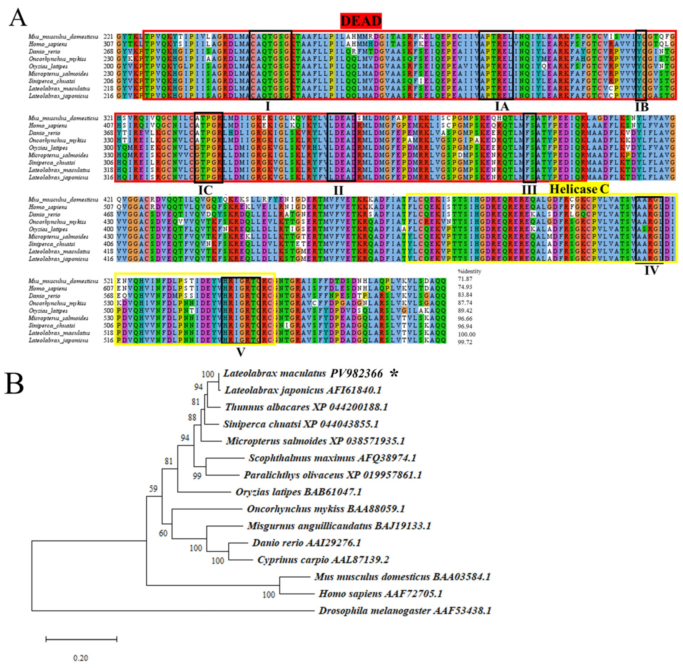
Figure 1. Multiple alignment and phylogenetic tree of vasa aa of spotted sea bass. (A) Conserved motifs are shown in the black block. The DEAD-box sequence is indicated in bright red, whereas Helicase C is in yellow. The eight domains within these superfamilies [I domain (AQTGSGKT), IA domain (PTREL), IB domain (GG), IC domain (TPGRL), II domain (DEAD), III domain (SAT), IV domain (ARGLD), and V domain (GRTGR)] are outlined with black rectangles. (B) Phylogenetic tree generated using the neighbor-joining method based on vasa. Other accession numbers are indicated. * represents the species that are studied.
Genes 16 01012 g001
Figure 2. Multiple alignment and phylogenetic tree of dnd aa of spotted sea bass. (A) Six conserved domains or motifs are indicated in the black block, including RNA recognition motif (RRM), N-terminal region (NR), and four C-terminal regions (CR1–4), which are typically present in dnd protein. (B) Phylogenetic tree generated using the neighbor-joining method based on dnd. Other accession numbers are indicated. * represents the species that are studied.
Genes 16 01012 g002
Figure 3. Spatial and temporal expression analyses of Lmvasa and Lmdnd isoforms detected by RT-PCR. (A) RT-qPCR of Lmvasa genes in different development stages of embryos. (B) RT-qPCR of Lmdnd genes in different development stages of embryos. β-actin as the control. (C) Values with the different superscript letters are significantly different (ANOVA, p < 0.05). Tissue specific expression of and Lmvasa and Lmdnd determined by RT-PCR. β-actin served as reference. M, DNA size marker.
Genes 16 01012 g003
Figure 4. Expression of spotted sea bass Lmvasa and Lmdnd proteins in the ovary and testis. (A,D) Histological sections of testis and ovary stained with Hematoxylin and Eosin (H&E). (B,C) Immunohistochemical sections of testis incubated with anti-Lmvasa and anti-dnd1 antibodies. Lmvasa and Lmdnd proteins were most intense in spermatogonia, faint in spermatocytes, and had disappeared in spermatids. (E,F) Immunohistochemical sections of ovary incubated with anti-Lmvasa and anti-dnd1 antibodies. In ovary, Lmvasa and Lmdnd were granular around the nucleus in stage II oocytes and weakly expressed in stage III oocytes. They showed weak diffusion in cytoplasm in stage IV oocytes and were evenly distributed in follicles in stage V oocytes. DU, sperm duct; LO, lobular lumen; Adiff, Type A differentiated spermatogonia; B, Type B spermatogonia; Ps, primary spermatocytes; Ss, secondary spermatocytes; St, Spermatids; GE, germinal epithelium; VA, vascuoles; II, primary growth oocyte; III, perinucleolar or previtellogenic oocyte; IV, late vitellogenic oocyte; and V, postvitellogenic oocyte, described by Yang et al. [44]. Scale bars: 200 μm (A,D), 10 μm (B,C), 100 μm (E,F).
Genes 16 01012 g004
Figure 5. Localization of Lmvasa mRNA during embryonic development based on WISH. (A) 1-cell; (B) 2-cell; (C) 4-cell; (D) 16-cell; (E) gastrula; (F) early gastrula; (G) late gastrula; and (H) somite stages. Black arrows indicate regions of Lmvasa transcript aggregation (dark purple). Magnification of the boxed area in (G). Scale bar: 200 μm. WISH, whole mount in situ hybridization.
Genes 16 01012 g005
Figure 6. Localization of Lmdnd mRNA during embryonic development based on WISH. (A) 2-cell; (B) 4-cell; (C) 8-cell; (D) 32-cell; (E) morula; (F), blastula; (G) early gastrula; (H) late gastrula; (I) early somite; (J) late somite; (K) heart beating; and (L) hatching stages; (M) 3 days after hatching. Black arrows indicate Lmdnd transcript aggregated regions (dark purple). Scale bar: 200 μm. WISH, whole mount in situ hybridization.
Genes 16 01012 g006
Figure 7. Schematic of Lmvasa or Lmdnd signal distribution during embryogenesis in spotted sea bass. Red spots indicate Lmvasa or Lmdnd signals. (A) 1-cell; (B) 2-cell; (C) 4-cell; (D) 8-cell; (E)16-cell; (F) 32-cell; (G) morula; (H) blastula; (I) early gastrula; (J) late gastrula; (K) early somite; (L) late somite stage; (M) heart beating; and (N) hatching stages. (O) 3 days after hatching.
Genes 16 01012 g007
Figure 8. Migration of labeled PGCs in spotted sea bass embryo. GFP-Lmvasa 3′UTR mRNA was injected into the vegetal pole of 1-to 4-cell stage embryos. (AC) Somite stage; (D) heart-beating stage. White arrows indicate GFP-positive cells (PGCs). Concentrations of injected mRNA were all 300 ng/μL. Scale bar: 200 μm.
Genes 16 01012 g008
Figure 9. Visualization of zebrafish PGCs following injection of GFP-Lmvasa 3′-UTR mRNA. (AC) Somite stage; (D) 1 day after hatching (1 dah). Magnification of the boxed areas in (D) showing localization of GFP-Lmvasa 3′UTR signals in PGCs. White arrows indicate GFP-positive cells (PGCs). Scale bar: 200 μm.
Genes 16 01012 g009
Figure 10. Visualization of medaka PGCs following injection of GFP-Lmvasa 3′-UTR mRNA. (A,B) Organogenesis stage. White arrows indicate GFP-positive cells (PGCs). Scale bar: 200 μm.
Genes 16 01012 g010
Table 1. Sequences of primers used in the present study a.
Primer NameSequence 5′-3′Size (bp)Temperature, °CPurpose
vasa oFATGGACGAATGGGAAGAAGAAGGAAC190556ORF
vasa oRCTACTCCCATTCTTCATCATCAGCTG
dnd oFATGGAGATGATGGAGAACAAGCGGAGC115856
dnd oRTCAGTGGGCAAACTGGTTATTGTACACC
5′RACE-vasaTTCCGATTCTCGTCACCACC29270RACE
3′RACE-vasaTATTGGAAGAACTGGCCGCT61968
5′RACE-dndCAGCTCCTGAACACGCTCAAGGT30070
3′RACE-dndGGATAACGGCCGCCTTTGAAGGGC41468
vasa qFGTGGAACACCAGGGAGACTG16960RT-qPCR
vasa qRTGACGGTTCTCTTTGGACGG
dnd qFGCACGGAGAAGAGACACCTC14560
dnd qRATGGCAGACACCCCCTCTAT
vasa FCCCACTATGAGACGGGCATC39456RT-PCR
vasa RCCAAAGGCAAACTTCCTGGC
dnd FACCTTGAGCGTGTTCAGGAG37756
dnd RGAGGTGTCTCTTCTCCGTGC
β-actin FCAACTGGGATGACATGGAGAAG11458
β-actin RTTGGCTTTGGGGTTCAGG
vasa-28a FTGGGTCGCGGATCCGAATTCATGGACGA
ATGGGAAGAAGAAG
194260WISH
vasa-28a RTGGTGGTGGTGGTGCTCGAGCTCCCATTCT
TCATCATCAGCT
WISH-vasa FGCAGCTGACTTTCTCAAGACGGA101356
WISH-vasa RAATTTTTCTTTTTTATTGGTGATC
WISH-dnd FATGGAGATGATGGAGAACAAGCGGAGC115856
WISH-dnd RTCAGTGGGCAAACTGGTTATTGTACACC
3′UTR-vasa FATGAACTATACAAACTCGAGAAGGAATATTAGAGAAGC327563′UTR
3′UTR-vasa RATGAACTATACAAATAACTCGAGAAACCCATTAACCAATTTTTCT
a The underlined bases are linearized vector end sequences.
Disclaimer/Publisher’s Note: The statements, opinions and data contained in all publications are solely those of the individual author(s) and contributor(s) and not of MDPI and/or the editor(s). MDPI and/or the editor(s) disclaim responsibility for any injury to people or property resulting from any ideas, methods, instructions or products referred to in the content.