Article

Phenotypic and Genetic Stability of the Aldrovanda vesiculosa L. Plants Regenerated in Tissue Culture

by
Marzena Parzymies
1,
Katarzyna Głębocka
2,
Magdalena Pogorzelec
3,*,
Barbara Banach-Albińska
4,
Alicja Świstowska
1 and
Michał Arciszewski
3
1
Institute of Horticultural Production, University of Life Sciences in Lublin, Głęboka 28, 20-612 Lublin, Poland
2
Institute of Plant Genetics, Breeding and Biotechnology, University of Life Sciences in Lublin, Akademicka 13, 20-950 Lublin, Poland
3
Department of Hydrobiology and Protection of Ecosystems, University of Life Sciences in Lublin, Dobrzańskiego 37, 20-262 Lublin, Poland
4
Department of Botany and Plant Physiology, University of Life Sciences in Lublin, Akademicka 13, 20-950 Lublin, Poland
*
Author to whom correspondence should be addressed.
Genes 2025, 16(9), 1003; https://doi.org/10.3390/genes16091003
Submission received: 31 July 2025 / Revised: 20 August 2025 / Accepted: 22 August 2025 / Published: 25 August 2025
(This article belongs to the Section Plant Genetics and Genomics)

Abstract

Background: Tissue culture might be a method supplementing traditional plant propagation in various fields, like agriculture, medicine, industry, and the active conservation of plant species. For the purpose of plant restoration, it is important that the obtained progenies are identical with the mother plants to ensure the true-to-typeness of the future population. Methods: In the present study, the stability of Aldrovanda vesiculosa regenerants obtained in vitro through phenotypic and genetic analysis was estimated. Clones of aldrovanda plants were cultivated in tissue culture in the 1/10 MS liquid medium under the same conditions for over a year, with five weeks of subculturing. Results: It was observed that two clones formed plants that displayed atypical growth structures, the shoots were shorter with many lateral shoots, and they had a lower fresh weight. They also formed fewer and smaller snap-traps, which, in the case of carnivorous plants, determines the capability of catching prey. The 35 in vitro regenerated plants and 5 specimens obtained from the natural habitat were subjected to genetic analyses with two molecular markers: start codon targeted (SCoT) polymorphism and sequence-related amplified polymorphism (SRAP). Despite the visible morphological variants, the genetic stability of all the regenerants with the individuals from natural stands was confirmed. All of them were monomorphic except three bands that were obtained for reference, where individuals were amplified with SCoT28 and me12-em13 SRAP primers. Conclusions: As shown in the presented research, it might be recommended to use different methods to evaluate the stability of in vitro cultivated plants.

1. Introduction

The preservation of genetic diversity in plant populations has emerged as a pivotal concern in the realm of nature conservation. Particular emphasis has been placed on preserving the gene pools of populations threatened with extinction [1]. Ex situ conservation is recognized as a significant tool that can complement traditional in situ conservation efforts, particularly in cases involving species inhabiting unstable environments or those with limited reproductive capacity. In such cases, the employment of tissue culture techniques may serve as a method that fosters active protection or restoration of populations [2,3]. Presently, in vitro propagation has become a prevalent method for addressing the challenges associated with the preservation of the gene pool of rare and endangered plant species [4]. Sarasan et al. [3] indicate that micropropagation facilitates the expeditious mass propagation of endangered plant species and their long-term storage. The significance of integrating micropropagation and cryoconservation as a gene banking approach for rare genotypes is also underscored. As demonstrated by Reed et al. [5] and Pence [6], in vitro techniques facilitate the generation of a substantial number of specimens that are both healthy and genetically homogenous.
A few examples of the use of micropropagated carnivorous plants in conservation include Drosera burmannii [7], Aldrovanda vesiculosa [8], Dionaea muscipula [9], Drosera rotundifolia [10,11], and Nepenthes khasiana [12]. However, according to Caldeira et al. [13], although tissue culture is an important biotechnological tool for this group of plants, there is a lack of protocol for mass propagation of the species in tissue culture, and scientific information on the subject is presented at a slow pace.
As Barhill-Dilling [14] demonstrates, the progeny plants produced through micropropagation may be used for effective introduction or reintroduction of threatened and endangered plant species. Nevertheless, the fundamental concern associated with tissue culture is the genetic stability of the propagated plants. The term ‘somaclonal variation’ is used to describe the presence of phenotypic and DNA variation among plant clones. This phenomenon may be caused by the process of tissue culture, and consequently, the term ‘tissue culture-induced variation’ is sometimes employed to indicate the effect of environment [15]. Somaclonal variation is particularly pronounced in the case of indirect organogenesis or when high doses of growth regulators are used [15,16]. Therefore, it is recommended to use direct organogenesis methods, avoiding the prolonged subculturing and the monitoring of genotypes with molecular markers (RAPD, ISSR, and AFLP) or flow cytometry.
One of the species for which tissue culture propagation is applied and then reintroduction of the obtained progeny specimens in the natural habitat is undertaken for the means of active protection is A. vesiculosa L., the waterwheel plant. It is an aquatic carnivorous plant belonging to the Drosearceae family. It inhabits shallow, standing dystrophic waters across Europe, Asia, Australia, and Africa. It forms rootless shoots free-floating just below the water surface [17]. The leaves are arranged in characteristic whorls of 4–9, with a maximum diameter of 23 mm. Each fully developed leaf forms a trap which is composed of a two-lobed lamina with a midrib and 3–6 long bristles [18,19]. When mechanically irritated by prey, traps close rapidly. Bristles probably guide prey toward the trap, and they might prevent casual objects from entering the trap [20]. The length of the stem (6–20 cm) depends on various factors, both related to the abiotic environment (physical and chemical parameters of the water of solar radiation intensity), and biocenotic (availability of potential prey). The growth season lasts from early–mid-spring to early autumn. Temperate waterwheel populations flower and set seeds under optimal conditions; however, such observations are seldom documented [21]. The propagation of plants is predominantly vegetative, characterized by the formation of shoots and subsequent branching [17,22,23]. In the middle of autumn, in response to adverse conditions, plants form turions, which are a type of dormant overwintering buds that sink to the bottom of water reservoirs. Turions are formed by the dense leaves growing at highly shortened internodes that surround and protect the apex from freezing [23,24,25,26,27]. It has been shown that the overwintering phenomenon is associated with a significant decline in population size (turions are often carried ashore or freeze to death), and in many cases, the plant survival rate has been reported at 20–30%. In spring, the surviving turions float to the surface and shoots resume the growth cycle [28].
According to Adamec [23], Euro-Asian temperate populations are distinct from Australian ones. The former produce morphologically distinct and highly dormant winter buds (turions), and they do not contain the pigment anthocyanin. In contrast, the latter produce only non-dormant winter shoot apices and contain anthocyanin. European plants exhibit low rates of flowering and rarely produce viable seeds [28,29]. They primarily propagate through vegetative reproduction via apical branching of the shoots [17,25].
Despite its widespread presence on a global scale, the population of A. vesiculosa is present in isolated habitats. Legislative protections have been instituted for this species in all countries within its distribution. The conservation status of A. vesiculosa is of concern on both a global and regional scale. This concern is primarily due to the plant’s rarity, habitat specificity, and ongoing threats. According to the IUCN Red List, the species has been designated as endangered (EN) in accordance with criteria that include a restricted area of occupancy, significantly fragmented populations, and a persistent deterioration in habitat quality [30,31]. It is also listed in the Bern Convention in the Conservation of European Wildlife and Natural Habitats Appendix I as a species requiring specific habitat conservation measures [32]. In Poland, it is stated as critically endangered (CR) on the Polish Red List of pteridophytes and flowering plants [33].
Historically, A. vesiculosa has been found to inhabit all continents of the Old World. In contemporary times, it has been acknowledged as being in markedly low abundance and has become extinct in numerous regions and countries [30,34]. At least seventy-nine historical sites of the species were recorded in Poland in the last 200 years, but only nine natural sites were confirmed between 2006 and 2013 [35,36]. In the 1970s, the reintroduction of aldrovanda in select locations in Poland was met with documented success. The plants were obtained through the process of tissue culture [35]. According to the IUCN, such actions remain the most effective measures of conservation for this species [30].
The primary objective of this study was to investigate somaclonal variation among A. vesiculosa specimens cultivated in tissue culture for over one year, based on the morphological features and genetic analysis (SCoT and SRAP molecular markers). The results of this investigation will be instrumental in the reintroduction activities that are implemented for the purpose of ensuring the active protection of the species.

2. Materials and Methods

2.1. Tissue Culture Conditions and Morphological Observation

The plant material for tissue culture establishment was A. vesiculosa individuals collected from two peat bog lakes located in the Łęczna-Włodawa Lakeland in Eastern Poland. The geographical coordinates of the two lakes in question are as follows: Lake Łukie: 51°24’.40.30″ N and 23°04’56.73″ E; Lake Orchowe: 51°29’27.63″ N and 23°34’26.12″ E. The mother plants selected for in vitro propagation were characterized by typical growth for the species, in terms of length of shoot (10–15 cm). In the laboratory, the shoots were defoliated, cut into 2 cm pieces, and then surface-sterilized with sodium hypochlorite (NaOCl, Chempur, Piekary Śląskie, Poland) at a concentration of 0.25% for five minutes. The disinfected shoot fragments were then placed individually in tubes containing 10 mL of the liquid medium, which consisted of Murashige and Skoog (MS) [37] macro- and microelements supplemented with 0.1 mg·dm−3 thiamine (vit. B1, Sigma-Aldrich, Saint Louis, MO, USA), 0.5 mg·dm−3 pyridoxine (vit. B6, Sigma-Aldrich, Saint Louis, MO, USA), 0.5 mg·dm−3 niacine (vit. PP, Sigma-Aldrich, Saint Louis, MO, USA), 2.0 mg·dm−3 glycine (Sigma-Aldrich, Saint Louis, MO, USA), 100 mg·dm−3 myo-inositol (Sigma-Aldrich, Saint Louis, MO, USA), and 20 g·dm−3 sucrose (Chempur, Piekary Śląskie, Poland). All the components were diluted 10 times. The medium composition was selected on the basis of the previous research findings [38]. The medium’s pH was established at 5.5. The regenerating individuals were placed in a fresh medium at 5-week intervals. The plants were cultivated in 450 mL jars, with 10 plants per jar, containing 200 mL of the medium, and covered with semi-transparent plastic lids. Each clone (i.e., progeny plants obtained from a single mother plant) was cultivated separately. The explant types, media, and conditions were the same for all the cultivated shoots. After a year, the observations on the morphology of the regenerants were conducted on 20 randomly selected plants from each of four separate clones (propagated from different mother plants).
The obtained results of the measurements were subjected to the statistical analysis performed with Arstat software (University of Life Sciences in Lublin, Poland), using one-way or two-way ANOVAs for a one-factorial design. The significance between means was estimated with Tukey’s confidence intervals at a 5% level of significance.

2.2. SCoT and SRAP Genetic Analysis

To conduct the genetic analyses, a total of forty plants were taken, of which 35 were regenerants obtained from in vitro. As DNA from the parent plants was not available, 5 randomly selected specimens from the natural habitat were used. The isolation of DNA was performed in accordance with the procedure described by Palfavi et al. [39], with minor modifications. The concentration and purity of the DNA were measured with a Nanodrop ND-1000 spectrophotometer (Thermo Fisher Scientific, Waltham, MA, USA). Each sample was then diluted to a concentration of 20 ng/µL. Two molecular marker methods were applied: start codon targeted (SCoT) polymorphism [40] and sequence-related amplified polymorphism (SRAP) [41]. Ten SCoT primers and 15 SRAP primer combinations were screened to select the ones that amplified clear and scorable bands. Sequences of SCoT and SRAP primers that were used in this study are shown in Table 1 and Table 2, respectively.
The volume and reagent concentrations of PCR were consistent in both methods. The reaction mixture was composed of 10 µL of the following components: 1× PCR buffer (100 mM Tris-HCl, 500 mM KCl, and detergent) (Thermo Fisher Scientific, Waltham, MA, USA); 0.5 µg/µL BSA; 2 mM MgCl2; 200 µM of each dNTP; 600 nM of primer (one primer in the case of the SCoT method and two primers in the case of SRAP); 0.5 U of Taq DNA polymerase (Thermo Fisher Scientific, Waltham, MA, USA); and 40 ng of DNA template. The thermal profiles of reactions are described in Table 3.
The PCR products were separated on 1.5% agarose gels stained with ethidium bromide, and their sizes were determined by comparing them with a fragment length standard (Thermo Fisher Scientific, GeneRuler 100 bp DNA Ladder Plus).

3. Results and Discussion

The growth and regeneration of plants in tissue culture is an asexual process involving the mitotic division of cells. Therefore, it is expected that the regenerating plants will be phenotypically and genetically identical to the stock plants [42]. However, some or even all of the progenies may be different from the donor plants [43]. The true-to-typeness of plants cultivated in vitro can be evaluated through phenotypic, cytological, and molecular analysis [44]. In the presented research, the phenotypic and molecular analyses were carried out to estimate the stability of A. vesiculosa progenies obtained in tissue culture.

3.1. Phenotypic Variation

During the cultivation, it was observed that two clones (marked as 6 and 7) formed shoots that displayed atypical growth characteristics. This phenomenon persisted throughout the entire cultivation period. The regenerated shoots were found to be shorter (5.07 and 4.76 on average, respectively), and they produced, on average, 7 and 4.7 lateral shoots of 2.2 and 2.54 mm (respectively) (Table 4). Two clones, marked 2 and 10, were selected from the remaining ones for the study as a reference, as they were characterized by typical growth for the species. Those clones produced longer shoots (10.63 and 10.28 on average), with fewer lateral shoots (1.60 and 2.30, respectively) that were formed at the top. Clones 6 and 7 shoots were also characterized by a lower fresh weight (17 and 7.89 mg, respectively) in comparison to clones 2 and 10 (31.67 and 31.53 mg, respectively). The morphology of the selected clones is illustrated in Figure 1.
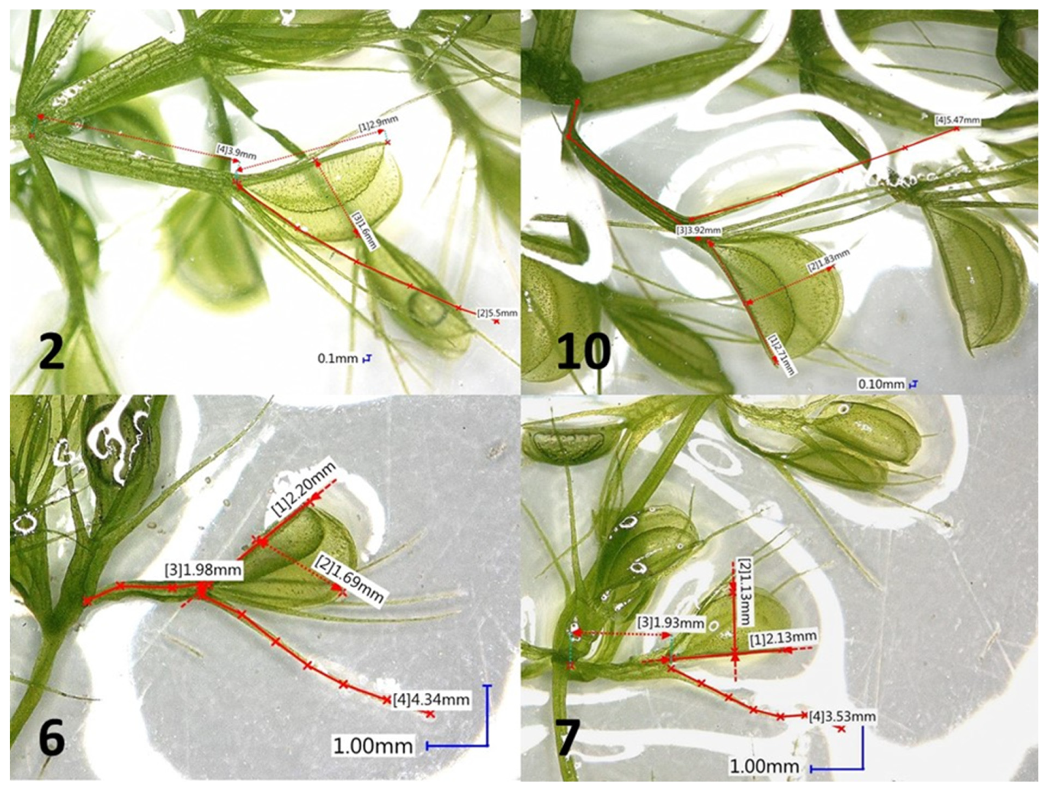
The A. vesiculosa is a carnivorous plant species with traps formed from two-lobed lamina with a midrib and long bristles. With regard to the capacity to capture prey, the structure and size of traps might be important features. Traps of natural plants range from 1.90 to 4.49 mm. Hortsmann et al. [45] studied the correlation between the trap size and prey type. However, due to partly digested prey in the traps, a reliable confirmation of that dependence was not possible. During in vitro cultivation, it was observed that the traps varied between the clones (Table 5). Significant differences in the number of whorls formed per centimeter of shoot were noted. Clones 2 and 10 formed significantly more whorls per 1 cm (3.0 both) when compared to clones 6 and 7 (2.3 and 2.1, respectively). The number of whorls is proportional to the number of traps. Furthermore, the size of traps (length and width) also varied between the clones. They were bigger in the case of clones 2 and 10 in comparison to clones 6 and 7. The appearance of traps is illustrated in Figure 2.
Morphological differences between plants can be easily detected on the basis of features such as plant structure, leaf morphology, or pigmentation [46]. They are usually detected during acclimatization or field cultivation [16]. The observed variations are usually genetically or environmentally induced [42]. According to Sharma et al. [47] and Karp [42], the direct formation of plant structures from axillary buds and shoot tips, without a callus phase, minimizes the risk of instability. The use of high doses of growth regulators, especially cytokinins, and increasing the number of subcultures and the culture age might also enhance the probability of somaclonal variation occurrence [16,48]. In the presented research, we can exclude any of the above factors that could influence the growth of plants. There are, however, some examples in the literature where more organized tissues, including meristem, presented somaclonal variation [16,42]. According to Podwyszyńska [49], the appearance of variants among lines cultured for the same time and under strictly identical culture conditions is apparently confusing.

3.2. SCoT and SRAP Genetic Analysis

Since the morphological changes in the case of two clones during cultivation in vitro were observed, we decided to perform molecular analysis to evaluate the genetic stability of A. vesiculosa regenerants obtained in tissue culture. SCoT and SRAP molecular markers were used in this research. Primers of both methods are designed in such a way that they tend to anneal to conserved regions of genes. In the case of SCoT, it is a region adjacent to the ATG start codon [40], and in the case of SRAP, CCGG (forward primer) and AATT (reverse primer) target exons and promoters or introns, respectively [41].
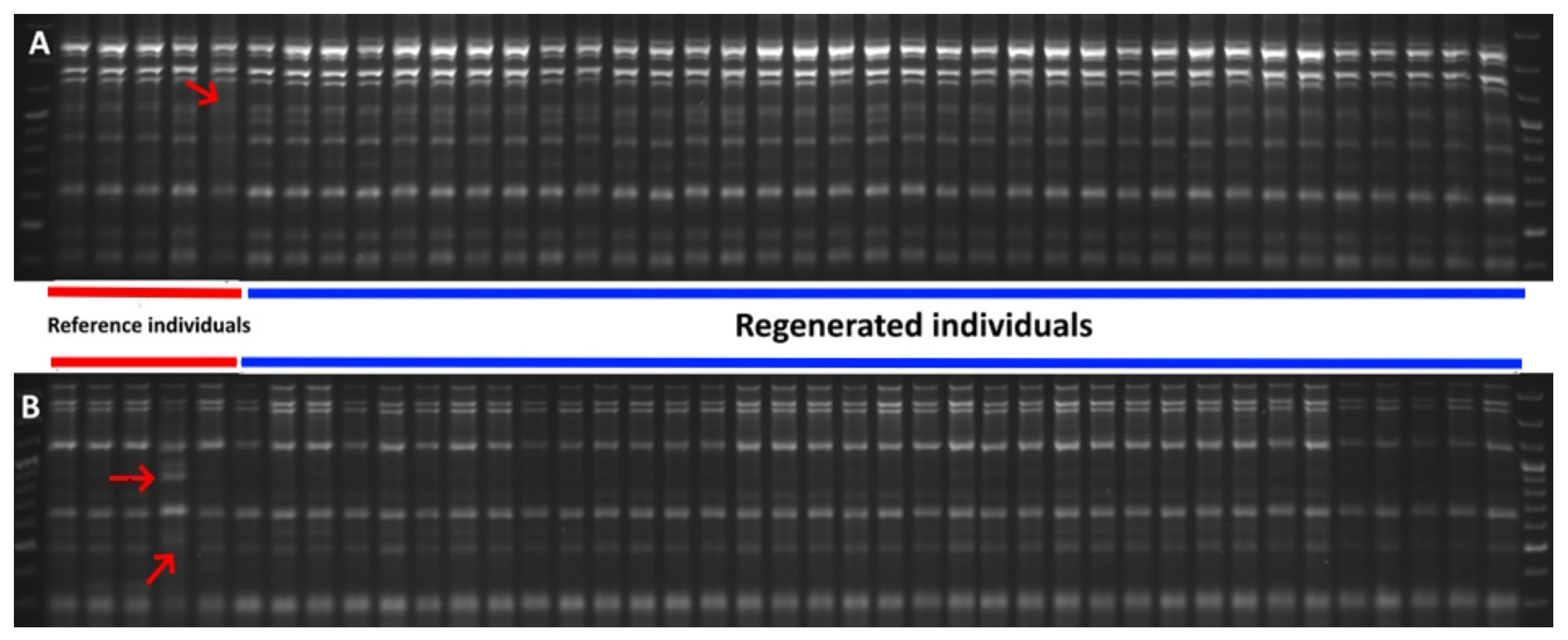
In the present study, altogether, 40 PCR products were obtained: 15 were SCoT fragments and 25 were SRAP fragments. The sizes of SCoT products ranged from 500 to 2000 bp, and SRAP products ranged from 250 to 2500 bp. All of them were monomorphic except three bands that were obtained for reference individuals amplified with SCoT28 and me12-em13 SRAP primers (Figure 3A,B).
When in vitro regenerated individuals are analyzed by molecular markers, usually one mother plant and a few regenerants are enclosed, and monomorphic products are expected because they all should be genetically identical [50,51,52]. A. vesiculosa is generally considered a genetically monomorphic species [29,53,54,55]. In the presented paper, in the case of the five plants obtained from the natural stands, when amplified altogether with eight primers/primer pairs, only three polymorphic bands were obtained. Taking into consideration the genetic analysis only, all regenerated individuals were monomorphic as expected, which indicated that they genetically fit the A. vesiculosa profile.
The use of both methods, phenotypic and genetic, to evaluate the stability of A. vesiculosa tissue culture regenerants increased the reliability of somaclonal variant detection. According to Harding [44], morphological methods are more sensitive for assessing variation in individual plants, which could be confirmed in the present study, as there were no proven genetic variations between the plants regenerated in vitro, while the phenotypic changes were observed between the clones. The observed variation could not be connected with the external factors, such as the use of PGRs or culture age, as the differences in the growth structure were observed from the beginning of the cultivation. There is little data available demonstrating that tissue culture itself can affect the frequency and nature of somaclonal variation [56,57] or that it can arise from mutations already present in the donor plant [42].
In practice, micropropagation is often used in the protection of many endangered plant species, like D. muscipula, Saussurea involucrate, and Orchid spp. [58,59]. A combination of reintroduction and in vitro propagation is, at present, considered the most effective method for preserving the genetic resources of plant species that are threatened or endangered with extinction. However, it is often recommended that the confirmation of the genetic true-to-typeness of in vitro propagated plants should be proven, especially before reintroduction into natural stands [60,61,62,63].
We conclude that tissue culture is an effective method supplementing the traditional active protection of plant species. However, it needs an accurate approach, together with monitoring the stability and true-to-typeness of the regenerants. The monitoring usually includes a molecular marker analysis of the obtained plants. As shown in the presented paper, in the case of A. vesiculosa plants cultivated in tissue culture, the often-underestimated morphological observations might also detect occurring somaclonal variations. Any specimens that differ from standard plants for the species should be eliminated from further propagation and reintroduction into the natural habitat.

Author Contributions

Conceptualization, M.P. (Marzena Parzymies) and M.P. (Magdalena Pogorzelec); methodology, M.P. (Marzena Parzymies) and K.G.; software, M.P. (Marzena Parzymies); validation, M.P. (Marzena Parzymies), M.P. (Magdalena Pogorzelec) and K.G.; formal analysis, M.P. (Marzena Parzymies) and B.B.-A.; investigation, M.P. (Marzena Parzymies), K.G. and M.A.; resources, M.A. and B.B.-A.; data curation, M.P. (Marzena Parzymies) and A.Ś.; writing—original draft preparation, M.P. (Marzena Parzymies) and K.G.; writing—review and editing, M.P. (Marzena Parzymies) and M.P. (Magdalena Pogorzelec); visualization, M.P. (Marzena Parzymies) and M.A.; supervision, M.P. (Marzena Parzymies); project administration, M.P. (Marzena Parzymies); funding acquisition, M.P. (Marzena Parzymies). All authors have read and agreed to the published version of the manuscript.

Funding

The research was funded in part by the European Union through the Infrastructure Environment Operational Programme, project title: Ochrona czynna aldrowandy pęcherzykowatej (Aldrovanda vesiculosa) na terenie Lubelszczyzny (Active conservation of Aldrovanda vesiculosa in Lubelszczyzna region), no. POIS.02.04.00-00-0034/18.

Institutional Review Board Statement

Not applicable.

Informed Consent Statement

Not applicable.

Data Availability Statement

All the required data related to the current study are included in this manuscript.

Conflicts of Interest

The authors declare no conflicts of interest.

Abbreviations

The following abbreviations are used in this manuscript:
MSMurashige and Skoog medium (1962)
PGRsPlant growth regulators

References

  1. Corlett, R.T. A bigger toolbox: Biotechnology in biodiversity conservation. Trends Biotechnol. 2017, 35, 55–65. [Google Scholar] [CrossRef]
  2. Fay, M.F. Conservation of rare and endangered plants using micropropagation. Vitr. Cell. Dev. Biol. Plant 1992, 28, 1–4. [Google Scholar] [CrossRef]
  3. Sarasan, V.; Cripps, R.; Ramsay, M.M.; Atherton, C.; McMichen, M.; Prendergast, G.; Rowntree, J.K. Conservation of rare and endangered plants using in vitro methods. Vitr. Cell. Dev. Biol. Plant 2006, 42, 206–214. [Google Scholar] [CrossRef]
  4. Chokheli, V.A.; Dmitriev, P.A.; Rajput, V.D.; Bakulin, S.D.; Azarov, A.S.; Varduni, T.V.; Stepanenko, V.V.; Tarigholizadeh, S.; Singh, R.K.; Verma, K.K.; et al. Recent development in micropropagation techniques for rare plant species. Plants 2020, 9, 1733. [Google Scholar] [CrossRef] [PubMed]
  5. Reed, B.M.; Sarasan, V.; Kane, M.; Bunn, E.; Pence, V.C. Biodiversity conservation and conservation biotechnology tools. Vitr. Cell. Dev. Biol. Plant 2011, 47, 1–4. [Google Scholar] [CrossRef]
  6. Pence, V.C. The possibilities and challenges of cryopreservation for conservation. Plant Cell Rep. 2011, 30, 241–250. [Google Scholar]
  7. Yanthan, J.S.; Kehie, M.; Kumaria, S.; Tnadon, P. In vitro regeneration of Drosera burmannii Vahl.: A carnivorous plant of north-east India. 3 Biotech 2017, 7, 124. [Google Scholar] [CrossRef]
  8. Kondo, K.; Kokubugata, G.; Varghese, S.B.; Itoyama, M.; Breckpot, C.; Kromer, K.; Kamiński, R. Conservation of endangered Aldrovanda vesiculosa by tissue culture. Carniv. Plant Newsl. 1997, 26, 89–92. [Google Scholar] [CrossRef]
  9. Jala, A. Colchicine and duration time on survival rate and micropropagation of Dionaea muscipula Ellis. Afr. J. Plant Sci. 2014, 8, 291–297. [Google Scholar] [CrossRef]
  10. Tkalec, M.; Doboš, M.; Babić, M.; Jurak, E. The acclimation of carnivorous round-leaved sundew (Drosera rotundifolia L.) to solar radiation. Acta Physiol. Plant 2015, 37, 78. [Google Scholar] [CrossRef]
  11. Jadczak, P.; Kulpa, D.; Zbrojewska, A. In vitro micropropagation of Drosera rotundifolia. World Sci. News 2017, 66, 75–85. [Google Scholar]
  12. Devi, S.P.; Kumaria, S.; Rao, S.R.; Tandon, P. Genetic fidelity assessment in micropropagated plants using cytogenetical analysis and heterochromatin distribution: A case study with Nepenthes khasiana Hook f. Protoplasma 2015, 252, 1305–1312. [Google Scholar] [CrossRef]
  13. Caldeira, M.M.M.; de Jesus, J.V.M.; Magalhães, H.S.; de Carvalho, M.A.S.; Andrade, M.S.; Nunes, C.F. Tissue culture applied to carnivorous species. Rev. Sci. Agrár. Parana. 2020, 19, 312–320. [Google Scholar] [CrossRef]
  14. Barnhill‐Dilling, S.K.; Delborne, J.A. Whose intentions? What consequences? Interrogating “Intended Consequences” for conservation with environmental biotechnology. Conserv. Sci. Pract. 2021, 3, e406. [Google Scholar] [CrossRef]
  15. Kaeppler, S.M.; Kaeppler, H.F.; Rhee, Y. Epigenetic aspects of somaclonal variation in plants. Plant Mol. Biol. 2000, 43, 179–188. [Google Scholar] [CrossRef]
  16. Bairu, M.W.; Aremu, A.O.; Van Staden, J. Somaclonal variation in plants: Causes and detection methods. Plant Growth Regul. 2011, 63, 147–173. [Google Scholar] [CrossRef]
  17. Adamec, L. Ecological requirements and the European distribution of the aquatic carnivorous plant Aldrovanda vesiculosa L.—A review. Folia Geobot. 1995, 30, 53–61. [Google Scholar] [CrossRef]
  18. Adamec, L. Biological flora of Central Europe: Aldrovanda vesiculosa L. Perspect. Plant Evol. Syst. 2018, 35, 8–21. [Google Scholar] [CrossRef]
  19. Cameron, K.M.; Wurdack, K.J.; Jobson, R.W. Molecular evidence for the common origin of snap-traps among carnivorous plants. Am. J. Bot. 2002, 89, 1503–1509. [Google Scholar] [CrossRef]
  20. Poppinga, S.; Bauer, U.; Speck, T.; Volkov, A.G. Motile traps. In Carnivorous Plants: Physiology, Ecology and Evolution; Ellison, A.M., Adamec, L., Eds.; Oxford University Press: Oxford, UK, 2018; p. 193. [Google Scholar]
  21. Cross, A.T.; Adamec, L.; Turner, S.R.; Dixon, K.W.; Merritt, D.J. Seed reproductive biology of the rare aquatic carnivorous plant Aldrovanda vesiculosa (Droseraceae). Bot. J. Linn. Soc. 2016, 180, 515–529. [Google Scholar] [CrossRef]
  22. Kamiński, R. Studies on the ecology of Aldrovanda vesiculosa L. I. Ecological differentiation of A. vesiculosa population under the influence of chemical factors in the habitat. Ekol. Pol. 1987, 35, 559–590. [Google Scholar]
  23. Adamec, L. The biology and cultivation of red Australian Aldrovanda vesiculosa. Carniv. Plant Newsl. 1999, 28, 128–132. [Google Scholar] [CrossRef]
  24. Adamec, L. Ecophysiological characteristics of turions of aquatic plants. A review. Aquat. Bot. 2018, 148, 64–77. [Google Scholar] [CrossRef]
  25. Adamec, L. Seasonal growth dynamics and overwintering of the aquatic carnivorous plant Aldrovanda vesiculosa at experimental field sites. Folia Geobot. 1999, 34, 287–297. [Google Scholar] [CrossRef]
  26. Adamec, L. Ecophysiological characterization of dormancy states in turions of the aquatic carnivorous plant Aldrovanda vesiculosa. Biol. Plant. 2003, 47, 395–402. [Google Scholar] [CrossRef]
  27. Cross, A.T. Turion development is an ecological trait in all populations of the aquatic carnivorous plant Aldrovanda vesiculosa. Carniv. Plant Newsl. 2013, 42, 57–61. [Google Scholar] [CrossRef]
  28. Adamec, L.; Lev, J. The introduction of the aquatic carnivorous plant Aldrovanda vesiculosa L. to new potential sites in the Czech Republic: A five-year investigation. Folia Geobot. 1999, 34, 299–305. [Google Scholar] [CrossRef]
  29. Adamec, L.; Tichý, M. Flowering of Aldrovanda vesiculosa in outdoor culture in the Czech Republic and isozyme variability of its European populations. Carniv. Plant Newsl. 1997, 26, 99–103. [Google Scholar] [CrossRef]
  30. Cross, A.; Adamec, L. Red List Assessment—Aldrovanda vesiculosa. In The IUCN Red List of Threatened Species; IUCN 2020: T162346A275442310; IUCN Red List: Cambridge, UK, 2020. [Google Scholar] [CrossRef]
  31. Bilz, M.; Kell, S.P.; Maxted, N.; Lansdown, R.V. European Red List of Vascular Plants; Publications Office of the European Union: Luxembourg, 2011; Available online: https://iucnredlist.org/resources/bilz2011 (accessed on 30 July 2025).
  32. Council of Europe. European Treaty Series—No. 104. Available online: https://rm.coe.int/1680078aff (accessed on 30 July 2025).
  33. Kaźmierczakowa, R.; Zarzycki, K.; Mirek, Z. Polska Czerwona Księga Roślin: Paprotniki i Rośliny Kwiatowe (Polish Red Data Book of Plants: Pteridophytes and Flowering Plants); Instytut Ochrony Przyrody PAN: Kraków, Poland, 2014; p. 895. [Google Scholar]
  34. Fleischmann, A.; Cross, A.T.; Gibson, R.; Gonella, P.M.; Dixon, K.W. Systematics and evolution of Droseraceae. In Carnivorous Plants: Physiology, Ecology and Evolution; Ellison, A., Adamec, L., Eds.; Oxford Academic: Oxford, UK, 2017; pp. 45–57. [Google Scholar]
  35. Kamiński, R. Restitution of Waterwheel plant (Aldrovanda vesiculosa L.) in Poland and recognition of factors deciding on its survival in temperate climate. Prace Ogrodu Bot. Univ. Wrocławskiego 2006, 8, 105. (In Polish) [Google Scholar]
  36. Kamiński, R. Aldrovanda vesiculosa L. In Polska Czerwona Księga Roślin: Paprotniki i Rośliny Kwiatowe, 3rd ed.; Polska Akademia Nauk: Kraków, Poland, 2014. [Google Scholar]
  37. Murashige, T.; Skoog, F. A revised medium for rapid growth and bioassays with tobacco tissue cultures. Physiol. Plant. 1962, 15, 473–497. [Google Scholar] [CrossRef]
  38. Parzymies, M.; Pogorzelec, M.; Świstowska, A. Optimization of propagation of the Polish strain of Aldrovanda vesiculosa in tissue culture. Biology 2022, 11, 1389. [Google Scholar] [CrossRef]
  39. Palfalvi, G.; Hackl, T.; Terhoeven, N.; Shibata, T.F.; Nishiyama, T.; Ankenbrand, M.; Becker, D.; Förster, F.; Freund, M.; Iosip, A.; et al. Genomes of the Venus flytrap and close relatives unveil the roots of plant carnivory. Curr. Biol. 2020, 30, 2312–2320. [Google Scholar] [CrossRef]
  40. Collard, B.C.Y.; Mackill, D.J. Start codon targeted (SCoT) polymorphism: A simple, novel DNA marker technique for generating gene-targeted markers in plants. Plant Mol. Biol. Rep. 2009, 27, 86–93. [Google Scholar] [CrossRef]
  41. Li, G.; Quiros, C.F. Sequence-related amplified polymorphism (SRAP), a new marker system based on a simple PCR reaction: Its application to mapping and gene tagging in Brassica. Theor. Appl. Genet. 2001, 103, 455–461. [Google Scholar] [CrossRef]
  42. Karp, A. Origins, causes and uses of variation in plant tissue cultures. In Plant Cell and Tissue Culture; Vasil, I.K., Thorpe, T.A., Eds.; Kluwer Academic Publishers: Dordrecht, The Netherlands, 1994; pp. 139–151. [Google Scholar]
  43. Skirvin, R.M.; McPheeters, K.D.; Norton, M. Sources and frequency of somaclonal variation. HortScience 1994, 29, 1232–1237. [Google Scholar] [CrossRef]
  44. Harding, K. Genetic integrity of cryopreserved plant cells: A review. CryoLetters 2004, 25, 3–22. [Google Scholar]
  45. Hortsmann, M.; Heier, L.; Kruppert, S.; Weiss, L.C.; Tollrian, R.; Adamec, L.; Westermeier, A.; Speck, T.; Poppinga, S. Comparative prey spectra analyses on the endangered aquatic carnivorous waterwheel plant (Aldrovanda vesiculosa, Droseraceae) at several naturalized microsites in the Czech Republic and Germany. Integr. Organismal Biol. 2019, 1, oby012. [Google Scholar] [CrossRef]
  46. Israeli, Y.; Reuveni, O.; Lahav, E. Qualitative aspects of somaclonal variations in banana propagated by in vitro techniques. Sci. Hortic. 1991, 48, 71–88. [Google Scholar] [CrossRef]
  47. Sharma, S.; Bryan, G.; Winfield, M.; Milliam, S. Stability of potato (Solanum tuberosum L.) plants regenerated via somatic embryos, axillary bud proliferated shoots, microtubers and true potato seeds: A comparative phenotypic, cytogenetic and molecular assessment. Planta 2007, 226, 1449–1458. [Google Scholar] [CrossRef]
  48. Etienne, H.; Bertrand, B. Somaclonal variation in Coffea arabica: Effects of genotype and embryogenic cell suspension age on frequency and phenotype of variants. Tree Physiol. 2003, 21, 1031–1038. [Google Scholar] [CrossRef]
  49. Podwyszyńska, M. Somaclonal variation in micropropagated tulips based on phenotype observation. J. Fruit Ornam. Plant Res. 2005, 13, 109–122. [Google Scholar]
  50. Rewers, M.; Kisiala, A.B.; Drouin, J.; Sliwinska, E.; Cholewa, E. In vitro-regenerated wetland sedge Eriophorum vaginatum L. is genetically stable. Acta Physiol. Plant. 2012, 34, 2197–2206. [Google Scholar] [CrossRef]
  51. Liu, F.; Huang, L.; Li, Y.; Reinhoud, P.; Jongsma, M.A.; Wang, C.-Y. Shoot organogenesis in leaf explants of Hydrangea macrophylla ‘Hyd1’ and assessing genetic stability of regenerants using ISSR markers. Plant Cell Tissue Organ Cult. 2011, 104, 111–117. [Google Scholar] [CrossRef]
  52. Babu, G.A.; Vinoth, A.; Ravindhran, R. Direct shoot regeneration and genetic fidelity analysis in finger millet using ISSR markers. Plant Cell Tissue Organ Cult. 2017, 132, 157–164. [Google Scholar] [CrossRef]
  53. Elansary, H.O.M.; Adamec, L.; Storchova, H. Uniformity of organellar DNA in Aldrovanda vesiculosa, an endangered aquatic carnivorous species, distributed across four continents. Aquat. Bot. 2010, 92, 214–220. [Google Scholar] [CrossRef]
  54. Maldonado San Martin, A.P.; Adamec, L.; Suda, J.; Mes, T.H.M.; Storchova, H. Genetic variation within the endangered species Aldrovanda vesiculosa (Droseraceae) as revealed by RAPD analysis. Aquat. Bot. 2003, 75, 159–172. [Google Scholar] [CrossRef]
  55. Nishihara, S.; Shiga, T.; Nishihiro, J. The discovery of a new locality for Aldrovanda vesiculosa (Droseraceae), a critically endangered free-floating plant in Japan. J. Asia-Pac. Biodivers. 2023, 16, 227–233. [Google Scholar] [CrossRef]
  56. Kawiak, A.; Łojkowska, E. Application of RAPD in the determination of genetic fidelity in micropropagated Drosera plantlets. Vitr. Cell. Dev. Biol. Plant 2004, 40, 592–595. [Google Scholar] [CrossRef]
  57. Chuang, S.J.; Chen, C.L.; Chen, J.J.; Chou, W.Y.; Sung, J.M. Detection of somaclonal variation in micro-propagated Echinacea purpurea using AFLP marker. Sci. Hortic. 2009, 120, 121–126. [Google Scholar] [CrossRef]
  58. Hirano, T.; Godo, T.; Mii, M. Conservation of endangered endemic plants of the Bonin Islands using micropropagation techniques. Vitr. Cell. Dev. Biol. Plant 2005, 41, 242–246. [Google Scholar] [CrossRef]
  59. Kułak, V.; Longboat, S.; Brunet, N.D.; Shukla, M.; Saxena, P. In vitro technology in plant conservation: Relevance to biocultural diversity. Plants 2022, 11, 503. [Google Scholar] [CrossRef] [PubMed]
  60. Ochatt, S.J.; Patat-Ochat, E.M.; Moessner, A. Ploidy level determination within the context of in vitro breeding. Plant Cell Tissue Organ Cult. (PCTOC) 2011, 104, 329–341. [Google Scholar] [CrossRef]
  61. Thiem, B.; Kikowska, M.; Krawczyk, A.; Więckowska, B.; Sliwinska, E. Phenolic acid and DNA contents of micropropagated Eryngium planum L. Plant Cell Tissue Organ Cult. (PCTOC) 2013, 114, 197–206. [Google Scholar] [CrossRef]
  62. Singh, S.R.; Dalal, S.; Singh, R.; Dhawan, A.K.; Kalia, R.K. Evaluation of genetic fidelity of in vitro raised plants of Dendrocalamus asper (Schult. & Schult. F.) Backer ex K. Heyne using DNA-based markers. Acta Physiol. Plant. 2012, 35, 419–430. [Google Scholar] [CrossRef]
  63. Nybom, H.; Weising, K.; Rotter, B. DNA fingerprinting in botany: Past, present, future. Investig. Genet. 2014, 5, 1. [Google Scholar] [CrossRef]
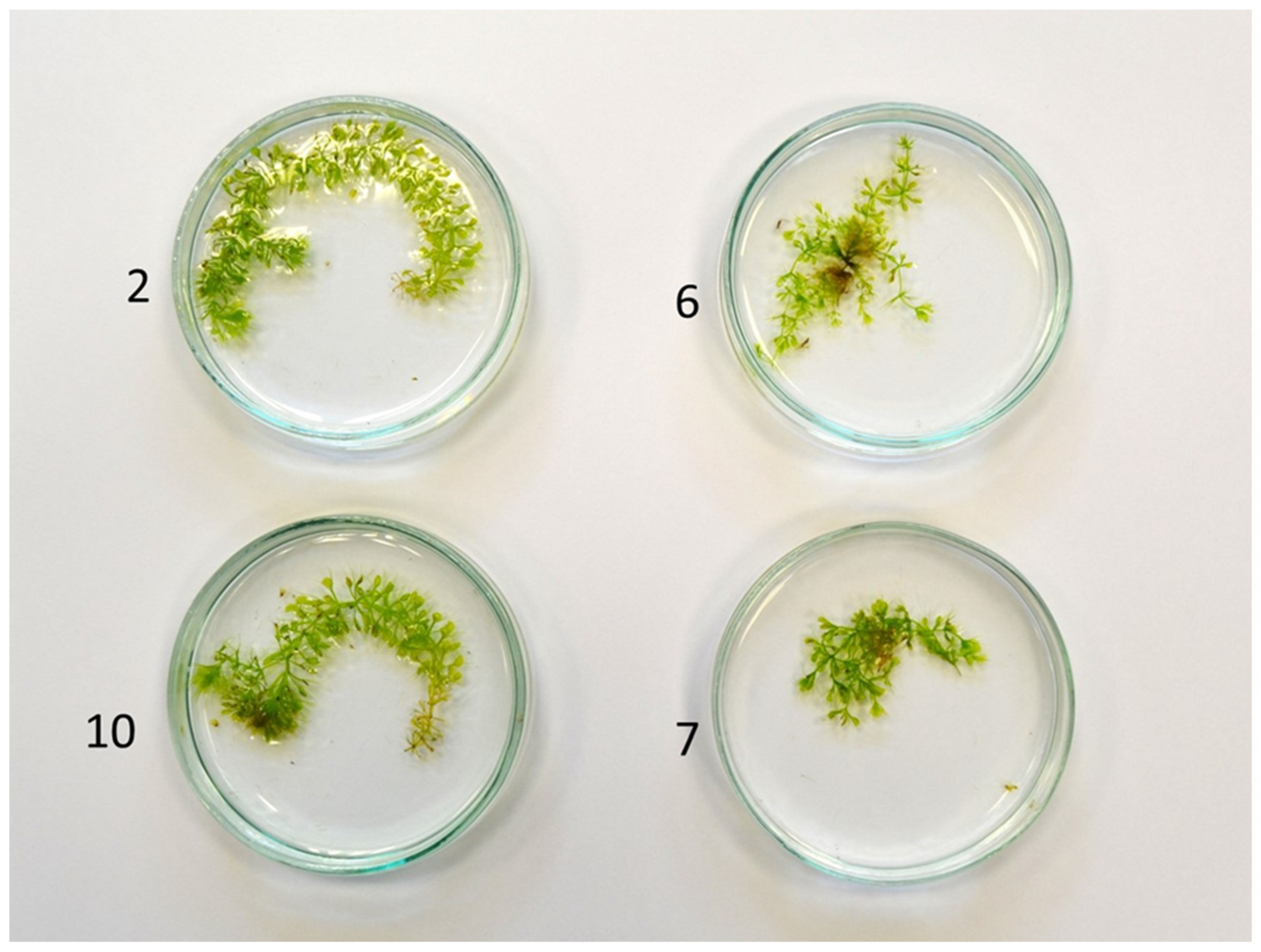
Figure 1. Morphology of the A. vesiculosa shoots propagated in vitro; clones 2 and 10—typical growth for the species; clones 6 and 7—branched growth.
Genes 16 01003 g001
Figure 2. The appearance and measurements of the example traps of A. vesiculosa shoots propagated in vitro from different clones (2 and 10—typical growth for the species; 6 and 7—branched growth).
Genes 16 01003 g002
Figure 3. PCR products obtained for SCoT28 (A) and me12-em13 SRAP (B) molecular markers. Red arrows indicate polymorphic products obtained for two reference individuals. Red lines indicate reference plants and blue lines indicate in vitro regenerated ones.
Genes 16 01003 g003
Table 1. SCoT primer sequences.
Primer NameSequence (5′ 3′)
SCoT18ACCATGGCTACCACCGCC
SCoT22AACCATGGCTACCACCAC
SCoT28CCATGGCTACCACCGCCA
Table 2. SRAP primer sequences and combinations applied in the study.
Forward
Primer
SequenceReverse
Primer
SequenceCombinations Used
me2TGAGTCCAAACCGGAGCem2GACTGCGTACGAATTTGCme2-m2
me5TGAGTCCAAACCGGAAGem5GACTGCGTACGAATTAACme5-em5
me10TGAGTCCAAACCGGAAAem11GACTGCGTACGAATTCTAme10-em12
me11TGAGTCCAAACCGGAACem12GACTGCGTACGAATTCTCme11-em11
me12TGAGTCCAAACCGGAGAem13GACTGCGTACGAATTCTGme12-em13
Table 3. Thermal profiles of SCoT and SRAP methods.
StepSCoTSRAP
Temperature (°C)Time
(s)
Number of CyclesTemperature (°C)Time
(s)
Number of
Cycles
Initial denaturation94180 94240
Denaturation94603594605
Annealing56603560
Elongation721207260
Denaturation 946036
Annealing 5060
Elongation 7260
Final elongation72300 72420
Table 4. Morphological features of four Aldrovanda vesiculosa clones propagated in vitro.
Clone MarkMain Shoot Length (mm)Number of Lateral Shoots/
Explant
Length of Lateral Shoots (mm)Fresh Weight of Plants (mg)
210.63 A *1.60 B4.15 A31.67 A
1010.28 A2.30 B4.34 A31.53 A
65.07 B7.00 A2.20 B17.0 AB
74.76 B4.70 AB2.54 B7.89 B
* Means followed by the same letter in columns do not differ significantly at p = 0.05.
Table 5. Morphological features of traps of A. vesiculosa plants propagated in vitro.
Clone MarkNo. of Whorls per 1 cm of ShootLength of Traps (mm)Width of Traps (mm)Length of Bristles (mm)Length of Petioles (mm)
23.0 A *2.77 A1.71 A5.67 A3.88 A
103.0 A2.98 A1.75 A5.64 A3.82 A
62.3 B1.93 B1.36 B4.09 B1.86 B
72.10 B2.11 B1.17 B3.61 B1.95 B
* Means followed by the same letter in columns do not differ significantly at p = 0.05.
Disclaimer/Publisher’s Note: The statements, opinions and data contained in all publications are solely those of the individual author(s) and contributor(s) and not of MDPI and/or the editor(s). MDPI and/or the editor(s) disclaim responsibility for any injury to people or property resulting from any ideas, methods, instructions or products referred to in the content.