Article

Characterization of the Four Rosa L. Species from Kazakhstan Based on Complete Plastomes and Nuclear Ribosomal Internal Transcribed Spacer (ITS) Sequences

by
Moldir Yermagambetova
1,
Akzhunis Imanbayeva
2,
Margarita Ishmuratova
3,
Aidar Sumbembayev
4 and
Shyryn Almerekova
1,*
1
Institute of Plant Biology and Biotechnology, Almaty 050040, Kazakhstan
2
Mangyshlak Experimental Botanical Garden, Aktau 130000, Kazakhstan
3
Biology and Geography Faculty, Karaganda Buketov University, Karaganda 100028, Kazakhstan
4
Altai Botanical Garden, Ridder 071300, Kazakhstan
*
Author to whom correspondence should be addressed.
Genes 2025, 16(8), 852; https://doi.org/10.3390/genes16080852
Submission received: 17 June 2025 / Revised: 21 July 2025 / Accepted: 21 July 2025 / Published: 22 July 2025
(This article belongs to the Section Plant Genetics and Genomics)

Abstract

Background: Rosa L. is an economically significant genus with species that are notable for their rich content of phenolic compounds. Despite its importance, the taxonomy of Rosa remains complex and unresolved. Methods: We sequenced, assembled, and performed comparative analyses of the complete plastomes of four Rosa species: R. acicularis, R. iliensis, R. laxa, and R. spinosissima. In addition to the plastome, we sequenced the nuclear ribosomal internal transcribed spacer (ITS). Results: Plastomes ranged in size from 157,148 bp (R. iliensis) to 157,346 bp (R. laxa). In each plastome, 136 genes were annotated, comprising 90 protein-coding, 38 tRNA, and eight rRNA genes. A total of 905 SSRs were identified, ranging from 224 (R. acicularis) to 229 in R. spinosissima. Nine highly variable regions were detected, including two coding genes (rps16 and ycf1) and seven intergenic spacers (ycf3-trnS(GGA), trnT(UGU)-trnL(UAA), rpl14-rpl16, trnR(UCU)-atpA, trnD(GUC), trnG(UCC)-trnfM(CAU), and psbE-petL). Maximum Likelihood (ML) phylogenetic analyses based on the complete plastome and ycf1 gene datasets consistently resolved the Rosa species into three major clades, with strong bootstrap support. In contrast, the ML tree based on ITS resolved species into four clades but showed lower bootstrap values, indicating reduced resolution compared to plastid datasets. Conclusions: Our findings underscore the value of plastome data in resolving phylogenetic relationships within the genus Rosa.

1. Introduction

The genus Rosa L., belonging to the Rosaceae Juss family, comprises approximately 200 species [1] primarily distributed across the Northern Hemisphere [2]. In Kazakhstan, the genus Rosa is represented by 25 species and is distributed throughout all regions of the country [3]. Among them, Rosa acicularis Lindl., Rosa laxa Retz., and Rosa spinosissima L. are widely distributed in the territory of the country, while Rosa iliensis Chrshan. is an endemic species [4]. The most recent classification of the genus Rosa was proposed by Wissemann (2017) [5], based on the earlier framework of Rehder (1940) [6]. This classification divides the genus into four subgenera: Rosa, Hulthemia (Dumort.) Focke, Platyrhodon (Hurst) Rehder, and Hesperhodos Cockerell. The largest subgenus, Rosa, is further divided into ten sections: Banksianae Lindl., Bracteatae Thory, Caninae (DC.) Ser., Carolinae Crép., Gallicanae (DC.) Ser., Indicae Thory, Laevigatae Thory, Pimpinellifoliae (DC.) Ser., Rosa (=Cinnamomeae (DC.) Ser.), and Synstylae DC [5,6].
Numerous phylogenetic studies have been conducted to clarify the taxonomy of the genus Rosa using molecular genetic approaches. For example, Rosa phylogenetic relationships have been investigated based on various genetic markers, including ITS-1 and the atpB-rbcL intergenic spacer [1], trnL, trnG, and the psbA-trnH intergenic spacer [7], as well as rpl16, trnL-F, and atpB-rbcL [8]. More recently, Debray et al. (2022) [9] employed phylogenomic analyses using 96 informative single-copy orthologous tags to resolve relationships within the genus. Furthermore, the genetic diversity and population structure of different Rosa species have been extensively investigated using a variety of molecular markers. Among these, randomly amplified polymorphic DNA (RAPD) [10,11], amplified fragment length polymorphism (AFLP) [12,13], simple sequence repeats (SSRs) [14,15,16], and single nucleotide polymorphisms (SNPs) [17,18] have been widely applied, offering valuable insights into intra- and interspecific genetic variation. Despite the studies that have been conducted, the assessment of phylogenetic relationships is often complicated by spontaneous hybridization events, which contribute to the formation of new species [19]. For instance, R. spinosissima has been proposed to have a hybrid origin [20]. In various phylogenetic studies, this species is positioned between Rosa and Pimpinellifoliae sections [7,9], suggesting its intermediate genetic background. The taxonomy of the genus remains challenging and continues to be a subject of debate and uncertainty.
Complete plastome sequences have also been employed to assess the phylogenetic relationships among Rosa species [21,22,23,24]. The highly informative coding regions ndhF, ycf1 ycf3-trnS, trnT-trnL, and psbE-petL have been identified within the plastomes of Rosa species and proposed as potential DNA barcoding markers for future phylogenetic studies [21,24]. It is well established that plastomes are a primary source of informative DNA barcoding markers, which are widely employed in phylogenetic analyses across a broad range of plant taxa [25,26]. In addition to their phylogenetic applications, plastomes also facilitate the identification of simple sequence repeat (SSR) markers, which are valuable tools in population genetic studies [27,28].
Plastomes of most angiosperms exhibit a conserved quadripartite structure consisting of a large single-copy (LSC) region, a small single-copy (SSC) region, and a pair of inverted repeats (IRs) [29]. The gene content and structural organization of plastomes are highly conserved, with genome sizes typically ranging from 120 to 160 kilobases [30,31]. With the advancement of sequencing technologies, plastome sequencing has become faster and more accessible, leading to its widespread application in phylogenetic studies [32,33].
Species of the genus Rosa are predominantly shrubs valued for their nutritional [34], ornamental [35], and medicinal [36] properties. Moreover, the fruits of Rosa species are widely used in food products such as jams, jellies, and herbal teas [37] and are well known for their high vitamin C content [38]. Phenolic compounds play a significant role in the activities of antidiabetic, anti-hypertensive, anti-tumor, anti-atherosclerotic, anti-inflammatory, and anti-aging properties [39,40,41,42]. Several studies [43,44,45] have investigated phenolic compounds from various parts of the Rosa species, with a particular focus on their chemical composition and antioxidant properties. Among these species, R. acicularis is a notable plant species for its rich diversity of phenolic compounds, which are distributed throughout its leaves, flowers, roots, and fruits. Ellagitannins and flavonoid glycosides are particularly abundant in this species [46]. Notably, leaf extracts of R. acicularis have demonstrated promising antidiabetic potential [46]. The chemical composition of four different extracts from R. laxa was quantitatively analyzed [47]. The results revealed that the aqueous extract exhibited the strongest antioxidant capacity and the highest contents of total triterpene acids, flavonoids, and polyphenols. These findings support the potential application of R. laxa in disease prevention and therapeutic strategies [47]. The qualitative and quantitative composition of anthocyanins in R. spinosissima fruit has been identified, highlighting its potential as a valuable natural source of anthocyanins for medicinal applications [48]. The composition of flower volatiles and seed fatty acids in R. iliensis populations from the Almaty Region in Kazakhstan was investigated [49]. A high abundance of oxygenated monoterpenes characterized the flower volatiles. In the seeds, linoleic, α-linolenic, and oleic acids were identified as the major fatty acid constituents [49]. The findings highlight that the Rosa species in this study (R. acicularis, R. iliensis, R. laxa, and R. spinosissima) are promising sources of bioactive compounds, underscoring their potential as valuable resources for medicinal applications.
In this study, we sequenced and assembled the complete plastomes of four Rosa species—R. acicularis, R. iliensis, R. laxa, and R. spinosissima—using next-generation sequencing technologies. The four species studied are dominant components of plant communities [3] in various regions of the country, possess high medicinal value [46,47,48,49], and have not been previously studied in Kazakhstan using plastome data. The complete plastomes of R. iliensis and R. laxa were sequenced for the first time in this study. The main objectives of this study were to (i) characterize the complete plastomes of the four Rosa species, (ii) perform comparative and phylogenetic analyses of these plastomes in conjunction with publicly available Rosa plastome sequences from the National Center for Biotechnology Information (NCBI) GenBank database, and (iii) identify additional molecular markers that may be informative for future phylogenetic and population genetic studies.

2. Materials and Methods

2.1. Plant Material Collection and DNA Extraction

Fresh leaf samples of R. acicularis were collected from the Karaganda Region, R. iliensis and R. laxa from the Mangystau Region, and R. spinosissima from the Western Altai Region (Figure 1). The collected fresh leaves were immediately dried in silica gel. Dried leaf material was subsequently used for total genomic DNA extraction using the DNeasy Plant Mini Kit (Qiagen, Valencia, CA, USA), following the manufacturer’s protocol. The concentration and quality of the extracted DNA were assessed using a NanoDrop 2000 spectrophotometer (Thermo Fisher Scientific, Wilmington, DE, USA) and by agarose gel electrophoresis.

2.2. PCR Amplification and Sequencing of the Internal Transcribed Spacer

The internal transcribed spacer (ITS) region of nuclear ribosomal DNA was utilized for phylogenetic analysis. Amplification of the ITS region was performed using the primers (Table 1) as described by White et al. (1990) [50].
The polymerase chain reaction (PCR) was conducted in a total reaction volume of 25 µL, containing 10× reaction buffer, 25 mM MgCl2, 2 µM of each primer, 4 mM of each dNTP, 1.6 units of Taq DNA polymerase, and 100 ng of genomic DNA. The volume was adjusted to 25 µL with nuclease-free water. The PCR was carried out using a SimpliAmp™ thermal cycler (Thermo Fisher Scientific, Carlsbad, CA, USA) under the following thermocycling conditions: initial denaturation at 94 °C for 3 min, followed by 40 cycles of denaturation at 94 °C for 30 s, annealing at 58 °C for 45 s, and extension at 72 °C for 1 min and 30 s, with a final extension at 72 °C for 10 min. PCR products were separated on a 1.5% agarose gel and subsequently purified using the ULTRAPrep® Agarose Gel Extraction Mini Prep Kit (AHN Biotechnologie GmbH, Nordhausen, Germany), following the manufacturer’s protocol. Purified amplicons were used for sequencing with both forward and reverse primers using the BigDye® Terminator Cycle Sequencing Kit (Applied Biosystems, Foster City, CA, USA). Sequencing was performed on an ABI 3130 Genetic Analyzer (Applied Biosystems, USA). Nucleotide sequences of the ITS of four Rosa species were deposited to the NCBI GenBank (https://www.ncbi.nlm.nih.gov/, accessed on 14 June 2025) under accession numbers PV789381, PV789382, PV789383, and PV789384.

2.3. Plastome Sequencing, Assembly, and Annotation

High-quality DNA samples met quality control standards for paired-end library preparation with the TruSeq Nano DNA Library Prep Kit (Illumina Inc., San Diego, CA, USA). Paired-end sequencing of the four Rosa species was performed on the Illumina NovaSeq 6000 platform (Illumina Inc., San Diego, CA, USA) at Macrogen Inc. (Seoul, Republic of Korea). The quality of the raw sequencing reads was evaluated using FastQC (https://www.bioinformatics.babraham.ac.uk/projects/fastqc/, accessed on 10 February 2025), and adapter sequences were removed using Trimmomatic v0.36 [51]. Cleaned reads were assembled de novo using NOVOPlasty v4.3.3 [52]. Plastome annotations were conducted using GeSeq [53], and the results were manually curated by comparison with reference sequences available in the NCBI GenBank. The annotated plastome sequences of R. acicularis, R. iliensis, R. laxa, and R. spinosissima have been deposited in GenBank (https://www.ncbi.nlm.nih.gov/, accessed on 10 March 2025) under accession numbers PV330080, PV330081, PV330082, and PV330083, respectively. Circular plastome maps were generated using Organellar Genome DRAW (OGDRAW) v1.3.1 [54].

2.4. Identification of the Simple Sequence Repeats

To identify simple sequence repeat (SSR or microsatellite) elements within the plastomes of four Rosa species (R. acicularis, R. iliensis, R. laxa, and R. spinosissima), the MISA (Microsatellite Identification Tool) online tool was utilized [55]. The minimum repeat thresholds were set to 8 for mononucleotide repeats, 4 for di- and trinucleotide repeats, and 3 for tetra-, penta-, and hexanucleotide repeats. All identified SSR loci were manually inspected to ensure accuracy; redundant or overlapping entries were removed from the final dataset.

2.5. Plastome Comparison

A comparative analysis of the plastomes from the four Rosa species was performed using the mVISTA tool [56] with the Shuffle-LAGAN mode. The plastomes of R. graciliflora (OQ992658) and R. acicularis (MK714016) were used as reference sequences. To further investigate the boundaries of the inverted repeat (IR) regions, the junction sites of the four plastomes were analyzed using the IRscope tool [57], employing the same reference genomes, R. graciliflora (OQ992658) and R. acicularis (MK714016).

2.6. Sliding Window Analysis and Phylogenetic Analysis

For sliding window analysis and phylogenetic analysis, plastome sequences of the Rosa samples were aligned using Geneious Prime® 2025.0.3. To evaluate nucleotide diversity, a sliding window analysis was conducted in DnaSP v6 [58], using a window size of 600 bp and a step size of 200 bp. This approach enabled the identification of highly variable regions across the aligned plastomes. For the phylogenetic analysis, three datasets were utilized: complete plastome nucleotide sequences, ycf1 gene sequences, and nuclear ribosomal internal transcribed spacer (ITS) sequences. Phylogenetic trees were reconstructed using the Maximum Likelihood (ML) method, implemented in IQ-TREE v2.2.2.6 [59]. The best-fit nucleotide substitution models, selected based on the Bayesian Information Criterion (BIC), were TVM+F+I+R4 for the complete plastomes, TVM+F+G4 for the ycf1 gene, and TIM+F+G4 for the ITS dataset. The resulting ML trees were visualized using FigTree v1.4.4 (http://tree.bio.ed.ac.uk/software/figtree/, accessed on 27 February 2025). The number of samples used in the phylogenetic analyses, based on different data sets, is provided in Table S1. The section names were assigned following Wisseman (2017) [5] and Rehder (1940) [6], while subclade designations were based on the work proposed by Debray et al. (2022) [9].

3. Results

3.1. General Characteristics of the Four Rosa Plastomes

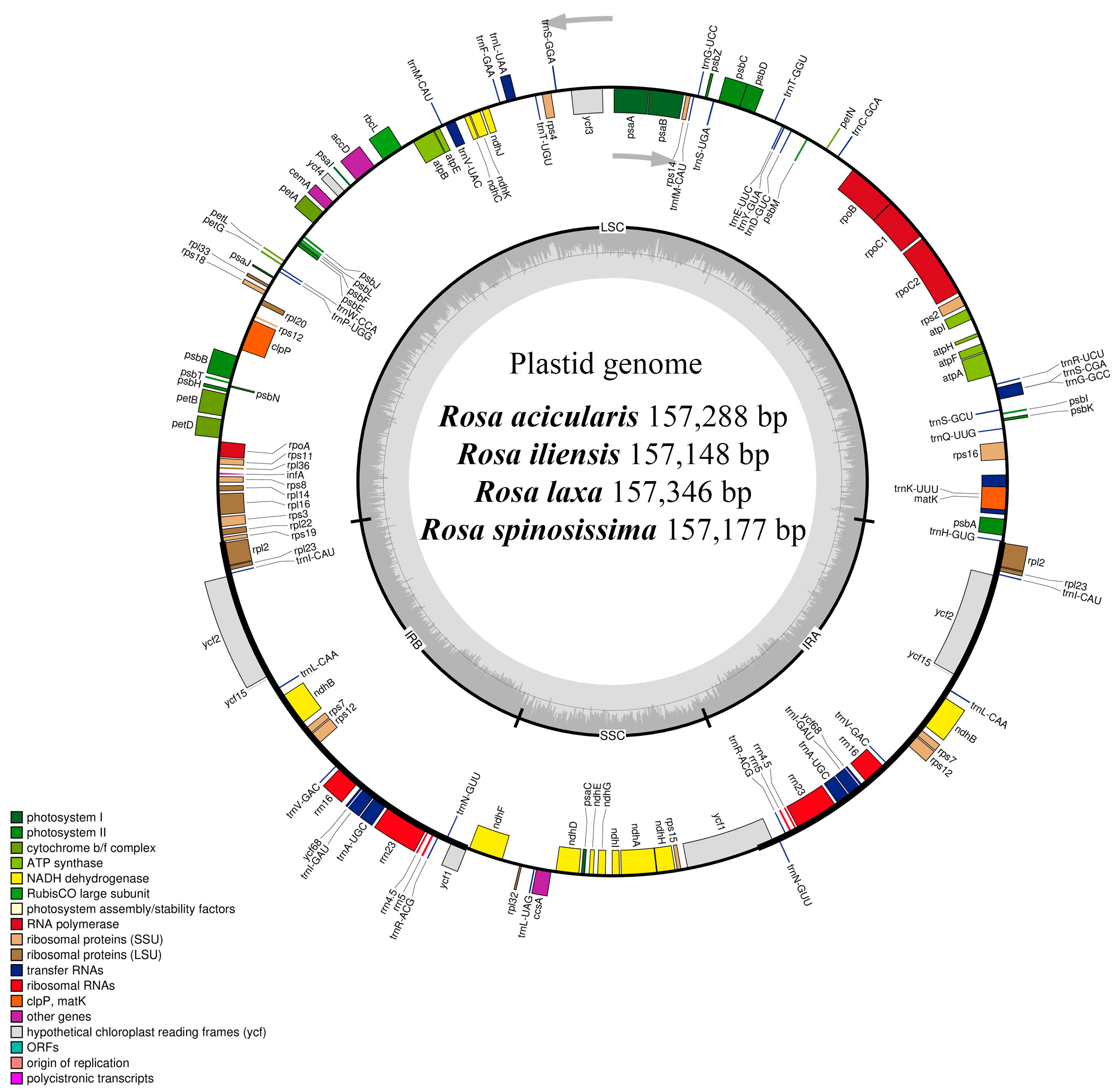
The complete plastome sizes of R. acicularis, R. iliensis, R. laxa, and R. spinosissima were determined to be 157,288 bp, 157,148 bp, 157,346 bp, and 157,177 bp, respectively (Table 2). All sequenced plastomes exhibited the typical quadripartite structure (Figure 2), comprising a large single-copy (LSC) region ranging from 86,269 (R. spinosissima) to 86,461 bp (R. laxa), a small single-copy (SSC) region varying between 18,778 (R. iliensis) and 18,791 bp (R. acicularis and R. laxa), and two inverted repeat (IR) regions ranging from 26,047 (R. laxa) to 26,062 bp (R. spinosissima). The overall GC content was consistent across all species, measured at 37.20% (Table 2).
The number of annotated genes was identical across all four plastomes, with a total of 136 genes identified. These included 90 protein-coding genes, 38 tRNA genes, and eight rRNA genes, including duplicated copies (Table 2). Twenty genes were duplicated within the IR regions, comprising nine protein-coding genes (ndhB, rpl2, rpl23, rps7, rps12, ycf1, ycf2, ycf15, and ycf68), seven tRNA genes (trnA-UGC, trnI-CAU, trnI-GAU, trnL-CAA, trnN-GUU, trnR-ACG, and trnV-GAC), and all four rRNA genes (rrn4.5, rrn5, rrn16, and rrn23). Among the annotated genes, seven tRNA genes (trnA-UGC, trnG-GCC, trnI-GAU, trnK-UUU, trnL-UAA, trnS-CGA, and trnV-UAC) and 11 protein-coding genes (rps12, rps16, rpl2, rpl16, rpoC1, ndhA, ndhB, petD, clpP, and ycf3) contained introns. The protein-coding genes rps12, clpP, and ycf3 each harbored two introns, while the others contained a single intron (Table 3).

3.2. Simple Sequence Repeat Analysis

Simple sequence repeats (SSRs), also known as microsatellites, were identified within the plastome sequences of the four Rosa species. A total of 905 SSRs were detected, with individual counts of 224, 227, 225, and 229 in R. acicularis, R. iliensis, R. laxa, and R. spinosissima, respectively. The identified SSRs ranged from mono- to hexanucleotide repeats, although not all six types were present in each species. Mononucleotide repeats were the most abundant, accounting for 601 SSRs (66.41%), and ranged from 147 in R. acicularis to 152 in R. spinosissima. These were predominantly A/T repeats across all plastomes. Dinucleotide repeats (AT/AT, AG/CT, and AC/GT) were the second most common, comprising 223 SSRs (24.64%), and were mainly represented by AT/AT motifs. Their abundance ranged from 55 in R. spinosissima to 56 in R. acicularis, R. iliensis, and R. laxa. Trinucleotide SSRs (AAT/ATT and AGC/CTG) and tetranucleotide SSRs (AAAT/ATTT, AATT/AATT, ACAT/ATGT, and ACCT/AGGT) were less frequent, with 27 (2.98%) and 43 (4.75%), respectively. Pentanucleotide repeats (AATAT/ATATT and AAAAG/CTTTT) were rare, with only three (0.33%) identified across three of the species, absent in R. iliensis. Hexanucleotide repeats (AAGTAG/ACTTCT) were found in all four plastomes, totaling eight repeats (0.88%) (Table 4). Detailed information on the identified SSRs is provided in Supplementary File S1.

3.3. Comparative Analysis of the Plastomes of Four Rosa Species

To assess sequence conservation and divergence across plastomes of four Rosa species, a mVISTA-based alignment was performed using the complete plastomes of R. graciliflora (OQ992658) and R. acicularis (MK714016) from NCBI GenBank as the references. The results revealed that the LSC and SSC regions are more variable than the IR regions, which showed higher sequence conservation across the examined Rosa samples. Overall, the alignment demonstrates a high degree of sequence conservation across the plastomes of Rosa samples, with notable divergence localized to non-coding regions (Figure 3).
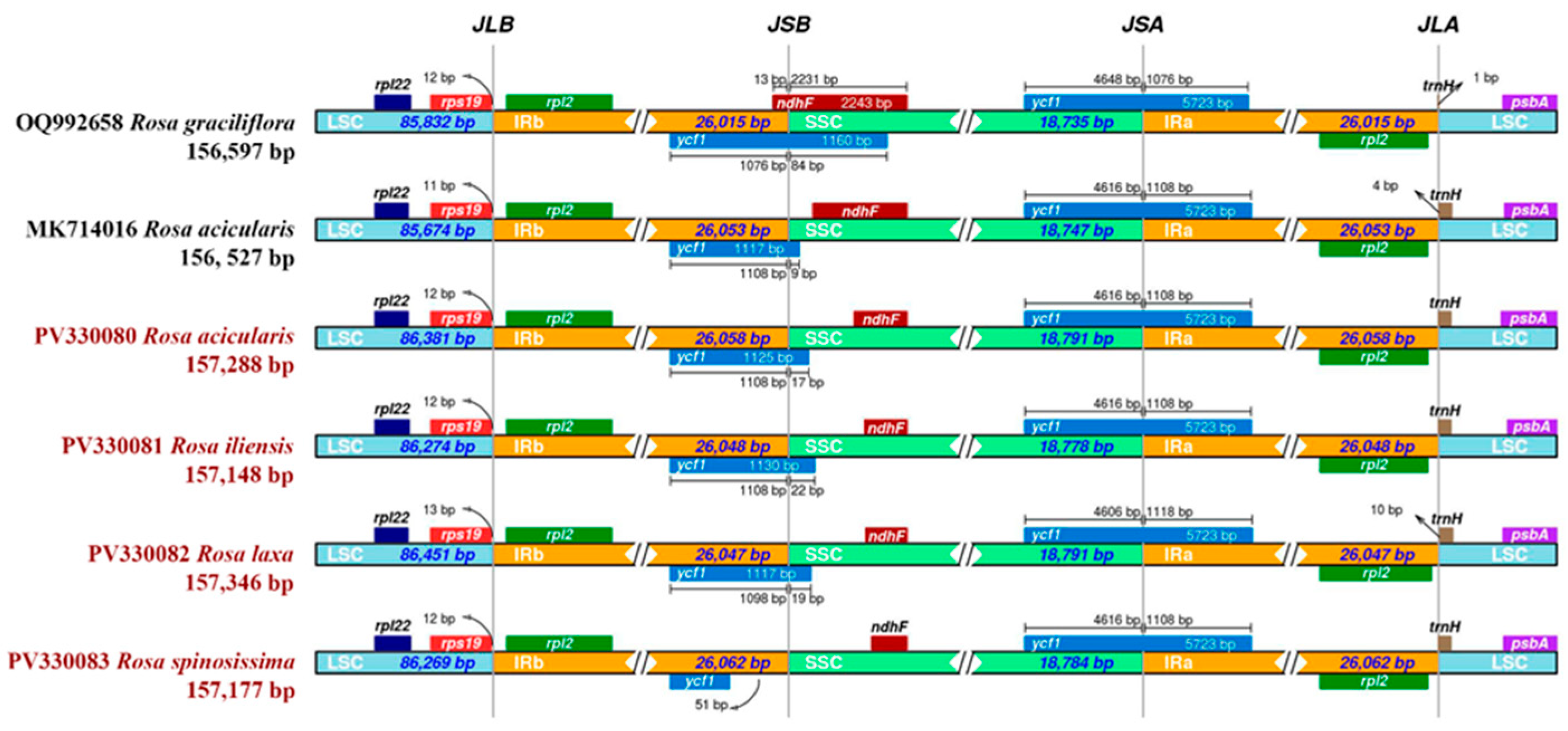
Next, the expansion and contraction of the IR regions at the LSC/IR/SSC junctions were analyzed using the reference plastomes of R. graciliflora (OQ992658) and R. acicularis (MK714016). The results revealed that the overall structure of the plastomes is conserved among the analyzed Rosa samples. However, minor differences in the positioning of boundary genes, such as rps19, ycf1, ndhF, and trnH, reflect subtle expansions or contractions of the IR regions. At the LSC/IRb junction (JLB), the rps19 gene consistently overlaps with the IRb region in all six samples, with an overlap ranging from 11 to 13 bp. The ndhF gene is located at the IRb/SSC junction (JSB) only in R. graciliflora (OQ992658) and is absent at this junction in the remaining five plastomes. The ycf1 gene spans the SSC/IRb junction (JSB) in five genomes but is absent at this boundary in R. spinosissima (PV330083). Additionally, a duplicated copy of the ycf1 gene is present at the IRa/SSC (JSA) junction in all six plastomes. The IRa/LSC junction (JLA) is relatively conserved, with the rpl2 gene located entirely within the IRa region, while the trnH gene is positioned downstream in the LSC, showing slight positional variation ranging from 1 to 10 bp. In terms of regional lengths, the LSC region ranges from 85,674 bp in R. acicularis (MK714016) to 86,451 bp in R. laxa (PV330082). The SSC region varies from 18,735 bp in R. graciliflora (OQ992658) to 18,791 bp in R. acicularis (PV330080) and R. laxa (PV330082). The length of each IR region (IRa and IRb) ranges from 26,015 bp in R. graciliflora to 26,062 bp in the R. spinosissima (PV330083) plastome (Figure 4).

3.4. Nucleotide Diversity Analysis by Sliding Window

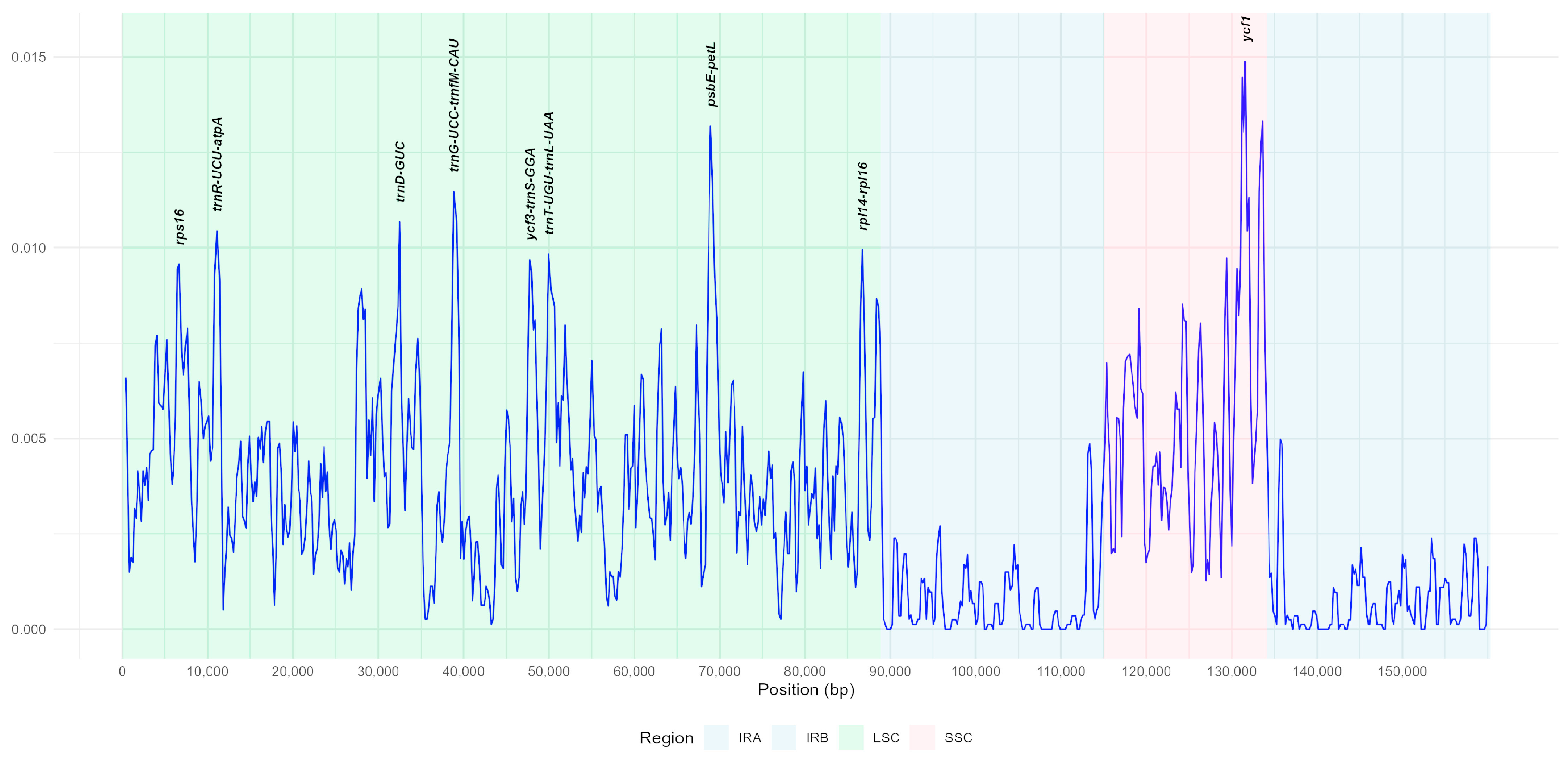
A nucleotide diversity analysis was conducted on the complete plastome sequences of 26 Rosa samples, including four studied species. The nucleotide diversity (Pi) values ranged from 0 to 0.01488. Nine highly variable regions were identified, exhibiting relatively elevated Pi values. These included two coding regions (rps16 and ycf1) and seven intergenic regions (ycf3-trnS(GGA), trnT(UGU)-trnL(UAA), rpl14-rpl16, trnR(UCU)-atpA, trnD(GUC), trnG(UCC)-trnfM(CAU), and psbE-petL). The highest nucleotide diversity (Pi = 0.01488) was observed in the ycf1 gene region of the SSC region. Overall, the LSC and SSC regions exhibited the greatest nucleotide variability, with the LSC region showing higher nucleotide diversity than the SSC region (Figure 5).

3.5. Phylogenetic Analysis

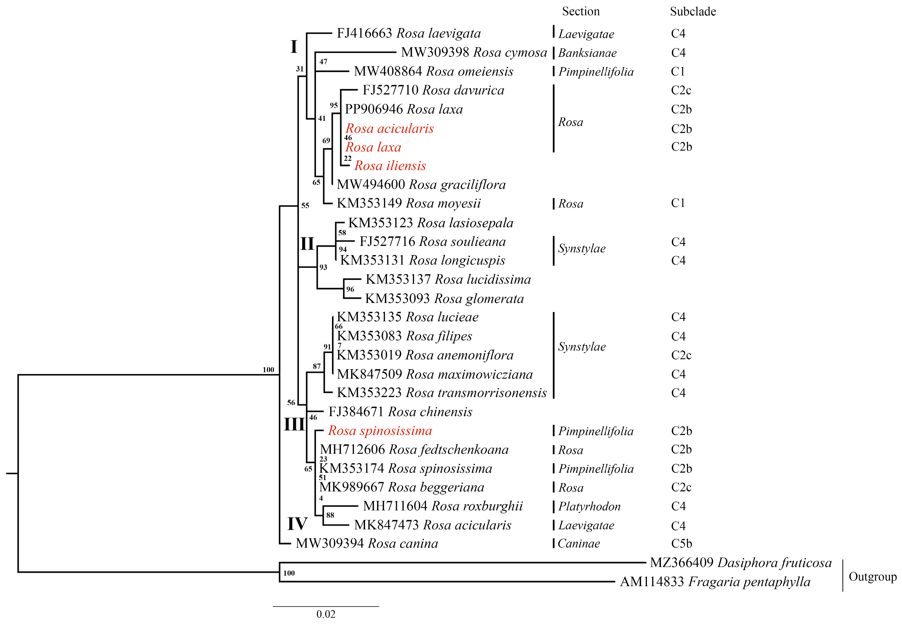
Phylogenetic trees were reconstructed using the Maximum Likelihood (ML) method based on three datasets: (1) nucleotide sequences of the complete plastome (Figure 6A), (2) nucleotide sequences of the ycf1 gene (Figure 6B), and (3) nucleotide sequences of the nuclear ribosomal internal transcribed spacer (ITS) (Figure 7). Fragaria pentaphylla and Dasiphora fruticosa were used as outgroups in all analyses. Based on the complete plastome and ycf1 gene datasets, the ML trees consistently resolved the Rosa species into three major clades (I, II, and III), with strong bootstrap support at most nodes. Clade I included R. omeiensis from the Pimpinellifoliae section. Clade II comprised species from sections Pimpinellifoliae and Rosa, forming subclades C1, C2b, and C2c. Notably, the species analyzed in this study, R. spinosissima, R. acicularis, R. laxa, and R. iliensis, are grouped within subclade C2b. Clade III was the most taxonomically diverse, including species from sections Synstylae, Banksianae, Caninae, Laevigatae, and Platyrhodon, primarily corresponding to clades C4 and C5b. An exception was R. anemoniiflora, which grouped within the subclade C2c (Figure 6). In contrast, the ML phylogenetic tree based on the ITS nucleotide sequences (Figure 7) resolved the species into four clades (I, II, III, and IV). The studied species R. acicularis, R. iliensis, and R. laxa were grouped in Clade I, along with R. davurica and R. laxa from GenBank, corresponding to subclade C2b. Meanwhile, R. spinosissima (C2b, this study) grouped in Clade III with GenBank samples of R. fedschenkoana, R. spinosissima, R. beggeriana (all C2b), R. roxburghii (C4), and R. acicularis (C4). The ITS tree showed lower bootstrap support than the plastome and ycf1 gene trees, indicating reduced resolution in the nuclear dataset (Figure 7).
Overall, the phylogenetic analysis based on the complete plastome and ycf1 gene nucleotide sequences revealed clear sectional affiliations and evolutionary relationships among the Rosa species, with strong bootstrap support indicating the robustness of the inferred topology.

4. Discussion

The development of low-cost, high-throughput sequencing technologies has greatly accelerated the sequencing of plant plastomes. In this study, we sequenced, assembled, and conducted a comparative analysis of the complete plastomes of four Rosa species from Kazakhstan: R. acicularis, R. iliensis, R. laxa, and R. spinosissima (Figure 1), using next-generation sequencing technologies. Consistent with the typical structure observed in most angiosperms [60,61], the plastomes of the studied Rosa species exhibited a quadripartite organization, comprising LSC and SSC regions, and two IR regions (Figure 2). The genome sizes ranged from 157,148 bp in R. iliensis to 157,346 bp in R. laxa (Table 2), closely aligning with the plastome lengths reported for R. glomerata Rehder & E.H. Wilson (157,064 bp) [62], R. praelucens Bijh. (157,186 bp) [63], and R. minutifolia Engelm. (157,396 bp) [64]. The GC content across all four species was 37.20%, which is consistent with the GC content of the R. xanthina Lindl. plastome (37.20%) [65].
Each genome contained 136 annotated genes, including 90 protein-coding genes, 38 tRNA genes, and eight rRNA genes. Twenty of these genes were duplicated within the IR regions: nine protein-coding genes (ndhB, rpl2, rpl23, rps7, rps12, ycf1, ycf2, ycf15, and ycf68), seven tRNA genes (trnA-UGC, trnI-CAU, trnI-GAU, trnL-CAA, trnN-GUU, trnR-ACG, and trnV-GAC), and all four rRNA genes (rrn4.5, rrn5, rrn16, and rrn23) (Table 3). The number of duplicated tRNA genes was consistent with previous reports from other Rosa species [63,66]. The plastomes of most land plants are highly conserved in gene content and structure [67]. However, certain genes exhibit high variability and have been entirely lost or pseudogenized during the evolution of angiosperms [31]. Among these, the infA gene stands out as one of the most dynamic, and it is frequently lost from the plastome and functionally transferred to the nuclear genome [68]. The loss of the infA gene through pseudogenization has been detected in some Rosa plastomes [21,69] and is reported as a common occurrence among Rosa species belonging to the section Synstylae. Additionally, the rps19 gene was also observed to be missing in some Rosa plastomes [69], further highlighting the variability of plastomes in this genus. The loss of infA, as well as other genes such as accD, rpl22, rps19, and several hypothetical open reading frame genes (ycf), has been previously reported across various angiosperm lineages [70,71,72,73]. These gene losses are often lineage-specific and may reflect functional gene transfer events or adaptive evolutionary pressures related to specific ecological conditions [74].
The mVISTA-based alignment of the plastomes revealed that the LSC and SSC regions exhibit greater sequence variability compared to the IR regions (Figure 3). This pattern is commonly observed in the plastomes of most angiosperms [75,76]. Overall, the alignment confirms a high level of sequence conservation among the studied Rosa plastomes, with the majority of sequence divergence occurring in non-coding regions. These findings are consistent with previous studies that reported similar patterns of variation and conservation in the plastomes of related taxa [77,78].
The expansion and contraction of the IR regions at the junctions with the LSC and SSC regions were analyzed using reference plastomes of R. graciliflora (OQ992658) and R. acicularis (MK714016) obtained from GenBank (Figure 4). The results demonstrate that the overall structure of the plastomes is conserved among the Rosa species examined, which aligns with previous findings on Rosa plastomes [21,24]. Notably, the ycf1 gene spans the SSC/IRb junction (JSB) in five of the analyzed genomes; however, this gene is absent at the corresponding boundary in the plastome of R. spinosissima (PV330083), as reported in the present study. This observation is consistent with earlier reports on the plastomes of R. roxburghii and R. odorata var. gigantea, where similar variations at the SSC/IRb junction have been identified [79].
SSRs, also known as microsatellites, are well recognized for their high polymorphism [80] and are widely utilized in studies of plant population genetic diversity [81,82]. Previous research has demonstrated the potential and significance of plastid-derived SSRs for assessing genetic variation in species from the genera Carya [83], Juglans [84], and Abies [85]. In this study, we identified a total of 905 SSRs across the plastomes of four Rosa species: R. acicularis—224 SSRs, R. iliensis—227 SSRs, R. laxa—225 SSRs, and R. spinosissima—229 SSRs (Table 3). Most of these SSRs were mononucleotide repeats (66.41%) and were predominantly located in non-coding LSC and SSC regions. This pattern is consistent with previous findings that show SSRs are more commonly found in non-coding rather than coding regions of plastomes [76,86]. Most of the SSRs identified in this study were composed of A/T or AT/AT motifs, which align with patterns observed in the plastomes of other plant species [87,88]. These SSRs may serve as valuable molecular markers for future population genetic studies within Rosa species and could also be potentially transferable across related taxa.
DNA barcoding markers, in addition to SSRs, serve as essential tools for evaluating plant phylogenetic relationships [89] and facilitating species identification [90,91]. Barcodes derived from the plastome have proven particularly valuable in studies of plant molecular evolution across a wide range of plant taxa [92,93]. Despite their utility, the informativeness of commonly used barcoding regions varies significantly among different plant groups, and not all markers provide sufficient resolution for phylogenetic inference [94]. Therefore, identifying highly informative, taxon-specific DNA barcoding markers is critical for accurately resolving phylogenetic relationships and improving species delimitation within complex plant lineages. Consequently, we performed nucleotide diversity analysis to identify the polymorphic regions of the plastomes of Rosa species. Nine highly variable regions were identified, including two coding regions (rps16 and ycf1) and seven intergenic regions (ycf3-trnS(GGA), trnT(UGU)-trnL(UAA), rpl14-rpl16, trnR(UCU)-atpA, trnD(GUC), trnG(UCC)-trnfM(CAU), and psbE-petL) (Figure 5). Consistent with previous research, the intergenic regions trnT(UGU)-trnL(UAA) [95], psbE-petL [74], trnT-trnL [96], and the coding region ycf1 [97] have been identified in various Rosaceae species as hotspots of nucleotide diversity. In addition, the coding regions rps16 (0,00957) and ycf1 (0,01488) were also identified across the plastomes of various Rosa species [21,24]. Due to their high variability and coding, intergenic regions have been proposed as strong candidates for DNA barcoding, potentially improving species discrimination and resolving phylogenetic relationships within families and genera.
In this study, in addition to the complete plastome nucleotide sequences (Figure 6A) and nuclear ribosomal ITS region nucleotide sequences (Figure 7), we also incorporated the ycf1 gene nucleotide sequences into our phylogenetic analysis (Figure 6B). The ML phylogenetic trees generated from the complete plastome and ycf1 datasets revealed congruent topologies (Figure 6), each resolving Rosa species into three major clades (Clades I, II, and III) with robust bootstrap support across most nodes. These topologies align closely with the ML tree constructed using single-copy orthologous tags, as reported by Debray et al. (2022) [9], underscoring the reliability of both the complete plastome and the ycf1 gene as effective molecular markers for resolving evolutionary relationships within the genus Rosa. In contrast, the ML phylogenetic tree constructed from ITS sequences (Figure 7) resolved the species into four clades (Clades I, II, III, and IV) but exhibited lower bootstrap support, indicating a reduced resolution power for the studied dataset. Although several reports on Rosa have employed ITS sequences [98,99], Wissemann and Ritz (2005) [1] have combined ITS with plastid markers, such as atpB–rbcL, to improve phylogenetic resolution. Additionally, several studies for phylogenetic resolution on different taxa have reported limitations of ITS sequences compared to complete plastomes [88,100]. Based on our findings and supporting evidence from the literature, we conclude that ITS sequences alone may not provide sufficient resolution to accurately delineate phylogenetic relationships among Rosa species. Instead, ITS should be used in combination with informative plastid markers, as recommended by earlier reports [21,24], to enhance the robustness of phylogenetic inference within this taxonomically complex genus Rosa.
Overall, our study demonstrates that plastome analyses offer valuable insights for future phylogenetic and taxonomic research within the genus Rosa. By comparing multiple datasets, including complete plastome sequences, the ycf1 gene, and nuclear ITS sequences, we highlight the superior resolution provided by plastome data, particularly in delineating major clades and uncovering evolutionary relationships. Despite the study conducted, further research is needed, including a wider range of Rosa species and the integration of morphological data, to improve evolutionary inferences within the genus Rosa.

5. Conclusions

In this study, we sequenced, assembled, and conducted comparative analyses of the complete plastomes of four Rosa species from Kazakhstan—R. acicularis, R. iliensis, R. laxa, and R. spinosissima. Sliding window analysis identified the ycf1 gene region as having a relatively high nucleotide diversity (Pi = 0.01488), highlighting its potential as a reliable DNA barcoding marker for the Rosa genus. Phylogenetic analyses based on the ycf1 gene and complete plastome data provided superior resolution compared to nuclear ITS sequences, effectively delineating major clades. For R. iliensis, we propose the possible section name Rosa, based on the results of phylogenetic analyses. These findings underscore the value of plastome data, particularly the ycf1 gene, for resolving taxonomic complexities within Rosa. The study’s results provide a valuable genomic resource for future phylogenetic and taxonomic investigations of the genus.

Supplementary Materials

The following supporting information can be downloaded at https://www.mdpi.com/article/10.3390/genes16080852/s1: Supplementary File S1: Information on the identified SSRs from the plastomes of four Rosa species; Table S1: The number of samples used in the phylogenetic analyses based on different data sets.

Author Contributions

Conceptualization, S.A.; methodology, S.A. and M.Y.; software, S.A.; validation, S.A. and M.Y.; formal analysis, S.A. and M.Y.; investigation, S.A., M.Y., A.I., M.I., and A.S.; resources, A.I., M.I., and A.S.; data curation, S.A. and M.Y.; writing—original draft preparation, S.A. and M.Y.; writing—review and editing, S.A.; visualization, S.A.; supervision, S.A.; project administration, S.A.; funding acquisition, A.I. All authors have read and agreed to the published version of the manuscript.

Funding

This research has been funded by the Committee of Science of the Ministry of Science and Higher Education of the Republic of Kazakhstan (grant no. BR21882166).

Institutional Review Board Statement

Not applicable.

Informed Consent Statement

Not applicable.

Data Availability Statement

Plastome data are available in the National Center for Biotechnology Information (NCBI) database under accession numbers PV330080, PV330081, PV330082, and PV330083. Nucleotide sequences of the ITS were also deposited in the NCBI under accession numbers PV789381, PV789382, PV789383, and PV789384.

Conflicts of Interest

The authors declare no conflicts of interest.

References

  1. Wissemann, V.; Ritz, C.M. The genus Rosa (Rosoideae, Rosaceae) revisited: Molecular analysis of nrITS-1 and atpB–rbcL intergenic spacer (IGS) versus conventional taxonomy. Bot. J. Linn. Soc. 2005, 147, 275–290. [Google Scholar] [CrossRef]
  2. Rehder, A. Bibliography of Cultivated Trees and Shrubs Hardy in the Cooler Temperate Regions of the Northern Hemisphere; The Arnold Arboretum of Harvard University: Boston, MA, USA, 1949. [Google Scholar]
  3. Pavlov, N.V. (Ed.) Flora of Kazakhstan; Publishing House of the Academy of Sciences of the Kazakh SSR: Almaty, Kazakhstan, 1961; Volume 4. [Google Scholar]
  4. Childibayeva, A.; Ametov, A.; Kurbatova, N.V.; Akhmetova, A.; Tynybekov, B.M.; Mukanova, G.A. Structural characteristics of Rosa iliensis Chrshan. under conditions of the floodplains of the Rivers Ili and Sharyn. J. Ecol. Eng. 2022, 23, 296–304. [Google Scholar] [CrossRef]
  5. Wissemann, V. Conventional Taxonomy (Wild Roses). 2017. Available online: https://www.researchgate.net/publication/312015123_Conventional_Taxonomy_Wild_Roses (accessed on 15 June 2025).
  6. Rehder, A. Rosa L. In Manual of Cultivated Trees and Shrubs Hardy in North America; Collier MacMillan Ltd.: London, UK, 1940. [Google Scholar]
  7. Fougère-Danezan, M.; Joly, S.; Bruneau, A.; Gao, X.F.; Zhang, L.B. Phylogeny and biogeography of wild roses with specific attention to polyploids. Ann. Bot. 2015, 115, 275–291. [Google Scholar] [CrossRef] [PubMed]
  8. Liu, C.; Wang, G.; Wang, H.; Xia, T.; Zhang, S.; Wang, Q.; Fang, Y. Phylogenetic relationships in the genus Rosa revisited based on rpl16, trnL-F, and atpB–rbcL sequences. HortScience 2015, 50, 1618–1624. [Google Scholar] [CrossRef]
  9. Debray, K.; Le Paslier, M.C.; Bérard, A.; Thouroude, T.; Michel, G.; Marie-Magdelaine, J.; Bruneau, A.; Foucher, F.; Malécot, V. Unveiling the patterns of reticulated evolutionary processes with phylogenomics: Hybridization and polyploidy in the genus. Rosa. Syst. Biol. 2022, 71, 547–569. [Google Scholar] [CrossRef] [PubMed]
  10. Frederick, C.; Wagner, A.; Morvillo, N. Randomly amplified polymorphic DNA (RAPD) analysis of the musk roses (Rosa moschata). Proc. Fla. State Hortic. Soc. 2002, 115, 117–119. [Google Scholar]
  11. Riaz, A.; Hameed, M.; Khan, A.I.; Younis, A.; Awan, F.S. Assessment of biodiversity based on morphological characteristics and RAPD markers among genotypes of wild rose species. Afr. J. Biotechnol. 2011, 10, 12520–12526. [Google Scholar] [CrossRef]
  12. De Riek, J.; De Cock, K.; Smulders, M.J.M.; Nybom, H. AFLP-based population structure analysis as a means to validate the complex taxonomy of dogroses (Rosa section Caninae). Mol. Phylogenet. Evol. 2013, 67, 547–559. [Google Scholar] [CrossRef] [PubMed]
  13. Koopman, W.J.M.; Wissemann, V.; De Cock, K.; Van Huylenbroeck, J.; De Riek, J.; Sabatino, G.J.; Visser, D.; Vosman, B.; Ritz, C.M.; Maes, B.; et al. AFLP markers as a tool to reconstruct complex relationships: A case study in Rosa (Rosaceae). Am. J. Bot. 2008, 95, 353–366. [Google Scholar] [CrossRef] [PubMed]
  14. Akond, M.; Jin, S.; Wang, X. Molecular characterization of selected wild species and miniature roses based on SSR markers. Sci. Hortic. 2012, 147, 89–97. [Google Scholar] [CrossRef]
  15. Gaurav, A.K.; Namita; Raju, D.V.S.; Ramkumar, M.K.; Singh, M.K.; Singh, B.; Krishnan, S.G.; Panwar, S.; Sevanthi, A.M. Genetic diversity analysis of wild and cultivated Rosa species of India using microsatellite markers and their comparison with morphology-based diversity. J. Plant Biochem. Biotechnol. 2021, 31, 61–70. [Google Scholar] [CrossRef]
  16. Yang, C.; Ma, Y.; Cheng, B.; Zhou, L.; Yu, C.; Luo, L.; Pan, H.; Zhang, Q. Molecular evidence for hybrid origin and phenotypic variation of Rosa section Chinenses. Genes 2020, 11, 996. [Google Scholar] [CrossRef] [PubMed]
  17. Heo, M.S.; Han, K.; Kwon, J.K.; Kang, B.C. Development of SNP markers using genotyping-by-sequencing for cultivar identification in rose (Rosa hybrida). Hortic. Environ. Biotechnol. 2017, 58, 292–302. [Google Scholar] [CrossRef]
  18. Xia, A.N.; Yang, A.A.; Meng, X.S.; Dong, G.Z.; Tang, X.J.; Lei, S.M.; Liu, Y.G. Development and application of rose (Rosa chinensis Jacq.) SNP markers based on SLAF-seq technology. Genet. Resour. Crop Evol. 2022, 69, 173–182. [Google Scholar] [CrossRef]
  19. Zhu, Z.M.; Gao, X.F.; Fougère-Danezan, M. Phylogeny of Rosa sections Chinenses and Synstylae (Rosaceae) based on chloroplast and nuclear markers. Mol. Phylogenet. Evol. 2015, 87, 50–64. [Google Scholar] [CrossRef] [PubMed]
  20. Roberts, A.V. Relationship between species in the genus Rosa, section Pimpinellifoliae. Bot. J. Linn. Soc. 1977, 74, 309–328. [Google Scholar] [CrossRef] [PubMed]
  21. Jeon, J.H.; Kim, S.C. Comparative analysis of the complete chloroplast genome sequences of three closely related East-Asian wild roses (Rosa sect. Synstylae; Rosaceae). Genes 2019, 10, 23. [Google Scholar] [CrossRef] [PubMed]
  22. Yan, H.; Liu, Y.; Wu, Z.; Yi, Y.; Huang, X. Phylogenetic relationships and characterization of the complete chloroplast genome of Rosa sterilis. Mitochondrial DNA B 2021, 6, 1544–1546. [Google Scholar] [CrossRef] [PubMed]
  23. Yin, X.; Liao, B.; Guo, S.; Liang, C.; Pei, J.; Xu, J.; Chen, S. The chloroplasts genomic analyses of Rosa laevigata, R. rugosa and R. canina. Chin. Med. 2020, 15, 18. [Google Scholar] [CrossRef] [PubMed]
  24. Gao, C.; Li, T.; Zhao, X.; Wu, C.; Zhang, Q.; Zhao, X.; Wu, M.; Lian, Y.; Li, Z. Comparative analysis of the chloroplast genomes of Rosa species and RNA editing analysis. BMC Plant Biol. 2023, 23, 318. [Google Scholar] [CrossRef] [PubMed]
  25. Appelhans, M.S.; Bayly, M.J.; Heslewood, M.M.; Groppo, M.; Verboom, G.A.; Forster, P.I.; Kallunki, J.A.; Duretto, M.F. A new subfamily classification of the Citrus family (Rutaceae) based on six nuclear and plastid markers. Taxon 2021, 70, 1035–1051. [Google Scholar] [CrossRef]
  26. Zhang, G.; Feng, C.; Kou, J.; Han, Y.; Zhang, Y.; Xiao, H. Phylogeny and divergence time estimation of the genus Didymodon (Pottiaceae) based on nuclear and chloroplast markers. J. Syst. Evol. 2023, 61, 115–126. [Google Scholar] [CrossRef]
  27. Ebert, D.; Peakall, R. Chloroplast simple sequence repeats (cpSSRs): Technical resources and recommendations for expanding cpSSR discovery and applications to a wide array of plant species. Mol. Ecol. Resour. 2009, 9, 673–690. [Google Scholar] [CrossRef] [PubMed]
  28. Song, S.L.; Lim, P.E.; Phang, S.M.; Lee, W.W.; Hong, D.D.; Prathep, A. Development of chloroplast simple sequence repeats (cpSSRs) for the intraspecific study of Gracilaria tenuistipitata (Gracilariales, Rhodophyta) from different populations. BMC Res. Notes 2014, 7, 77. [Google Scholar] [CrossRef] [PubMed]
  29. Kolodner, R.; Tewari, K.K. Inverted repeats in chloroplast DNA from higher plants. Proc. Natl. Acad. Sci. USA 1979, 76, 41–45. [Google Scholar] [CrossRef] [PubMed]
  30. Wambugu, P.W.; Brozynska, M.; Furtado, A.; Waters, D.L.; Henry, R.J. Relationships of wild and domesticated rices (Oryza AA genome species) based upon whole chloroplast genome sequences. Sci. Rep. 2015, 5, 13957. [Google Scholar] [CrossRef] [PubMed]
  31. Wicke, S.; Schneeweiss, G.M.; Depamphilis, C.W.; Müller, K.F.; Quandt, D. The evolution of the plastid chromosome in land plants: Gene content, gene order, gene function. Plant Mol. Biol. 2011, 76, 273–297. [Google Scholar] [CrossRef] [PubMed]
  32. Almerekova, S.; Yermagambetova, M.; Ivaschenko, A.; Turuspekov, Y.; Abugalieva, S. Comparative analysis of plastome sequences of seven Tulipa L. (Liliaceae Juss.) species from section Kolpakowskianae Raamsd. ex Zonn and Veldk. Int. J. Mol. Sci. 2024, 25, 7874. [Google Scholar] [CrossRef] [PubMed]
  33. Nyamgerel, N.; Baasanmunkh, S.; Munkhtulga, D.; Tugsbilguun, T.; Oyuntsetseg, B.; Xiang, C.L.; Choi, H.J. Characterization of the complete chloroplast genome of Dracocephalum ruyschiana (Lamiaceae) and its phylogenetic analysis. Korean J. Plant Taxon. 2025, 55, 44–51. [Google Scholar] [CrossRef]
  34. Singh, K.; Gairola, S. Nutritional Potential of Wild Edible Rose Hips in India for Food Security. In Wild Food Plants for Zero Hunger and Resilient Agriculture; Springer: Cham, Switzerland, 2023; pp. 163–179. [Google Scholar]
  35. Pekamwar, S.S.; Kalyankar, T.M.; Jadhav, A.C. Hibiscus rosa-sinensis: A review on ornamental plant. World J. Pharm. Pharm. Sci. (WJPPS) 2013, 2, 4719–4727. [Google Scholar]
  36. Takahashi, N. Rose (Rosa sp.) More Than Just Beautiful: Exploring the Therapeutic Properties of the Rose Species. In Advances in Medicinal and Aromatic Plants; Apple Academic Press: Oakville, 2025; Volume 2, p. 263. [Google Scholar]
  37. Butkevičiūtė, A.; Urbštaitė, R.; Liaudanskas, M.; Obelevičius, K.; Janulis, V. Phenolic content and antioxidant activity in fruit of the genus Rosa L. Antioxidants 2022, 11, 912. Antioxidants 2022, 11, 912. [Google Scholar] [CrossRef] [PubMed]
  38. Chrubasik, C.; Roufogalis, B.D.; Müller-Ladner, U.; Chrubasik, S. A systematic review on the Rosa canina effect and efficacy profiles. Phytother. Res. 2008, 22, 725–733. [Google Scholar] [CrossRef] [PubMed]
  39. Akhtar, N.; Mirza, B. Phytochemical analysis and comprehensive evaluation of antimicrobial and antioxidant properties of 61 medicinal plant species. Arab. J. Chem. 2018, 11, 1223–1235. [Google Scholar] [CrossRef]
  40. Cagle, P.; Idassi, O.; Carpenter, J.; Minor, R.; Goktepe, I.; Martin, P. Effect of rosehip (Rosa canina) extracts on human brain tumor cell proliferation and apoptosis. J. Cancer Ther. 2012, 3, 534–545. [Google Scholar] [CrossRef]
  41. Jian, C.; Lu, W.; Tang, X.; Huang, X.; Chen, H. Study on the anti-atherosclerosis effect of Rosa roxburghii Tratt. Asia-Pac. Tradit. Med. 2015, 11, 10–11. [Google Scholar]
  42. Wu, H.; Li, M.; Yang, X.; Wei, Q.; Sun, L.; Zhao, J.; Shang, H. Extraction optimization, physicochemical properties and antioxidant and hypoglycemic activities of polysaccharides from roxburgh rose (Rosa roxburghii Tratt.) leaves. Int. J. Biol. Macromol. 2020, 165, 517–529. [Google Scholar] [CrossRef] [PubMed]
  43. Liaudanskas, M.; Noreikienė, I.; Zymonė, K.; Juodytė, R.; Žvikas, V.; Janulis, V. Composition and antioxidant activity of phenolic compounds in fruit of the genus Rosa L. Antioxidants 2021, 10, 545. [Google Scholar] [CrossRef] [PubMed]
  44. Chroho, M.; Bouymajane, A.; Oulad El Majdoub, Y.; Cacciola, F.; Mondello, L.; Aazza, M.; Zair, T.; Bouissane, L. Phenolic composition, antioxidant and antibacterial activities of extract from flowers of Rosa damascena from Morocco. Separations 2022, 9, 247. [Google Scholar] [CrossRef]
  45. Ni, M.; Chen, J.; Fu, M.; Li, H.; Bu, S.; Hao, X.; Gu, W. UPLC-ESI-MS/MS-based analysis of various edible Rosa fruits concerning secondary metabolites and evaluation of their antioxidant activities. Foods 2024, 13, 796. [Google Scholar] [CrossRef] [PubMed]
  46. Olennikov, D.N.; Chemposov, V.V.; Chirikova, N.K. Metabolites of prickly rose: Chemodiversity and digestive-enzyme-inhibiting potential of Rosa acicularis and the main ellagitannin rugosin D. Plants 2021, 10, 2525. [Google Scholar] [CrossRef] [PubMed]
  47. Guo, Y.; Zhao, W.; He, Y.; Li, A.; Feng, Q.; Tian, L. Research on the pharmacognostic characteristics, physicochemical properties and in vitro antioxidant potency of Rosa laxa Retz. flos. Microsc. Res. Tech. 2024, 87, 2487–2503. [Google Scholar] [CrossRef] [PubMed]
  48. Novikov, O.O.; Pisarev, D.I.; Zhilyakova, E.T.; Novikova, M.U.; Bondarenko, E.V.; Fadeeva, D.A.; Bezmenova, M.D. Phytochemical study of the north caucasian Rosa spinossima L. fruit. Bull. Exp. Biol. Med. 2011, 152, 216–218. [Google Scholar] [CrossRef] [PubMed]
  49. Özek, G.; Chidibayeva, A.; Ametov, A.; Nurmahanova, A.; Özek, T. Chemical composition of flower volatiles and seeds fatty acids of Rosa iliensis Chrshan, an endemic species from Kazakhstan. Rec. Nat. Prod. 2022, 16, 3. [Google Scholar] [CrossRef]
  50. White, T.J.; Bruns, T.; Lee, S.; Taylor, J. Amplification and Direct Sequencing of Fungal Ribosomal RNA Genes for Phylogenetics. In PCR Protocols: A Guide to Methods and Applications; Academic Press: Cambridge, MA, USA, 1990; Volume 18, pp. 315–322. [Google Scholar]
  51. Bolger, A.M.; Lohse, M.; Usadel, B. Trimmomatic: A flexible trimmer for Illumina sequence data. Bioinformatics 2014, 30, 2114–2120. [Google Scholar] [CrossRef] [PubMed]
  52. Dierckxsens, N.; Mardulyn, P.; Smits, G. NOVOPlasty: De novo assembly of organelle genomes from whole genome data. Nucleic Acids Res. 2017, 45, e18. [Google Scholar] [PubMed]
  53. Tillich, M.; Lehwark, P.; Pellizzer, T.; Ulbricht-Jones, E.S.; Fischer, A.; Bock, R.; Greiner, S. GeSeq—Versatile and accurate annotation of organelle genomes. Nucleic Acids Res. 2017, 45, W6–W11. [Google Scholar] [CrossRef] [PubMed]
  54. Lohse, M.; Drechsel, O.; Bock, R. OrganellarGenomeDRAW (OGDRAW): A tool for the easy generation of high-quality custom graphical maps of plastid and mitochondrial genomes. Curr. Genet. 2007, 52, 267–274. [Google Scholar] [CrossRef] [PubMed]
  55. Beier, S.; Thiel, T.; Münch, T.; Scholz, U.; Mascher, M. MISA-web: A web server for microsatellite prediction. Bioinformatics 2017, 33, 2583–2585. [Google Scholar] [CrossRef] [PubMed]
  56. Frazer, K.A.; Pachter, L.; Poliakov, A.; Rubin, E.M.; Dubchak, I. VISTA: Computational tools for comparative genomics. Nucleic Acids Res. 2004, 32, W273–W279. [Google Scholar] [CrossRef] [PubMed]
  57. Amiryousefi, A.; Hyvönen, J.; Poczai, P. IRscope: An online program to visualize the junction sites of chloroplast genomes. Bioinformatics 2018, 34, 3030–3031. [Google Scholar] [CrossRef] [PubMed]
  58. Rozas, J.; Sánchez-DelBarrio, J.C.; Messeguer, X.; Rozas, R. DnaSP, DNA polymorphism analyses by the coalescent and other methods. Bioinformatics 2003, 19, 2496–2497. [Google Scholar] [CrossRef] [PubMed]
  59. Nguyen, L.T.; Schmidt, H.A.; Von Haeseler, A.; Minh, B.Q. IQ-TREE: A fast and effective stochastic algorithm for estimating maximum-likelihood phylogenies. Mol. Biol. Evol. 2015, 32, 268–274. [Google Scholar] [CrossRef] [PubMed]
  60. Nguyen, H.Q.; Nguyen, T.N.L.; Doan, T.N.; Nguyen, T.T.N.; Phạm, M.H.; Le, T.L.; Sy, D.T.; Chu, H.H.; Chu, H.M. Complete chloroplast genome of novel Adrinandra megaphylla Hu species: Molecular structure, comparative and phylogenetic analysis. Sci. Rep. 2021, 11, 11731. [Google Scholar] [CrossRef] [PubMed]
  61. Oyuntsetseg, D.; Nyamgerel, N.; Baasanmunkh, S.; Oyuntsetseg, B.; Bayarmaa, G.A.; Choi, H.J. The complete chloroplast genome of Swertia obtusa (Gentianaceae) in Mongolia. J. Asia Pac. Biodivers. 2024. [Google Scholar] [CrossRef]
  62. Chen, J.F.; Wang, S.Q.; Cai, H.W.; Zhu, Z.M. Characteristics and phylogenetic analysis of the complete chloroplast genome of Rosa glomerata (Rosaceae). Mitochondrial DNA B 2022, 7, 1579–1580. [Google Scholar] [CrossRef] [PubMed]
  63. Jian, H.; Zhang, S.; Zhang, T.; Qiu, X.; Yan, H.; Li, S.; Wang, Q.; Tang, K. Characterization of the complete chloroplast genome of a critically endangered decaploid rose species, Rosa praelucens (Rosaceae). Conserv. Genet. Resour. 2018, 10, 851–854. [Google Scholar] [CrossRef]
  64. Zhao, X.; Gao, C. The complete chloroplast genome sequence of Rosa minutifolia. Mitochondrial DNA B 2020, 5, 3320–3321. [Google Scholar] [CrossRef] [PubMed]
  65. Gao, C.; Wu, C.; Zhang, Q.; Wu, M.; Chen, R.; Zhao, Y.; Guo, A.; Li, Z. Sequence and phylogenetic analysis of the chloroplast genome for Rosa xanthina. Mitochondrial DNA B 2020, 5, 2922–2923. [Google Scholar] [CrossRef] [PubMed]
  66. Jian, H.Y.; Zhang, Y.H.; Yan, H.J.; Qiu, X.Q.; Wang, Q.G.; Li, S.B.; Wang, Q.-G.; Tang, K.-X. The complete chloroplast genome of a key ancestor of modern roses, Rosa chinensis var. spontanea, and a comparison with congeneric species. Molecules 2018, 23, 389. [Google Scholar] [CrossRef] [PubMed]
  67. Yang, T.; Sahu, S.K.; Yang, L.; Liu, Y.; Mu, W.; Liu, X.; Strube, M.L.; Liu, H.; Zhong, B. Comparative analyses of 3,654 plastid genomes unravel insights into evolutionary dynamics and phylogenetic discordance of green plants. Front. Plant Sci. 2022, 13, 808156. [Google Scholar] [CrossRef] [PubMed]
  68. Millen, R.S.; Olmstead, R.G.; Adams, K.L.; Palmer, J.D.; Lao, N.T.; Heggie, L.; Kavanagh, T.A.; Hibberd, J.M.; Gray, J.C.; Morden, C.W.; et al. Many parallel losses of infA from chloroplast DNA during angiosperm evolution with multiple independent transfers to the nucleus. Plant Cell 2001, 13, 645–659. [Google Scholar] [CrossRef] [PubMed]
  69. Gao, W.; Zhou, X.; Yu, Q.; Lin, G.; Fu, C.; Kang, T.; Zeng, H. Assembly and comparative analyses of the chloroplast genomes of the threatened plant Rosa anemoniflora. Forests 2024, 15, 940. [Google Scholar] [CrossRef]
  70. Jansen, R.K.; Saski, C.; Lee, S.B.; Hansen, A.K.; Daniell, H. Complete plastid genome sequences of three rosids (Castanea, Prunus, Theobroma): Evidence for at least two independent transfers of rpl22 to the nucleus. Mol. Biol. Evol. 2011, 28, 835–847. [Google Scholar] [CrossRef] [PubMed]
  71. Tangphatsornruang, S.; Uthaipaisanwong, P.; Sangsrakru, D.; Chanprasert, J.; Yoocha, T.; Jomchai, N.; Tragoonrung, S. Characterization of the complete chloroplast genome of Hevea brasiliensis reveals genome rearrangement, RNA editing sites and phylogenetic relationships. Gene 2011, 475, 104–112. [Google Scholar] [CrossRef] [PubMed]
  72. Liu, T.J.; Zhang, C.Y.; Yan, H.F.; Zhang, L.; Ge, X.J.; Hao, G. Complete plastid genome sequence of Primula sinensis (Primulaceae): Structure comparison, sequence variation and evidence for accD transfer to nucleus. PeerJ 2016, 4, e2101. [Google Scholar] [CrossRef] [PubMed]
  73. Yang, L.; Deng, S.; Zhu, Y.; Da, Q. Comparative chloroplast genomics of 34 species in subtribe Swertiinae (Gentianaceae) with implications for its phylogeny. BMC Plant Biol. 2023, 23, 164. [Google Scholar] [CrossRef] [PubMed]
  74. Lei, W.; Ni, D.; Wang, Y.; Shao, J.; Wang, X.; Yang, D.; Wang, J.; Chen, H.; Liu, C. Intraspecific and heteroplasmic variations, gene losses and inversions in the chloroplast genome of Astragalus membranaceus. Sci. Rep. 2016, 6, 21669. [Google Scholar] [CrossRef] [PubMed]
  75. Li, C.; Cai, C.; Tao, Y.; Sun, Z.; Jiang, M.; Chen, L.; Li, J. Variation and evolution of the whole chloroplast genomes of Fragaria spp. (Rosaceae). Front. Plant Sci. 2021, 12, 754209. [Google Scholar] [CrossRef] [PubMed]
  76. Niu, Z.; Lin, Z.; Tong, Y.; Chen, X.; Deng, Y. Complete plastid genome structure of 13 Asian Justicia (Acanthaceae) species: Comparative genomics and phylogenetic analyses. BMC Plant Biol. 2023, 23, 564. [Google Scholar] [CrossRef] [PubMed]
  77. Asaf, S.; Khan, A.L.; Khan, M.A.; Shahzad, R.; Lubna; Kang, S.M.; Al-Harrasi, A.; Al-Rawahi, A.; Lee, I.-J.; Budak, H. Complete chloroplast genome sequence and comparative analysis of loblolly pine (Pinus taeda L.) with related species. PLoS ONE 2018, 13, e0192966. [Google Scholar] [CrossRef] [PubMed]
  78. Nazareno, A.G.; Carlsen, M.; Lohmann, L.G. Complete chloroplast genome of Tanaecium tetragonolobum: The first Bignoniaceae plastome. PLoS ONE 2015, 10, e0129930. [Google Scholar] [CrossRef] [PubMed]
  79. Li, C.; Zheng, Y.; Huang, P. Molecular markers from the chloroplast genome of rose provide a complementary tool for variety discrimination and profiling. Sci. Rep. 2020, 10, 12188. [Google Scholar] [CrossRef] [PubMed]
  80. Hu, L.; Wang, J.; Wang, X.; Zhang, D.; Sun, Y.; Lu, T.; Shi, W. Development of SSR markers and evaluation of genetic diversity of endangered plant Saussurea involucrata. Biomolecules 2024, 14, 1010. [Google Scholar] [CrossRef] [PubMed]
  81. Yermagambetova, M.; Almerekova, S.; Turginov, O.; Sultangaziev, O.; Abugalieva, S.; Turuspekov, Y. Genetic diversity and population structure of Juniperus seravschanica Kom. collected in Central Asia. Plants 2023, 12, 2961. [Google Scholar] [CrossRef] [PubMed]
  82. Ye, L.; Shavvon, R.S.; Qi, H.; Wu, H.; Fan, P.; Shalizi, M.N.; Khurram, S.; Davletbek, M.; Turuspekov, Y.; Liu, J. Population genetic insights into the conservation of common walnut (Juglans regia) in Central Asia. Plant Divers. 2024, 46, 600–610. [Google Scholar] [CrossRef] [PubMed]
  83. Nagel, J.C.; Poletto, T.; Muniz, M.F.B.; Poletto, I.; de Oliveira, J.N.M.; Stefenon, V.M. Species-specific plastid SSR markers reveal evidence of cultivar misassignments in Brazilian pecan [Carya illinoinensis (Wangenh.) K. Koch] orchards. Genet. Resour. Crop Evol. 2023, 70, 971–980. [Google Scholar] [CrossRef]
  84. Götz, J.; Caré, O.; Beck, W.; Gailing, O.; Hosius, B.; Leinemann, L. A novel set of chloroplast SSR markers for the genus Juglans reveals within species differentiation. Silvae Genet. 2024, 73, 120–125. [Google Scholar] [CrossRef]
  85. Götz, J.; Leinemann, L.; Gailing, O.; Hardtke, A.; Caré, O. Development of a highly polymorphic chloroplast SSR set in Abies grandis with transferability to other conifer species—A promising toolkit for gene flow investigations. Ecol. Evol. 2024, 14, e11593. [Google Scholar] [CrossRef] [PubMed]
  86. Liu, H.; Jacquemyn, H.; Wang, Y.; Hu, Y.; He, X.; Zhang, Y.; Zhang, Y.; Huang, Y.; Chen, W. Comparative analysis of the chloroplast genomes of Cypripedium: Assessing the roles of SSRs and TRs in the non-coding regions of LSC in shaping chloroplast genome size. Int. J. Mol. Sci. 2025, 26, 3691. [Google Scholar] [CrossRef] [PubMed]
  87. Alharbi, S.A.; Albokhari, E.J. Complete chloroplast genomes of Desmidorchis penicillata (Deflers) Plowes and Desmidorchis retrospiciens Ehrenb.: Comparative and phylogenetic analyses among subtribe Stapeliinae (Ceropegieae, Asclepiadoideae, Apocynaceae). Nord. J. Bot. 2025, 2025, e04526. [Google Scholar] [CrossRef]
  88. Almerekova, S.; Yermagambetova, M.; Osmonali, B.; Vesselova, P.; Turuspekov, Y.; Abugalieva, S. Complete plastid genome sequences of four Salsoleae s.l. species: Comparative and phylogenetic analyses. Biomolecules 2024, 14, 890. [Google Scholar] [CrossRef] [PubMed]
  89. Almerekova, S.; Shchegoleva, N.; Abugalieva, S.; Turuspekov, Y. The molecular taxonomy of three endemic Central Asian species of Ranunculus (Ranunculaceae). PLoS ONE 2020, 15, e0240121. [Google Scholar] [CrossRef] [PubMed]
  90. Antil, S.; Abraham, J.S.; Sripoorna, S.; Maurya, S.; Dagar, J.; Makhija, S.; Bhagat, P.; Gupta, R.; Sood, U.; Lal, R.; et al. DNA barcoding, an effective tool for species identification: A review. Mol. Biol. Rep. 2023, 50, 761–775. [Google Scholar] [CrossRef] [PubMed]
  91. Jamdade, R.; Mosa, K.A.; El-Keblawy, A.; Al Shaer, K.; Al Harthi, E.; Al Sallani, M.; Al Jasmi, M.; Gairola, S.; Shabana, H.; Mahmoud, T. DNA barcodes for accurate identification of selected medicinal plants (Caryophyllales): Toward barcoding flowering plants of the United Arab Emirates. Diversity 2022, 14, 262. [Google Scholar] [CrossRef]
  92. Cabelin, V.L.D.; Santor, P.J.S.; Alejandro, G.J.D. Evaluation of DNA barcoding efficiency of cpDNA barcodes in selected Philippine Leea L. (Vitaceae). Acta Bot. Gall. 2015, 162, 317–324. [Google Scholar] [CrossRef]
  93. Sevindik, E.; Korkom, Y.; Murathan, Z.T. Evaluating DNA barcoding using cpDNA matK and rbcL for species identification and phylogenetic analysis of Prunus armeniaca L. (Rosaceae) genotypes. Genet. Resour. Crop Evol. 2024, 71, 1825–1835. [Google Scholar] [CrossRef]
  94. Roy, S.; Tyagi, A.; Shukla, V.; Kumar, A.; Singh, U.M.; Chaudhary, L.B.; Datt, B.; Bag, S.K.; Singh, P.K.; Nair, N.K.; et al. Universal plant DNA barcode loci may not work in complex groups: A case study with Indian Berberis species. PLoS ONE 2010, 5, e13674. [Google Scholar] [CrossRef] [PubMed]
  95. Zhang, S.Y.; Yan, H.F.; Wei, L.; Liu, T.J.; Chen, L.; Hao, G.; Wu, X.; Zhang, Q.L. Plastid genome and its phylogenetic implications of Asiatic Spiraea (Rosaceae). BMC Plant Biol. 2024, 24, 23. [Google Scholar] [CrossRef] [PubMed]
  96. Tang, C.; Chen, X.; Deng, Y.; Geng, L.; Ma, J.; Wei, X. Complete chloroplast genomes of Sorbus sensu stricto (Rosaceae): Comparative analyses and phylogenetic relationships. BMC Plant Biol. 2022, 22, 495. [Google Scholar] [CrossRef] [PubMed]
  97. Lu, Q.; Tian, Q.; Gu, W.; Yang, C.X.; Wang, D.J.; Yi, T.S. Comparative genomics on chloroplasts of Rubus (Rosaceae). Genomics 2024, 116, 110845. [Google Scholar] [CrossRef] [PubMed]
  98. Wu, S.; Ueda, Y.; Nishihara, S.; Matsumoto, S. Phylogenetic analysis of Japanese Rosa species using DNA sequences of nuclear ribosomal internal transcribed spacers (ITS). J. Hortic. Sci. Biotechnol. 2001, 76, 127–132. [Google Scholar] [CrossRef]
  99. Tuong, L.Q.; Tam, T.V.; Dong, D.V.; Duong, T.D.; Cuc, D.T.K.; Thu, P.T.L.; Luong, D.T.; Tuyen, V.T.M.; Giang, N.V.; Tuan, N.T. Identification of Vietnamese native rose species by using internal transcribed spacers (ITS) sequencing. Plant Cell Biotech. Mol. Biol. 2020, 21, 1–10. [Google Scholar]
  100. Lee, S.Y.; Xu, K.W.; Huang, C.Y.; Lee, J.H.; Liao, W.B.; Zhang, Y.H. Molecular phylogenetic analyses based on the complete plastid genomes and nuclear sequences reveal Daphne (Thymelaeaceae) to be non-monophyletic as current circumscription. Plant Divers. 2022, 44, 279–289. [Google Scholar] [CrossRef] [PubMed]
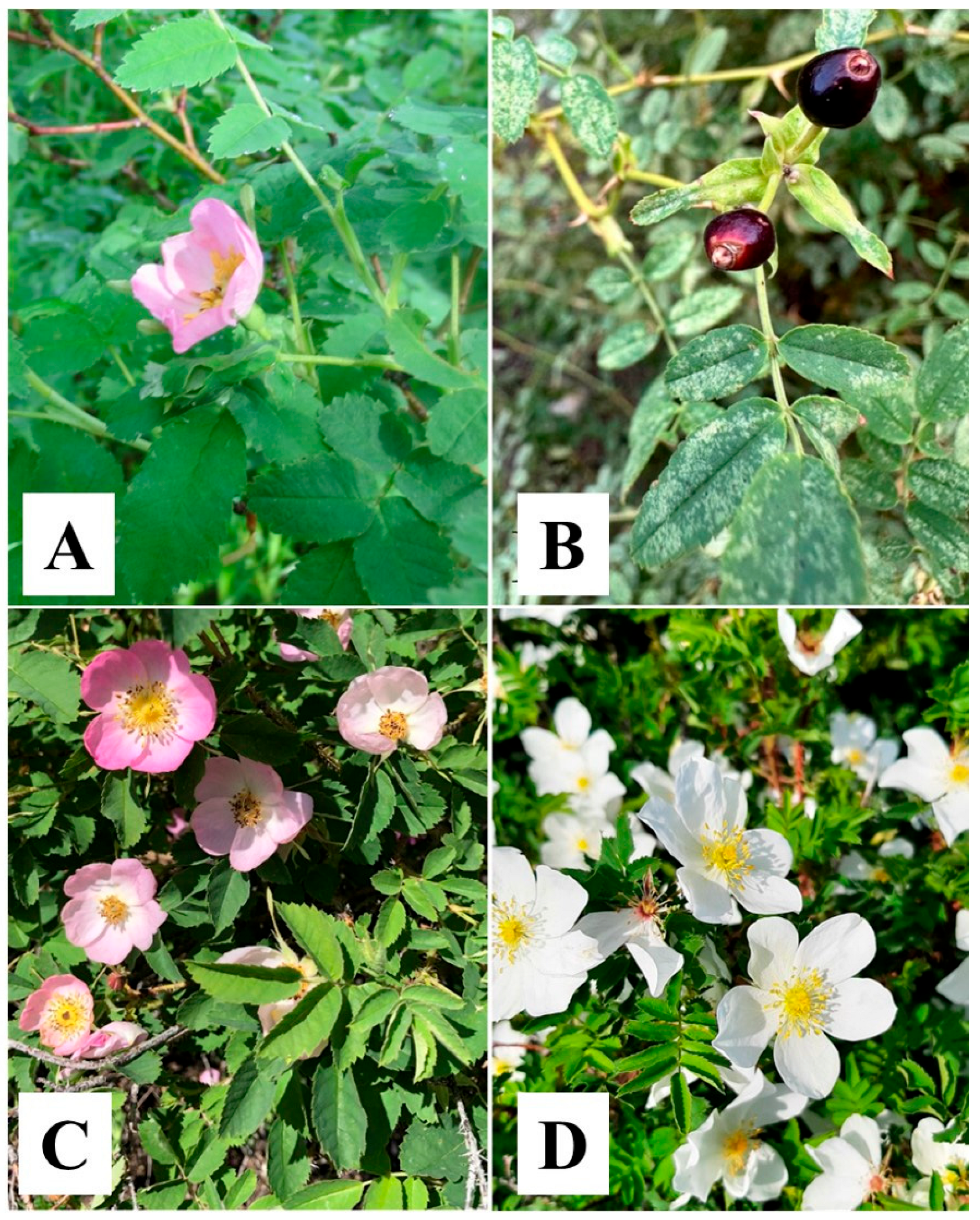
Figure 1. Pictures of the Rosa acicularis (A), Rosa iliensis (B), Rosa laxa (C), and Rosa spinosissima (D) species in nature.
Genes 16 00852 g001
Figure 2. The plastome map of the Rosa acicularis, Rosa iliensis, Rosa laxa, and Rosa spinosissima species. The plastome map highlights the large single-copy (LSC) region, the small single-copy (SSC) region, and the inverted repeat regions (IRA and IRB). Genes located outside the outer circle are transcribed in a counterclockwise direction, whereas those within the circle are transcribed in a clockwise direction. The inner circle illustrates the content of GC (darker gray) and AT (lighter gray). Genes are color-coded based on their functional categories.
Genes 16 00852 g002
Figure 3. Comparison of the plastomes of Rosa species using mVISTA. The plastomes of Rosa graciliflora (OQ992658) and Rosa acicularis (MK714016) were used as the references. Species names highlighted in red represent those sequenced in this study. Coding regions (exons), untranslated regions (UTRs), and conserved non-coding sequences (CNSs) are represented in purple, blue, and red, respectively. The lower and upper horizontal axis indicates genome coordinates in kilobases (kb) and gene annotations, respectively. The vertical scale represents percent identity, ranging from 50% to 100%.
Genes 16 00852 g003
Figure 4. Comparison of junctions between large single-copy (LSC), small single-copy (SSC), and inverted repeat regions (IRa and IRb). Junctions are labeled as follows: JLB (LSC/IRb), JSB (IRb/SSC), JSA (SSC/IRa), and JLA (IRa/LSC). Samples highlighted in red represent the species sequenced in this study.
Genes 16 00852 g004
Figure 5. Nucleotide diversity (Pi) across the plastomes of 26 Rosa samples, estimated using sliding window analysis. The Pi values for each window are plotted on the Y-axis, while the corresponding positions are represented on the X-axis.
Genes 16 00852 g005
Figure 6. Maximum Likelihood phylogenetic trees based on (A) nucleotide sequences of the complete plastome and (B) the ycf1 gene. Species names highlighted in red represent sequenced samples from this study. Section names are provided according to the classification of Wissemann (2017) [5] and Rehder (1940) [6], and subclade designations (C1, C2b, C4, and C5b) follow the subclade structure inferred from Debray et al. (2022) [9]. Roman numerals (I–III) indicate major clades. Numbers at the nodes represent bootstrap support values.
Genes 16 00852 g006
Figure 7. Maximum Likelihood phylogenetic tree based on nucleotide sequences of the nuclear ribosomal internal transcribed spacer. Species names highlighted in red represent sequenced samples from this study. Section names are provided according to the classification of Wissemann (2017) [5] and Rehder (1940) [6], and subclade designations (C1, C2b, C4, and C5b) follow the subclade structure inferred from Debray et al. (2022) [9]. Roman numerals (I–IV) indicate major clades. Numbers at the nodes represent bootstrap support values.
Genes 16 00852 g007
Table 1. Information on primers used for amplification.
PrimerSequence (5′-3′)DirectionTemperature (°C)Reference
ITS1nFAGAAGTCGTAACAAGGTTTCCGTAGGForward58[50]
ITS4nRTCCTCCGCTTATTGATATGCReverse58[50]
Table 2. Summary of plastome characteristics of four Rosa species.
SpeciesRosa acicularisRosa iliensisRosa laxaRosa spinosissima
GenBank accession numberPV330080PV330081PV330082PV330083
Genome size (bp)157,288157,148157,346157,177
LSC length (bp)86,38186,27486,46186,269
SSC length (bp)18,79118,77818,79118,784
IRA length (bp)26,05826,04826,04726,062
IRB length (bp)26,05826,04826,04726,062
Total GC content (%)37.2037.2037.2037.20
Number of total genes (unique)136 (116)136 (116)136 (116)136 (116)
Total protein-coding genes (unique)90 (81)90 (81)90 (81)90 (81)
Total tRNA genes (unique)38 (31)38 (31)38 (31)38 (31)
Total rRNA genes (unique)8 (4)8 (4)8 (4)8 (4)
Table 3. List of the annotated genes in the Rosa acicularis, Rosa iliensis, Rosa laxa, and Rosa spinosissima plastomes.
Group of GenesName of Genes
Self-replication
Ribosomal RNArrn4.5 (×2), rrn5 (×2), rrn16 (×2), rrn23 (×2)
Transfer RNAtrnA-UGC * (×2), trnC-GCA, trnD-GUC, trnE-UUC, trnF-GAA, trnfM-CAU, trnG-GCC *, trnG-UCC, trnH-GUG, trnI-CAU (×2), trnI-GAU * (×2), trnK-UUU *, trnL-CAA (×2), trnL-UAA *, trnL-UAG, trnM-CAU, trnN-GUU (×2), trnP-UGG, trnQ-UUG, trnR-ACG (×2), trnR-UCU, trnS-CGA *, trnS-GCU, trnS-GGA, trnS-UGA, trnT-GGU, trnT-UGU, trnV-GAC (×2), trnV-UAC *, trnW-CCA, trnY-GUA
Small subunit of ribosomerps2, rps3, rps4, rps7 (×2), rps8, rps11, rps12 ** (×2), rps14, rps15, rps16 *, rps18, rps19
Large subunit of ribosomerpl2 * (×2), rpl14, rpl16 *, rpl20, rpl22, rpl23 (×2), rpl32, rpl33, rpl36
RNA polymeraserpoA, rpoB, rpoC1 *, rpoC2
Translation initiation factorinfA
Photosynthesis
ATP synthaseatpA, atpB, atpE, atpF, atpH, atpI
NADH dehydrogenasendhA *, ndhB * (×2), ndhC, ndhD, ndhE, ndhF, ndhG, ndhH, ndhI, ndhJ, ndhK
Subunits of cytochromepetA, petB *, petD *, petG, petL, petN
Photosystem IpsaA, psaB, psaC, psaI, psaJ
Photosystem IIpsbA, psbB, psbC, psbD, psbE, psbF, psbH, psbI, psbJ, psbK, psbL, psbM, psbN, psbT, psbZ
RubiscorbcL
Other genes
MaturasematK
ProteaseclpP **
Envelope membrane proteincemA
Subunit of acetyl-CoA-carboxylaseaccD
C-type cytochrome synthesis geneccsA
Genes of unknown function
Conserved hypothetical chloroplast ORFycf1 (×2), ycf2 (×2), ycf3 **, ycf4, ycf15 (×2), ycf68 (×2)
A single asterisk (*) indicates genes containing one intron, while a double asterisk (**) indicates genes with two introns. Genes marked with (×2) are duplicated and located in the inverted repeat (IR) regions.
Table 4. The type and number of identified simple sequence repeats in four Rosa plastomes.
TypeRepeat UnitRosa acicularisRosa iliensisRosa laxaRosa spinosissimaTotal%
Mono-A/T13814413814356366.41
C/G91010938
Di-AT/AT3939393815524.64
AG/CT1616161664
AC/GT11114
Tri-AAT/ATT7577262.98
AGC/CTG00011
Tetra-AAAT/ATTT6666244.75
AATT/AATT21227
ACAT/ATGT11114
ACCT/AGGT22228
Penta-AATAT/ATATT101020.33
AAAAG/CTTTT00011
Hexa-AAGTAG/ACTTCT222280.88
Total224227225229905100.00
Disclaimer/Publisher’s Note: The statements, opinions and data contained in all publications are solely those of the individual author(s) and contributor(s) and not of MDPI and/or the editor(s). MDPI and/or the editor(s) disclaim responsibility for any injury to people or property resulting from any ideas, methods, instructions or products referred to in the content.