Article

A Novel Recurrent 200 kb CRYL1 Deletion Underlies DFNB1A Hearing Loss in Patients from Northwestern Spain

1
Fundación Centro Médico de Asturias, 33193 Oviedo, Spain
2
Instituto de Medicina Oncológica y Molecular de Asturias (IMOMA), 33193 Oviedo, Spain
3
Hospital Centro Médico de Asturias, 33193 Oviedo, Spain
4
Institute of Molecular Oncology and Functional Genomics, School of Medicine, Technische Universität München, 81675 Munich, Germany
5
Hospital Universitario Central de Asturias, 33011 Oviedo, Spain
6
Cabanillas Precision Consulting, 8620 Zürich, Switzerland
*
Author to whom correspondence should be addressed.
Genes 2025, 16(6), 670; https://doi.org/10.3390/genes16060670
Submission received: 8 May 2025 / Revised: 23 May 2025 / Accepted: 28 May 2025 / Published: 30 May 2025
(This article belongs to the Section Human Genomics and Genetic Diseases)

Abstract

Background/Objectives: Pathogenic recessive GJB2 variants are the main genetic cause of non-syndromic sensorineural hearing loss. However, following GJB2 testing, a significant proportion of deaf patients are only found to be heterozygous carriers of pathogenic GJB2 alleles. Five large deletions not affecting GJB2 but encompassing a minimal common 62 kb region within the neighbouring CRYL1 gene have been described to cause loss of cis GJB2 expression and, as a result, produce hearing loss when in trans with pathogenic GJB2 variants. We describe the identification and characterization of a novel deletion of this type in deaf patients from northwestern Spain. Methods: We used panel NGS sequencing to detect the deletion, MLPA to validate it, whole-genome sequencing to map its breakpoints, PCR + Sanger sequencing to finely characterize it and triple-primer PCR to screen for it. Results: We identified a novel 200 kb deletion spanning the whole CRYL1 gene in two unrelated deaf patients from Asturias (in northwestern Spain) who were heterozygous for the pathogenic GJB2 c.35delG variant. Although the large deletion was absent from gnomAD v4.1.0 and 2052 local control alleles, screening for it in 20 additional deaf carriers of monoallelic pathogenic GJB2 variants detected it in another patient from Galicia (also in northwestern Spain). The novel deletion, termed del(200 kb)insATTATA, explained hearing loss in 3/43 (7%) deaf patients from our cohort that were otherwise heterozygous for pathogenic GJB2 variants. Conclusions: This work highlights the importance of comprehensively testing all genomic regions known to be clinically relevant for a given genetic condition, including thorough CRYL1 CNV screening for DFNB1A diagnostics.

1. Introduction

Hearing loss (HL) is the most common sensory disorder, affecting approximately 5% of the world population [1]. Sensorineural hearing loss (SNHL), the condition with the highest prevalence, is caused by damage to the cochlea, the auditory nerve or the brain’s central processing centres. Although most deaf patients are adults, HL is present congenitally in 1–2 out of 1000 newborns [2]. Genetic causes account for 50–60% of childhood HL cases in developed countries, and 70% of these correspond to non-syndromic SNHL (NS-SNHL). With regard to inheritance patterns, 75–80% of such cases are autosomal recessive (AR), while 20% are autosomal dominant (AD), close to 2% are X-linked and less than 1% are mitochondrial [3].
In spite of the high genetic heterogeneity of SNHL, with hundreds of genes involved, pathogenic variants in the GJB2 gene alone, encoding connexin 26, are responsible for up to half of severe to profound NS-SNHL in many western populations [4]. To date, more than 100 pathogenic GJB2 variants resulting in HL have been identified. The majority of these are loss-of-function variants associated with the autosomal recessive 1A deafness (DFNB1A) phenotype (MIM #220290). In Caucasians, GJB2 (NM_004004.6) c.35delG is the most frequent DFNB1A pathogenic variant, comprising 70% of pathogenic DFNB1A alleles in some cohorts, and with a carrier rate of 1–3% in the general population [5].
Apart from small (single nucleotide and indels) and large (copy-number) loss-of-function variants directly affecting the GJB2 locus, to date, five different large deletions not spanning the GJB2 locus have been described to cause hearing loss when they occur in compound heterozygosity with another pathogenic DFNB1A variant (Table 1 and Figure 1A) [6,7,8,9,10]. The first two such deletions identified, del(GJB6-D13S1830) (spanning 309 kb) and del(GJB6-D13S1854) (spanning 232 kb), affected the GJB6 locus, immediately telomeric to GJB2, leading to the assumption that GJB6 was also involved in non-syndromic hearing loss in a digenic fashion with GJB2 [6,7]. However, subsequent functional studies indicated that the hearing loss associated with the large GJB6 deletions was not caused by a loss of function in GJB6, but by loss of GJB2 expression [11,12,13]. In parallel, two novel large neighbouring deletions (spanning 131 kb and 179 kb, respectively) were identified in deaf patients with monoallelic pathogenic GJB2 variants [8,9]. These novel deletions did not affect the GJB6 locus. Instead, they overlapped with the telomeric halves of del(GJB6-D13S1830) and del(GJB6-D13S1854), defining a 95.4 kb region shared by the four of them and affecting the CRYL1 locus. Finally, recent work studying hearing loss in an East-Asian cohort (all previous CRYL1 deletion had been originally identified in individuals with European ancestries), detected a recurrent 125 kb CRYL1 deletion shared by several unrelated deaf patients with monoallelic pathogenic GJB2 variants [10]. The minimal common region (MCR) shared by all these deletions is 62 kb long and spans the 3′ half of CRYL1 (which is in the minus strand as GJB2 and GJB6) plus 5.3 kb further downstream [chr13(GRCh38):20398370-20460629] (Figure 1A). Such MCR includes a candidate 1043 bp regulatory region termed C3 [chr13(GRCh38):20419404-20420446], containing potential enhancers that have been functionally demonstrated to affect GJB2 expression [14].
In this work, combining the OTOgenicsTM NGS panel [15], Whole Genome Sequencing (WGS) and breakpoint-specific PCR, we have identified and genomically characterized a novel 200 kb CRYL1 deletion present in heterozygosis in three unrelated deaf patients from northwestern Spain who were also heterozygotes for the frequent GJB2 (NM_004004.6) c.35delG pathogenic variant. The novel deletion includes the 62 kb MCR, is not present in individuals from gnomADv4 nor in 2052 control local chromosomes and segregates with hearing loss when in compound heterozygosity with GJB2 (NM_004004.6) c.35delG.

2. Materials and Methods

2.1. Patients

We identified the novel deletion in two patients (Patients #1 and #2) with hearing loss who underwent NGS with a gene panel comprising > 230 genes associated with sensorineural or mixed, syndromic or non-syndromic, HL [15]. The deleted region included 10 probes distributed across the CRYL1 locus (in the minus strand), plus 1 probe upstream and 5 probes downstream of it (affected probes shown in red in Figure 1A). Then, we tested for the presence of the novel CRYL1 deletion in deaf patients (either wild-type for GJB2 or monoallelic for NM_004004.6: c.35delG) with no genetic diagnosis after previous analyses based on Sanger sequencing or the NGS panel. Samples from normal hearing individuals were taken from our lab research sample collections. All involved individuals had provided written informed consent.

2.2. Novel Deletion Detection, Validation and Characterization

The novel large CRYL1 deletion was detected in our laboratory using the previously described OTOgenicsTM panel [15]. Orthogonal validation was initially performed through MLPA on Patient #1’s and Patient #2’s germline DNA, using the P163-E1 GJB-WFS1-POU3F4 kit and following the manufacturer’s instructions (MRC Holland, Netherlands). The CRYL1 deletion breakpoint regions were identified through WGS of Patient 2’s germline DNA at Macrogen (Macrogen Inc., Seoul, South Korea), and they were then finely mapped by PCR using two primers flanking the breakpoint regions (5′-TTTCCTCATCCAACTGCCCA-3′ and 5′-TTCGAAGGTACAGGGGAGAC-3′), followed by Sanger sequencing. That PCR, performed using MegaMix-Double reagent (Microzone, Stourbridge, UK) under the manufacturer’s suggested conditions, generating an 897 bp fragment in the presence of the deletion-containing allele.

2.3. del(GJB6-D13S1854), del(GJB6-D13S18530) and CRYL1 del(200 kb)insATTATA Deletion Screening

del(GJB6-D13S1854) and del(GJB6-D13S1830) were screened for as previously described [6,7]. For evaluating the presence of the novel CRYL1 del(200 kb)insATTATA, we designed a triple-primer PCR that generates two differential amplicons depending on the allele, which are easily discerned in an electrophoresis gel. We used two primers flanking the deletion breakpoint (5′-GATTGTGGAATGCCCAAAGT-3′ and 5′-TGCTCAATTGACACCAACAA-3′) that amplify a 436 bp-product from genomic DNA bearing the deletion (including the 6 bp from the ATTATA insertion) and a 200 kb fragment from the wild-type allele (not amplifiable in normal PCR conditions); the third primer (5′-TTTACTGAGGCTGCCGGTAT-3′) anneals within in the deleted region and amplifies, together with the first primer, a 702 bp product that includes sequence from the deleted interval. For the PCR we used MegaMix-Double reagent (Microzone, Stourbridge, UK) following the manufacturer’s suggested conditions, with an annealing temperature of 63 °C, and we visualized the products after electrophoresis in an 1.2% agarose gel.

2.4. GJB2 c.35delG Genotyping

To determine the presence/absence of GJB2 (NM_004004.6) c.35delG in the two brothers of Patient #2, we used Megamix Double (Microzone, UK) and chimeric primers with sequences complementary to M13 and human GJB2 (5′-tgtaaaacgacggccagtCCCAGCACAGCAAATTTTTA-3′ and 5′-tgtaaaacgacggccagtCCCAGCACAGCAAATTTTTA-3′; lowercase: oligonucleotide part specific M13D or M13R sequencing primers, respectively; uppercase: oligonucleotide part specific for human GJB2) to amplify a 723 bp amplicon, that we Sanger-sequenced with M13D and M13R primers at the Scientific and Technical Services of University of Oviedo (Spain).

3. Results

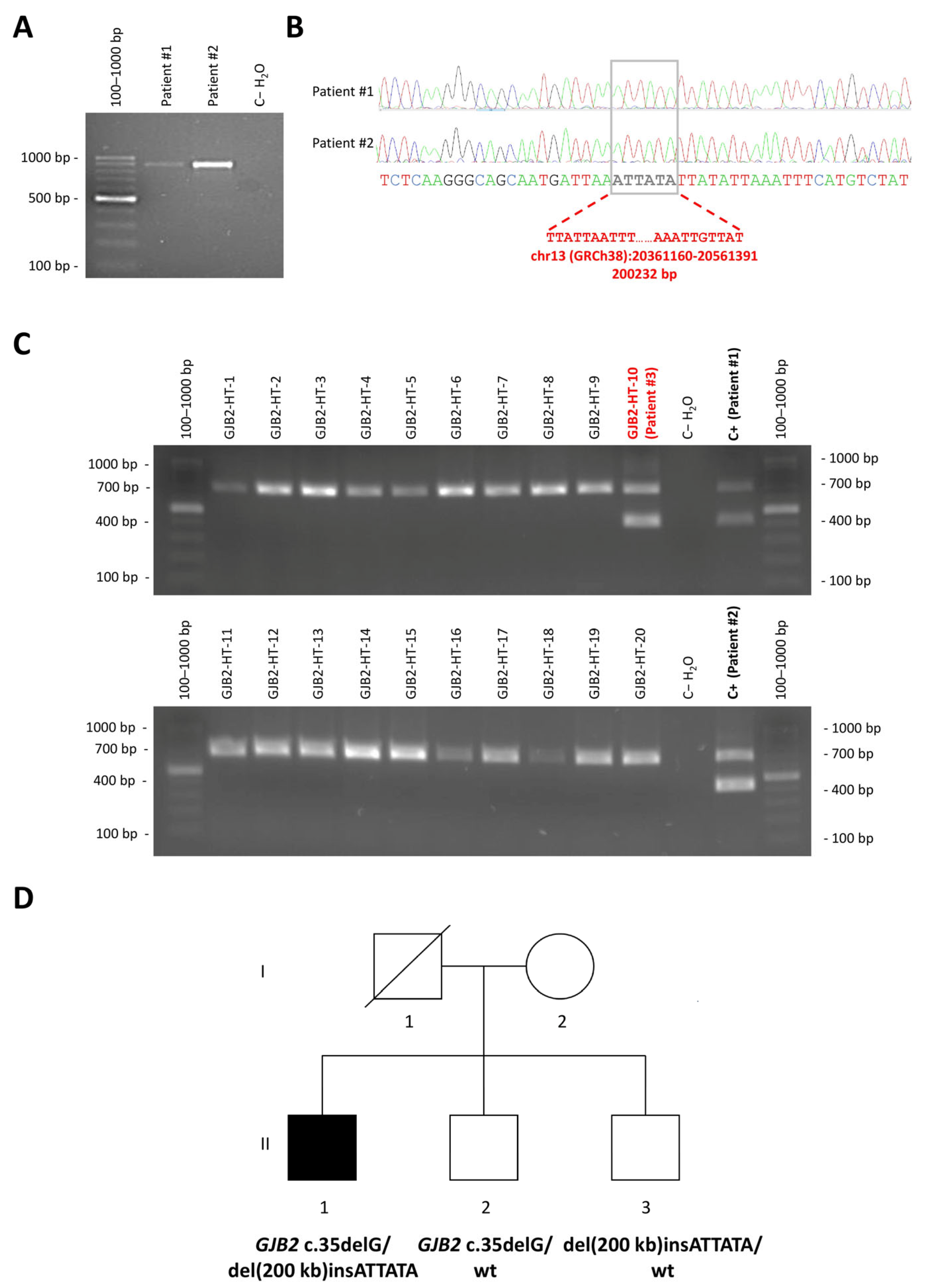
3.1. Detection of a Novel CRYL1 Deletion in Hearing Loss Patients Using an NGS Panel

With the aim of identifying the genetic cause of deafness in Patient #1, diagnosed with profound sensorineural hearing loss at the age of 5 months, we used the OTOgenicsTM NGS panel (v4) to sequence 231 genes associated with hearing loss in genomic DNA from peripheral blood cells. The panel contains probes to capture exons and exon-intron boundaries from all target genes, as well as a selection of additional probes to capture genomic regions bearing previously described non-coding pathogenic variants. The latter include 27 probes (281–505 nt) scattered from the intergenic region upstream of GJB6 to the intergenic region upstream of CRYL1 intron (both genes lying on the minus strand of chromosome 13), with an average separation of 10,888 (6120–22,320) bp between them (Figure 1A). Sequence analysis detected a pathogenic GJB2 (NM_004004.6) c.35delG variant in heterozygosis in Patient #1 as the only clinically relevant variant directly affecting GJB2. However, copy-number analysis of NGS results detected a potential 50% loss in the relative read depth of all 10 probes targeting CRYL1 intronic regions as well as the first 5 intergenic probes placed between CRYL1 and GJB6 (Figure 1B). The presence of a heterozygous deletion affecting CRYL1 but not GJB6 was confirmed by MLPA (Figure 1C). The pattern of probes showing decreased read depth indicates that this is a novel CRYL1 deletion, not matching any of the large deletions previously described in this genomic region (Figure 1A,B). As this variant encompassed the minimal common region shared by all previously reported pathogenic CRYL1 variants, and taking into account the published evidence on large deletions contained within the probes that detected 50% read depth loss in our patient, we classified it as pathogenic according to ACMG guidelines (PM3_VeryStrong, PP1_Strong, PM2_Supporting) [8,10,16,17], and issued a clinical report considering it as causative of DFNB1A in the patient in conjunction with GJB2 (NM_004004.6) c.35delG, provided that the variants affected different alleles (trans configuration).
A few months later, we found an identical copy-number pattern from the OTOgenicsTM (v5) NGS results of a new deaf patient (Patient #2). Patient #2, unrelated to Patient #1, was diagnosed with profound sensorineural hearing loss at the age of 2 years. He also had visual impairment and intellectual disability, which may have been the result of complications during childbirth. Patient #2 was heterozygous for GJB2 (NM_004004.6) c.35delG and his gDNA showed the same regional read depth (and MLPA) profiles as Patient #1’s (Figure 1B,C). We hypothesized then that we might have found a local/regional CRYL1 deletion that could be responsible for other unexplained hearing loss cases in our area (northwestern Spain), so we set out to genomically characterize it with the aim of designing a quick and cost-effective PCR assay to screen for it in other deaf patients with heterozygous pathogenic/likely pathogenic GJB2 variants.

3.2. Mapping of the Breakpoints of the New CRYL1 Deletion

The pattern for relative read depth on the probes situated around GJB6 and CRYL1 suggested that the centromeric breakpoint of the deletion was placed within the chr13(GRCh38/hg38): 20352212-20362132 interval. However, it did not provide information to infer a candidate region for the telomeric end, as the most telomeric probe also showed read depth loss (Figure 1A,B). Thus, we performed WGS on gDNA from Patient #2 and focused on the genomic area under study, revealing a clear reduction in coverage depth and absence of heterozygosity that spanned approximately chr13(GRCh38/hg38):20360000-20560000 (Figure 2A). Closer examination of the boundaries of such region allowed detection of both discordant-read pairs and split reads supporting the existence of a centromeric breakpoint at chr13(GRCh38/hg38):20361159-20361160 and a telomeric breakpoint at chr13(GRCh38/hg38):20561391-20561392 (Figure 2B).
We designed PCR primers (chr13(GRCh38/hg38):20360608-20360627, plus strand; chr13(GRCh38/hg38):20561711-20561730, minus strand) surrounding the candidate breakpoints to amplify a deletion-specific amplicon from both Patient #2 and Patient #1 (Figure 1A, Figure 2B and Figure 3A). Sanger sequencing of the amplicon confirmed the breakpoints at chr13(GRCh38/hg38):20361159-20361160 and chr13(GRCh38/hg38): 20561391-20561392 and revealed a 6-base (ATTATA) insertion (Figure 3 A,B). Thus, HGVS nomenclature of the novel deletion is chr13(GRCh38/hg38):20361160_20561391delinsATTATA. A search in the structural variant dataset of the gnomAD v4.1.0 database, able to identify all other large deletions represented in Figure 1A except for del(179 kb), did not detect the novel CRYL1 del(200 kb)insATTATA variant, indicating that this is a previously undescribed alteration.

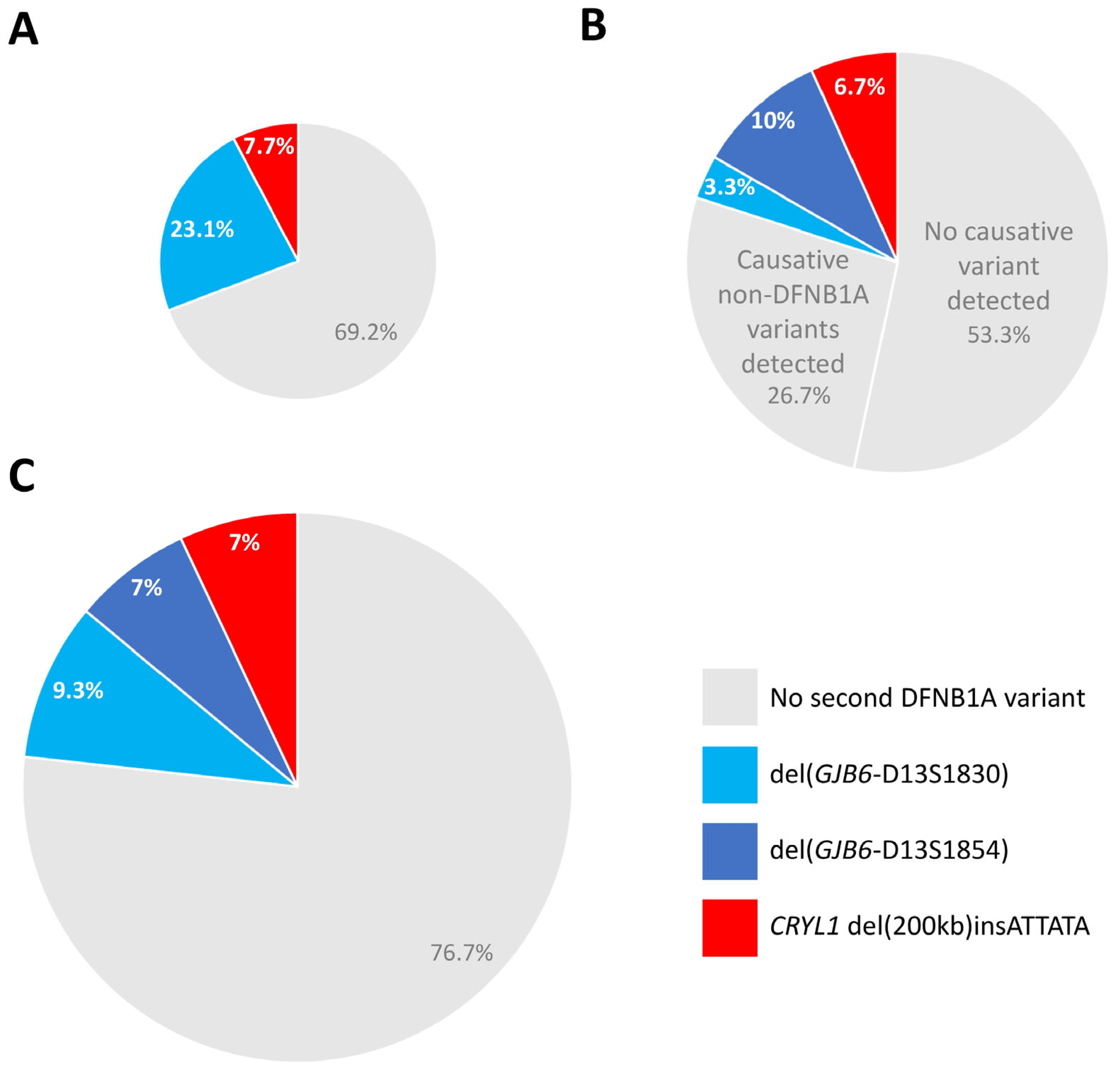
3.3. Novel Deletion Screen in Unexplained Hearing Loss Cases and in the Normal Hearing Population

As Patient #1 and Patient #2 were not family-related, we hypothesized that the novel deletion might also be the second hit responsible for hearing loss in other deaf patients. Thus, to screen for it, we designed a novel pair of primers closer to the breakpoints [chr13(GRCh38/hg38):20360924-20360943, plus strand; chr13(GRCh38/hg38):20561566-20561585, minus strand]. Introduction of a second reverse primer (chr13[GRCh38/hg38]:20361606-20361625, minus strand) annealing 3′ to the 5′ breakpoint allowed us to design a triple-primer PCR expected to produce a 702 bp amplicon for the wt allele and a 436 bp amplicon (including the 6 bases from the ATTATA insertion) from the deleted allele (Figure 1A, Figure 2B and Figure 3C). We used this triple-primer PCR assay to analyze gDNA from 20 patients previously evaluated in our laboratory who had not been tested for CRYL1 deletions and who were heterozygous for a pathogenic/likely pathogenic GJB2 variant and negative for the previously described del(GJB6-D13S1830) and del(GJB6-D13S1854) large deletions. As a result, we obtained the expected amplicon from another GJB2 c.35delG heterozygous patient (Patient #3, from Galicia) who had been referred to our laboratory for genetic testing due to childhood hearing loss (Figure 3C). Finally, we did the same triple-primer PCR on DNA samples from 14 deaf patients previously analyzed in our laboratory with versions of the OTOgenics panel that contained probes against the CRYL1 region, that were found to be heterozygous for pathogenic recessive GJB2 variants, negative for del(GJB6-D13S1830) and del(GJB6-D13S1854) and in whom CRYL1 deletions had not been detected through the NGS assay, obtaining (as expected) only the wild-type band (Supplementary Figure S1). Genetic analysis of Patient #2’s siblings showed that del(200 kb)insATTATA segregates with hearing loss when in compound heterozygosity with GJB2 (NM_004004.6) c.35delG (Figure 3D), adding more evidence in favour of the PM3 ACMG criterion previously considered for its classification as a pathogenic variant [8,10,16,17].
We then used this assay to test gDNAs from 285 other deaf patients previously evaluated for suspected hereditary hearing loss in our laboratory, for whom a genetic cause had not being identified, and who were not known to be carriers of pathogenic DFNB1A variants. We found that all of them were negative for the novel deletion. After that, to explore the regional population frequency of chr13(GRCh38/hg38): 20361160_20561391delinsATTATA, we analyzed saliva gDNA from 1026 local individuals (2052 alleles) without reported suspected hereditary hearing loss, none of which was a carrier of the novel variant (allele frequency < 0.00049). Thus, we have identified a novel, large, rare CRYL1 deletion, found in heterozygosis in three unrelated deaf patients from northwestern Spain (2 from Asturias and 1 from Galicia) who are also heterozygous for GJB2 (NM_004004.6) c.35delG.

3.4. Diagnostic Relevance of CRYL1 del(200 kb)insATTATA Within Deaf Patients with Monoallelic Pathogenic GJB2 Variants

To estimate the potential diagnostic impact of CRYL1 del(200 kb)insATTATA in our population, we determined its prevalence within deaf patients with monoallelic pathogenic GJB2 variants that had followed different diagnostic paths, and compared it with the prevalence of del(GJB6-D13S1830), of del(GJB6-D13S1854) and of causative variants in non-DFNB1A genes.
In total, we considered 43 deaf patients with monoallelic pathogenic GJB2 variants. In 13 of them, no NGS panel testing had been requested (Figure 4A). Of those 13, heterozygous del(GJB6-D13S1830) was found in three (23.1%), whereas heterozygous del(200 kb)insATTATA was detected in one (7.7%) (Patient #3). NGS panel testing had been requested for the other 30 (Figure 4B). Of those, four patients (13.3%) were found to have heterozygous GJB6 deletions [one del(GJB6-D13S1830) (3.3%) and three del(GJB6-D13S1854) (10%)], and two patients (6.6%) were found to have heterozygous del(200 kb)insATTATA (Patients #1 and #2). Causative variants in non-DFNB1A genes were found in 8 of the 30 patients with monoallelic pathogenic GJB2 variants than underwent panel testing (26.6%) (Figure 4B).
Thus, within 43 deaf carriers of monoallelic pathogenic GJB2 variants, heterozygous large deletions affecting CRYL1 explained hearing loss in 10/43 (23.3%): 4/43 (9.3%) being del(GJB6-D13S1830, 3/43 (7%) being del(GJB6-D13S1854) and 3/43 (7%) being del(200 kb)insATTATA (Figure 4C). These results show that deletions affecting CRYL1 different from del(GJB6-D13S1830) and del(GJB6-D13S1854) have a relevant impact in DFNB1A diagnosis and, thus, should not be overlooked, but actively screened for, particularly in deaf carriers of monoallelic pathogenic GJB2 variants.

4. Discussion

Genetic alterations causing GJB2 dysfunction are the most prevalent cause of hereditary hearing loss worldwide. These include not only genetic variants directly affecting the GJB2 locus, but also distant rearrangements that eliminate cis-acting regulatory elements necessary for GJB2 expression. In this work, we identified a novel 200 kb deletion spanning more than 20 kb each side of the CRYL1 locus and present in heterozygosis in three unrelated hearing loss patients from northwestern Spain who are also heterozygous for the most prevalent pathogenic GJB2 mutation in Europeans (c.35delG). This novel CRYL1 deletion has not been detected in individuals from gnomADv4.1.0, nor in 2052 alleles from a control local population, and its presence in compound heterozygosity with c.35delG segregates with hearing loss.
This study, together with previous works describing other large deletions involving CRYL1 (Figure 1) in DFNB1A patients, demonstrates that genetic testing of the GJB2 locus, even if complemented with approaches targeting the known GJB6 and CRYL1 deletions already described to alter GJB2 expression, may leave a significant proportion of DFNB1A patients undiagnosed. Thus, del(200 kb)insATTATA explained 3 out of 43 (7%) DFNB1A cases, in which, otherwise, only a monoallelic pathogenic/likely pathogenic GJB2 variant had been detected in our laboratory. This is the same number of cases as those explained by the known del(GJB6-D13S1854) deletion in this very cohort (Figure 4C). In our opinion, all genetic diagnostic strategies for hereditary deafness that claim to be comprehensive should be able to detect any copy number alteration affecting the 62 kb CRYL1 MCR. As illustrated in Figure 1, MLPA might not be sufficient for this, as only one of the two probes included in the commercial kit used in this work overlaps with the MCR. Moreover, this probe is close to its telomeric end and more than 10 kb away from the C3 region.
In line with EMQN Best Practice recommendations for population-tailored initial GJB2 testing [5] and leveraging our previously validated OTOgenics™ NGS panel [15], we advocate for a two-step DFNB1A workflow. The first step should be GJB2 sequencing plus detection of known large deletions spanning the CRYL1 MCR. Unsolved cases after the first step would be analyzed by a comprehensive NGS panel able to detect all relevant CRYL1 deletions, to maximize diagnostic yield of hearing loss in clinical settings.
In conclusion, in this work, we have identified a novel large CRYL1 deletion that, in combination with heterozygous GJB2 c.35delG, causes DFNB1A hearing loss in deaf patients from northwestern Spain, and we describe a simple triple-primer PCR assay that could be used to detect the novel large deletion. Considering the historically relevant migratory patterns of people from Asturias and Galicia to other regions of Spain, Europe and Latin America, this variant might be responsible for DFNB1A cases in several geographically distant populations.
Consequently, we invite clinical genetics laboratories with sample collections from deaf patients of possible Spanish ancestry without a genetic diagnosis who are carriers of monoallelic DFNB1A pathogenic/likely pathogenic variants to screen them for the presence of the CRYL1 del(200 kb)insATTATA deletion described herein.

Supplementary Materials

The following supporting information can be downloaded at https://www.mdpi.com/article/10.3390/genes16060670/s1: Supplementary Figure S1. Absence of del(200 kb)insATTATA in patients analyzed with versions of the OTOgenicsTM platform designed to detect CRYL1 deletions and that had been found to be heterozygotes for single pathogenic GJB2 variants, based on results from the NGS panel.

Author Contributions

Conceptualization, J.C.; Funding acquisition, J.C.; Investigation, G.A.C., A.F. and J.C.; Methodology, G.A.C., M.D., A.R.H., R.C. (Raquel Capín), A.S., A.A.R.V., D.C., E.L.M., B.A., A.F. and M.C.; Project administration, J.C.; Supervision, J.C.; Validation, G.A.C., M.D. and A.R.H.; Visualization, A.A.R.V. and J.C.; Writing—original draft, G.A.C. and J.C.; Writing—review and editing, G.A.C., A.R.H., R.C. (Raquel Capín), D.C., A.F., R.R., R.C. (Rubén Cabanillas) and J.C. All authors have read and agreed to the published version of the manuscript.

Funding

This research was funded by Fundación María Cristina Masaveu Peterson.

Institutional Review Board Statement

The study was conducted in accordance with the Declaration of Helsinki, and approved by the Comité de Ética de la Investigación con Medicamentos del Principado de Asturias (protocol code CEImPA 2022.432; date of approval: 20 October 2022).

Informed Consent Statement

Specific research-related informed consent was obtained from all study participants who had not previously consented to DFNB1A genetic testing, as well as from the patient who underwent whole-genome sequencing. The ethics committee waived the need for specific research consent from patients who had already given their consent for genetic testing that included DFNB1A analyses.

Data Availability Statement

The data presented in this study are available on request from the corresponding author due to institutional limitations on publishing patient-derived whole genome sequencing results.

Acknowledgments

We thank the patients and their relatives for their kind collaboration. Research work at Fundación Centro Medico de Asturias and IMOMA is supported by Fundación María Cristina Masaveu Peterson.

Conflicts of Interest

Author Rubén Cabanillas was employed by the company Cabanillas Precision Consulting. The remaining authors declare that the research was conducted in the absence of any commercial or financial relationships that could be construed as a potential conflict of interest.

References

  1. Sheffield, A.M.; Smith, R.J.H. The Epidemiology of Deafness. Cold Spring Harb Perspect. Med. 2019, 9, a033258. [Google Scholar] [CrossRef] [PubMed]
  2. Butcher, E.; Dezateux, C.; Cortina-Borja, M.; Knowles, R.L. Prevalence of permanent childhood hearing loss detected at the universal newborn hearing screen: Systematic review and meta-analysis. PLoS ONE 2019, 14, e0219600. [Google Scholar] [CrossRef] [PubMed]
  3. Shibata, S.B.; Shearer, A.E.; Smith, R.J.H. Genetic sensorineural hearing loss. In Cummings Otolaryngology Head and Neck Surgery; Flint, P.W., Ed.; Elsevier: Philadelphia, PA, USA, 2015; pp. 2285–2300. [Google Scholar]
  4. Lucotte, G. High prevalences of carriers of the 35delG mutation of connexin 26 in the Mediterranean area. Int. J. Pediatr. Otorhinolaryngol. 2007, 71, 741–746. [Google Scholar] [CrossRef] [PubMed]
  5. Hoefsloot, L.H.; Roux, A.F.; Bitner-Glindzicz, M. EMQN Best Practice guidelines for diagnostic testing of mutations causing non-syndromic hearing impairment at the DFNB1 locus. Eur. J. Hum. Genet. 2013, 21, 1325–1329. [Google Scholar] [PubMed]
  6. del Castillo, I.; Villamar, M.; Moreno-Pelayo, M.A.; del Castillo, F.J.; Alvarez, A.; Telleria, D.; Menendez, I.; Moreno, F. A deletion involving the connexin 30 gene in nonsyndromic hearing impairment. N. Engl. J. Med. 2002, 346, 243–249. [Google Scholar] [CrossRef] [PubMed]
  7. del Castillo, F.J.; Rodriguez-Ballesteros, M.; Alvarez, A.; Hutchin, T.; Leonardi, E.; de Oliveira, C.A.; Azaiez, H.; Brownstein, Z.; Avenarius, M.R.; Marlin, S.; et al. A novel deletion involving the connexin-30 gene, del(GJB6-d13s1854), found in trans with mutations in the GJB2 gene (connexin-26) in subjects with DFNB1 non-syndromic hearing impairment. J. Med. Genet. 2005, 42, 588–594. [Google Scholar] [CrossRef] [PubMed]
  8. Wilch, E.; Azaiez, H.; Fisher, R.A.; Elfenbein, J.; Murgia, A.; Birkenhager, R.; Bolz, H.; Da Silva-Costa, S.M.; Del Castillo, I.; Haaf, T.; et al. A novel DFNB1 deletion allele supports the existence of a distant cis-regulatory region that controls GJB2 and GJB6 expression. Clin. Genet. 2010, 78, 267–274. [Google Scholar] [CrossRef] [PubMed]
  9. Tayoun, A.N.; Mason-Suares, H.; Frisella, A.L.; Bowser, M.; Duffy, E.; Mahanta, L.; Funke, B.; Rehm, H.L.; Amr, S.S. Targeted Droplet-Digital PCR as a Tool for Novel Deletion Discovery at the DFNB1 Locus. Hum. Mutat. 2016, 37, 119–126. [Google Scholar] [CrossRef] [PubMed]
  10. Lin, Z.; Xiang, J.; Sun, X.; Song, N.; Liu, X.; Cai, Q.; Yang, J.; Ye, H.; Xu, J.; Zhang, H.; et al. Genome sequencing unveils the role of copy number variants in hearing loss and identifies novel deletions with founder effect in the DFNB1 Locus. Hum. Mutat. 2024, 2024, 9517114. [Google Scholar] [CrossRef] [PubMed]
  11. Common, J.E.; Bitner-Glindzicz, M.; O’Toole, E.A.; Barnes, M.R.; Jenkins, L.; Forge, A.; Kelsell, D.P. Specific loss of connexin 26 expression in ductal sweat gland epithelium associated with the deletion mutation del(GJB6-D13S1830). Clin. Exp. Dermatol. 2005, 30, 688–693. [Google Scholar] [CrossRef] [PubMed]
  12. Rodriguez-Paris, J.; Schrijver, I. The digenic hypothesis unraveled: The GJB6 del(GJB6-D13S1830) mutation causes allele-specific loss of GJB2 expression in cis. Biochem. Biophys. Res. Commun. 2009, 389, 354–359. [Google Scholar] [CrossRef] [PubMed]
  13. Rodriguez-Paris, J.; Tamayo, M.L.; Gelvez, N.; Schrijver, I. Allele-specific impairment of GJB2 expression by GJB6 deletion del(GJB6-D13S1854). PLoS ONE 2011, 6, e21665. [Google Scholar] [CrossRef] [PubMed]
  14. Le Nabec, A.; Blotas, C.; Briset, A.; Collobert, M.; Ferec, C.; Moisan, S. 3D Chromatin Organization Involving MEIS1 Factor in the cis-Regulatory Landscape of GJB2. Int. J. Mol. Sci. 2022, 23, 6964. [Google Scholar] [CrossRef] [PubMed]
  15. Cabanillas, R.; Dineiro, M.; Cifuentes, G.A.; Castillo, D.; Pruneda, P.C.; Alvarez, R.; Sanchez-Duran, N.; Capin, R.; Plasencia, A.; Viejo-Diaz, M.; et al. Comprehensive genomic diagnosis of non-syndromic and syndromic hereditary hearing loss in Spanish patients. BMC Med. Genom. 2018, 11, 58. [Google Scholar] [CrossRef] [PubMed]
  16. Richards, S.; Aziz, N.; Bale, S.; Bick, D.; Das, S.; Gastier-Foster, J.; Grody, W.W.; Hegde, M.; Lyon, E.; Spector, E.; et al. Standards and guidelines for the interpretation of sequence variants: A joint consensus recommendation of the American College of Medical Genetics and Genomics and the Association for Molecular Pathology. Genet. Med. 2015, 17, 405–424. [Google Scholar] [CrossRef] [PubMed]
  17. Oza, A.M.; DiStefano, M.T.; Hemphill, S.E.; Cushman, B.J.; Grant, A.R.; Siegert, R.K.; Shen, J.; Chapin, A.; Boczek, N.J.; Schimmenti, L.A.; et al. Expert specification of the ACMG/AMP variant interpretation guidelines for genetic hearing loss. Hum. Mutat. 2018, 39, 1593–1613. [Google Scholar] [CrossRef] [PubMed]
Figure 1. Identification of the novel 200 kb CRYL1 deletion. (A). Schematic representation of chromosome 13 genomic regions affected by DFNB1A-causative large deletions that involve CRYL1 (+/− GJB6) but not GJB2. Light-blue [6], dark-blue [7], orange [8], beige [9] and yellow [10] segments correspond to previously described deletions, whereas the novel 200 kb CRYL1 deletion described in this work is shown in red. The double headed dotted arrow represents the 62 kb overlapping segment. Short vertical green/red segments indicate the positions of the probes included in our capture design to detect large DFNB1A deletions (red ones: affected by the novel deletion; green ones: unaffected), the short orange horizontal segment indicates the C3 region and the short red horizontal segments signal the position of the two CRYL1 probes (targeting exon 8 and exon 3) contained in the MLPA kit used for validation. Black arrowheads mark the positions and orientations of the primers used in the first PCR designed to amplify the breakpoint region, whereas blue, green and red arrowheads represent the oligonucleotides used in the triple-primer PCR screening assay. GRCh38/hg38 and GRCh37/hg19 genomic coordinate scales are shown at the bottom. Note that GJB2, GJB6 and CRYL1 are all in the minus strand. (B). NGS read depths (as seen on IGV) obtained by OTOgenics panel sequencing of germline DNAs from Patient #1 and Patient #2 on the deleted region (captured by the probes represented at the bottom by red segments) and the adjacent non-deleted region centromeric to CRYL1 (captured by the probes represented at the bottom by green segments), each compared to those from a control individual (not affected by the CRYL1 deletion) sequenced in the same run and with a similar total number of reads. (C). MLPA analysis of Patient #1 and Patient #2 germline DNA. Red bars correspond to probes targeted to CRYL1 exon 3 and exon 8 (showing heterozygous deletion in both patients). The purple bar corresponds to a probe targeting the wild-type region affected by the GJB2 c.35delG pathogenic variant (heterozygous in both patients). Blue bars correspond to probes targeting the POU3F4 gene, located in the X chromosome (both Patient #1 and Patient #2 are males).
Genes 16 00670 g001
Figure 2. Visual analysis of WGS reads at the deleted CRYL1 region. (A). IGV view of WGS reads from Patient #2 on the deleted region. The red box on the q12.11 chromosome 13 band indicates the position of the 272 kb window shown just below. Horizontal blue bars represent the whole CRYL1 locus and part of the IFT188 locus. Blue segments at the bottom correspond to the positions of OTOgenicsTM probes in the region. (B). Close-up of A at the centromeric (left) and telomeric (right) breakpoint regions. Black arrowheads mark the positions and orientations of the primers used in the first PCR designed to amplify the breakpoint region. Blue, green and red arrowheads represent the oligonucleotides used in the triple-primer PCR screening assay (as in Figure 1A).
Genes 16 00670 g002
Figure 3. Characterization and screening of the novel 200 kb CRYL1 deletion. (A). Agarose gel electrophoresis of products from first PCR designed to amplify the breakpoint region on germline DNA from Patient #1, Patient #2 and a control individual without the large 200 kb CRYL1 deletion. An 897 bp band is observed only in the presence of the deletion. (B). Electropherograms corresponding to Sanger sequencing of the 897 bp bands obtained in A at the region of the breakpoints. The initial and final nucleotides of the deletion are shown in bold red characters. The inserted sequence (ATTATA) is shown in bold grey characters. (C). Agarose gel electrophoresis of products from the triple-primer PCR designed to screen for del(200 kb)insATTATA in 20 deaf patients previously evaluated in our laboratory, in whom CRYL1 deletions had not been tested for, and who were heterozygous for a pathogenic/likely pathogenic GJB2 variant. A 436 bp band is amplified from the allele affected by the deletion, whereas a 702 bp fragment is obtained from the wild-type allele. The DNA sample GJB2-HT16, corresponding to Patient #3 and labelled in bold red characters, contained 1 copy of del(200 kb)insATTATA. (D). Genogram of Patient #2. I and II indicate first and second generation, respectively. 1, 2 and 3 indicate first, second and third individual within the generation. Patient #2 is represented as II.1.
Genes 16 00670 g003
Figure 4. Relative contribution of the novel 200 kb CRYL1 deletion to hearing loss in deaf patients with monoallelic pathogenic GJB2 variants. Pie charts comparing the contribution of CRYL1 del(200 kb)insATTATA (red), GJB6-D13S1854 (dark blue) and GJB6-D13S1830 (light blue) to the genetic diagnosis of deaf monoallelic carriers of pathogenic GJB2 variants. Patients with no second DFNB1A variant detected are represented in grey. (A) Thirteen patients with no NGS panel testing requested. (B) Thirty patients with NGS panel testing requested (note the two distinct grey sectors corresponding to patients with no causative variant detected after panel testing and patients with causative non-DFNB1A variants detected). (C). Forty-three patients from panels A and B combined.
Genes 16 00670 g004
Table 1. Large deletions not spanning the GJB2 locus that cause hearing loss in compound heterozygosity with another pathogenic DFNB1A variant (see also Figure 1A).
DeletionGenome VersionCentromeric BreakpointTelomeric BreakpointReference
GJB6-D13S1830GRCh38/hg38chr13:20223038chr13:20531828[6]
GRCh37/hg19chr13:20797177chr13:21105967
GJB6-D13S1854GRCh38/hg38chr13:20228587chr13:20460616[7]
GRCh37/hg19chr13:20802726chr13:21034755
del(131 kb)GRCh38/hg38chr13:20365205chr13:20496559[8]
GRCh37/hg19chr13:20939344chr13:21070698
del(179 kb)GRCh38/hg38chr13:20347572chr13:20526976[9]
GRCh37/hg19chr13:20921711chr13:21101115
del(125kb)GRCh38/hg38chr13:20398369chr13:20523824[10]
GRCh37/hg19chr13:20972508chr13:21097963
del(200 kb)insATTATAGRCh38/hg38chr13:20361160chr13:20561391This study
GRCh37/hg19chr13:20935299chr13:21135530
Disclaimer/Publisher’s Note: The statements, opinions and data contained in all publications are solely those of the individual author(s) and contributor(s) and not of MDPI and/or the editor(s). MDPI and/or the editor(s) disclaim responsibility for any injury to people or property resulting from any ideas, methods, instructions or products referred to in the content.

