
The Prognostic Value and Immunomodulatory Role of Spsb2, a Novel Immune Checkpoint Molecule, in Hepatocellular Carcinoma


Abstract
1. Introduction
2. Materials and Methods
2.1. Data Sources and Preprocessing
2.2. Differential Gene Expression Analysis and Correlation Analysis
2.3. Differential Expression Analysis of SPSB2
2.4. Clinical Correlation Analysis and Survival Prognosis of SPSB2 Expression
2.5. Functional Enrichment Analysis
2.6. Immunoinfiltration Analysis
2.7. Differential Analysis of SPSB2 Protein Expression Levels in LIHC
2.8. Cell Lines and Culture
2.9. Cell Transfection
2.10. Real-Time-PCR
2.11. Cell Proliferation Assay
2.12. Colony Formation Assay
2.13. Wound-Healing Assay
2.14. Transwell Migration Assay
2.15. Statistical Analysis
3. Results
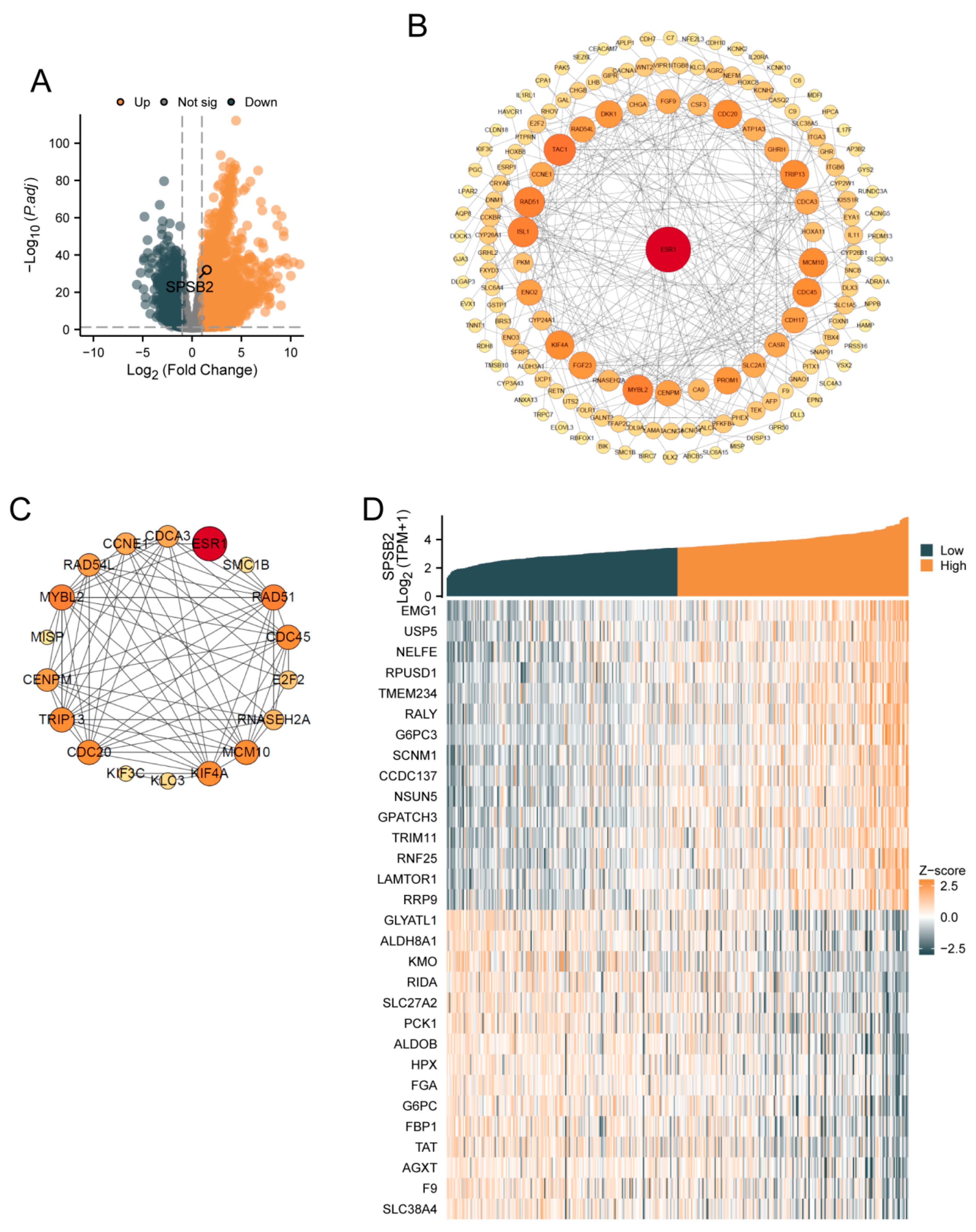
3.1. Differential Gene Expression Analysis and Correlation Analysis of SPSB2
3.2. Differential Expression of SPSB2 in Pancancer and LIHC
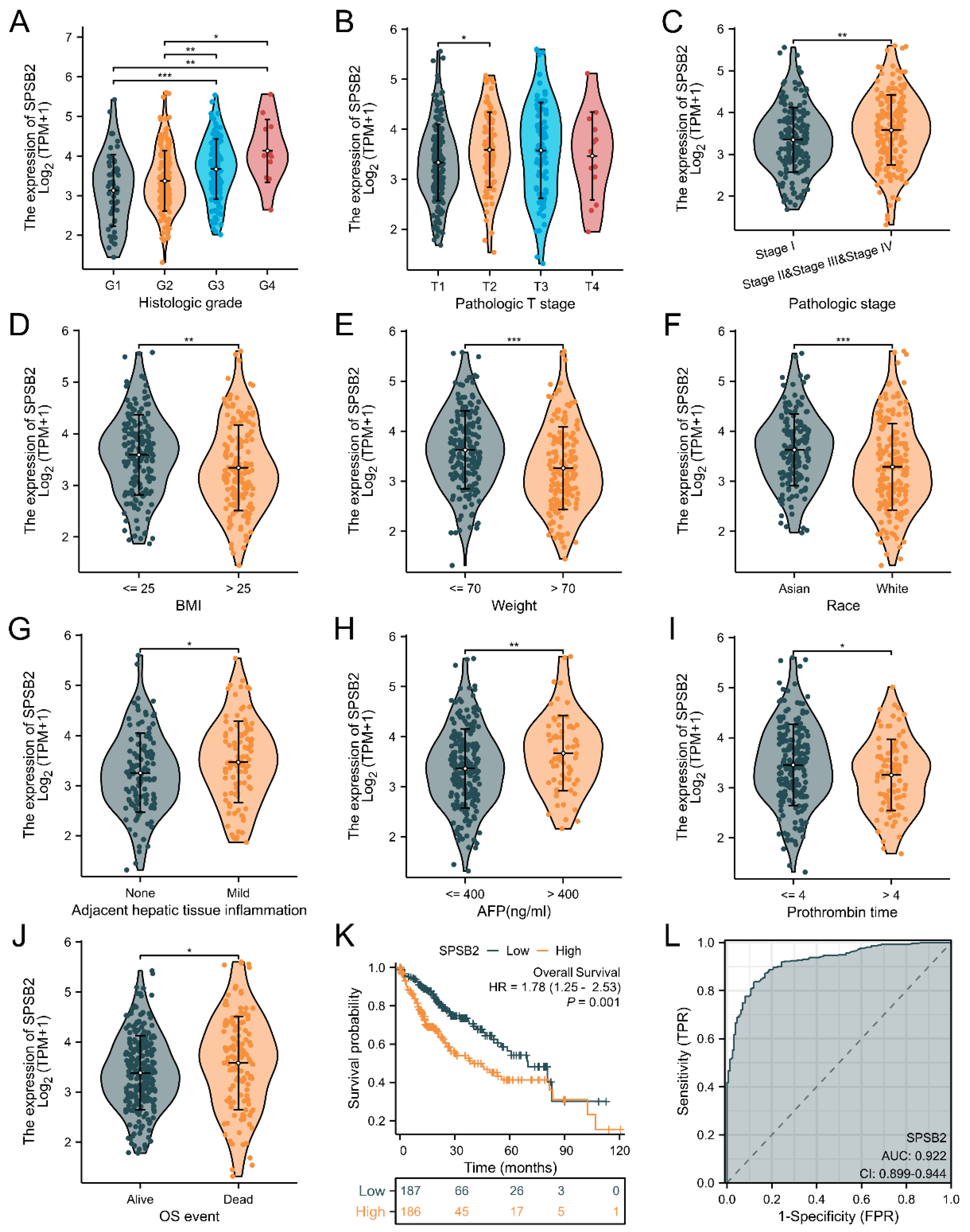
3.3. Correlation of SPSB2 Expression with Clinicopathologic Parameters
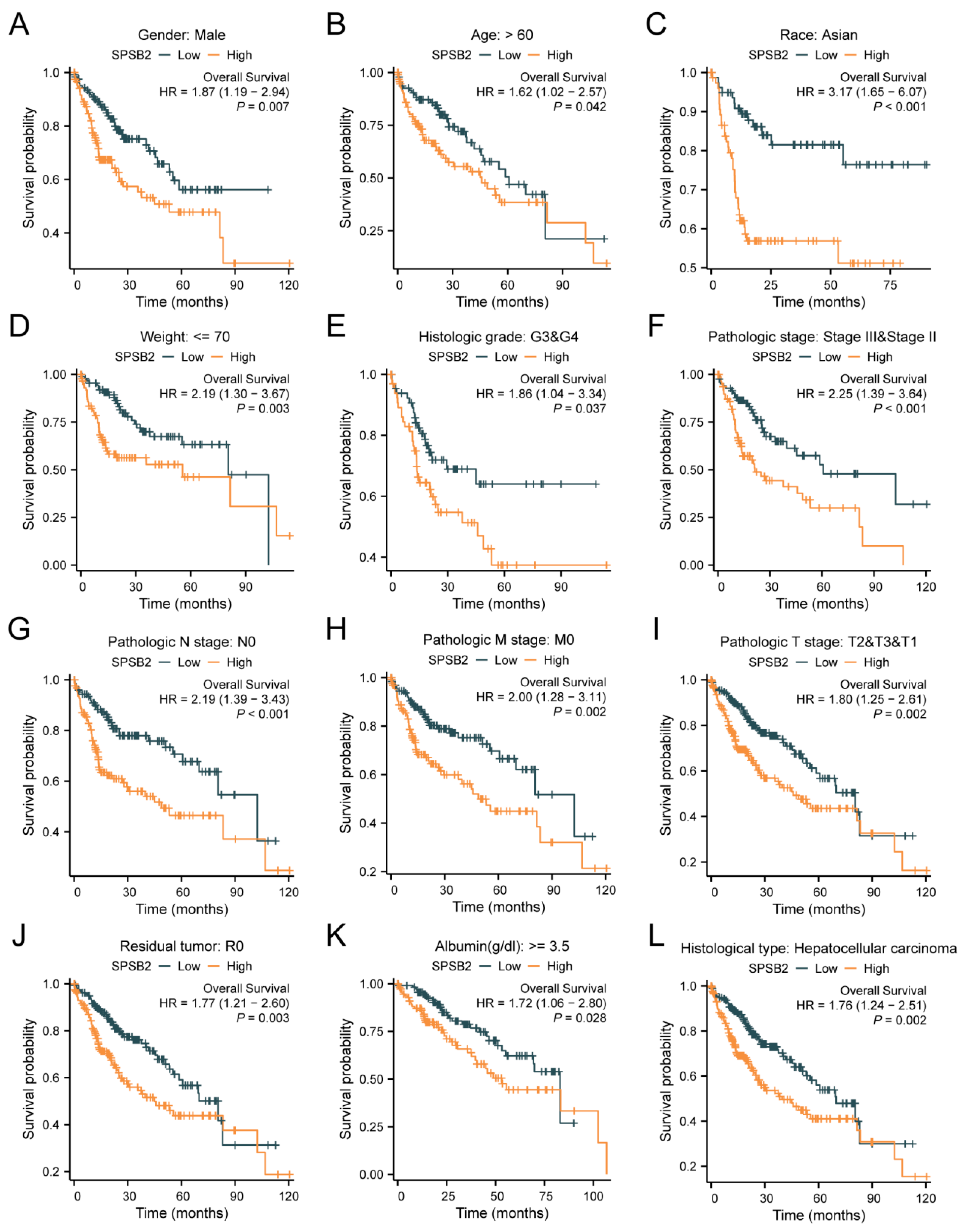
3.4. Subgroup Analysis of Survival Prognosis of SPSB2 Expression
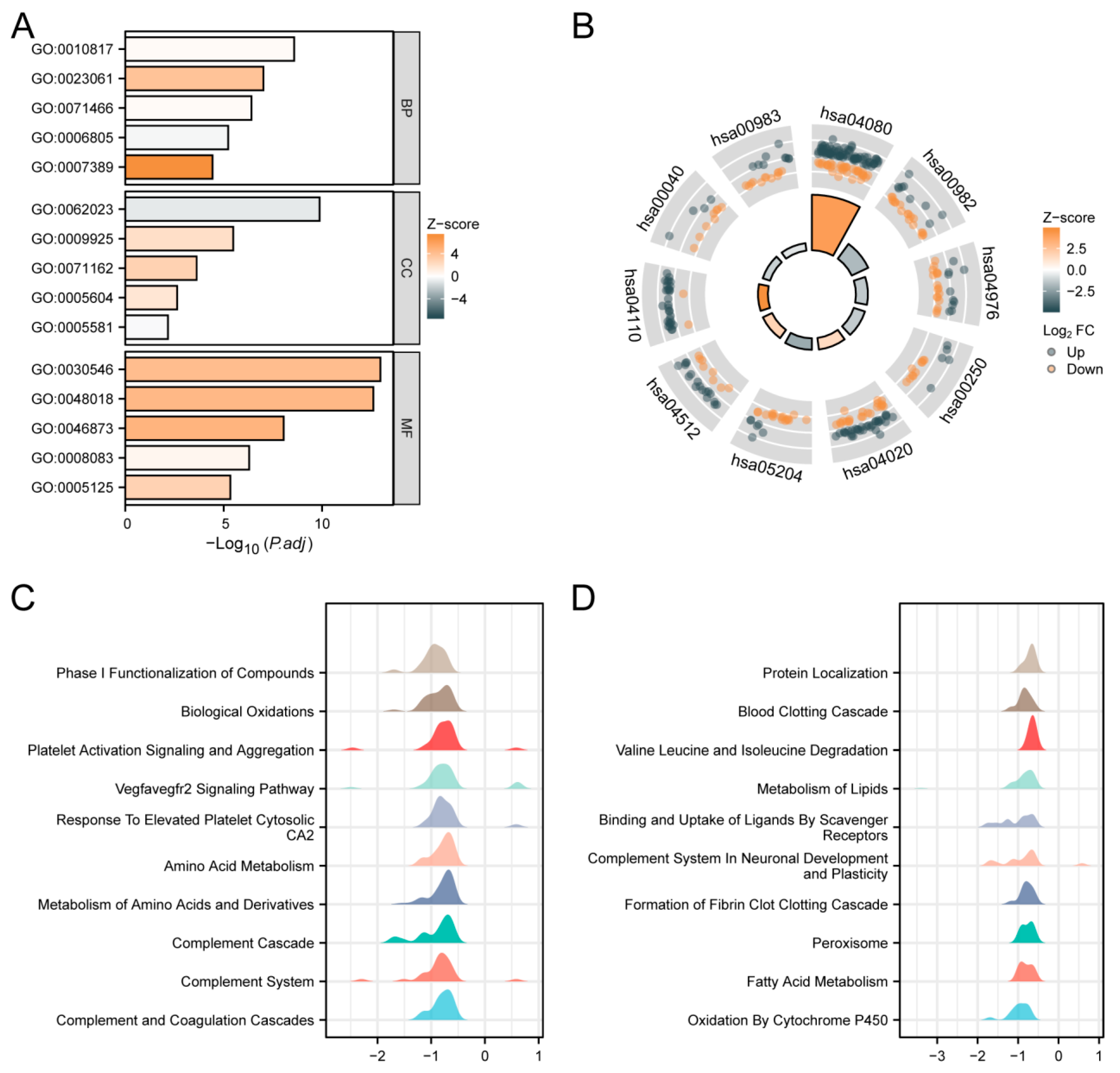
3.5. Functional Enrichment Analysis of SPSB2 in LIHC
3.6. Immunoinfiltration Analysis of SPSB2
3.7. Correlation Between SPSB2 Expression and Immune Checkpoints
3.8. Evaluation of SPSB2 Expression
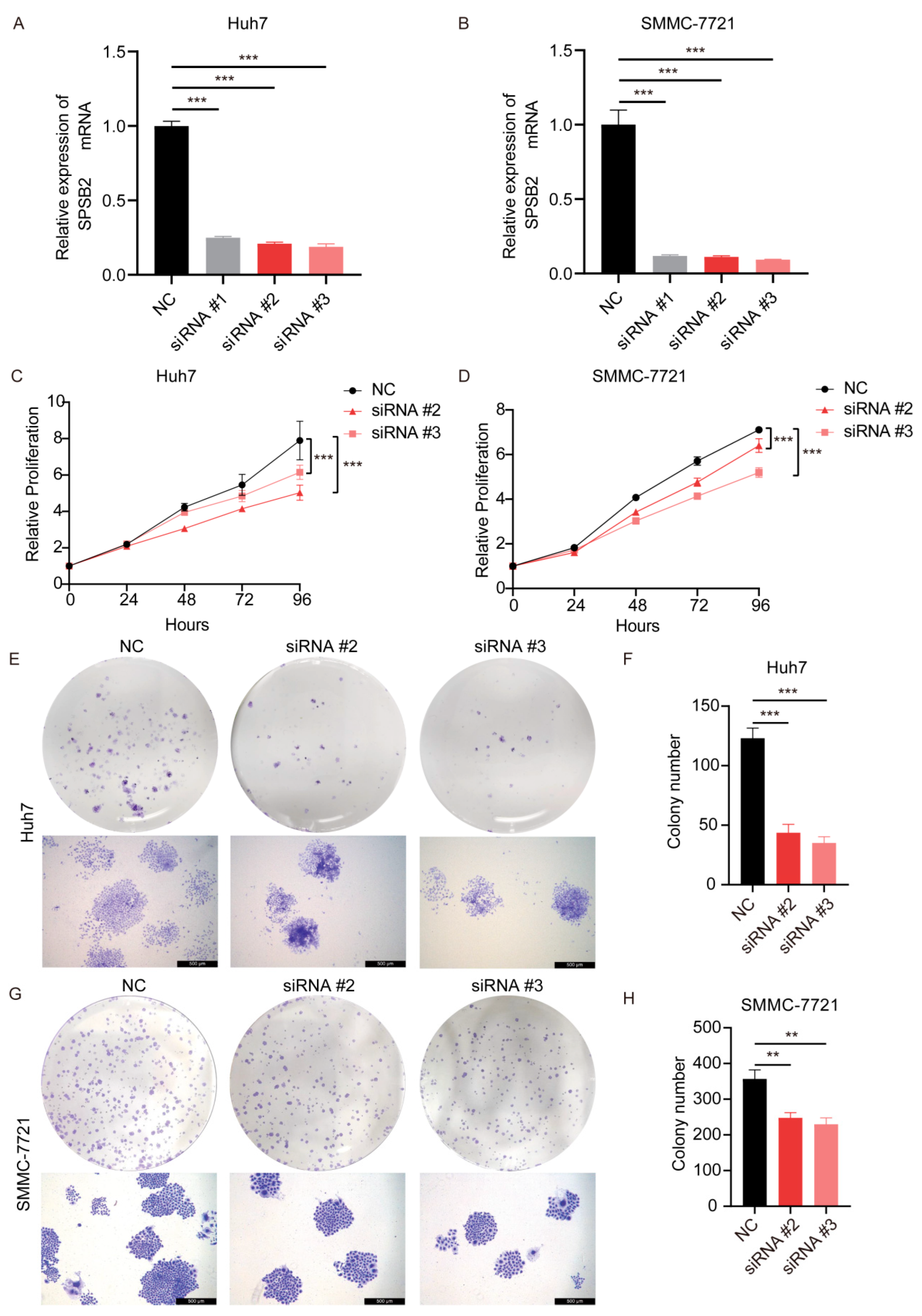
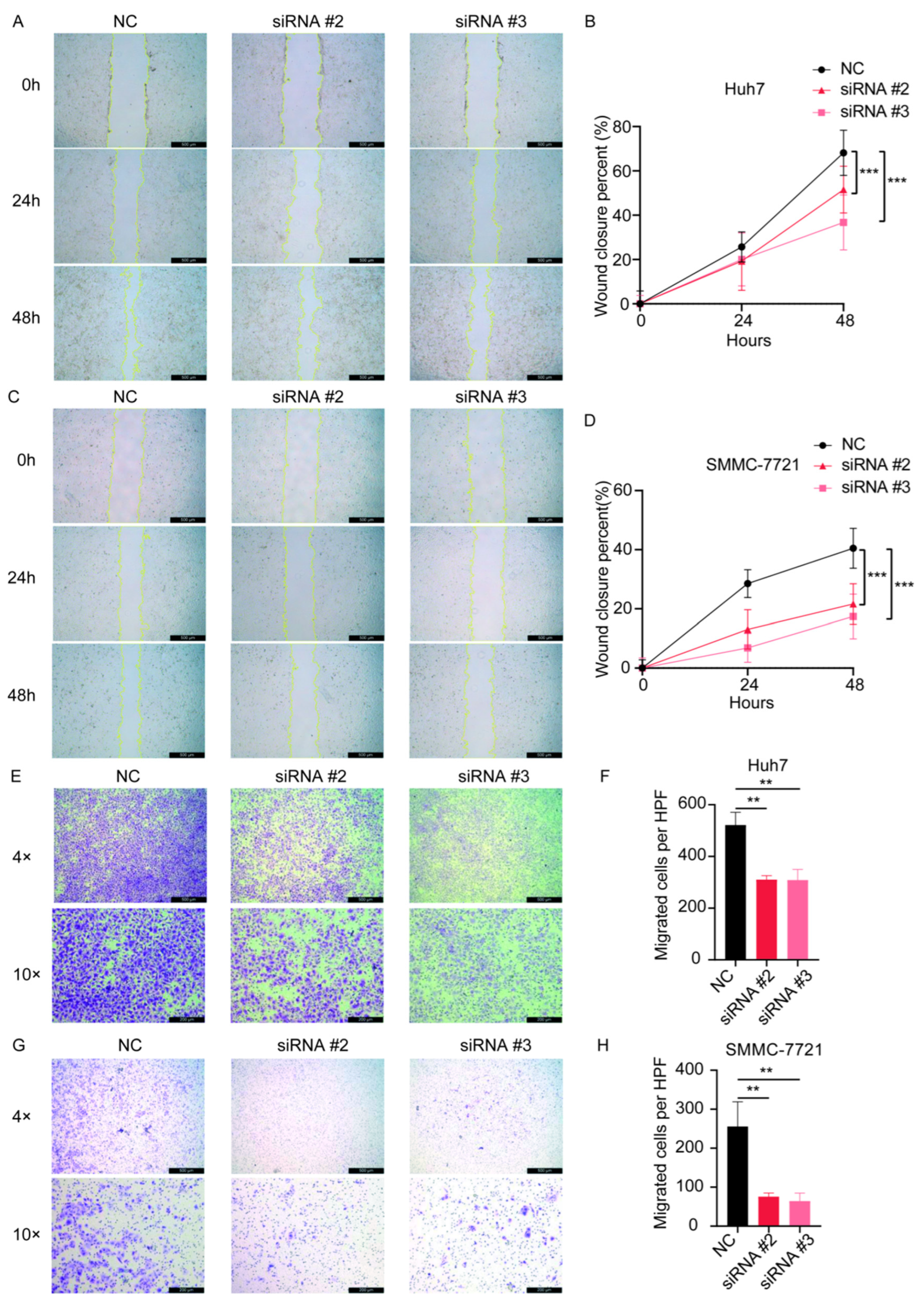
3.9. Effect of Knockdown of SPSB2 on the Biological Function of Hepatocellular Carcinoma Cells
4. Discussion
5. Conclusions
Author Contributions
Funding
Institutional Review Board Statement
Informed Consent Statement
Data Availability Statement
Conflicts of Interest
Abbreviations
| LIHC | Liver Hepatocellular Carcinoma |
| TCGA | The Cancer Genome Atlas |
| GEO | Gene Expression Omnibus |
| GO | Gene Ontology |
| KEGG | Kyoto Encyclopedia of Genes and Genomes |
| GSEA | Gene set enrichment analysis |
| OS | Overall survival |
| ANOVA | One-factor analysis of variance |
References
- Xia, S.; Pan, Y.; Liang, Y.; Xu, J.; Cai, X. The microenvironmental and metabolic aspects of sorafenib resistance in hepatocellular carcinoma. EBioMedicine 2020, 51, 102610. [Google Scholar] [CrossRef]
- European Association for the Study of the Liver. EASL Clinical Practice Guidelines: Management of hepatocellular carcinoma. J. Hepatol. 2018, 69, 182–236. [Google Scholar] [CrossRef] [PubMed]
- Marrero, J.A.; Kulik, L.M.; Sirlin, C.B.; Zhu, A.X.; Finn, R.S.; Abecassis, M.M.; Roberts, L.R.; Heimbach, J.K. Diagnosis, Staging, and Management of Hepatocellular Carcinoma: 2018 Practice Guidance by the American Association for the Study of Liver Diseases. Hepatology 2018, 68, 723–750. [Google Scholar] [CrossRef] [PubMed]
- Llovet, J.M.; Kelley, R.K.; Villanueva, A.; Singal, A.G.; Pikarsky, E.; Roayaie, S.; Lencioni, R.; Koike, K.; Zucman-Rossi, J.; Finn, R.S. Hepatocellular carcinoma. Nat. Rev. Dis. Primers 2021, 7, 6. [Google Scholar] [CrossRef]
- Chaabna, K.; Dashzeveg, D.; Shagdarsuren, T.; Al-Rifai, R.H. Prevalence and genotype distribution of hepatitis C virus in Mongolia: Systematic review and meta-analysis. Int. J. Infect. Dis. 2021, 105, 377–388. [Google Scholar] [CrossRef]
- Jiang, Y.; Han, Q.; Zhao, H.; Zhang, J. The Mechanisms of HBV-Induced Hepatocellular Carcinoma. J. Hepatocell. Carcinoma 2021, 8, 435–450. [Google Scholar] [CrossRef] [PubMed]
- Santoni, M.; Rizzo, A.; Kucharz, J.; Mollica, V.; Rosellini, M.; Marchetti, A.; Tassinari, E.; Monteiro, F.S.M.; Soares, A.; Molina-Cerrillo, J.; et al. Complete remissions following immunotherapy or immuno-oncology combinations in cancer patients: The MOUSEION-03 meta-analysis. Cancer Immunol. Immunother. 2023, 72, 1365–1379. [Google Scholar] [CrossRef]
- Facciorusso, A.; Tartaglia, N.; Villani, R.; Serviddio, G.; Ramai, D.; Mohan, B.P.; Chandan, S.; Abd El Aziz, M.A.; Evangelista, J.; Cotsoglou, C.; et al. Lenvatinib versus sorafenib as first-line therapy of advanced hepatocellular carcinoma: A systematic review and meta-analysis. Am. J. Transl. Res. 2021, 13, 2379–2387. [Google Scholar]
- Sheng, H.; Huang, Y.; Xiao, Y.; Zhu, Z.; Shen, M.; Zhou, P.; Guo, Z.; Wang, J.; Wang, H.; Dai, W.; et al. ATR inhibitor AZD6738 enhances the antitumor activity of radiotherapy and immune checkpoint inhibitors by potentiating the tumor immune microenvironment in hepatocellular carcinoma. J. Immunother. Cancer 2020, 8, e000340. [Google Scholar] [CrossRef]
- Wang, J.; Li, Y.; Zhang, C.; Chen, X.; Zhu, L.; Luo, T. Characterization of diagnostic and prognostic significance of cell cycle-linked genes in hepatocellular carcinoma. Transl. Cancer Res. 2021, 10, 4636–4651. [Google Scholar] [CrossRef]
- Hilton, D.J.; Richardson, R.T.; Alexander, W.S.; Viney, E.M.; Willson, T.A.; Sprigg, N.S.; Starr, R.; Nicholson, S.E.; Metcalf, D.; Nicola, N.A. Twenty proteins containing a C-terminal SOCS box form five structural classes. Proc. Natl. Acad. Sci. USA 1998, 95, 114–119. [Google Scholar] [CrossRef] [PubMed]
- Mahrour, N.; Redwine, W.B.; Florens, L.; Swanson, S.K.; Martin-Brown, S.; Bradford, W.D.; Staehling-Hampton, K.; Washburn, M.P.; Conaway, R.C.; Conaway, J.W. Characterization of Cullin-box sequences that direct recruitment of Cul2-Rbx1 and Cul5-Rbx2 modules to Elongin BC-based ubiquitin ligases. J. Biol. Chem. 2008, 283, 8005–8013. [Google Scholar] [CrossRef] [PubMed]
- Kuang, Z.; Lewis, R.S.; Curtis, J.M.; Zhan, Y.; Saunders, B.M.; Babon, J.J.; Kolesnik, T.B.; Low, A.; Masters, S.L.; Willson, T.A.; et al. The SPRY domain-containing SOCS box protein SPSB2 targets iNOS for proteasomal degradation. J. Cell Biol. 2010, 190, 129–141. [Google Scholar] [CrossRef]
- Lewis, R.S.; Kolesnik, T.B.; Kuang, Z.; D’Cruz, A.A.; Blewitt, M.E.; Masters, S.L.; Low, A.; Willson, T.; Norton, R.S.; Nicholson, S.E. TLR regulation of SPSB1 controls inducible nitric oxide synthase induction. J. Immunol. 2011, 187, 3798–3805. [Google Scholar] [CrossRef] [PubMed]
- Nishiya, T.; Matsumoto, K.; Maekawa, S.; Kajita, E.; Horinouchi, T.; Fujimuro, M.; Ogasawara, K.; Uehara, T.; Miwa, S. Regulation of inducible nitric-oxide synthase by the SPRY domain- and SOCS box-containing proteins. J. Biol. Chem. 2011, 286, 9009–9019. [Google Scholar] [CrossRef]
- Wang, M.; Wang, Y.; Liu, Y.; Wang, H.; Xin, X.; Li, J.; Hao, Y.; Han, L.; Yu, F.; Zheng, C.; et al. SPSB2 inhibits hepatitis C virus replication by targeting NS5A for ubiquitination and degradation. PLoS ONE 2019, 14, e0219989. [Google Scholar] [CrossRef]
- Yamada, M.; Motoike, I.N.; Kojima, K.; Fuse, N.; Hozawa, A.; Kuriyama, S.; Katsuoka, F.; Tadaka, S.; Shirota, M.; Sakurai, M.; et al. Genetic loci for lung function in Japanese adults with adjustment for exhaled nitric oxide levels as airway inflammation indicator. Commun. Biol. 2021, 4, 1288. [Google Scholar] [CrossRef]
- Liu, S.; Nheu, T.; Luwor, R.; Nicholson, S.E.; Zhu, H.J. SPSB1, a Novel Negative Regulator of the Transforming Growth Factor-β Signaling Pathway Targeting the Type II Receptor. J. Biol. Chem. 2015, 290, 17894–17908. [Google Scholar] [CrossRef]
- I V, A.N.; Nair, A.S. Bioinformatics screening of ETV4 transcription factor oncogenes and identifying small-molecular anticancer drugs. Chem. Biol. Drug Des. 2022, 99, 277–285. [Google Scholar] [CrossRef]
- Giannos, P.; Kechagias, K.S.; Gal, A. Identification of Prognostic Gene Biomarkers in Non-Small Cell Lung Cancer Progression by Integrated Bioinformatics Analysis. Biology 2021, 10, 1200. [Google Scholar] [CrossRef]
- Vivian, J.; Rao, A.A.; Nothaft, F.A.; Ketchum, C.; Armstrong, J.; Novak, A.; Pfeil, J.; Narkizian, J.; Deran, A.D.; Musselman-Brown, A.; et al. Toil enables reproducible, open source, big biomedical data analyses. Nat. Biotechnol. 2017, 35, 314–316. [Google Scholar] [CrossRef] [PubMed]
- Day, R.S.; McDade, K.K.; Chandran, U.R.; Lisovich, A.; Conrads, T.P.; Hood, B.L.; Kolli, V.S.; Kirchner, D.; Litzi, T.; Maxwell, G.L. Identifier mapping performance for integrating transcriptomics and proteomics experimental results. BMC Bioinform. 2011, 12, 213. [Google Scholar] [CrossRef]
- Day, R.S.; McDade, K.K. A decision theory paradigm for evaluating identifier mapping and filtering methods using data integration. BMC Bioinform. 2013, 14, 223. [Google Scholar] [CrossRef] [PubMed]
- Davis, S.; Meltzer, P.S. GEOquery: A bridge between the Gene Expression Omnibus (GEO) and BioConductor. Bioinformatics 2007, 23, 1846–1847. [Google Scholar] [CrossRef]
- Ritchie, M.E.; Phipson, B.; Wu, D.; Hu, Y.; Law, C.W.; Shi, W.; Smyth, G.K. limma powers differential expression analyses for RNA-sequencing and microarray studies. Nucleic Acids Res. 2015, 43, e47. [Google Scholar] [CrossRef] [PubMed]
- Love, M.I.; Huber, W.; Anders, S. Moderated estimation of fold change and dispersion for RNA-seq data with DESeq2. Genome Biol. 2014, 15, 550. [Google Scholar] [CrossRef]
- Jensen, L.J.; Kuhn, M.; Stark, M.; Chaffron, S.; Creevey, C.; Muller, J.; Doerks, T.; Julien, P.; Roth, A.; Simonovic, M.; et al. STRING 8—A global view on proteins and their functional interactions in 630 organisms. Nucleic Acids Res. 2009, 37, D412–D416. [Google Scholar] [CrossRef]
- Liu, J.; Lichtenberg, T.; Hoadley, K.A.; Poisson, L.M.; Lazar, A.J.; Cherniack, A.D.; Kovatich, A.J.; Benz, C.C.; Levine, D.A.; Lee, A.V.; et al. An Integrated TCGA Pan-Cancer Clinical Data Resource to Drive High-Quality Survival Outcome Analytics. Cell 2018, 173, 400–416.e11. [Google Scholar] [CrossRef]
- Yu, G.; Wang, L.G.; Han, Y.; He, Q.Y. clusterProfiler: An R package for comparing biological themes among gene clusters. Omics 2012, 16, 284–287. [Google Scholar] [CrossRef]
- Walter, W.; Sánchez-Cabo, F.; Ricote, M. GOplot: An R package for visually combining expression data with functional analysis. Bioinformatics 2015, 31, 2912–2914. [Google Scholar] [CrossRef]
- Subramanian, A.; Tamayo, P.; Mootha, V.K.; Mukherjee, S.; Ebert, B.L.; Gillette, M.A.; Paulovich, A.; Pomeroy, S.L.; Golub, T.R.; Lander, E.S.; et al. Gene set enrichment analysis: A knowledge-based approach for interpreting genome-wide expression profiles. Proc. Natl. Acad. Sci. USA 2005, 102, 15545–15550. [Google Scholar] [CrossRef] [PubMed]
- Hänzelmann, S.; Castelo, R.; Guinney, J. GSVA: Gene set variation analysis for microarray and RNA-seq data. BMC Bioinform. 2013, 14, 7. [Google Scholar] [CrossRef] [PubMed]
- Bindea, G.; Mlecnik, B.; Tosolini, M.; Kirilovsky, A.; Waldner, M.; Obenauf, A.C.; Angell, H.; Fredriksen, T.; Lafontaine, L.; Berger, A.; et al. Spatiotemporal dynamics of intratumoral immune cells reveal the immune landscape in human cancer. Immunity 2013, 39, 782–795. [Google Scholar] [CrossRef]
- Gu, Z.; Gu, L.; Eils, R.; Schlesner, M.; Brors, B. circlize Implements and enhances circular visualization in R. Bioinformatics 2014, 30, 2811–2812. [Google Scholar] [CrossRef] [PubMed]
- Ahn, H.R.; Baek, G.O.; Yoon, M.G.; Son, J.A.; Yoon, J.H.; Cheong, J.Y.; Cho, H.J.; Kang, H.C.; Eun, J.W.; Kim, S.S. Hypomethylation-mediated upregulation of the WASF2 promoter region correlates with poor clinical outcomes in hepatocellular carcinoma. J. Exp. Clin. Cancer Res. 2022, 41, 158. [Google Scholar] [CrossRef]
- Liu, L.; Wang, Q.; Wu, L.; Zhang, L.; Huang, Y.; Yang, H.; Guo, L.; Fang, Z.; Wang, X. Overexpression of POLA2 in hepatocellular carcinoma is involved in immune infiltration and predicts a poor prognosis. Cancer Cell Int. 2023, 23, 138. [Google Scholar] [CrossRef]
- Akinyemiju, T.; Abera, S.; Ahmed, M.; Alam, N.; Alemayohu, M.A.; Allen, C.; Al-Raddadi, R.; Alvis-Guzman, N.; Amoako, Y.; Artaman, A.; et al. The Burden of Primary Liver Cancer and Underlying Etiologies From 1990 to 2015 at the Global, Regional, and National Level: Results From the Global Burden of Disease Study 2015. JAMA Oncol. 2017, 3, 1683–1691. [Google Scholar]
- Rahib, L.; Smith, B.D.; Aizenberg, R.; Rosenzweig, A.B.; Fleshman, J.M.; Matrisian, L.M. Projecting cancer incidence and deaths to 2030: The unexpected burden of thyroid, liver, and pancreas cancers in the United States. Cancer Res. 2014, 74, 2913–2921. [Google Scholar] [CrossRef]
- Xu, F.; Jin, T.; Zhu, Y.; Dai, C. Immune checkpoint therapy in liver cancer. J. Exp. Clin. Cancer Res. 2018, 37, 110. [Google Scholar] [CrossRef]
- Oura, K.; Morishita, A.; Tani, J.; Masaki, T. Tumor Immune Microenvironment and Immunosuppressive Therapy in Hepatocellular Carcinoma: A Review. Int. J. Mol. Sci. 2021, 22, 5801. [Google Scholar] [CrossRef]
- Tekpli, X.; Lien, T.; Røssevold, A.H.; Nebdal, D.; Borgen, E.; Ohnstad, H.O.; Kyte, J.A.; Vallon-Christersson, J.; Fongaard, M.; Due, E.U.; et al. An independent poor-prognosis subtype of breast cancer defined by a distinct tumor immune microenvironment. Nat. Commun. 2019, 10, 5499. [Google Scholar] [CrossRef]
- Nilsson, S.; Mäkelä, S.; Treuter, E.; Tujague, M.; Thomsen, J.; Andersson, G.; Enmark, E.; Pettersson, K.; Warner, M.; Gustafsson, J.A. Mechanisms of estrogen action. Physiol. Rev. 2001, 81, 1535–1565. [Google Scholar] [CrossRef] [PubMed]
- Yager, J.D.; Davidson, N.E. Estrogen carcinogenesis in breast cancer. N. Engl. J. Med. 2006, 354, 270–282. [Google Scholar] [CrossRef] [PubMed]
- Robinson, D.R.; Wu, Y.M.; Vats, P.; Su, F.; Lonigro, R.J.; Cao, X.; Kalyana-Sundaram, S.; Wang, R.; Ning, Y.; Hodges, L.; et al. Activating ESR1 mutations in hormone-resistant metastatic breast cancer. Nat. Genet. 2013, 45, 1446–1451. [Google Scholar] [CrossRef]
- Merenbakh-Lamin, K.; Ben-Baruch, N.; Yeheskel, A.; Dvir, A.; Soussan-Gutman, L.; Jeselsohn, R.; Yelensky, R.; Brown, M.; Miller, V.A.; Sarid, D.; et al. D538G mutation in estrogen receptor-α: A novel mechanism for acquired endocrine resistance in breast cancer. Cancer Res. 2013, 73, 6856–6864. [Google Scholar] [CrossRef]
- Shang, L.; Chen, X.; Zhu, T.; Chong, S.; Liu, H.; Huang, W.; Fu, W.; She, H.; Shen, X. Cancer-Associated Fibroblast-Secreted Exosomes Promote Gastric Cancer Cell Migration and Invasion via the IL-32/ESR1 Axis. Appl. Biochem. Biotechnol. 2024, 196, 6045–6058. [Google Scholar] [CrossRef] [PubMed]
- Wang, M.; Guo, H.; Zhang, B.; Shang, Y.; Zhang, S.; Liu, X.; Cao, P.; Fan, Y.; Tan, K. Transcription factors-related molecular subtypes and risk prognostic model: Exploring the immunogenicity landscape and potential drug targets in hepatocellular carcinoma. Cancer Cell Int. 2024, 24, 9. [Google Scholar] [CrossRef]
- Lautert-Dutra, W.; Melo, C.M.; Chaves, L.P.; Souza, F.C.; Crozier, C.; Sundby, A.E.; Woroszchuk, E.; Saggioro, F.P.; Avante, F.S.; Dos Reis, R.B.; et al. Identification of tumor-agnostic biomarkers for predicting prostate cancer progression and biochemical recurrence. Front. Oncol. 2023, 13, 1280943. [Google Scholar] [CrossRef]
- Hamilton, D.H.; Griner, L.M.; Keller, J.M.; Hu, X.; Southall, N.; Marugan, J.; David, J.M.; Ferrer, M.; Palena, C. Targeting Estrogen Receptor Signaling with Fulvestrant Enhances Immune and Chemotherapy-Mediated Cytotoxicity of Human Lung Cancer. Clin. Cancer Res. 2016, 22, 6204–6216. [Google Scholar] [CrossRef]
- Song, D.; Zhang, D.; Chen, S.; Wu, J.; Hao, Q.; Zhao, L.; Ren, H.; Du, N. Identification and validation of prognosis-associated DNA repair gene signatures in colorectal cancer. Sci. Rep. 2022, 12, 6946. [Google Scholar] [CrossRef]
- Wu, T.; Liao, L.; Wu, T.; Chen, S.; Yi, Q.; Xu, M. IGF2BP2 promotes glycolysis and hepatocellular carcinoma stemness by stabilizing CDC45 mRNA via m6A modification. Cell Cycle 2023, 22, 2245–2263. [Google Scholar] [CrossRef]
- Wang, Y.Y.; Cheng, K.H.; Hung, A.C.; Lo, S.; Chen, P.Y.; Wu, Y.C.; Hou, M.F.; Yuan, S.F. Differential impact of cytoplasmic vs. nuclear RAD51 expression on breast cancer progression and patient prognosis. Int. J. Oncol. 2024, 64, 12. [Google Scholar] [CrossRef] [PubMed]
- Wang, Y.; Dong, S.; Hu, K.; Xu, L.; Feng, Q.; Li, B.; Wang, G.; Chen, G.; Zhang, B.; Jia, X.; et al. The novel norcantharidin derivative DCZ5417 suppresses multiple myeloma progression by targeting the TRIP13-MAPK-YWHAE signaling pathway. J. Transl. Med. 2023, 21, 858. [Google Scholar] [CrossRef]
- Pan, B.; Wan, T.; Zhou, Y.; Huang, S.; Yuan, L.; Jiang, Y.; Zheng, X.; Liu, P.; Xiang, H.; Ju, M.; et al. The MYBL2-CCL2 axis promotes tumor progression and resistance to anti-PD-1 therapy in ovarian cancer by inducing immunosuppressive macrophages. Cancer Cell Int. 2023, 23, 248. [Google Scholar] [CrossRef] [PubMed]
- Wu, F.; Wang, M.; Zhong, T.; Xiao, C.; Chen, X.; Huang, Y.; Wu, M.; Yu, J.; Chen, D. Inhibition of CDC20 potentiates anti-tumor immunity through facilitating GSDME-mediated pyroptosis in prostate cancer. Exp. Hematol. Oncol. 2023, 12, 67. [Google Scholar] [CrossRef]
- Arteaga, C.L.; Engelman, J.A. ERBB receptors: From oncogene discovery to basic science to mechanism-based cancer therapeutics. Cancer Cell 2014, 25, 282–303. [Google Scholar] [CrossRef]
- Pollak, M. Insulin and insulin-like growth factor signalling in neoplasia. Nat. Rev. Cancer 2008, 8, 915–928. [Google Scholar] [CrossRef] [PubMed]
- Wang, G.; Biswas, A.K.; Ma, W.; Kandpal, M.; Coker, C.; Grandgenett, P.M.; Hollingsworth, M.A.; Jain, R.; Tanji, K.; Lόpez-Pintado, S.; et al. Metastatic cancers promote cachexia through ZIP14 upregulation in skeletal muscle. Nat. Med. 2018, 24, 770–781. [Google Scholar] [CrossRef]
- Drabsch, Y.; ten Dijke, P. TGF-β signalling and its role in cancer progression and metastasis. Cancer Metastasis Rev. 2012, 31, 553–568. [Google Scholar] [CrossRef]
- Yang, J.D.; Nakamura, I.; Roberts, L.R. The tumor microenvironment in hepatocellular carcinoma: Current status and therapeutic targets. Semin. Cancer Biol. 2011, 21, 35–43. [Google Scholar] [CrossRef]
- Bissell, M.J.; Hines, W.C. Why don’t we get more cancer? A proposed role of the microenvironment in restraining cancer progression. Nat. Med. 2011, 17, 320–329. [Google Scholar] [CrossRef] [PubMed]
- Stratton, M.R.; Campbell, P.J.; Futreal, P.A. The cancer genome. Nature 2009, 458, 719–724. [Google Scholar] [CrossRef]
- Robil, N.; Petel, F.; Kilhoffer, M.C.; Haiech, J. Glioblastoma and calcium signaling--analysis of calcium toolbox expression. Int. J. Dev. Biol. 2015, 59, 407–415. [Google Scholar] [CrossRef] [PubMed]
- Kwong, E.; Li, Y.; Hylemon, P.B.; Zhou, H. Bile acids and sphingosine-1-phosphate receptor 2 in hepatic lipid metabolism. Acta Pharm. Sin. B 2015, 5, 151–157. [Google Scholar] [CrossRef]
- Kerbel, R.; Folkman, J. Clinical translation of angiogenesis inhibitors. Nat. Rev. Cancer 2002, 2, 727–739. [Google Scholar] [CrossRef]
- Ferrara, N. VEGF as a therapeutic target in cancer. Oncology 2005, 69 (Suppl. 3), 11–16. [Google Scholar] [CrossRef] [PubMed]
- Liu, J.; Deng, G.H.; Zhang, J.; Wang, Y.; Xia, X.Y.; Luo, X.M.; Deng, Y.T.; He, S.S.; Mao, Y.Y.; Peng, X.C.; et al. The effect of chronic stress on anti-angiogenesis of sunitinib in colorectal cancer models. Psychoneuroendocrinology 2015, 52, 130–142. [Google Scholar] [CrossRef]
- Mayers, J.R.; Wu, C.; Clish, C.B.; Kraft, P.; Torrence, M.E.; Fiske, B.P.; Yuan, C.; Bao, Y.; Townsend, M.K.; Tworoger, S.S.; et al. Elevation of circulating branched-chain amino acids is an early event in human pancreatic adenocarcinoma development. Nat. Med. 2014, 20, 1193–1198. [Google Scholar] [CrossRef]
- Pandya, P.H.; Murray, M.E.; Pollok, K.E.; Renbarger, J.L. The Immune System in Cancer Pathogenesis: Potential Therapeutic Approaches. J. Immunol. Res. 2016, 2016, 4273943. [Google Scholar] [CrossRef]
- Diao, D.; Wang, Z.; Cheng, Y.; Zhang, H.; Guo, Q.; Song, Y.; Zhu, K.; Li, K.; Liu, D.; Dang, C. D-dimer: Not just an indicator of venous thrombosis but a predictor of asymptomatic hematogenous metastasis in gastric cancer patients. PLoS ONE 2014, 9, e101125. [Google Scholar] [CrossRef]
- Buisseret, L.; Pommey, S.; Allard, B.; Garaud, S.; Bergeron, M.; Cousineau, I.; Ameye, L.; Bareche, Y.; Paesmans, M.; Crown, J.P.A.; et al. Clinical significance of CD73 in triple-negative breast cancer: Multiplex analysis of a phase III clinical trial. Ann. Oncol. 2018, 29, 1056–1062. [Google Scholar] [CrossRef]
- Powell, D.R.; Huttenlocher, A. Neutrophils in the Tumor Microenvironment. Trends Immunol. 2016, 37, 41–52. [Google Scholar] [CrossRef]
- Xu, L.N.; Zou, S.Q.; Wang, J.M. Action and mechanism of Fas and Fas ligand in immune escape of gallbladder carcinoma. World J. Gastroenterol. 2005, 11, 3719–3723. [Google Scholar] [CrossRef] [PubMed]
- Sharma, A.; Rajappa, M.; Saxena, A.; Sharma, M. Cytokine profile in Indian women with cervical intraepithelial neoplasia and cancer cervix. Int. J. Gynecol. Cancer 2007, 17, 879–885. [Google Scholar] [CrossRef] [PubMed]
- Johnson, S.D.; De Costa, A.M.; Young, M.R. Effect of the premalignant and tumor microenvironment on immune cell cytokine production in head and neck cancer. Cancers 2014, 6, 756–770. [Google Scholar] [CrossRef] [PubMed]
- Facciabene, A.; Peng, X.; Hagemann, I.S.; Balint, K.; Barchetti, A.; Wang, L.P.; Gimotty, P.A.; Gilks, C.B.; Lal, P.; Zhang, L.; et al. Tumour hypoxia promotes tolerance and angiogenesis via CCL28 and T(reg) cells. Nature 2011, 475, 226–230. [Google Scholar] [CrossRef]
- Liu, P.; Chen, L.; Zhang, H. Natural Killer Cells in Liver Disease and Hepatocellular Carcinoma and the NK Cell-Based Immunotherapy. J. Immunol. Res. 2018, 2018, 1206737. [Google Scholar] [CrossRef]
- Croft, M. Control of immunity by the TNFR-related molecule OX40 (CD134). Annu. Rev. Immunol. 2010, 28, 57–78. [Google Scholar] [CrossRef]
- Dong, H.; Strome, S.E.; Salomao, D.R.; Tamura, H.; Hirano, F.; Flies, D.B.; Roche, P.C.; Lu, J.; Zhu, G.; Tamada, K.; et al. Tumor-associated B7-H1 promotes T-cell apoptosis: A potential mechanism of immune evasion. Nat. Med. 2002, 8, 793–800. [Google Scholar] [CrossRef]
- Iwai, Y.; Ishida, M.; Tanaka, Y.; Okazaki, T.; Honjo, T.; Minato, N. Involvement of PD-L1 on tumor cells in the escape from host immune system and tumor immunotherapy by PD-L1 blockade. Proc. Natl. Acad. Sci. USA 2002, 99, 12293–12297. [Google Scholar] [CrossRef]
- Roncador, G.; Garcia, J.F.; Garcia, J.F.; Maestre, L.; Lucas, E.; Menarguez, J.; Ohshima, K.; Nakamura, S.; Banham, A.H.; Piris, M.A. FOXP3, a selective marker for a subset of adult T-cell leukaemia/lymphoma. Leukemia 2005, 19, 2247–2253. [Google Scholar] [CrossRef] [PubMed]
- Karube, K.; Aoki, R.; Sugita, Y.; Yoshida, S.; Nomura, Y.; Shimizu, K.; Kimura, Y.; Hashikawa, K.; Takeshita, M.; Suzumiya, J.; et al. The relationship of FOXP3 expression and clinicopathological characteristics in adult T-cell leukemia/lymphoma. Mod. Pathol. 2008, 21, 617–625. [Google Scholar] [CrossRef] [PubMed]
- Okazawa, H.; Motegi, S.; Ohyama, N.; Ohnishi, H.; Tomizawa, T.; Kaneko, Y.; Oldenborg, P.A.; Ishikawa, O.; Matozaki, T. Negative regulation of phagocytosis in macrophages by the CD47-SHPS-1 system. J. Immunol. 2005, 174, 2004–2011. [Google Scholar] [CrossRef]
- Oldenborg, P.A.; Zheleznyak, A.; Fang, Y.F.; Lagenaur, C.F.; Gresham, H.D.; Lindberg, F.P. Role of CD47 as a marker of self on red blood cells. Science 2000, 288, 2051–2054. [Google Scholar] [CrossRef]
- Leach, D.R.; Krummel, M.F.; Allison, J.P. Enhancement of antitumor immunity by CTLA-4 blockade. Science 1996, 271, 1734–1736. [Google Scholar] [CrossRef] [PubMed]


| Characteristics | Low Expression of SPSB2 | High Expression of SPSB2 | p Value |
|---|---|---|---|
| n | 187 | 187 | |
| Age, n (%) | 0.109 | ||
| ≤60 | 81 (21.7%) | 96 (25.7%) | |
| >60 | 106 (28.4%) | 90 (24.1%) | |
| Weight, n (%) | <0.001 | ||
| ≤70 | 72 (20.8%) | 112 (32.4%) | |
| >70 | 101 (29.2%) | 61 (17.6%) | |
| Gender, n (%) | 0.320 | ||
| Female | 65 (17.4%) | 56 (15%) | |
| Male | 122 (32.6%) | 131 (35%) | |
| Race, n (%) | <0.001 | ||
| Asian | 62 (17.1%) | 98 (27.1%) | |
| Black or African American | 7 (1.9%) | 10 (2.8%) | |
| White | 112 (30.9%) | 73 (20.2%) | |
| Pathologic T stage, n (%) | 0.017 | ||
| T1 | 106 (28.6%) | 77 (20.8%) | |
| T2 | 38 (10.2%) | 57 (15.4%) | |
| T3 | 34 (9.2%) | 46 (12.4%) | |
| T4 | 6 (1.6%) | 7 (1.9%) | |
| Pathologic N stage, n (%) | 0.094 | ||
| N0 | 115 (44.6%) | 139 (53.9%) | |
| N1 | 4 (1.6%) | 0 (0%) | |
| Pathologic M stage, n (%) | 0.710 | ||
| M0 | 126 (46.3%) | 142 (52.2%) | |
| M1 | 1 (0.4%) | 3 (1.1%) | |
| Pathologic stage, n (%) | 0.041 | ||
| Stage I | 98 (28%) | 75 (21.4%) | |
| Stage II | 34 (9.7%) | 53 (15.1%) | |
| Stage III | 38 (10.9%) | 47 (13.4%) | |
| Stage IV | 2 (0.6%) | 3 (0.9%) | |
| Tumor status, n (%) | 0.312 | ||
| Tumor free | 106 (29.9%) | 96 (27%) | |
| With tumor | 72 (20.3%) | 81 (22.8%) | |
| Histological type, n (%) | 0.117 | ||
| Hepatocellular carcinoma | 182 (48.7%) | 182 (48.7%) | |
| Fibrolamellar carcinoma | 3 (0.8%) | 0 (0%) | |
| Hepatocholangiocarcinoma (mixed) | 2 (0.5%) | 5 (1.3%) | |
| BMI, n (%) | <0.001 | ||
| ≤25 | 68 (20.2%) | 109 (32.3%) | |
| >25 | 98 (29.1%) | 62 (18.4%) | |
| Residual tumor, n (%) | 0.970 | ||
| R0 | 162 (47%) | 165 (47.8%) | |
| R1&R2 | 9 (2.6%) | 9 (2.6%) | |
| Histologic grade, n (%) | <0.001 | ||
| G1 | 33 (8.9%) | 22 (6%) | |
| G2 | 105 (28.5%) | 73 (19.8%) | |
| G3 | 42 (11.4%) | 82 (22.2%) | |
| G4 | 2 (0.5%) | 10 (2.7%) | |
| AFP(ng/mL), n (%) | 0.024 | ||
| ≤400 | 117 (41.8%) | 98 (35%) | |
| >400 | 25 (8.9%) | 40 (14.3%) | |
| Albumin(g/dL), n (%) | 0.908 | ||
| <3.5 | 35 (11.7%) | 34 (11.3%) | |
| ≥3.5 | 119 (39.7%) | 112 (37.3%) | |
| Prothrombin time, n (%) | 0.281 | ||
| ≤4 | 105 (35.4%) | 103 (34.7%) | |
| >4 | 51 (17.2%) | 38 (12.8%) | |
| Child-Pugh grade, n (%) | 0.200 | ||
| A | 108 (44.8%) | 111 (46.1%) | |
| B&C | 14 (5.8%) | 8 (3.3%) | |
| Fibrosis ishak score, n (%) | 0.695 | ||
| 0 | 44 (20.5%) | 31 (14.4%) | |
| 1/2 | 14 (6.5%) | 17 (7.9%) | |
| 3/4 | 14 (6.5%) | 14 (6.5%) | |
| 5 | 5 (2.3%) | 4 (1.9%) | |
| 6 | 42 (19.5%) | 30 (14%) | |
| Adjacent hepatic tissue inflammation, n (%) | 0.164 | ||
| None | 72 (30.4%) | 46 (19.4%) | |
| Mild | 49 (20.7%) | 52 (21.9%) | |
| Severe | 9 (3.8%) | 9 (3.8%) |
| Characteristics | Total (N) | Univariate Analysis | Multivariate Analysis | ||
|---|---|---|---|---|---|
| Hazard Ratio (95% CI) | p Value | Hazard Ratio (95% CI) | p Value | ||
| Gender | 373 | ||||
| Female | 121 | Reference | |||
| Male | 252 | 0.793 (0.557–1.130) | 0.200 | ||
| Race | 361 | ||||
| Asian | 159 | Reference | |||
| Black or African American | 17 | 1.585 (0.675–3.725) | 0.290 | ||
| White | 185 | 1.323 (0.909–1.928) | 0.144 | ||
| Age | 373 | ||||
| ≤60 | 177 | Reference | |||
| >60 | 196 | 1.205 (0.850–1.708) | 0.295 | ||
| Weight | 345 | ||||
| ≤70 | 184 | Reference | |||
| >70 | 161 | 0.941 (0.657–1.346) | 0.738 | ||
| BMI | 336 | ||||
| ≤25 | 177 | Reference | |||
| >25 | 159 | 0.798 (0.550–1.158) | 0.235 | ||
| Adjacent hepatic tissue inflammation | 236 | ||||
| None | 118 | Reference | |||
| Mild | 101 | 1.204 (0.723–2.007) | 0.476 | ||
| Severe | 17 | 1.144 (0.447–2.930) | 0.779 | ||
| Pathologic T stage | 370 | ||||
| T1&T2 | 277 | Reference | Reference | ||
| T3 | 80 | 2.355 (1.618–3.430) | <0.001 | 2.439 (1.551–3.836) | <0.001 |
| T4 | 13 | 4.733 (2.424–9.241) | <0.001 | 4.753 (1.769–12.771) | 0.002 |
| Pathologic N stage | 258 | ||||
| N0 | 254 | Reference | |||
| N1 | 4 | 2.029 (0.497–8.281) | 0.324 | ||
| Pathologic M stage | 272 | ||||
| M0 | 268 | Reference | Reference | ||
| M1 | 4 | 4.077 (1.281–12.973) | 0.017 | 0.946 (0.224–3.996) | 0.940 |
| Residual tumor | 344 | ||||
| R0 | 326 | Reference | |||
| R1&R2 | 18 | 1.604 (0.812–3.169) | 0.174 | ||
| Histologic grade | 368 | ||||
| G1 | 55 | Reference | |||
| G2 | 178 | 1.162 (0.686–1.969) | 0.576 | ||
| G3 | 123 | 1.185 (0.683–2.057) | 0.545 | ||
| G4 | 12 | 1.681 (0.621–4.549) | 0.307 | ||
| AFP(ng/mL) | 279 | ||||
| ≤400 | 215 | Reference | |||
| >400 | 64 | 1.075 (0.658–1.759) | 0.772 | ||
| Albumin(g/dL) | 299 | ||||
| <3.5 | 69 | Reference | |||
| ≥3.5 | 230 | 0.897 (0.549–1.464) | 0.662 | ||
| Prothrombin time | 296 | ||||
| ≤4 | 207 | Reference | |||
| >4 | 89 | 1.335 (0.881–2.023) | 0.174 | ||
| Vascular invasion | 317 | ||||
| No | 208 | Reference | |||
| Yes | 109 | 1.344 (0.887–2.035) | 0.163 | ||
| Fibrosis ishak score | 214 | ||||
| 0 | 75 | Reference | |||
| 1/2 | 31 | 0.936 (0.437–2.002) | 0.864 | ||
| 3/4 | 28 | 0.699 (0.288–1.695) | 0.428 | ||
| 5 | 9 | 0.764 (0.181–3.228) | 0.714 | ||
| 6 | 71 | 0.734 (0.401–1.345) | 0.317 | ||
| SPSB2 | 373 | ||||
| Low | 187 | Reference | Reference | ||
| High | 186 | 1.781 (1.253–2.534) | 0.001 | 1.965 (1.234–3.129) | 0.004 |
| Ontology | ID | Description | GeneRatio | BgRatio | p Value | p.adjust | Zscore |
|---|---|---|---|---|---|---|---|
| BP | GO:0010817 | Regulation of hormone levels | 121/2389 | 496/18,800 | 4.48 × 10⁻¹³ | 2.62 × 10⁻⁹ | 0.2727273 |
| BP | GO:0023061 | Signal release | 107/2389 | 451/18,800 | 6.18 × 10⁻¹¹ | 9.59 × 10⁻⁸ | 3.7702723 |
| BP | GO:0071466 | Cellular response to xenobiotic stimulus | 52/2389 | 168/18,800 | 3.95 × 10⁻¹⁰ | 3.84 × 10⁻⁷ | 0.2773501 |
| BP | GO:0006805 | Xenobiotic metabolic process | 36/2389 | 108/18,800 | 2.25 × 10⁻⁸ | 5.95 × 10⁻⁶ | −0.3333333 |
| BP | GO:0007389 | Pattern specification process | 98/2389 | 463/18,800 | 1.82 × 10⁻⁷ | 3.67 × 10⁻⁵ | 7.4751288 |
| CC | GO:0062023 | Collagen-containing extracellular matrix | 111/2532 | 429/19,594 | 2.17 × 10⁻¹³ | 1.34 × 10⁻¹⁰ | −0.8542422 |
| CC | GO:0009925 | Basal plasma membrane | 64/2532 | 251/19,594 | 4.74 × 10⁻⁸ | 3.25 × 10⁻⁶ | 2.0000000 |
| CC | GO:0071162 | CMG complex | 8/2532 | 11/19,594 | 8.83 × 10⁻⁶ | 0.0002 | 2.8284271 |
| CC | GO:0005604 | Basement membrane | 26/2532 | 95/19,594 | 0.0001 | 0.0023 | 1.5689291 |
| CC | GO:0005581 | Collagen trimer | 23/2532 | 86/19,594 | 0.0004 | 0.0068 | −0.2085144 |
| MF | GO:0030546 | Signaling receptor activator activity | 134/2437 | 496/18,410 | 1.02 × 10⁻¹⁶ | 1.08 × 10⁻¹³ | 4.1465684 |
| MF | GO:0048018 | Receptor ligand activity | 131/2437 | 489/18,410 | 4.7 × 10⁻¹⁶ | 2.49 × 10⁻¹³ | 4.4558907 |
| MF | GO:0046873 | metal ion Transmembrane transporter activity | 107/2437 | 428/18,410 | 2.53 × 10⁻¹¹ | 8.93 × 10⁻⁹ | 4.7370088 |
| MF | GO:0008083 | Growth factor activity | 50/2437 | 162/18,410 | 3.85 × 10⁻⁹ | 5.1 × 10⁻⁷ | 0.5656854 |
| MF | GO:0005125 | Cytokine activity | 62/2437 | 235/18,410 | 4.75 × 10⁻⁸ | 4.58 × 10⁻⁶ | 2.7940028 |
| Ontology | ID | Description | GeneRatio | BgRatio | p Value | p.adjust | Zscore |
|---|---|---|---|---|---|---|---|
| KEGG | hsa04080 | Neuroactive ligand-receptor interaction | 111/1118 | 362/8164 | 8.99 × 10⁻¹⁸ | 2.94 × 10⁻¹⁵ | 4.0813794 |
| KEGG | hsa00982 | Drug metabolism—cytochrome P450 | 27/1118 | 72/8164 | 3.47 × 10⁻⁷ | 3 × 10⁻⁵ | −1.7320508 |
| KEGG | hsa04976 | Bile secretion | 27/1118 | 89/8164 | 3.45 × 10⁻⁵ | 0.0014 | −1.3471506 |
| KEGG | hsa00250 | Alanine, aspartate and glutamate metabolism | 15/1118 | 37/8164 | 4.96 × 10⁻⁵ | 0.0016 | −1.2909944 |
| KEGG | hsa04020 | Calcium signaling pathway | 55/1118 | 240/8164 | 5.82 × 10⁻⁵ | 0.0017 | 1.4832397 |
| KEGG | hsa05204 | Chemical carcinogenesis—DNA adducts | 22/1118 | 69/8164 | 7.9 × 10⁻⁵ | 0.0019 | −2.1320072 |
| KEGG | hsa04512 | ECM-receptor interaction | 25/1118 | 88/8164 | 0.0002 | 0.0044 | 1.8000000 |
| KEGG | hsa04110 | Cell cycle | 32/1118 | 126/8164 | 0.0003 | 0.0052 | 4.9497475 |
| KEGG | hsa00040 | Pentose and glucuronate interconversions | 12/1118 | 35/8164 | 0.0016 | 0.0206 | −1.1547005 |
| KEGG | hsa00983 | Drug metabolism—other enzymes | 21/1118 | 80/8164 | 0.0020 | 0.0235 | −0.6546537 |
Disclaimer/Publisher’s Note: The statements, opinions and data contained in all publications are solely those of the individual author(s) and contributor(s) and not of MDPI and/or the editor(s). MDPI and/or the editor(s) disclaim responsibility for any injury to people or property resulting from any ideas, methods, instructions or products referred to in the content. |
© 2025 by the authors. Licensee MDPI, Basel, Switzerland. This article is an open access article distributed under the terms and conditions of the Creative Commons Attribution (CC BY) license (https://creativecommons.org/licenses/by/4.0/).
Share and Cite
Tian, L.; Wang, Y.; Guan, J.; Zhang, L.; Fan, J. The Prognostic Value and Immunomodulatory Role of Spsb2, a Novel Immune Checkpoint Molecule, in Hepatocellular Carcinoma. Genes 2025, 16, 346. https://doi.org/10.3390/genes16030346
Tian L, Wang Y, Guan J, Zhang L, Fan J. The Prognostic Value and Immunomodulatory Role of Spsb2, a Novel Immune Checkpoint Molecule, in Hepatocellular Carcinoma. Genes. 2025; 16(3):346. https://doi.org/10.3390/genes16030346
Chicago/Turabian StyleTian, Lv, Yiming Wang, Jiexin Guan, Lu Zhang, and Jun Fan. 2025. "The Prognostic Value and Immunomodulatory Role of Spsb2, a Novel Immune Checkpoint Molecule, in Hepatocellular Carcinoma" Genes 16, no. 3: 346. https://doi.org/10.3390/genes16030346
APA StyleTian, L., Wang, Y., Guan, J., Zhang, L., & Fan, J. (2025). The Prognostic Value and Immunomodulatory Role of Spsb2, a Novel Immune Checkpoint Molecule, in Hepatocellular Carcinoma. Genes, 16(3), 346. https://doi.org/10.3390/genes16030346