Review

MicroRNAs in the Mitochondria–Telomere Axis: Novel Insights into Cancer Development and Potential Therapeutic Targets

by
José Alfonso Cruz-Ramos
1,2,*,†,
Emmanuel de la Mora-Jiménez
2,*,†,
Beatriz Alejandra Llanes-Cervantes
3 and
Miguel Ángel Damián-Mejía
4
1
Departamento de Clínicas Médicas, Instituto de Patología Infecciosa y Experimental, Centro Universitario de Ciencias de la Salud, Universidad de Guadalajara, Guadalajara 44340, Mexico
2
Dirección de Desarrollo Institucional, Instituto Jalisciense de Cancerología, Zapopan 45060, Mexico
3
Licenciatura en Nutrición, Centro Universitario de Ciencias de la Salud, Universidad de Guadalajara, Guadalajara 44340, Mexico
4
Licenciatura en Médico Cirujano y Partero, Centro Universitario del Sur, Universidad de Guadalajara, Ciudad Guzmán 49000, Mexico
*
Authors to whom correspondence should be addressed.
These authors contributed equally to this work.
Genes 2025, 16(3), 268; https://doi.org/10.3390/genes16030268
Submission received: 29 January 2025 / Revised: 15 February 2025 / Accepted: 21 February 2025 / Published: 25 February 2025
(This article belongs to the Special Issue The Role of miRNAs in Human Cancer)

Abstract

:
The mitochondria–telomere axis is recognized as an important factor in the processes of metabolism, aging and oncogenesis. MicroRNAs (miRNAs) play an essential function in this complex interaction, having an impact on aspects such as cellular homeostasis, oxidative responses and apoptosis. In recent years, miRNAs have been found to be crucial for telomeric stability, as well as for mitochondrial behavior, factors that influence cell proliferation and viability. Furthermore, mitochondrial miRNAs (mitomiRs) are associated with gene expression and the activity of the cGAS/STING pathway activity, linking mitochondrial DNA recognition to immune system responses. Hence, miRNAs maintain a link to mitochondrial biogenesis, metabolic changes in cancer and cellular organelles. This review focuses on the roles of a variety of miRNAs in cancer progression and their potential application as biomarkers or therapeutic agents.

1. Introduction

Mitochondria regulate the metabolism and many cellular processes, while telomeres are responsible for controlling the lifespan of the cell; impaired function in either can trigger several pathways to failure, leading to a cascade of dysfunctional events within the cell [1]. Consequently, the interaction between telomeres and mitochondria has been implicated in maintaining cellular homeostasis, contributing to the development of neoplasms and oncogenic processes, emerging as an important field of research [2].
On the other hand, it has been described that the biological behavior of a cell is mediated by the interaction of ribonucleic acid (RNA) with telomeres, specifically through telomeric repeat-containing RNA (TERRA), a non-coding RNA (ncRNA) which acquires an important function in the regulation and maintenance of telomeres [3].
This has led to research into the importance of ncRNAs as regulators in cell processes, emerging as a strategy for the modulation of diverse cellular pathways, such as miRNAs, which have demonstrated active participation in messenger RNA (mRNA) regulation, by binding to the 3’untranslated region (3’ UTR) [4,5]. miRNAs consist of small sequences of about 21 to 23 nucleotides, and their coding genes are transcribed by RNA polymerase II [5].
During malignant cellular transformation, miRNAs play key roles in cell differentiation by regulating cancer stem cells and tumor development [6]. Additionally, the molecular heterogeneity of cancer and its variability in the response to medical treatment may contribute to the investigation of novel therapies and diagnostic models, since even single-nucleotide polymorphisms modulate miRNA expression in some cancers, for example breast cancer, demonstrating a wide field of research [7,8].
Understanding the mechanisms involved in the complex axis between miRNAs, telomeres and mitochondria is indispensable for the development of novel approaches to cancer treatment strategies.

2. Cellular Organization and Energy Metabolism

Cellular organelles in eukaryotes are highly complex and maintain complex bidirectional signaling relationships with each other and with other cellular structures both functionally and structurally. The signaling pathways result in cellular metabolism regulation and evolution towards a healthy state, cell death, or malignant transformation [9]. Smaller molecules have the advantage of being able to diffuse more easily across the cell membrane compared to larger molecules. However, within the cell, specifically in the mitochondria, membranes exhibit a highly complex structure, which is closely related to their unique energy production capacity. This structural complexity not only supports energy generation but is also fundamental for cellular metabolism and the determination of cell fate, whether tending towards apoptosis, carcinogenesis, or senescence, among other cellular processes [10]. Mitochondrial membranes interact in a distinct manner with molecules and mediators, compared to the cytoplasmic membrane. In mitochondria, the membrane folds or cristae increase the surface area for adenosine triphosphate (ATP) production during electron transport in the mitochondrial respiratory chain, as well as other forms of metabolic regulation and cell proliferation [11].
Within the mitochondrial matrix resides the mitochondrial genome, which is structurally distinct from nuclear DNA. The mitochondrial DNA (mtDNA) in mammals is a small, circular genome of about 16.5 kb, encoding 13 subunits of the oxidative phosphorylation (OXPHOS) system, as well as the transfer RNAs (tRNAs) and ribosomal (rRNAs) crucial for their synthesis. The mtDNA system, which resembles bacterial DNA in its organization, is capable of synthesizing proteins that operate in close coordination with nuclear-encoded proteins imported into the mitochondria [12].
The electron transport complexes (I through V) in the inner mitochondrial membrane generate ATP mostly through OXPHOS. This process results in the formation of reactive oxygen species (ROS) as a byproduct of biochemical reactions in ATP production [13]. Additionally, mitochondria are associated with several pathways outside the tricarboxylic acid (TCA) cycle, including fatty acid oxidation and amino acid metabolism. The varying energy requirements should in principle be met by the cell through plasticity in metabolic pathways to maintain homeostasis and survive in changing local microenvironments. In this sense, the basic canonical mechanisms observed under normal conditions undergo modifications that lead to metabolic shifts in energy systems. These alterations, which are characteristic of cancer cells, include increased aerobic glycolysis and lactic acid production. Such metabolic reprogramming supports tumor progression and enhances the survival of neoplastic cells [14].

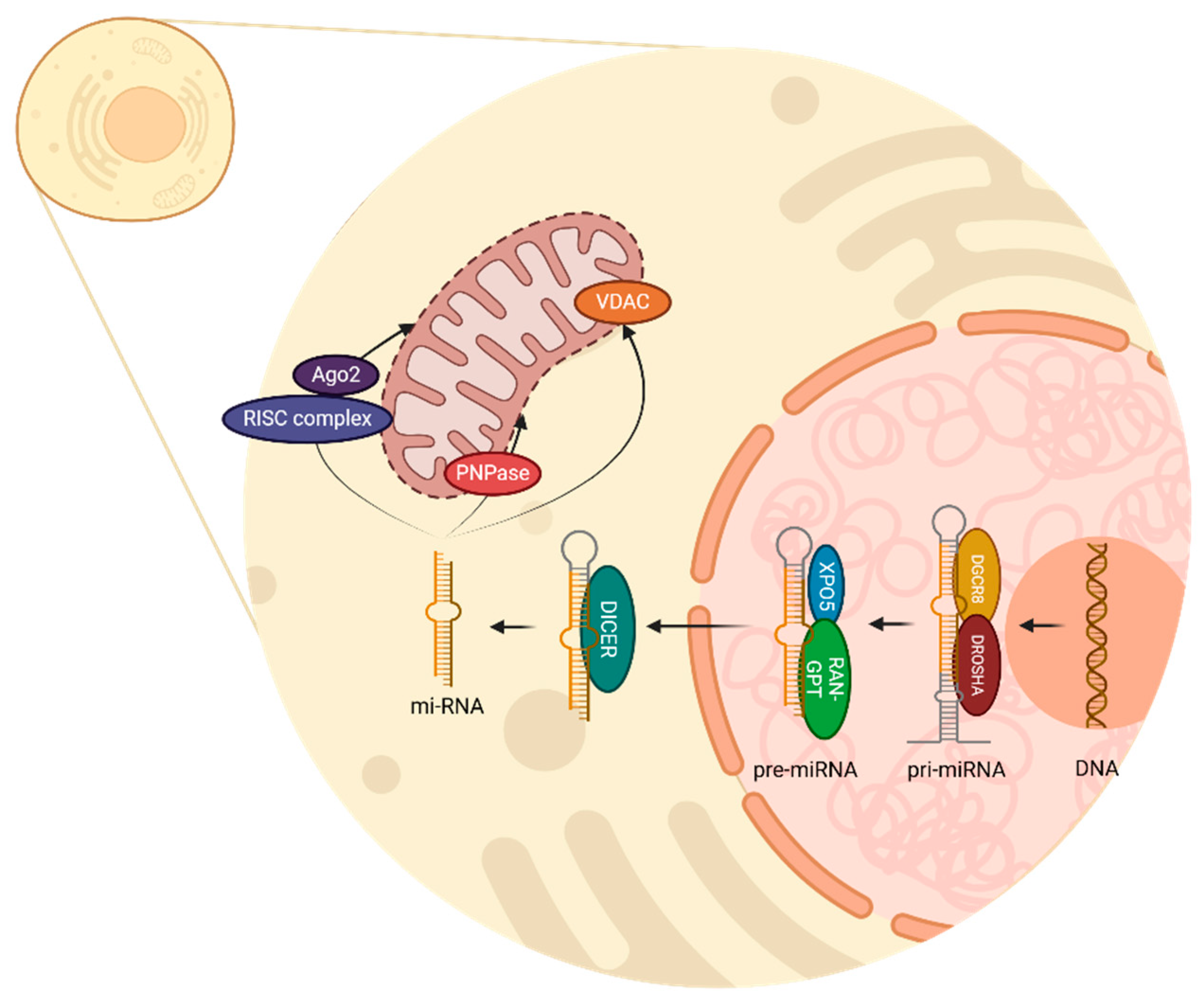
3. The miRNA: Biogenesis in Animal Cells and Its Relationship with Mitochondria

The miRNAs have been described in different species such as viruses, animals, and plants [15]. Nomenclature to classify miRNAs remains undefined; however, it has been described that miRNAs with identical sequences at nucleotides 2–8 of the mature miRNA will appertain to the same family [16].
Furthermore, to maintain a consensus for miRNA characterization, specific criteria are considered in addition to length; there must be a relationship with the Argonaute proteins, the synthesis must be mediated by a ribonuclease (RNase) type III, and there must be phylogenetic conservation, an abundance of sequence reads and the homogeneous presence of the 5’ end [15,16].
Several studies have elucidated these processes by studying various animal species, mainly humans, Caenorhabditis elegans (C. elegans), Drosophila melanogaster, and Giardia intestinalis [15,16]. Moreover, phylogenetic conservation among mammals has been demonstrated [16,17].
However, it is important to mention the differences between miRNA biogenesis in animals and plants. In plants, it has been observed that the processing takes place entirely in the nucleus and unlike in animals, and there is no homolog of Drosha and DiGeorge syndrome critical region gene 8 (DGCR8); therefore, most of the processes are mediated by DICER-LIKE 1, another type III RNase [16]. In this review, we describe miRNA biogenesis in animal cells, focusing on human cells.
miRNA biogenesis begins in the cell nucleus with the transcription of a DNA strand to produce primary miRNA (pri-miRNA) by RNA polymerase II [16]. This primary transcription is mediated by the complex of Drosha and DGCR8, which acts as an enzyme and removes the non-structured endings of the pri-miRNA, therefore creating pre-miRNA as a precursor [16,18]. Drosha is an RNase type III which cuts pri-miRNA; meanwhile, DGCR8 acts as a cofactor through which to recognize and stabilize pri-miRNA structure [19].
Once pre-miRNA is recognized by exportin 5 (XPO5), and Ras-related nuclear protein bound with Guanosine Triphosphate (RAN-GTP) provides the necessary energy, pre-miRNA is transported from the nucleus to the cytoplasm. Then, the RNase type III DICER, recognizes and processes the chain by cutting the hairpin; thus, a double-stranded miRNA of approximately 22 nucleotides is generated [16,18]. This miRNA duplex has two strands, the mature miRNA, which will be the functional guide strand, and the passenger strand, which will be degraded.
In the cytoplasm, miRNA can conjugate with various complexes and regulate important functions. When incorporated into the RNA-induced silencing complex (RISC) and Argonaute 2 (Ago2) protein, miRNA can identify complementary sequences in mRNA and regulate two mechanisms: mRNA degradation and translation inhibition [20]. Additionally, it is known that several miRNAs can be found inside the mitochondria, accomplishing regulatory processes in mitochondrial genes. Polynucleotide phosphorylase (PNPase) facilitates the importation of miRNA into mitochondria, and the voltage-dependent anion channel (VDAC) participates in RNA and protein transportation through the external mitochondrion membrane [21,22]. All these processes are illustrated in Figure 1.
Notably, most of the miRNAs known are synthesized outside the mitochondrion, in other words, they are nuclear miRNAs with targets in the mitochondrion. Nevertheless, the difference in the process of miRNAs synthesized within the mitochondria compared to the process of those encoded in the nucleus remains unclear [23]. Once localized inside the mitochondrion, the presence of miRNAs can modulate its dynamics, which could lead to structural changes, resulting in alterations in fission or fusion, causing damage to the organism [24].

4. Fusion and Fission Mitochondrial Processes

Mitochondria are complex organelles that can fuse and divide to some degree, similarly to bacterial cell division. Under certain conditions, mitochondria fuse specifically depending on the context, microenvironment, and cellular stress [10,25,26]. Golgi apparatus-dependent mitochondrial fusion is achieved through GTPases such as Mitofusin 1 (MFN1) and Mitofusin 2 (MFN2), which coordinate outer mitochondrial membrane fusion. On the other hand, Optic atrophy 1 (OPA1) protein is responsible for mediating inner mitochondrial membrane fusion [9,10,25].
Dysregulation of fusion and fission promotes the survival of malignant cells; dysfunctional mitochondria lead to abnormalities in both mitochondrial morphology and function [25,26]. Fusion processes are particularly important when metabolic demand is high, and therefore mitochondria can fuse and share components to maintain their functionality and energy efficiency [10,26].
The mitochondrial fission machinery relies on dynamin-related protein 1 (DRP1) and its receptors FIS1, MFF (mitochondrial gene-specific protein), and MiD49/51 to preserve mitochondrial morphology and their distribution in the cell matrix [9,10,25]. In cancer cells, aberrant fission is observed, which further promotes the proliferation and survival of malignant neoplastic cells, making it a stress response process [9,25,26].

5. Mitochondrial–Telomere Communications in Cancer

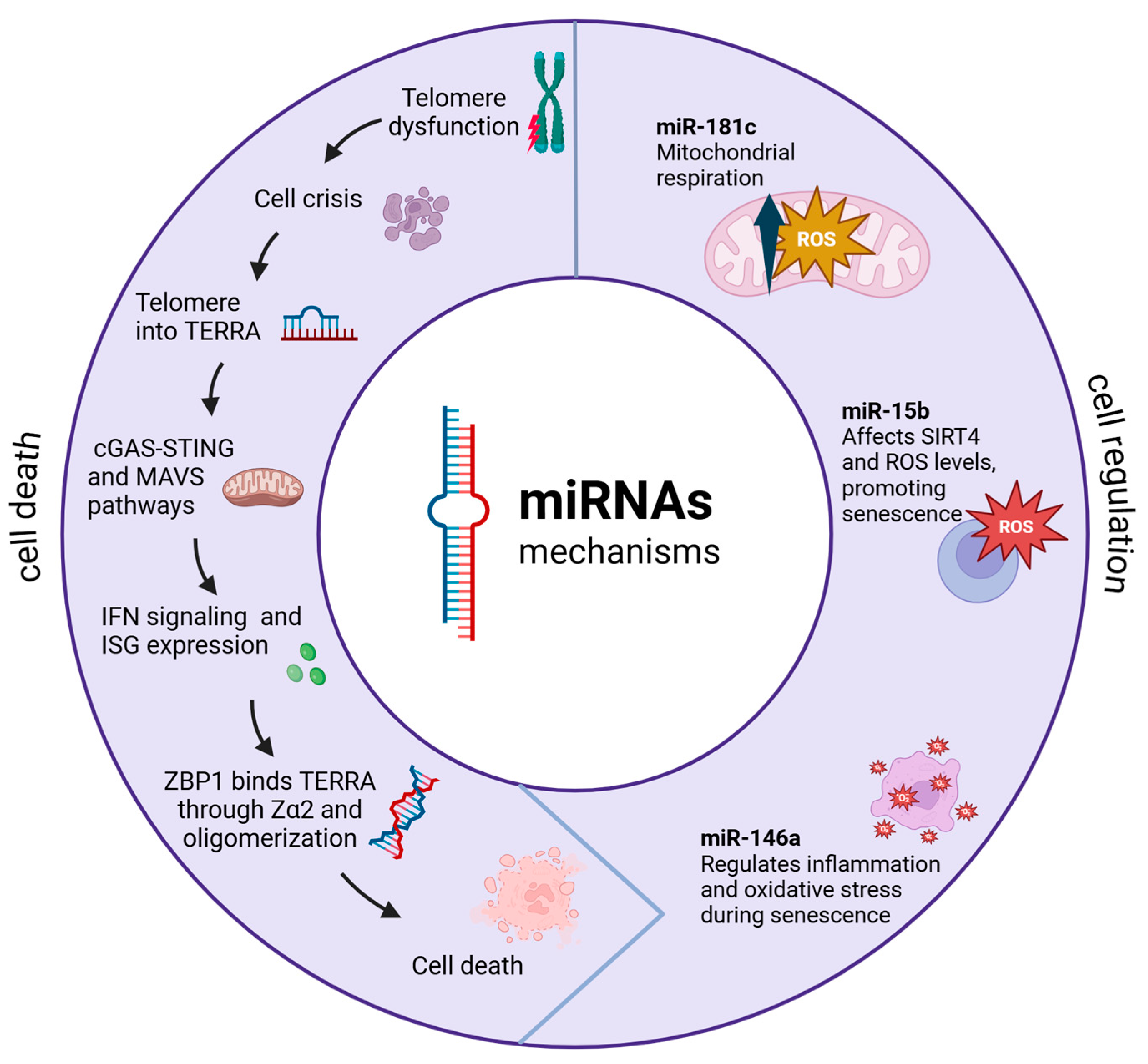
5.1. Molecular Basis of Crosstalk Communication Between the Mitochondria and Telomeres

The bidirectional interactions between mitochondria and telomeres at the molecular level contribute in a fundamental manner to cellular homeostasis and adaptation. This interaction is more complex than previously thought, particularly regarding oxidative stress and cellular signaling pathways [27,28].
A significant breakthrough in understanding this relationship comes from studies showing that mitochondrial H2O2 release and ROS production affect nuclear DNA and telomeres through sophisticated signaling mechanisms rather than direct oxidative damage, challenging previous assumptions about these interactions [27,29].
The telomere–mitochondria axis is particularly relevant in cellular senescence and aging. It has been shown that telomere damage influences mitochondrial function through specific signaling pathways, notably the p53-peroxisome proliferator-activated receptor gamma 1α coactivator (PGC-1α) pathway [1]. Furthermore, Sung et al. have shown that interventions targeting this axis, such as metformin treatment, can influence both telomere stability and mitochondrial function, potentially mitigating cellular senescence [30].
This complex interplay between telomeres and mitochondria represents a critical regulatory mechanism in cellular aging and disease progression, where dysfunction in either component can trigger a cascade of cellular responses affecting both structures [1,27]. Understanding these interactions has important implications for developing targeted therapeutic strategies for age-related diseases and cancer [29,30].

5.2. Mitochondrial–Telomere Communication via Non-Coding RNAs

ncRNAs are key in mediating communication among mitochondria and telomeres. Nassour et al. research has revealed sophisticated mechanisms through which these RNA molecules coordinate various cellular processes, particularly in the context of senescence and cancer [31].
A key discovery involves TERRA, which are long non-coding RNAs (lncRNAs) transcribed from dysfunctional telomeres [32]. These TERRA transcripts have been shown to interact specifically with ZBP1 (Z-DNA binding protein 1) on the outer mitochondria membrane, where they form distinct oligomeric structures [33,34].
Dysfunctional telomeres trigger cellular senescence through the activation of DNA damage response pathways. While senescence is mediated by p53 and retinoblastoma tumor suppressor (RB) pathways, cells with disrupted checkpoints bypass this protective mechanism [35]. Therefore, these cells enter a replicative crisis characterized by transcriptional changes due to an overlap of upregulated genes [31,32]. During crisis, telomeres undergo active transcription, producing TERRA: lncRNAs sequences containing UUAGGG repeats and subtelomeric-derived RNA [32]. TERRA has been involved in the sensing innate immune system pathways through the induction of interferon-stimulated genes (ISGs) [31]. Among ISG products, ZBP1 protein has arisen as a main mediator in cell death regulation and innate immunity through the induction of type I interferons (IFNs).
The synthesis of ZBP1 seems to be linked to the cyclic GMP-AMP synthase/stimulator of interferon genes (cGAS/STING) pathway, which becomes activated upon the cytosolic accumulation of nucleic acids. Furthermore, cGAS/STING-mediated DNA sensing amplifies ZBP1 expression [31,36].
The conformation of the ZBP1–TERRA complex through the Zα2 domain leads to the expression of mitochondrial antiviral signaling protein (MAVS), which leads to the activation of nuclear factor kappa–B (NF-kB) and interferon regulatory factor (IRF) 3/7 signaling 170 pathways, resulting in ISG expression [31,37]. Additionally, the complete activation of IFN-dependent ZBP1 requires the previous upregulation of ZBP1 by cGAS/STING and a signal from dysfunctional telomeres. When these conditions are fulfilled, an inflammatory loop is established, leading to enhanced ISG expression and to cell death [31]. This interaction triggers a cascade of events that can lead to programmed cell death in cancer cells, representing a novel tumor-suppressive mechanism; this mechanism is represented in Figure 2.
In this sense, miRNAs, particularly those targeting mitochondrial functions (mitomiRs), have emerged as important regulators of cellular metabolism and cancer progression. These mitochondria-localized miRNAs can originate from both nuclear and mitochondrial genomes, impacting various aspects of energy metabolism and cellular defense mechanisms [38]. Feng et al. have shown that specific mitomiRs are differentially expressed in various cancer types, affecting both mitochondrial function and the metabolic reprogramming of cancer cells [39].

5.3. Mitochondrial Nuclear-Encoded MitomiRs

mitomiRs act as essential post-transcriptional regulators. Kuo et al. have shown that mitomiRs require specific targeting mechanisms and specialized transport systems for proper mitochondrial localization [40]. Maurya et al. have demonstrated that these regulatory molecules play crucial roles in maintaining mitochondrial function and cellular homeostasis [41].
The regulation of mitochondrial function by nuclear-encoded mitomiRs occurs through multiple pathways. A well-documented example is miR-181c, which has been shown to significantly affect mitochondrial function by modulating ROS synthesis and glucose oxidation patterns in cancer cells. Luo et al. have identified its function as a tumor suppressor and its influence on drug responsiveness, highlighting that processes such as telomere attrition, oxidative stress, activation of tumor-suppressing pathways and mitochondrial impairment serve as primary triggers of cellular senescence [20,42].
Lang et al., in recent research, reported a link between miR-15b and ROS, where the inhibition of miR-15b induces mitochondrial ROS generation and reduces the mitochondrial membrane potential-dependent upregulation of SIRT4 [43]. On the other hand, miR-146a has been shown to have an important role in regulation processes regarding ROS production and inflammation, by interacting with elements of the NF-kB pathway and NADPH oxidase 4 (NOX4), where it functions as an anti-inflammatory and antioxidant, resulting in a decrease in oxidative stress [44].
Disruptions in the transport, assembly, or localization of mitochondrial elements may severely affect cellular homeostasis. For example, in tongue squamous cell carcinoma, an upregulation of miR-2392 has been demonstrated to specifically hinder mtDNA transcription, markedly decreasing the expression of COX1, ND4, and CYTB, ultimately inhibiting OXPHOS [20,45].
Additionally, a decline in miR-107 levels can result in mitochondrial dysfunction, characterized by diminished mitochondrial membrane potential and impaired electron transport chain (ETC) activity. Such alterations may profoundly influence key cellular processes, including resistance to drugs, inflammatory responses, and aging [20].
Alterations in mitomiRs expression are closely linked to cancer metabolism and mitochondrial stress signaling. The latest evidence, according to Maurya et al., demonstrates that tumor cells process mitochondrial ROS regulation to interact with various components in the tumor microenvironment, thereby affecting cancer progression [41]. Furthermore, Dasgupta et al. investigated a novel regulatory mechanism in mtDNA-less cells. Their analysis revealed a reduced expression of several mitochondrial RNAs, including COX1-3, 12S rRNA, and ND4-5. In contrast, Cyto b, ND1 and ND3 were entirely absent in 206 ρ° cells. This imbalance in mitochondrial RNA transcription indicates that post-transcriptional cleavage and processing mechanisms may be involved in controlling mitochondrial gene expression within these cells [46].
By partially base pairing with complementary sites in the cytosol, miRNAs are enabled to regulate mitochondrial RNA expression by aiming for specific mRNAs. This regulation of mitochondrial gene expression has significant implications for cellular energy metabolism [46,47].

5.4. Mitochondria-Encoded miRNAs

Mitochondrial-encoded miRNAs have recently begun to be studied, and while not much is known, it is understood that they are encoded in mtDNA and are processed by fine and specific machinery within the mitochondria [9]. These unique miRNAs have been shown to have distinct processing mechanisms that differ from their nuclear counterparts, suggesting specialized regulatory functions [20].
Initially, their existence was doubted; however, several studies have confirmed not only their existence but also their crucial participation in cellular metabolism and mitochondria–nucleus communication [46]. Yumeng et al. have emphasized the critical role of miRNAs in regulating both nuclear and mitochondrial proteins, thereby establishing a complex network of cellular regulations. Notably, abnormal methylation of mitochondrial RNA can impact mtDNA function by altering transcription stability and structure. Ribosomal RNA (rRNA) methylation is primarily regulated by mitochondrial rRNA methyltransferases, which include enzymes targeting both large (MRM1, MRM2, MRM3, TRMT61B) and small (TFB1M, TRMT2B, NSUN4, METTL15) ribosomal subunits [48,49].
Their processing is not traditional like nuclear miRNAs, suggesting a divergent evolution of these processing mechanisms. This evolutionary adaptation appears specifically aimed at regulating mitochondrial processes and facilitating communication with the nucleus [9,48]. Luo et al. have shown that these mitochondrial-specific miRNAs play crucial roles in maintaining cellular homeostasis and energy metabolism [20].

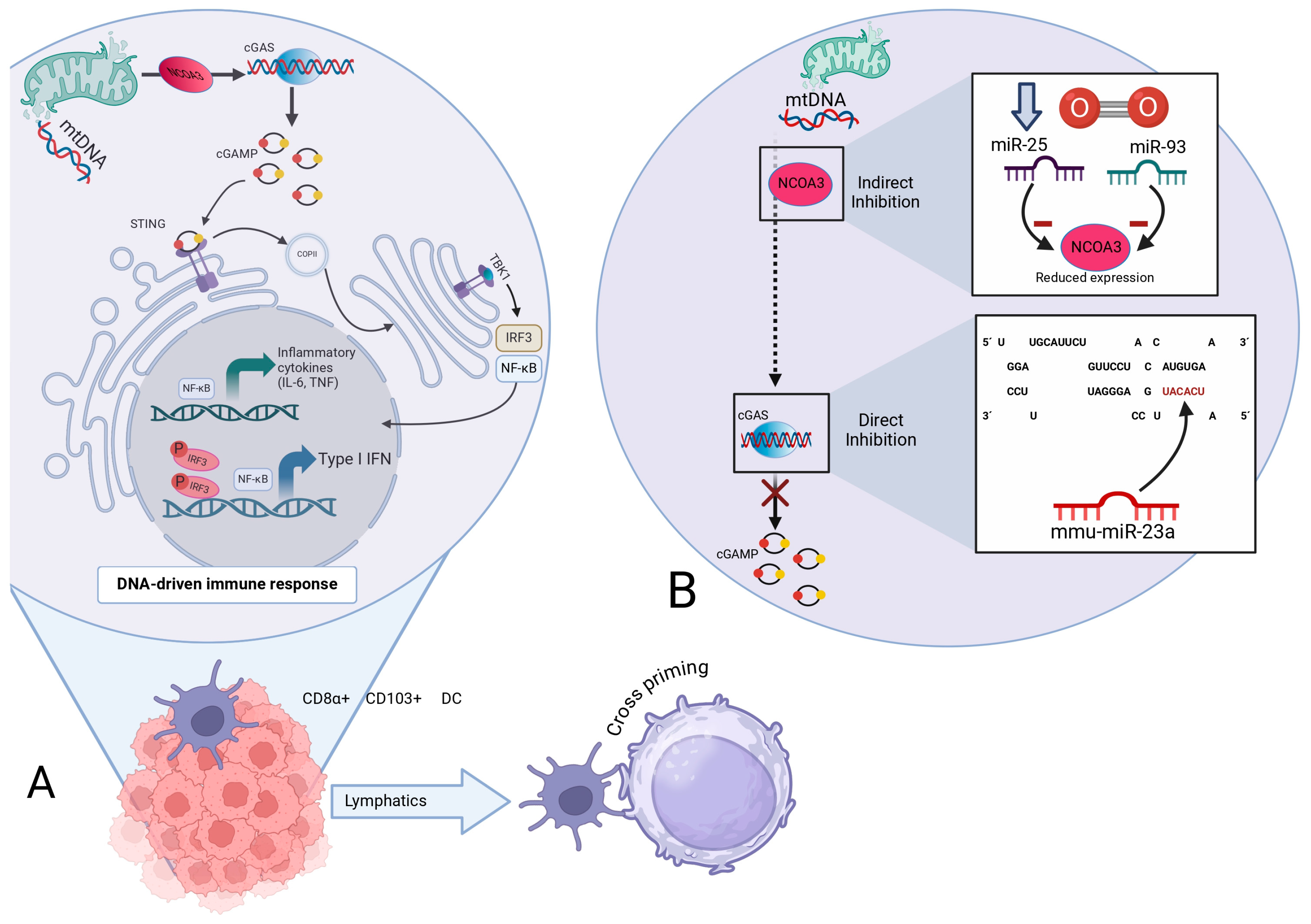
5.5. RNA-Dependent Modulation of the cGAS/STING Axis: Convergence of Mitochondrial Dynamics, Telomeric Integrity, and Programmed Cell Death Pathways

Pattern recognition receptors (PRRs) are of great importance in the innate immune system. PRRs recognize pathogen-associated molecular patterns (PAMPs) and host damage-associated molecular patterns (DAMPs) on the surface of innate immune cells [50].
The PRRs that have been mainly related to the cGAS/STING pathways are AIM2-like receptors (ALRs) and 2’-5’oligoadenylate synthetase-like receptors (OLRs) [51]. Once DNA is detected in the cell, ALR activates the STING-dependent ISG pathway. This mechanism was originally studied in antiviral responses [51,52]. OLRs, when activated in the cytoplasm by double-stranded nucleic acids, generate second messenger molecules like cGAMP. These molecules bind to and activate STING, triggering a downstream innate immune response [53].
cGAS activation enables the synthesis of cGAMP from GTP and ATP, which then binds to STING dimers localized in the endoplasmic reticulum (ER) membrane, resulting in their incorporation into coatomer protein complex II (COPII); then, it recruits and promotes the autophosphorylation of TANK-binding kinase 1 (TBK1), STING phosphorylation at Ser366, and the recruitment and further phosphorylation of IRF3, which is required for the induction of genes encoding IL-6 and IL-12. IRF3 dimerizes and translocates to the nucleus, ultimately leading to the expression of type I IFN and ISGs, boosting the cross-presentation of antigens to CD8+ T cells [54,55].
It was demonstrated that mitochondria experiencing minority mitochondrial outer-membrane permeability (miMOP) release mtDNA into the cytosol [56]. The release of mtDNA into the cell triggers the mtDNA-dependent cGAS/STING pathway, leading to the synthesis of type I IFN and inflammatory markers, which leads to the senescence-associated secretory phenotype (SASP) and inflammation [55,56].
It can be inferred that mtDNA-dependent cGAS/STING signaling is of great importance not only in senescence but also in certain illnesses such as cancer, where it has been shown to be key for antitumor immunity. STING activation is believed to promote tumor rejection by eliciting CD8+ T cell responses [57].
It promotes the enlistment of T cells and natural killer (NK) cells, into the malignancy microenvironment. Additionally, activation of the cGAS/STING signaling helps suppress the tumor metastasis capacity; as these cells attempt to spread, active STING signaling enhances immune recognition and destruction, thereby restricting the formation of new tumors [57,58,59].
miRNAs play an important role in regulating the cGAS/STING pathways. The 3’UTRs of cGAS/STING mRNA carry probable miRNA binding sites [51]. Yu et al. have stated that miRNAs can suppress the immune response through different mechanisms. For instance, certain miRNAs, such as miR-23a/b, directly bind to the 3’ UTR of cGAS, inhibiting its expression and thereby suppressing the cGAS-mediated innate immune response [60]. Indirectly, during hypoxia, miR-25 and miR-93 regulate cGAS expression by targeting NCOA3, a crucial epigenetic factor for maintaining cGAS expression levels. This regulation results in the downregulation of cGAS mRNA levels, facilitating hypoxic tumor cells to evade immune detection by modulating the cGAS/STING pathway [61], Figure 3.

6. Metabolic Reprogramming in Cancer

Malignant cells are profoundly reprogrammed in terms of energy metabolism; these changes are linked to and affect telomere and mitochondrial function. Aerobic glycolysis is prominently used by tumor cells; this has been called the Warburg effect [62]. Cancer cells maintain a balance between aerobic glycolysis, which involves glucose fermentation even in the presence of oxygen, and OXPHOS, which produces ATP through the oxidation of glucose carbon bridges [63].
The balance in energy acquisition is directed towards the prolonged cellular survival characteristics of certain malignant tumor clones. This type of mixed metabolism favors the maintenance of telomere integrity, characteristic of cancer cells [64]. Thus, this type of cancer cell metabolism promotes proliferation and telomere length maintenance through the modification of the TCA cycle, amino acid metabolism, and fatty acid oxidation [64].
The AMP-activated protein kinase (AMPK)–mammalian target of rapamycin (mTOR) axis regulates cellular energy metabolism, mitochondrial function, and telomere maintenance. This pathway is tightly dysregulated in cancer cells; therefore, these changes support both metabolism and cell survival [65,66]. Also, sirtuins (SIRT), particularly SIRT1 and SIRT3, act as metabolism-associated sensors that regulate mitochondria and telomeres in cellular stress responses and cancer cell adaptation to specific contexts [67].
Central to cellular metabolism regulation is the mTOR pathway, which acts aberrantly in cancer cells and has multiple effects on cellular metabolism. Indeed, mTOR and AMPK are regarded as metabolic sensors that relay cell survival signals under stress, and such crosstalk with mitochondrial function and telomere maintenance is essential for promoting cancer [68,69]. This means that cancer cell metabolic plasticity, mitochondrial and telomere structure and function changes play an important role in the survival of neoplastic cells and their proliferation [65].
The SIRT system modulates mitochondrial and telomere physiology through interaction with multiple molecules via histone protein deacetylation and the regulation of mitochondrial function and apoptosis. These proteins regulate cell proliferation and differentiation through various catalytic activities that depend on NAD+. Additionally, due to their deacetylase activity, they interact closely with telomeres, preventing their erosion and supporting their repair. Therefore, the processes of oncogenesis and tumor progression are closely linked to these axes and metabolic pathways related to AMPK, mTOR, and SIRTs, which in turn are precisely regulated by the large family of miRNAs. The complexity of these systems is very high, and their effects are dependent on the cellular microenvironment. Energy metabolism is strongly linked to the regulation of these highly specific molecular systems, with pleiotropic functions in most organs and systems, both under healthy and diseased conditions [70,71].
In hepatocellular carcinoma (HCC), SIRT1 is overexpressed and correlates with tumor grade, predicting adverse clinical outcomes and poor prognosis. miR-34a, decreased in severe HCC cases, has been shown, in mice, to induce tumor regression and prevent recurrence when expressed. Similarly, miRNA-29c acts as a tumor suppressor by inhibiting SIRT1 in HCC [72,73]. However, SIRTs also have antitumoral effects or dual effects (pro- and anti-tumoral) that can be context-dependent. For example, SIRT1 is associated with protective effects against tumor growth and promotes the cell death of tumor cells. It inhibits the proliferation of various types of cancer, such as ovarian, lung, and colorectal cancer [74].
Regarding the link between miRNA activity and metabolic reprogramming, it is closely associated with cancer cell transformation. The miR–200c–SIRT2 interaction axis plays a crucial role in establishing the metabolic shift known as the Warburg effect by regulating glycolytic enzymes [75].
In summary, metabolic reprogramming in cancer is regulated by miRNAs, which in turn regulate the canonical metabolic pathways of malignant transformation and progression such as mTOR, AMPK, and SIRT. These pathways reversely modulate cell cycle and fate, impacting mitochondrial structure and function and telomeric maintenance, confirming that the elucidation of these processes intrinsically involves investigating cancer reprogramming or the Warburg effect, demonstrating that the mitochondria-telomere axis is fundamental in this regulation, with miRNAs serving as the way that all these processes are fine-tuned.

7. Clinical Implications and Therapeutic Applications in Cancer

The clinical applications of miRNA appear to cover a vast range, and yet are largely unexplored. miRNAs play crucial roles in various diseases. Additionally, numerous miRNAs have been associated in degenerative diseases and cellular senescence, including increased expression of miR-34a, miR-21-22, miR-26b, miR-29 and miR-2010, as well as decreased expression of miR-106a, miR-19b, miR-20a and miR-320c [76].
Specifically, in the field of cancer research, miRNAs have garnered significant attention for their potential diagnostic, prognostic, and therapeutic applications.
The relationship between mitochondria and telomeres and their connection to senescence and malignant transformation processes represent opportunities for the diagnosis and monitoring of multiple diseases, especially oncological diseases. RNA species associated with oncological processes and cellular senescence can be useful as diagnostic markers and for monitoring disease progression, potentially resulting in increased specificity to traditional oncological markers [77,78].
In this sense, miRNAs are known to interact with several pathological conditions; high miR-181c expression is associated with mitochondrial dysfunction, and differences in miR-138 levels are associated with telomere abnormalities [76]. miRNAs, as an entire family of transcriptional regulators and molecular receptors, are likely to be useful in clinical assessments for the identification of improved subtypes and prognostication of oncological diseases [78].
In this regard, these molecules can also be used in RNA-based therapy, which can be directed at specific therapeutic targets and be used as regulators of mitochondria and telomeres. This represents a new frontier in personalized medicine, as miRNAs can be used to regulate metabolic pathways that can be utilized according to the context of the tumor type and the pathways that are altered [76,78].
The goal is to restore cellular functions, specifically mitochondria and telomeres, through synthetic RNA or regulatory molecules of these RNAs. In the case of miR-34a, mimicking molecules can be developed for the treatment of certain neoplasms to regulate mitochondrial functions and telomere maintenance, thereby restoring the homeostasis of both organelles [79,80].

8. Current Challenges and Future Directions

Developing miRNA-based therapeutics is challenging and needs to address the intrinsic stability of miRNA-based therapeutic agents and their delivery to the cellular compartments in which they are needed to modulate cellular functions. Nanoparticle delivery and other cell-specific transport methods have been applied for reduce side effects and enhance therapeutic efficacy [81,82].
Basic and clinical research is required to understand the complex network of molecular interactions of miRNAs with other RNA species. Bioinformatics and the development of highly complex and sophisticated computational models can shed light on the network of interactions between RNA species, telomeres, and mitochondria, thereby enabling the development of personalized therapies [83,84].
Moreover, integrating artificial intelligence methods and machine learning can lead to precise predictions and characterizations of different types of RNA and their relationship with mitochondrial and telomeric function and structure [85,86].

Author Contributions

Conceptualization, J.A.C.-R.; methodology, J.A.C.-R.; software, B.A.L.-C.; investigation, B.A.L.-C. and M.Á.D.-M.; resources, E.d.l.M.-J. and J.A.C.-R.; data curation, B.A.L.-C.; writing—original draft preparation, J.A.C.-R. and E.d.l.M.-J.; writing—review and editing, B.A.L.-C., E.d.l.M.-J. and M.Á.D.-M.; supervision, E.d.l.M.-J.; funding acquisition, E.d.l.M.-J. All authors have read and agreed to the published version of the manuscript.

Funding

The APC was financed through the APPAC-VII 2025.

Acknowledgments

We thank Dulce Lizbeth Valdez Sánchez, student of Universidad de Guadalajara, for the editing of this manuscript and for the provision of the figures.

Conflicts of Interest

The authors declare no conflicts of interest.

References

  1. Gao, X.; Yu, X.; Zhang, C.; Wang, Y.; Sun, Y.; Sun, H.; Zhang, H.; Shi, Y.; He, X. Telomeres and Mitochondrial Metabolism: Implications for Cellular Senescence and Age-Related Diseases. Stem Cell Rev. Rep. 2022, 18, 2315–2327. [Google Scholar] [CrossRef] [PubMed]
  2. Assalve, G.; Lunetti, P.; Rocca, M.S.; Cosci, I.; Di Nisio, A.; Ferlin, A.; Zara, V.; Ferramosca, A. Exploring the Link Between Telomeres and Mitochondria: Mechanisms and Implications in Different Cell Types. Int. J. Mol. Sci. 2025, 26, 993. [Google Scholar] [CrossRef] [PubMed]
  3. Cusanelli, E.; Chartrand, P. Telomeric Repeat-Containing RNA TERRA: A Noncoding RNA Connecting Telomere Biology to Genome Integrity. Front. Genet. 2015, 6, 143. [Google Scholar] [CrossRef]
  4. Brosnan, C.A.; Voinnet, O. The Long and the Short of Noncoding RNAs. Curr. Opin. Cell Biol. 2009, 21, 416–425. [Google Scholar] [CrossRef]
  5. Slack, F.J.; Chinnaiyan, A.M. The Role of Non-Coding RNAs in Oncology. Cell 2019, 179, 1033–1055. [Google Scholar] [CrossRef]
  6. Khan, A.Q.; Ahmed, E.I.; Elareer, N.R.; Junejo, K.; Steinhoff, M.; Uddin, S. Role of miRNA-Regulated Cancer Stem Cells in the Pathogenesis of Human Malignancies. Cells 2019, 8, 840. [Google Scholar] [CrossRef]
  7. Lehmann, B.D.; Jovanović, B.; Chen, X.; Estrada, M.V.; Johnson, K.N.; Shyr, Y.; Moses, H.L.; Sanders, M.E.; Pietenpol, J.A. Refinement of Triple-Negative Breast Cancer Molecular Subtypes: Implications for Neoadjuvant Chemotherapy Selection. PLoS ONE 2016, 11, e0157368. [Google Scholar] [CrossRef]
  8. Lehmann, B.D.; Pietenpol, J.A.; Tan, A.R. Triple-Negative Breast Cancer: Molecular Subtypes and New Targets for Therapy. Am. Soc. Clin. Oncol. Educ. Book 2015, 35, e31–e39. [Google Scholar] [CrossRef]
  9. Zong, Y.; Li, H.; Liao, P.; Chen, L.; Pan, Y.; Zheng, Y.; Zhang, C.; Liu, D.; Zheng, M.; Gao, J. Mitochondrial Dysfunction: Mechanisms and Advances in Therapy. Sig Transduct. Target. Ther. 2024, 9, 124. [Google Scholar] [CrossRef]
  10. Chen, W.; Zhao, H.; Li, Y. Mitochondrial Dynamics in Health and Disease: Mechanisms and Potential Targets. Signal Transduct. Target. Ther. 2023, 8, 333. [Google Scholar] [CrossRef]
  11. Cai, X.; Ng, C.P.; Jones, O.; Fung, T.S.; Ryu, K.W.; Li, D.; Thompson, C.B. Lactate Activates the Mitochondrial Electron Transport Chain Independently of Its Metabolism. Mol. Cell 2023, 83, 3904–3920.e7. [Google Scholar] [CrossRef] [PubMed]
  12. Gustafsson, C.M.; Falkenberg, M.; Larsson, N.-G. Maintenance and Expression of Mammalian Mitochondrial DNA. Annu. Rev. Biochem. 2016, 85, 133–160. [Google Scholar] [CrossRef] [PubMed]
  13. Yang, Y.; An, Y.; Ren, M.; Wang, H.; Bai, J.; Du, W.; Kong, D. The Mechanisms of Action of Mitochondrial Targeting Agents in Cancer: Inhibiting Oxidative Phosphorylation and Inducing Apoptosis. Front. Pharmacol. 2023, 14, 1243613. [Google Scholar] [CrossRef]
  14. Zou, W.; Rohatgi, N.; Brestoff, J.R.; Li, Y.; Barve, R.A.; Tycksen, E.; Kim, Y.; Silva, M.J.; Teitelbaum, S.L. Ablation of Fat Cells in Adult Mice Induces Massive Bone Gain. Cell Metab. 2020, 32, 801–813.e6. [Google Scholar] [CrossRef]
  15. Griffiths-Jones, S.; Saini, H.K.; van Dongen, S.; Enright, A.J. miRBase: Tools for microRNA Genomics. Nucleic Acids Res. 2008, 36, D154–D158. [Google Scholar] [CrossRef]
  16. Ha, M.; Kim, V.N. Regulation of microRNA Biogenesis. Nat. Rev. Mol. Cell Biol. 2014, 15, 509–524. [Google Scholar] [CrossRef]
  17. Chiang, H.R.; Schoenfeld, L.W.; Ruby, J.G.; Auyeung, V.C.; Spies, N.; Baek, D.; Johnston, W.K.; Russ, C.; Luo, S.; Babiarz, J.E.; et al. Mammalian microRNAs: Experimental Evaluation of Novel and Previously Annotated Genes. Genes. Dev. 2010, 24, 992–1009. [Google Scholar] [CrossRef]
  18. O’Brien, J.; Hayder, H.; Zayed, Y.; Peng, C. Overview of MicroRNA Biogenesis, Mechanisms of Actions, and Circulation. Front. Endocrinol. 2018, 9, 402. [Google Scholar] [CrossRef]
  19. Han, J.; Lee, Y.; Yeom, K.-H.; Kim, Y.-K.; Jin, H.; Kim, V.N. The Drosha-DGCR8 Complex in Primary microRNA Processing. Genes. Dev. 2004, 18, 3016–3027. [Google Scholar] [CrossRef]
  20. Luo, L.; An, X.; Xiao, Y.; Sun, X.; Li, S.; Wang, Y.; Sun, W.; Yu, D. Mitochondrial-Related microRNAs and Their Roles in Cellular Senescence. Front. Physiol. 2024, 14, 1279548. [Google Scholar] [CrossRef]
  21. Shoshan-Barmatz, V.; De Pinto, V.; Zweckstetter, M.; Raviv, Z.; Keinan, N.; Arbel, N. VDAC, a Multi-Functional Mitochondrial Protein Regulating Cell Life and Death. Mol. Asp. Med. 2010, 31, 227–285. [Google Scholar] [CrossRef] [PubMed]
  22. Wang, G.; Chen, H.-W.; Oktay, Y.; Zhang, J.; Allen, E.L.; Smith, G.M.; Fan, K.C.; Hong, J.S.; French, S.W.; McCaffery, J.M.; et al. PNPASE Regulates RNA Import into Mitochondria. Cell 2010, 142, 456–467. [Google Scholar] [CrossRef] [PubMed]
  23. Kuthethur, R.; Shukla, V.; Mallya, S.; Adiga, D.; Kabekkodu, S.P.; Ramachandra, L.; Saxena, P.U.P.; Satyamoorthy, K.; Chakrabarty, S. Expression Analysis and Function of Mitochondrial Genome-Encoded microRNAs. J. Cell Sci. 2022, 135, jcs258937. [Google Scholar] [CrossRef]
  24. Li, P.; Jiao, J.; Gao, G.; Prabhakar, B.S. Control of Mitochondrial Activity by miRNAs. J. Cell Biochem. 2012, 113, 1104–1110. [Google Scholar] [CrossRef]
  25. Wu, Z.; Xiao, C.; Long, J.; Huang, W.; You, F.; Li, X. Mitochondrial Dynamics and Colorectal Cancer Biology: Mechanisms and Potential Targets. Cell Commun. Signal 2024, 22, 91. [Google Scholar] [CrossRef]
  26. Darvin, P.; Sasidharan Nair, V. Editorial: Understanding Mitochondrial Dynamics and Metabolic Plasticity in Cancer Stem Cells: Recent Advances in Cancer Treatment and Potential Therapeutic Approaches. Front. Oncol. 2023, 13, 1155774. [Google Scholar] [CrossRef]
  27. van Soest, D.M.K.; Polderman, P.E.; den Toom, W.T.F.; Keijer, J.P.; van Roosmalen, M.J.; Leyten, T.M.F.; Lehmann, J.; Zwakenberg, S.; De Henau, S.; van Boxtel, R.; et al. Mitochondrial H2O2 Release Does Not Directly Cause Damage to Chromosomal DNA. Nat. Commun. 2024, 15, 2725. [Google Scholar] [CrossRef]
  28. Jagaraj, C.J.; Shadfar, S.; Kashani, S.A.; Saravanabavan, S.; Farzana, F.; Atkin, J.D. Molecular Hallmarks of Ageing in Amyotrophic Lateral Sclerosis. Cell. Mol. Life Sci. 2024, 81, 111. [Google Scholar] [CrossRef]
  29. Zeinoun, B.; Teixeira, M.T.; Barascu, A. Hog1 Acts in a Mec1-Independent Manner to Counteract Oxidative Stress Following Telomerase Inactivation in Saccharomyces Cerevisiae. Commun. Biol. 2024, 7, 761. [Google Scholar] [CrossRef]
  30. Sung, J.Y.; Kim, S.G.; Park, S.-Y.; Kim, J.-R.; Choi, H.C. Telomere Stabilization by Metformin Mitigates the Progression of Atherosclerosis via the AMPK-Dependent p-PGC-1α Pathway. Exp. Mol. Med. 2024, 56, 1967–1979. [Google Scholar] [CrossRef]
  31. Nassour, J.; Aguiar, L.G.; Correia, A.; Schmidt, T.T.; Mainz, L.; Przetocka, S.; Haggblom, C.; Tadepalle, N.; Williams, A.; Shokhirev, M.N.; et al. Telomere-to-Mitochondria Signalling by ZBP1 Mediates Replicative Crisis. Nature 2023, 614, 767–773. [Google Scholar] [CrossRef] [PubMed]
  32. Azzalin, C.M.; Reichenbach, P.; Khoriauli, L.; Giulotto, E.; Lingner, J. Telomeric Repeat–Containing RNA and RNA Surveillance Factors at Mammalian Chromosome Ends. Science 2007, 318, 798–801. [Google Scholar] [CrossRef] [PubMed]
  33. Strzyz, P. From Shortening Telomeres to Replicative Crisis. Nat. Rev. Mol. Cell Biol. 2023, 24, 239. [Google Scholar] [CrossRef]
  34. Gaela, V.M.; Chen, L.-Y. Ends End It via Mitochondria: A Telomere-Dependent Tumor Suppressive Mechanism Acts during Replicative Crisis. Mol. Cell 2023, 83, 1027–1029. [Google Scholar] [CrossRef]
  35. Nassour, J.; Radford, R.; Correia, A.; Fusté, J.M.; Schoell, B.; Jauch, A.; Shaw, R.J.; Karlseder, J. Autophagic Cell Death Restricts Chromosomal Instability during Replicative Crisis. Nature 2019, 565, 659–663. [Google Scholar] [CrossRef]
  36. Zhan, J.; Wang, J.; Liang, Y.; Wang, L.; Huang, L.; Liu, S.; Zeng, X.; Zeng, E.; Wang, H. Apoptosis Dysfunction: Unravelling the Interplay between ZBP1 Activation and Viral Invasion in Innate Immune Responses. Cell Commun. Signal. 2024, 22, 149. [Google Scholar] [CrossRef]
  37. Chen, Y.; Shi, Y.; Wu, J.; Qi, N. MAVS: A Two-Sided CARD Mediating Antiviral Innate Immune Signaling and Regulating Immune Homeostasis. Front. Microbiol. 2021, 12, 744348. [Google Scholar] [CrossRef]
  38. Wallace, L.; Aikhionbare, K.; Banerjee, S.; Peagler, K.; Pitts, M.; Yao, X.; Aikhionbare, F. Differential Expression Profiles of Mitogenome Associated MicroRNAs Among Colorectal Adenomatous Polyps. Cancer Res. J. 2021, 9, 23–33. [Google Scholar] [CrossRef]
  39. Feng, Y.; Huang, W.; Paul, C.; Liu, X.; Sadayappan, S.; Wang, Y.; Pauklin, S. Mitochondrial Nucleoid in Cardiac Homeostasis: Bidirectional Signaling of Mitochondria and Nucleus in Cardiac Diseases. Basic. Res. Cardiol. 2021, 116, 49. [Google Scholar] [CrossRef]
  40. Kuo, C.-L.; Lin, Y.-C.; Lo, Y.K.; Lu, Y.-Z.; Babuharisankar, A.P.; Lien, H.-W.; Chou, H.-Y.; Lee, A.Y.-L. The Mitochondrial Stress Signaling Tunes Immunity from a View of Systemic Tumor Microenvironment and Ecosystem. iScience 2024, 27, 110710. [Google Scholar] [CrossRef]
  41. Maurya, A.K.; Rabina, P.; Kumar, V.B.S. In-Silico Analysis of Important Mitochondrial microRNAs and Their Differential Expression in Mitochondria. bioRxiv 2024. [Google Scholar] [CrossRef]
  42. Liu, R.-M. Aging, Cellular Senescence, and Alzheimer’s Disease. Int. J. Mol. Sci. 2022, 23, 1989. [Google Scholar] [CrossRef] [PubMed]
  43. Lang, A.; Grether-Beck, S.; Singh, M.; Kuck, F.; Jakob, S.; Kefalas, A.; Altinoluk-Hambüchen, S.; Graffmann, N.; Schneider, M.; Lindecke, A.; et al. MicroRNA-15b Regulates Mitochondrial ROS Production and the Senescence-Associated Secretory Phenotype through Sirtuin 4/SIRT4. Aging 2016, 8, 484–505. [Google Scholar] [CrossRef] [PubMed]
  44. Zhang, L.; Wang, I.C.; Meng, S.; Xu, J. miR-146a Decreases Inflammation and ROS Production in Aged Dermal Fibroblasts. Int. J. Mol. Sci. 2024, 25, 6821. [Google Scholar] [CrossRef]
  45. Fan, S.; Tian, T.; Chen, W.; Lv, X.; Lei, X.; Zhang, H.; Sun, S.; Cai, L.; Pan, G.; He, L.; et al. Mitochondrial miRNA Determines Chemoresistance by Reprogramming Metabolism and Regulating Mitochondrial Transcription. Cancer Res. 2019, 79, 1069–1084. [Google Scholar] [CrossRef]
  46. Dasgupta, N.; Peng, Y.; Tan, Z.; Ciraolo, G.; Wang, D.; Li, R. miRNAs in mtDNA-Less Cell Mitochondria. Cell Death Discov. 2015, 1, 15004. [Google Scholar] [CrossRef]
  47. Das, S.; Ferlito, M.; Kent, O.A.; Fox-Talbot, K.; Wang, R.; Liu, D.; Raghavachari, N.; Yang, Y.; Wheelan, S.J.; Murphy, E.; et al. Nuclear miRNA Regulates the Mitochondrial Genome in the Heart. Circ. Res. 2012, 110, 1596–1603. [Google Scholar] [CrossRef]
  48. Lin, Y.; Yang, B.; Huang, Y.; Zhang, Y.; Jiang, Y.; Ma, L.; Shen, Y.-Q. Mitochondrial DNA-Targeted Therapy: A Novel Approach to Combat Cancer. Cell Insight 2023, 2, 100113. [Google Scholar] [CrossRef]
  49. Lopez Sanchez, M.I.G.; Cipullo, M.; Gopalakrishna, S.; Khawaja, A.; Rorbach, J. Methylation of Ribosomal RNA: A Mitochondrial Perspective. Front. Genet. 2020, 11, 761. [Google Scholar] [CrossRef]
  50. Qin, X.-W.; He, J.; Yu, Y.; Liu, C.; Luo, Z.-Y.; Li, Z.-M.; Weng, S.-P.; Guo, C.-J.; He, J.-G. The Roles of Mandarin Fish STING in Innate Immune Defense against Infectious Spleen and Kidney Necrosis Virus Infections. Fish. Shellfish. Immunol. 2020, 100, 80–89. [Google Scholar] [CrossRef]
  51. Gareev, I.; de Jesus Encarnacion Ramirez, M.; Goncharov, E.; Ivliev, D.; Shumadalova, A.; Ilyasova, T.; Wang, C. MiRNAs and lncRNAs in the Regulation of Innate Immune Signaling. Noncoding RNA Res. 2023, 8, 534–541. [Google Scholar] [CrossRef] [PubMed]
  52. Gray, E.E.; Winship, D.; Snyder, J.M.; Child, S.J.; Geballe, A.P.; Stetson, D.B. The AIM2-like Receptors Are Dispensable for the Interferon Response to Intracellular DNA. Immunity 2016, 45, 255–266. [Google Scholar] [CrossRef] [PubMed]
  53. Hornung, V.; Hartmann, R.; Ablasser, A.; Hopfner, K.-P. OAS Proteins and cGAS: Unifying Concepts in Sensing and Responding to Cytosolic Nucleic Acids. Nat. Rev. Immunol. 2014, 14, 521–528. [Google Scholar] [CrossRef] [PubMed]
  54. Sun, L.; Wu, J.; Du, F.; Chen, X.; Chen, Z.J. Cyclic GMP-AMP Synthase Is a Cytosolic DNA Sensor That Activates the Type I Interferon Pathway. Science 2013, 339, 786–791. [Google Scholar] [CrossRef]
  55. Decout, A.; Katz, J.D.; Venkatraman, S.; Ablasser, A. The cGAS–STING Pathway as a Therapeutic Target in Inflammatory Diseases. Nat. Rev. Immunol. 2021, 21, 548–569. [Google Scholar] [CrossRef]
  56. Izquierdo, J.M. Mitochondria-cGAS-STING Axis Is a Potential Therapeutic Target for Senescence-Dependent Inflammaging-Associated Neurodegeneration. Neural Regen. Res. 2025, 20, 805–807. [Google Scholar] [CrossRef]
  57. He, X.; Wedn, A.; Wang, J.; Gu, Y.; Liu, H.; Zhang, J.; Lin, Z.; Zhou, R.; Pang, X.; Cui, Y. IUPHAR ECR Review: The cGAS-STING Pathway: Novel Functions beyond Innate Immune and Emerging Therapeutic Opportunities. Pharmacol. Res. 2024, 201, 107063. [Google Scholar] [CrossRef]
  58. Nicolai, C.J.; Wolf, N.; Chang, I.-C.; Kirn, G.; Marcus, A.; Ndubaku, C.O.; McWhirter, S.M.; Raulet, D.H. NK Cells Mediate Clearance of CD8+ T Cell-Resistant Tumors in Response to STING Agonists. Sci. Immunol. 2020, 5, eaaz2738. [Google Scholar] [CrossRef]
  59. Nakamura, T.; Miyabe, H.; Hyodo, M.; Sato, Y.; Hayakawa, Y.; Harashima, H. Liposomes Loaded with a STING Pathway Ligand, Cyclic Di-GMP, Enhance Cancer Immunotherapy against Metastatic Melanoma. J. Control. Release 2015, 216, 149–157. [Google Scholar] [CrossRef]
  60. Yu, Q.; Chu, L.; Li, Y.; Wang, Q.; Zhu, J.; Wang, C.; Cui, S. miR-23a/b Suppress cGAS-Mediated Innate and Autoimmunity. Cell Mol. Immunol. 2021, 18, 1235–1248. [Google Scholar] [CrossRef]
  61. Wu, M.-Z.; Cheng, W.-C.; Chen, S.-F.; Nieh, S.; O’Connor, C.; Liu, C.-L.; Tsai, W.-W.; Wu, C.-J.; Martin, L.; Lin, Y.-S.; et al. miR-25/93 Mediates Hypoxia-Induced Immunosuppression by Repressing cGAS. Nat. Cell Biol. 2017, 19, 1286–1296. [Google Scholar] [CrossRef] [PubMed]
  62. Warburg, O. The Metabolism of Carcinoma Cells1. J. Cancer Res. 1925, 9, 148–163. [Google Scholar] [CrossRef]
  63. Schiliro, C.; Firestein, B.L. Mechanisms of Metabolic Reprogramming in Cancer Cells Supporting Enhanced Growth and Proliferation. Cells 2021, 10, 1056. [Google Scholar] [CrossRef] [PubMed]
  64. Hanahan, D.; Weinberg, R.A. Hallmarks of Cancer: The next Generation. Cell 2011, 144, 646–674. [Google Scholar] [CrossRef] [PubMed]
  65. Huang, M.; Liu, M.; Wang, R.; Man, Y.; Zhou, H.; Xu, Z.-X.; Wang, Y. The Crosstalk between Glucose Metabolism and Telomerase Regulation in Cancer. Biomed. Pharmacother. 2024, 175, 116643. [Google Scholar] [CrossRef]
  66. Herzig, S.; Shaw, R.J. AMPK: Guardian of Metabolism and Mitochondrial Homeostasis. Nat. Rev. Mol. Cell Biol. 2018, 19, 121–135. [Google Scholar] [CrossRef]
  67. Wu, Q.-J.; Zhang, T.-N.; Chen, H.-H.; Yu, X.-F.; Lv, J.-L.; Liu, Y.-Y.; Liu, Y.-S.; Zheng, G.; Zhao, J.-Q.; Wei, Y.-F.; et al. The Sirtuin Family in Health and Disease. Sig Transduct. Target. Ther. 2022, 7, 402. [Google Scholar] [CrossRef]
  68. Wang, H.; Guo, M.; Wei, H.; Chen, Y. Targeting P53 Pathways: Mechanisms, Structures and Advances in Therapy. Sig Transduct. Target. Ther. 2023, 8, 92. [Google Scholar] [CrossRef]
  69. Mantovani, F.; Collavin, L.; Del Sal, G. Mutant P53 as a Guardian of the Cancer Cell. Cell Death Differ. 2019, 26, 199–212. [Google Scholar] [CrossRef]
  70. Mei, Z.; Zhang, X.; Yi, J.; Huang, J.; He, J.; Tao, Y. Sirtuins in Metabolism, DNA Repair and Cancer. J. Exp. Clin. Cancer Res. 2016, 35, 182. [Google Scholar] [CrossRef]
  71. Yamakuchi, M. MicroRNA Regulation of SIRT1. Front. Physio. 2012, 3, 68. [Google Scholar] [CrossRef] [PubMed]
  72. Lou, W.; Chen, Q.; Ma, L.; Liu, J.; Yang, Z.; Shen, J.; Cui, Y.; Bian, X.; Qian, C. Oncolytic Adenovirus Co-Expressing miRNA-34a and IL-24 Induces Superior Antitumor Activity in Experimental Tumor Model. J. Mol. Med. 2013, 91, 715–725. [Google Scholar] [CrossRef] [PubMed]
  73. Bae, H.J.; Noh, J.H.; Kim, J.K.; Eun, J.W.; Jung, K.H.; Kim, M.G.; Chang, Y.G.; Shen, Q.; Kim, S.-J.; Park, W.S.; et al. MicroRNA-29c Functions as a Tumor Suppressor by Direct Targeting Oncogenic SIRT1 in Hepatocellular Carcinoma. Oncogene 2014, 33, 2557–2567. [Google Scholar] [CrossRef] [PubMed]
  74. Shen, H.; Qi, X.; Hu, Y.; Wang, Y.; Zhang, J.; Liu, Z.; Qin, Z. Targeting Sirtuins for Cancer Therapy: Epigenetics Modifications and Beyond. Theranostics 2024, 14, 6726–6767. [Google Scholar] [CrossRef] [PubMed]
  75. Cha, Y.; Han, M.-J.; Cha, H.-J.; Zoldan, J.; Burkart, A.; Jung, J.H.; Jang, Y.; Kim, C.-H.; Jeong, H.-C.; Kim, B.-G.; et al. Metabolic Control of Primed Human Pluripotent Stem Cell Fate and Function by the miR-200c–SIRT2 Axis. Nat. Cell Biol. 2017, 19, 445–456. [Google Scholar] [CrossRef] [PubMed]
  76. Ibragimova, M.; Kussainova, A.; Aripova, A.; Bersimbaev, R.; Bulgakova, O. The Molecular Mechanisms in Senescent Cells Induced by Natural Aging and Ionizing Radiation. Cells 2024, 13, 550. [Google Scholar] [CrossRef]
  77. Roy, B.; Dwivedi, Y. An Insight into the Sprawling Microverse of microRNAs in Depression Pathophysiology and Treatment Response. Neurosci. Biobehav. Rev. 2023, 146, 105040. [Google Scholar] [CrossRef]
  78. Pistritto, G.; Trisciuoglio, D.; Ceci, C.; Garufi, A.; D’Orazi, G. Apoptosis as Anticancer Mechanism: Function and Dysfunction of Its Modulators and Targeted Therapeutic Strategies. Aging 2016, 8, 603–619. [Google Scholar] [CrossRef]
  79. Zhou, Y.; Tao, L.; Qiu, J.; Xu, J.; Yang, X.; Zhang, Y.; Tian, X.; Guan, X.; Cen, X.; Zhao, Y. Tumor Biomarkers for Diagnosis, Prognosis and Targeted Therapy. Signal Transduct. Target. Ther. 2024, 9, 132. [Google Scholar] [CrossRef]
  80. Boccardi, V.; Marano, L. Aging, Cancer, and Inflammation: The Telomerase Connection. Int. J. Mol. Sci. 2024, 25, 8542. [Google Scholar] [CrossRef]
  81. Witten, J.; Hu, Y.; Langer, R.; Anderson, D.G. Recent Advances in Nanoparticulate RNA Delivery Systems. Proc. Natl. Acad. Sci. USA 2024, 121, e2307798120. [Google Scholar] [CrossRef] [PubMed]
  82. Paunovska, K.; Loughrey, D.; Dahlman, J.E. Drug Delivery Systems for RNA Therapeutics. Nat. Rev. Genet. 2022, 23, 265–280. [Google Scholar] [CrossRef] [PubMed]
  83. Bhatia, S.N.; Dahlman, J.E. RNA Delivery Systems. Proc. Natl. Acad. Sci. USA 2024, 121, e2315789121. [Google Scholar] [CrossRef] [PubMed]
  84. Winkle, M.; El-Daly, S.M.; Fabbri, M.; Calin, G.A. Noncoding RNA Therapeutics—Challenges and Potential Solutions. Nat. Rev. Drug Discov. 2021, 20, 629–651. [Google Scholar] [CrossRef]
  85. Zarnack, K.; Eyras, E. Artificial Intelligence and Machine Learning in RNA Biology. Brief. Bioinform. 2023, 24, bbad415. [Google Scholar] [CrossRef]
  86. Martinelli, D.D. From Sequences to Therapeutics: Using Machine Learning to Predict Chemically Modified siRNA Activity. Genomics 2024, 116, 110815. [Google Scholar] [CrossRef]
Figure 1. In human cells, miRNA biogenesis begins in the nucleus, where RNA polymerase II transcribes DNA into pri-miRNA. The DROSHA–DGCR8 complex then cleaves pri-miRNA, generating pre-miRNA, which is subsequently transported to the cytoplasm by XPO5 and RAN-GTP. Once in the cytoplasm, DICER processes the hairpin structure into a 22-nucleotide double-stranded miRNA, consisting of a functional mature strand and a degradable passenger strand. To modulate cellular functions, miRNA associates with RISC and Ago2, either promoting mRNA degradation or inhibiting its translation. Additionally, miRNAs are found within the mitochondria, where they influence gene expression, with PNPase and VDAC facilitating their transport.
Genes 16 00268 g001
Figure 2. Telomere dysfunction triggers a complex molecular signaling cascade integrating innate immune responses. Telomere erosion and sheltering loss induce TERRA RNA transcription, which acts through two parallel pathways, including the formation of complexes with ZBP1 (Via Zα2 domain) that activate MAVS at the mitochondrial surface. These converging pathways lead to type I interferon signaling and pro-inflammatory cytokine production. The response is amplified through feedback loops involving mitochondrial ROS production and sustained IRF3/7 and NF-κB activation. This mechanism serves as a tumor suppression pathway where telomere dysfunction couples to innate immunity through mitochondrial signaling, ultimately triggering cellular senescence or cell death programs based on telomeric damage severity.
Genes 16 00268 g002
Figure 3. (A). mtDNA-dependent cGAS/STING signaling pathways and its suspected role in cancer. A schematic detailing how double-stranded mtDNA is released into the cell due to miMOP. NCOA3 aids in maintaining cGAS expression. cGAS dimers gather on mtDNA following the activation and synthesis of cGAMP. CGAMP then binds to stimulators of STING dimers contained at the ERr, inducing STING oligomerization and promoting COPII vesicle formation. STING then recruits TBK1, enabling its autophosphorylation: STING phosphorylation at Ser366; recruitment and phosphorylation of IRF 3 followed by its dimerization of and translocation to the nucleus, inducing the gene expression of type I IFN, as well as genes encoding inflammatory cytokines (IL-6), finally enhancing the DNA-driven immune response in dendritic cells (DCs), leading to cross priming. (B). Role of miRNAs in mtDNA-dependent cGAS/STING signaling. miR-25 and miR-93 interact with NCOA3 in the context of hypoxia, indirectly affecting its ability to positively regulate cGAS expression. On the other hand, miR-23a/b regulates the cGAS/STING pathway by binding directly to the 3’ region of cGAS, resulting in restrictions of the synthesis of cGAMP and therefore of Type I IFN and pro-inflammatory cytokines.
Genes 16 00268 g003
Disclaimer/Publisher’s Note: The statements, opinions and data contained in all publications are solely those of the individual author(s) and contributor(s) and not of MDPI and/or the editor(s). MDPI and/or the editor(s) disclaim responsibility for any injury to people or property resulting from any ideas, methods, instructions or products referred to in the content.

