Article

Goat miR-92a-3p Targets APOL6 Gene to Regulate the Differentiation of Intramuscular Precursor Adipocytes

1
Key Laboratory of Qinghai-Tibetan Plateau Animal Genetic Resource Reservation and Utilization of Education Ministry, Southwest Minzu University, Chengdu 610041, China
2
Key Laboratory of Qinghai-Tibetan Plateau Animal Genetic Resource Reservation and Exploitation of Sichuan Province, Southwest Minzu University, Chengdu 610041, China
3
College of Animal & Veterinary Science, Southwest Minzu University, Chengdu 610041, China
4
College of Animal Science and Technology, Tarim University, Alar 843301, China
*
Author to whom correspondence should be addressed.
These authors contributed equally to this work.
Genes 2024, 15(1), 57; https://doi.org/10.3390/genes15010057
Submission received: 9 December 2023 / Revised: 23 December 2023 / Accepted: 26 December 2023 / Published: 30 December 2023
(This article belongs to the Special Issue Genetics and Genomics of Sheep and Goat)

Abstract

:
The quality of lamb meat is positively correlated with intramuscular fat content. In recent years, a large number of studies have shown that miRNAs play an important role in the proliferation and differentiation of adipocytes. In this study, we aimed to explore the effect of miR-92a-3p on the differentiation of goat intramuscular preadipocytes. The results showed that the expression level of miR-92a-3p was low in the early stage of differentiation, reached the highest level on the third day of differentiation, and then decreased. And miR-92a-3p can inhibit the accumulation of lipid droplets and down-regulate the determinants of adipogenic differentiation. Mechanistically, by predicting target genes, we found that miR-92a-3p affects the differentiation of goat intramuscular preadipocytes and the accumulation of lipid droplets by regulating the expression of goat gene APOL6. This study provides important new information to better understand the relationship between miRNAs and the differentiation of goat intramuscular preadipocytes, thus providing a new reference for goat intramuscular adipogenesis.

1. Introduction

Lamb meat is rich in essential amino acids, vitamins, and minerals, which makes it popular among consumers [1]. The content of intramuscular fat (IMF) is one of the key determinants of meat quality [2,3], and IMF has a great influence on the flavor of meat [4,5,6,7,8]. In recent years, there has been an increasing demand for lamb with high IMF content [9]. Intramuscular fat deposition in animals is a process in which preadipocytes accumulate fat and differentiate into mature adipocytes [10]. Adipocyte differentiation can be divided into two stages. In the differentiation decision stage, multifunctional stem cells differentiate directionally into adipose progenitor cells and further form precursor adipocytes. Subsequently, in the terminal differentiation stage, precursor adipocytes gradually differentiate into mature adipocytes with lipid droplets accumulation [11]. The differentiation process of precursor adipocytes is regulated by a complex network of transcription factors that orchestrate the expression of nearly one hundred proteins that are responsible for establishing the mature adipocyte phenotype. At the center of this network are two major adipogenic factors, peroxisome proliferator-activated receptor γ (PPARγ) and CCAAT/enhancer binding protein (C/EBPα), which oversee the entire process of terminal differentiation [12]. PPARγ, in particular, is recognized as a major regulator of adipogenesis [13]. In addition, preadipocyte differentiation is regulated by many signaling pathways [14]. For instance, adipocyte lipid metabolism-related genes SREBP1, FAS, and LPL have been shown to play a regulatory role in intramuscular fat deposition in goats.
MicroRNAs (miRNAs) are important post-transcriptional regulators of gene expression and play important roles in cell proliferation and differentiation, apoptosis, oncogene expression and inhibition, and lipid metabolism [15]. Many miRNAs have been found to regulate adipocyte differentiation and lipid deposition. For instance, in a study of 3T3-L1 cells, Li et al. found that miR-103 promoted adipogenesis in 3T3-L1 cells by targeting MEF2D [16]. In addition, another study reported that miR-429 promotes porcine preadipocyte differentiation and inhibits cell cycle progression by binding to the 3′UTR of KLF9 and p27 [17]. For ruminants, it has been shown that miR-27a regulates the synthesis and accumulation of lipid droplets in preadipocytes by targeting CPT1B, and that overexpression of CPT1B leads to a significant increase in lipid accumulation in sheep precursor adipocytes [18]. Moreover, in studies of subcutaneous fat, Shan et al. [19] found that miR-218-5p has an inhibitory effect in porcine preadipocyte differentiation by suppressing ACSL1 expression. However, how miRNAs regulate intramuscular adipocyte differentiation in goats remains unclear.
APOL6 is a member of the apolipoprotein L (APOL) family and is a widely expressed lipid-binding protein [20]. Intracellular APOL6 may affect the movement of lipids or allow lipids to bind to organelles [21]. APOL6 is a lipid-binding protein with the structural domain of BH3, and lipid second messengers play an important role in initiating the apoptotic pathway; the current study suggests that APOL6 plays an important role in apoptosis. However, no study has been reported on the role of APOL6 in the differentiation of intramuscular adipocytes in goats. Our previous work showed that miR-92a-3p was differentially expressed before and after intramuscular adipocyte differentiation. Through the Target Gene Online Prediction Software, we found that there might be a targeting relationship between differentially expressed miR-92a-3p and APOL6. Whether miR-92a-3p affects intramuscular adipocyte differentiation by regulating APOL6 expression is unclear
In order to identify the miRNAs’ function in goat adipocyte, differentially expressed miR-92a-3p screened by whole transcriptome sequencing (RNA-seq) of goat intramuscular adipocytes during differentiation, was selected as the research object. In this study, we used overexpression, interferences, Bodipy and Oil red O staining, dual luciferase report assay, and qPCR to explore the effect of miR-92a-3p on the differentiation of goat intramuscular adipocytes. And we revealed that the inhibition of goat intramuscular preadipocyte differentiation by miR-92a-3p was mediated by targeting and regulating the expression of APOL6.

2. Materials and Methods

2.1. Cell Isolation, Culture, and Induction of Differentiation

This experiment complies with the requirements of ethical handling of experimental animals in China and meets the requirements of the Catalog of Ethical Handling of Experimental Animals in China. The samples were obtained from the intramuscular fat of the longissimus dorsi muscle of three 7-day-old male Jianzhou big-eared goats purchased from Sichuan Tiandi Sheep Industry Co., Ltd. (Chengdu, Sichuan, China). Goat intramuscular preadipocytes were isolated as described by Xu Q et al. [22]. DMEM-F12 (Gibco, Carlsbad, CA, USA) containing 10% fetal bovine serum, 1% penicillin-streptomycin (Gibco, Carlsbad, CA, USA), and 50 μmol·L−1 oleic acid (Sigma, St Louis, MO, USA) was used to induce intramuscular preadipocytes to differentiate into mature intramuscular adipocytes.

2.2. Quantitative Real-Time Polymerase Chain Reaction (qRT-PCR)

Total RNA was extracted according to the manufacture of Trizol (TaKaRa, Japan). The RNAs were then reverse transcribed using the RevertAid First Strand cDNA Synthesis Kit (Thermo, America) according to the protocol. Then, qPCR was performed using amplification primers with SYBR Green PCR Master Mix (TaKaRa). UXT or U6 were used as the internal control genes. The primer information is listed in Table 1.

2.3. Vector Construction

The mature sequence of goat miR-92a-3p was found on miRBase online program and compared with the laboratory sequencing results; after the results were consistent, the mimics and inhibitor of miR-92a-3p were synthesized according to the obtained mature sequence by Shanghai Gimo Pharmaceutical Technology Co. Named as m miR-92a-3p (miR-92a-3p mimics), i miR-92a-3p (miR-92a-3p inhibitor), and Negative Control were named NC (mimics NC) and iNC (inhibitor NC), respectively. The primers’ information is in Table 2.
The 3′UTR of APOL6 was cloned by 30RACE kit (Takara, Tokyo, Japan): obtain the 3′UTR sequence of APOL6 and analyze its cleavage site using DNAMAN software, and then combine with the sequence of pmirGLO vector. Two fast cleavage enzymes, SacI and XhoI, were selected for the double cleavage reaction. Double digestion system: 1 μg of gel recovery product (or 1 μg of pmirGLO plasmid), 1 μL of Q.cut SacI, 1 μL of Q.cut XhoI, 2 μL of 10 × Q.cut Buffer, supplemented with ddH2O up to 20 μL, and then purified after digestion at 37 °C for 0.5 h. After purified, the target fragment was ligated with T4 ligase with pmirGLO in a molar ratio of 5:1, and ligated for 10 h at 16 °C. After purification, the target fragment and pmirGLO were ligated with T4 ligase at a molar ratio of 5:1, and the constructed plasmid was named pmirGLO-APOL6 WT. According to the instructions of Mut Express ‖Fast Mutagenesis Kit V2 kit, the amplification primers of mutant vectors for miR-92a-3p were designed and named 92a-S and 92a-A (the information of the primers is shown in Table 3). The wild-type pmirGLO-APOL6 WT plasmid was used as the plasmid. The APOL6 WT plasmid was used as a template and the mutant vector plasmid, named pmirGLO-APOL6 92aMT, was obtained according to the kit instructions.
For construction of APOL6 overexpression vector, the primers were designed according to the sequence of APOL6 CDS region provided by NCBI (MK886491.1), and the primer sequences are shown in Table 3. The pM19T-APOL6 plasmid kept in the laboratory was used as the template, and amplified with the primers, the gel was recovered and purified, and the concentration was detected, then the plasmid was double cleaved by NheI and NotI, and at the same time the pcDNA-3.1 plasmid was double cleaved by the same enzyme. At the same time, the pcDNA-3.1 plasmid was double digested with the same enzyme, and the plasmid was extracted and stored at −20 °C; after the double digestion, it was verified that the vector was constructed correctly. Then, si-APOL6 was synthesized by Invitrogen. The primer information is listed in Table 3.

2.4. Luciferase Reporter Assaying

Resuscitate 293T cells, inoculate the F2 generation cells in 24-well plates, and perform co-transfection when the cell fusion reaches 60~70%. Four experimental groups were set up, and the specific groupings are shown in Table 4. Single-well co-transfection mix: 500 ng of vector plasmid, 2 μL of NC/miR-92a-3P mimics, and 2 μL of TurboFect transfection reagent added to 200 μL of Opti MEM for dilution and incubation. After 15 min of incubation, the reagent mixture was slowly added to each well of the cell culture plate, and after 12–20 h of transfection, the liquid mixture in the wells was discarded and replaced with complete medium. Then, 48 h later, the complete medium was aspirated off, rinsed gently twice with PBS, and 100 μL of 1 × PLB was added to each well, which was placed on the shaker at 180 r/min−1 for 30 min to fully lyse the cells, then the cells were lysed in accordance with the Dual-Luciferase® Reporter Assay System with kit instructions followed to detect the reporter gene activity.

2.5. Lipid Droplets Staining

The main function of Bodipy staining and Oil Red O staining is to stain lipid droplets in the cells. And after Oil Red O staining, the OD value of the lipid droplets was measured by microplate reader to obtain the content of the lipid droplets, which is the quantitative result of Oil Red O staining.
Discard the medium in the 24-well plate with intramuscular adipocytes, rinse gently with PBS 2 times, fix with 10% formaldehyde solution for 30 min, discard the formaldehyde solution, rinse with PBS 2–3 times (try to rinse close to the wall), add 200 μL of Oil Red O working solution to each well (whichever is sufficient to cover the cells completely), stain for 30 min, then discard, and rinse with PBS several times until no precipitate can be seen in the wells, and then rinse with PBS. After no precipitate was visible, the 24-well plate was placed under a microscope to observe the morphology and number of lipid droplets in the cells. Finally, 1 mL of isopropanol was added to each well to extract intracellular lipids, and 200 μL of lipid extract was added to each well of a 96-well plate to measure the absorbance at 490 nm.
The Bodipy stock solution was prepared with distilled water at a ratio of 1:1000. The Bodipy stock solution was added to ddH2O and then quickly blown up with a pipette gun, and then immediately added into 24-well cell culture plates at 200 μL per well (avoiding light during the preparation). The cell culture plate was wrapped with tinfoil to protect it from light and placed under a microscope for rapid observation of the morphology and number of lipid droplets in goat intramuscular adipocytes.

2.6. Statistical Analysis

All data were presented as “means ± SD”. Variance of data was analyzed by SPSS 17.0. To assess the significance of differences between 2 groups, we used Student’s t-tests. Differences between multiple groups were assessed by one-way ANOVA analysis. * means the 0.01 ≤ p < 0.05, whereas ** means p < 0.01. All experiments in our study were carried out three times at least.

3. Results

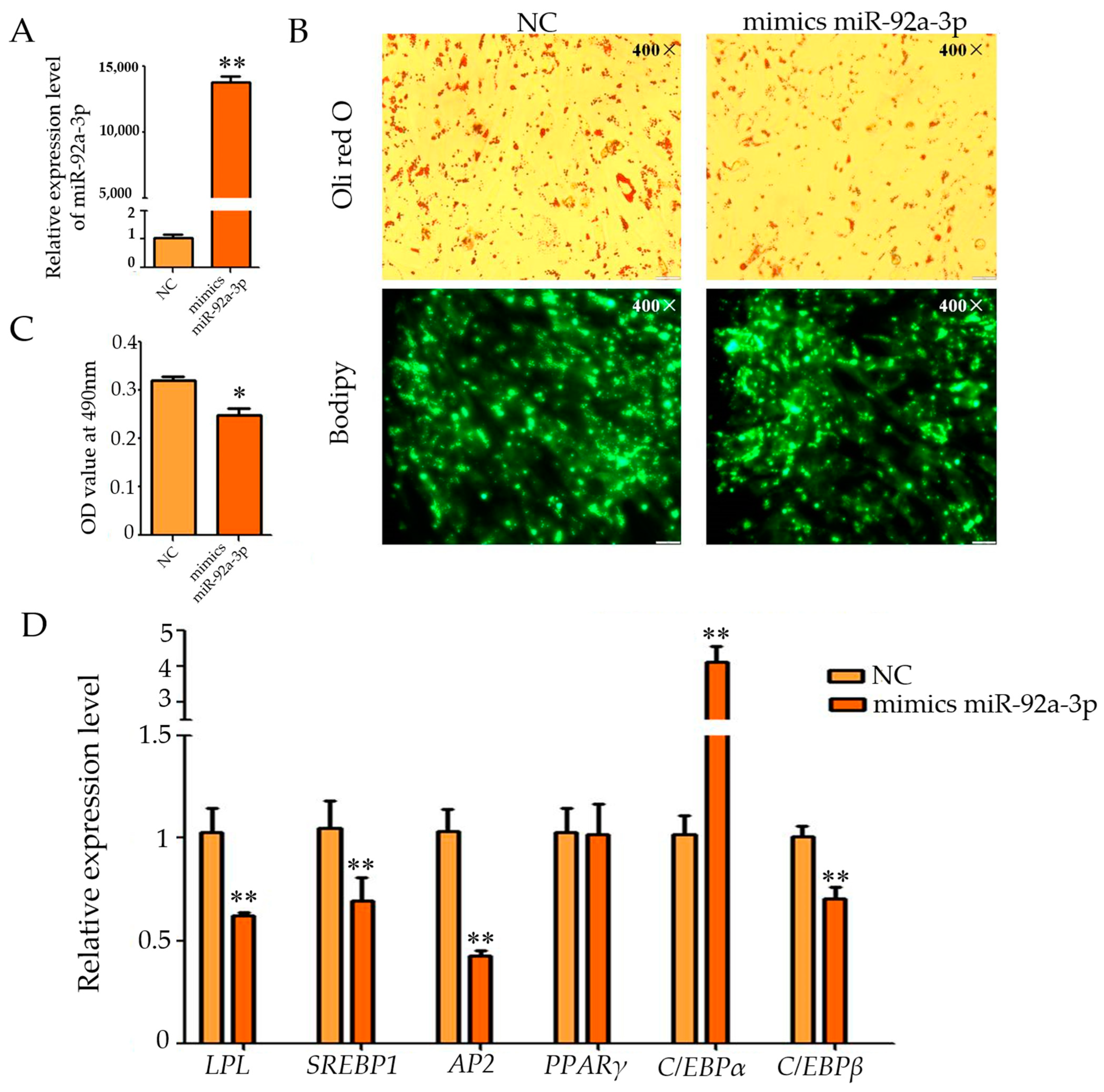
3.1. Effect of Overexpression of miR-92a-3p on the Differentiation of Intramuscular Adipocytes in Goats

The qPCR results showed that the expression of miR-92a-3p declined from 0 to 24 h during the differentiation of goat intramuscular adipocyte, whereas the expression gradually increased from 24 to 72 h, reaching a peak at 72 h of differentiation, and then the expression of miR-92a-3p gradually declined from 72 to 120 h of differentiation (Figure S1). Subsequently, miR-92a-3p mimics were utilized to up-regulate its expression in cells, and its overexpression efficiency was detected using qPCR technology. The results showed that mimics caused miR-92a-3p to be highly expressed in the m miR-92a-3p group (overexpression of miR-92a-3p group) and it was higher than that in the control group (p < 0.01, Figure 1A). Oil Red O and Bodipy staining showed that intracellular lipid droplets were significantly reduced during overexpression of miR-92a-3p (Figure 1B), and the real-time fluorescence quantitative PCR. The results were consistent with the above observations, and the OD value of the m miR-92a-3p group was significantly lower than that of the NC group (p < 0.05, Figure 1C). Furthermore, the relative expression of the marker genes of lipid differentiation, such as LPL, AP2, SREBP1, and C/EBPβ, were significantly inhibited during the overexpression of miR-92a-3p (all p < 0.01), while the expression of C/EBPα was highly promoted (p < 0.01), and no significant effect was found for the expression of PPARγ (p > 0.05, Figure 1D). These results suggest that overexpression of miR-92a-3p inhibits differentiation of goat intramuscular adipocytes.

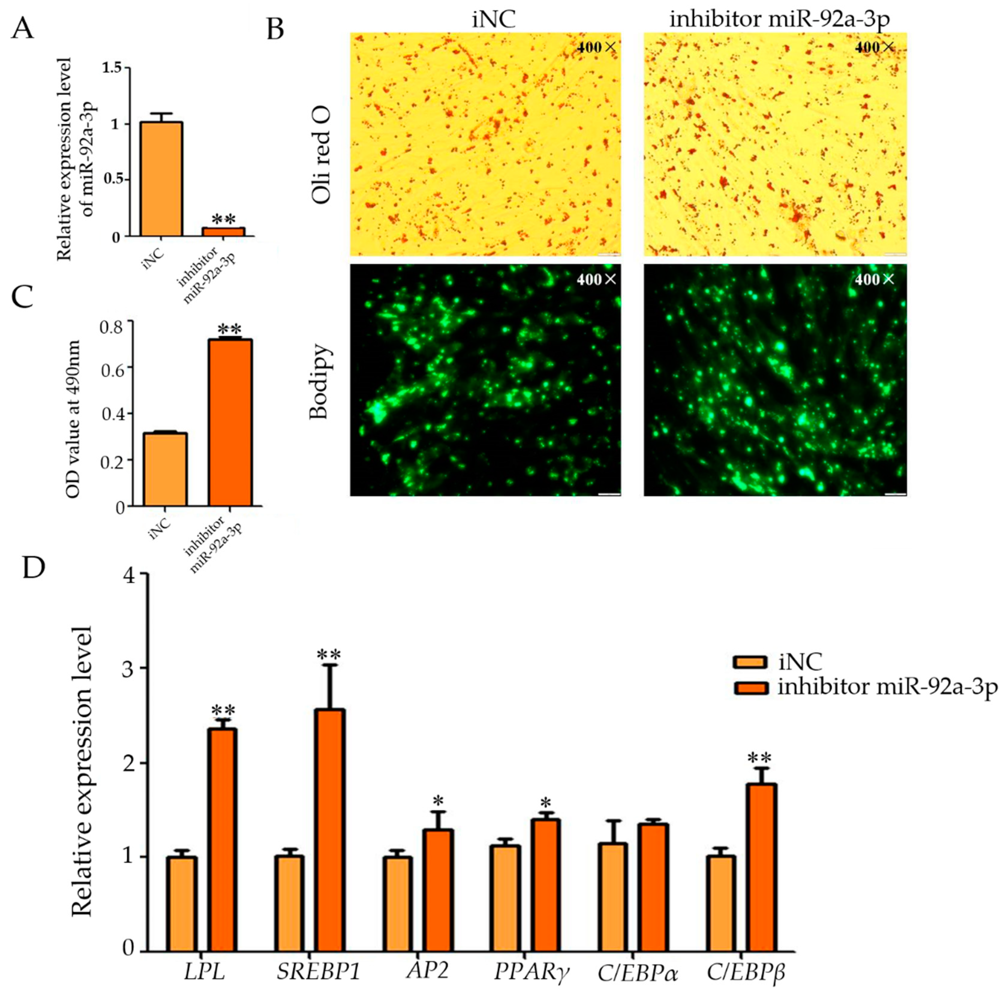
3.2. Effect of miR-92a-3p Inhibition on Intramuscular Adipocyte Differentiation in Goats

We synthesized the inhibitor based on the sequence of miR-92a-3p to inhibit its expression. The qRT-PCR results showed that the inhibitor successfully inhibited the expression of miR-92a-3p in the cells, and the miR-92a-3p expression in the experimental group was much lower than that in the control group (p < 0.01, Figure 2A). Oil Red O staining showed that inhibition of miR-92a-3p expression resulted in an increase in intracellular lipid droplets (Figure 2B), and qPCR results showed that the OD value of the i miR-92a-3p group (miR-92a-3p inhibition group) was highly significantly increased when compared with that of the iNC group (control group) (p < 0.01, Figure 2C). The qRT-PCR results showed that the expression levels of LPL, SREBP1, C/EBPβ, AP2, and PPARγ were significantly up-regulated after inhibiting the expression of miR-92a-3p (p < 0.05), whereas the relative expression levels of C/EBPα did not change significantly (Figure 2D). These results suggest that inhibition of miR-92a-3p promotes differentiation of goat intramuscular adipocytes.

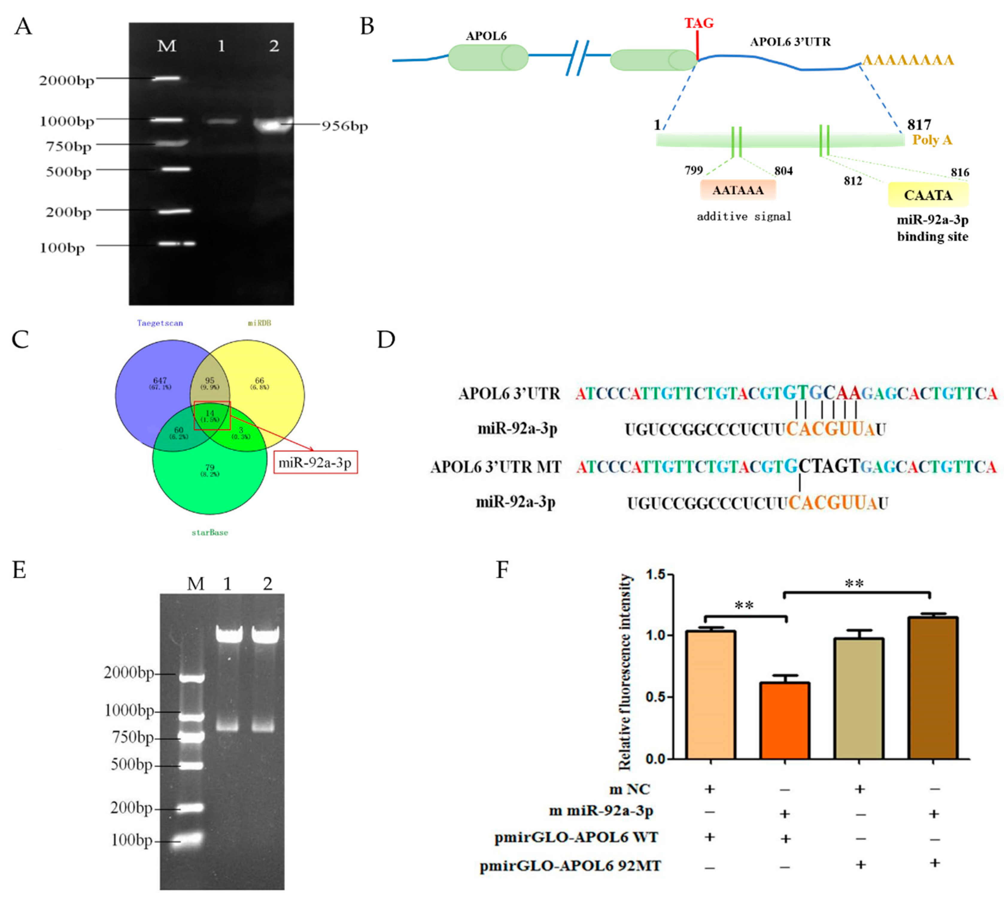
3.3. Effect of miR-92a-3p on the Expression of APOL6

In order to further reveal the regulation mechanism of miR-92a-3p, we used TargetScan software to predict that APOL6 may be a target gene of miRNAs. And then, we further investigated the gene APOL6, and the relative expression levels of APOL6 were examined after miR-92a-3p expression was up-regulated or inhibited. The results showed that the expression level of APOL6 was significantly decreased during overexpressing of miR-92a-3p (p < 0.01, Figure S2A), and the expression level of APOL6 was significantly increased after miR-92a-3p inhibition (p < 0.01). To determine the targeting relationship between miR-92a-3p and APOL6, we performed a comparative analysis of the temporal expression profiles of miR-92a-3p and APOL6. The qPCR results showed that during the differentiation of goat intramuscular adipocytes, the expression level of APOL6 increased and then decreased from 0 to 120 h, increased at 96 h, and decreased at 96 h. The expression level of APOL6 in goat intramuscular adipocytes was also found to decrease from 0 to 120 h, and then increased at 96 h (Figure S2B).

3.4. miR-92a-3p Targets APOL6 3′UTR Binding

The sequence of goat APOL6 3′UTR was obtained by cloning using a 3′RACE kit, and the sequence length was 956 bp (Figure 3A), including 817 bp of APOL6 3′UTR, a poly A tail of 9 bp in length. Bioinformatics analysis of the obtained goat APOL6 3′UTR sequence revealed the presence of a classical plus-tail signal (AATAAA) at 799~804 bp of the goat APOL6 3′UTR, and the binding site of miR-92a-3p was located at 812~816 bp (Figure 3B). Using three different online programs (TargetScan, starBase, miRDB) to reverse predict the miRNAs that might target the goat APOL6 3′UTR, and using Veeny 2.1 to draw a Wayne diagram, the results showed (Figure 3C) that miR-92a-3p was located in the intersection of all three sets of software results. And the binding site of APOL6 3′UTR with miR-92a-3p was predicted using the above software (Figure 3C). In this study, we first constructed a wild-type dual luciferase reporter vector for APOL6 3′UTR (pmirGLO-APOL6 WT) and mutated miR-92a-3p at the predicted binding site of miR-92a-3p and APOL6 3′UTR (Figure 3D), and successfully constructed a mutant vector (pmirGLO-APOL6 92aMT). The two constructed vectors were identified by gel electrophoresis after double digestion, and the results showed that the size of the inserted fragment was the same as the length of the APOL6 3′UTR (Figure 3E), and the sequencing results also showed that the mutation of the site was successful, which proved that the two vector vectors were constructed successfully.
The results of the dual luciferase reporter gene detection system showed (Figure 3F) that transfection of miR-92a-3p mimics in goat intramuscular adipocytes, along with the pmirGLO-APOL6 WT plasmid, resulted in a highly significant reduction in the activity of the 3′UTR of APOL6 (p < 0.01); co-transfection of miR-92a-3p in goat intramuscular adipocytes with miR-92a-3p mimics and pmirGLO-APOL6 92aMT plasmid, on the other hand, had no significant effect on APOL6 3′UTR activity.

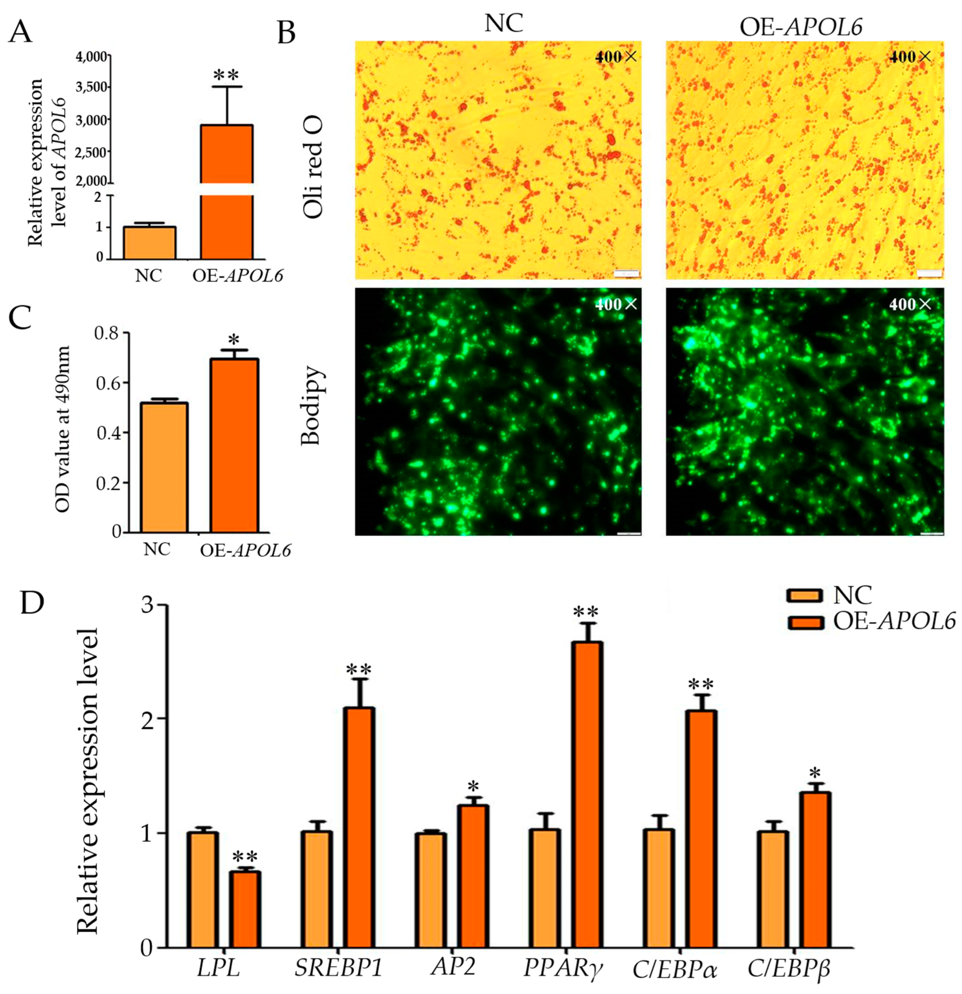
3.5. Effect of Overexpression of APOL6 on the Differentiation of Intramuscular Adipocytes in Goats

The qPCR results showed that after transfection with the overexpression vector plasmid, the expression level of APOL6 in the experimental group’s cells was significantly higher than the control group’s cells (p < 0.01, Figure 4A), and Oil red O and Bodipy staining showed a significant increase in lipid droplets (Figure 4B). The OD value of the OE-APOL6 group was significantly increased compared to that of the Negative Control group (control group) (p < 0.05, Figure 4C). Meanwhile, qPCR results showed that the expression of PPARγ, SREBP1, and C/EBPAα was highly significantly up-regulated after overexpression of APOL6 (p < 0.01), and that the expression levels of AP2 and C/EBPβ were significantly up-regulated (p < 0.05), while the expression levels of LPL were significantly down-regulated (p < 0.01, Figure 4D). These results suggest that overexpression of APOL6 promotes differentiation of goat intramuscular adipocytes.

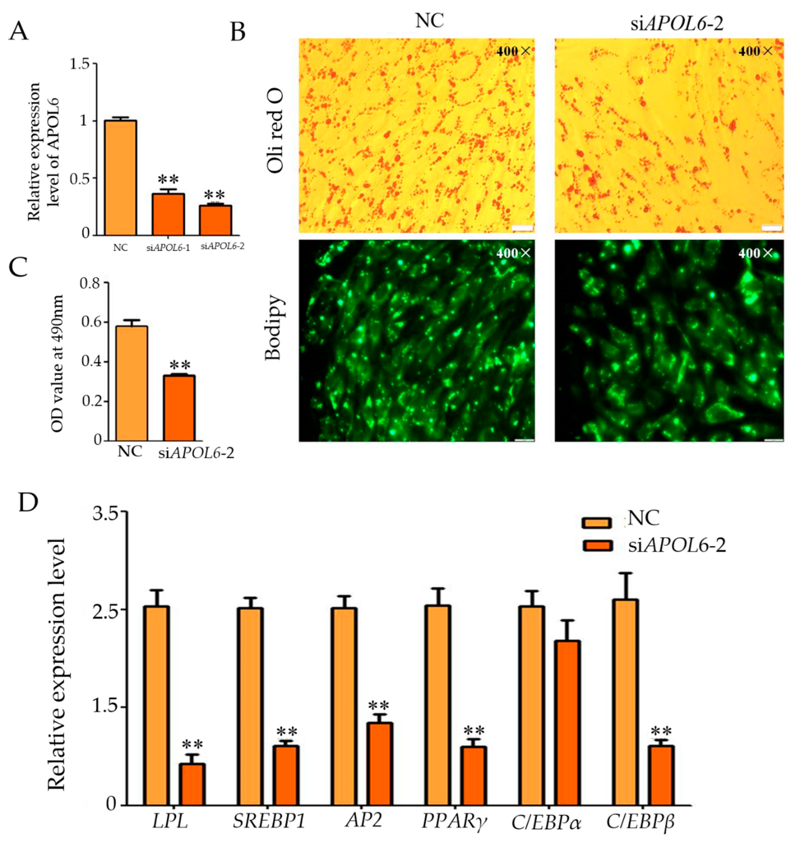
3.6. Effect of Inhibition of APOL6 on the Differentiation of Intramuscular Adipocytes in Goats

In order to further confirm the experimental result that overexpression of APOL6 promotes the differentiation of goat intramuscular adipocytes, interference experiments were also designed in this study. The qPCR results showed that the knockdown effect of specific siRNAs was good, and the expression of APOL6 was reduced by 64% in the siAPOL6-1 group, and 74% in the siAPOL6-2 group, compared with that of the NC group (Figure 5A). The interference effect of siAPOL6-2 group was relatively better, so siAPOL6-2 was chosen for the subsequent experiments. Oil red O and Bodipy staining results showed that compared with the NC group, the intracellular lipid droplets in the siAPOL6-2 group were significantly reduced (Figure 5B), and the OD value at 490 nm was extremely significantly reduced (p < 0.01) (Figure 5C). The qPCR results showed that after interfering with APOL6, the relative expression levels of PPARγ, LPL, C/EBPβ, AP2, and SREBP1 also decreased (p < 0.01, Figure 5D). These results suggest that inhibition of APOL6 suppressed differentiation of goat intramuscular adipocytes.

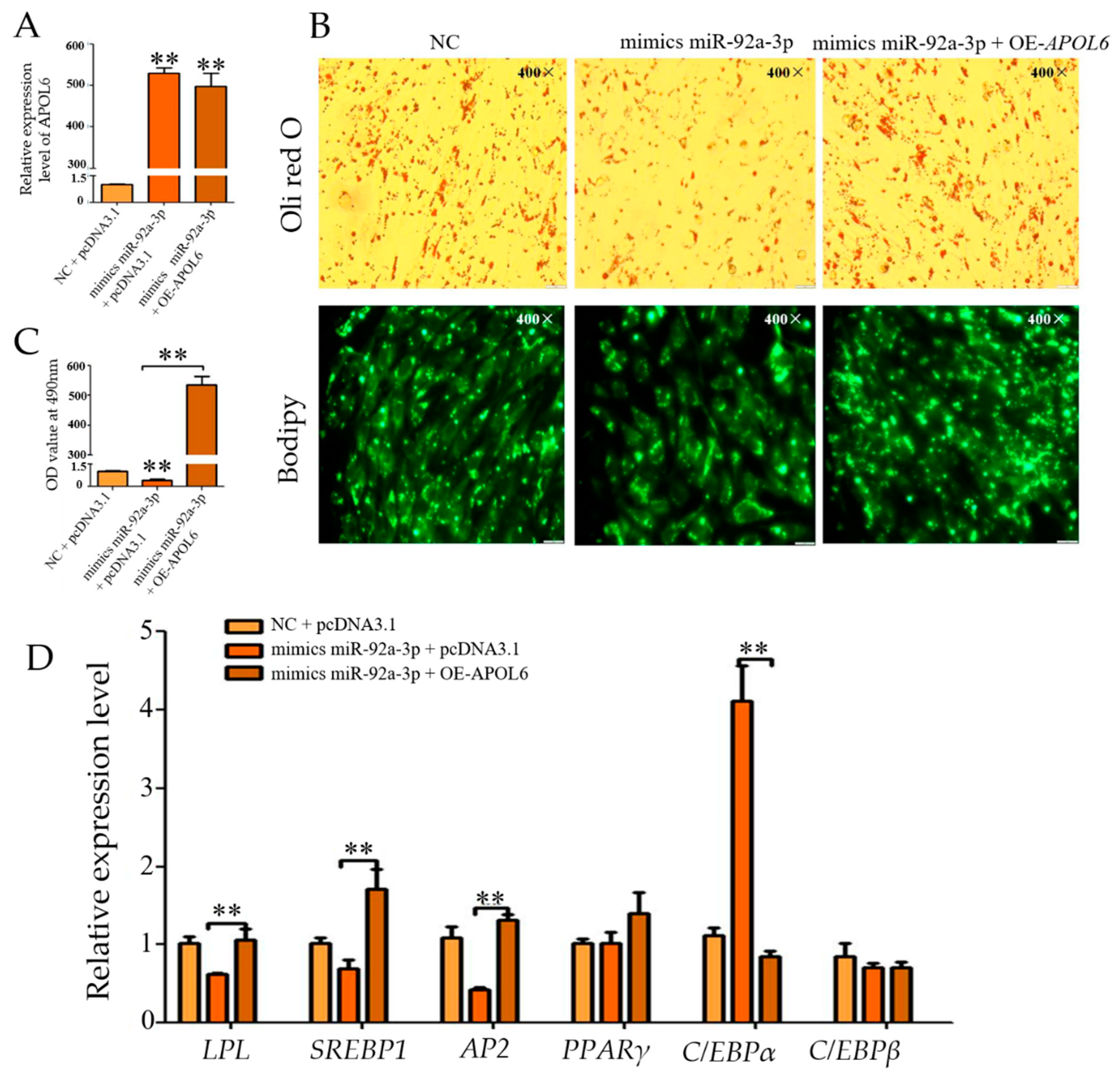
3.7. Effect of Overexpression of miR-92a-3p Followed by Overexpression of APOL6 on the Differentiation of Goat Intramuscular Adipocytes

The qPCR results showed (Figure 6A) that the expression of miR-92a-3p was significantly increased in both the mmiR-92a-3p group and the mmiR-92a-3p + OEA group (p < 0.01). The qPCR results showed (Figure 6C) that compared with the m miR-92a-3p group, the expression of APOL6 in the m miR-92a-3p + OEA group increased significantly (p < 0.01). Oil Red O and Bodipy staining results showed (Figure 6B) that compared with the m miR-92a-3p group, the accumulation of intracellular lipid droplets in the m miR-92a-3p + OEA group increased. With overexpression of APOL6 after the overexpression of miR-92a-3p, the expression levels of SREBP1 and AP2 were significantly increased (p < 0.01), the expression of LPL was significantly increased (p < 0.05), and the expression of C/EBPβ was extremely significantly decreased (p < 0.01) (Figure 6D).

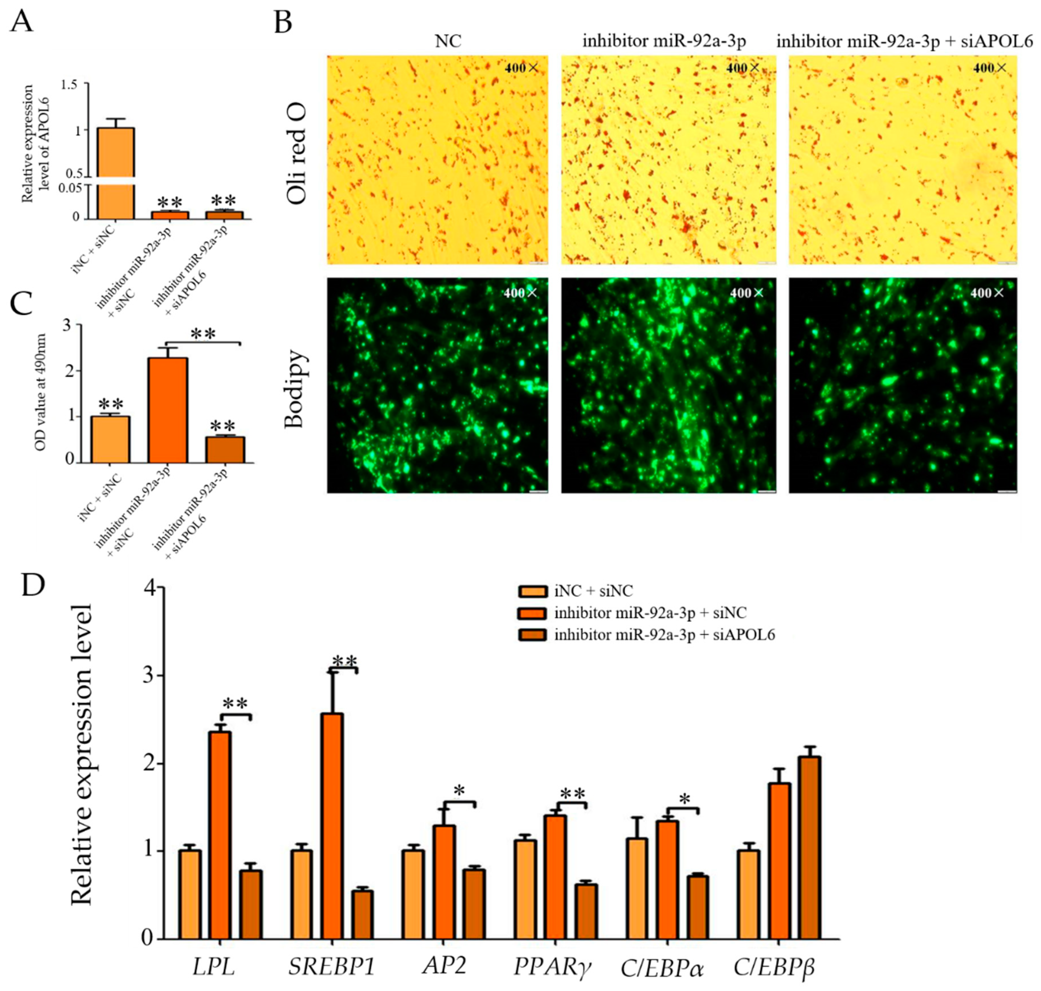
3.8. Effect of Inhibition of miR-92a-3p Followed by Interference with APOL6 on Goat Intramuscular Adipocyte Differentiation

The qPCR results showed (Figure 7A) that the expression of miR-92a-3p was successfully suppressed in the i miR-92a-3p group versus the i miR-92a-3p + siAPOL6 group (p < 0.01). Subsequently, the expression of APOL6 was detected, and the qPCR results showed (Figure 7B) that the expression of APOL6 in the i miR-92a-3p + siAPOL6 group was extremely significantly decreased compared with that in the i miR-92a-3p group (p < 0.01). Oil red O and Bodipy staining results showed (Figure 7C) that intracellular lipid droplet aggregation was reduced in the i miR-92a-3p + siAPOL6 group compared with the i miR-92a-3p group. After miR-92a-3p inhibition interfered with APOL6, the expression levels of LPL, SREBP1, and PPARγ were highly significantly down-regulated (p < 0.01), and the expression of AP2 and C/EBPβ was significantly decreased (p < 0.05, Figure 7D).

4. Discussion

Adipocyte differentiation is a complex biological process that involves numerous molecular events and requires the synergistic action of multiple factors such as transcription factors, functional genes, and signaling pathways. Ma et al. [23] found that miRNAs play a role in the transcriptional regulation of mammalian preadipocyte differentiation. The authors of [24] first demonstrated experimentally that miR-143 could promote adipocyte differentiation through mitogen-activated protein kinase 5, and subsequently, more and more miRNAs were experimentally verified to interact with transcription factors or signaling molecules important for adipocyte differentiation to regulate adipogenesis [25]. A large number of studies have found that miRNAs are involved in the differentiation process of adipocytes. From this, it is reasonable to speculate that miR-92a-3p may also be involved in the differentiation process of adipocytes in goat muscle. However, there is no report on the differentiation process of adipocytes in goat muscle. Therefore, in this experiment, we firstly constructed the cellular time-series expression profile of miR-92a-3p. Although the expression level of miR-92a-3p decreased in the late stage of cell differentiation, it was still higher than that in the pre-differentiation stage in general, so it was hypothesized that it might inhibit the differentiation of goat intramuscular adipocytes. However, its specific mechanism of action remains to be experimentally determined.
To further elucidate the exact role of miR-92a-3p in goat intramuscular adipocyte differentiation, we synthesized a mimic/inhibitor of miR-92a-3p and overexpressed/inhibited it in goat intramuscular adipocytes cultured in vitro, and the results showed that miR-92a-3p suppressed the production of lipid droplets, as well as down-regulating LPL, AP2, SREBP1, and C/EBPβ expression, while C/EBPα was significantly up-regulated. Among them, C/EBPβ is a positive regulator of adipocyte differentiation, and overexpression of C/EBPβ in 3T3-L1 precursor adipocytes or NIH-3T3 cells promotes adipogenic differentiation in the absence of hormone-induced differentiation [26]. Secondly, PPARγ plays a key role in adipocyte differentiation as a ligand-activated transcription factor [27]. PPARγ activation initiates adipocyte differentiation and lipogenic gene expression, leading to the synthesis and sustained accumulation of triglycerides, which in turn regulates a variety of biological processes in adipocytes from development to metabolism [28,29]. SREBP1, also known as adipocyte determinant and differentiation-dependent factor 1, is a transcription factor associated with adipocyte differentiation and compartmentalization. Studies have shown that SREBP1 plays a role in adipocyte gene expression by regulating the expression of FAS and LPL, important genes involved in fatty acid metabolism; meanwhile, SREBP1 also promotes adipogenic differentiation by inducing the expression of PPARγ [30]. Therefore, the significant down-regulation of SREBP1 and C/EBPβ may be the reason for the significant reduction in lipid droplets after overexpression of miR-92a-3p, and the up-regulation of the expression of C/EBPα, one of the factors promoting adipocyte differentiation, but after overexpression of miR-92a-3p, it may be due to the degree of inhibition of adipocyte differentiation after overexpression of miR-92a-3p is beyond the normal range, resulting in a kind of negative feedback regulation.
Apolipoprotein L (APOL) is a novel plasma protein belonging to the family of lipid-transporting proteins, and it is a novel high-density lipoprotein. Liu et al. [31] found that APOL6 overexpression could induce apoptosis in tumor cells and interact with lipid/fatty acid components in the apoptosis-inducing process, but APOL6 with the BH3 structure removed lost its apoptosis-inducing function. Zhaorigetu et al. [32] found that high expression of APOL6 induced apoptosis in cells such as macrophages, endothelial cells, and vascular smooth muscle cells, accelerating the progression of atherosclerosis. The role of APOL6 in adipocyte differentiation should not be ignored, Tan et al. [33] found that APOL6 is a target gene of miR-10b-5p, and the up-regulation of APOL6 expression after inhibition of miR-10b-5p also promoted the differentiation of 3T3-L1 cells, which suggests that APOL6 can play a role as a target gene of some miRNAs in the process of regulating adipocyte differentiation. However, no study has been reported on the role of APOL6 on the differentiation of intramuscular adipocytes in goats.
In this study, by constructing APOL6 overexpression vector and synthesizing siRNAs of APOL6, the results showed that overexpression of APOL6 promoted goat intramuscular adipocyte differentiation and intracellular lipid droplet production, while up-regulating the expression of C/EBPβ, PPARγ, SREBP1, and AP2, which was consistent with the finding of Tan et al. [34] that up-regulation of APOL6 expression promoted 3T3-L1 cell differentiation and adipogenesis; whereas, after interfering with APOL6, intracellular lipid droplet production was significantly reduced, while the expression of LPL, C/EBPα, PPARγ, SREBP1, and AP2 was significantly down-regulated, suggesting that down-regulation of APOL6 expression inhibited goat intramuscular adipocyte differentiation. Differentiated adipocytes were characterized as containing lipid droplets and expressing adipocyte marker genes such as C/EBPβ, PPARγ, LPL, lipocalin, and leptin [35]. It was found that LPL inhibited lipolysis, and the expression of LPL gradually increased during adipocyte differentiation, promoting the generation and accumulation of intracellular lipid droplets [36,37,38]; the inhibition of APOL6 resulted in a decrease in the expression of LPL and a reduction in intracellular lipid droplets, which was hypothesized to promote lipolysis through the inhibition of LPL expression, leading to a reduction in intracellular lipid droplets and the inhibition of goat intramuscular adipocyte differentiation. C/EBPβ is an early transcription factor that induces adipogenesis, and activates PPARγ, which is a major inducer of adipogenesis and a positive regulator of adipocyte differentiation, by regulating the transcription of its neighboring promoter [10]. C/EBPα has been shown to be a key gene in adipocyte differentiation in a previous study, and furthermore, it can regulate AP2 expression, which promotes the aggregation of lipid droplets, and can also combine with C/EBPβ and PPARγ to regulate the expression of fatty acid synthase (FAS). Taken together, APOL6 may promote adipocyte differentiation by promoting lipogenesis and by up-regulating the expression levels of C/EBPβ, PPARγ, SREBP1, and AP2, whereas interfering with APOL6 inhibits adipocyte differentiation and lipid droplet generation by repressing the expression of LPL, C/EBPα, PPARγ, SREBP1, and AP2. In this study, we found that overexpression of APOL6 promoted the differentiation of goat intramuscular adipocytes, suggesting that APOL6 positively regulates IMF deposition in goats. However, although the regulatory role of APOL6 in goat intramuscular adipocyte differentiation is unknown, the specific regulatory mechanisms need to be further investigated.

5. Conclusions

In summary, our study reveals that miR-92a-3p is a novel regulator of intramuscular preadipocyte differentiation in goats, and miR-92a-3p inhibits intramuscular preadipocyte differentiation in goats by targeting the APOL6 gene. These findings contribute to a better understanding of miRNA-regulated adipogenesis.

Supplementary Materials

The following supporting information can be downloaded at: https://www.mdpi.com/article/10.3390/genes15010057/s1, Figure S1: The temporal expression levels of miR-92a-3p in intramuscular adipocytes of goats; Figure S2. Effect of miR-92a-3p on the expression of APOL6 and Cellular temporal expression profile of APOL6. A: Effects of miR-92A-3p on APOL6 expression; B: The temporal expression levels of APOL6 in intramuscular adipocytes of goats.

Author Contributions

Conceptualization, W.Q., Y.L. (Yaqiu Lin) and J.Z.; methodology, W.Q., Y.L. (Yaqiu Lin) and J.Z.; software and formal analysis, W.L. (Wei Liu), W.Q., J.Z., Y.X., Y.W. (Youli Wang), Y.L. (Yanyan Li) and Y.L. (Yaqiu Lin); validation, W.Q., J.Z. and G.X.; resources, Y.L. (Yaqiu Lin) and W.Q.; writing original draft preparation, W.Q. and J.Z.; writing—review and editing, Y.L. (Yaqiu Lin) and J.Z.; supervision, Y.W. (Youli Wang), Y.W. (Yong Wang) and G.X. All authors have read and agreed to the published version of the manuscript.

Funding

This work was supported by National Natural Sciences Foundation of China (32072723), Sichuan Science and Technology Program (2022JDTD0030), and Southwest Minzu University Double World-Class Project (XM2023011).

Institutional Review Board Statement

The animal study protocol was approved by the Institutional Ethics Committee of Southwest Minzu University (No. 18032, 2018).

Informed Consent Statement

Not applicable.

Data Availability Statement

The data used to support this study are available from the corresponding author on reasonable request.

Conflicts of Interest

The authors declare, there are no competing interests.

References

  1. Stanišić, N.; Žujović, M.; Tomić, Z.; Maksimović, N.; Bijelić, Z.; Ivanović, S.; Memiši, N. The Effects of Crossing Balkan and Saanen Goat Breeds on Carcass Traits and Certain Quality Parameters of Kid Meat. Ann. Anim. Sci. 2012, 12, 53–62. [Google Scholar] [CrossRef]
  2. Xu, K.; Ji, M.; Huang, X.; Peng, Y.; Wu, W.; Zhang, J. Differential regulatory roles of MicroRNAs in porcine intramuscular and subcutaneous adipocytes. J. Agric. Food Chem. 2020, 68, 3954–3962. [Google Scholar] [CrossRef] [PubMed]
  3. Zhou, J.; Zhang, Y.; Wu, J.; Qiao, M.; Xu, Z.; Peng, X.; Mei, S. Proteomic and lipidomic analyses reveal saturated fatty acids, phosphatidylinositol, phosphatidylserine, and associated proteins contributing to intramuscular fat deposition. J. Proteom. 2021, 241, 104235. [Google Scholar] [CrossRef] [PubMed]
  4. Frank, D.; Ball, A.; Hughes, J.; Krishnamurthy, R.; Piyasiri, U.; Stark, J.; Watkins, P.; Warner, R. Sensory and flavor chemistry characteristics of Australian beef: Influence of intramuscular fat, feed, and breed. J. Agric. Food Chem. 2016, 64, 4299–4311. [Google Scholar] [CrossRef] [PubMed]
  5. Frank, D.; Watkins, P.; Ball, A.; Krishnamurthy, R.; Piyasiri, U.; Sewell, J.; Ortuno, J.; Stark, J.; Warner, R. Impact of brassica and lucerne finishing feeds and intramuscular fat on lamb eating quality and flavor. A cross-cultural study using Chinese and Non-Chinese Australian consumers. J. Agric. Food Chem. 2016, 64, 6856–6868. [Google Scholar] [CrossRef] [PubMed]
  6. Gui, L.S.; Raza, S.H.A.; Sun, Y.G.; Khan, R.; Ullah, I.; Han, Y.C. Detection of polymorphisms in the promoter of bovine SIRT1 gene and their effects on intramuscular fat content in Chinese indigenous cattle. Gene 2019, 700, 47–51. [Google Scholar] [CrossRef]
  7. Li, X.; Fu, X.; Yang, G.; Du, M. Review: Enhancing intramuscular fat development via targeting fibro-adipogenic progenitor cells in meat animals. Animal 2020, 14, 312–321. [Google Scholar] [CrossRef]
  8. Migdał, W.; Kawęcka, A.; Sikora, J.; Migdał, Ł. Meat quality of the native Carpathian goat breed in comparison with the Saanen breed. Animals 2021, 11, 2220. [Google Scholar] [CrossRef]
  9. Realini, C.E.; Pavan, E.; Johnson, P.L.; Font-I-Furnols, M.; Jacob, N.; Agnew, M.; Craigie, C.R.; Moon, C.D. Consumer liking of M. longissimus lumborum from New Zealand pasture-finished lamb is influenced by intramuscular fat. Meat Sci. 2021, 173, 108380. [Google Scholar] [CrossRef]
  10. Mota de Sá, P.; Richard, A.J.; Hang, H.; Stephens, J.M. Transcriptional Regulation of Adipogenesis. Compr. Physiol. 2017, 7, 635–674. [Google Scholar]
  11. Covas, D.T.; Panepucci, R.A.; Fontes, A.M.; Silva, W.A., Jr.; Orellana, M.D.; Freitas, M.C.; Neder, L.; Santos, A.R.; Peres, L.C.; Jamur, M.C.; et al. Multipotent mesenchymal stromal cells obtained from diverse human tissues share functional properties and gene-expression profile with CD146+ perivascular cells and fibroblasts. Exp. Hematol. 2008, 36, 642–654. [Google Scholar] [CrossRef] [PubMed]
  12. Qin, Y.; Chen, H.; Zhang, Y.; Zhu, C.; Gao, B.; Yin, Y.; Li, W.; Shi, Q.; Zheng, M.; Xu, Q.; et al. Cloning of the Xuhuai goat PPARγ gene and the preparation of Transgenic sheep. Biochem. Genet. 2013, 51, 543–553. [Google Scholar] [CrossRef] [PubMed]
  13. Guo, H.; Khan, R.; Raza, S.H.A.; Nurgulsim, K.; Suhail, S.M.; Rahman, A.; Ahmed, I.; Ijaz, A.; Ahmad, I.; Linsen, Z. Transcriptional regulation of adipogenic marker genes for the improvement of intramuscular fat in Qinchuan beef cattle. Anim. Biotechnol. 2022, 33, 776–795. [Google Scholar]
  14. Zhou, L.; Chen, S.Y.; Han, H.J.; Sun, J.Q. Lactate augments intramuscular triglyceride accumulation and mitochondrial biogenesis in rats. J. Biol. Regul. Homeost. Agents 2021, 35, 105–115. [Google Scholar] [PubMed]
  15. Deb, B.; Uddin, A.; Chakraborty, S. miRNAs and ovarian cancer: An overview. Cell Physiol. 2018, 233, 3846–3854. [Google Scholar] [CrossRef]
  16. Li, M.; Liu, Z.; Zhang, Z.; Liu, G.; Sun, S. Sun C miR-103 promotes 3T3-L1 cell adipogenesis through AKT/mTOR signal pathway with its target being MEF2D. Biol Chem. 2015, 396, 235–244. [Google Scholar] [CrossRef] [PubMed]
  17. Peng, Y.; Chen, F.F.; Ge, J.; Zhu, J.Y.; Shi, X.E.; Li, X.; Yu, T.Y.; Chu, G.Y.; Yang, G.S. miR-429 Inhibits Differentiation and Promotes Proliferation in Porcine Preadipocytes. Int. J. Mol. Sci. 2016, 17, 2047. [Google Scholar] [CrossRef]
  18. Li, B.; Huang, X.; Yang, C.; Ge, T.; Zhao, L.; Zhang, X.; Tian, L.; Zhang, E. miR-27a regulates sheep adipocyte differentiation by targeting CPT1B gene. Animals 2021, 12, 28. [Google Scholar] [CrossRef]
  19. Shan, B.; Yan, M.; Yang, K.; Lin, W.; Yan, J.; Wei, S.; Wei, W.; Chen, J.; Zhang, L. MiR-218-5p affects subcutaneous adipogenesis by targeting ACSL1, a novel candidate for pig fat deposition. Genes 2022, 13, 260. [Google Scholar] [CrossRef]
  20. Page, N.M.; Butlin, D.J.; Lomthaisong, K.; Lowry, P.J. The human apolipoprotein L gene cluster: Identification, classification, and sites of distribution. Genomics. 2001, 74, 71–78. [Google Scholar] [CrossRef]
  21. Schroeder, F.; Gallegos, A.M.; Atshaves, B.P.; Storey, S.M.; Mcintosh, A.L.; Petrescu, A.D.; Huang, H.; Starodub, O.; Chao, H.; Yang, H.; et al. Recent advances in membrane microdomains: Rafts, caveolae, and intracellular cholesterol trafficking. Exp. Biol. Med. 2001, 226, 873–890. [Google Scholar] [CrossRef]
  22. Xu, Q.; Lin, S.; Wang, Y.; Zhu, J.; Lin, Y. Fibroblast growth factor 10 (FGF10) promotes the adipogenesis of intramuscular preadipocytes in goat. Mol. Biol. Rep. 2018, 45, 1881–1888. [Google Scholar] [CrossRef] [PubMed]
  23. Booth, A.D.; Magnuson, A.M.; Fouts, J.; Wei, Y.; Wang, D.; Pagliassotti, M.J.; Foster, M.T. Subcutaneous adipose tissue accumulation protects systemic glucose tolerance and muscle metabolism. Adipocyte 2018, 7, 261–272. [Google Scholar] [CrossRef] [PubMed]
  24. Esau, C.; Kang, X.; Peralta, E.; Hanson, E.; Marcusson, E.G.; Ravichandran, L.V.; Sun, Y.; Koo, S.; Perera, R.J.; Jain, R.; et al. MicroRNA-143 regulates adipocyte differentiation. J. Biol. Chem. 2004, 279, 52361–52365. [Google Scholar] [CrossRef] [PubMed]
  25. Kim, C.; Lee, H.; Cho, Y.M.; Kwon, O.J.; Kim, W.; Lee, E.K. TNFα-induced miR-130 resulted in adipocyte dysfunction during obesity-related inflammation. FEBS Lett. 2013, 587, 3853–3858. [Google Scholar] [CrossRef]
  26. Zhao, N.Q.; Li, X.Y.; Wang, L.; Feng, Z.L.; Li, X.F.; Wen, Y.F.; Han, J.X. Palmitate induces fat accumulation by activating C/EBPβ-mediated G0S2 expression in HepG2 cells. World J. Gastroenterol. 2017, 23, 7705–7715. [Google Scholar] [CrossRef]
  27. Stachecka, J.; Nowacka-Woszuk, J.; Kolodziejski, P.A.; Szczerbal, I. The importance of the nuclear positioning of the PPARG gene for its expression during porcine in vitro adipogenesis. Chromosome Res. 2019, 27, 271–284. [Google Scholar] [CrossRef]
  28. Lefterova, M.I.; Haakonsson, A.K.; Lazar, M.A.; Mandrup, S. PPARγ and the global map of adipogenesis and beyond. Trends Endocrinol. Metab. 2014, 25, 293–302. [Google Scholar] [CrossRef]
  29. Lee, J.E.; Ge, K. Transcriptional and epigenetic regulation of PPARγ expression during adipogenesis. Cell Biosci. 2014, 4, 29. [Google Scholar] [CrossRef]
  30. Fajas, L.; Schoonjans, K.; Gelman, L.; Kim, J.B.; Najib, J.; Martin, G.; Fruchart, J.C.; Briggs, M.; Spiegelman, B.M.; Auwerx, J. Regulation of peroxisome proliferator-activated receptor gamma expression by adipocyte differentiation and determination factor 1/sterol regulatory element binding protein 1: Implications for adipocyte differentiation and metabolism. Mol. Cell Biol. 1999, 19, 5495–5503. [Google Scholar] [CrossRef]
  31. Liu, Z.; Lu, H.; Jiang, Z.; Pastuszyn, A.; Hu, C.A. Apolipoprotein l6, a novel proapoptotic Bcl-2 homology 3-only protein, induces mitochondria-mediated apoptosis in cancer cells. Mol. Cancer Res. 2005, 3, 21–31. [Google Scholar] [CrossRef] [PubMed]
  32. Zhaorigetu, S.; Yang, Z.; Toma, I.; McCaffrey, T.A.; Hu, C.A. Apolipoprotein L6, induced in atherosclerotic lesions, promotes apoptosis and blocks Beclin 1-dependent autophagy in atherosclerotic cells. J. Biol. Chem. 2011, 286, 27389–27398. [Google Scholar] [CrossRef] [PubMed]
  33. Han, H.; Gu, S.; Chu, W.; Sun, W.; Wei, W.; Dang, X.; Tian, Y.; Liu, K.; Chen, J. miR-17-5p Regulates Differential Expression of NCOA3 in Pig Intramuscular and Subcutaneous Adipose Tissue. Lipids. 2017, 52, 939–949. [Google Scholar] [CrossRef] [PubMed]
  34. Tan, Y.; Gan, M.; Fan, Y.; Li, L.; Zhong, Z.; Li, X.; Bai, L.; Zhao, Y.; Niu, L.; Shang, Y.; et al. miR-10b-5p regulates 3T3-L1 cells differentiation by targeting Apol6. Gene. 2019, 687, 39–46. [Google Scholar] [CrossRef] [PubMed]
  35. Salmerón, C. Adipogenesis in fish. J. Exp. Biol. 2018, 221 (Suppl. S1), jeb161588. [Google Scholar] [CrossRef]
  36. Tang, Q.Q.; Lane, M.D. Activation and centromeric localization of CCAAT/enhancer-binding proteins during the mitotic clonal expansion of adipocyte differentiation. Genes Dev. 1999, 13, 2231–2241. [Google Scholar] [CrossRef]
  37. Zhang, F.; Pan, T.; Nielsen, L.D.; Mason, R.J. Lipogenesis in fetal rat lung: Importance of C/EBPalpha, SREBP-1c, and stearoyl-CoA desaturase. Am. J. Respir. Cell Mol. Biol. 2004, 30, 174–183. [Google Scholar] [CrossRef]
  38. Tontonoz, P.; Hu, E.; Spiegelman, B.M. Stimulation of adipogenesis in fibroblasts by PPAR gamma 2, a lipid-activated transcription factor. Cell 1995, 79, 1147–1156. [Google Scholar] [CrossRef]
Figure 1. Effect of overexpression of miR-92a-3p on the differentiation of intramuscular adipocytes in goats (after transfection was completed, cells were collected after 48 h of oleic acid-induced differentiation and subjected to relevant tests and assays, * means the 0.01 ≤ p < 0.05, whereas ** means p < 0.01). (A): The overexpression efficiency of miR-92a-3p; (B): Oil red O staining and Bodipy staining figure (400×); (C): The result of OD value (490 nm); (D): The relative expression level of differentiation marker genes of goat intramuscular adipocytes after overexpressed miR-92a-3p.
Genes 15 00057 g001
Figure 2. Effect of miR-92a-3p inhibition on intramuscular adipocyte differentiation in goats (after transfection was completed, cells were collected after 48 h of oleic acid-induced differentiation and subjected to relevant tests and assays, * means the 0.01 ≤ p < 0.05, whereas ** means p < 0.01). (A): The inhibiting efficiency of miR-92a-3p; (B): Oil red O staining and Bodipy staining figure (400×); (C): The result of OD value (490 nm); (D): The relative expression level of differentiation marker genes of goat intramuscular adipocytes after inhibited miR-92a-3p.
Genes 15 00057 g002
Figure 3. miR-92a-3p targets APOL6 3′UTR binding (** means p < 0.01). (A): PCR amplification result of APOL6 3′UTR in goat (M: DL2000 Marker; 1~2: APOL6 3′UTR); (B): APOL6 3′UTR sequence analysis diagram; (C): miRNAs might target APOL6 3′UTR; (D): APOL6 3′UTR wild-type and mutant sequence information; (E): Dual-luciferase identification results, M: DL2000 Marker, 1: pmirGLO-APOL6 WT, 2: pmirGLO-APOL6 92aMT; (F): Dual-luciferase reporter assay experiment results.
Genes 15 00057 g003
Figure 4. Effect of overexpression of APOL6 on the differentiation of intramuscular adipocytes in goats (after transfection was completed, cells were collected after 48 h of oleic acid-induced differentiation and subjected to relevant tests and assays, * means the 0.01 ≤ p < 0.05, whereas ** means p < 0.01). (A): APOL6 overexpression efficiency; (B): Oil red O staining and Bodipy staining figure (400×); (C): The result of OD value (490 nm); (D): The relative expression levels of goat intramuscular adipocyte differentiation marker genes after overexpression of APOL6.
Genes 15 00057 g004
Figure 5. Effect of inhibition of APOL6 on the differentiation of intramuscular adipocytes in goats (after transfection was completed, cells were collected after 48 h of oleic acid-induced differentiation and subjected to relevant tests and assays, ** means p < 0.01). (A): The knockdown efficiency of APOL6; (B): Oil red O staining and Bodipy staining figure (400×); (C): The result of OD value (490 nm); (D): The relative expression levels of goat intramuscular adipocyte differentiation marker genes after knockdown of APOL6.
Genes 15 00057 g005
Figure 6. Effect of overexpression of miR-92a-3p followed by overexpression of APOL6 on the differentiation of goat intramuscular adipocytes (after transfection was completed, cells were collected after 48 h of oleic acid-induced differentiation and subjected to relevant tests and assays, whereas ** means p < 0.01). (A): The overexpression efficiency of miR-92a-3p; (B): Relative expression level of; (C): Oil red O staining and Bodipy staining figure (400×); (D): The relative expression levels of goat intramuscular adipocyte differentiation marker genes.
Genes 15 00057 g006
Figure 7. Effect of inhibition of miR-92a-3p followed by interference with APOL6 on goat intramuscular adipocyte differentiation (after transfection was completed, cells were collected after 48 h of oleic acid-induced differentiation and subjected to relevant tests and assays, * means the 0.01 ≤ p < 0.05, whereas ** means p < 0.01). (A): The inhibiting efficiency of miR-92a-3p; (B): Relative expression level of; (C): Oil red O staining and Bodipy staining figure (400×); (D): The relative expression levels of goat intramuscular adipocyte differentiation marker genes.
Genes 15 00057 g007
Table 1. Primer sequence information.
Gene/miRNAAccession NumberSequence (5′~3′)Tm/°CPurposeProduct Length
U6S: TGGAACGCTTCACGAATTTGCGNR_138,085.160qPCR68
A: GGAACGATACAGAGAAGATTAGC
UXTS: GCAAGTGGATTTGGGCTGTAACXM_005700842.260qPCR180
A: ATGGAGTCCTTGGTGAGGTTGT
miR-92a-3pS: CTGGAGTATTGCACTTGTCCCG 60qPCR
A: GTGCAGGGTCCGAGGT
LPLS: TCCTGGAGTGACGGAATCTGTNM_001285607.160qPCR156
A: GACAGCCAGTCCACCACGAT
C/EBPαS: CCGTGGACAAGAACAGCAACXM_01806227858qPCR142
A: AGGCGGTCATTGTCACTGGT
PPARγS: AAGCGTCAGGGTTCCACTATGNM_00128565860qPCR197
A: GAACCTGATGGCGTTATGAGAC
SREBP1S: AAGTGGTGGGCCTCTCTGANM_00128575558qPCR127
A: GCAGGGGTTTCTCGGACT
C/EBPβS: CAAGAAGACGGTGGACAAGCXM_018058020.165qPCR204
A: AACAAGTTCCGCAGGGTG
AP2S: TGAAGTCACTCCAGATGACAGGNM_001285623.158qPCR143
A: TGACACATTCCAGCACCAGC
Table 2. Sequence information of mimics and inhibitor.
miRNASequenceClassification
NCUUCUCCGAACGUGUCACGUTTmimics
CAGUACUUUUGUGUAGUACAAinhibitor
miR-92a-3pUAUUGCACUUGUCCCGGCCUGUmimics
ACAGGCCGGGACAAGUGCAAUAinhibitor
Table 3. The sequence information of overexpression and interference primers.
GenePrimer Sequence (5′~3′)Purpose
APOL6S: CGGCTAGCATGGACAAAATGAGCATGRT-PCR
A: TTGCGGCCGCCTAAAATAGGAGCTGG
si-NCS: UUCUCCGAACGUGUCACGUTTInterference
A: ACGUGACACGUUCGGAGAATT
siAPOL6-1S: CAUGCCCUUGCAGACCACAUUGACAInterference
A: UGUCAAUGUGGUCUGCAAGGGCAUG
siAPOL6-2S: UGGCCGCUGGAAAGGUGAUUCAGAAInterference
A: UUCUGAAUCACCUUUCCAGCGGCCA
Table 4. Experiment group.
Group NameTreatment
NC WTpmirGLO-APOL6 WT + mimics NC
NC MTpmirGLO-APOL6 28MT + mimics NC
92a WTpmirGLO-APOL6 WT + miR-92a-3p mimics
92a MTpmirGLO-APOL6 92aMT + miR-92a-3p mimics
Disclaimer/Publisher’s Note: The statements, opinions and data contained in all publications are solely those of the individual author(s) and contributor(s) and not of MDPI and/or the editor(s). MDPI and/or the editor(s) disclaim responsibility for any injury to people or property resulting from any ideas, methods, instructions or products referred to in the content.

