Article

MiRNA Profiling and Its Potential Roles in Rapid Growth of Velvet Antler in Gansu Red Deer (Cervus elaphus kansuensis)

1
Qinghai Provincial Key Laboratory of Adaptive Management on Alpine Grassland, Academy of Animal Science and Veterinary Medicine, Qinghai University, Xining 810016, China
2
College of Eco–Environmental Engineering, Qinghai University, Xining 810016, China
*
Author to whom correspondence should be addressed.
Genes 2023, 14(2), 424; https://doi.org/10.3390/genes14020424
Submission received: 6 December 2022 / Revised: 31 January 2023 / Accepted: 2 February 2023 / Published: 7 February 2023
(This article belongs to the Section Animal Genetics and Genomics)

Abstract

:
A significant variety of cell growth factors are involved in the regulation of antler growth, and the fast proliferation and differentiation of various tissue cells occur during the yearly regeneration of deer antlers. The unique development process of velvet antlers has potential application value in many fields of biomedical research. Among them, the nature of cartilage tissue and the rapid growth and development process make deer antler a model for studying cartilage tissue development or rapid repair of damage. However, the molecular mechanisms underlying the rapid growth of antlers are still not well studied. MicroRNAs are ubiquitous in animals and have a wide range of biological functions. In this study, we used high-throughput sequencing technology to analyze the miRNA expression patterns of antler growth centers at three distinct growth phases, 30, 60, and 90 days following the abscission of the antler base, in order to determine the regulatory function of miRNA on the rapid growth of antlers. Then, we identified the miRNAs that were differentially expressed at various growth stages and annotated the functions of their target genes. The results showed that 4319, 4640, and 4520 miRNAs were found in antler growth centers during the three growth periods. To further identify the essential miRNAs that could regulate fast antler development, five differentially expressed miRNAs (DEMs) were screened, and the functions of their target genes were annotated. The results of KEGG pathway annotation revealed that the target genes of the five DEMs were significantly annotated to the “Wnt signaling pathway”, “PI3K-Akt signaling pathway”, “MAPK signaling pathway”, and “TGF-β signaling pathway”, which were associated with the rapid growth of velvet antlers. Therefore, the five chosen miRNAs, particularly ppy-miR-1, mmu-miR-200b-3p, and novel miR-94, may play crucial roles in rapid antler growth in summer.

1. Introduction

The appendages of male deer, velvet antlers, are rich in blood vessels and nerves and covered with velvet skin [1]. It is the only appendage organ in mammals which can regenerate every year [2]. Androgen-regulated antlers undergo annual cyclic shedding and regrowth, which is distinct from cellular dedifferentiation in lower organisms but based on the differentiation process in stem cells [3]. Antler’s growth is a very complex process. During rapid growth, the cells proliferate rapidly but remain organized and not neoplastic. The antlers eventually undergo complete ossification, resulting in the death of velvet antler tissue and effectively preventing the uncontrolled growth of the antler [4]. However, as an ideal growth system model for cartilage damage repair and cell proliferation and differentiation without carcinogenesis, the mechanism of antler rapid growth remain unclear.
The growing season of antlers varies from being slow in spring to being exponentially accelerated in summer to being slow in autumn with a typical S curve [5]. In summer, antlers grow fast and are covered in velvet skin. In autumn, due to the rapid increase in the androgen level in deer, antlers are rapidly ossified, and the velvet skin is gradually dried up and shed. In winter, completely ossified and dead antlers are tightly attached to the living tissue antler pedicle. The following spring, the antlers fall off, and a new cycle of antler regeneration begins [6]. The whole process of antler growth and development is divided into anagenesis and ossification, with the regeneration of new blood vessels and nerves. The morphology of an antler consists of anagenesis between a tiny saddle (30 days) and a three-branched antler (90 days), which is mostly made up of growth and a sluggish ossification level. Between these two points, the growth of a two-bar antler (60 days) represents a period of rapid growth, with a growth rate of up to 2 cm/d. When the three-branched antler enters the ossification period, the growth rate decreases, and the ossification level increases [6]. The rapid growth of an antler is closely related to the proliferation, differentiation, and hypertrophy of apical chondrocytes, with the abundant blood supply in the pre-cartilage and cartilage areas and a higher level of angiogenesis in the pre-cartilage area [7,8]. Antler growth is primarily driven and controlled by the apical growth center through cartilaginous internalization [1]. The growth center of an antler mainly includes the dermis, mesenchyme, and cartilage tissue [9,10]. The apical tissues of velvet antlers have been the subjects of research to understand better the biological mechanisms underlying the rapid growth of antlers and to discover important genes [11,12,13].
MicroRNAs are endogenous small single-stranded non-coding RNAs generated by the transcriptional processing of non-coding regions of eukaryotic genomes. They are abundantly distributed across all eukaryotes, including unicellular algae, and their structural details have been remarkably conserved throughout biological history [14]. After extensive processing and maturation, microRNAs are around 21–25 nt in length and bind to the mRNA 3′ non-coding region (3′-UTR) of target genes in a totally or partially complementary way, then affecting the expression of the target genes at the transcriptional or post-transcriptional level [15]. MicroRNAs can inhibit the translation of their target mRNAs without affecting mRNA transcription [16]. They play a crucial role in the formation of the body, organogenesis, a variety of illnesses, and other processes and are potent regulators of numerous cellular functions, such as cell proliferation, differentiation, development, and apoptosis [17,18]. Additionally, research in the field of deer antler biology has demonstrated that velvet antler growth and development can be influenced by miRNAs either directly or indirectly [19,20,21,22,23,24].
At present, the molecular mechanism of the rapid growth of velvet antlers has not been studied in detail. In this study, the miRNA expression at different growth stages in Gansu Red deer antler growth centers was studied via high-throughput sequencing. By predicting and analyzing the functions of miRNA target genes, it was possible to explore the mechanism of miRNAs participation in the growth and development of antlers. With these initiatives, we hope to learn more about miRNA expression patterns in antler growth and develop a theoretical framework for future research regarding the molecular mechanisms of miRNAs in the rapid growth and regeneration of antlers.

2. Materials and Methods

2.1. Sample Collection

Antlers of three healthy adult individuals (4–5 years old) from the Shandan Farm in Gansu Province, China, were collected for this study. At different growth stages (30, 60, and 90 days), the apical (~5 cm) portions of the antlers were harvested and cut into thin slices which were approximately 2 mm longitudinally along the sagittal plane, and then, the antler growth centers (dermis, mesenchyme, and anterior cartilage layer) were separated according to Chunyi Li’s method [9]. In each phase, samples were taken from three different individuals. The collected samples were cleaned with PBS, frozen with liquid nitrogen, and brought back to the lab to be stored at −80 °C for RNA extraction. After the deer were anesthetized, the antlers were collected, and once they were cut, hemostatic procedures were promptly carried out. After the deer awoke, they were returned to the deer herd.

2.2. RNA Extraction, Library Preparation, and Sequencing

Total RNA was extracted using the TRIzol reagent (Thermo Fisher Scientific, Waltham, MA, USA) in accordance with the manufacturer’s instructions. DNase I (Promega, Madison, WI, USA) was used to clean up extracted RNA and remove contaminants. The concentration and integrity of the RNA samples were assessed using a NanoDrop 2000 spectrophotometer (Thermo Fisher Scientific, USA), RNA Nano 6000 analysis kit, and Bioanalyzer 2100 system (Agilent Technologies, Santa Clara, CA, USA) to confirm that they complied with sequencing requirements. It was found that the extracted total RNA satisfied the quality standards and could therefore be utilized for the construction of small RNA libraries if 1.8 < OD260/OD280 < 2.0, RIN > 7.0, and rRNA 28S/18S ≥ 0.7. Utilizing the NEBNext® Ultra™ RNA Library Prep Kit for Illumina® (New England Biolabs, Ipswich, MA, USA), sequencing libraries were created. A Bioanalyzer 2100 (Agilent Technologies) was used to test the quality of the libraries. The miRNA libraries were sequenced using the HiSeq™ 2500 platform (Illumina, USA).

2.3. Data Processing and Analysis

By eliminating spliced sequences, reads with poly(A) tails, low-quality bases, and sequences with less than 18 or more than 30 nucleotides, raw data were initially processed to obtain clean reads. Additionally, the cleaned dataset’s Q20 and Q30 scores, GC content (%), and levels of sequence repetitions were calculated. The Trinity software [25] was used to carry out de novo assembly. Secondly, clean reads were aligned with Silva (https://www.arb-silva.de, accessed on 15 September 2022), genomic tRNA (GtRNAdb; http://lowelab.ucsc.edu/GtRNAdb/, accessed on 15 September 2022), Rfam (http://rfam.xfam.org/, accessed on 16 September 2022), and Repbase (www.girinst.org/Repbase, accessed on 16 September 2022) databases using Bowtie to remove sequences matching rRNA, tRNA, SNRNA, SnRNA, other NCRNA, and repetitive sequences. Thirdly, the secondary structures of novel miRNAs were predicted using the Randfold program (http://www.aquafold.com, accessed on 19 September 2022). Fourth, the known miRNAs were determined via the miRBase v21.0 database (http://www.mirbase.org/, accessed on 20 September 2022) using either 0 or 1 mismatches. The novel miRNAs were predicted using RNAfold software [26] for sequences that could not be aligned to the miRBase database but could be mapped to the red deer genome. Expression profiles of miRNA were computed using the normalized approach [27]. DEseq software [28] was used to identify miRNAs that were differentially expressed across the three libraries based on normalized counts. The differentially expressed miRNAs (DEMs) were detected using |log2(Fold Change, FC)| ≥ 1.00 and False Discovery Rate (FDR) ≤ 0.01 as the screening criteria. Then, data from the three sample groups were compared in pairs (d30 versus d60, d30 versus d90, and d60 versus d90), and the DEMs of each comparison group were screened, respectively.
We used the default settings of the software programs Miranda v33 (http://www.microrna.org, accessed on 25 September 2022) and TargetScan 7.2 (http://www.targetscan.org, accessed on 25 September 2022) to estimate possible target genes of miRNAs. Both approaches’ predictions of genes were taken into consideration when choosing possible targets for certain miRNAs. The Kyoto Encyclopedia of Genes and Genomes (KEGG) pathway analysis and GO enrichment (Gene ontology) were used to annotate these candidate genes with default parameters functionally.

2.4. Real-Time PCR to Verify Differentially Expressed microRNAs

The DEMs in the antler growth centers of Gansu red deer during the three growth periods were detected via qPCR. The U6 gene was used as the internal reference, and the primers of the DEMs and U6 gene are listed in Table 1. RNA was extracted from antler growth centers using the TaKaRa MiniBEST Universal RNA Extraction Kit (TaKaRa, Code No. 9767), and 1 ug of total RNA from each sample was reverse transcribed to synthesize cDNA using PrimeScript™ RT Master Mix (Perfect Real Time) (TaKaRa, Code No. RR036Q), which used as the template for qPCR. qPCR was performed with TB Green® Premix Ex Taq™ II (Tli RNaseH Plus) (TaKaRa, Code No. RR820A). qPCR reaction system (20 μL): 2 μL of cDNA, 1 μL of forward primer (10 μM), 1 μL of reverse primer (10 μM), 10 μL of TB Green Premix Ex Taq II (Tli RNaseH Plus) (2×), and 6 μL of ddH2O. qPCR reaction conditions: pre-denaturation at 95 °C for 10 min; denaturation at 95 °C for 15 s, and annealing at 60 °C for 30 s for 40 cycles; extension at 72 °C for 5 min; storage at 4 °C. The relative expression results were calculated and analyzed via the 2−ΔΔCt method.

3. Results

3.1. Sequencing Data Processing

The sequencing results showed that 14,971,388, 16,985,192, and 15,732,412 raw reads were obtained from the 30-, 60-, and 90-day growth centers of red deer velvet antlers, respectively. Further processing of the raw data resulted in 9,203,007, 12,661,360, and 10,346,245 clean reads in three groups (d30, d60, and d90), with Q30 percentages higher than 98.00% for each sample. After removing rRNA, tRNA, SNRNA, SnRNA, other NCRNAs, and repeat sequences, 8,083,271, 11,313,839, and 9,089,417 unannotated reads from the three groups were compared to the red deer reference genome; then 5,047,174, 6,834,924, and 5,384,420 mapped reads were obtained in the three groups, which accounted for 52.09%, 45.12%, and 46.46% of their unannotated reads, respectively. Finally, the mapped reads were aligned with the miRBase database for miRNA identification. Based on these results, the clean reads were reliable, and data quality was consistent and good, which meant that it could be used for subsequent analysis.

3.2. MiRNA Identification

The mapped reads were aligned with the miRBase database for the identification of conserved miRNAs, and miRDeep2 software was used for the prediction of novel miRNAs. In total, 4103, 4429, and 4307 known miRNAs were identified, and 216, 211, and 213 novel miRNAs were predicted in each of the three groups. The length distribution of the identified known miRNAs and new miRNAs showed that the miRNAs detected in this study were 18–28 bp in length, and most of them were concentrated at 20–24 bp, with 22 bp being predominant (Figure 1). The expression abundance data were generated by homogenizing miRNA expression across different samples; Table 2 and Table 3 show the top 20 known and novel miRNAs with the highest levels of expression in all three groups. Among all the identified miRNAs, 4136 were expressed in d30, d60, and d90, and 41, 161, and 91 miRNAs were specifically expressed in groups d30, d60, and d90, respectively (Figure 2).

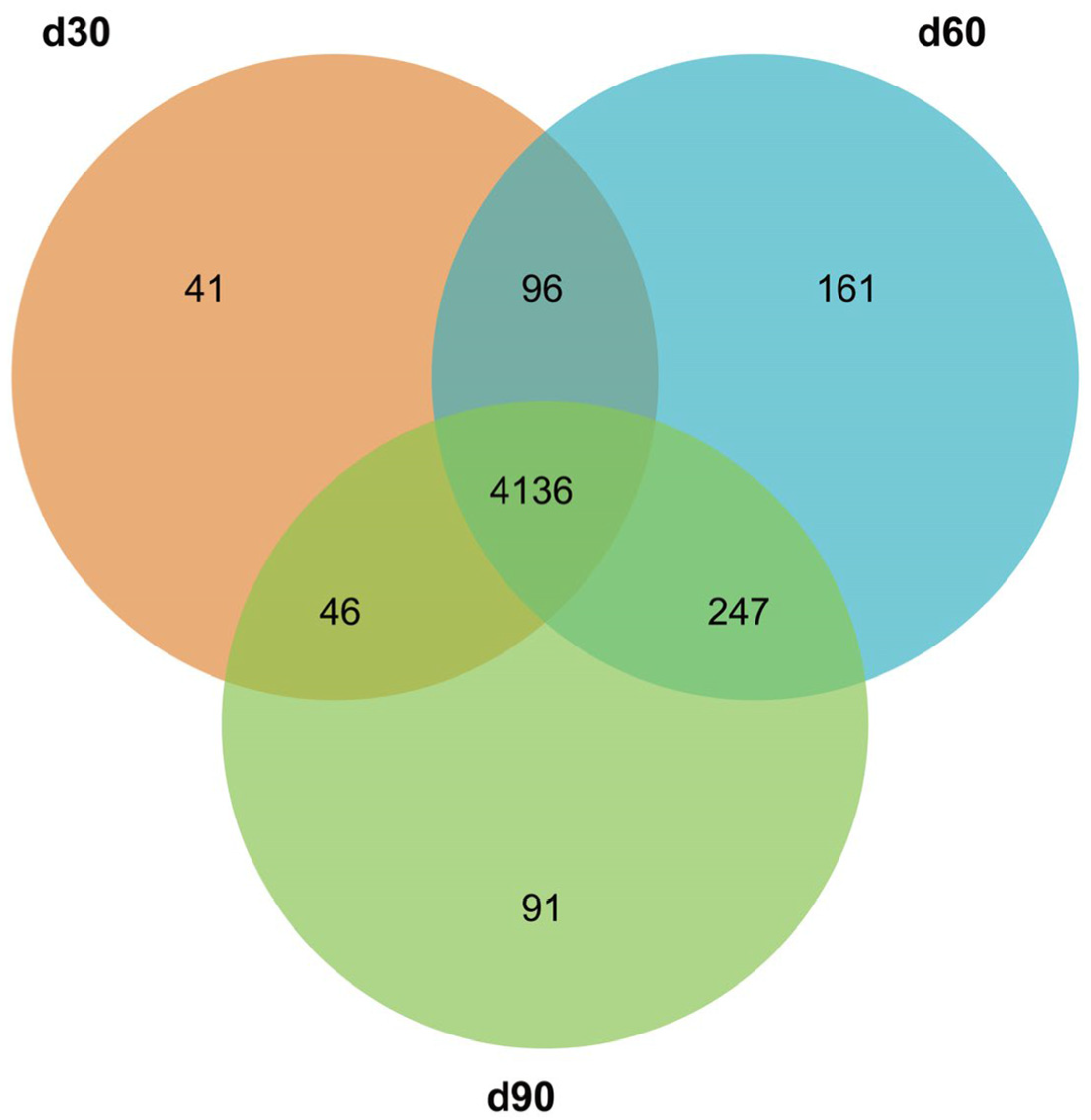
3.3. Screening for Differentially Expressed miRNAs

The miRNAs that were differentially expressed in the different comparison groups (d30 vs. d60, d90 vs. d60, and d30 vs. d90) were filtered out. A total of 292, 102, and 60 miRNAs were found to be differentially expressed in the three comparison groups, respectively. In total, 122 DEMs were significantly up-regulated in d30 compared to the expression of miRNAs in d60, and 170 DEMs were significantly down-regulated. Compared with the expression of miRNA in d90, 38 DEMs were significantly up-regulated in d30, 64 DEMs were significantly down-regulated in d30; the expression of 40 DEMs was significantly up-regulated in d60, and 20 DEMs were significantly down-regulated in d60 (Figure 3, Supplementary Table S1).

3.4. Target Genes’ Prediction of Differentially Expressed miRNAs

The target genes of DEMs in the three comparison groups were predicted using two algorithms, TargetScan and miRanda. A total of 14,050 target genes were identified after the intersection, of which 13,086 genes were predicted for conserved miRNAs, and 9584 genes were predicted for novel miRNAs. Specifically, 13,162, 13,807, and 13,725 target genes were obtained in antler growth centers at days 30, 60, and 90, respectively.
In terms of differentially expressed miRNAs, there were 3419, 3719, and 3665 in the d30 vs. d60, d30 vs. d90, and d60 vs. d90 comparison groups, respectively. Specifically, in the 30d vs. 60d comparison group, up-regulated DEMs predicted 1189 target genes, and down-regulated DEMs predicted 2663 target genes. In the d30 vs. d90 comparison group, up-regulated DEMs predicted 3308 target genes, and down-regulated DEMs predicted 730 target genes. In the d60 vs. d90 comparison group, up-regulated DEMs predicted 3603 target genes, and down-regulated DEMs predicted 115 target genes.

3.5. Target Genes’ Functional Analysis of Differentially Expressed miRNAs

We examined the roles played by the target genes of the DEMs to better understand the roles of miRNA target genes in antler growth centers during various growth phases. The results of the GO enrichment analysis showed that the target genes of differentially expressed miRNAs were significantly enriched in 30 GO categories (p < 0.05) in all three comparison groups, including “cellular process”, “biological regulation”, and “single-organism process” in Biological Process (BP); “cell part”, “cell”, and “organelle” in Cellular Component (CC); and “binding” and “catalytic activity” in Molecular Function (MF) (Figure 4).
The KEGG pathway enrichment analysis revealed that in the d30 vs. d60 comparison group, the functions of the target genes of DEMs were significantly enriched in 232 KEGG pathways, including “Type II diabetes mellitus”, “Axon guidance”, and “Small cell lung cancer”, among others. The target genes of DEMs were enriched in 230 KEGG pathways in the comparison group between d30 and d90, with target genes significantly enriched in “Basal cell carcinoma”, “Axon guidance”, “Endocytosis”, etc. In the d60 vs. d90 comparison group, differential target genes were significantly enriched in “Basal cell carcinoma”, “Endocytosis”, “Axon guidance”, etc. (p < 0.05). Figure 5 shows the top 20 signaling pathways with the highest confidence of enrichment significance (i.e., the smallest Q value) across the three comparison groups.
Additionally, we were intrigued by the molecular processes that underlining the quick development of velvet antlers in the rapid growth stage (about 60 days). As miRNAs usually negatively regulate the expression of their target genes, in this study, we screened four DEMs that were lowly expressed in antler growth centers at 60 days and highly expressed in antler growth center tissues at 30 and 90 days, including ppy-miR-1, mmu-miR-200b-3p, novel miR-6, and novel miR-94. In addition, there was another miRNA (novel miR-10) with expression characteristics that were opposite to those of the above four miRNAs. Target gene prediction results showed that ppy-miR-1, mmu-miR-200b-3p, novel miR-6, novel miR-94, and novel miR-10 predicted 821, 1154, 121, 458, and 737 target genes, respectively. The results of the KEGG pathway annotation showed that the target genes of ppy-miR-1, mmu-miR-200b-3p, novel miR-6, novel miR-94, and novel miR-10 were significantly annotated to “Ras signaling pathway”, “Neurotrophin signaling pathway”, “Glioma”, “Pathways in cancer”, and “Thyroid hormone signaling pathway” (p < 0.05), respectively. Furthermore, the target genes of these miRNAs were also annotated to the “Wnt signaling pathway”, “PI3K-Akt signaling pathway”, “MAPK signaling pathway”, “TGF-β signaling pathway”, and “mTOR signaling pathway”, and other classical signaling pathways (Supplementary Table S2). It is possible that these miRNAs may play important roles in the rapid growth of velvet antlers, which needs to be further verified.

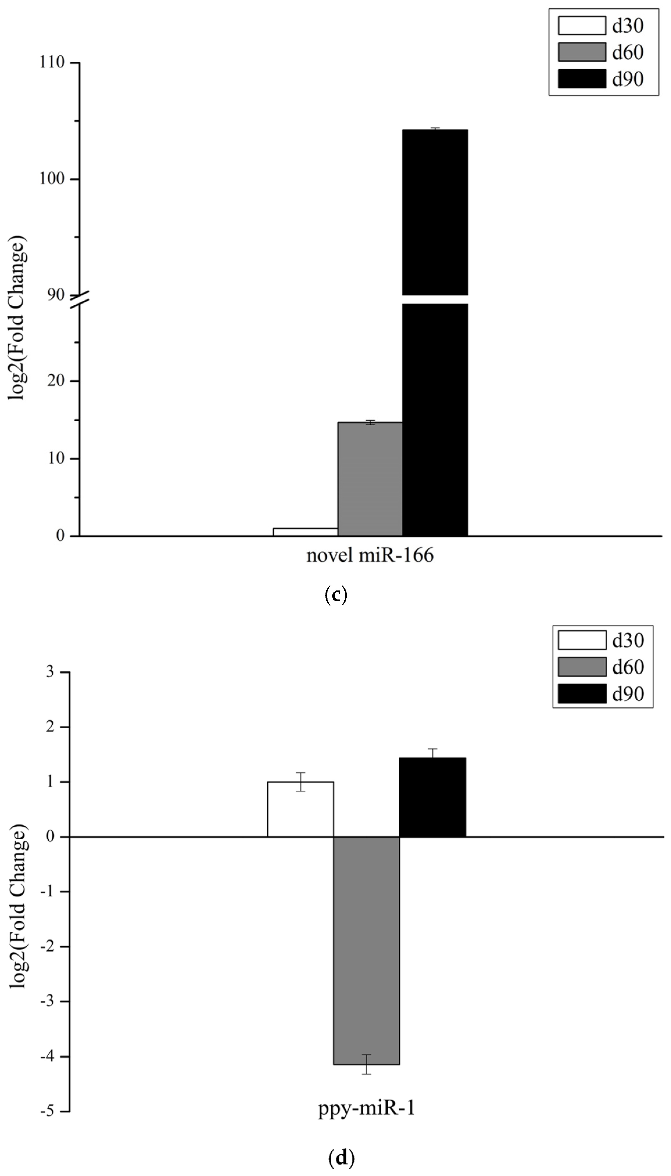
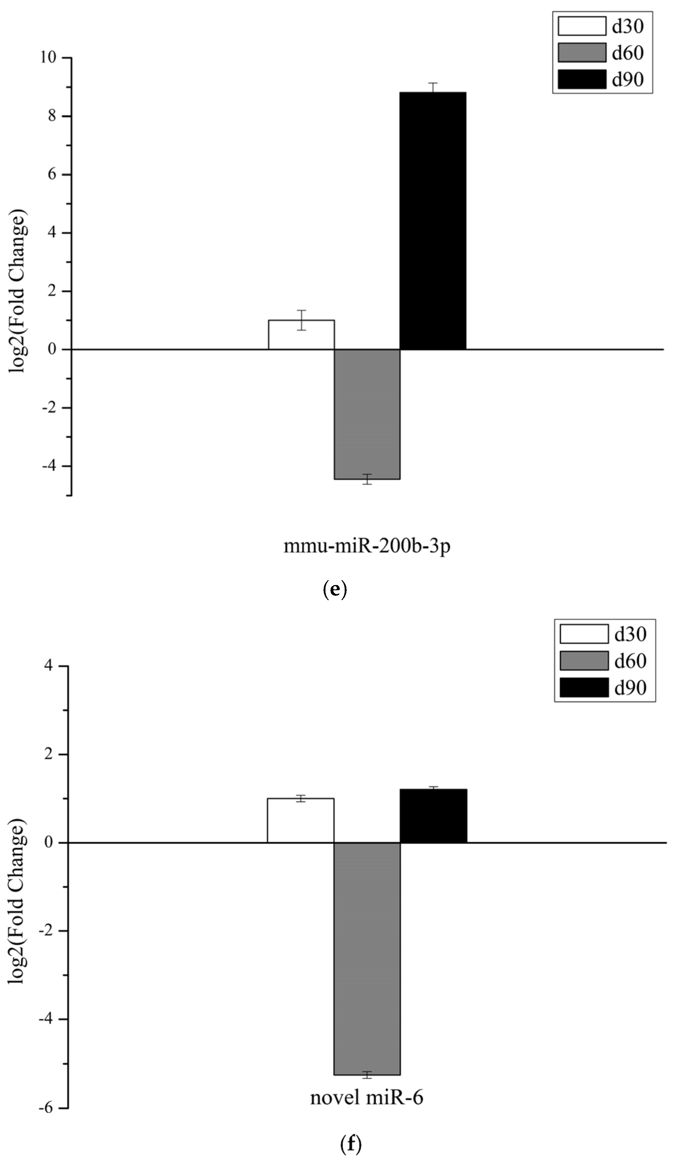
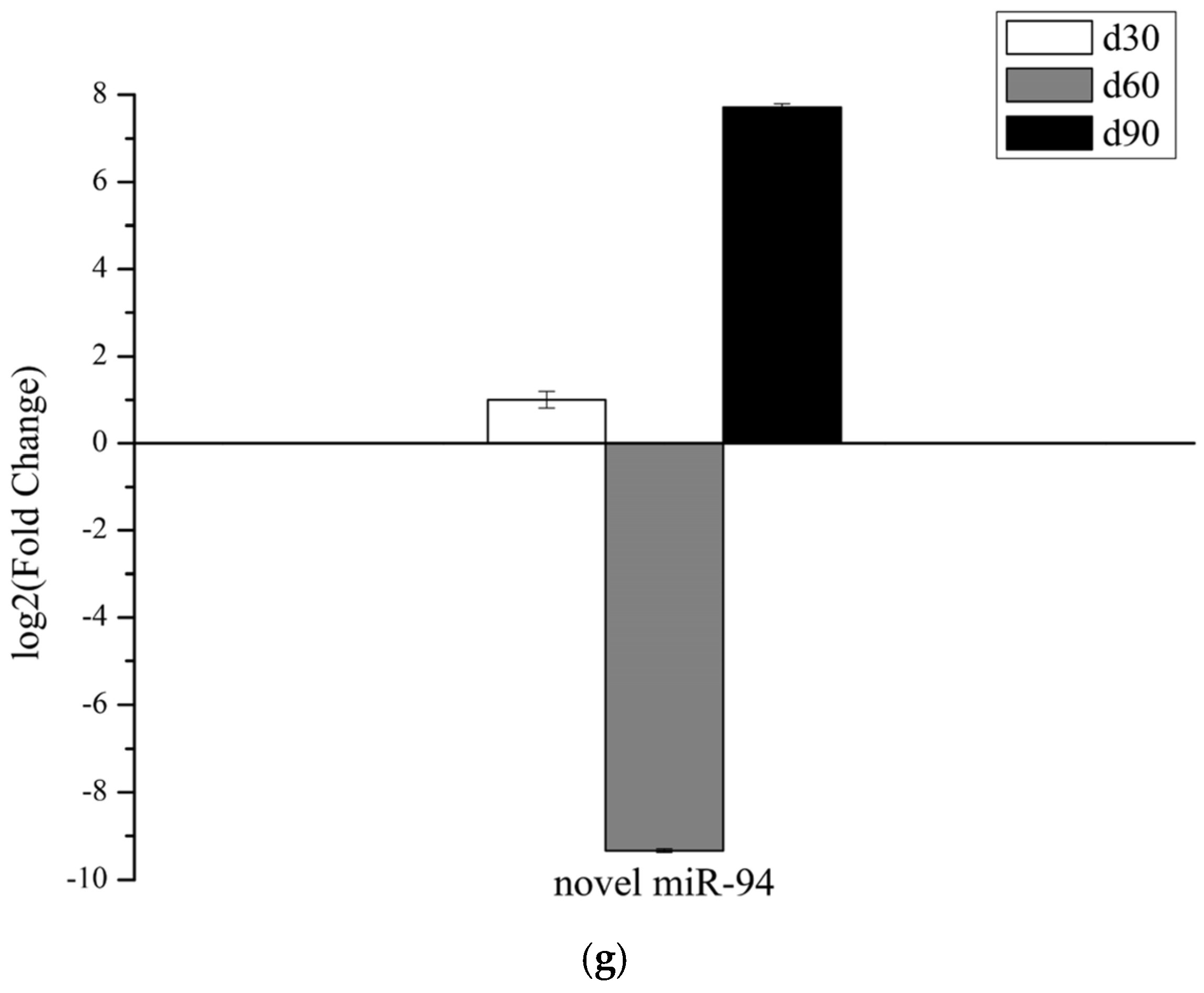
3.6. Validation of miRNAs Expression

The Venn Diagram package of R language was used to analyze the differential miRNAs, and there were 13 common DEMs in the three comparison groups. After removing the miRNAs with consistent, mature sequences, three DEMs (bta-miR-1298, bta-miR-486, and novel miR-166) were retained for verification via stem-loop RT-qPCR. In addition, to further investigate the function of the four DEMs (ppy-miR-1, mmu-miR-200b-3p, novel miR-6, and novel miR-94), their relative expression in the growth centers of velvet antlers in three growth stages was also detected.
The expression levels of bta-miR-1298 and novel miR-166 increased with antler growth, and the expression levels of bta-miR-486 showed a rising then falling trend, while the expression levels of ppy-miR-1, mmu-miR-200b-3p, novel miR-6, and novel miR-94 showed a falling then rising trend, which was in line with the expression trend in the sequencing data, showing that the small RNA sequencing results were accurate (Figure 6).

4. Discussion

Antler growth is primarily driven and controlled by the antler apical growth center—which is made up of velvet skin, mesenchyme, and cartilage tissue—by the process of endochondral ossification [1,9]. The antler skin and cartilage tissues in the antler growth center are rich in vascular tissue and differ greatly from normal skin and cartilage tissues, which is inevitably associated with the astonishing growth rate of the antler and its capacity for repair and regeneration [29]. Therefore, the investigation of changes in gene expression in antler growth centers during different growth periods is highly significant in order to reveal the mechanism of rapid antler growth. The majority of organ growth is driven by the combinatorial effects of cell proliferation and cell expansion [30].
The purpose of this study was to deep sequence for miRNAs in velvet antler growth centers at different growth stages. In total, 292, 102, and 60 miRNAs were found to be differentially expressed in the d30 vs. d60, d30 vs. d90, and d60 vs. d90 comparison groups, respectively. Furthermore, 10 miRNAs were differentially expressed in the d30 vs. d60 and d60 vs. d90 comparison groups but not in the d30 vs. d90 group, among which four differentially expressed miRNAs (ppy-miR-1, mmu-miR-200b-3p, novel miR-6, and novel miR-94) with high expression levels in the antler growth center of 30 and 90 days and low expression in the antler growth center of 60 days. Meanwhile, the reverse was true for another miRNA (novel miR-10). Given that miRNAs typically function by down-regulating the expression of their target genes [31], five differentiated miRNAs were screened, which could play important roles in the rapid growth of velvet antlers. Further validation is required to determine whether miRNAs that are highly or lowly expressed in antler growth centers at 60 days of growth promote antler cell proliferation by suppressing the expression of their target genes.
It has been reported that miR-1 has a broad range of biological functions, e.g., in modulating skeletal muscle proliferation and differentiation [32,33], playing roles in heart disease [34,35,36,37], sex determination [38], and diabetes [39]. MiR-1 also plays critical roles in chondrogenesis [40]; the proliferation, differentiation, and hypertrophy of chondrocytes [41,42]; and the development of osteoarthritis [43,44]. The growth center of the antler tip drives the longitudinal endochondral ossification process of velvet antlers [45,46]. It is probable that miR-1 has a significant impact on this procedure. In one study, it was discovered that miR-1 was capable of suppressing the proliferation of chondrocytes in Chinese sika deer by specifically targeting the 3′UTR of IGF-1 [47]. In the present study, ppy-miR-1 was lowly expressed in the growth center of antlers at 60 days of growth and highly expressed at 30 and 90 days of growth, which may be closely related to the rapid growth and ossification of antlers.
MiR-200b is a miRNA with a wide range of functions, including functions in multiple cancers or tumors [48,49,50,51], endotheliogenesis [52,53,54], angiogenesis [55], etc. It has been reported that a decreased level of miR-200b-3p may result in angiogenesis in HCC [56]. The growing tips of velvet antlers are rich in blood vessels [4,19,57,58,59]. The antler grows quickly, including its interior components, such as the blood vessels, nerves, and skin that join it [59]. Mmu-miR-200b-3p may promote angiogenesis in antlers during the rapid growth phase, which would provide nutritional and other types of support for the rapid growth of antlers.
The novel miR-94, the target genes of which are strongly annotated to the “Pathways in cancer” and “Wnt signaling pathway”, is the miRNA in which we are most interested. It was shown that the proliferation of velvet antler cells was faster than that of cancer cells during the rapid growth stage of antlers [23,60,61]. Academics refer to this phenomenon as cancer-like growth [62,63]. “Pathways in cancer” is a complicated signaling pathway, forming a complex network with the “ERK signaling pathway”, “PI3K-AKT signaling pathway”, “Wnt signaling pathway”, “Notch signaling pathway”, “TGF-β signaling pathway”, “HIF-1 signaling pathway”, and other signaling pathways. Studies showed that the “ERK signaling pathway” [64], “PI3K-AKT signaling pathway” [65,66,67], “Notch signaling pathway” [68,69], “TGF-β signaling pathway” [70,71], “Wnt signaling pathway” [72,73,74], and “HIF-1 signaling pathway” [66] were involved in the development and regeneration of velvet antlers. Thus, more research is necessary to determine if novel miR-94 and its target genes have a major impact on the quick development and even the regeneration of velvet antlers.
The target genes of novel miR-6 annotated fewer signaling pathways than ppy-miR-1, mmu-miR-200b-3p, and novel miR-94, and the target genes are significantly enriched in “mTOR signaling pathway”, “Pathways in cancer”, “Rap1 signaling pathway” and “signaling pathways regulating pluripotency of stem cells”. mTOR is a vital regulator of multiple cellular functions, including cell proliferation, differentiation, and protein synthesis [75]. The mTOR signaling pathway can stimulate the protein synthesis of chondrocytes to regulate mammalian limb skeletal growth [76]. Studies have indicated that mTOR is an important regulator of cartilage development [77,78,79,80]. The Rap1 signaling pathway is a cancer-related signaling pathway with a wide range of biological functions [81,82,83,84]. The longitudinal section of the antler was divided into velvet skin, mesenchyme, pre-cartilage, and cartilage from top to bottom, which clearly reflected the dynamic process of antler ossification, indicating that the growth and development of antler is essentially the process of bone tissue growth and development [85]. All factors that theoretically play a relevant role in bone tissue development may be involved in the regulation of antler growth and development. Studies on the regulation of antler growth by the mTOR signaling pathway and Rap1 signaling pathway have not been reported yet. Whether novel miR-6 regulates the proliferation of antler cells through these two pathways remains to be further verified. The antler mesenchyme is filled with mesenchymal cells (reserve mesenchyme cells, RMCs) that have the ability to divide vigorously and differentiate towards chondrocytes, which ultimately secrete the bone matrix to complete the ossification [7]. RMCs are stem cells that drive the rapid growth of velvet antlers and are stromal cells capable of self-replication and have the potential to differentiate into chondrocytes, etc. [86]. Target genes of novel miR-6 annotated to the “Signaling pathways regulating pluripotency of stem cells”, this miRNA and its target genes may be involved in the differentiation of RMCs into chondrocytes. Taken together, the screened DEMs have a certain significance for the study of the rapid growth of velvet antlers, and the regulatory mechanism remains to be further investigated.

5. Conclusions

In summary, we detected miRNAs that were differentially expressed in antler growth centers at different stages of growth using an RNA-seq approach. Following the comparison and analysis, we screened five differentially expressed miRNAs with target genes that were significantly enriched in the “Wnt signaling pathway”, “PI3K-Akt signaling pathway”, “MAPK signaling pathway”, “TGF-β signaling pathway”, “mTOR signaling pathway”, and other classical signaling pathways. These miRNAs, especially ppy-miR-1, mmu-miR-200b-3p, and novel miR-94, could be extremely important for the rapid development of antlers.

Supplementary Materials

The following supporting information can be downloaded at: https://www.mdpi.com/article/10.3390/genes14020424/s1. Table S1: All differentially expressed miRNAs; Table S2: KEGG pathway annotation of target genes for five selected differentially expressed miRNAs.

Author Contributions

Conceptualization, Y.C. and Z.Z.; methodology, Z.Z.; software, Z.Z.; validation, C.H., C.B. and Z.L.; formal analysis, Z.Z.; investigation, Z.Z.; resources, Y.C.; data curation, Y.C.; writing—original draft preparation, Z.Z.; writing—review and editing, Y.C. visualization, W.J.; supervision, C.L.; project administration, Y.C.; funding acquisition, Y.C. All authors have read and agreed to the published version of the manuscript.

Funding

This research was funded by the Science and Technology Innovation Project of Three RiverSource First-class Discipline, College of Eco–Environment Engineering, Qinghai University, grant number 2021-JS-02, and The Doctoral Scientific Research Start-up Foundation of Qinghai University, grant number 2021-BS-093.

Institutional Review Board Statement

All experimental protocols were approved by the Institutional Animal Care and Use Committee of Qinghai University (Xining, China), and all methods were carried out to approved guidelines and regulations (Code: SL-2022024, Approval Date: 17 March 2022).

Informed Consent Statement

Not applicable.

Data Availability Statement

Raw data were submitted to the National Center for Biotechnology Information (NCBI) Sequence Read Archive (SRA) database (registry number SUB12298775, BioProject: PRJNA903551). Available online: https://submit.ncbi.nlm.nih.gov/subs/bioproject/SUB12298775/overview.

Acknowledgments

We are grateful to Qinghai University for the financial support of this research in the form of a doctoral fellowship for Yanxia Chen. We would like to thank Shandan Farm for the sample resources.

Conflicts of Interest

The authors declare no conflict of interest.

References

  1. Price, J.S.; Allen, S.; Faucheux, C.; Althnaian, T.; Mount, J.G. Deer antlers: A zoological curiosity or the key to understanding organ regeneration in mammals? J. Anat. 2005, 207, 603–618. [Google Scholar] [CrossRef] [PubMed]
  2. Goss, R.J. Future directions in antler research. Anat. Rec. 1995, 241, 291–302. [Google Scholar] [CrossRef] [PubMed]
  3. Li, C.; Yang, F.; Sheppard, A. Adult stem cells and mammalian epimorphic regeneration-insights from studying annual renewal of deer antlers. Curr. Stem Cell Res. Ther. 2009, 4, 237–251. [Google Scholar] [CrossRef]
  4. Dong, Z.; Coates, D. Bioactive Molecular Discovery Using Deer Antlers as a Model of Mammalian Regeneration. J. Proteome Res. 2021, 20, 2167–2181. [Google Scholar] [CrossRef]
  5. Li, C.; Suttie, J.M.; Clark, D.E. Morphological observation of antler regeneration in red deer (Cervus elaphus). J. Morphol. 2004, 262, 731–740. [Google Scholar] [CrossRef] [PubMed]
  6. Fennessy, P.F. Deer Antlers: Regeneration, function and evolution. J. R. Soc. N. Z. 1984, 14, 290–291. [Google Scholar] [CrossRef]
  7. Gyurjan, I., Jr.; Molnar, A.; Borsy, A.; Steger, V.; Hackler, L., Jr.; Zomborszky, Z.; Papp, P.; Duda, E.; Deak, F.; Lakatos, P.; et al. Gene expression dynamics in deer antler: Mesenchymal differentiation toward chondrogenesis. Mol. Genet. Genom. 2007, 277, 221–235. [Google Scholar] [CrossRef]
  8. Waldo, C.M.; Wislocki, G.B.; Fawcett, D.W. Observations on the blood supply of growing antlers. Am. J. Anat. 1949, 84, 27–61. [Google Scholar] [CrossRef]
  9. Li, C.; Clark, D.E.; Lord, E.A.; Stanton, J.A.; Suttie, J.M. Sampling technique to discriminate the different tissue layers of growing antler tips for gene discovery. Anat. Rec. 2002, 268, 125–130. [Google Scholar] [CrossRef]
  10. Price, J.; Allen, S. Exploring the mechanisms regulating regeneration of deer antlers. Philos. Trans. R. Soc. Lond. B Biol. Sci. 2004, 359, 809–822. [Google Scholar] [CrossRef]
  11. Yao, B.; Zhao, Y.; Wang, Q.; Zhang, M.; Liu, M.; Liu, H.; Li, J. De novo characterization of the antler tip of Chinese Sika deer transcriptome and analysis of gene expression related to rapid growth. Mol. Cell. Biochem. 2012, 364, 93–100. [Google Scholar] [CrossRef]
  12. Zhao, Y.; Yao, B.; Zhang, M.; Wang, S.; Zhang, H.; Xiao, W. Comparative analysis of differentially expressed genes in Sika deer antler at different stages. Mol. Biol. Rep. 2013, 40, 1665–1676. [Google Scholar] [CrossRef] [PubMed]
  13. Yao, B.; Zhao, Y.; Zhang, H.; Zhang, M.; Liu, M.; Liu, H.; Li, J. Sequencing and de novo analysis of the Chinese Sika deer antler-tip transcriptome during the ossification stage using Illumina RNA-Seq technology. Biotechnol. Lett. 2012, 34, 813–822. [Google Scholar] [CrossRef]
  14. Ho, P.T.B.; Clark, I.M.; Le, L.T.T. MicroRNA-Based Diagnosis and Therapy. Int. J. Mol. Sci. 2022, 23, 7167. [Google Scholar] [CrossRef]
  15. Lu, T.X.; Rothenberg, M.E. MicroRNA. J. Allergy Clin. Immunol. 2018, 141, 1202–1207. [Google Scholar] [CrossRef]
  16. Bartel, D.P. MicroRNAs: Genomics, biogenesis, mechanism, and function. Cell 2004, 116, 281–297. [Google Scholar] [CrossRef] [PubMed]
  17. Saliminejad, K.; Khorram Khorshid, H.R.; Soleymani Fard, S.; Ghaffari, S.H. An overview of microRNAs: Biology, functions, therapeutics, and analysis methods. J. Cell. Physiol. 2019, 234, 5451–5465. [Google Scholar] [CrossRef]
  18. Bhaskaran, M.; Mohan, M. MicroRNAs. Vet. Pathol. 2013, 51, 759–774. [Google Scholar] [CrossRef] [PubMed]
  19. Hu, P.; Wang, T.; Liu, H.; Xu, J.; Wang, L.; Zhao, P.; Xing, X. Full-length transcriptome and microRNA sequencing reveal the specific gene-regulation network of velvet antler in sika deer with extremely different velvet antler weight. Mol. Genet. Genom. 2019, 294, 431–443. [Google Scholar] [CrossRef]
  20. Yao, B.; Zhang, M.; Liu, M.; Lu, B.; Leng, X.; Hu, Y.; Zhao, D.; Zhao, Y.U. Identification of the miRNA-mRNA regulatory network of antler growth centers. J. Biosci. 2019, 44, 11. [Google Scholar] [CrossRef]
  21. Wang, Y.; Hu, W. Progress of Noncoding RNA Regulating the Growth and Development of Antler Tissue Research. Biomed Res. Int. 2022, 2022, 3541577. [Google Scholar] [CrossRef]
  22. Chen, Y.; Zhang, Z.; Zhang, J.; Chen, X.; Guo, Y.; Li, C. RNA sequencing-based identification of microRNAs in the antler cartilage of Gansu red deer (Cervus elaphus kansuensis). PeerJ 2022, 10, e13947. [Google Scholar] [CrossRef] [PubMed]
  23. Jia, B.; Zhang, L.; Zhang, Y.; Ge, C.; Yang, F.; Du, R.; Ba, H. Integrated analysis of miRNA and mRNA transcriptomic reveals antler growth regulatory network. Mol. Genet. Genom. 2021, 296, 689–703. [Google Scholar] [CrossRef]
  24. Ba, H.; Wang, D.; Li, C. MicroRNA profiling of antler stem cells in potentiated and dormant states and their potential roles in antler regeneration. Mol. Genet. Genom. 2016, 291, 943–955. [Google Scholar] [CrossRef]
  25. Grabherr, M.G.; Haas, B.J.; Yassour, M.; Levin, J.Z.; Thompson, D.A.; Amit, I.; Adiconis, X.; Fan, L.; Raychowdhury, R.; Zeng, Q.; et al. Full-length transcriptome assembly from RNA-Seq data without a reference genome. Nat. Biotechnol. 2011, 29, 644–652. [Google Scholar] [CrossRef] [PubMed]
  26. Hofacker, I.L. Vienna RNA secondary structure server. Nucleic Acids Res. 2003, 31, 3429–3431. [Google Scholar] [CrossRef]
  27. Zhao, S.; Ye, Z.; Stanton, R. Misuse of RPKM or TPM normalization when comparing across samples and sequencing protocols. RNA 2020, 26, 903–909. [Google Scholar] [CrossRef] [PubMed]
  28. Anders, S.; Huber, W. Differential expression analysis for sequence count data. Genome Biol. 2010, 11, R106. [Google Scholar] [CrossRef]
  29. Clark, D.E.; Li, C.; Wang, W.; Martin, S.K.; Suttie, J.M. Vascular localization and proliferation in the growing tip of the deer antler. Anat. Rec. A Discov. Mol. Cell. Evol. Biol. 2006, 288, 973–981. [Google Scholar] [CrossRef]
  30. Rodriguez, R.E.; Schommer, C.; Palatnik, J.F. Control of cell proliferation by microRNAs in plants. Curr. Opin. Plant Biol. 2016, 34, 68–76. [Google Scholar] [CrossRef]
  31. Hill, M.; Tran, N. miRNA interplay: Mechanisms and consequences in cancer. Dis. Model. Mech. 2021, 14, dmm047662. [Google Scholar] [CrossRef] [PubMed]
  32. Chen, J.F.; Mandel, E.M.; Thomson, J.M.; Wu, Q.; Callis, T.E.; Hammond, S.M.; Conlon, F.L.; Wang, D.Z. The role of microRNA-1 and microRNA-133 in skeletal muscle proliferation and differentiation. Nat. Genet. 2006, 38, 228–233. [Google Scholar] [CrossRef]
  33. Wu, N.; Gu, T.; Lu, L.; Cao, Z.; Song, Q.; Wang, Z.; Zhang, Y.; Chang, G.; Xu, Q.; Chen, G. Roles of miRNA-1 and miRNA-133 in the proliferation and differentiation of myoblasts in duck skeletal muscle. J. Cell. Physiol. 2019, 234, 3490–3499. [Google Scholar] [CrossRef]
  34. Wang, J.D.; Qiu, F.; Li, W. miRNA-1: A novel, potential therapeutic target in the treatment of acute myocardial infarction? Int. J. Cardiol. 2021, 333, 53. [Google Scholar] [CrossRef]
  35. Liu, Z.; Liu, Y.; Zhu, Y.; Gong, J. HOTAIR/miRNA-1/Cx43: A potential mechanism for treating myocardial ischemia-reperfusion injury. Int. J. Cardiol. 2020, 308, 11. [Google Scholar] [CrossRef]
  36. Jayawardena, E.; Medzikovic, L.; Ruffenach, G.; Eghbali, M. Role of miRNA-1 and miRNA-21 in Acute Myocardial Ischemia-Reperfusion Injury and Their Potential as Therapeutic Strategy. Int. J. Mol. Sci. 2022, 23, 1512. [Google Scholar] [CrossRef]
  37. Cai, Q.; Gao, M.L.; Huang, L.S.; Pan, L.H. lncRNA H19/miRNA-1: Another potential mechanism for treating myocardial ischemia-reperfusion injury. Int. J. Cardiol. 2021, 322, 57. [Google Scholar] [CrossRef] [PubMed]
  38. Peng, W.; Yu, S.; Handler, A.M.; Tu, Z.; Saccone, G.; Xi, Z.; Zhang, H. miRNA-1-3p is an early embryonic male sex-determining factor in the Oriental fruit fly Bactrocera dorsalis. Nat. Commun. 2020, 11, 932. [Google Scholar] [CrossRef]
  39. Feng, B.; Cao, Y.; Chen, S.; Ruiz, M.; Chakrabarti, S. miRNA-1 regulates endothelin-1 in diabetes. Life Sci. 2014, 98, 18–23. [Google Scholar] [CrossRef] [PubMed]
  40. Li, P.; Wei, X.; Guan, Y.; Chen, Q.; Zhao, T.; Sun, C.; Wei, L. MicroRNA-1 regulates chondrocyte phenotype by repressing histone deacetylase 4 during growth plate development. FASEB J. 2014, 28, 3930–3941. [Google Scholar] [CrossRef] [Green Version]
  41. Chen, T.; Che, X.; Han, P.; Lu, J.; Wang, C.; Liang, B.; Hou, Z.; Wei, X.; Wei, L.; Li, P. MicroRNA-1 promotes cartilage matrix synthesis and regulates chondrocyte differentiation via post-transcriptional suppression of Ihh expression. Mol. Med. Rep. 2020, 22, 2404–2414. [Google Scholar] [CrossRef]
  42. Cong, L.; Jiang, P.; Wang, H.; Huang, L.; Wu, G.; Che, X.; Wang, C.; Li, P.; Duan, Q.; Guo, X.; et al. MiR-1 is a critical regulator of chondrocyte proliferation and hypertrophy by inhibiting Indian hedgehog pathway during postnatal endochondral ossification in miR-1 overexpression transgenic mice. Bone 2022, 165, 116566. [Google Scholar] [CrossRef] [PubMed]
  43. Che, X.; Chen, T.; Wei, L.; Gu, X.; Gao, Y.; Liang, S.; Li, P.; Shi, D.; Liang, B.; Wang, C.; et al. MicroRNA-1 regulates the development of osteoarthritis in a Col2a1-Cre-ERT2/GFPfl/fl-RFP-miR-1 mouse model of osteoarthritis through the downregulation of Indian hedgehog expression. Int. J. Mol. Med. 2020, 46, 360–370. [Google Scholar] [CrossRef] [PubMed]
  44. Yang, Y.; Wang, Y.; Jia, H.; Li, B.; Xing, D.; Li, J.J. MicroRNA-1 Modulates Chondrocyte Phenotype by Regulating FZD7 of Wnt/β-Catenin Signaling Pathway. Cartilage 2021, 13, 1019S–1029S. [Google Scholar] [CrossRef] [PubMed]
  45. Zhang, R.; Li, Y.; Xing, X. Comparative antler proteome of sika deer from different developmental stages. Sci. Rep. 2021, 11, 10484. [Google Scholar] [CrossRef]
  46. Dong, Z.; Li, C.; Coates, D. PTN-PTPRZ signalling is involved in deer antler stem cell regulation during tissue regeneration. J. Cell. Physiol. 2021, 236, 3752–3769. [Google Scholar] [CrossRef]
  47. Hu, W.; Meng, X.; Lu, T.; Wu, L.; Li, T.; Li, M.; Tian, Y. MicroRNA1 inhibits the proliferation of Chinese sika deerderived cartilage cells by binding to the 3’-untranslated region of IGF1. Mol. Med. Rep. 2013, 8, 523–528. [Google Scholar] [CrossRef]
  48. Ren, S.; Liu, J.; Feng, Y.; Li, Z.; He, L.; Li, L.; Cao, X.; Wang, Z.; Zhang, Y. Knockdown of circDENND4C inhibits glycolysis, migration and invasion by up-regulating miR-200b/c in breast cancer under hypoxia. J. Exp. Clin. Cancer Res. 2019, 38, 388. [Google Scholar] [CrossRef]
  49. Guan, W.; Cui, H.; Huang, P.; Chun, W.J.; Lee, J.W.; Kim, H.; Zou, H. miR-200b/200a/429 Cluster Stimulates Ovarian Cancer Development by Targeting ING5. J. Oncol. 2020, 2020, 3404059. [Google Scholar] [CrossRef]
  50. Chen, S.; Tu, Y.; Yuan, H.; Shi, Z.; Guo, Y.; Gong, W.; Tu, S. Regulatory functions of miR200b3p in tumor development (Review). Oncol. Rep. 2022, 47, 1–9. [Google Scholar] [CrossRef]
  51. Zhang, Z.; Xing, T.; Chen, Y.; Xiao, J. Exosome-mediated miR-200b promotes colorectal cancer proliferation upon TGF-beta1 exposure. Biomed. Pharmacother. 2018, 106, 1135–1143. [Google Scholar] [CrossRef] [PubMed]
  52. Zhang, F.; Cheng, N.; Du, J.; Zhang, H.; Zhang, C. MicroRNA-200b-3p promotes endothelial cell apoptosis by targeting HDAC4 in atherosclerosis. BMC Cardiovasc. Disord. 2021, 21, 172. [Google Scholar] [CrossRef] [PubMed]
  53. Yamazaki-Takai, M.; Takai, H.; Iwai, Y.; Noda, K.; Mezawa, M.; Tsuruya, Y.; Yamaguchi, A.; Nakayama, Y.; Ogata, Y. MiR-200b suppresses TNF-α-induced AMTN production in human gingival epithelial cells. Odontology 2021, 109, 403–410. [Google Scholar] [CrossRef]
  54. Tamagawa, S.; Enomoto, K.; Gunduz, E.; Gunduz, M.; Sato, F.; Uchino, S.; Muragaki, Y.; Hotomi, M. MicroRNA 200b promotes mesenchymal-to-epithelial transition in anaplastic thyroid carcinoma. Oncol. Lett. 2020, 20, 3. [Google Scholar] [CrossRef]
  55. Sinha, M.; Ghatak, S.; Roy, S.; Sen, C.K. microRNA-200b as a Switch for Inducible Adult Angiogenesis. Antioxid. Redox Signal. 2015, 22, 1257–1272. [Google Scholar] [CrossRef]
  56. Moh-Moh-Aung, A.; Fujisawa, M.; Ito, S.; Katayama, H.; Ohara, T.; Ota, Y.; Yoshimura, T.; Matsukawa, A. Decreased miR-200b-3p in cancer cells leads to angiogenesis in HCC by enhancing endothelial ERG expression. Sci. Rep. 2020, 10, 10418. [Google Scholar] [CrossRef]
  57. Li, C. Deer antler regeneration: A stem cell-based epimorphic process. Birth Defects Res. C Embryo Today 2012, 96, 51–62. [Google Scholar] [CrossRef] [PubMed]
  58. Li, C.; Stanton, J.A.; Robertson, T.M.; Suttie, J.M.; Sheard, P.W.; Harris, A.J.; Clark, D.E. Nerve growth factor mRNA expression in the regenerating antler tip of red deer (Cervus elaphus). PLoS ONE 2007, 2, e148. [Google Scholar] [CrossRef]
  59. Wei, G.; Qin, T.; Li, X.; Wang, Z.; Wang, Y.; Guan, Q.; Shi, W.; Xie, L.; Zhao, S.; Sun, H. Constructing the in vitro culture system of the sika deer (cervus nippon) antler periosteal cell to detect its function on antler regeneration. Front. Biosci. (Landmark Ed.) 2022, 27, 69. [Google Scholar] [CrossRef]
  60. Han, R.; Han, L.; Wang, S.; Li, H. Whole Transcriptome Analysis of Mesenchyme Tissue in Sika Deer Antler Revealed the CeRNAs Regulatory Network Associated With Antler Development. Front. Genet. 2019, 10, 1403. [Google Scholar] [CrossRef] [Green Version]
  61. Chen, Y.; Zhang, Z.; Jin, W.; Li, Z.; Bao, C.; He, C.; Guo, Y.; Li, C. Integrative Analyses of Antler Cartilage Transcriptome and Proteome of Gansu Red Deer (Cervus elaphus kansuensis) at Different Growth Stages. Animals 2022, 12, 934. [Google Scholar] [CrossRef] [PubMed]
  62. Goss, R.J. Tumor-like growth of antlers in castrated fallow deer: An electron microscopic study. Scanning Microsc. 1990, 4, 715–720; discussion 720–711. [Google Scholar]
  63. Li, C.; Waldrup, K.A.; Corson, I.D.; Littlejohn, R.P.; Suttie, J.M. Histogenesis of antlerogenic tissues cultivated in diffusion chambers in vivo in red deer (Cervus elaphus). J. Exp. Zool. 1995, 272, 345–355. [Google Scholar] [CrossRef] [PubMed]
  64. Wei, J.; Xu, M.; Zhang, X.; Meng, S.; Wang, Y.; Zhou, T.; Ma, Q.; Han, B.; Wei, Y.; Deng, X. Enhanced Osteogenic Behavior of ADSCs Produced by Deproteinized Antler Cancellous Bone and Evidence for Involvement of ERK Signaling Pathway. Tissue Eng. Part A 2015, 21, 1810–1821. [Google Scholar] [CrossRef] [PubMed]
  65. Liu, Z.; Zhao, H.; Wang, D.; McMahon, C.; Li, C. Differential effects of the PI3K/AKT pathway on antler stem cells for generation and regeneration of antlers in vitro. Front. Biosci. (Landmark Ed.) 2018, 23, 1848–1863. [Google Scholar] [CrossRef]
  66. Dong, Z.; Ba, H.; Zhang, W.; Coates, D.; Li, C. iTRAQ-Based Quantitative Proteomic Analysis of the Potentiated and Dormant Antler Stem Cells. Int. J. Mol. Sci. 2016, 17, 1778. [Google Scholar] [CrossRef]
  67. Weng, R.; Lin, H.; Li, Z.; Chen, D.; Lin, X.; Zhang, Z.; Chen, Q.; Yao, Y.; Li, W. Mechanisms of Intervertebral Disc Degeneration Treatment with Deer Antlers Based on Network Pharmacology and Molecular Docking. Evid. Based Complement. Altern. Med. 2022, 2022, 8092848. [Google Scholar] [CrossRef]
  68. Chen, D.Y.; Yang, M.; Sun, Z.T.; Song, M.M.; Yao, H.B.; Long, G.H.; Hu, W. Notch4 affects the proliferation and differentiation of deer antler chondrocytes through the Smad3/lncRNA27785.1 axis. Cell. Signal. 2022, 98, 110429. [Google Scholar] [CrossRef]
  69. Ma, L.; Duan, C.C.; Yang, Z.Q.; Ding, J.L.; Liu, S.; Yue, Z.P.; Guo, B. Crosstalk between Activin A and Shh signaling contributes to the proliferation and differentiation of antler chondrocytes. Bone 2019, 123, 176–188. [Google Scholar] [CrossRef]
  70. Ma, L.; Yang, Z.Q.; Ding, J.L.; Liu, S.; Guo, B.; Yue, Z.P. Function and regulation of transforming growth factor beta1 signalling in antler chondrocyte proliferation and differentiation. Cell Prolif. 2019, 52, e12637. [Google Scholar] [CrossRef]
  71. Chen, D.Y.; Li, Y.J.; Jiang, R.F.; Li, Y.T.; Feng, J.; Hu, W. Effects and mechanism of lncRNA-27785.1 that regulates TGF-beta1 of Sika deer on antler cell proliferation. J. Cell. Physiol. 2021, 236, 5742–5756. [Google Scholar] [CrossRef]
  72. Zhang, H.L.; Yang, Z.Q.; Duan, C.C.; Geng, S.; Wang, K.; Yu, H.F.; Yue, Z.P.; Guo, B. WNT4 acts downstream of BMP2 to mediate the regulation of ATRA signaling on RUNX1 expression: Implications for terminal differentiation of antler chondrocytes. J. Cell. Physiol. 2018, 233, 1129–1145. [Google Scholar] [CrossRef]
  73. Ba, H.; Wang, D.; Yau, T.O.; Shang, Y.; Li, C. Transcriptomic analysis of different tissue layers in antler growth Center in Sika Deer (Cervus nippon). BMC Genom. 2019, 20, 173. [Google Scholar] [CrossRef] [PubMed]
  74. Mount, J.G.; Muzylak, M.; Allen, S.; Althnaian, T.; McGonnell, I.M.; Price, J.S. Evidence that the canonical Wnt signalling pathway regulates deer antler regeneration. Dev. Dyn. 2006, 235, 1390–1399. [Google Scholar] [CrossRef]
  75. Kim, Y.C.; Guan, K.L. mTOR: A pharmacologic target for autophagy regulation. J. Clin. Investig. 2015, 125, 25–32. [Google Scholar] [CrossRef] [PubMed]
  76. Chen, J.; Long, F. mTORC1 signaling controls mammalian skeletal growth through stimulation of protein synthesis. Development 2014, 141, 2848–2854. [Google Scholar] [CrossRef] [PubMed]
  77. Ma, L.; Zhang, R.; Li, D.; Qiao, T.; Guo, X. Fluoride regulates chondrocyte proliferation and autophagy via PI3K/AKT/mTOR signaling pathway. Chem. Biol. Interact. 2021, 349, 109659. [Google Scholar] [CrossRef]
  78. Kim, M.S.; Wu, K.Y.; Auyeung, V.; Chen, Q.; Gruppuso, P.A.; Phornphutkul, C. Leucine restriction inhibits chondrocyte proliferation and differentiation through mechanisms both dependent and independent of mTOR signaling. Am. J. Physiol. Endocrinol. Metab. 2009, 296, E1374. [Google Scholar] [CrossRef] [PubMed]
  79. Feng, F.B.; Qiu, H.Y. Effects of Artesunate on chondrocyte proliferation, apoptosis and autophagy through the PI3K/AKT/mTOR signaling pathway in rat models with rheumatoid arthritis. Biomed. Pharmacother. 2018, 102, 1209–1220. [Google Scholar] [CrossRef]
  80. Xu, K.; He, Y.; Moqbel, S.A.A.; Zhou, X.; Wu, L.; Bao, J. SIRT3 ameliorates osteoarthritis via regulating chondrocyte autophagy and apoptosis through the PI3K/Akt/mTOR pathway. Int. J. Biol. Macromol. 2021, 175, 351–360. [Google Scholar] [CrossRef]
  81. Wang, Y.; Chu, F.; Lin, J.; Li, Y.; Johnson, N.; Zhang, J.; Gai, C.; Su, Z.; Cheng, H.; Wang, L.; et al. Erianin, the main active ingredient of Dendrobium chrysotoxum Lindl, inhibits precancerous lesions of gastric cancer (PLGC) through suppression of the HRAS-PI3K-AKT signaling pathway as revealed by network pharmacology and in vitro experimental verification. J. Ethnopharmacol. 2021, 279, 114399. [Google Scholar] [CrossRef]
  82. Hu, L.; Xie, X.; Xue, H.; Wang, T.; Panayi, A.C.; Lin, Z.; Xiong, Y.; Cao, F.; Yan, C.; Chen, L.; et al. MiR-1224-5p modulates osteogenesis by coordinating osteoblast/osteoclast differentiation via the Rap1 signaling target ADCY2. Exp. Mol. Med. 2022, 54, 961–972. [Google Scholar] [CrossRef] [PubMed]
  83. Liu, M.; Banerjee, R.; Rossa, C., Jr.; D’Silva, N.J. RAP1-RAC1 Signaling Has an Important Role in Adhesion and Migration in HNSCC. J. Dent. Res. 2020, 99, 959–968. [Google Scholar] [CrossRef] [PubMed]
  84. Li, H.; Liang, J.; Wang, J.; Han, J.; Li, S.; Huang, K.; Liu, C. Mex3a promotes oncogenesis through the RAP1/MAPK signaling pathway in colorectal cancer and is inhibited by hsa-miR-6887-3p. Cancer Commun. 2021, 41, 472–491. [Google Scholar] [CrossRef] [PubMed]
  85. Li, C.; Suttie, J. Morphogenetic aspects of deer antler development. Front. Biosci. (Elite Ed.) 2012, 4, 1836–1842. [Google Scholar] [CrossRef]
  86. Prockop, D.J. Marrow stromal cells as stem cells for nonhematopoietic tissues. Science 1997, 276, 71–74. [Google Scholar] [CrossRef] [Green Version]
Figure 1. Length distribution of all identified miRNAs. The sequence length of mature miRNA in most species is about 22 nt. The length distribution of miRNAs identified in this study was 18–27 nt, mainly ranging from 20 to 24 nt.
Genes 14 00424 g001
Figure 2. Venn diagram of all miRNAs. It has identified 4818 miRNAs in antler growth centers of Cervus elaphus kansuensis at three development stages (30 d, 60 d, 90 d). Most of the miRNAs (4136) were co-expressed in three libraries. Only 41, 161, and 91 miRNAs were specially expressed in d30, d60, and d90 antler growth centers.
Genes 14 00424 g002
Figure 3. Venn diagram of differentially expressed miRNAs. In the Venn diagram, the number of overlapping triangular regions represents the number of common DEMs among the corresponding comparison combinations (d30 vs. d60, d90 vs. d60, and d30 vs. d90), and the non-overlapping region represents the unique differential genes in each comparison combination. (a) The up-regulated DEMs in three comparison groups; (b) The down-regulated DEMs in three comparison groups.
Genes 14 00424 g003
Figure 4. GO classification of target genes for DEMs in different comparison groups. (a) GO classification of target genes for DEMs in d30 vs. d60; (b) GO classification of target genes for DEMs in d30 vs. d90; (c) GO classification of target genes for DEMs in d60 vs. d90.
Genes 14 00424 g004aGenes 14 00424 g004b
Figure 5. KEGG pathway enrichment of target genes for DEMs in different comparison groups. (a) KEGG pathway enrichment of target genes for DEMs in d30 vs. d60; (b) KEGG pathway enrichment of target genes for DEMs in d30 vs. d90; (c) KEGG pathway enrichment of target genes for DEMs in d60 vs. d90.
Genes 14 00424 g005aGenes 14 00424 g005b
Figure 6. Results of qPCR validation. MicroRNA expression was assessed by qPCR in growth centers of velvet antlers in three growth stages. The relative expression levels were calculated by the 2−ΔΔCt method. (a) bta−miR−1298; (b) bta−miR−486; (c) novel miR−166; (d) ppy−miR−1; (e) mmu−miR−200b−3p; (f) novel miR−6; (g) novel miR−94.
Genes 14 00424 g006aGenes 14 00424 g006bGenes 14 00424 g006cGenes 14 00424 g006d
Table 1. Primers used for reverse transcription and stem-loop real-time qPCR.
Gene IDmiRNA SequencePrimersSequence
U6 RTPAACGCTTCACGAATTTGCGT
Forward primerCTCGCTTCGGCAGCACA
Reverse primerAACGCTTCACGAATTTGCG
bta-miR-1298UUCAUUCGGCUGUCCAGAUGUARTPCTCAACTGGTGTCGTGGAGTCGGCAATTCAGTTGAG TACATCTG
Forward primerACACTCCAGCTGGGUUCAUUCGGCUGUCCA
Reverse primerTGTCGTGGAGTCGGCAATTCAG
bta-miR-486UCCUGUACUGAGCUGCCCCGAGRTPCTCAACTGGTGTCGTGGAGTCGGCAATTCAGTTGAG CTCGGGGC
Forward primerACACTCCAGCTGGGUCCUGUACUGAGCUGC
Reverse primerTGTCGTGGAGTCGGCAATTCAG
novel miR-166GUGGGCUUCCCUGGUGGCUCAGARTPCTCAACTGGTGTCGTGGAGTCGGCAATTCAGTTGAG TCTGAGCC
Forward primerACACTCCAGCTGGGGUGGGCUUCCCUGGUGG
Reverse primerTGTCGTGGAGTCGGCAATTCAG
ppy-miR-1UGGAAUGUAAAGAAGUAUGUAURTPCTCAACTGGTGTCGTGGAGTCGGCAATTCAGTTGAG ATACATAC
Forward primerACACTCCAGCTGGG UGGAAUGUAAAGAAGU
Reverse primerTGTCGTGGAGTCGGCAATTCAG
mmu-miR-200b-3pUAAUACUGCCUGGUAAUGAUGARTPCTCAACTGGTGTCGTGGAGTCGGCAATTCAGTTGAG TCATCATT
Forward primerACACTCCAGCTGGGUAAUACUGCCUGGUAA
Reverse primerTGTCGTGGAGTCGGCAATTCAG
Novel miR-6CAAAUUCGUGAAGCGUUCCAUAUUURTPCTCAACTGGTGTCGTGGAGTCGGCAATTCAGTTGAG AAATATGG
Forward primerACACTCCAGCTGGGCAAAUUCGUGAAGCGUUCC
Reverse primerTGTCGTGGAGTCGGCAATTCAG
Novel miR-94GAAUUAUAGGAAUUGAACCRTPCTCAACTGGTGTCGTGGAGTCGGCAATTCAGTTGAG GGTTCAAT
Forward primerACACTCCAGCTGGGGAAUUAUAGGAAU
Reverse primerTGTCGTGGAGTCGGCAATTCAG
Table 2. The top 20 known miRNAs with the highest expression abundance in antler growth centers at different growth stages.
d30 d60 d90
miRNA IDCountTPMmiRNA IDCountTPMmiRNA IDCountTPM
xla-miR-148a-3p141,30822,996.78xla-miR-148a-3p154,29017,939.06xla-miR-148a-3p143,63721,242.91
chi-miR-148a-3p131,46421,394.74chi-miR-148a-3p144,24916,771.61chi-miR-148a-3p131,08819,387
mmu-miR-148a-3p131,46421,394.74mmu-miR-148a-3p144,24916,771.61mmu-miR-148a-3p131,08819,387
ggo-miR-148a131,46121,394.25ggo-miR-148a144,24816,771.5ggo-miR-148a131,08719,386.85
rno-miR-148a-3p131,46121,394.25rno-miR-148a-3p144,24816,771.5rno-miR-148a-3p131,08719,386.85
oan-miR-148-3p131,42321,388.07oan-miR-148-3p144,24016,770.57oan-miR-148-3p131,06219,383.16
bta-miR-148a131,37821,380.75bta-miR-148a144,21316,767.43bta-miR-148a131,04619,380.79
cgr-miR-148a131,37821,380.75cgr-miR-148a144,21316,767.43cgr-miR-148a131,04619,380.79
eca-miR-148a131,37821,380.75eca-miR-148a144,21316,767.43eca-miR-148a131,04619,380.79
hsa-miR-148a-3p131,37821,380.75hsa-miR-148a-3p144,21316,767.43hsa-miR-148a-3p131,04619,380.79
mdo-miR-148-3p131,37821,380.75mdo-miR-148-3p144,21316,767.43mdo-miR-148-3p131,04619,380.79
mml-miR-148a-3p131,37821,380.75mml-miR-148a-3p144,21316,767.43mml-miR-148a-3p131,04619,380.79
oar-miR-148a131,37821,380.75oar-miR-148a144,21316,767.43oar-miR-148a131,04619,380.79
ppy-miR-148a131,37821,380.75ppy-miR-148a144,21316,767.43ppy-miR-148a131,04619,380.79
ssc-miR-148a-3p131,37821,380.75ssc-miR-148a-3p144,21316,767.43ssc-miR-148a-3p131,04619,380.79
ptr-miR-148a131,33621,373.91ptr-miR-148a144,18916,764.64ptr-miR-148a131,01719,376.5
ppa-miR-148a130,30521,206.12ppa-miR-148a143,46716,680.69ppa-miR-148a130,18219,253.01
cfa-miR-148a128,86720,972.1cfa-miR-148a142,16116,528.84cfa-miR-148a128,90819,064.59
ocu-miR-148a-3p128,86720,972.1ocu-miR-148a-3p142,16116,528.84ocu-miR-148a-3p128,90819,064.59
tch-miR-148a-3p128,86720,972.1tch-miR-148a-3p142,16116,528.84tch-miR-148a-3p128,90819,064.59
Table 3. The top 20 novel miRNAs with the highest expression abundance in antler growth centers at different growth stages.
d30 d60 d90
miRNA IDCountTPMmiRNA IDCountTPMmiRNA IDCountTPM
novel miR-249683111.1529novel miR-2491171136.1504novel miR-2491008149.0762
novel miR-77654106.4334novel miR-7778290.92195novel miR-77684101.1588
novel miR-9327444.59137novel miR-9352160.57588novel miR-16645767.58711
novel miR-1518730.4328novel miR-1531136.1595novel miR-26745667.43922
novel miR-11417728.80537novel miR-11420724.06758novel miR-545667.43922
novel miR-613822.45843novel miR-5716419.06803novel miR-8645667.43922
novel miR-15012119.69181novel miR-2111813.71968novel miR-9341361.07982
novel miR-5710216.59971novel miR-15011513.37088novel miR-5229042.88898
novel miR-9110216.59971novel miR-4411513.37088novel miR-1522633.42382
novel miR-4810116.43696novel miR-1011213.02207novel miR-11418327.06442
novel miR-849415.29777novel miR-9110812.557novel miR-5714221.00081
novel miR-839114.80954novel miR-4810211.85939novel miR-9411817.45138
novel miR-726410.4155novel miR-2659811.39431novel miR-4811416.85981
novel miR-1936210.09002novel miR-849811.39431novel miR-611416.85981
novel miR-21619.927275novel miR-247829.534016novel miR-15010916.12034
novel miR-94609.764533novel miR-41778.952673novel miR-26510014.7893
novel miR-71579.276307novel miR-107768.836405novel miR-919313.75405
novel miR-107558.950822novel miR-83768.836405novel miR-818212.12723
novel miR-41558.950822novel miR-71687.906257novel miR-447210.6483
novel miR-90497.974369novel miR-6647.441183novel miR-727210.6483
Disclaimer/Publisher’s Note: The statements, opinions and data contained in all publications are solely those of the individual author(s) and contributor(s) and not of MDPI and/or the editor(s). MDPI and/or the editor(s) disclaim responsibility for any injury to people or property resulting from any ideas, methods, instructions or products referred to in the content.

