Review

Molecular Dissection of Phagocytosis by Proteomic Analysis in Entamoeba histolytica

by
Natsuki Watanabe
1,
Kumiko Nakada-Tsukui
2 and
Tomoyoshi Nozaki
1,*
1
Department of Biomedical Chemistry, Graduate School of Medicine, The University of Tokyo, Tokyo 113-0033, Japan
2
Department of Parasitology, National Institute of Infectious Diseases, Tokyo 113-0033, Japan
*
Author to whom correspondence should be addressed.
Genes 2023, 14(2), 379; https://doi.org/10.3390/genes14020379
Submission received: 23 December 2022 / Revised: 24 January 2023 / Accepted: 27 January 2023 / Published: 31 January 2023
(This article belongs to the Special Issue Parasite Omics: Applications, Tools and Databases)

Abstract

:
Entamoeba histolytica is the enteric protozoan parasite responsible for amebiasis. Trophozoites of E. histolytica ingest human cells in the intestine and other organs, which is the hallmark of its pathogenesis. Phagocytosis and trogocytosis are pivotal biological functions for its virulence and also contribute to the proliferation of nutrient uptake from the environment. We previously elucidated the role of a variety of proteins associated with phagocytosis and trogocytosis, including Rab small GTPases, Rab effectors, including retromer, phosphoinositide-binding proteins, lysosomal hydrolase receptors, protein kinases, and cytoskeletal proteins. However, a number of proteins involved in phagocytosis and trogocytosis remain to be identified, and mechanistic details of their involvement must be elucidated at the molecular level. To date, a number of studies in which a repertoire of proteins associated with phagosomes and potentially involved in phagocytosis have been conducted. In this review, we revisited all phagosome proteome studies we previously conducted in order to reiterate information on the proteome of phagosomes. We demonstrated the core set of constitutive phagosomal proteins and also the set of phagosomal proteins recruited only transiently or in condition-dependent fashions. The catalogs of phagosome proteomes resulting from such analyses can be a useful source of information for future mechanistic studies as well as for confirming or excluding a possibility of whether a protein of interest in various investigations is likely or is potentially involved in phagocytosis and phagosome biogenesis.

1. Introduction

Entamoeba histolytica is the enteric protozoan parasite that causes amoebiasis in humans. Phagocytosis and trogocytosis (simply expressed as phagocytosis, indicating both processes hereinafter) play a crucial role in the biology and pathogenesis of E. histolytica. E. histolytica trophozoites ingest dead cells to remove apoptotic and necrotic corpses by phagocytosis and invade into host tissue [1]. Trogocytosis is needed for killing host cells and cross-dressing (decorating its own surface with incorporated human cell membrane proteins) to protect itself from lysis by human serum [2]. A number of proteins involved in the process of phagocytosis have been identified and characterized toward the elucidation of a cascade of events during phagocytosis. However, since proteins and lipids involved in phagocytosis are regulated (i.e., recruited to and dissociated from the phagocytic site and the phagosome) in a spatiotemporal manner, it is difficult to have a global view of an entire panel of phagosomal proteins. In order to elucidate the mechanisms of phagocytosis, it is essential to carefully examine a panel of proteins identified by different biochemical and cell biological approaches. In this short review, we revisited our previous proteomic studies conducted in the last fifteen years and took advantage of the diversity of the methods and conditions used in these studies. These proteome data of phagosomes, which were isolated and purified using different protocols, were independently analyzed and never comprehensively discussed in the past. In this review, we made a list of proteins that were identified by all five independent studies of phagosome proteomics using latex beads as bait and buoyancy floating for phagosome isolation and also from magnetic isolation, using paramagnetic serum-coated beads as bait. These two methods allow us to differentiate the proteins strongly associated with phagosomes and those only transiently or weakly associated. Note that in the protocol using paramagnetic serum-coated beads, cells that internalized beads were treated by a cross-linker prior to pulling down, and thus, transiently or weakly associated proteins were also detected. The comprehensive analysis summarized in this review allowed us to identify common denominators, including Rab small GTPases, particularly the ones conserved among eukaryotes, but not those specific to Entamoeba, hydrolytic degrading enzymes, such as cysteine proteases, from all five sets of phagosome proteomes. Furthermore, Rho, filopodin, Hgl of Gal/GalNAc lectin, V-ATPase, and SyntaxinB were also reproducibly detected from phagosomes, irrespective of isolation protocols. In contrast, the proteins that are likely associated with phagosomes only transiently, including cytoskeletal proteins (actin-related protein 2/3 complex subunit, myosin heavy chain, F-actin-capping protein subunit α) and membrane traffic-related proteins [Vacuolar protein sorting (Vps) 29, Vps26, Vps35, adaptor protein family proteins] were identified only from the phagosomes with chemical crosslinking. Furthermore, some proteins, including α-amylase, cysteine protease 2, and myosine IB, were identified more abundantly by specific repression of phagocytosis-associated proteins, such as Atg8 or cysteine protease binding proteins (CPBF6), as well as under the condition of phosphatidylinositol 3-phosphate (PI3P) being depleted. These lists should be used as references for various cell biology studies, such as organelle and exo- (and ect-) some isolation, as well as protein–protein interaction studies. Altogether, our list of phagosome proteomes should not only serve as the baseline information to elucidate molecular mechanisms of phagocytosis in this parasite but be a useful reference to phagosome proteomics of other phagocytic pathogens as well as professional phagocytes of multicellular organisms.

2. Two General Methods for Phagosome Isolation from E. histolytica Trophozoites

To purify the phagosomes from E. histolytica trophozoites, two methods have been used: a density-gradient method using carboxylated latex beads [1,2] and magnetic isolation of phagosomes using tosyl-activated paramagnetic beads coated with the human serum [3,4,5,6]. In our laboratory, phagosome proteomic analysis has been repeatedly performed several times by using both methods (Table 1). In general, a lower number of proteins were detected from phagosomes purified by the density-gradient floating centrifugation method compared to those identified by using the magnetic isolation method (with the criteria for selection as phagosome proteins being the quantitative value (QV) over 0.7). In the previous review [1], we offered a summary based on one set of phagosome proteome data using phagosomes purified by the density-gradient method. This method can collect only the phagosomal proteins which are strongly and stably associated with phagosomes. On the other hand, by the magnetic-bead method, we could collect the proteins that were weakly or transiently associated with phagosomes. Table 1 and Table 2 summarize the Proteome 1–3 [1,2,6] or Proteome 4 and 5 of the control reference strain, but not overexpressed, or gene silenced strains of any particular genes. These data suggest two possibilities: the density-gradient centrifugation method yields isolated phagosomes with higher purity, or the method may fail to identify phagosome-associated proteins that either transiently or loosely bind to phagosomes. This is most likely due to chemical cross-linking by dithiobis (DSP) used in the paramagnetic-bead protocol, which allows the copurification of weakly associated proteins together with phagosomes. Furthermore, paramagnetic beads were also coated with human serum, which may induce the recruitment of serum component-dependent phagosomal proteins. Needless to say, the proteins which were not detected in the control (mock control of each experiment) were removed. Forty-one proteins were commonly identified in six independent phagosome proteomic studies and categorized into several functional groups.

3. Phagosomal Proteins Detected in All Proteomic Studies Represent a Core Set of Constitutive Proteins Necessary for Phagosome Biogenesis

Forty-one proteins were commonly detected in all phagosome proteome surveys irrespective of the methods of isolation of phagosomes and the presence or absence of the crosslinking reagent and serum coating of the beads. They are classified into eight categories (Supplemental Table S1). Among them, small G-proteins and lysosomal degrading enzymes represent more than a quarter (eleven) of 41 proteins. Small G-proteins are a family of GTP-binding proteins of 20–30 kDa and are conserved throughout eukaryotes. They are present in two forms, GTP-bound active and GDP-bound inactive forms, and play a pivotal role in the signaling of many cellular functions. There are five subfamilies, Ras, Rho, Rab, Afr, and Ran. In the list of the 41 proteins, three kinds of small G-proteins, eight Rab, two Rho, and one Ras, were found. Small G-proteins that belong to the Rho family regulate cell motility via cytoskeleton reorganization. Rab family G-proteins are involved in membrane trafficking between cellular compartments, such as the trans-Gogi network (TGN)–endosome, endosome–plasma membrane (PM), PM–endosome, and endosome–lysosome. Lysosomal hydrolytic degrading enzymes are known to be central to the pathogenesis and pathophysiology of amebiasis and also essential for digesting internalized prey, such as intestinal bacteria. Therefore, it is plausible that the small G-proteins, including Rho, Rab, and degrading enzymes, were commonly detected by all proteomic studies and play fundamental roles in phagocytosis and phagosome biogenesis.
Small G-proteins detected in all five studies are listed in Table 2. Several Rab proteins, which have been previously demonstrated to be involved in endocytosis, phagocytosis, and CP secretion, and also implicated in the destruction of mammalian cells, are included (Rab7A, Rab7D, and Rab11B) [8,9,10]. As stated above, Rab proteins are essential for vesicular transport (i.e., membrane traffic). Homo sapiens has around 60 Rab genes, while E. histolytica has more than 100 Rab genes [9]. The list contains eight Rabs including Rab7A, 7B, 7D, 11B, 11C, 1A, C1, and C3. Rab7A is known as the regulator of lysosome functions, particularly CP activity in the E. histolytica [8]. Thus, Rab7A is involved in phagocytosis via the regulation of phagosome maturation and lysosome fusion to phagosomes. Rab7A was also suggested to be involved in the degradation of transferrin in the lysosomes [11]. Rab7B is involved in the late-phase phagosome maturation in a coordinated fashion with Rab7A [12]. Rab5 and Rab7A are localized on pre-phagosomal vacuole (PPV), which is the preparatory compartment that emerges upon host cell attachment and demonstrated only in E. histolytica until today, and controls phagosome acidification by fusion to phagosomes in the later stage of phagosome biogenesis [13]. It was previously shown that Rab5 is associated with the PPV and replaced with Rab7A soon after PPV maturation prior to fusion with the phagosome [13]; thus, it is reasonable that Rab5 was not detected in Proteomes 1-3. Rab7D is also localized on lysosomes or PPV and inhibits phagocytosis when Rab7D is overexpressed [9].
Rab11B is partially associated with non-acidified vesicles, such as recycling endosomes. Intracellular and secreted cysteine protease activity is increased by Rab11B overexpression. Overexpression of Rab11B also enhanced exocytosis of the fluid-phase marker [10]. Rab11C expression increases throughout encystation, suggesting Rab11C is involved in the cyst formation [14]. However, the role of Rab11C in phagosome function in trophozoites and during encystation remains elusive. RabC1 and RabC3 were detected from all phagosome proteomes. It may be worth noting that E. invadens has two isotypes of RabC3, while E. histolytica has a single isotype [15]. However, no experimental data is available for the RabC family so far. Rab8A is known to be involved in the transport of a surface receptor or receptors from the ER to the PM [16] via binding to Cdc50 and a presumable lipid flipase P4 ATPase in E. histolytica [17]. If Rab8A is mainly engaged with biosynthetic and secretory pathways, it is reasonable that Rab8 was not detected from Proteomes 1-3.
Lysosomal digestive enzymes (listed in Table 2) are essential for the decomposition of internalized particles and substances by phagocytosis or micropinocytosis and are implicated in the pathogenesis of E. histolytica [18,19,20]. It has been established that expression levels of cysteine proteases correlate well with the apparent virulence [21]. CP-A1, CP-A2, and CP-A5 are known as the major CPs in E. histolytica and are important for tissue destruction and invasion and induction of macrophage proinflammatory response [22]. Importantly, CP-A1 and CP-A5 are absent from E. dispar, which is a non-virulent sibling species and cannot invade tissues and manifest disease symptoms [21]. EhCP2 is involved in the chemotaxis of leucocytes, i.e., modulation of leucocyte migration, by chemokine cleavage [15]. It has also been demonstrated that the expression level of CP-A4 in E. histolytica during amoebic liver abscess (ALA) formation was increased, suggesting that CP-A4 is associated with pathogenesis in the liver [23]. CP-A1, CP-A4, and CP-A5 were detected from all phagosome proteomes in E. histolytica. While the causal connection between the absence of CP-A1 and CP-A5 and the loss of apparent virulence in E. dispar is still elusive, the phagosome maturation (e.g., acidification) and degradation are significantly different between two sibling species as experimentally demonstrated [24,25]. The precise role of individual CPs in phagosome biogenesis remains unknown.
Lysozymes, α-amylases, and β-hexosaminidases (Table 2) were shown to be involved in the degradation of ingested bacteria in phagosomes of professional phagocytes from mammals [26,27,28]. In E. histolytica, lysozyme I, II, α-amylase, and β-hexosaminidase have been demonstrated to bind to the cysteine protease binding protein family (CPBF8) in E. histolytica. CPBF proteins are a family of eleven transporting receptors of lysosomal hydrolytic enzymes, including cysteine proteases, amylases, hexosaminidases, and lysozymes [29]. CPBF6 and CPBF8 are the most highly expressed CPBFs in E. histolytica trophozoites. Both CPBF6 and 8 are localized on phagosomes and lysosomes. CPBF8 gene silencing abolished transport of a panel of lysosomal enzymes to phagosomes reducing the destruction of CHO cells and ingestion of bacteria [30], suggesting that those transporters may also mediate signaling from phagosomes to the cell surface where adherence occurs prior to ingestion.
Glycoprotein (Gp) 63 is known as a surface protein from Leishmania major and, as integrated into exosomes, is involved in the evasion from host defense; thus, the crucial virulence factor [31,32,33] (Table 2). Gp63 suppresses the expression of the receptors of natural killer (NK) cells which assist in the immune response towards T helper type 1 in coordination with interferon-γ [34]. Gp63 is also known to affect the host immune systems via a selection of cargoes of exosomes. It was shown that inflammation and immune modulation were not controlled when Gp63 knockout L. donovani strain infected mice. Gp63 from L. donovani changes the expression level of surface proteins in NK cell [35]. In E. histolytica, two proteins are encoded by Gp63 genes (Gp63-1, EHI_200230; Gp63-2, EHI_042870). The expression level of Gp63-1 is approximately 300 times higher than Gp63-2. Gp63-1 was detected from all phagosome proteomes, while Gp63-2 was detected from only one proteome with a low quantitative value (proteome No. 4, under GFP-FYVE expressing condition). Gp63-1 was also detected from exosomes from E. histolytica [36] (Santos et al., unpublished data). Identification of Gp63-1 in the core phagosome proteome indicates two possibilities: (1) Gp63-1 is involved in phagosome biogenesis; (2) the phagosome may have cross-talk with the late endosomes and the multivesicular bodies in which the exosomes are likely formed.

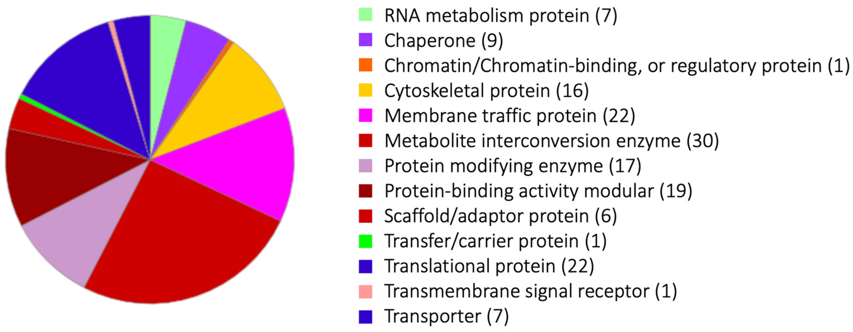
4. Phagosome Proteins Detected Exclusively by the Magnetic Bead Isolation Method, Combined with Bead Serum Coating and Chemical Cross-Linking, Represent Those Involved in Serum-Dependent Phagocytosis or Transiently Associated with Phagosomes

A panel of phagosome proteins was exclusively identified by the magnetic bead isolation method, combined with serum coating of beads and chemical cross-linking (Proteome 4 and 5). We believe that the two lists of phagosomal proteins created based on two protocols show functional and temporal (i.e., strength and timing of the interaction with phagosomes) differences in their association with phagosomes. As stated above, a few key differences between the two methods are: (1) paramagnetic beads were coated by a human serum to mimic target human cells; (2) beads were collected by either magnetic separation using paramagnetic beads or by density gradient centrifugation; (3) chemical cross-linking was applied to the magnetic isolation method. Proteomes 4 and 5 (using the paramagnetic beads isolation method) detected twice to thrice as many proteins as Proteomes 1–3, which were gained by the density-gradient centrifugation protocol. The average number of proteins of Proteomes 1–3 was 227, while that of Proteomes 4 and 5 was 811 (Table 1). Two hundred thirty-one phagosome proteins were differentially detected in Proteomes 4 and 5 but not in Proteomes 1, 2, and 3 (Supplemental Table S2), and were categorized by PANTHER (http://pantherdb.org/about.jsp, accessed on 21 December 2022) (Figure 1). One hundred and fifty-eight out of 231 proteins were classified into thirteen groups. The largest group, “metabolite interconversion enzyme”, included two long-chain-fatty-acid-CoA ligases, phosphatidylserine synthase, phosphoglycerate kinase, and diacylglycerol O-acyltransferase. Six out of thirty proteins are lipid metabolism-related enzymes (Supplemental Table S3). The second largest groups are the “membrane traffic protein” and “translational protein” categories. The third to fifth largest groups are the “protein-binding activity modular”, “protein modifying enzyme”, and “cytoskeletal protein” categories, respectively.
The largest group of “metabolite interconversion enzymes” includes the enzymes involved in phospholipid metabolism (EHI_009800: phosphatidylserine synthase; EHI_068320: glycerophosphoryl diester phosphodiesterase, EHI_070720: inositol-3-phosphate synthase), a glycolytic enzyme (EHI_188180: phosphoglycerate kinase), and general lipid metabolism-related enzymes (EHI_113590: diacylglycerol O-acyltransferase; EHI_079300 and EHI_188590: long-chain-fatty-acid-CoA ligase) were detected. These lipid metabolism- and (phospho) lipid metabolism-related enzymes identified in Proteomes 4 and 5 suggest that these metabolic pathways are transiently or weakly associated with phagosomes during phagosome biogenesis, as previously suggested [37]. Lipid metabolic enzymes may be needed for the elongation of the phagosomal membrane.
In the “membrane traffic protein” category, vacuolar protein sorting (Vps) 26-1 (EHI_062490, Watanebe et al., unpublished), 29 (EHI_025270), 35-1, and 35-2 (EHI_002990, EHI_041950) were detected from Proteomes 4 and 5. These proteins make a retromer complex, which is known to be important for the retrograde transport of hydrolase receptors from lysosomes (endosomes) to the TGN and the surface transmembrane receptors from endosomes to the PM [38]. Vps26 localization on the phagosome in E. histolytica was confirmed by immunofluorescence assay [4,8]. The finding that the retromer components were exclusively identified from phagosomes after beads serum coating and cross-linking is consistent with the notion that the interaction of the retromer with phagosomes is transient and time-dependent. Vps26 was characterized as a Rab7A effector protein and co-localized with Rab7A [8]. Rab7A is localized on the phagosome stably; however, Vps26 localized with it transiently. This fact may be caused by the alteration of the GTP and GDP of Rab7A. Vps26 can bind to GTP type Rab7A, which suggests that Rab7A transiently changes to GTP Rab7A and Vps26 binds to it at that time.
In addition to six E. histolytica Rabs that have orthologs in other species and are broadly conserved in eukaryotes (Rab1A, 7A, 7B, 7D, 11B, and 11C) and two Entamoeba-specific Rabs (RabC1 and C3), six additional Entamoeba-specific Rabs (RabA, RabC5, RabC7, RabK1, RabI2, and RabP2) (note that Entamoeba-specific Rabs are named primarily in alphabetic order, followed by a numerical order for isotypes), were detected only from Proteomes 4 and 5. RabC7 and RabP2 were also detected in an independent proteome study [3] using serum-coated magnetic beads. It suggests that these two Rabs may be recruited only when the bait is coated with human serum. RabA has been shown to be involved in motility, polarization [15,39], and transport of the Gal/GalNAc lectin [15,40]. According to the phylogeny reconstruction of Entamoeba Rabs, after E. histolytica-specific Rabs (RabX), including RabX16, 17, and 32, branched off the main tree, the RabC group was formed. RabC group was then divided into two subgroups, RabC1/C3 and RabC5/C7 [15]. It may be worth rephrasing that RabC1 and RabC3 are constitutively (or tightly) associated with phagosomes, whereas similar isotypes RabC5 and RabC7 are weakly or transiently associated. RabI is evolutionarily relatively close to the Rab7 subfamily, indicating that RabI may also play an important role in phagosome biogenesis similar to Rab7 isotypes. Note that RabP2 is present in E. dispar but absent in E. invadens [15]. The role and localization of RabK1 and RabI2 remain uncharacterized. Rab21 is known to be involved in endocytosis and phagocytosis in general [15,41]. Rab21 is found in Proteomes 4 and 5 (Supplemental Tables S2 and S3); however, this is a canonical type of Rab that is conserved in eukaryotes.
Twenty-two proteins were categorized into “translational proteins”. These proteins include ribosomal proteins, translation initiation factors, and tRNA synthetases, which are localized in ribosomes, and potentially associated with the ER. These ER-localized proteins were also detected in phagosomes from mammalian cells. The phagosome and the ER form a membrane contact site (MCS), which is needed to transport the ER-resident proteins to the phagosomes [42]. Therefore, the detection of these proteins from phagosomes is reasonable.
“Protein modifying enzyme” includes seventeen proteins. Five out of seventeen are ubiquitin-proteasome-related proteins. The ubiquitin-proteasome system is known as a selective proteolytic pathway and is different from autophagy, which is a non-selective protein degradative pathway. However, it has been reported that the ubiquitinated proteins are also delivered via vesicles to the late endosome and are degraded by the lysosome [43]. It is not clear whether this ubiquitinated lysosome degradation pathway is conserved in E. histolytica. The discovery of the ubiquitin–proteasome system in Proteomes 4 and 5 suggests that the ubiquitin–proteasome system interacts with phagosomes or, alternatively, the system is entirely engulfed by autophagosomes in E. histolytica. The interaction between ubiquitin–proteasome system and phagosomes has not been explored in this organism.
Hemolysin, which is categorized as a “transmembrane signal receptor”, is usually secreted from bacteria, and has been shown to degrade red blood cells and induce apoptosis of immune cells. The role of hemolysin in E. histolytica remains controversial, while the gene is used as the target for diagnostic PCR [44].
PANTHER analysis indicates that sixteen proteins are categorized into “cytoskeletal proteins”. These proteins include actin-related protein, actin-binding protein, actin, and F-actin-capping protein. Actin is known to be involved in many essential cellular processes of E. histolytica, such as motility, pseudopod formation, adherence, endo- and exocytosis, and phagocytosis [45,46]. In E. histolytica, 390 actin-binding proteins (ABPs) were identified by in silico analysis based on their domain arrangements [45]. In humans, only 162 ABPs are encoded, suggesting that E. histolytica ABPs are more diversified within the species [47]. ABPs are known to diversify the function of actin by modifying actin functions, as actin is extremely conserved throughout eukaryotes’ function. Therefore, six ABPs found in Proteomes 4 and 5 may modulate actin participation during phagosome biogenesis in a lineage-specific fashion. Actin and phagosome-associated ABPs may be involved in pathogenesis via phagocytosis-related functions. Rho1 and Rho7 were detected from all phagosome proteomes (Table 1). This result supports the importance of Rho-regulated actin rearrangement during phagocytosis and phagosome biogenesis. Furthermore, filopodin, one of the actin-related proteins, was detected in all phagosome proteomes.
“RNA metabolism proteins” and “chromatin binding proteins“ are involved in translation and gene expression. These proteins were unexpectedly detected from phagosome proteomes. The proteins detected from phagosomes and isolated by the magnetic bead method were categorized by PANTHER (Supplemental Table S3) and subsequently, analyzed by STRING (https://string-db.org) (Supplemental Figure S1). STRING analysis indicates that the proteins that belong to “RNA metabolism proteins” and “chromatin-binding proteins” have relationships with ribosomal proteins or chaperons, which are localized in the ER. It is well known, as stated above, that phagosomes and the ER make a physical interaction via MCS [42].
Furthermore, proteins categorized into “metabolite interconversion enzyme” were unexpectedly detected from phagosome proteomes 4 and 5. STRING analysis also indicates that a good number of proteins that are categorized into “metabolite interconversion enzymes” were also linked to ER localizing proteins. For example, EHI_188180 (phosphoglycerate kinase), a glycolytic enzyme, is linked to a transcriptional protein (EHI_125650) and ER ATPase (cell division cycle 48: EHI_045120). KEGG analysis (https://www.genome.jp/kegg/pathway.html, accessed on 21 December 2022) of the proteins of “metabolite interconversion enzyme” indicates predominant roles of phagosome-associated metabolism in “Carbohydrate metabolism” (12 proteins) and “Lipid metabolism” (8 proteins) (Supplemental Table S4). These observations are consistent with the role of phagosomes as a nutrient provider.

5. Conclusions

In this review, we summarized all the phagosome proteome data that had been created using two different phagosome isolation methods in our own laboratory and categorized them into two major categories: the core constitutive proteome and the proteome of transiently or weakly associated constituents (Supplemental Figure S2). The catalog of the phagosome proteomes should be a useful reference to easily validate whether proteins of interest are involved in phagosome biogenesis directly or indirectly. As far as we are concerned, this is the first review to systematically compare phagosome proteomes obtained by different methods in a single laboratory. By comparing data obtained by two methods, we gained a new overall picture of phagosome proteomes. For example, broadly conserved canonical Rabs were constitutively detected from all phagosome proteomes, while Entamoeba-specific Rabs six were detected only by magnetic isolation method combined with cross-linking, suggesting that Entamoeba-specific Rabs may access to the phagosomes only transiently and weakly, and presumably control the multiple fate of phagosomes, which well illustrates diversified vesicular trafficking mechanisms in E. histolytica. Other than expected players, unexpected proteins, including Gp63 and lipid metabolism enzymes, were detected from phagosomes. Furthermore, a panel of cysteine proteases and hydrolases reinforces their central role in phagosome biogenesis. “Metabolite interconversion enzyme” is one of the largest categories. This may suggest that many unknown metabolic enzymes/pathways interact with phagosomes during phagosome biogenesis.

Supplementary Materials

The following supporting information can be downloaded at: https://www.mdpi.com/article/10.3390/genes14020379/s1, Supplemental Tables S1–S3 (Excel file). Table S1: The list of phagosomal proteins detected in all five proteome studies. These phagosomal proteins were categorized by their function. Forty proteins are listed. Table S2: The list of phagosomal proteins detected only in two proteome studies (Proteome 4 and 5) in which the magnetic-bead method was used for phagosome isolation. Two hundred thirty proteins are listed. Table S3: The table of PANTHER-categorized phagosomal proteins is shown in Supplemental Table S2. One hundred and fifty-eight proteins categorized by PANTHER are listed. Note that the remaining seventy-two proteins are not categorized (no PANTHER category is assigned) and are omitted from the list. Table S4: KEGG analysis of thirty phagosomal proteins categorized into “metabolite interconversion enzyme” (https://www.genome.jp/kegg/pathway.html, accessed on 21 December 2022). Figure S1: STRING analysis (https://string-db.org) of the data set of Supplemental Table S3. The relationships among listed proteins were visualized by STRING. The following parameters were chosen: “Multiple proteins”; EHI_numbers for “List Of Names”; and “Entamoeba histolytica” for “Organisms”. Figure S2. Overview of the major categories of identified phagosome proteins by two phagosome isolation methods: “density-gradient method” and “magnetic bead method”. Representative proteins discussed in this review are given after the category names.

Author Contributions

Conceptualization, N.W. and T.N.; methodology, N.W.; software, N.W.; formal analysis, N.W.; resources, T.N.; data curation, N.W.; writing—original draft preparation, N.W.; writing—review and editing, T.N.; visualization, N.W.; supervision, K.N.-T.; project administration, T.N.; funding acquisition, N.W. and T.N. All authors have read and agreed to the published version of the manuscript.

Funding

This research was funded by Grant-in-Aid for a Research Activity start-up (JP20K22758 to N.W.), Sasagawa Scientific Research Grant from The Japan Science Society (2020-4044 to N.W.). Grants-in-Aid for Scientific Research (B) (JP21H02723 to T.N.), Grant for research on emerging, re-emerging infectious diseases from Japan Agency for Medical Research and Development (AMED, JP20fk0108138 to T.N.), Scientific Research on Innovative Areas (JP19H03463 and JP20H05353 to KN-T), Promotion of Joint International Research (Fostering Joint International Research (B)) (21KK0139 to KN-T), and Grant for research on emerging and re-emerging infectious diseases from Japan Agency for Medical Research and Development (AMED, JP20fk0108139 to KN-T).

Institutional Review Board Statement

Not applicable.

Informed Consent Statement

Not applicable.

Data Availability Statement

Not applicable.

Acknowledgments

We want to thank all previous and current Nozaki lab members who contributed to phagosome proteomics, including Mami Okada and Atsushi Furukawa.

Conflicts of Interest

The authors declare no conflict of interest. The funders had no role in the design of this study, in the collection, analyses, or interpretation of data, in the writing of the manuscript, or in the decision to publish the results.

References

  1. Okada, M.; Nozaki, T. New insights into molecular mechanisms of phagocytosis in Entamoeba histolytica by proteomic analysis. Arch. Med. Res. 2006, 37, 244–251. [Google Scholar] [CrossRef] [PubMed]
  2. Okada, M.; Huston, C.D.; Oue, M.; Mann, B.J.; Petri, W.A.; Kita, K.; Nozaki, T. Kinetics and strain variation of phagosome proteins of Entamoeba histolytica by proteomic analysis. Mol. Biochem. Parasitol. 2006, 145, 171–183. [Google Scholar] [CrossRef] [PubMed]
  3. Marion, S.; Laurent, C.; Guillén, N. Signalization and cytoskeleton activity through myosin IB during the early steps of phagocytosis in Entamoeba histolytica: A proteomic approach. Cell. Microbiol. 2005, 7, 1504–1518. [Google Scholar] [CrossRef] [PubMed]
  4. Watanabe, N.; Nakada-Tsukui, K.; Nozaki, T. Two isotypes of phosphatidylinositol 3-phosphate-binding sorting nexins play distinct roles in trogocytosis in Entamoeba histolytica. Cell Microbiol. 2020, 22, e13144. [Google Scholar] [CrossRef] [Green Version]
  5. Nakada-tsukui, K.; Watanabe, N.; Shibata, K.; Wahyuni, R.; Miyamoto, E.; Nozaki, T. Proteomic analysis of Atg8-dependent recruitment of phagosomal proteins in the enteric protozoan parasite Entamoeba histolytica. Front. Cell Infect. Microbiol. 2022, 12, 1–14. [Google Scholar] [CrossRef]
  6. Furukawa, A.; Nakada-Tsukui, K.; Nozaki, T. Cysteine protease-binding protein family 6 mediates the trafficking of amylases to phagosomes in the enteric protozoan entamoeba histolytica. Infect. Immun. 2013, 81, 1820–1829. [Google Scholar] [CrossRef] [Green Version]
  7. Okada, M.; Huston, C.D.; Mann, B.J.; Petri, W.A.; Kita, K.; Nozaki, T. Proteomic analysis of phagocytosis in the enteric protozoan parasite Entamoeba histolytica. Eukaryot. Cell 2005, 4, 827–831. [Google Scholar] [CrossRef] [Green Version]
  8. Nakada-Tsukui, K.; Saito-Nakano, Y.; Ali, V.; Nozaki, T. A Retromerlike Complex Is a Novel Rab7 Effector That Is Involved in the Transport of the Virulence Factor Cysteine Protease in the Enteric Protozoan Parasite Entamoeba histolytica Kumiko. Mol. Biol. Cell 2005, 16, 5294–5303. [Google Scholar] [CrossRef] [Green Version]
  9. Saito-Nakano, Y.; Wahyuni, R.; Nakada-Tsukui, K.; Tomii, K.; Nozaki, T. Rab7D small GTPase is involved in phago-, trogocytosis and cytoskeletal reorganization in the enteric protozoan Entamoeba histolytica. Cell Microbiol. 2021, 23, e13267. [Google Scholar] [CrossRef]
  10. Mitra, B.N.; Saito-Nakano, Y.; Nakada-Tsukui, K.; Sato, D.; Nozaki, T. Rab11B small GTPase regulates secretion of cysteine proteases in the enteric protozoan parasite Entamoeba histolytica. Cell Microbiol. 2007, 9, 2112–2125. [Google Scholar] [CrossRef]
  11. Verma, K.; Saito-Nakano, Y.; Nozaki, T.; Datta, S. Insights into endosomal maturation of human holo-transferrin in the enteric parasite Entamoeba histolytica: Essential roles of Rab7A and Rab5 in biogenesis of giant early endocytic vacuoles. Cell Microbiol. 2015, 17, 1779–1796. [Google Scholar] [CrossRef] [Green Version]
  12. Saito-nNkano, Y.; Mitra, B.N.; Nakada-tsukui, K.; Sato, D.; Nozaki, T. Two Rab7 isotypes, Eh Rab7A and Eh Rab7B, play distinct roles in biogenesis of lysosomes and phagosomes in the enteric protozoan parasite Entamoeba histolytica. Cell Microbiol. 2007, 9, 1796–1808. [Google Scholar] [CrossRef]
  13. Saito-Nakano, Y.; Yasuda, T.; Nakada-Tsukui, K.; Leippe, M.; Nozaki, T. Rab5-associated vacuoles play a unique role in phagocytosis of the enteric protozoan parasite Entamoeba histolytica. J. Biol. Chem. 2004, 279, 49497–49507. [Google Scholar] [CrossRef] [Green Version]
  14. Herman, E.; Siegesmund, M.A.; Bottery, M.J.; Van Aerle, R.; Shather, M.M.; Caler, E.; Dacks, J.B.; Van Der Giezen, M. Membrane Trafficking Modulation during Entamoeba Encystation. Sci. Rep. 2017, 7, 12854. [Google Scholar] [CrossRef] [Green Version]
  15. Nakada-Tsukui, K.; Saito-Nakano, Y.; Husain, A.; Nozaki, T. Conservation and function of Rab small GTPases in Entamoeba: Annotation of E. invadens Rab and its use for the understanding of Entamoeba biology. Exp. Parasitol. 2010, 126, 337–347. [Google Scholar] [CrossRef]
  16. Hanadate, Y.; Saito-Nakano, Y.; Nakada-Tsukui, K.; Nozaki, T. Endoplasmic reticulum-resident Rab8A GTPase is involved in phagocytosis in the protozoan parasite Entamoeba histolytica. Cell Microbiol. 2016, 18, 1358–1373. [Google Scholar] [CrossRef] [Green Version]
  17. Hanadate, Y.; Saito-Nakano, Y.; Nakada-Tsukui, K.; Nozaki, T. Identification and characterization of the entamoeba histolytica rab8a binding protein: A Cdc50 homolog. Int. J. Mol. Sci. 2018, 19, 3831. [Google Scholar] [CrossRef] [Green Version]
  18. Bansal, D.; Ave, P.; Kerneis, S.; Frileux, P.; Boché, O.; Baglin, A.C.; Dubost, G.; Leguern, A.S.; Prevost, M.C.; Bracha, R.; et al. An ex-vivo human intestinal model to study Entamoeba histolytica pathogenesis. PLoS Negl. Trop. Dis. 2009, 3, 551. [Google Scholar] [CrossRef]
  19. Que, X.; Reed, S.L. Cysteine proteinases and the pathogenesis of amebiasis. Clin. Microbiol. Rev. 2000, 13, 196–206. [Google Scholar] [CrossRef]
  20. Temesvari, L.A.; Harris, E.N.; Stanley, S.L.; Cardelli, J.A. Early and late endosomal compartments of Entamoeba histolytica are enriched in cysteine proteases, acid phosphatase and several Ras-related Rab GTPases. Mol. Biochem. Parasitol. 1999, 103, 225–241. [Google Scholar] [CrossRef]
  21. Freitas, M.A.R.; Fernandes, H.C.; Calixto, V.C.; Martins, A.S.; Silva, E.F.; Pesquero, J.L.; Gomes, M.A. Entamoeba histolytica: Cysteine proteinase activity and virulence. Focus on cysteine proteinase 5 expression levels. Exp. Parasitol. 2009, 122, 306–309. [Google Scholar] [CrossRef] [PubMed]
  22. Siqueira-Neto, J.L.; Debnath, A.; McCall, L.I.; Bernatchez, J.A.; Ndao, M.; Reed, S.L.; Rosenthal, P.J. Cysteine proteases in protozoan parasites. PLoS Negl. Trop. Dis. 2018, 12, 1–20. [Google Scholar] [CrossRef] [PubMed] [Green Version]
  23. Matthiesen, J.; Bär, A.K.; Bartels, A.K.; Marien, D.; Ofori, S.; Biller, L.; Tannich, E.; Lotter, H.; Bruchhaus, I. Overexpression of specific cysteine peptidases confers pathogenicity to a nonpathogenic Entamoeba histolytica clone. MBio 2013, 4, 00072-13. [Google Scholar] [CrossRef] [PubMed] [Green Version]
  24. Mitra, B.N.; Yasuda, T.; Kobayashi, S.; Saito-Nakano, Y.; Nozaki, T. Differences in morphology of phagosomes and kinetics of acidification and degradation in phagosomes between the pathogenic Entamoeba histolytica and the non-pathogenic Entamoeba dispar. Cell Motil. Cytoskelet. 2005, 62, 84–99. [Google Scholar] [CrossRef]
  25. Mitra, B.N.; Kobayashi, S.; Saito-Nakano, Y.; Nozaki, T. Entamoeba histolytica: Differences in phagosome acidification and degradation between attenuated and virulent strains. Exp. Parasitol. 2006, 114, 57–61. [Google Scholar] [CrossRef]
  26. Ragland, S.A.; Criss, A.K. From bacterial killing to immune modulation: Recent insights into the functions of lysozyme. PLoS Pathog. 2017, 13, 1–22. [Google Scholar] [CrossRef] [Green Version]
  27. Fleming, D.; Chahin, L.; Rumbaugh, K. Glycoside hydrolases degrade polymicrobial bacterial biofilms in wounds. Antimicrob. Agents Chemother. 2017, 61, 1–9. [Google Scholar] [CrossRef] [Green Version]
  28. Koo, I.C.; Ohol, Y.M.; Wu, P.; Morisaki, J.H.; Cox, J.S.; Brown, E.J. Role for lysosomal enzyme β-hexosaminidase in the control of mycobacteria infection. Proc. Natl. Acad. Sci. USA 2008, 105, 710–715. [Google Scholar] [CrossRef] [Green Version]
  29. Marumo, K.; Nakada-Tsukui, K.; Tomii, K.; Nozaki, T. Ligand heterogeneity of the cysteine protease binding protein family in the parasitic protist Entamoeba histolytica. Int. J. Parasitol. 2014, 44, 625–635. [Google Scholar] [CrossRef] [Green Version]
  30. Furukawa, A.; Nakada-Tsukui, K.; Nozaki, T. Novel transmembrane receptor involved in phagosome transport of lysozymes and β-hexosaminidase in the enteric protozoan Entamoeba histolytica. PLoS Pathog. 2012, 8, e1002539. [Google Scholar] [CrossRef]
  31. Isnard, A.; Shio, M.T.; Olivier, M. Impact of Leishmania metalloprotease GP63 on macrophage signaling. Front. Cell Infect. Microbiol. 2012, 2, 72. [Google Scholar] [CrossRef] [Green Version]
  32. da Silva Lira Filho, A.; Fajardo, E.F.; Chang, K.P.; Clément, P.; Olivier, M. Leishmania Exosomes/Extracellular Vesicles Containing GP63 Are Essential for Enhance Cutaneous Leishmaniasis Development Upon Co-Inoculation of Leishmania amazonensis and Its Exosomes. Front. Cell Infect. Microbiol. 2022, 11, 1–14. [Google Scholar] [CrossRef]
  33. Castro Neto, A.L.; Brito, A.N.A.L.M.; Rezende, A.M.; Magalhães, F.B.; De Melo Neto, O.P. In silico characterization of multiple genes encoding the GP63 virulence protein from Leishmania braziliensis: Identification of sources of variation and putative roles in immune evasion. BMC Genomics 2019, 20, 118. [Google Scholar] [CrossRef] [Green Version]
  34. Lieke, T.; Nylén, S.; Eidsmo, L.; McMaster, W.R.; Mohammadi, A.M.; Khamesipour, A.; Berg, L.; Akuffo, H. Leishmania surface protein gp63 binds directly to human natural killer cells and inhibits proliferation. Clin. Exp. Immunol. 2008, 153, 221–230. [Google Scholar] [CrossRef]
  35. Hassani, K.; Shio, M.T.; Martel, C.; Faubert, D.; Olivier, M. Absence of metalloprotease GP63 alters the protein content of leishmania exosomes. PLoS One 2014, 9, 95007. [Google Scholar] [CrossRef] [Green Version]
  36. Sharma, M.; Morgado, P.; Zhang, H.; Ehrenkaufer, G.; Manna, D.; Singh, U. Characterization of Extracellular Vesicles from Entamoeba histolytica Identifies Roles in Intracellular Communication That Regulates Parasite Growth and Development. Infect. Immun. 2020, 88, e00349-20. [Google Scholar] [CrossRef]
  37. Saharan, O.; Mehendale, N.; Kamat, S.S. Phagocytosis: A (Sphingo) Lipid Story. Curr. Res. Chem. Biol. 2022, 2, 100030. [Google Scholar] [CrossRef]
  38. Williams, E.T.; Chen, X.; Moore, D.J. VPS35, the retromer complex and Parkinson’s disease. J. Parkinsons. Dis. 2017, 7, 219–233. [Google Scholar] [CrossRef] [Green Version]
  39. Welter, B.H.; Temesvari, L.A. A unique Rab GTPase, EhRabA, of Entamoeba histolytica, localizes to the leading edge of motile cells. Mol. Biochem. Parasitol. 2004, 135, 185–195. [Google Scholar] [CrossRef]
  40. Welter, B.H.; Temesvari, L.A. Overexpression of a mutant form of EhRabA, a unique rab GTPase of entamoeba histolytica, alters endoplasmic reticulum morphology and localization of the Gal/GalNAc adherence lectin. Eukaryot. Cell 2009, 8, 1014–1026. [Google Scholar] [CrossRef] [PubMed]
  41. Simpson, J.C.; Griffiths, G.; Wessling-Resnick, M.; Fransen, J.A.M.; Bennett, H.; Jones, A.T. A role for the small GTPase Rab21 in the early endocytic pathway. J. Cell Sci. 2004, 117, 6297–6311. [Google Scholar] [CrossRef] [PubMed] [Green Version]
  42. Levin-Konigsberg, R.; Grinstein, S. Phagosome-endoplasmic reticulum contacts: Kissing and not running. Traffic 2020, 21, 172–180. [Google Scholar] [CrossRef] [PubMed]
  43. Jo, E.K.; Yuk, J.M.; Shin, D.M.; Sasakawa, C. Roles of autophagy in elimination of intracellular bacterial pathogens. Front. Immunol. 2013, 4, 1–9. [Google Scholar] [CrossRef] [Green Version]
  44. Zindrou, S.; Orozco, E.; Linder, E.; Téllez, A.; Björkman, A. Specific detection of Entamoeba histolytica DNA by hemolysin gene targeted PCR. Acta Trop. 2001, 78, 117–125. [Google Scholar] [CrossRef]
  45. Rath, P.P.; Gourinath, S. The actin cytoskeleton orchestra in Entamoeba histolytica. Proteins Struct. Funct. Bioinforma. 2020, 88, 1361–1375. [Google Scholar] [CrossRef]
  46. Bharadwaj, R.; Sharma, S.; Arya, R.; Bhattacharya, S.; Bhattacharya, A. EhRho1 regulates phagocytosis by modulating actin dynamics through EhFormin1 and EhProfilin1 in Entamoeba histolytica. Cell Microbiol. 2018, 20, e12851. [Google Scholar] [CrossRef]
  47. Dos Remedios, C.G.; Chhabra, D.; Kekic, M.; Dedova, I.V.; Tsubakihara, M.; Berry, D.A.; Nosworthy, N.J. Actin binding proteins: Regulation of cytoskeletal microfilaments. Physiol. Rev. 2003, 83, 433–473. [Google Scholar] [CrossRef]
Figure 1. Gene ontology analysis of phagosome proteins isolated by the magnetic bead isolation method. The pie chart of thirteen “protein classes” of the proteins detected from the phagosomes collected by the magnetic bead isolation method. The list of proteins is in Supplemental Table S3. The numbers after the categories in Figure 1 are detected protein numbers.
Genes 14 00379 g001
Table 1. The list of phagosome proteome performed in our laboratory. Phagosomes were purified by two different methods: the density-gradient and magnetic-bead methods. In a magnetic-bead method, beads were coated with human serum, and the samples were treated with DSP for cross-linking.
NumberJournalYearMethodDetected ProteinsReference
1Mol. Biochem. Parasitol.2006Density-gradient155[2]
2Arch. Med. Res.2006Density-gradient151[1]
3Infect. Immun.2013Density-gradient375[6]
4Cell. Microbiol.2019Magnetic beads
(serum coated; cross linking)
884[4]
5Front. Cell. Infect. Microbiol.2022Magnetic beads
(serum coated; cross-linking)
738[5]
One phagosome proteomic study from our laboratory [7] was not included in this review because the proteome was largely identical to what was described in [2].
Table 2. Small G-proteins and degrading enzymes detected in all proteomes, and Small G-proteins detected in Proteomes 4 and 5 and categorized into “protein-binding activity modulators”.
AmoebaDB
Annotation
EHI NumberCommon NamePhagosome Isolation MethodProteome NumberGeneral Function
Rab family GTPaseEHI_108610Rab1ADensity-gradient/
Magnetic beads
1,2,3,4,5Membrane traffic
Small GTPase Rab7AEHI_192810Rab7ADensity-gradient/
Magnetic beads
1,2,3,4,5Early phagosome maturation
EhRab7B proteinEHI_081330Rab7BDensity-gradient/
Magnetic beads
1,2,3,4,5Membrane traffic
EhRab7D proteinEHI_082070Rab7DDensity-gradient/
Magnetic beads
1,2,3,4,5Phagosome maturation
Small GTPase Rab11BEHI_107250Rab11BDensity-gradient/
Magnetic beads
1,2,3,4,5Phagosome maturation (in Eh)/CP secretion (in Eh)/endosome recycling
Rab family GTPaseEHI_161030Rab11CDensity-gradient/
Magnetic beads
1,2,3,4,5Endosome recycling
Rab family GTPaseEHI_153690RabC1Density-gradient/
Magnetic beads
1,2,3,4,5Membrane traffic
Rab family GTPaseEHI_143650RabC3Density-gradient/
Magnetic beads
1,2,3,4,5Membrane traffic
Rho family GTPaseEHI_070730Rho1Density-gradient/
Magnetic beads
1,2,3,4,5Cell motility
Rho family GTPaseEHI_129750Rho7Density-gradient/
Magnetic beads
1,2,3,4,5Cell motility
Ras family GTPaseEHI_058090 Density-gradient/
Magnetic beads
1,2,3,4,5Signal transduction
Cell surface protease gp63, putativeEHI_200230Gp63Density-gradient/
Magnetic beads
1,2,3,4,5Degrading enzyme
Lysozyme, putative EHI_199110Lysozyme IDensity-gradient/
Magnetic beads
1,2,3,4,5Degrading enzyme
Lysozyme, putative EHI_096570Lysozyme IIDensity-gradient/
Magnetic beads
1,2,3,4,5Degrading enzyme
Cysteine proteinase 1 EHI_074180CP-A1Density-gradient/
Magnetic beads
1,2,3,4,5Degrading enzyme
HistolysainEHI_033710CP-A2Density-gradient/
Magnetic beads
1,2,3,4,5Degrading enzyme
Cysteine proteinase, putativeEHI_050570CP-A4Density-gradient/
Magnetic beads
1,2,3,4,5Degrading enzyme
Cysteine proteinaseEHI_168240CP-A5Density-gradient/
Magnetic beads
1,2,3,4,5Degrading enzyme
Dipeptidyl-peptidaseEHI_136440 Density-gradient/
Magnetic beads
1,2,3,4,5Degrading enzyme
α-amylase family proteinEHI_023360 Density-gradient/
Magnetic beads
1,2,3,4,5Degrading enzyme
β-hexosaminidaseEHI_007330 Density-gradient/
Magnetic beads
1,2,3,4,5Degrading enzyme
Serine carboxypeptidase (S28) family proteinEHI_054530 Density-gradient/
Magnetic beads
1,2,3,4,5Degrading enzyme
Rab family GTPaseEHI_168600RabAMagnetic beads4,5Membrane traffic
Rab family GTPaseEHI_079890RabC7Magnetic beads4,5Membrane traffic
Rab family GTPaseEHI_122730RabC5Magnetic beads4,5Membrane traffic
Rab family GTPaseEHI_053420RabI2Magnetic beads4,5Membrane traffic
Rab family GTPaseEHI_024680RabK1Magnetic beads4,5Membrane traffic
Rab family GTPaseEHI_117890RabP2Magnetic beads4,5Membrane traffic
Ras-related proteinEHI_129330Rab21Magnetic beads4,5Membrane traffic
Rho family GTPaseEHI_190440Rho10Magnetic beads4,5Cell motility
Rho family GTPaseEHI_135450Rho13Magnetic beads4,5Cell motility
Ras family GTPaseEHI_198330Not assignedMagnetic beads4,5Signal transduction
Disclaimer/Publisher’s Note: The statements, opinions and data contained in all publications are solely those of the individual author(s) and contributor(s) and not of MDPI and/or the editor(s). MDPI and/or the editor(s) disclaim responsibility for any injury to people or property resulting from any ideas, methods, instructions or products referred to in the content.

