The Genetic Mechanisms Underlying the Concerted Expression of the yellow and tan Genes in Complex Patterns on the Abdomen and Wings of Drosophila guttifera

 , ,
, ,
Abstract

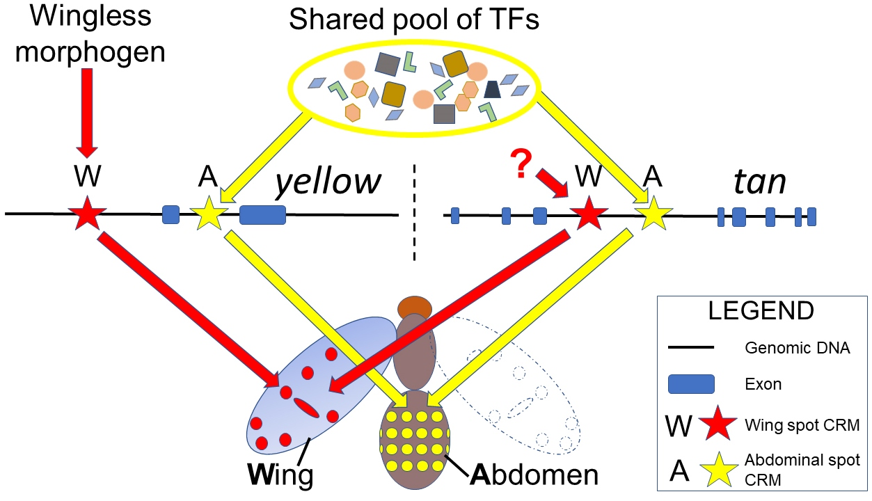
1. Introduction
2. Materials and Methods
2.1. Fruit Fly Stocks and Genomic DNA
2.2. RNA In Situ Hybridization
2.3. PCR Primers
- gut t wing CRM: Forward- 5’-GCAGCATCGACAGTCGAGTTA-3’ and Reverse: 5’-CAACAAACGCTACGTTGACC-3’.
- gut t body CRM: Forward: 5’-GCTTACCATATCGAAGCCGAC-3’ and Reverse: 5’-GGAAGTTGAACTTCCATAACTCG-3’.
- gut t wing spot CRM: Forward: 5’-CTCGTTATAGGCGAGTGCCAAT-3’ and Reverse: 5’-AGATGTGCAAAAGTCCCACG-3’.
- gut t abdominal spot CRM: Forward: 5’-CGGTGGAGTATGGTGATTAAAG-3’ and Reverse: 5’-AACAGCCGATTCGATATAGC-3’.
- gut t core stripe CRM: Forward: 5’-GCGTGCACTTAATTGTCCAAC-3’ and Reverse: 5’-AACAGCCGATTCGATATAGC-3’.
2.4. Transgenic Assays
3. Results
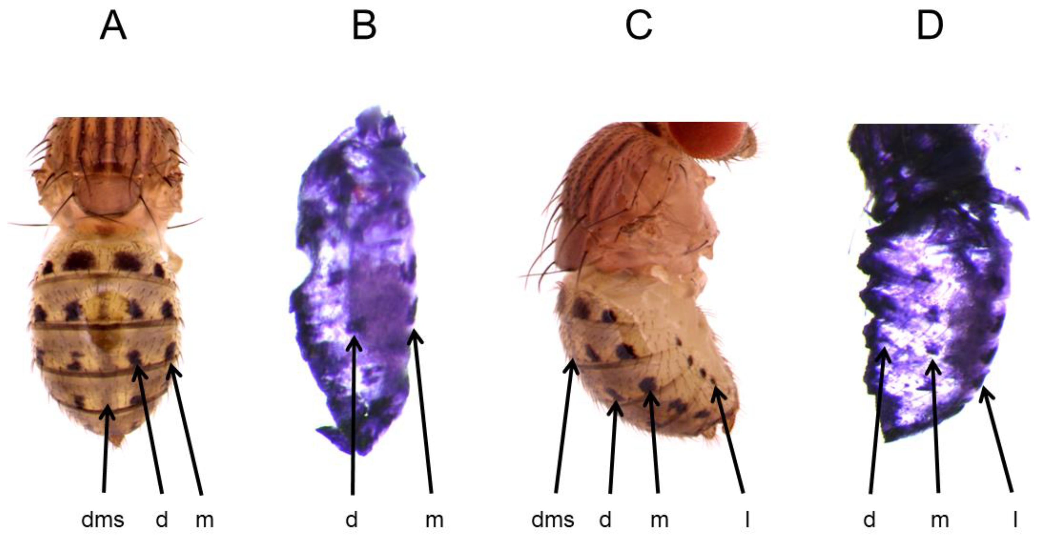
3.1. t Gene Expression Foreshadows the Adult Abdominal Melanin Pattern of D. guttifera
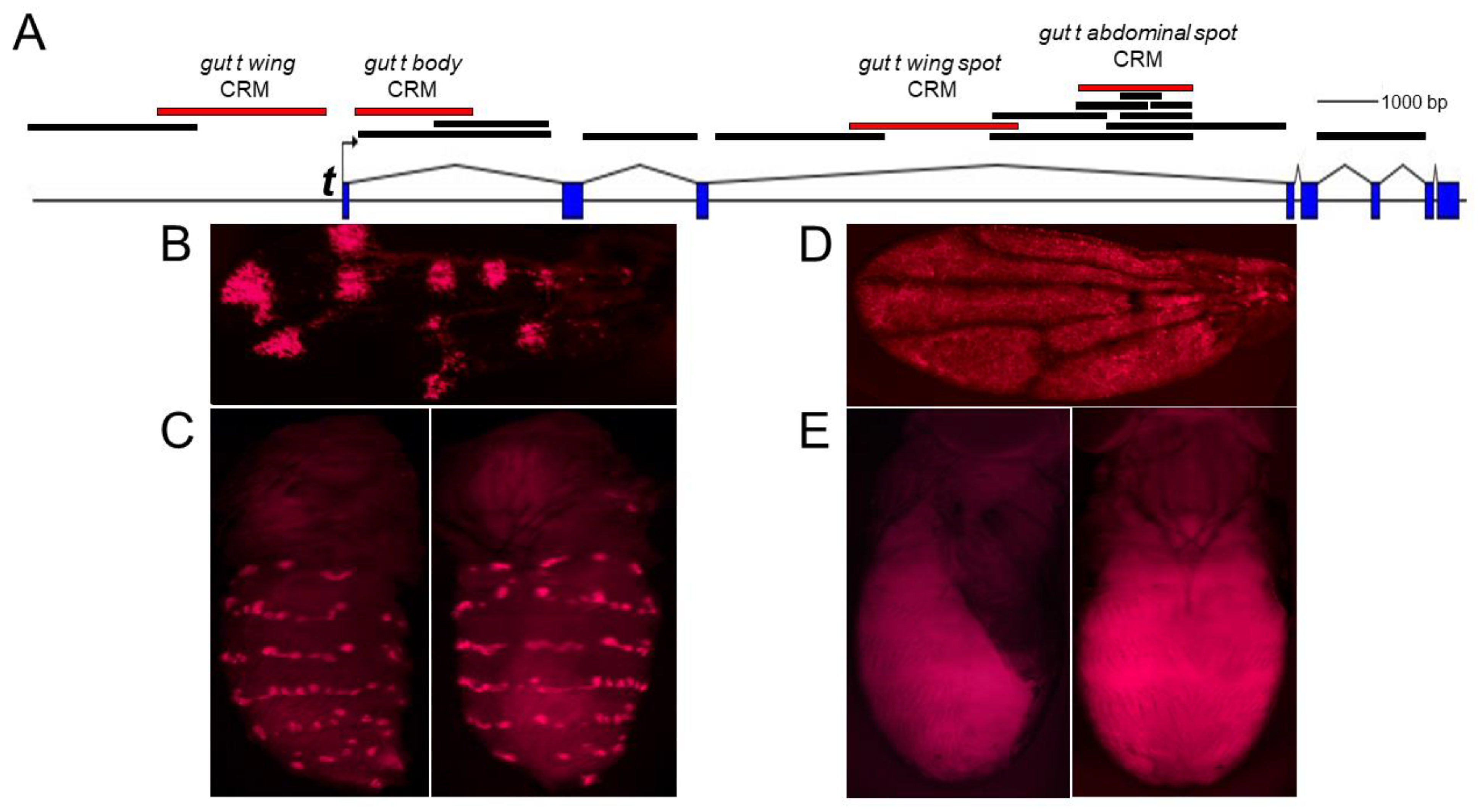
3.2. The CRM Controlling the Abdominal Spot Pattern of t Is Located in the Third Intron of t
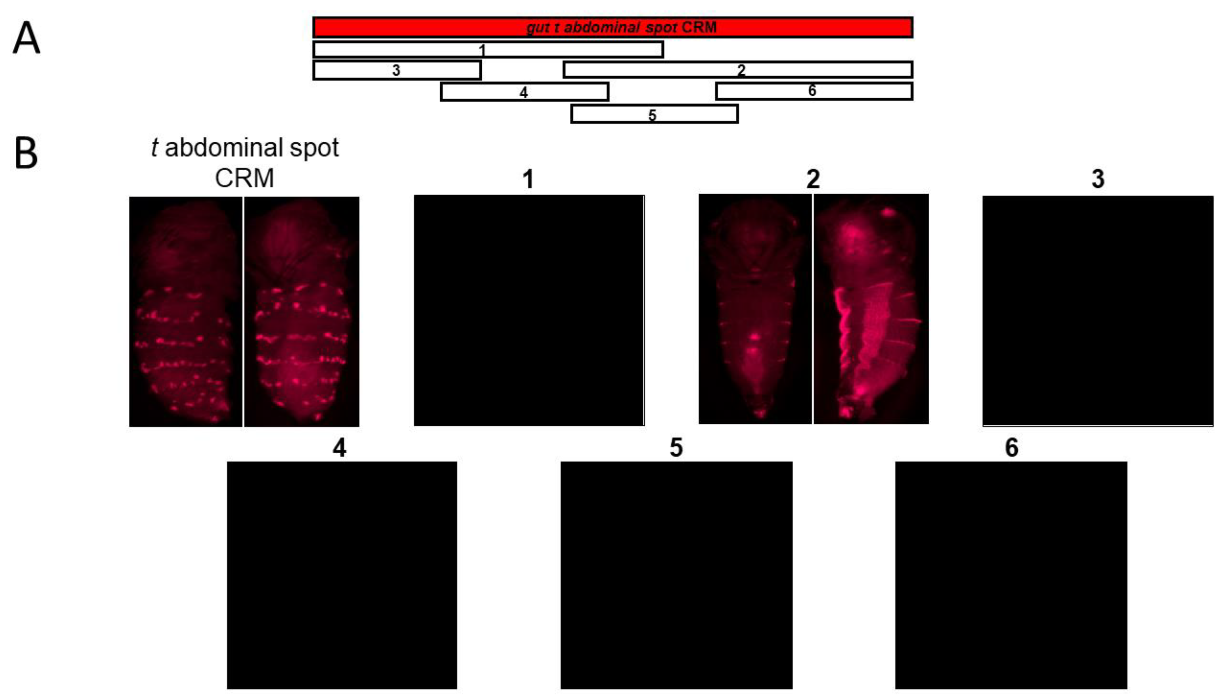
3.3. The Dissection of the Gut t Abdominal Spot CRM Reveals a Core Stripe CRM
3.4. The Gut t Abdominal Spot CRM Is Inactive in Transgenic D. melanogaster
3.5. Transcription Factor Binding Site Analysis of the Abdominal “t spot” CRM Sequence
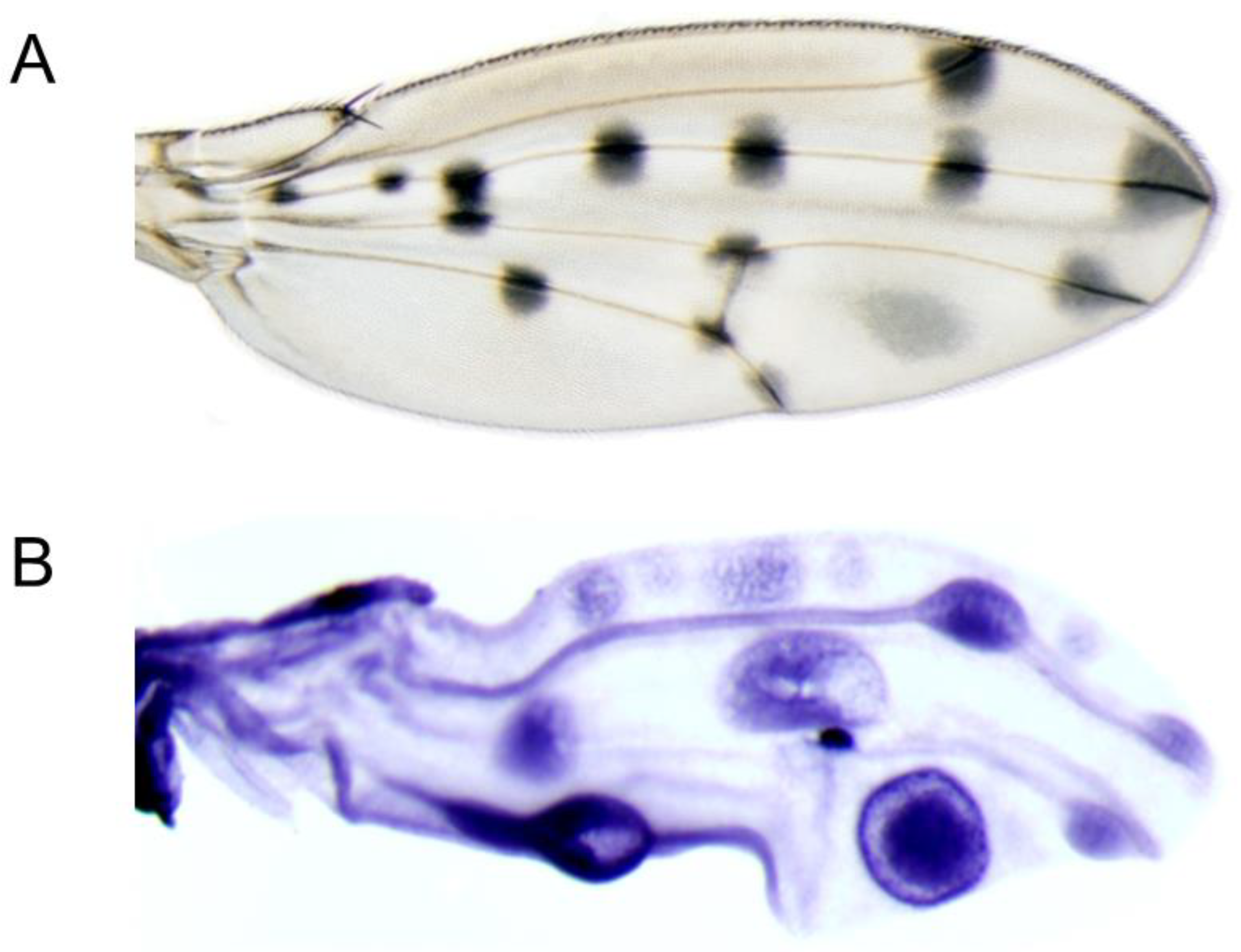
3.6. t Is Partially Co-Expressed with y in a Complex Spotted Wing Pattern
4. Discussion
5. Conclusions
Supplementary Materials
Author Contributions
Funding
Institutional Review Board Statement
Informed Consent Statement
Data Availability Statement
Acknowledgments
Conflicts of Interest
References
- Raja, K.K.B.; Shittu, M.O.; Nouhan, P.M.E.; Steenwinkel, T.E.; Bachman, E.A.; Kokate, P.P.; McQueeney, A.; Mundell, E.A.; Armentrout, A.A.; Nugent, A.; et al. The regulation of a pigmentation gene in the formation of complex color patterns in Drosophila abdomens. PLoS ONE 2022, 17, e0279061. [Google Scholar] [CrossRef] [PubMed]
- Werner, T.; Koshikawa, S.; Williams, T.M.; Carroll, S.B. Generation of a novel wing colour pattern by the Wingless morphogen. Nature 2010, 464, 1143–1148. [Google Scholar] [CrossRef] [PubMed]
- Adams, M.D.; Celniker, S.E.; Holt, R.A.; Evans, C.A.; Gocayne, J.D.; Amanatides, P.G.; Scherer, S.E.; Li, P.W.; Hoskins, R.A.; Galle, R.F.; et al. The genome sequence of Drosophila melanogaster. Science 2000, 287, 2185–2195. [Google Scholar] [CrossRef] [PubMed]
- Chinwalla, A.T.; Cook, L.L.; Delehaunty, K.D.; Fewell, G.A.; Fulton, L.A.; Fulton, R.S. Initial sequencing and comparative analysis of the mouse genome. Nature 2002, 420, 520–562. [Google Scholar]
- Venter, J.C.; Adams, M.D.; Myers, E.W.; Li, P.W.; Mural, R.J.; Sutton, G.G. The sequence of the human genome. Science 2001, 291, 1304–1351. [Google Scholar] [CrossRef] [PubMed]
- Carroll, S.B. Endless forms: The evolution of gene regulation and morphological diversity. Cell 2000, 101, 577–580. [Google Scholar] [CrossRef]
- Gostling, N.J. From DNA to Diversity: Molecular Genetics and the Evolution of Animal Design. Heredity 2002, 89, 411. [Google Scholar] [CrossRef][Green Version]
- Wasik, B.R.; Rose, D.J.; Moczek, A.P. Beetle horns are regulated by the Hox gene, Sex combs reduced, in a species- and sex-specific manner. Evol. Dev. 2010, 12, 353–362. [Google Scholar] [CrossRef]
- Weatherbee, S.D.; Nijhout, H.F.; Grunert, L.W.; Halder, G.; Galant, R.; Selegue, J.; Carroll, S. Ultrabithorax function in butterfly wings and the evolution of insect wing patterns. Curr. Biol. 1999, 9, 109–115. [Google Scholar] [CrossRef]
- Zhang, L.; Mazo-Vargas, A.; Reed, R.D. Single master regulatory gene coordinates the evolution and development of butterfly color and iridescence. Proc. Natl. Acad. Sci. USA 2017, 114, 10707–10712. [Google Scholar] [CrossRef]
- Stoehr, A.M.; Walker, J.F.; Monteiro, A. Spalt expression and the development of melanic color patterns in pierid butterflies. Evodevo 2013, 4, 6. [Google Scholar] [CrossRef] [PubMed]
- Zhang, L.; Reed, R.D. Genome editing in butterflies reveals that spalt promotes and Distal-less represses eyespot colour patterns. Nat. Commun. 2016, 7, 11769. [Google Scholar] [CrossRef] [PubMed]
- Martin, A.; Papa, R.; Nadeau, N.J.; Hill, R.I.; Counterman, B.A.; Halder, G.; Jiggins, C.D.; Kronforst, M.R.; Long, A.D.; McMillan, W.O.; et al. Diversification of complex butterfly wing patterns by repeated regulatory evolution of a Wnt ligand. Proc. Natl. Acad. Sci. USA 2012, 109, 12632–12637. [Google Scholar] [CrossRef] [PubMed]
- Murugesan, S.N.; Connahs, H.; Matsuoka, Y.; Das Gupta, M.; Tiong, G.J.; Huq, M.; Gowri, V.; Monroe, S.; Deem, K.D.; Werner, T.; et al. Butterfly eyespots evolved via cooption of an ancestral gene-regulatory network that also patterns antennae, legs, and wings. Proc. Natl. Acad. Sci. USA 2022, 119, e2108661119. [Google Scholar] [CrossRef]
- Arnoult, L.; Su, K.F.Y.; Manoel, D.; Minervino, C.; Magriña, J.; Gompel, N.; Prud’homme, B. Emergence and Diversification of Fly Pigmentation Through Evolution of a Gene Regulatory Module. Science 2013, 339, 1423–1426. [Google Scholar] [CrossRef]
- Gompel, N.; Prud’homme, B.; Wittkopp, P.J.; Kassner, V.A.; Carroll, S.B. Chance caught on the wing: Cis-regulatory evolution and the origin of pigment patterns in Drosophila. Nature 2005, 433, 481–487. [Google Scholar] [CrossRef]
- Jeong, S.; Rokas, A.; Carroll, S.B. Regulation of body pigmentation by the Abdominal-B Hox protein and its gain and loss in Drosophila evolution. Cell 2006, 125, 1387–1399. [Google Scholar] [CrossRef]
- Williams, T.M.; Selegue, J.E.; Werner, T.; Gompel, N.; Kopp, A.; Carroll, S.B. The regulation and evolution of a genetic switch controlling sexually dimorphic traits in Drosophila. Cell 2008, 134, 610–623. [Google Scholar] [CrossRef]
- Dion, W.A.; Steenwinkel, T.E.; Werner, T. From Aedes to Zeugodacus: A review of dipteran body coloration studies regarding evolutionary developmental biology, pest control, and species discovery. Curr. Opin. Genet. Dev. 2021, 69, 35–41. [Google Scholar] [CrossRef]
- True, J.R.; Yeh, S.D.; Hovemann, B.T.; Kemme, T.; Meinertzhagen, I.A.; Edwards, T.N.; Liou, S.R.; Han, Q.; Li, J. Drosophila tan encodes a novel hydrolase required in pigmentation and vision. PLoS Genet. 2005, 1, e63. [Google Scholar] [CrossRef]
- Massey, J.H.; Akiyama, N.; Bien, T.; Dreisewerd, K.; Wittkopp, P.J.; Yew, J.Y.; Takahashi, A. Pleiotropic Effects of ebony and tan on Pigmentation and Cuticular Hydrocarbon Composition in Drosophila melanogaster. Front. Physiol. 2019, 10, 518. [Google Scholar] [CrossRef] [PubMed]
- Ordway, A.J.; Hancuch, K.N.; Johnson, W.; Wiliams, T.M.; Rebeiz, M. The expansion of body coloration involves coordinated evolution in cis and trans within the pigmentation regulatory network of Drosophila prostipennis. Dev. Biol. 2014, 392, 431–440. [Google Scholar] [CrossRef] [PubMed]
- Camino, E.M.; Butts, J.C.; Ordway, A.; Vellky, J.E.; Rebeiz, M.; Williams, T.M. The evolutionary origination and diversification of a dimorphic gene regulatory network through parallel innovations in cis and trans. PLoS Genet. 2015, 11, e1005136. [Google Scholar] [CrossRef] [PubMed]
- Dion, W.A.; Shittu, M.O.; Steenwinkel, T.E.; Raja, K.K.; Kokate, P.P.; Werner, T. The modular expression patterns of three pigmentation genes prefigure unique abdominal morphologies seen among three Drosophila species. Gene Expr. Patterns 2020, 38, 119132. [Google Scholar] [CrossRef] [PubMed]
- Ferguson, L.C.; Maroja, L.; Jiggins, C.D. Convergent, modular expression of ebony and tan in the mimetic wing patterns of Heliconius butterflies. Dev. Genes Evol. 2011, 221, 297–308. [Google Scholar] [CrossRef]
- Futahashi, R.; Banno, Y.; Fujiwara, H. Caterpillar color patterns are determined by a two-phase melanin gene prepatterning process: New evidence from tan and laccase2. Evol. Dev. 2010, 12, 157–167. [Google Scholar] [CrossRef]
- Jeong, S.; Rebeiz, M.; Andolfatto, P.; Werner, T.; True, J.; Carroll, S.B. The evolution of gene regulation underlies a morphological difference between two Drosophila sister species. Cell 2008, 132, 783–793. [Google Scholar] [CrossRef]
- Yassin, A.; Bastide, H.; Chung, H.; Veuille, M.; David, J.R.; Pool, J.E. Ancient balancing selection at tan underlies female colour dimorphism in Drosophila erecta. Nat. Commun. 2016, 7, 10400. [Google Scholar] [CrossRef]
- Wittkopp, P.J.; Stewart, E.E.; Arnold, L.L.; Neidert, A.H.; Haerum, B.K.; Thompson, E.M.; Akhras, S.; Smith-Winberry, G.; Shefner, L. Intraspecific polymorphism to interspecific divergence: Genetics of pigmentation in Drosophila. Science 2009, 326, 540–544. [Google Scholar] [CrossRef]
- Dembeck, L.M.; Huang, W.; Carbone, M.A.; Mackay, T.F. Genetic basis of natural variation in body pigmentation in Drosophila melanogaster. Fly 2015, 9, 75–81. [Google Scholar] [CrossRef]
- Endler, L.; Betancourt, A.J.; Nolte, V.; Schlotterer, C. Reconciling Differences in Pool-GWAS Between Populations: A Case Study of Female Abdominal Pigmentation in Drosophila melanogaster. Genetics 2016, 202, 843–855. [Google Scholar] [CrossRef] [PubMed]
- Endler, L.; Gibert, J.M.; Nolte, V.; Schlotterer, C. Pleiotropic effects of regulatory variation in tan result in correlation of two pigmentation traits in Drosophila melanogaster. Mol. Ecol. 2018, 27, 3207–3218. [Google Scholar] [CrossRef] [PubMed]
- Erives, A.; Levine, M. Coordinate enhancers share common organizational features in the Drosophila genome. Proc. Natl. Acad. Sci. USA 2004, 101, 3851–3856. [Google Scholar] [CrossRef] [PubMed]
- Crocker, J.; Tamori, Y.; Erives, A. Evolution acts on enhancer organization to fine-tune gradient threshold readouts. PLoS Biol. 2008, 6, e263. [Google Scholar] [CrossRef] [PubMed]
- Werner, T.; Steenwinkel, T.E.; Jaenike, J. The Encyclopedia of North American Drosophilids Volume 2: Drosophilids of the Southeast; Michigan Tech: Houghton, MI, USA, 2020. [Google Scholar]
- Koshikawa, S.; Giorgianni, M.W.; Vaccaro, K.; Kassner, V.A.; Yoder, J.H.; Werner, T.; Carroll, S.B. Gain of cis-regulatory activities underlies novel domains of wingless gene expression in Drosophila. Proc. Natl. Acad. Sci. USA 2015, 112, 7524–7529. [Google Scholar] [CrossRef]
- Werner, T.; Steenwinkel, T.; Jaenike, J. The Encyclopedia of North American Drosophilids Volume 1: Drosophilids of the Midwest and Northeast; Michigan Tech: Houghton, MI, USA, 2018. [Google Scholar]
- Gloor, G.B.; Preston, C.R.; Johnson-Schlitz, D.M.; Nassif, N.A.; Phillis, R.W.; Benz, W.K.; Robertson, H.M.; Engels, W.R. Type I repressors of P element mobility. Genetics 1993, 135, 81–95. [Google Scholar] [CrossRef]
- Shittu, M.; Steenwinkel, T.; Dion, W.; Ostlund, N.; Raja, K.; Werner, T. RNA In Situ Hybridization for Detecting Gene Expression Patterns in the Abdomens and Wings of Drosophila Species. Methods Protoc. 2021, 4, 20. [Google Scholar] [CrossRef] [PubMed]
- Fukutomi, Y.; Kondo, S.; Toyoda, A.; Shigenobu, S.; Koshikawa, S. Transcriptome analysis reveals wingless regulates neural development and signaling genes in the region of wing pigmentation of a polka-dotted fruit fly. FEBS J. 2021, 288, 115–126. [Google Scholar] [CrossRef]
- Horn, C.; Wimmer, E.A. A versatile vector set for animal transgenesis. Dev. Genes Evol. 2000, 210, 630–637. [Google Scholar] [CrossRef]
- Shittu, M.; Steenwinkel, T.; Koshikawa, S.; Werner, T. The Making of Transgenic Drosophila guttifera. Methods Protoc. 2020, 3, 31. [Google Scholar] [CrossRef]
- Spicer, G.S.; Jaenike, J. Phylogenetic Analysis of Breeding Site Use and alpha-Amanitin Tolerance Within the Drosophila Quinaria Species Group. Evolution. Int. J. Org. Evol. 1996, 50, 2328–2337. [Google Scholar]
- Mathelier, A.; Zhao, X.; Zhang, A.W.; Parcy, F.; Worsley-Hunt, R.; Arenillas, D.J.; Buchman, S.; Chen, C.Y.; Chou, A.; Ienasescu, H.; et al. JASPAR 2014: An extensively expanded and updated open-access database of transcription factor binding profiles. Nucleic Acids Res. 2014, 42, D142–D147. [Google Scholar] [CrossRef] [PubMed]
- Sekelsky, J.J.; Newfeld, S.J.; Raftery, L.A.; Chartoff, E.H.; Gelbart, W.M. Genetic characterization and cloning of mothers against dpp, a gene required for decapentaplegic function in Drosophila melanogaster. Genetics 1995, 139, 1347–1358. [Google Scholar] [CrossRef] [PubMed]
- Raftery, L.A.; Twombly, V.; Wharton, K.; Gelbart, W.M. Genetic screens to identify elements of the decapentaplegic signaling pathway in Drosophila. Genetics 1995, 139, 241–254. [Google Scholar] [CrossRef] [PubMed]
- Dufour, H.D.; Koshikawa, S.; Finet, C. Temporal flexibility of gene regulatory network underlies a novel wing pattern in flies. Proc. Natl. Acad. Sci. USA 2020, 117, 11589–11596. [Google Scholar] [CrossRef] [PubMed]
- Monteiro, A.; Werner, T. Editorial overview: Cool, colorful, and complex animal systems. Curr. Opin. Genet. Dev. 2021, 69, iii–v. [Google Scholar] [CrossRef]
- Bray, M.; Werner, T.; Dyer, K. Two genomic regions together cause dark abdominal pigmentation in Drosophila tenebrosa. Heredity 2014, 112, 454–462. [Google Scholar] [CrossRef]
- Allocco, D.J.; Kohane, I.S.; Butte, A.J. Quantifying the relationship between co-expression, co-regulation and gene function. BMC Bioinform. 2004, 5, 18. [Google Scholar] [CrossRef]
- Chua, G.; Robinson, M.D.; Morris, Q.; Hughes, T.R. Transcriptional networks: Reverse-engineering gene regulation on a global scale. Curr. Opin. Microbiol. 2004, 7, 638–646. [Google Scholar] [CrossRef]
- Ruprecht, C.; Vaid, N.; Proost, S.; Persson, S.; Mutwil, M. Beyond Genomics: Studying Evolution with Gene Coexpression Networks. Trends Plant Sci. 2017, 22, 298–307. [Google Scholar] [CrossRef]
- Marco, A.; Konikoff, C.; Karr, T.L.; Kumar, S. Relationship between gene co-expression and sharing of transcription factor binding sites in Drosophila melanogaster. Bioinformatics 2009, 25, 2473–2477. [Google Scholar] [CrossRef] [PubMed]
- Tanaka, S. Hormonal Control of Body-Color Polyphenism in the American Grasshopper, Schistocerca americana: A Function of [His7]-Corazonin. Ann. Entomol. Soc. Am. 2004, 97, 302–309. [Google Scholar] [CrossRef]
- Linnen, C.R.; O’Quin, C.T.; Shackleford, T.; Sears, C.R.; Lindstedt, C. Genetic Basis of Body Color and Spotting Pattern in Redheaded Pine Sawfly Larvae (Neodiprion lecontei). Genetics 2018, 209, 291–305. [Google Scholar] [CrossRef] [PubMed]


| EFFECTOR | JASPAR Sites |
|---|---|
| Wg (TCF/pan) | 3 |
| Abd-A | 53 |
| Zen | 35 |
| Mad (Dpp) | 1 |
| Ci (Hh) | 0 |
| En | 62 |
Disclaimer/Publisher’s Note: The statements, opinions and data contained in all publications are solely those of the individual author(s) and contributor(s) and not of MDPI and/or the editor(s). MDPI and/or the editor(s) disclaim responsibility for any injury to people or property resulting from any ideas, methods, instructions or products referred to in the content. |
© 2023 by the authors. Licensee MDPI, Basel, Switzerland. This article is an open access article distributed under the terms and conditions of the Creative Commons Attribution (CC BY) license (https://creativecommons.org/licenses/by/4.0/).
Share and Cite
Raja, K.K.B.; Bachman, E.A.; Fernholz, C.E.; Trine, D.S.; Hobmeier, R.E.; Maki, N.J.; Massoglia, T.J.; Werner, T. The Genetic Mechanisms Underlying the Concerted Expression of the yellow and tan Genes in Complex Patterns on the Abdomen and Wings of Drosophila guttifera. Genes 2023, 14, 304. https://doi.org/10.3390/genes14020304
Raja KKB, Bachman EA, Fernholz CE, Trine DS, Hobmeier RE, Maki NJ, Massoglia TJ, Werner T. The Genetic Mechanisms Underlying the Concerted Expression of the yellow and tan Genes in Complex Patterns on the Abdomen and Wings of Drosophila guttifera. Genes. 2023; 14(2):304. https://doi.org/10.3390/genes14020304
Chicago/Turabian StyleRaja, Komal K. B., Evan A. Bachman, Catrina E. Fernholz, David S. Trine, Rebecca E. Hobmeier, Nathaniel J. Maki, Timothy J. Massoglia, and Thomas Werner. 2023. "The Genetic Mechanisms Underlying the Concerted Expression of the yellow and tan Genes in Complex Patterns on the Abdomen and Wings of Drosophila guttifera" Genes 14, no. 2: 304. https://doi.org/10.3390/genes14020304
APA StyleRaja, K. K. B., Bachman, E. A., Fernholz, C. E., Trine, D. S., Hobmeier, R. E., Maki, N. J., Massoglia, T. J., & Werner, T. (2023). The Genetic Mechanisms Underlying the Concerted Expression of the yellow and tan Genes in Complex Patterns on the Abdomen and Wings of Drosophila guttifera. Genes, 14(2), 304. https://doi.org/10.3390/genes14020304