Communication

Nonalcoholic Fatty Liver Disease in Patients with Inherited and Sporadic Motor Neuron Degeneration

1
Neurogenetics Branch, National Institute of Neurological Disorders and Stroke, NIH, Bethesda, MD 20892, USA
2
Radiology and Imaging Sciences, Clinical Center, NIH, Bethesda, MD 20892, USA
3
Mid-Atlantic Permanente Research Institute, Rockville, MD 20852, USA
*
Author to whom correspondence should be addressed.
Genes 2022, 13(6), 936; https://doi.org/10.3390/genes13060936
Submission received: 6 May 2022 / Revised: 20 May 2022 / Accepted: 22 May 2022 / Published: 24 May 2022
(This article belongs to the Special Issue Genetics of Motor Neuron Diseases)

Abstract

:
We describe evidence of fatty liver disease in patients with forms of motor neuron degeneration with both genetic and sporadic etiology compared to controls. A group of 13 patients with motor neuron disease underwent liver imaging and laboratory analysis. The cohort included five patients with hereditary spastic paraplegia, four with sporadic amyotrophic lateral sclerosis (ALS), three with familial ALS, and one with primary lateral sclerosis. A genetic mutation was reported in nine of the thirteen motor neuron disease (MND) patients. Fatty liver disease was detected in 10 of 13 (77%) MND patients via magnetic resonance spectroscopy, with an average dome intrahepatic triacylglycerol content of 17% (range 2–63%, reference ≤5.5%). Liver ultrasound demonstrated evidence of fatty liver disease in 6 of the 13 (46%) patients, and serum liver function testing revealed significantly elevated alanine aminotransferase levels in MND patients compared to age-matched controls. Fatty liver disease may represent a non-neuronal clinical component of various forms of MND.

1. Introduction

Motor neuron diseases (MNDs) are widely heterogeneous due to both the phenotypic diversity of the affected cell types and variation in underlying molecular defects [1]. Studies of MND patients have increasingly revealed evidence of non-neuronal involvement [2,3]. To comprehensively address the management of MND patients, a better understanding of non-neuronal MND manifestations is needed in a cohort with diverse features of MND with both genetic and sporadic etiologies.
Non-alcoholic fatty liver disease (NAFLD) is the most common liver disease in the world, with a global prevalence of nearly 25% [4]. The hallmark of the disease is hepatic steatosis with the accumulation of excess fat in the liver and metabolic dysfunction. Abnormalities in fat accumulation and fatty acid metabolism have previously been identified in patients with MND affecting both upper (UMNs) and lower motor neurons (LMNs), such as amyotrophic lateral sclerosis (ALS) [2], as well as spinal muscular atrophy (SMA) from deletion in the SMN1 gene, which selectively targets LMNs [3]. Recently, we reported an increased incidence of NAFLD in patients with inherited LMN involvement from spinal and bulbar muscular atrophy (SBMA) [5]. The aim of the current study was to investigate the prevalence and features of NAFLD in a broader cohort of patients with MND.

2. Materials and Methods

Patients were recruited under the NIH Combined Neuroscience Institutional Review Board protocol NCT02124057, and informed written consent was obtained. Participants were screened by a neurologist and were required to have a diagnosis of MND other than SBMA. We enrolled male patients with MND who were age-matched (±5 years) to SBMA male patients and male controls from a previous study [5]. Fasting blood was collected and analyzed at the NIH Clinical Center Department of Laboratory Medicine. Liver fat content was measured by means of 1H magnetic resonance spectroscopy, and triacylglycerol was quantified at the dome of the right hepatic lobe, right inferior lobe, and left lobe as previously described [5].

3. Results

Thirteen male patients (mean age: 56 ± 13 years) with four different types of MND (five hereditary spastic paraplegia (HSP), four sporadic ALS, three familial ALS, one primary lateral sclerosis (PLS)) underwent clinical evaluation, including liver imaging and laboratory testing. A genetic etiology of disease was reported in nine of the thirteen MND patients. Ten patients in the MND cohort (77%) had evidence of NAFLD according to one or more imaging modality. Magnetic resonance spectroscopy (MRS) showed elevated (>5.5%) liver dome intrahepatic triacylglycerol (IHTG) in 8 of 11 (73%) subjects receiving MRI, with an average liver dome IHTG of 17.3% (SD: 18.4, range 1.8–63.1, Table 1). Evidence of elevated IHTG was also detected in the left and right lobes of the liver.
Features of NAFLD were observed in each of the four types of MND evaluated and in seven of the nine subjects with inherited forms of disease. Liver ultrasound showed evidence of hepatic steatosis in 46% (6/13) of MND patients, with four assessed as moderate and two assessed as mild. The MND cohort showed increased liver dome IHTG compared to our previously published cohort of 14 male controls (p = 0.0086, Figure 1A) [5].
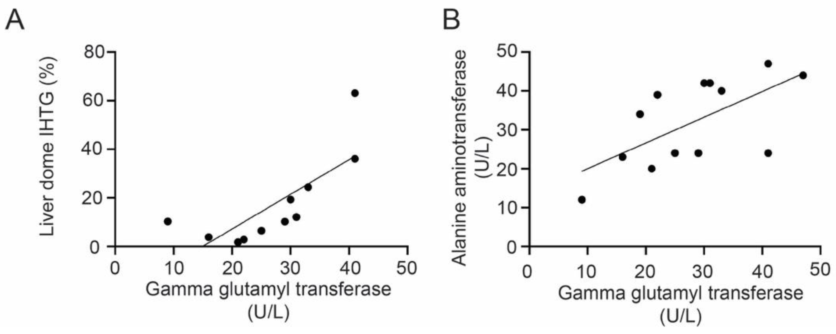
Upon laboratory evaluation, ALT was significantly greater in the MND cohort compared to male controls, with seven subjects having elevated (>30 U/L) values (mean MND ALT: 31.9 ± 11.2 U/L, control ALT: 20.6 ± 6.3 U/L, p = 0.0031). Elevation of the Homeostatic Model Assessment for Insulin Resistance (HOMA-IR) was detected in eight (62%) of the MND cohort, seven of whom had liver imaging consistent with NAFLD. MND patient serum γ-glutamyl transferase (GGT) levels positively correlated with both liver dome IHTG (r = 0.58, p = 0.0063, Figure 2A) and ALT (r = 0.41, p = 0.018, Figure 2B), although no difference in GGT levels was observed between the MND and control cohorts. Other laboratory measures, including serum creatine kinase (CK), total bilirubin, and free fatty acids, were normal (Table 1).

4. Discussion

We report evidence of NAFLD in a cohort of MND patients, indicating that NAFLD may be a part of the clinical spectrum in sporadic and inherited forms of MND. We observed liver dome IHTG elevation in MND patients that was comparable to levels reported previously in SBMA [5]. We observed elevated ALT and evidence of insulin resistance in MND patients compared to controls. Interestingly, both liver dome IHTG and serum ALT were positively correlated with serum GGT in the MND cohort. Previous studies have reported associations between GGT levels and the onset of neurodegenerative diseases such as Parkinson’s disease [6] and dementia [7]. It is unclear if fatty deposition precedes GGT elevation in MND.
The presentation of NAFLD in both MND and SBMA may be a consequence of alterations in lipid metabolism in these diseases. Most of the MND subjects in our study were found to have evidence of both insulin resistance and NAFLD, indicating a potential link between these processes. In a previous study, evidence of hepatic steatosis was reported in 76% of ALS patients and only 19% of non-MND neurological disease cases, which included patients with movement disorders and dementia, suggesting that the metabolic findings may be unique to those with motor neuron degeneration [2]. Additionally, our previous study of NAFLD in SBMA showed distinct hepatic gene expression changes in SBMA patient liver samples compared to others with non-alcoholic steatohepatitis [5].
Several therapeutic strategies have been identified for patients with NAFLD. Emerging evidence suggests that anti-diabetic drugs can reduce fatty accumulation and decrease liver enzyme levels in NAFLD [8]. Studies have shown the benefit of antioxidants on fatty liver progression [9], and antioxidants such as vitamin E [10,11] have also been investigated as potential therapies for ALS.
Since the present study was a cross-sectional analysis of NAFLD in MND, we are unable to report on the age of NAFLD onset in this cohort. NAFLD onset is challenging to detect as it typically results in few or no symptoms [4]. One potential mechanism of NAFLD in our cohort may be the reduction in activity levels associated with muscle atrophy and functional deficits. Cross-sectional studies have highlighted a strong positive association between sedentary time and NAFLD [12,13], suggesting that MND patients may develop NAFLD due to reduced overall mobility. Other evidence from animal models of MND, such as mouse models of SMA from SMN1 deletion, have suggested that the NAFLD phenotype may precede findings of motor dysfunction [3]. Further research is necessary to understand the prevalence of NAFLD in MND and to characterize the relationship of this process with the underlying disease mechanisms.

Author Contributions

Conceptualization, A.K., K.H.F. and C.G.; methodology, A.K., K.H.F. and C.G.; formal analysis, B.J. and C.G.; investigation, A.K., N.G., E.A.S., C.B. and C.G.; data curation, C.G.; writing—original draft preparation, B.J. and C.G.; writing—review and editing, A.K., N.G., E.A.S., C.B. and K.H.F.; visualization, B.J. and C.G.; supervision, K.H.F. and C.G.; funding acquisition, K.H.F. All authors have read and agreed to the published version of the manuscript.

Funding

This research was supported by intramural funds from the National Institute of Neurological Disorders and Stroke.

Institutional Review Board Statement

The study was conducted in accordance with the Declaration of Helsinki and approved by the Institutional Review Board of the National Institutes of Neurological Disorders and Stroke, National Institutes of Health, protocol NCT02124057, approval 28 April 2014.

Informed Consent Statement

Informed consent was obtained from all subjects involved in the study.

Data Availability Statement

All data generated or analyzed during this study are included in this published article.

Acknowledgments

The authors thank Elizabeth Hartnett, MSW (NINDS, National Institutes of Health) for her help in scheduling participants during the study, the clinical staff of the outpatient NIH neurology clinic for their assistance during the participants’ visits, and the research participants who made this study possible.

Conflicts of Interest

The authors declare no conflict of interest.

References

  1. Kanning, K.C.; Kaplan, A.; Henderson, C.E. Motor Neuron Diversity in Development and Disease. Annu. Rev. Neurosci. 2010, 33, 409–440. [Google Scholar] [CrossRef] [PubMed]
  2. Nodera, H.; Takamatsu, N.; Muguruma, N.; Ukimoto, K.; Nishio, S.; Oda, M.; Izumi, Y.; Kaji, R. Frequent Hepatic Steatosis in Amyotrophic Lateral Sclerosis: Implication for Systemic Involvement. Neurol. Clin. Neurosci. 2015, 3, 58–62. [Google Scholar] [CrossRef]
  3. Deguise, M.O.; Baranello, G.; Mastella, C.; Beauvais, A.; Michaud, J.; Leone, A.; de Amicis, R.; Battezzati, A.; Dunham, C.; Selby, K.; et al. Abnormal Fatty Acid Metabolism Is a Core Component of Spinal Muscular Atrophy. Ann. Clin. Transl. Neurol. 2019, 6, 1519–1532. [Google Scholar] [CrossRef] [PubMed] [Green Version]
  4. Cotter, T.G.; Rinella, M. Nonalcoholic Fatty Liver Disease 2020: The State of the Disease. Gastroenterology 2020, 158, 1851–1864. [Google Scholar] [CrossRef] [PubMed]
  5. Guber, R.D.; Takyar, V.; Kokkinis, A.; Fox, D.A.; Alao, H.; Kats, I.; Bakar, D.; Remaley, A.T.; Hewitt, S.M.; Kleiner, D.E.; et al. Nonalcoholic Fatty Liver Disease in Spinal and Bulbar Muscular Atrophy. Neurology 2017, 89, 2481–2490. [Google Scholar] [CrossRef] [Green Version]
  6. Yoo, D.; Kim, R.; Jung, Y.J.; Han, K.; Shin, C.M.; Lee, J.-Y. Serum Gamma-Glutamyltransferase Activity and Parkinson’s Disease Risk in Men and Women. Sci. Rep. 2020, 10, 1258. [Google Scholar] [CrossRef] [Green Version]
  7. Kunutsor, S.K.; Laukkanen, J.A. Gamma Glutamyltransferase and Risk of Future Dementia in Middle-aged to Older Finnish Men: A New Prospective Cohort Study. Alzheimers Dement. 2016, 12, 931–941. [Google Scholar] [CrossRef] [PubMed] [Green Version]
  8. Ferguson, D.; Finck, B.N. Emerging Therapeutic Approaches for the Treatment of NAFLD and Type 2 Diabetes Mellitus. Nat. Rev. Endocrinol. 2021, 17, 484–495. [Google Scholar] [CrossRef]
  9. Ferramosca, A.; di Giacomo, M.; Zara, V. Antioxidant Dietary Approach in Treatment of Fatty Liver: New Insights and Updates. World J. Gastroenterol. 2017, 23, 4146–4157. [Google Scholar] [CrossRef]
  10. Veldink, J.H.; Kalmijn, S.; Groeneveld, G.-J.; Wunderink, W.; Koster, A.; de Vries, J.H.M.; van der Luyt, J.; Wokke, J.H.J.; van den Berg, L.H. Intake of Polyunsaturated Fatty Acids and Vitamin E Reduces the Risk of Developing Amyotrophic Lateral Sclerosis. J. Neurol. Neurosurg. Psychiatry 2007, 78, 367–371. [Google Scholar] [CrossRef] [Green Version]
  11. Carrera-Juliá, S.; Moreno, M.L.; Barrios, C.; de la Rubia Ortí, J.E.; Drehmer, E. Antioxidant Alternatives in the Treatment of Amyotrophic Lateral Sclerosis: A Comprehensive Review. Front. Physiol. 2020, 11, 63. [Google Scholar] [CrossRef] [PubMed] [Green Version]
  12. Hallsworth, K.; Thoma, C.; Moore, S.; Ploetz, T.; Anstee, Q.M.; Taylor, R.; Day, C.P.; Trenell, M.I. Non-Alcoholic Fatty Liver Disease Is Associated with Higher Levels of Objectively Measured Sedentary Behaviour and Lower Levels of Physical Activity than Matched Healthy Controls. Frontline Gastroenterol. 2015, 6, 44–51. [Google Scholar] [CrossRef] [PubMed] [Green Version]
  13. Joo, J.H.; Kim, H.J.; Park, E.C.; Jang, S.I. Association between Sitting Time and Non-Alcoholic Fatty Live Disease in South Korean Population: A Cross-Sectional Study. Lipids Health Dis. 2020, 19, 212. [Google Scholar] [CrossRef] [PubMed]
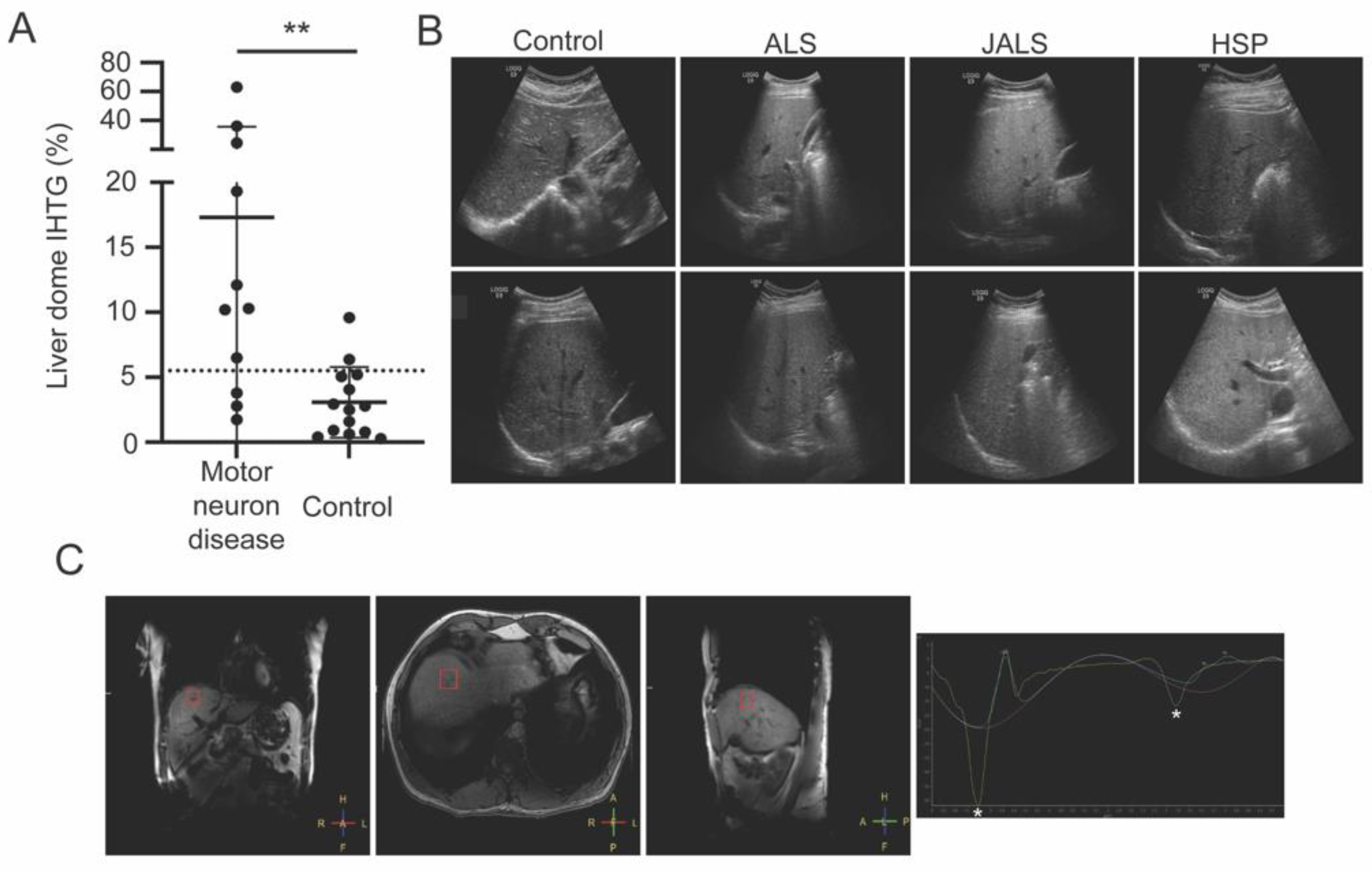
Figure 1. (A) 1H magnetic resonance spectroscopic imaging showed that the motor neuron disease group had a significantly higher intrahepatic triacylglycerol (IHTG) content in the dome of the liver compared to controls (** p = 0.0086); (B) increased echogenicity on liver ultrasound showing steatosis in patients with amyotrophic lateral sclerosis (ALS), juvenile ALS (JALS) with mutation in senataxin, and hereditary spastic paraplegia (HSP). Two patients from each category are shown; (C) voxel placement (red square) in the liver dome of a patient with HSP and spectroscopy trace showing the peaks for water (left asterisk) and triacylglycerol (right asterisk).
Genes 13 00936 g001
Figure 2. There was a significant correlation between (A) the liver dome intrahepatic triacylglycerol (IHTG) content and the serum γ-glutamyl transferase (r = 0.58, p = 0.0063) and (B) serum alanine aminotransferase and serum γ-glutamyl transferase (r = 0.41, p = 0.018) in the motor neuron disease group.
Genes 13 00936 g002
Table 1. Motor neuron disease cohort characteristics.
SubjectAge/
Onset (years)
BMI (kg/m2)DiseaseGeneMedications *CK (U/L)Total Bilirubin (mg/dL)ALT (U/L)HOMA-IRGGT (U/L)Cholesterol (mg/dL)Triglyc-erides
(mg/dL)
Free Fatty Acid (mEq/L)Liver Dome IHTG (%)Right Lobe IHTG (%)Left Lobe IHTG (%)Ultrasound
MND0159/49N/APLSN/ATizanidine1090.524N/A25187800.56.58.24.3Normal
MND0268/13N/AALS4SETXAtorvastatin3140.4247.7291541161.010.24.85.8Normal
MND0367/6733ALSN/AAtorvastatin, (AMX0035
research compound)
3860.6393.122134760.52.86.34.0Normal
MND0474/4924HSPSPG31Atorvastatin, solifenacin
succinate
2850.7231.226127550.33.83.310.6Normal
MND0556/30N/AHSPSPG4Diltiazem1070.9201.421269640.31.83.05.1Normal
MND0656/5424ALSN/ARiluzole4300.44219.1301302200.319.312.95.9Moderate hepatic steatosis
MND0729/19N/AHSPKIF1ANone1480.6474.6411971970.863.18.610.3Minimal hepatic steatosis
MND0840/1326ALS4SETXNone2000.5402.833137800.324.417.63.6Moderate hepatic steatosis
MND0954/5027HSPSPG4None1891.6242.7412291330.536.19.338.4Mild
hepatic steatosis
MND1067/3021HSPSPG4Duloxetine2500.5121.59155550.510.30.03.3Normal
MND1159/5824ALSN/ARiluzole12750.9420.931217790.512.17.68.7Normal
MND1237/1333ALS4SETXNone1770.5444.9471751000.4# Diffuse hepatic steatosis# Diffuse hepatic steatosis# Diffuse hepatic steatosisModerate hepatic steatosis
MND1357/54N/AALSC9orf72None2340.4344.019204860.8N/AN/AN/AModerate hepatic steatosis
* Medications listed are those which meet the criteria for the highest drug-induced liver injury (DILI) concern. Subjects MND06 and MND11 were on riluzole. # Spectroscopy not available and determination of hepatic steatosis made based on MRI imaging. Abbreviations: amyotrophic lateral sclerosis (ALS), alanine aminotransferase (ALT), body mass index (BMI), creatine kinase (CK), γ-glutamyl transferase (GGT), homeostatic model assessment insulin resistance index (HOMA-IR), hereditary spastic paraplegia (HSP), intrahepatic triacylglycerol (IHTG), kinesin family member 1A (KIF1A), primary lateral sclerosis (PLS), senataxin (SETX), spastic paraplegia 4 (SPG4), spastic paraplegia 31 (SPG31). N/A: Not Applicable.
Publisher’s Note: MDPI stays neutral with regard to jurisdictional claims in published maps and institutional affiliations.