Article

Utility of Select Gene Mutation Detection in Tumors by the Idylla Rapid Multiplex PCR Platform in Comparison to Next-Generation Sequencing

Department of Pathology and Laboratory Medicine, University of Rochester School of Medicine & Dentistry, Rochester, NY 14642, USA
*
Author to whom correspondence should be addressed.
Genes 2022, 13(5), 799; https://doi.org/10.3390/genes13050799
Submission received: 10 February 2022 / Revised: 24 April 2022 / Accepted: 26 April 2022 / Published: 29 April 2022
(This article belongs to the Special Issue Next Generation Sequencing in Clinical Diagnostics)

Abstract

:
Testing of tumors by next generation sequencing (NGS) is impacted by relatively long turnaround times and a need for highly trained personnel. Recently, Idylla oncology assays were introduced to test for BRAF, EGFR, KRAS, and NRAS common hotspot mutations that do not require specialized trained personnel. Moreover, the interpretation of results is fully automated, with rapid turnaround time. Though Idylla testing and NGS have been shown to have high concordance in identifying EGFR, BRAF, KRAS, and NRAS hotspot mutations, there is limited experience on optimal ways the Idylla system can be used in routine practice. We retrospectively evaluated all cases with EGFR, BRAF, KRAS, or NRAS mutations identified in clinical specimens sequenced on two different NGS panels at the University of Rochester Medical Center (URMC) molecular diagnostics laboratory between July 2020 and July 2021 and assessed if these mutations would be detected by the Idylla cartridges if used. We found that the Idylla system could accurately identify Tier 1 or 2 actionable genomic alterations in select associated disease pathologies if used. Yet, in a minority of cases, we would have been unable to detect NGS-identified pathogenic mutations due to their absence on the Idylla panels. We derived algorithmic practice guidelines for the use of the Idylla cartridges. Overall, Idylla molecular testing could be implemented either as a first-line standalone diagnostic tool in select indications or for orthogonal confirmation of uncertain results.

1. Introduction

Development of personalized therapies for cancer patients in the last two decades has had a substantial impact on the treatment strategies of different tumor types [1]. Testing to detect genomic alterations in tumors is standard of care in establishing diagnosis, for calculating prognosis, and for selecting optimal therapies. Some of the commonly identified mutations associated with targeted therapies include, e.g., mutations in BRAF in cutaneous melanoma; KRAS, BRAF, and NRAS in colorectal cancer; and EGFR mutations and ALK and ROS1 gene rearrangements in lung adenocarcinomas [2,3,4].
The gold standard for detecting different genomic alterations in tumors is using next generation sequencing (NGS)-based assays [5]. Institutions implement different NGS workflow plans depending on the NGS test available to them (i.e., gene panel list) and sample volume. However, relatively low sample volume for NGS testing and the specifics of standard NGS testing platforms typically requires sample batching, with suboptimal turnaround times of about 12–15 days. These are also high-complexity, labor-intensive tests that require highly trained specialized personnel; thus, they are not immediately available at smaller institutions and hospitals.
Recently, IdyllaTM oncology assays (Biocartis, Mechelen, Belgium) were launched to complement NGS testing. The Idylla system is an allele-specific qPCR-based assay platform in which all wet-bench steps are automated and confined to a single use cartridge. The assay starts after the insertion of sample-loaded gene(s) specific cartridges into the unit that are connected to a user interface console that displays result summaries. qPCR curve results generated from the assay are visualized through a secure web-based interface. The Idylla BRAF, EGFR, KRAS, and NRAS/BRAF mutation tests (cartridges) on the Idylla platform allow the detection of EGFR, BRAF, NRAS and KRAS hotspot mutations with rapid turnaround times (<3 h). Unlike NGS-based assays, the Idylla system can directly use formalin-fixed paraffin-embedded (FFPE) tissue sections without the need for DNA preparation and extraction, with interpretation of the results being fully automated. Several studies have demonstrated the validity and accuracy of the Idylla system in comparison to NGS in detecting EGFR, BRAF, KRAS, and NRAS hotspot mutations [6,7,8,9,10,11,12]. Yet, despite the high concordance of the Idylla and the NGS results, little has been done to formally assess how to best use the Idylla system clinically as a first-line diagnostic tool.
The overall objective of this study has been to start developing a formalized workflow on the optimal use of the Idylla system as a first-line diagnostic tool for the detection of different actionable genomic mutations in routine clinical practice. To this end, we retrospectively identified cases with EGFR, BRAF, KRAS, or NRAS mutations detected in specimens that were sequenced on two different NGS panels at the University of Rochester Medical Center (URMC) molecular diagnostics laboratory and assessed if the mutations would be detected on the Idylla system cartridges. The EGFR, BRAF, KRAS, or NRAS NGS detected mutations were compared to mutations that could be identified by the Idylla cartridges if used. We classified gene mutations identified by the NGS panels according to their tier grouping and evaluated to what extent the Idylla platform would have detected these mutations if utilized. Furthermore, we selected NGS-diagnosed BRAF or NRAS mutation positive cutaneous melanomas, BRAF mutation positive hairy cell leukemia, KRAS or EGFR mutation positive lung adenocarcinomas, and BRAF, KRAS or NRAS mutation positive colorectal cancer cases to assess what proportion of the identified mutations would have been recognized on the Idylla platform. Lastly, we also uncovered a proportion of NGS-evaluated cases with mutations that would have been detected on the Idylla system but had additional, Idylla non-identifiable mutations.

2. Materials and Methods

2.1. Clinical Specimens for Amplicon Based NGS Testing

We retrieved NGS results from all specimens referred for routine testing at the URMC Molecular diagnostics laboratory between July 2020 and July 2021. These samples were tested using laboratory-developed NGS-based assays using either the ThermoFisher’s Oncomine Focus Assay panel (Table S1) or the Illumina TruSightTM Myeloid sequencing panel (Table S1). From the database, we grouped the results to analyze the identified gene mutations in BRAF, KRAS, EGFR, and NRAS genes. The nucleotide changes identified for the four genes were compared to detectable BRAF, KRAS, EGFR, and NRAS variants on the Idylla cartridges.

2.2. cBioPortal for Cancer Genomics

Point mutation data was collected from the “cBioPortal for Cancer Genomics” website (https://www.cbioportal.org/datasets, accessed on or about 15 December 2021) for all studies with greater than 500 recorded point mutations. These data included the following studies: [13,14,15,16,17,18,19,20] Zehir et al. [13], Zhang et al. [14], Bolton et al. [15], Pereira et al. [16], Stopsack et al. [17], Razavi et al. [18], Samstein et al. [19], Nguyen et al. [20], Myelodysplastic (MSKCC, 2020), Cancer Cell Line Encyclopedia (Broad, 2019), MSK-IMPACT and MSK-ACCESS Mixed Cohort (MSK, 2021), Li et al. [21], Campbell et al. [22], Yaeger et al. [23], Pediatric Neuroblastoma (TARGET, 2018), Breast Invasive Carcinoma (TCGA, PanCancer Atlas), Ho et al. [24], Barretina et al. [25], Armenia et al. [26], Jonsson et al. [27], Reddy et al. [28], Jordan et al. [29], Grobner et al. [30], Ciriello et al. [31], Ceccarelli et al. [24,25,26,27,28,29,30,31,32], Mature B-cell malignancies (MD Anderson Cancer Center), Melanoma (MSKCC, 2018), Tyner et al. [33], Giannakis et al. [34], Lung Adenocarcinoma (MSKCC, 2020), Lung Adenocarcinoma (TCGA, PanCancer Atlas), Landau et al. [35], Colorectal Adenocarcinoma (TCGA, PanCancer Atlas), Ovarian Serous Cystadenocarcinoma (TCGA, PanCancer Atlas), Kim et al. [36], Uterine Corpus Endometrial Carcinoma (TCGA, PanCancer Atlas), Head and Neck Squamous Cell Carcinoma (TCGA, PanCancer Atlas), Brain Lower Grade Glioma (TCGA, PanCancer Atlas), and Head and Neck Squamous Cell Carcinoma (TCGA, Firehose Legacy), Koboldt et al. [37], Puente et al. [38], Abida et al. [39]. Records from these studies were aggregated and filtered, with only data columns for ‘study name’, ‘specimen ID’, ‘gene target’, ‘codon mutation name’ (example: c.901C>G), ‘long protein mutation name’ (example: p.Ala379Val), and ‘short protein mutation name’ (example: p.A379V) retained. Duplicate entries were filtered out, leaving approximately 3.7 million point-mutation records. The data was further filtered to include only four genes of interest: BRAF, EGFR, KRAS, and NRAS. This created a list of 11,420 unique ‘study-sample’ concatenation records.
A list of Idylla-identifiable point mutation targets was created based on package insert materials (comprising a list of 196 unique point mutations and indels spread across the 4 target genes). We designed a Python programming language script to compare each of the Idylla-identifiable mutations against each entry in the ‘four-target-gene-only’ data aggregate from the selected cBioPortal studies. We included successful matches in a list of ‘study-sample’ string-concatenations. Lastly, we filtered these results for unique values to avoid over-counting specimens which may have had multiple Idylla-identifiable mutations. We then compared the portion of Idylla-identifiable point mutations to the total number of ‘four-target-genes-only’ mutations found in the prepared cBioPortal data.

2.3. Statistical Analysis

Data analysis and figures were created by using Microsoft Excel, GraphPad 8.3, and CorelDraw 2019 software programs.

3. Results

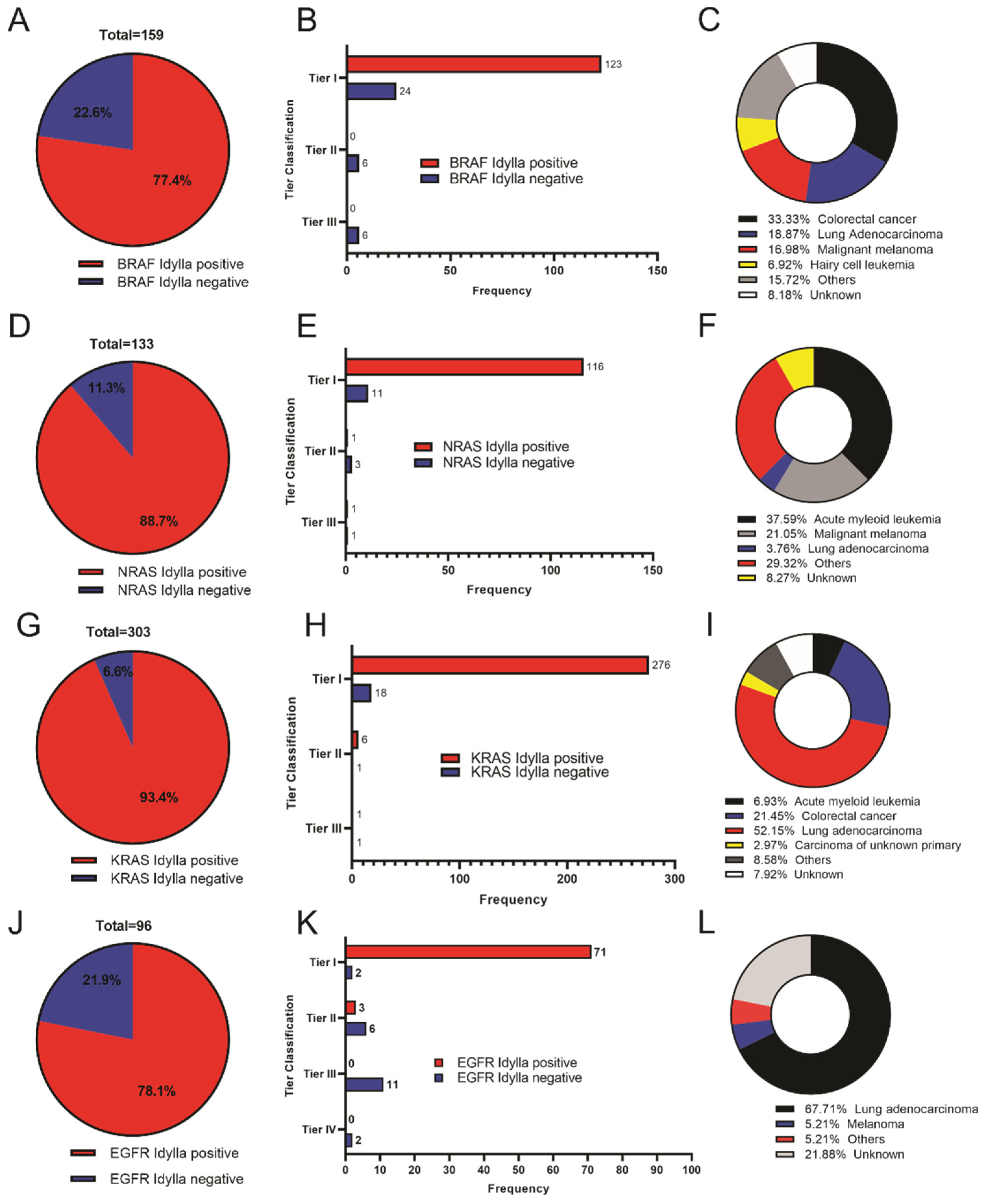
3.1. Idylla Cartridges Can Detect Most NGS Test Identified Tier 1 and Tier 2 Hotspot Mutations in EGFR, BRAF, KRAS, and NRAS

In the Idylla system, all assay cartridges are ready-to-use and contain the necessary reagents to perform sample preparation and real-time PCR amplification and detection, starting from insertion of FFPE tissue sections into the cartridges. The detectable variants for each target gene by the Idylla BRAF, KRAS, EGFR, and NRAS/BRAF cartridges are listed in Table S2 or on technical data sheets from https://www.biocartis.com/en-US/meet-idylla/idylla-oncology-assays (accessed on 29 December 2021).
We collated a total of 159 tumor samples bearing BRAF mutations, 133 with NRAS mutations, 303 with KRAS mutations, and 96 with EGFR mutations from the URMC NGS database from June 2020 to July 2021. We compared the NGS assay-detected nucleotide (protein) changes on these genes to the Idylla-detectable variants for these four genes (Table S2).
For BRAF, 77.4% of the variants identified on the NGS panels could potentially be identified by the Idylla system, while the remaining 22.6% could not (Figure 1A). We categorized 93% (147 of 159) of these BRAF mutations as Tier 1 mutations, of which 84% (123 of 147) could likewise be identified by the Idylla system (Figure 1B) if used. The majority of the samples with BRAF mutations were from colorectal cancers (approximately 33%) followed by lung adenocarcinoma (19%) and malignant melanoma (17%) (Figure 1C).
For the identified NRAS mutations, 89% (118 of 133) of the variants identified by NGS testing could be identified on the Idylla system as well (Figure 1D). 91% of the Tier 1 NRAS mutations (116 of 127) could be detected by the Idylla NRAS cartridge if used (Figure 1E). Most specimens with NRAS mutations were from acute myeloid leukemia patients (approximately 38%), followed by malignant melanoma (21%) and lung adenocarcinoma (4%) (Figure 1F).
KRAS mutation analysis revealed that 93% (283 of 303) of the NGS-detected mutations could be identified by the Idylla system (Figure 1G). We classified 294 of the KRAS mutations to be Tier 1, of which 276 (94%) would be detected on the Idylla system if utilized (Figure 1H). The majority of the samples were from lung adenocarcinoma (~52%), followed by colorectal cancer (21%) (Figure 1I).
EGFR mutation evaluation showed that 78% (76 of 96) of the NGS-identified EGFR mutations could likewise be detected by the Idylla system if run (Figure 1J). Furthermore, the Idylla system, if utilized, is predicted to have identified 97% (71 of 73) of the NGS-detected Tier 1 EGFR mutations (as shown by the upper bars of Figure 1K). Most of the EGFR mutations were from lung adenocarcinoma, at about 68%, and malignant melanoma, approximately 5% (Figure 1L).
Taken together, our results show that the four DNA extraction-based Idylla cartridges could identify most of the Tier 1 mutations detected by routine NGS testing if used.

3.2. Idylla Cartridges Identify Most High Tier Mutations in Samples from cBioportal for Cancer Database

Additionally, we compared the single nucleotide variant (SNV) and small indel profiles identifiable by NGS testing at the URMC Molecular Diagnostics laboratory for EGFR, BRAF, KRAS, and NRAS genes to their comprehensive variant profiles, using data for these genes compiled from the cBioPortal for Cancer Genomics website (https://www.cbioportal.org/datasets, accessed on 29 December 2021). The data comprised a total of 11,420 unique samples with either EGFR, BRAF, KRAS, or NRAS gene mutation profiles, spread across 44 studies (see Methods). This data set contained 2483 samples with BRAF mutations and 2960 with EGFR mutations, of which 1346 (54%) BRAF mutations and 1122 (38%) EGFR mutations could be identified by the Idylla system if used (Table 1). Furthermore, a total of 4667 samples with KRAS and 1636 with NRAS mutations from the cBioPortal data were compared to the Idylla system-detectable targets. The Idylla platform could have identified 4232 (91%) KRAS and 1423 (87%) NRAS mutations as positive if utilized (Table 1).

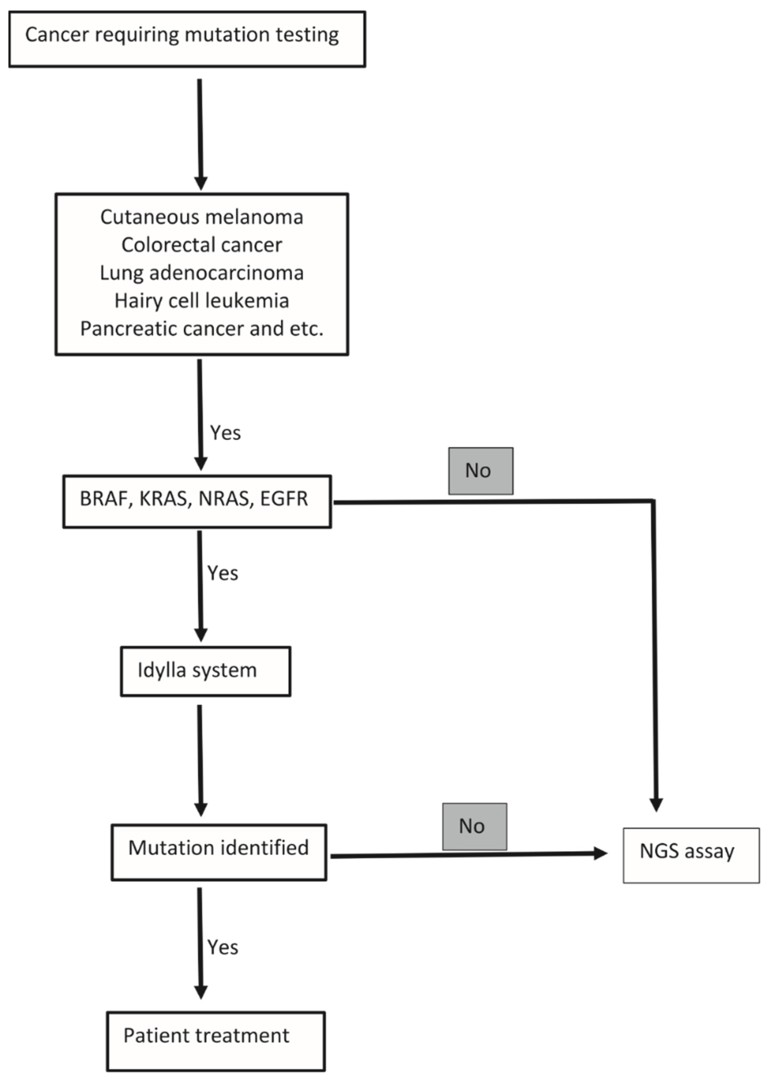
3.3. Assessment of the Idylla System with Mutations Linked to Specific Disease Pathologies
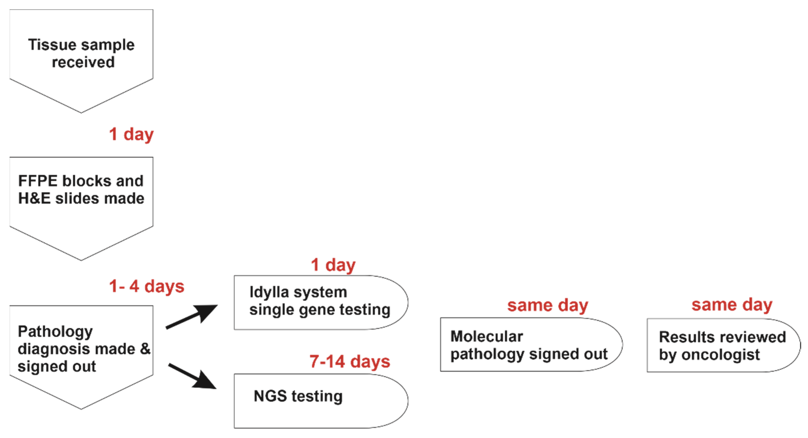
The utility of the Idylla system may vary from one tumor type to another. Thus, to determine the type and frequency of gene mutations that would be missed by the Idylla system, we next grouped gene alterations with their associated disease pathologies. We hypothesized that associating the NGS-identified gene mutations to their specific associated disease pathologies (e.g., lung adenocarcinoma with EGFR mutations or cutaneous melanoma with NRAS or BRAF mutations) would demonstrate higher concordance with hotspot mutations recognized by the Idylla system and may also uncover additional clinically significant NGS-identified gene mutations not detected by Idylla. The comprehensive results are detailed in Tables S3–S10.
We identified 27 cutaneous melanoma cases with BRAF mutations, of which 21 (78%) could also be detected by the Idylla BRAF and BRAF/NRAS cartridges if run (Table 2). Eight (33%) of the cutaneous melanoma samples had additional NGS-identified mutations (Table 2), of which four possessed BRAF variants that cannot be identified by the Idylla system, two of which were Tier 1 mutations. In the remaining three cases, one had a Tier 1 MAP2K1 mutation while the rest of the variants were mostly classified as Tier 3 (Table 2 and Table S3).
NRAS mutations were identified in 28 cutaneous melanoma cases, of which 26 (93%) could be also detected if run on the Idylla system (Table 2). Twelve (43%) NRAS mutation bearing cutaneous melanoma cases had additional mutations, with two harboring Tier 1 NRAS mutations that cannot be identified by the Idylla system (Table 2). Five of the other extra mutations were classified as Tier 1, while the remaining four variants were Tier 3 mutations (Table 2 and Table S4).
BRAF V600E mutations are present in almost all cases of hairy cell leukemia (HCL) at diagnosis [40]. Indeed, ten of the eleven HCL cases had BRAF V600E mutations that would be detected on the Idylla system if utilized (Table 2). Three (30%) HCL cases had additional genomic alterations, two of which (a TET2 and SF3B1 mutation) were classified as Tier 1 (Table 2 and Table S5).
We identified 53 patients with BRAF mutation bearing colorectal cancer (CRC), of which 51 (96%) had NGS identified BRAF mutations that would be detected on the Idylla system if performed (Table 2). Seventeen (32%) cases were observed to have additional mutations. Eight of these cases had one or more Tier 1 mutation(s) while the rest of the cases mostly had Tier 3 mutations (Table 2 and Table S6).
KRAS mutations were also identified in 65 CRC cases, of which 62 (95%) could be identified on the Idylla system if run (Table 2). We identified 24 (37%) cases with additional mutations (Table 2). Seventeen of these cases had one or more Tier 1 mutation(s), with PIK3CA alterations being the mostly frequently observed (Table 2 and Table S7).
NRAS mutations were also identified in three cases with CRC and two (66%) of the identified genomic alterations could be identified on the Idylla system (Table 2). We identified two cases with additional Tier 1 BRAF and PIK3CA mutations (Table 2 and Table S8).
Mutated KRAS and EGFR are common oncogenic drivers of lung adenocarcinoma with predictive value for targeted therapies [41,42]. Most EGFR mutations occur within its kinase domain, encoded by exons 18–21 [43]. We observed 65 cases of lung adenocarcinoma with EGFR mutations, of which 56 (86%) could be identified using the Idylla EGFR cartridge (Table 2) if used. Twenty-one (32%) samples harbored additional mutations, of which nine cases harbored a Tier 1 mutation (Table 2 and Table S9).
KRAS mutations were identified in 158 lung adenocarcinoma samples, of which 147 (93%) could be identified using the Idylla KRAS cartridge (Table 2). Twenty-five (13%) of the cases had extra mutations identified (Table 2), of which twelve possessed a Tier 1 mutation (Table 2 and Table S10).
Altogether, these data show that NGS identified EGFR, BRAF, KRAS, and NRAS mutations with select associated disease pathologies displayed high, but imperfect, concordance with mutations that would be detected by the Idylla cartridges if used, as a substantial minority of Idylla positive cases harbored additional NGS-identified Tier 1 or 2 gene mutations.

4. Discussion

NGS assays represent the gold standard for identifying genomic mutations in tumors [5]. However, introducing novel, closed-system technologies, such as the fully automated Idylla system, is of paramount importance because they can shorten the turnaround time and do not require specially trained laboratory personnel for their use.
Idylla cartridges are intended to identify common, clinically relevant Tier 1 and 2 mutations, hence they do not detect rare or complex variants. Our analyses and the results of other studies verified the Idylla system’s capacity for highly accurate detection of Tier 1 or 2 BRAF, NRAS, KRAS, and EGFR mutations within different malignancies [9,12,44,45,46]. Similarly, Van Haele et al. showed that BRAF, EGFR, and KRAS mutation testing on the Idylla system had a very high overall concordance with the TruSight Tumor26 NGS panel on actionable genomic mutations [10]. The lower detection levels for BRAF and EGFR mutations on the Idylla system with the cBioPortal for Cancer Genomics dataset can be attributed to the fact that most of the identified variants are rare and/or non-pathogenic. Thus, these variants would be outside the Idylla mutation profile range. Our data further demonstrated that NGS-detected BRAF, NRAS, KRAS, and EGFR mutations with select associated disease pathologies had high similarity with mutations that could be identified by Idylla cartridges. These data suggest that the Idylla system can be implemented as a standalone diagnostic tool or can be used as an orthogonal tool to confirm results from other molecular diagnostic assays.
From this, we derive a proposed algorithm on how the Idylla can be best utilized as a standalone diagnostic tool when testing for these select gene mutations (Figure 2). We also outline an algorithm of processing tumor samples for routine molecular testing (Figure 3). Pathologists in smaller and/or rural hospitals without an on-site molecular laboratory can easily incorporate and perform these molecular tests on FFPE tissue sections, obviating the need for DNA preparation and extraction while also offering the fully-automated interpretation of these results [47]. If no mutation is identified, the sample can be reflexed to send-out NGS testing. Alternatively, in large academic medical centers, the Idylla system can be used for orthogonal confirmation of ambiguous results from other (NGS) testing methods or for on-demand single gene testing when rapid identification of BRAF, NRAS, KRAS, and EGFR mutations for diagnosis and/or therapeutic decisions is required (Figure 3). Indeed, studies have shown that the Idylla molecular testing report can be reported within a day while initiating, e.g., EGFR-targeted treatments in NSCLC [48].
Though the Idylla system is a valuable tool for identifying select pathogenic mutations, it also has some considerable limitations. Tumor samples bearing complex mutations pose a challenge, as the Idylla system only identifies a limited number of pathogenic mutations while other pathogenic mutations outside the hotspots of the listed genes would not be identified. We have also shown that a substantial minority of samples have more than one Tier 1 and/or 2 mutations where the Idylla system would not identify all pathogenic mutations present in the sample. Samples with such mutation patterns present a conundrum: whether all samples initially run on the Idylla system should be reflexed for NGS testing, despite detecting an actionable mutation. For example, the IDH1 mutations we detected by NGS in Idylla-detectable BRAF or KRAS mutation positive CRC specimens (Tables S6 and S7) are associated with mucinous or signet ring cell adenocarcinoma, thus providing molecular support for the correct diagnosis that would not be possible from the Idylla results [49]. The Idylla system has also been shown to be ineffective in identifying some drug-resistant mutations when present at low variant allele levels within the tumor [48]. An additional shortcoming of the Idylla system is that it can test only one sample at a time per instrument, limiting throughput [9]. It is also worth noting that the choice to utilize the Idylla molecular testing can be influenced by sample size. Scant tissue samples may necessitate conventional NGS protocols.
Taken together, our analyses have shown that the Idylla platform is able to identify the majority of common pathogenic mutations in the BRAF, NRAS, KRAS, and EGFR genes, has a very fast turnaround time, and does not require specialized personnel training—all of which makes it an ideal diagnostic system in small hospital/remote area settings. In contrast, for larger institutions with relatively fast NGS capabilities, the Idylla system can be best utilized to support select morphologic diagnoses (e.g., identifying BRAF V600E mutation to support diagnosis of hairy cell leukemia) or for orthogonal confirmation of uncertain or very low allele frequency mutation results.

Supplementary Materials

The following supporting information can be downloaded at: https://www.mdpi.com/article/10.3390/genes13050799/s1, Tables S1–S10. Table S1: (A) Oncomine Focus Assay (OFA) hotspot gene list; (B) TruSeq myeloid panel gene list; Table S2: (A) Idylla BRAF cartridge detectable mutations; (B) Idylla EGFR cartridge detectable mutations; (C) Idylla KRAS cartridge detectable mutations; (D) Idylla NRAS-BRAF cartridge detectable mutations; Table S3: Cutaneous melanoma specimens with Idylla-identifiable BRAF mutations and/or with additional genetic alterations; Table S4: Cutaneous melanoma specimens with Idylla-identifiable NRAS mutations and/or with additional genetic alterations; Table S5: Hairy cell leukemia specimens with Idylla-identifiable BRAF mutations and/or with additional genetic alterations; Table S6: Colorectal cancer specimens with Idylla-identifiable BRAF mutations and/or with additional genetic alterations; Table S7: Colorectal cancer specimens with Idylla-identifiable KRAS mutations and/or with additional genetic alterations; Table S8: Colorectal cancer specimens with Idylla-identifiable NRAS mutations and/or with additional genetic alterations; Table S9: Lung adenocarcinoma specimens with Idylla-identifiable EGFR mutations and/or with additional genetic alterations; Table S10: Lung adenocarcinoma specimens with Idylla-identifiable KRAS mutations and/or with additional genetic alterations.

Author Contributions

D.N., V.L.C. and Z.N.O. designed the studies. D.N., C.R.S. and V.L.C. performed the analyses. D.N., V.L.C. and Z.N.O. wrote the paper. All authors endorse the results of this investigation. All authors have read and agreed to the published version of the manuscript.

Funding

This research received no external funding.

Informed Consent Statement

All patients signed the institution-approved, standard consent for clinical diagnostic testing by NGS, including agreement to the opt in/out clause for use of genetic and other diagnostic information for research purposes. This consent mechanism does not allow for sharing of genetic and other diagnostic information beyond that clinically relevant and reported in the manuscript.

Data Availability Statement

The consent documentation signed by the patients do not expressly allow submission of full sequencing data (FASTQ, BAM/BAI, VCF) to external data repositories.

Acknowledgments

We thank Paul Rothberg (University of Rochester) for comments on the manuscript.

Conflicts of Interest

The authors declare no conflict of interest.

References

  1. Gandara, D.R.; Lara, P.N., Jr.; Mack, P.; Scagliotti, G. Individualizing therapy for non-small-cell lung cancer: A paradigm shift from empiric to integrated decision-making. Clin. Lung Cancer 2009, 10, 148–150. [Google Scholar] [CrossRef] [PubMed]
  2. Benson, A.B.; Venook, A.P.; Cederquist, L.; Chan, E.; Chen, Y.-J.; Cooper, H.S.; Deming, D.; Engstrom, P.F.; Enzinger, P.C.; Fichera, A.; et al. Colon Cancer, Version 1.2017, NCCN Clinical Practice Guidelines in Oncology. J. Natl. Compr. Cancer Netw. 2017, 15, 370–398. [Google Scholar] [CrossRef] [PubMed]
  3. Coit, D.G.; Thompson, J.A.; Algazi, A.; Andtbacka, R.; Bichakjian, C.K.; Carson, W.E.; Daniels, G.A.; DiMaio, D.; Fields, R.C.; Fleming, M.D.; et al. NCCN Guidelines Insights: Melanoma, Version 3.2016. J. Natl. Compr. Cancer Netw. 2016, 14, 945–958. [Google Scholar] [CrossRef] [PubMed]
  4. Lindeman, N.I.; Cagle, P.T.; Aisner, D.L.; Arcila, M.E.; Beasley, M.B.; Bernicker, E.H.; Colasacco, C.; Dacic, S.; Hirsch, F.R.; Kerr, K.; et al. Updated Molecular Testing Guideline for the Selection of Lung Cancer Patients for Treatment with Targeted Tyrosine Kinase Inhibitors: Guideline from the College of American Pathologists, the International Association for the Study of Lung Cancer, and the Association for Molecular Pathology. Arch. Pathol. Lab. Med. 2018, 142, 321–346. [Google Scholar] [CrossRef] [PubMed] [Green Version]
  5. Salto-Tellez, M.; de Castro, D.G. Next-generation sequencing: A change of paradigm in molecular diagnostic validation. J. Pathol. 2014, 234, 5–10. [Google Scholar] [CrossRef]
  6. Colling, R.; Wang, L.M.; Soilleux, E. Validating a fully automated real-time PCR-based system for use in the molecular diagnostic analysis of colorectal carcinoma: A comparison with NGS and IHC. J. Clin. Pathol. 2017, 70, 610–614. [Google Scholar] [CrossRef]
  7. Johnston, L.; Power, M.; Sloan, P.; Long, A.; Silmon, A.; Chaffey, B.; Lisgo, A.J.; Little, L.; Vercauteren, E.; Steiniche, T.; et al. Clinical performance evaluation of the Idylla NRAS-BRAF mutation test on retrospectively collected formalin-fixed paraffin-embedded colorectal cancer tissue. J. Clin. Pathol. 2018, 71, 336–343. [Google Scholar] [CrossRef]
  8. Melchior, L.; Grauslund, M.; Bellosillo, B.; Montagut, C.; Torres, E.; Moragón, E.; Micalessi, I.; Frans, J.; Noten, V.; Bourgain, C.; et al. Multi-center evaluation of the novel fully-automated PCR-based Idylla™ BRAF Mutation Test on formalin-fixed paraffin-embedded tissue of malignant melanoma. Exp. Mol. Pathol. 2015, 99, 485–491. [Google Scholar] [CrossRef] [Green Version]
  9. Uguen, A.; Troncone, G. A review on the Idylla platform: Towards the assessment of actionable genomic alterations in one day. J. Clin. Pathol. 2018, 71, 757–762. [Google Scholar] [CrossRef]
  10. Van Haele, M.; Vander Borght, S.; Ceulemans, A.; Wieërs, M.; Metsu, S.; Sagaert, X.; Weynand, B. Rapid clinical mutational testing of KRAS, BRAF and EGFR: A prospective comparative analysis of the Idylla technique with high-throughput next-generation sequencing. J. Clin. Pathol. 2020, 73, 35–41. [Google Scholar] [CrossRef]
  11. Yeo, M.-K.; Jung, M.-K.; Lee, S.-Y.; Lee, Y.-M.; Hur, G.M.; Kim, J.-M. The usefulness of a novel fully automated PCR-based Idylla test for detection of the BRAF V600E mutation in thyroid tissue: Comparison with PNA-clamping PCR, real-time PCR and pyrosequencing. J. Clin. Pathol. 2017, 70, 260–265. [Google Scholar] [CrossRef] [PubMed]
  12. Momeni-Boroujeni, A.; Salazar, P.; Zheng, T.; Mensah, N.; Rijo, I.; Dogan, S.; Yao, J.; Moung, C.; Vanderbilt, C.; Benhamida, J.; et al. Rapid EGFR Mutation Detection Using the Idylla Platform: Single-Institution Experience of 1200 Cases Analyzed by an In-House Developed Pipeline and Comparison with Concurrent Next-Generation Sequencing Results. J. Mol. Diagn. 2021, 23, 310–322. [Google Scholar] [CrossRef] [PubMed]
  13. Zehir, A.; Benayed, R.; Shah, R.H.; Syed, A.; Middha, S.; Kim, H.R.; Srinivasan, P.; Gao, J.; Chakravarty, D.; Devlin, S.M.; et al. Mutational landscape of metastatic cancer revealed from prospective clinical sequencing of 10,000 patients. Nat. Med. 2017, 23, 703–713. [Google Scholar] [CrossRef] [PubMed]
  14. Zhang, Y.; Yao, Y.; Xu, Y.; Li, L.; Gong, Y.; Zhang, K.; Zhang, M.; Guan, Y.; Chang, L.; Xia, X.; et al. Pan-cancer circulating tumor DNA detection in over 10,000 Chinese patients. Nat. Commun. 2021, 12, 11. [Google Scholar] [CrossRef]
  15. Bolton, K.L.; Ptashkin, R.N.; Gao, T.; Braunstein, L.; Devlin, S.M.; Kelly, D.; Patel, M.; Berthon, A.; Syed, A.; Yabe, M.; et al. Cancer therapy shapes the fitness landscape of clonal hematopoiesis. Nat. Genet. 2020, 52, 1219–1226. [Google Scholar] [CrossRef]
  16. Pereira, B.; Chin, S.-F.; Rueda, O.M.; Vollan, H.-K.M.; Provenzano, E.; Bardwell, H.A.; Pugh, M.; Jones, L.; Russell, R.; Sammut, S.-J.; et al. The somatic mutation profiles of 2,433 breast cancers refine their genomic and transcriptomic landscapes. Nat. Commun. 2016, 7, 11479. [Google Scholar] [CrossRef] [Green Version]
  17. Stopsack, K.H.; Nandakumar, S.; Arora, K.; Nguyen, B.; Vasselman, S.E.; Nweji, B.; McBride, S.M.; Morris, M.J.; Rathkopf, D.E.; Slovin, S.F.; et al. Differences in prostate cancer genomes by self-reported race: Contributions of genetic ancestry, modifiable cancer risk factors, and clinical factors. Clin. Cancer Res. 2022, 28, 318–326. [Google Scholar] [CrossRef]
  18. Razavi, P.; Chang, M.T.; Xu, G.; Bandlamudi, C.; Ross, D.S.; Vasan, N.; Cai, Y.; Bielski, C.M.; Donoghue, M.T.A.; Jonsson, P.; et al. The Genomic Landscape of Endocrine-Resistant Advanced Breast Cancers. Cancer Cell 2018, 34, 427–438.e426. [Google Scholar] [CrossRef] [Green Version]
  19. Samstein, R.M.; Lee, C.-H.; Shoushtari, A.N.; Hellmann, M.D.; Shen, R.; Janjigian, Y.Y.; Barron, D.A.; Zehir, A.; Jordan, E.J.; Omuro, A.; et al. Tumor mutational load predicts survival after immunotherapy across multiple cancer types. Nat. Genet. 2019, 51, 202–206. [Google Scholar] [CrossRef]
  20. Nguyen, B.; Mota, J.M.; Nandakumar, S.; Stopsack, K.H.; Weg, E.; Rathkopf, D.; Morris, M.J.; Scher, H.I.; Kantoff, P.W.; Gopalan, A.; et al. Pan-cancer Analysis of CDK12 Alterations Identifies a Subset of Prostate Cancers with Distinct Genomic and Clinical Characteristics. Eur. Urol. 2020, 78, 671–679. [Google Scholar] [CrossRef]
  21. Li, Q.; Jiang, B.; Guo, J.; Shao, H.; Del Priore, I.S.; Chang, Q.; Kudo, R.; Li, Z.; Razavi, P.; Liu, B.; et al. INK4 tumor suppressor proteins mediate resistance to CDK4/6 kinase inhibitors. Cancer Discov. 2022, 12, 356–371. [Google Scholar] [CrossRef] [PubMed]
  22. Campbell, J.D.; Alexandrov, A.; Kim, J.; Wala, J.; Berger, A.H.; Pedamallu, C.S.; Shukla, S.A.; Guo, G.; Brooks, A.N.; Murray, B.A.; et al. Distinct patterns of somatic genome alterations in lung adenocarcinomas and squamous cell carcinomas. Nat. Genet. 2016, 48, 607–616. [Google Scholar] [CrossRef] [PubMed] [Green Version]
  23. Yaeger, R.; Chatila, W.K.; Lipsyc, M.D.; Hechtman, J.F.; Cercek, A.; Sanchez-Vega, F.; Jayakumaran, G.; Middha, S.; Zehir, A.; Donoghue, M.T.A.; et al. Clinical Sequencing Defines the Genomic Landscape of Metastatic Colorectal Cancer. Cancer Cell 2018, 33, 125–136.e123. [Google Scholar] [CrossRef] [Green Version]
  24. Ho, A.S.; Ochoa, A.; Jayakumaran, G.; Zehir, A.; Valero Mayor, C.; Tepe, J.; Makarov, V.; Dalin, M.G.; He, J.; Bailey, M.; et al. Genetic hallmarks of recurrent/metastatic adenoid cystic carcinoma. J. Clin. Investig. 2019, 129, 4276–4289. [Google Scholar] [CrossRef] [PubMed]
  25. Barretina, J.; Caponigro, G.; Stransky, N.; Venkatesan, K.; Margolin, A.A.; Kim, S.; Wilson, C.J.; Lehár, J.; Kryukov, G.V.; Sonkin, D.; et al. The Cancer Cell Line Encyclopedia enables predictive modelling of anticancer drug sensitivity. Nature 2012, 483, 603–607. [Google Scholar] [CrossRef]
  26. Armenia, J.; Wankowicz, S.A.M.; Liu, D.; Gao, J.; Kundra, R.; Reznik, E.; Chatila, W.K.; Chakravarty, D.; Han, G.C.; Coleman, I.; et al. The long tail of oncogenic drivers in prostate cancer. Nat. Genet. 2018, 50, 645–651. [Google Scholar] [CrossRef]
  27. Jonsson, P.; Lin, A.L.; Young, R.J.; DiStefano, N.M.; Hyman, D.M.; Li, B.T.; Berger, M.F.; Zehir, A.; Ladanyi, M.; Solit, D.B.; et al. Genomic Correlates of Disease Progression and Treatment Response in Prospectively Characterized Gliomas. Clin. Cancer Res. 2019, 25, 5537–5547. [Google Scholar] [CrossRef] [Green Version]
  28. Reddy, A.; Zhang, J.; Davis, N.S.; Moffitt, A.B.; Love, C.L.; Waldrop, A.; Leppa, S.; Pasanen, A.; Meriranta, L.; Karjalainen-Lindsberg, M.-L.; et al. Genetic and Functional Drivers of Diffuse Large B Cell Lymphoma. Cell 2017, 171, 481–494.e415. [Google Scholar] [CrossRef] [PubMed] [Green Version]
  29. Jordan, E.J.; Kim, H.R.; Arcila, M.E.; Barron, D.; Chakravarty, D.; Gao, J.; Chang, M.T.; Ni, A.; Kundra, R.; Jonsson, P.; et al. Prospective Comprehensive Molecular Characterization of Lung Adenocarcinomas for Efficient Patient Matching to Approved and Emerging Therapies. Cancer Discov. 2017, 7, 596–609. [Google Scholar] [CrossRef] [Green Version]
  30. Gröbner, S.N.; Worst, B.C.; Weischenfeldt, J.; Buchhalter, I.; Kleinheinz, K.; Rudneva, V.A.; Johann, P.D.; Balasubramanian, G.P.; Segura-Wang, M.; Brabetz, S.; et al. The landscape of genomic alterations across childhood cancers. Nature 2018, 555, 321–327. [Google Scholar] [CrossRef] [Green Version]
  31. Ciriello, G.; Gatza, M.L.; Beck, A.H.; Wilkerson, M.D.; Rhie, S.K.; Pastore, A.; Zhang, H.; McLellan, M.; Yau, C.; Kandoth, C.; et al. Comprehensive Molecular Portraits of Invasive Lobular Breast Cancer. Cell 2015, 163, 506–519. [Google Scholar] [CrossRef] [PubMed] [Green Version]
  32. Ceccarelli, M.; Barthel, F.P.; Malta, T.M.; Sabedot, T.S.; Salama, S.R.; Murray, B.A.; Morozova, O.; Newton, Y.; Radenbaugh, A.; Pagnotta, S.M.; et al. Molecular Profiling Reveals Biologically Discrete Subsets and Pathways of Progression in Diffuse Glioma. Cell 2016, 164, 550–563. [Google Scholar] [CrossRef] [PubMed] [Green Version]
  33. Tyner, J.W.; Tognon, C.E.; Bottomly, D.; Wilmot, B.; Kurtz, S.E.; Savage, S.L.; Long, N.; Schultz, A.R.; Traer, E.; Abel, M.; et al. Functional genomic landscape of acute myeloid leukaemia. Nature 2018, 562, 526–531. [Google Scholar] [CrossRef] [PubMed]
  34. Giannakis, M.; Mu, X.J.; Shukla, S.A.; Qian, Z.R.; Cohen, O.; Nishihara, R.; Bahl, S.; Cao, Y.; Amin-Mansour, A.; Yamauchi, M.; et al. Genomic Correlates of Immune-Cell Infiltrates in Colorectal Carcinoma. Cell Rep. 2016, 15, 857–865. [Google Scholar] [CrossRef] [Green Version]
  35. Landau, D.A.; Tausch, E.; Taylor-Weiner, A.N.; Stewart, C.; Reiter, J.G.; Bahlo, J.; Kluth, S.; Bozic, I.; Lawrence, M.; Böttcher, S.; et al. Mutations driving CLL and their evolution in progression and relapse. Nature 2015, 526, 525–530. [Google Scholar] [CrossRef] [Green Version]
  36. Kim, J.; Bowlby, R.; Mungall, A.J.; Robertson, A.G.; Odze, R.D.; Cherniack, A.D.; Shih, J.; Pedamallu, C.S.; Cibulskis, C.; Dunford, A.; et al. Integrated genomic characterization of oesophageal carcinoma. Nature 2017, 541, 169–175. [Google Scholar] [CrossRef] [Green Version]
  37. Koboldt, D.C.; Fulton, R.S.; McLellan, M.D.; Schmidt, H.; Kalicki-Veizer, J.; McMichael, J.F.; Fulton, L.L.; Dooling, D.J.; Ding, L.; Mardis, E.R.; et al. Comprehensive molecular portraits of human breast tumours. Nature 2012, 490, 61–70. [Google Scholar] [CrossRef] [Green Version]
  38. Puente, X.S.; Beà, S.; Valdés-Mas, R.; Villamor, N.; Gutiérrez-Abril, J.; Martín-Subero, J.I.; Munar, M.; Rubio-Pérez, C.; Jares, P.; Aymerich, M.; et al. Non-coding recurrent mutations in chronic lymphocytic leukaemia. Nature 2015, 526, 519–524. [Google Scholar] [CrossRef]
  39. Abida, W.; Armenia, J.; Gopalan, A.; Brennan, R.; Walsh, M.; Barron, D.; Danila, D.; Rathkopf, D.; Morris, M.; Slovin, S.; et al. Prospective Genomic Profiling of Prostate Cancer Across Disease States Reveals Germline and Somatic Alterations That May Affect Clinical Decision Making. JCO Precis Oncol 2017, 2017, PO.17.00029. [Google Scholar] [CrossRef]
  40. Falini, B.; Martelli, M.P.; Tiacci, E. BRAF V600E mutation in hairy cell leukemia: From bench to bedside. Blood 2016, 128, 1918–1927. [Google Scholar] [CrossRef] [Green Version]
  41. Riely, G.J.; Ladanyi, M. KRAS mutations: An old oncogene becomes a new predictive biomarker. J. Mol. Diagn 2008, 10, 493–495. [Google Scholar] [CrossRef] [PubMed]
  42. Sequist, L.V.; Bell, D.W.; Lynch, T.J.; Haber, D.A. Molecular predictors of response to epidermal growth factor receptor antagonists in non-small-cell lung cancer. J. Clin. Oncol. 2007, 25, 587–595. [Google Scholar] [CrossRef] [PubMed]
  43. Shigematsu, H.; Lin, L.; Takahashi, T.; Nomura, M.; Suzuki, M.; Wistuba, I.I.; Fong, K.M.; Lee, H.; Toyooka, S.; Shimizu, N.; et al. Clinical and biological features associated with epidermal growth factor receptor gene mutations in lung cancers. J. Natl. Cancer Inst. 2005, 97, 339–346. [Google Scholar] [CrossRef] [PubMed] [Green Version]
  44. Delgado-García, M.; Weynand, B.; Gómez-Izquierdo, L.; Hernández, M.J.; Blanco, Á.M.; Varela, M.; Matias-Guiu, X.; Nadal, E.; Márquez-Lobo, B.; Alarcão, A.; et al. Clinical performance evaluation of the Idylla™ EGFR Mutation Test on formalin-fixed paraffin-embedded tissue of non-small cell lung cancer. BMC Cancer 2020, 20, 275. [Google Scholar] [CrossRef] [PubMed] [Green Version]
  45. Evrard, S.M.; Taranchon-Clermont, E.; Rouquette, I.; Murray, S.; Dintner, S.; Nam-Apostolopoulos, Y.C.; Bellosillo, B.; Varela-Rodriguez, M.; Nadal, E.; Wiedorn, K.H.; et al. Multicenter Evaluation of the Fully Automated PCR-Based Idylla EGFR Mutation Assay on Formalin-Fixed, Paraffin-Embedded Tissue of Human Lung Cancer. J. Mol. Diagn. 2019, 21, 1010–1024. [Google Scholar] [CrossRef]
  46. Tsongalis, G.J.; Al Turkmani, M.R.; Suriawinata, M.; Babcock, M.J.; Mitchell, K.; Ding, Y.; Scicchitano, L.; Tira, A.; Buckingham, L.; Atkinson, S.; et al. Comparison of Tissue Molecular Biomarker Testing Turnaround Times and Concordance Between Standard of Care and the Biocartis Idylla Platform in Patients with Colorectal Cancer. Am. J. Clin. Pathol. 2020, 154, 266–276. [Google Scholar] [CrossRef]
  47. Le Flahec, G.; Guibourg, B.; Marcorelles, P.; Uguen, A. Financial implications of Idylla testing in colorectal cancer, lung cancer and melanoma: A French laboratory point of view. J. Clin. Pathol. 2017, 70, 906–907. [Google Scholar] [CrossRef]
  48. Lambros, L.; Caumont, C.; Guibourg, B.; Barel, F.; Quintin-Roué, I.; Marcorelles, P.; Merlio, J.-P.; Uguen, A. Evaluation of a fast and fully automated platform to diagnose EGFR and KRAS mutations in formalin-fixed and paraffin-embedded non-small cell lung cancer samples in less than one day. J. Clin. Pathol. 2017, 70, 544–549. [Google Scholar] [CrossRef]
  49. Huang, J.; Tseng, L.-H.; Parini, V.; Lokhandwala, P.M.; Pallavajjala, A.; Rodriguez, E.; Xian, R.; Chen, L.; Gocke, C.D.; Eshleman, J.R.; et al. IDH1 and IDH2 Mutations in Colorectal Cancers. Am. J. Clin. Pathol. 2021, 156, 777–786. [Google Scholar] [CrossRef]
Figure 1. Proportion of gene mutations identified on NGS that would be positive on Idylla molecular testing. (AC) Percentage of BRAF mutations identified on NGS panel that would be detected on Idylla system and associated Tier classification and primary site diagnosis. (DF) Percentage of NRAS mutations identified on NGS panel that would be detected on Idylla system and associated Tier classification and primary site diagnosis. (GI) Percentage of KRAS mutations identified on NGS panel that would be detected on Idylla system and associated Tier classification and primary site diagnosis. (JL) Percentage of EGFR mutations identified on NGS panel that would be detected on Idylla system and associated Tier classification and primary site diagnosis.
Genes 13 00799 g001
Figure 2. Generic algorithm for the clinical use of the Idylla platform.
Genes 13 00799 g002
Figure 3. Algorithm for processing of tumor samples for molecular testing.
Genes 13 00799 g003
Table 1. cBioPortal for Cancer Genomics and associated gene mutations.
GeneIdylla PositiveIdylla Negative
BRAF1346 (54%)1137 (46%)
EGFR1122 (38%)1838 (62%)
KRAS4232 (91%)435 (9%)
NRAS1423 (87%)213 (13%)
Table 2. Gene mutations and associated specific disease.
Gene Mutation and DiagnosisCase
Numbers
Idylla
Positive
Idylla
Negative
Cases with Extra
Mutations
Cases with Extra and Tier 1 Mutations Not Detected by Idylla
BRAF melanoma2721 (78%)6 (22%)8 (30%)2 (8%)
NRAS melanoma2826 (93%)2 (7%)12 (43%)5 (18%)
BRAF HCL1110 (91%)1(9%)3 (27%)3 (27%)
BRAF CRC5351 (96%)2 (4%)17 (32%)9 (17%)
KRAS CRC6562 (95%)3 (5%)24 (37%)17 (26%)
NRAS CRC32 (66%)1 (33%)22 (66%)
EGFR lung adenocarcinoma6556 (86%)9 (7%)21 (32%)7 (11%)
KRAS lung adenocarcinoma158147 (93%)11 (7%)25 (16%)12 (8%)
Publisher’s Note: MDPI stays neutral with regard to jurisdictional claims in published maps and institutional affiliations.

