Review

The Astonishing Large Family of HSP40/DnaJ Proteins Existing in Leishmania

1
Centro de Biología Molecular Severo Ochoa (CSIC-UAM), Departamento de Biología Molecular, Instituto Universitario de Biología Molecular (IUBM), Universidad Autónoma de Madrid, 28049 Madrid, Spain
2
Centro de Investigación Biomédica en Red de Enfermedades Infecciosas (CIBERINFEC), Instituto de Salud Carlos III, 28029 Madrid, Spain
3
WHO Collaborating Centre for Leishmaniasis, Centro Nacional de Microbiología, Instituto de Salud Carlos III, Majadahonda, 28220 Madrid, Spain
4
Centro de Biología Molecular Severo Ochoa (CSIC-UAM), Genomic and NGS Facility (GENGS), 28049 Madrid, Spain
*
Author to whom correspondence should be addressed.
Genes 2022, 13(5), 742; https://doi.org/10.3390/genes13050742
Submission received: 31 March 2022 / Revised: 14 April 2022 / Accepted: 21 April 2022 / Published: 23 April 2022
(This article belongs to the Special Issue Genetic Mechanisms Involved in Microbial Stress Responses)

Abstract

:
Abrupt environmental changes are faced by Leishmania parasites during transmission from a poikilothermic insect vector to a warm-blooded host. Adaptation to harsh environmental conditions, such as nutrient deprivation, hypoxia, oxidative stress and heat shock needs to be accomplished by rapid reconfiguration of gene expression and remodeling of protein interaction networks. Chaperones play a central role in the maintenance of cellular homeostasis, and they are responsible for crucial tasks such as correct folding of nascent proteins, protein translocation across different subcellular compartments, avoiding protein aggregates and elimination of damaged proteins. Nearly one percent of the gene content in the Leishmania genome corresponds to members of the HSP40 family, a group of proteins that assist HSP70s in a variety of cellular functions. Despite their expected relevance in the parasite biology and infectivity, little is known about their functions or partnership with the different Leishmania HSP70s. Here, we summarize the structural features of the 72 HSP40 proteins encoded in the Leishmania infantum genome and their classification into four categories. A review of proteomic data, together with orthology analyses, allow us to postulate cellular locations and possible functional roles for some of them. A detailed study of the members of this family would provide valuable information and opportunities for drug discovery and improvement of current treatments against leishmaniasis.

1. Introduction

Life is the result of an orchestrated interplay between the biomolecules constituting cells and environmental signals, which are of either biological, physical or chemical nature. Inside the cells, proteins are continuously being synthesized, and they have to reach a proper folding to perform their biological functions, but these processes occur in a ‘crowded marketplace’. In this context, it is not surprising that molecular chaperones were among the first inhabitants of living cells. These proteins, many of them also known as heat shock proteins (HSPs) because they are involved in protecting cells from the effects derived of anomalously high temperatures, assist the folding and assembly of most de novo synthetized polypeptides. Moreover, molecular chaperones are involved in cell-signaling cascades through modulating interactions between different biomolecules. There are several families of evolutionarily conserved chaperones, but central is the HSP70 chaperone, which is considered the master player in protein homeostasis [1]. Nevertheless, the functional versatility of Hsp70, its precise localization within the cell and the specificity of substrate binding are substantially dependent on its interaction with a family of co-chaperones named either HSP40s, JDPs (DnaJ-domain-containing proteins) or J-proteins [2]. This review presents an overview of the structural features and functional roles of the HSP40 family members in Leishmania, a parasite possessing one of the largest compendiums of different HSP40s.

2. The Stressful Life of Leishmania Parasites

Leishmania parasites are the causative agent of the leishmaniases, a group of diverse diseases ranging in severity from a spontaneously healing skin ulcer to disfiguring mucocutaneous lesions or visceral disease, the latter often fatal if untreated [3]. More than 12 million people suffer some form of the disease in tropical and subtropical regions of the world, including Central and South America, the Indian subcontinent, the Middle East, Central Asia, East Africa and the Mediterranean basin [4].
This protist is transmitted to mammalians by insects; to complete its life cycle, therefore, the parasite requires adaptation to two different hosts. The promastigote stage (easily recognized by its flagellum) lives in the gut of phlebotomines (a.k.a., sand flies), mainly of the genera Phlebotomus, Lutzomyia and Psychodopygus. When the sand fly vector bites to feed on the mammalian host, promastigotes are inoculated into the dermis. Then, promastigotes are phagocytized by macrophages, but the parasites are able to survive inside the mature phagolysosome compartment, where they differentiate to the non-motile amastigote stage. Therefore, along its life cycle, Leishmania faces dramatic changes in the environmental conditions, including temperature upshifts, acidic pH, oxidative stress and nutrient deprivation [5]. Moreover, these changes are cyclical and reversible, when the parasite is transmitted back from the mammalian to the insect vector. Most of the environmental changes faced by the parasite might have a deleterious impact on structure and function of many Leishmania proteins. In order to counteract these effects, cells have evolved the stress response, in which molecular chaperones are central components. As many molecular chaperones were first identified as being induced by heat shock, they are also known as heat shock proteins (HSPs) or in general stress proteins [6]. In Leishmania parasites, because of their stressful life cycle, a robust and versatile chaperone system was developed in order not only to act as cytoprotector against different stresses but also to tune stage differentiation and virulence development [7].

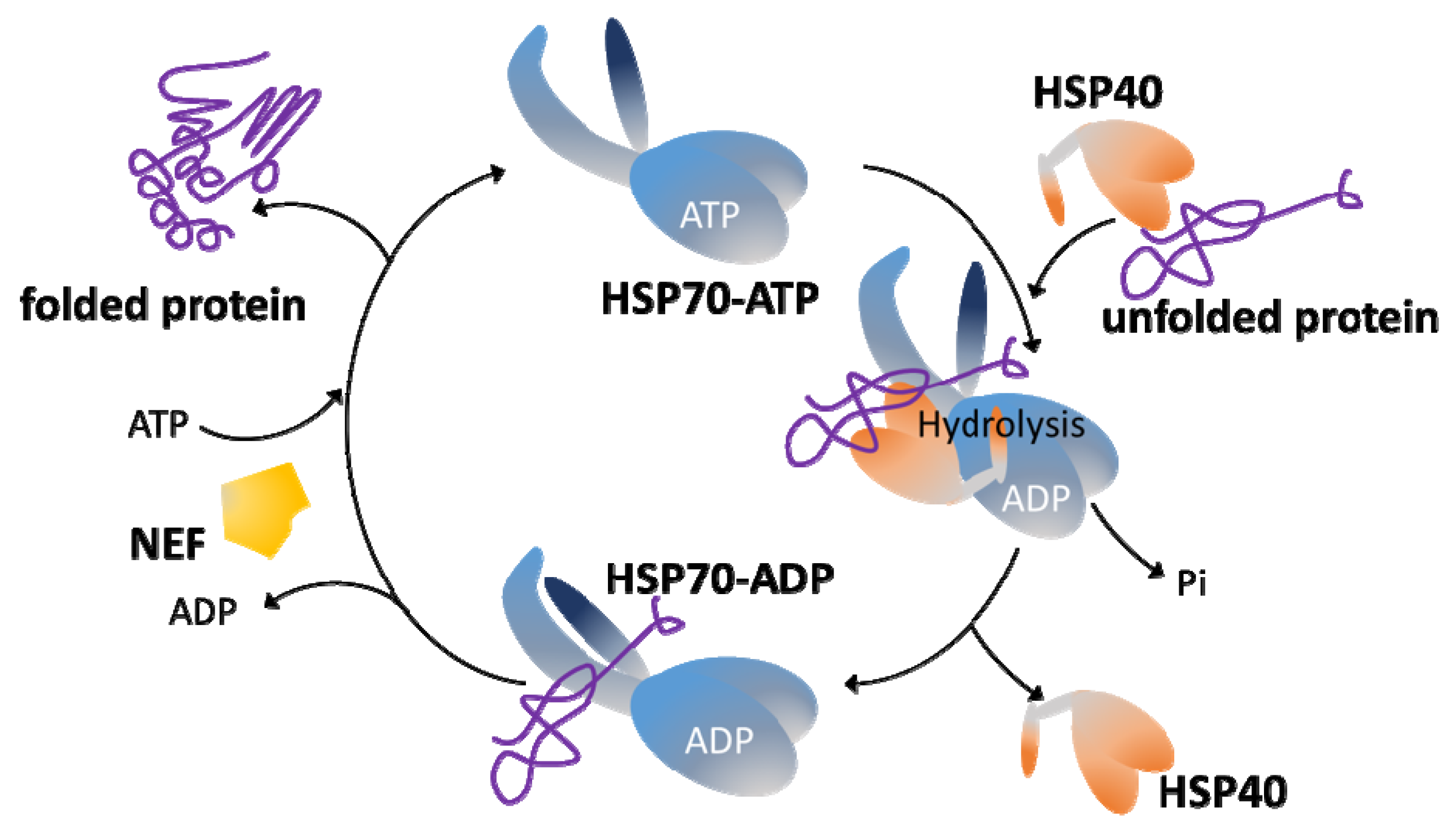
3. The HSP70/HSP40 Chaperone System

Protein quality control is paramount for all molecular processes mediated by protein interactions that occur along the cell life. Together with protease systems and cellular mechanisms such as autophagy and lysosomal degradation, chaperones ensure that proteins are correctly folded and functional at the right place and time [8]. Molecular chaperones are nanomachines specially engineered to interact with non-native conformations of proteins to avoid protein aggregation and assist protein folding. Usually, they bind to hydrophobic residues that are transiently exposed during initial folding but also present in damaged or denatured proteins; in their absence, protein aggregation may occur. The main classes of molecular chaperones are grouped in families, named according to the molecular weights of their prototypical members: small HPS (sHSPs), HSP40, HSP60, HSP70, HSP90 and HSP100 [8,9].
Among molecular chaperones, members of the HSP70 family are central hubs of many cellular processes [10]. Because of this, it is not surprising that prototypical HSP70 (named DnaK in bacteria) is the most conserved protein present in all organisms [11]. Moreover, HSP70 chaperones are key in protein quality control mechanisms. Thus, HSP70s interact with nascent polypeptides at the ribosome to assist de novo protein folding and with mis-folded or stress-denatured proteins; also, they are involved in the stabilization of partially unfolded proteins prior their translocation across membranes [12,13,14].
HSP70s have two functional domains, a substrate binding domain (SBD) and a nucleotide-binding domain (NBD). These domains work in a coordinate manner to accommodate the diverse functional roles played by HSP70s. Thus, the SBD moiety interacts with the target proteins, but this binding is transient, being modulated by an allosteric mechanism that involves cycles of ATP hydrolysis and ADP/ATP exchange in the NBD moiety (Figure 1). Briefly, in the ATP-bound state, the HSP70 presents low affinity and fast exchange rates for the polypeptide substrates, while in the ADP-bound state, HSP70 has high affinity and low exchange rates for substrates [10]. When ATP binds to the NBD, the entire HSP70 structure is affected, and the substrate-binding cleft of SBD opens to release the substrate (polypeptide). The subsequent ATP hydrolysis leads to a new conformational alteration (ADP-bound state) in the HSP70 structure that results in the trapping and tight binding of the target protein [10]. Thus, binding and release alternating steps are important to stabilize non-native proteins and to promote their correct folding. Remarkably, HSP70s possess a weak intrinsic ATPase activity, but this is activated upon interaction with co-chaperones of the family HSP40/JDP. In fact, JDPs play a double role: they favor the binding of substrate proteins to HSP70 in its ATP-bound stage, but concomitantly a direct JDP-HSP70 interaction triggers the ATP hydrolysis and the transition to the ADP-bound state of HSP70, which has high affinity for the specific polypeptide brought over by a particular JDP. Another crucial player is the nucleotide exchange factor (NEF), which induces ADP dissociation and binding of a new ATP molecule, modifying HSP70 structure to the low-affinity state and consequently leading to substrate release [15].

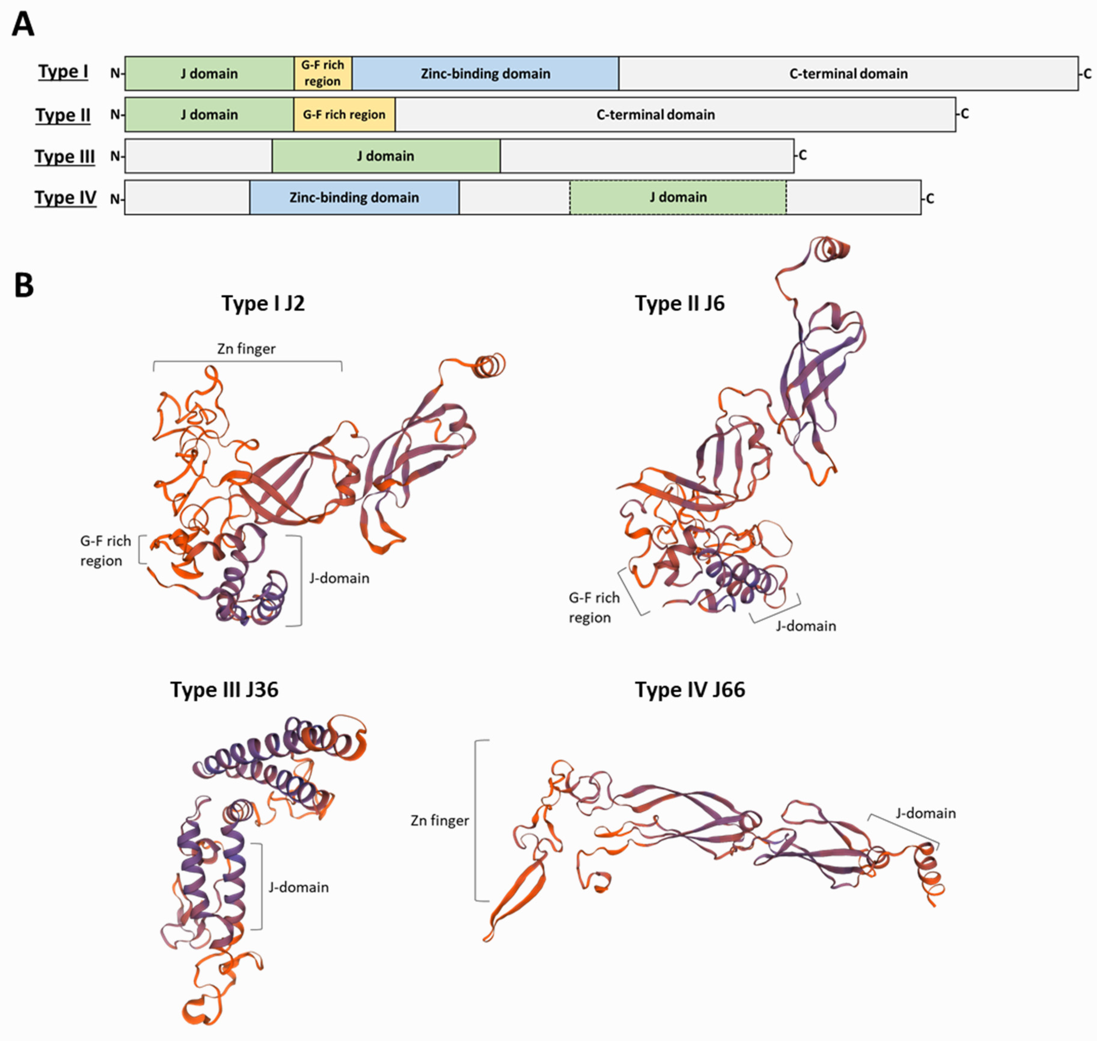
4. J-Domain Proteins

HSP40s, also known as J-domain proteins (JDP), DNAJ-like or J proteins, are essential partners for HSP70 chaperones [16]. These proteins are grouped because of the presence of a J-domain, whose prototype is that defined in Escherichia coli DnaJ protein [17]. JDPs can bind to substrate polypeptides by themselves, and their J-domain promotes ATP hydrolysis by the HSP70 protein, favoring the binding of polypeptides by the HSP70 [18]. Remarkably, but not surprisingly as they are responsible for substrate specificity, HSP40s, in a cell or into cellular compartments, outnumber HSP70 family members [19]. Thus, 6 HSP40s have been found in E. coli, 22 in Saccharomyces cerevisiae, 41 in humans [20], 49 in Plasmodium falciparum [21] and 69 in L. major [7]. In contrast, only three distinct DnaK genes exist in E. coli; six distinct HSP70s are present in P. falciparum, nine in Leishmania and ten in humans [7].
As mentioned above, the prototypical and founding member of this superfamily is the E. coli DnaJ protein [17], whose structure and functions have been elucidated in great detail [22]. DnaJ contains four structural domains: an N-terminal J-domain, followed by a Gly/Phe (G-F)-rich domain, a Zn2+-finger domain and a less-conserved C-terminal domain. The J-domain region is comprised of approximately 70 amino acids that fold into four α-helices (I–IV). The existence of a highly conserved His-Pro-Asp (HPD) tripeptide motif in the loop region between helices II and III is another structural feature of the J-domain; this motif is essential for the stimulation of HSP70 ATP hydrolysis [23]. It is believed that J-domain only interacts with the ATP-bound HSP70 conformation at the interface between NBD and SBD moieties. Then, the HDP motif contacts key residues of the HSP70 ATP catalytic site, remodeling the NBD lobes to orientate the catalytic residues to a position optimal for ATP hydrolysis. Furthermore, the J-domain interacts with residues of the HSP70 SBD, promoting high affinity for the HSP70 ADP-bound state and efficient trapping of substrates [10,22]. Moreover, many J-proteins directly bind substrates, favoring the specific interactions of HSP70 with particular polypeptides and linking in turn the HSP70 functions to particular cellular processes [2]. Apart from the common J-domain, which is the distinctive feature of HSP40s, this class of proteins show a large structural divergence among them. Different HSP40s interact with a particular member of the HSP70 family; hence, HSP40s are contributing to the multi-functionality of the HSP70 machinery by specifying the target substrates and cellular processes in which the HSP70 chaperone activity is requested [20,24].
Based on the similarity to the domain architecture of the DnaJ, HSP40s have been grouped into four classes (Figure 2). Type I proteins share the four characteristic domains of DnaJ: the N-terminal domain constituted by the J-domain; a glycine-phenylalanine (G-F)-rich linker segment; two β-sandwich C-terminal domains, which contain four repeats of the CxxCxGxG type (zinc finger-like region); and a dimerization domain involved in binding to the client proteins. Examples of proteins belonging to this class are the yeast Ydj1 and the human DnaJA1-4. Type II proteins share the J-domain, the G-F-rich linker and the C-terminal substrate binding domain but lack the zinc-finger domain; examples are the yeast Sis1 and the human DnaJB-1, -4 and -5 [24]. The G-F-rich region is also involved in determining the specificity of HSP40s for target proteins [25]. Additionally, in class II JDPs, the G-F-rich region would be involved in an autoinhibitory mechanism, in which the G-F region initially blocks J-domain binding to HSP70 [15]. Type III proteins are heterogeneous in sequence and share only the J-domain with DnaJ; more often, they contain domains involved in specifying target interaction or sub-cellular localization [20]. As mentioned above, within the J-domain, there exists the highly conserved HPD motif; however, for some J-domain containing proteins, a strict HPD sequence is not found. To denote this feature, Louw and coworkers proposed a fourth group (type IV) of HSP40s to include those proteins lacking the HPD sequence in their J-domains [26]. Unlike type I and type II HSP40s, in which the J-domain always has an N-terminal location, in type III proteins the J-domain can be in any position along their sequence. It has been suggested that type I and type II HSP40s form complexes (dimers or tetramers) and are able to interact promiscuously with nascent polypeptides; also, they recognize mis-folded or aggregated proteins and cooperate with HSP70s in protein disaggregation [10,27]. In contrast, type III HSP40s would have evolved to specifically interact with a limited number of HSP70 substrates or alternatively acting directly as a bait to locate an HSP70 to particular cellular places [10,20]. Regarding type IV HSP40s, some authors have questioned whether they must be considered either members of the JDP family or only JDP-like proteins [28]. Nevertheless, they should be considered to understand the evolutionary history of the family and its functional diversification. Moreover, for some HSP40s, the maintenance of their co-chaperone functions in the absence of a canonical J-domain has been reported [29].

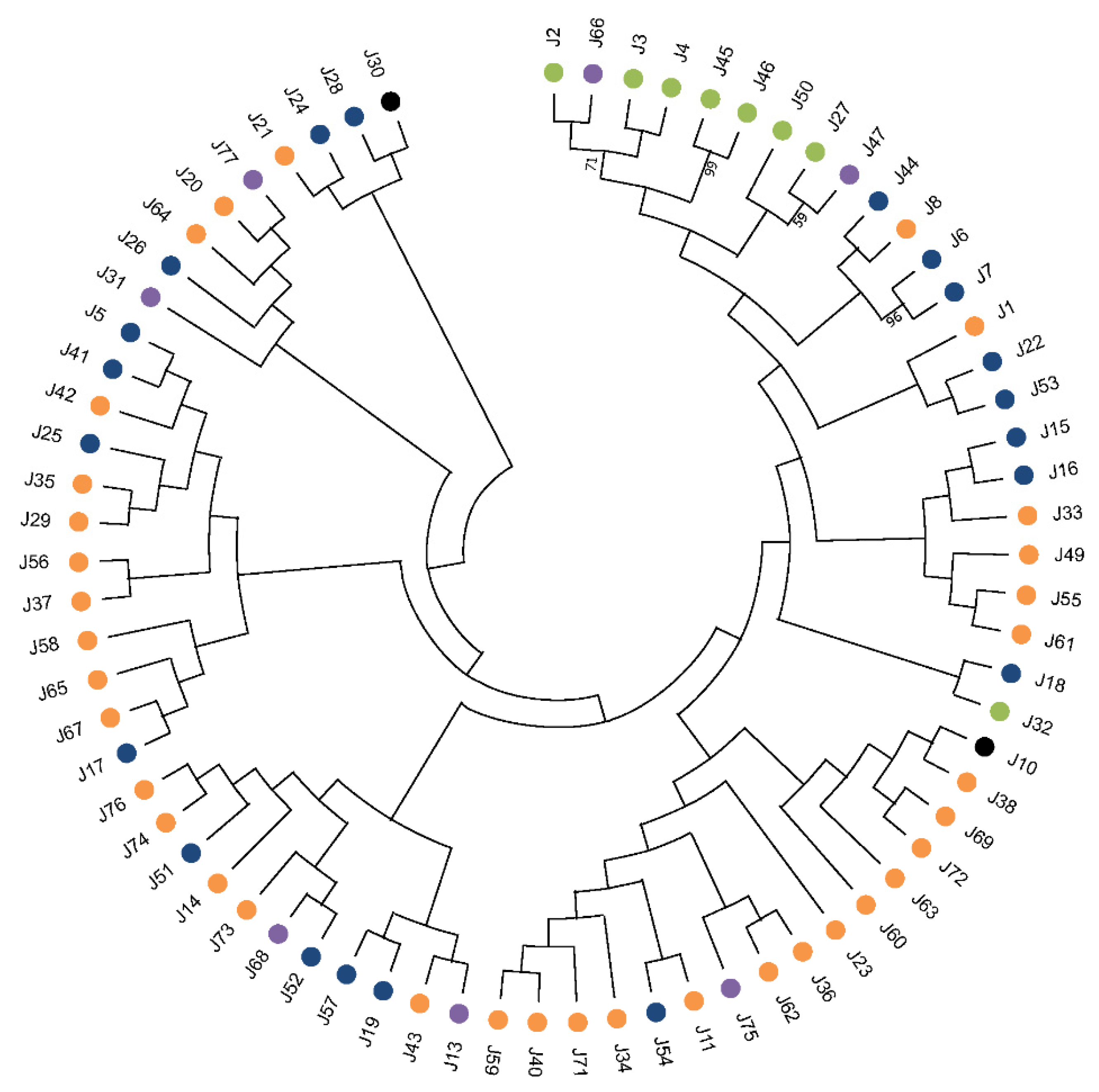
5. Appraisal and Updating of the Compendium of HSP40s in L. infantum

In a previous work, we found 69 different HSP40s to be encoded in the L. major genome [7]. At that time (2015), the L. major genome was the best assembled one for the genus Leishmania; however, in 2017, an improved genome assembly was attained by the combination of second- and third-generation sequencing methodologies for the L. infantum genome [30]. Hence, in this review, we analyzed the L. infantum genome for genes encoding J-domain containing proteins. All the 69 previously identified proteins for the HSP40 family in L. major were found to be also present in the L. infantum genome. Moreover, three new members were uncovered, amounting to a total of 72 different JDPs (Figure 3 and Table 1). Following the nomenclature proposed by Folgueira and Requena [31], they were named J75 (JDP75), J76 (JDP76) and J77 (JDP77). Thus, Leishmania possesses one of the largest HSP40 families among the organisms in which this family has been studied. To our knowledge, a larger HSP40 family has only been found in pepper, which contains 76 annotated HSP40 genes in its genome [32]. According to the presence/absence of the prototypical DnaJ-domains (Figure 2), the 72 Leishmania JDPs could be grouped into the four established classes (Table 1). Thus, eight L. infantum HSP40 proteins belong to the type I category, since they contain all typical domains found in the prototypical DnaJ molecule; these are J2, J3, J4, J27, J32, J45, J46 and J50. Another 18 HSP40s belong to the type II, as they have Gly/Phe-rich region close to the J-domain but lack a zinc-binding domain. The largest category (type III) is that formed by 38 proteins containing only the J-domain. Of note, the protein J30 (gene LINF_070013700), as currently annotated at TriTrypDB, lacks the J-domain; however, its ortholog in Trypanosoma brucei (Tb927.8.1010) contains the J-domain at the N-terminal moiety. Thus, we analyzed the LINF_070013700 transcript sequence and found that the gene was mis-annotated, and the coding sequence can be extended 783 nucleotides at its 5′-end. Thus, the new predicted protein would be 261 amino acids longer and then would contain the complete J-domain [33]. The L. infantum J10 protein (gene LINF_170010900) was found to be N-terminal truncated related to its L. major ortholog, and, as a consequence, the J-domain is incomplete, although the HPD motif is present; in this case, a possible mis-annotation of the coding sequence was not evidenced [34]. Within the type IV HSP40 group, six proteins were found to be encoded in the L. infantum genome: J31, J47, J66, J68, J75 and J77. Of them, J47, J66 and J75 have also the Zn-finger domain (Table 1).
Among the members of the Leishmania HSP40 family, as observed in other organisms, there is little sequence conservation beyond the characteristic J-domain. A phylogenetic analysis was conducted with the 72 JDPs to determine possible evolutionary relationships (Figure 3). Only type I HSP40s (except J32) grouped in the same branch of the tree, but the small bootstrap values supporting most of the branches did not allow for definition of clear evolutionary relationships among them. Exceptions are the pairs J45 and J46 (bootstrap 99%) and J6 and J7 (96%), proteins that probably resulted from a recent duplication of an ancestral gene. Proteins of the type II, III and IV show marked divergences in their structure and sequence; even the J-domain (typically found at the N-terminal region in DnaJ and type I-HSP40s) is located in the middle of the sequence or even at the C-terminal region in the proteins J20, J21, J25, J28, J29, J35, J41, J42, J51, J52, J53, J64, J65, J67 and J75 (Table 1). This atypical location of J-domain has been also reported for HSP40s in other organisms [37]. A remarkable feature is that Leishmania J14 contains two J-domains.
Leishmania protists belong to the Euglenozoa phylum, which represent one of the earliest extant branches of the eukaryotic lineage [38]. Thus, the enormous evolutionary distance separating Leishmania from the model eukaryotes makes it difficult to establish straightforward orthologous relationships between Leishmania proteins and the human or yeast ones; in fact, in databases, around half of the Leishmania genes are annotated as coding for proteins of unknown functions because no clear orthologous proteins exist in model eukaryotes. Nevertheless, based on the hypothesis that the identification of potential orthologues in the well-characterized human and/or yeast proteomes would give clues about the functions of the Leishmania proteins, we performed a detailed search of sequence conservation with the 72 L. infantum HSP40s against protein databases found in the servers PantherDB (http://www.pantherdb.org/, accessed on 22 April 2022), Expasy (https://prosite.expasy.org/, accessed on 22 April 2022), InterPro (https://www.ebi.ac.uk/interpro/, accessed on 22 April 2022) and UniProt (https://www.uniprot.org/blast/, accessed on 22 April 2022). As a result, a possible orthologous relationship among the Leishmania type I J2, J3 and J4 (these three proteins may share an evolutionary origin, see Figure 3) and the members DnaJA-1, -2 and -4 of the human HSP40 family was deduced. Similarly, J2 may be ortholog to S. cerevisiae mas5 protein (YDJ1 gene), which is involved in mitochondrial protein import [39]. Also, Leishmania type I-JDPs J45, J46 and J50 have substantial sequence conservation with the human DnaJA2 and S. cerevisiae SCJ1 proteins, which are located at the endoplasmic reticulum in those organisms [40,41].
Within the group of type IV HSP40s, J66 has a probable evolutionary relationship with L. infantum type I-JDPs and particularly with J2 (Figure 3). Thus, J2 and J66 may be considered paralogs, and in turn both would be orthologs to human DnaJA2. Another type IV-JDP, J47, shares sequence conservation with the type I-JDP J27 (Figure 3), and both are possible orthologs to human DnaJA3 and S. cerevisiae Mdj1 proteins, which were located at the mitochondrion [42,43].
J6 and J7, belonging to the type II group, might be orthologs to human DnaJB4, DnaJB5 and DnaJB1 HSP40s and to S. cerevisiae Sis1 protein, which is required for nuclear migration during mitosis and initiation of translation [44,45]. J10 (and to some extent J34) may be orthologue to Sec63, an essential HSP40 protein involved in post-translational translocation of proteins across the endoplasmic reticulum [46,47]. Another endoplasmic reticulum located proteins, human DnaJC3/ERdj6 and S. cerevisiae JEM1 [48,49], might be the orthologs of Leishmania J53. Moreover, we postulate that Leishmania J13 is ortholog of human DnaJC24, a protein involved in diphthamide biosynthesis, a post-translational modification of histidines that have been found in the translation elongation factor-2 [50]. A possible ortholog of J16 would be the well-known human zuotin/DnaJC2 protein. Zuotin is a component of the ribosome-associated complex involved in maintaining nascent polypeptides in a folding-competent state [51]. The type I-JDP J27 may be an ortholog of the human DnaJA3/Tid1 protein, a mitochondrial molecular chaperone [42,52]. A plausible mitochondrial location of Leishmania J31 is suggested on its sequence similarity with human DnaJC11 [53]. J36 might be ortholog of human DnaJC20/HscB and S. cerevisiae JAC1 proteins, which are involved in iron–sulfur cluster biosynthesis [54,55]. Leishmania J68 might be a component of the mitochondrial Tim translocase because of its structural and sequence similarities with human DnaJC15 and S. cerevisisae Pam18 proteins [56]. Human DnaJC21 and S. cerevisiae JJJ1 are involved in rRNA biogenesis [57], and they may be orthologs of Leishmania J32. A histone chaperone function may be suggested for Leishmania J33 based on its sequence similarity with human DnaJC9 and Schizosaccharomyces pombe C1071.09c proteins [58]. J59 is a really long protein (2451 amino acids) that might be orthologue of the human DnaJC13/RME-8 protein, which is involved in endosome organization and regulation [59]. In addition to all possible orthologs mentioned above, another several Leishmania HSP40s share remarkable sequence identity with genes coding for HSP40s in fungi and plant species. Nevertheless, these are not commented here, as they remain still uncharacterized, and no information about their functional roles is available.
According to structural features and the putative orthologs identified (see above), some Leishmania HSP40s could be associated to distinct sub-cellular locations such as mitochondrion (J27, J31, J36, J47 and J68), endoplasmic reticulum (J10, J22, J34, J45, J46, J53, J66 and J72), flagellar pocket (J11, J51 and J54), endosomes (J59) and the nucleus (J14, J33 and J56) (Table 2). Of note, several Leishmania HSP40s (J2, J3, J6, J8, J27, J50 and J54) have been localized in the L. donovani glycosomes following a proteomic approach [60] (Table 2). Glycosomes are specialized peroxisomes, existing in Leishmania and other trypanosomatids, that contain key enzymes involved on the glycolytic pathway and purine salvage [61]. Moreover, at least 23 HSP40s in Leishmania may play functional roles in cellular membranes as they possess transmembrane domains, including members from group I (J45, J46), group II (J5, J19, J22, J28, J44, J53 and J54) and group III (Table 1). In these locations, putative functions for these Leishmania JDPs may be providing co-translational chaperone assistance, transport across organelle membranes and unfolding/refolding after transportation from one cellular compartment to another. For example, J22 is a possible membrane protein related to endoplasmic reticulum (ER) membrane-located S. pombe pi041 and human DnaJB12 proteins that are involved in ER-associated degradation of misfolded proteins [62].
In addition, TPR (tetratrico-peptide repeat) domains have been found in eight JDP proteins: J51, J52, J53, J67 and J76 contain two or more TPR domains each, whereas a sole TPR domain is present in J42, J65 and J76 (Table 1). TPR domain has been found to be a docking site, interacting with the EEVD motif present at the C-termini of some members of the HSP70 family and in the C-terminus of the HSP83/90 protein [73,74].
Despite the outstanding number of HSP40s existing in Leishmania, none of these proteins have been biochemically characterized to date, and consequently, nothing is known about the potential HSP70-HSP40 partnerships and their role in the Leishmania life cycle. However, evidence of their existence and levels of stage specific expression are available now from transcriptomics and proteomics data. Thus, out of the 72 putative HSP40s identified in Leishmania, 37 have been experimentally reported in proteomic studies of the Leishmania promastigote [62,66,72] and/or amastigote stages [67,70]. Moreover, some of them have been found in the proteomes derived from specific sub-cellular fractions of the parasite such as the glycosome [60] and the flagellum [72]. A list of the experimentally detected HSP40s in Leishmania proteomes is included in Supplementary Materials, Table S1. For example, J2 protein has been experimentally reported in L. infantum and L. donovani promastigotes and amastigotes [60,63,66], in extracellular vesicles of L. infantum [68] and in the L. braziliensis secreted proteome [75]. Remarkably, J2 has been found to be phosphorylated on Serine-89, and the phosphorylation ratio of this residue increased by 3.5-fold after 2.5 h of promastigote-to-amastigote differentiation, reaching an increase in phosphorylation up to 22-fold in full differentiated amastigotes [76]. Such a dramatic increase in phosphorylation suggests a relevant role for this protein in the differentiation process from promastigote to amastigote stage. Phosphorylation on serine residues is the most common post-transcriptional modification in HSP40s (Supplementary Materials, Table S1) and may be related to a general regulatory mechanism induced during the stress response [77]. Other relevant protein modifications found in HSP40s are acetylation and methylation. Particularly relevant may be the N-terminal acetylation on threonine-39 of J7 [78], because this modification would be changing its chemical properties and having marked biological consequences on protein function and cellular localization [64]. In addition, methylation on glutamic acid-134 in J6 and J50 proteins would increase the hydrophobicity of this protein [79].

6. Concluding Remarks and Future Work

The HSP40 proteins play essential roles in cellular metabolism by regulating interactions between HSP70 chaperones and their client proteins. We identified 72 HSP40 coding genes in the Leishmania genome, making up an exceptionally large protein family for this parasite compared to other eukaryotes. However, unlike other insect-transmitted parasites such as Plasmodium [80], the crucial role of HSP40s in the survival and pathogenesis of Leishmania parasites remains unexplored. Future studies should address the molecular characterization of the HSP40 family members and the identification of their HSP70 counterparts. Different HSP40s can bind and deliver client proteins to a single HSP70 and establish unique HSP70–HSP40 pairs with specific activities at distinct cellular locations. Thus, studies aimed to analyze the co-localization of particular HSP70 family members and HSP40s in sub-cellular compartments would be a suitable strategy to establish HSP70–HSP40 partnerships. For this purpose, high throughput tagging based on CRISPR/Cas9 tools would be a strategy for those localization studies [81]. Another appealing approach to define specific HSP40 location would be the use of protein phase separation methods; in fact, many HSP40s have been found as components of membraneless organelles such as nucleolus or stress granules [63]. Also, CRISPR-Cas9 genome editing methods would be useful for analyzing essential roles played by this family of proteins, following the strategies used to screen proteins involved in Leishmania flagellar architecture and function [72] or to define the essential kinases comprising the Leishmania kinome [82]. In addition, as HSP40s regulate the activity of the HSP70 chaperone system, the analysis of changes in gene expression of HSP40 genes along the Leishmania life cycle, which includes a drastic morphological differentiation and adaptations to stressful environments, would inform on the relevance role played by every HSP40 at particular points of the life cycle. These studies also might be useful to detect changes in the parasite fitness related to drug resistance. Thus, for instance, it has been reported that transcription of the J57 gene is upregulated in an L. donovani line resistant to liposomal amphotericin B (AmBisome) drug, the most effective anti-Leishmania treatment [83]. In summary, we are just starting the fascinating road to comprehend the roles that this group of proteins are playing in the differentiation, survival and pathogenesis of Leishmania parasites.

Supplementary Materials

The following supporting information can be downloaded at: https://www.mdpi.com/article/10.3390/genes13050742/s1, Supplementary file (Excel) containing: Table S1, Leishmania HSP40 proteins experimentally detected in proteomic studies.

Author Contributions

Conceptualization, J.M.R. and J.C.S.; methodology, J.M.R. and J.C.S.; software, J.C.S. and L.B.; validation, J.M.R.; formal analysis, J.C.S., L.B. and J.M.R.; investigation, J.C.S. and L.B.; resources, J.M.R.; data curation, J.M.R. and J.C.S.; writing—original draft preparation, J.C.S., L.B. and J.M.R.; writing—review and editing, J.M., B.A. and J.M.R.; visualization, J.C.S., L.B. and J.M.R.; supervision, J.M.R.; project administration, J.M.R.; funding acquisition, J.M., B.A. and J.M.R. All authors have read and agreed to the published version of the manuscript.

Funding

This research was supported by the Spanish Ministerio de Ciencia, Innovación (MICINN), Agencia Estatal de Investigación (AEI), grant number PID2020-117916RB-I00, and Instituto de Salud Carlos III, grant CB21/13/00018 (CIBERINFEC). An institutional grant from Fundacion Ramon Areces is also acknowledged.

Institutional Review Board Statement

Not applicable.

Data Availability Statement

The data supporting reported results can be found at Leish-ESP (http://leish-esp.cbm.uam.es/, accessed on 22 April 2022), TriTrypDB (https://tritrypdb.org/, accessed on 22 April 2022), UniProt (https://www.uniprot.org/, accessed on 22 April 2022) and Wikidata datasets (https://www.wikidata.org/, accessed on 22 April 2022).

Conflicts of Interest

The authors declare no conflict of interest.

References

  1. Fernández-Fernández, M.R.; Valpuesta, J.M. Hsp70 chaperone: A master player in protein homeostasis. F1000Research 2018, 7, 1497. [Google Scholar] [CrossRef] [PubMed] [Green Version]
  2. Craig, E.A.; Marszalek, J. How Do J-Proteins Get Hsp70 to Do So Many Different Things? Trends Biochem. Sci. 2017, 42, 355–368. [Google Scholar] [CrossRef] [PubMed] [Green Version]
  3. Burza, S.; Croft, S.L.; Boelaert, M. Leishmaniasis. Lancet 2018, 392, 951–970. [Google Scholar] [CrossRef]
  4. Sasidharan, S.; Saudagar, P. Leishmaniasis: Where are we and where are we heading? Parasitol. Res. 2021, 120, 1541–1554. [Google Scholar] [CrossRef]
  5. Requena, J.M. The Stressful Life of pathogenic Leishmania species. In Stress Response in Microbiology; Requena, J.M., Ed.; Caister Academic Press: Norfolk, UK, 2012; pp. 323–346. [Google Scholar]
  6. Lindquist, S.; Craig, E.A. THE HEAT-SHOCK PROTEINS. Annu. Rev. Genet. 1988, 22, 631–677. [Google Scholar] [CrossRef]
  7. Requena, J.M.; Montalvo, A.M.; Fraga, J. Molecular Chaperones of Leishmania: Central Players in Many Stress-Related and -Unrelated Physiological Processes. Biomed. Res. Int. 2015, 2015, 301326. [Google Scholar] [CrossRef] [Green Version]
  8. Saibil, H. Chaperone machines for protein folding, unfolding and disaggregation. Nat. Rev. Mol. Cell Biol. 2013, 14, 630–642. [Google Scholar] [CrossRef] [Green Version]
  9. Hartl, F.U.; Hayer-Hartl, M. Converging concepts of protein folding in vitro and in vivo. Nat. Struct. Mol. Biol. 2009, 16, 574–581. [Google Scholar] [CrossRef]
  10. Mayer, M.P.; Gierasch, L.M. Recent advances in the structural and mechanistic aspects of Hsp70 molecular chaperones. J. Biol. Chem. 2019, 294, 2085–2097. [Google Scholar] [CrossRef] [Green Version]
  11. Gupta, R.S. Protein Phylogenies and Signature Sequences: A Reappraisal of Evolutionary Relationships among Archaebacteria, Eubacteria, and Eukaryotes. Microbiol. Mol. Biol. Rev. 1998, 62, 1435–1491. [Google Scholar] [CrossRef] [Green Version]
  12. Mayer, M.P.; Bukau, B. Hsp70 chaperones: Cellular functions and molecular mechanism. Cell. Mol. Life Sci. 2005, 62, 670. [Google Scholar] [CrossRef] [PubMed] [Green Version]
  13. Preissler, S.; Deuerling, E. Ribosome-associated chaperones as key players in proteostasis. Trends Biochem. Sci. 2012, 37, 274–283. [Google Scholar] [CrossRef] [PubMed] [Green Version]
  14. Rosenzweig, R.; Nillegoda, N.B.; Mayer, M.P.; Bukau, B. The Hsp70 chaperone network. Nat. Rev. Mol. Cell Biol. 2019, 20, 665–680. [Google Scholar] [CrossRef] [PubMed]
  15. Faust, O.; Rosenzweig, R. Structural and Biochemical Properties of Hsp40/Hsp70 Chaperone System. Adv. Exp. Med. Biol. 2020, 1243, 3–20. [Google Scholar] [CrossRef]
  16. Qiu, X.B.; Shao, Y.M.; Miao, S.; Wang, L. The diversity of the DnaJ/Hsp40 family, the crucial partners for Hsp70 chaperones. Cell. Mol. Life Sci. CMLS 2006, 63, 2560–2570. [Google Scholar] [CrossRef]
  17. Langer, T.; Lu, C.; Echols, H.; Flanagan, J.; Hayer, M.K.; Hartl, F.U. Successive action of DnaK, DnaJ and GroEL along the pathway of chaperone-mediated protein folding. Nature 1992, 356, 683–689. [Google Scholar] [CrossRef]
  18. Hennessy, F.; Nicoll, W.S.; Zimmermann, R.; Cheetham, M.E.; Blatch, G.L. Not all J domains are created equal: Implications for the specificity of Hsp40-Hsp70 interactions. Protein Sci. A Publ. Protein Soc. 2005, 14, 1697–1709. [Google Scholar] [CrossRef] [Green Version]
  19. Craig, E.A.; Huang, P.; Aron, R.; Andrew, A. The diverse roles of J-proteins, the obligate Hsp70 co-chaperone. In Reviews of Physiology, Biochemistry and Pharmacology; Springer: Berlin/Heidelberg, Germany, 2006; Volume 156, pp. 1–21. [Google Scholar] [CrossRef]
  20. Kampinga, H.H.; Craig, E.A. The HSP70 chaperone machinery: J proteins as drivers of functional specificity. Nat. Rev. Mol. Cell Biol. 2010, 11, 579–592. [Google Scholar] [CrossRef] [Green Version]
  21. Njunge, J.M.; Ludewig, M.H.; Boshoff, A.; Pesce, E.R.; Blatch, G.L. Hsp70s and J proteins of Plasmodium parasites infecting rodents and primates: Structure, function, clinical relevance, and drug targets. Curr. Pharm. Des. 2013, 19, 387–403. [Google Scholar] [CrossRef]
  22. Kityk, R.; Kopp, J.; Mayer, M.P. Molecular Mechanism of J-Domain-Triggered ATP Hydrolysis by Hsp70 Chaperones. Mol. Cell 2018, 69, 227–237.e224. [Google Scholar] [CrossRef]
  23. Tsai, J.; Douglas, M.G. A conserved HPD sequence of the J-domain is necessary for YDJ1 stimulation of Hsp70 ATPase activity at a site distinct from substrate binding. J. Biol. Chem. 1996, 271, 9347–9354. [Google Scholar] [CrossRef] [PubMed] [Green Version]
  24. Liu, Q.; Liang, C.; Zhou, L. Structural and functional analysis of the Hsp70/Hsp40 chaperone system. Protein Sci. A Publ. Protein Soc. 2020, 29, 378–390. [Google Scholar] [CrossRef] [PubMed]
  25. Yan, W.; Craig, E.A. The glycine-phenylalanine-rich region determines the specificity of the yeast Hsp40 Sis1. Mol. Cell. Biol. 1999, 19, 7751–7758. [Google Scholar] [CrossRef] [PubMed] [Green Version]
  26. Louw, C.A.; Ludewig, M.H.; Mayer, J.; Blatch, G.L. The Hsp70 chaperones of the Tritryps are characterized by unusual features and novel members. Parasitol. Int. 2010, 59, 497–505. [Google Scholar] [CrossRef]
  27. Nillegoda, N.B.; Kirstein, J.; Szlachcic, A.; Berynskyy, M.; Stank, A.; Stengel, F.; Arnsburg, K.; Gao, X.; Scior, A.; Aebersold, R.; et al. Crucial HSP70 co-chaperone complex unlocks metazoan protein disaggregation. Nature 2015, 524, 247–251. [Google Scholar] [CrossRef] [Green Version]
  28. Kampinga, H.H.; Andreasson, C.; Barducci, A.; Cheetham, M.E.; Cyr, D.; Emanuelsson, C.; Genevaux, P.; Gestwicki, J.E.; Goloubinoff, P.; Huerta-Cepas, J.; et al. Function, evolution, and structure of J-domain proteins. Cell Stress Chaperones 2019, 24, 7–15. [Google Scholar] [CrossRef]
  29. Ajit Tamadaddi, C.; Sahi, C. J domain independent functions of J proteins. Cell Stress Chaperones 2016, 21, 563–570. [Google Scholar] [CrossRef] [Green Version]
  30. Gonzalez-de la Fuente, S.; Peiro-Pastor, R.; Rastrojo, A.; Moreno, J.; Carrasco-Ramiro, F.; Requena, J.M.; Aguado, B. Resequencing of the Leishmania infantum (strain JPCM5) genome and de novo assembly into 36 contigs. Sci. Rep. 2017, 7, 18050. [Google Scholar] [CrossRef]
  31. Folgueira, C.; Requena, J.M. A postgenomic view of the heat shock proteins in kinetoplastids. FEMS Microbiol. Rev. 2007, 31, 359–377. [Google Scholar] [CrossRef]
  32. Fan, F.; Yang, X.; Cheng, Y.; Kang, Y.; Chai, X. The DnaJ Gene Family in Pepper (Capsicum annuum L.): Comprehensive Identification, Characterization and Expression Profiles. Front. Plant Sci. 2017, 8, 689. [Google Scholar] [CrossRef] [Green Version]
  33. Requena, J.M.; Solana, J.C. LINF_070013700. In Mendeley Data, V2; Universidad Autonoma de Madrid: Madrid, Spain, 2022. [Google Scholar] [CrossRef]
  34. Requena, J.M.; Solana, J.C. LINF_170010900. In Mendeley Data, V1; Universidad Autonoma de Madrid: Madrid, Spain, 2021. [Google Scholar] [CrossRef]
  35. Jones, D.T.; Taylor, W.R.; Thornton, J.M. The rapid generation of mutation data matrices from protein sequences. Comput. Appl. Biosci. CABIOS 1992, 8, 275–282. [Google Scholar] [CrossRef] [PubMed]
  36. Tamura, K.; Stecher, G.; Kumar, S. MEGA11: Molecular Evolutionary Genetics Analysis Version 11. Mol. Biol. Evol. 2021, 38, 3022–3027. [Google Scholar] [CrossRef] [PubMed]
  37. Walsh, P.; Bursać, D.; Law, Y.C.; Cyr, D.; Lithgow, T. The J-protein family: Modulating protein assembly, disassembly and translocation. EMBO Rep. 2004, 5, 567–571. [Google Scholar] [CrossRef] [PubMed] [Green Version]
  38. Hamilton, P.B.; Stevens, J.R.; Gaunt, M.W.; Gidley, J.; Gibson, W.C. Trypanosomes are monophyletic: Evidence from genes for glyceraldehyde phosphate dehydrogenase and small subunit ribosomal RNA. Int. J. Parasitol. 2004, 34, 1393–1404. [Google Scholar] [CrossRef]
  39. Xie, J.L.; Bohovych, I.; Wong, E.O.Y.; Lambert, J.P.; Gingras, A.C.; Khalimonchuk, O.; Cowen, L.E.; Leach, M.D. Ydj1 governs fungal morphogenesis and stress response, and facilitates mitochondrial protein import via Mas1 and Mas2. Microb. Cell 2017, 4, 342–361. [Google Scholar] [CrossRef]
  40. Kim Chiaw, P.; Hantouche, C.; Wong, M.J.H.; Matthes, E.; Robert, R.; Hanrahan, J.W.; Shrier, A.; Young, J.C. Hsp70 and DNAJA2 limit CFTR levels through degradation. PLoS ONE 2019, 14, e0220984. [Google Scholar] [CrossRef] [Green Version]
  41. Schlenstedt, G.; Harris, S.; Risse, B.; Lill, R.; Silver, P.A. A yeast DnaJ homologue, Scj1p, can function in the endoplasmic reticulum with BiP/Kar2p via a conserved domain that specifies interactions with Hsp70s. J. Cell Biol. 1995, 129, 979–988. [Google Scholar] [CrossRef]
  42. Elwi, A.N.; Lee, B.; Meijndert, H.C.; Braun, J.E.; Kim, S.W. Mitochondrial chaperone DnaJA3 induces Drp1-dependent mitochondrial fragmentation. Int. J. Biochem. Cell Biol. 2012, 44, 1366–1376. [Google Scholar] [CrossRef]
  43. Deloche, O.; Liberek, K.; Zylicz, M.; Georgopoulos, C. Purification and biochemical properties of Saccharomyces cerevisiae Mdj1p, the mitochondrial DnaJ homologue. J. Biol. Chem. 1997, 272, 28539–28544. [Google Scholar] [CrossRef] [Green Version]
  44. Zhong, T.; Arndt, K.T. The yeast SIS1 protein, a DnaJ homolog, is required for the initiation of translation. Cell 1993, 73, 1175–1186. [Google Scholar] [CrossRef]
  45. Klaips, C.L.; Gropp, M.H.M.; Hipp, M.S.; Hartl, F.U. Sis1 potentiates the stress response to protein aggregation and elevated temperature. Nat. Commun. 2020, 11, 6271. [Google Scholar] [CrossRef] [PubMed]
  46. Young, B.P.; Craven, R.A.; Reid, P.J.; Willer, M.; Stirling, C.J. Sec63p and Kar2p are required for the translocation of SRP-dependent precursors into the yeast endoplasmic reticulum in vivo. EMBO J. 2001, 20, 262–271. [Google Scholar] [CrossRef] [PubMed] [Green Version]
  47. Goldshmidt, H.; Sheiner, L.; Butikofer, P.; Roditi, I.; Uliel, S.; Gunzel, M.; Engstler, M.; Michaeli, S. Role of protein translocation pathways across the endoplasmic reticulum in Trypanosoma brucei. J. Biol. Chem. 2008, 283, 32085–32098. [Google Scholar] [CrossRef] [PubMed] [Green Version]
  48. Daverkausen-Fischer, L.; Pröls, F. Dual topology of co-chaperones at the membrane of the endoplasmic reticulum. Cell Death Discov. 2021, 7, 203. [Google Scholar] [CrossRef]
  49. Nishikawa, S.; Endo, T. The yeast JEM1p is a DnaJ-like protein of the endoplasmic reticulum membrane required for nuclear fusion. J. Biol. Chem. 1997, 272, 12889–12892. [Google Scholar] [CrossRef] [Green Version]
  50. Webb, T.R.; Cross, S.H.; McKie, L.; Edgar, R.; Vizor, L.; Harrison, J.; Peters, J.; Jackson, I.J. Diphthamide modification of eEF2 requires a J-domain protein and is essential for normal development. J. Cell Sci. 2008, 121, 3140–3145. [Google Scholar] [CrossRef] [Green Version]
  51. Lee, K.; Ziegelhoffer, T.; Delewski, W.; Berger, S.E.; Sabat, G.; Craig, E.A. Pathway of Hsp70 interactions at the ribosome. Nat. Commun. 2021, 12, 5666. [Google Scholar] [CrossRef]
  52. Ng, A.C.; Baird, S.D.; Screaton, R.A. Essential role of TID1 in maintaining mitochondrial membrane potential homogeneity and mitochondrial DNA integrity. Mol. Cell. Biol. 2014, 34, 1427–1437. [Google Scholar] [CrossRef] [Green Version]
  53. Ioakeimidis, F.; Ott, C.; Kozjak-Pavlovic, V.; Violitzi, F.; Rinotas, V.; Makrinou, E.; Eliopoulos, E.; Fasseas, C.; Kollias, G.; Douni, E. A splicing mutation in the novel mitochondrial protein DNAJC11 causes motor neuron pathology associated with cristae disorganization, and lymphoid abnormalities in mice. PLoS ONE 2014, 9, e104237. [Google Scholar] [CrossRef] [Green Version]
  54. Voisine, C.; Cheng, Y.C.; Ohlson, M.; Schilke, B.; Hoff, K.; Beinert, H.; Marszalek, J.; Craig, E.A. Jac1, a mitochondrial J-type chaperone, is involved in the biogenesis of Fe/S clusters in Saccharomyces cerevisiae. Proc. Natl. Acad. Sci. USA 2001, 98, 1483–1488. [Google Scholar] [CrossRef] [Green Version]
  55. Maio, N.; Ghezzi, D.; Verrigni, D.; Rizza, T.; Bertini, E.; Martinelli, D.; Zeviani, M.; Singh, A.; Carrozzo, R.; Rouault, T.A. Disease-Causing SDHAF1 Mutations Impair Transfer of Fe-S Clusters to SDHB. Cell Metab. 2016, 23, 292–302. [Google Scholar] [CrossRef] [PubMed] [Green Version]
  56. D’Silva, P.D.; Schilke, B.; Walter, W.; Andrew, A.; Craig, E.A. J protein cochaperone of the mitochondrial inner membrane required for protein import into the mitochondrial matrix. Proc. Natl. Acad. Sci. USA 2003, 100, 13839–13844. [Google Scholar] [CrossRef] [PubMed] [Green Version]
  57. Kaschner, L.A.; Sharma, R.; Shrestha, O.K.; Meyer, A.E.; Craig, E.A. A conserved domain important for association of eukaryotic J-protein co-chaperones Jjj1 and Zuo1 with the ribosome. Biochim. Biophys. Acta 2015, 1853, 1035–1045. [Google Scholar] [CrossRef] [PubMed] [Green Version]
  58. Hammond, C.M.; Bao, H.; Hendriks, I.A.; Carraro, M.; García-Nieto, A.; Liu, Y.; Reverón-Gómez, N.; Spanos, C.; Chen, L.; Rappsilber, J.; et al. DNAJC9 integrates heat shock molecular chaperones into the histone chaperone network. Mol. Cell 2021, 81, 2533–2548.e2539. [Google Scholar] [CrossRef] [PubMed]
  59. Fujibayashi, A.; Taguchi, T.; Misaki, R.; Ohtani, M.; Dohmae, N.; Takio, K.; Yamada, M.; Gu, J.; Yamakami, M.; Fukuda, M.; et al. Human RME-8 is involved in membrane trafficking through early endosomes. Cell Struct. Funct. 2008, 33, 35–50. [Google Scholar] [CrossRef] [PubMed] [Green Version]
  60. Jamdhade, M.D.; Pawar, H.; Chavan, S.; Sathe, G.; Umasankar, P.K.; Mahale, K.N.; Dixit, T.; Madugundu, A.K.; Prasad, T.S.; Gowda, H.; et al. Comprehensive proteomics analysis of glycosomes from Leishmania donovani. Omics 2015, 19, 157–170. [Google Scholar] [CrossRef] [PubMed] [Green Version]
  61. Bauer, S.; Morris, M.T. Glycosome biogenesis in trypanosomes and the de novo dilemma. PLoS Negl. Trop. Dis. 2017, 11, e0005333. [Google Scholar] [CrossRef]
  62. Yamamoto, Y.H.; Kimura, T.; Momohara, S.; Takeuchi, M.; Tani, T.; Kimata, Y.; Kadokura, H.; Kohno, K. A novel ER J-protein DNAJB12 accelerates ER-associated degradation of membrane proteins including CFTR. Cell Struct. Funct. 2010, 35, 107–116. [Google Scholar] [CrossRef] [Green Version]
  63. Gu, J.; Liu, Z.; Zhang, S.; Li, Y.; Xia, W.; Wang, C.; Xiang, H.; Liu, Z.; Tan, L.; Fang, Y.; et al. Hsp40 proteins phase separate to chaperone the assembly and maintenance of membraneless organelles. Proc. Natl. Acad. Sci. USA 2020, 117, 31123–31133. [Google Scholar] [CrossRef]
  64. Deng, S.; Marmorstein, R. Protein N-terminal Acetylation: Structural Basis, Mechanism, Versatility, and Regulation. Trends Biochem. Sci. 2021, 46, 15–27. [Google Scholar] [CrossRef]
  65. Ito, N.; Kamiguchi, K.; Nakanishi, K.; Sokolovskya, A.; Hirohashi, Y.; Tamura, Y.; Murai, A.; Yamamoto, E.; Kanaseki, T.; Tsukahara, T.; et al. A novel nuclear DnaJ protein, DNAJC8, can suppress the formation of spinocerebellar ataxia 3 polyglutamine aggregation in a J-domain independent manner. Biochem. Biophys. Res. Commun. 2016, 474, 626–633. [Google Scholar] [CrossRef] [PubMed]
  66. Moffatt, N.S.; Bruinsma, E.; Uhl, C.; Obermann, W.M.; Toft, D. Role of the cochaperone Tpr2 in Hsp90 chaperoning. Biochemistry 2008, 47, 8203–8213. [Google Scholar] [CrossRef] [PubMed]
  67. Tsigankov, P.; Gherardini, P.F.; Helmer-Citterich, M.; Späth, G.F.; Zilberstein, D. Phosphoproteomic analysis of differentiating Leishmania parasites reveals a unique stage-specific phosphorylation motif. J. Proteome Res. 2013, 12, 3405–3412. [Google Scholar] [CrossRef] [PubMed]
  68. Douanne, N.; Dong, G.; Douanne, M.; Olivier, M.; Fernandez-Prada, C. Unravelling the proteomic signature of extracellular vesicles released by drug-resistant Leishmania infantum parasites. PLoS Negl. Trop. Dis. 2020, 14, e0008439. [Google Scholar] [CrossRef]
  69. Martin, J.L.; Yates, P.A.; Soysa, R.; Alfaro, J.F.; Yang, F.; Burnum-Johnson, K.E.; Petyuk, V.A.; Weitz, K.K.; Camp, D.G., 2nd; Smith, R.D.; et al. Metabolic reprogramming during purine stress in the protozoan pathogen Leishmania donovani. PLoS Pathog. 2014, 10, e1003938. [Google Scholar] [CrossRef] [Green Version]
  70. Brotherton, M.C.; Racine, G.; Foucher, A.L.; Drummelsmith, J.; Papadopoulou, B.; Ouellette, M. Analysis of stage-specific expression of basic proteins in Leishmania infantum. J. Proteome Res. 2010, 9, 3842–3853. [Google Scholar] [CrossRef]
  71. Sanchiz, A.; Morato, E.; Rastrojo, A.; Camacho, E.; Gonzalez-de la Fuente, S.G.; Marina, A.; Aguado, B.; Requena, J.M. The Experimental Proteome of Leishmania infantum Promastigote and Its Usefulness for Improving Gene Annotations. Genes 2020, 11, 1036. [Google Scholar] [CrossRef]
  72. Beneke, T.; Demay, F.; Hookway, E.; Ashman, N.; Jeffery, H.; Smith, J.; Valli, J.; Becvar, T.; Myskova, J.; Lestinova, T.; et al. Genetic dissection of a Leishmania flagellar proteome demonstrates requirement for directional motility in sand fly infections. PLoS Pathog. 2019, 15, e1007828. [Google Scholar] [CrossRef] [Green Version]
  73. Blatch, G.L.; Lässle, M. The tetratricopeptide repeat: A structural motif mediating protein-protein interactions. BioEssays News Rev. Mol. Cell. Dev. Biol. 1999, 21, 932–939. [Google Scholar] [CrossRef]
  74. Scheufler, C.; Brinker, A.; Bourenkov, G.; Pegoraro, S.; Moroder, L.; Bartunik, H.; Hartl, F.U.; Moarefi, I. Structure of TPR domain-peptide complexes: Critical elements in the assembly of the Hsp70-Hsp90 multichaperone machine. Cell 2000, 101, 199–210. [Google Scholar] [CrossRef]
  75. Rodríguez-Vega, A.; Losada-Barragán, M.; Berbert, L.R.; Mesquita-Rodrigues, C.; Bombaça, A.C.S.; Menna-Barreto, R.; Aquino, P.; Carvalho, P.C.; Padrón, G.; de Jesus, J.B.; et al. Quantitative analysis of proteins secreted by Leishmania (Viannia) braziliensis strains associated to distinct clinical manifestations of American Tegumentary Leishmaniasis. J. Proteom. 2021, 232, 104077. [Google Scholar] [CrossRef] [PubMed]
  76. Tsigankov, P.; Gherardini, P.F.; Helmer-Citterich, M.; Späth, G.F.; Myler, P.J.; Zilberstein, D. Regulation dynamics of Leishmania differentiation: Deconvoluting signals and identifying phosphorylation trends. Mol. Cell. Proteom. 2014, 13, 1787–1799. [Google Scholar] [CrossRef] [PubMed] [Green Version]
  77. Morales, M.A.; Watanabe, R.; Dacher, M.; Chafey, P.; Osorio y Fortéa, J.; Scott, D.A.; Beverley, S.M.; Ommen, G.; Clos, J.; Hem, S.; et al. Phosphoproteome dynamics reveal heat-shock protein complexes specific to the Leishmania donovani infectious stage. Proc. Natl. Acad. Sci. USA 2010, 107, 8381–8386. [Google Scholar] [CrossRef] [PubMed] [Green Version]
  78. Rosenzweig, D.; Smith, D.; Myler, P.J.; Olafson, R.W.; Zilberstein, D. Post-translational modification of cellular proteins during Leishmania donovani differentiation. Proteomics 2008, 8, 1843–1850. [Google Scholar] [CrossRef]
  79. Sprung, R.; Chen, Y.; Zhang, K.; Cheng, D.; Zhang, T.; Peng, J.; Zhao, Y. Identification and validation of eukaryotic aspartate and glutamate methylation in proteins. J. Proteome Res. 2008, 7, 1001–1006. [Google Scholar] [CrossRef] [Green Version]
  80. Dutta, T.; Pesce, E.R.; Maier, A.G.; Blatch, G.L. Role of the J Domain Protein Family in the Survival and Pathogenesis of Plasmodium falciparum. Adv. Exp. Med. Biol. 2021, 1340, 97–123. [Google Scholar] [CrossRef]
  81. Yagoubat, A.; Corrales, R.M.; Bastien, P.; Lévêque, M.F.; Sterkers, Y. Gene Editing in Trypanosomatids: Tips and Tricks in the CRISPR-Cas9 Era. Trends Parasitol. 2020, 36, 745–760. [Google Scholar] [CrossRef]
  82. Baker, N.; Catta-Preta, C.M.C.; Neish, R.; Sadlova, J.; Powell, B.; Alves-Ferreira, E.V.C.; Geoghegan, V.; Carnielli, J.B.T.; Newling, K.; Hughes, C.; et al. Systematic functional analysis of Leishmania protein kinases identifies regulators of differentiation or survival. Nat. Commun. 2021, 12, 1244. [Google Scholar] [CrossRef]
  83. Rastrojo, A.; Garcia-Hernandez, R.; Vargas, P.; Camacho, E.; Corvo, L.; Imamura, H.; Dujardin, J.C.; Castanys, S.; Aguado, B.; Gamarro, F.; et al. Genomic and transcriptomic alterations in Leishmania donovani lines experimentally resistant to antileishmanial drugs. Int. J. Parasitol. Drugs Drug Resist. 2018, 8, 246–264. [Google Scholar] [CrossRef]
Figure 1. Overview of the HSP70/HSP40 chaperone system. An unfolded protein (the substrate) binds to an HSP40 member, and both form a complex with the HSP70 chaperone in its ATP-state, which has low affinity for polypeptides. The HSP40–HSP70 interaction triggers ATP hydrolysis and promotes a conformational change in the HSP70 (ADP-bound state) that results in a structure with high affinity for the protein substrate, which is bound into the substrate binding domain (SBD) of HSP70. Following substrate transfer, HSP40 leaves the complex, and the nucleotide exchange factor (NEF) is recruited to the HSP70–polypeptide complex, stimulating the ADP-by-ATP exchange. ATP binding induces both release of NEF and the folded polypeptide and leaves HSP70 ready for a new cycle.
Genes 13 00742 g001
Figure 2. Classification of HSP40s into four types and representative L. infantum proteins for each type. (A) Type I proteins have in their structure the four typical domains, starting with a J-domain in the N-terminal end and a short G-F-rich region, followed by a zinc-binding domain that ends in the substrate-binding C-terminal domain. Type II proteins share the J-domain and the G-F-rich linker but lack the zinc-finger domain. Type III proteins show remarkable structure divergence and only share the J-domain, which is frequently located in the middle of the sequence. Type IV HSP40s include those proteins lacking the highly conserved HPD motif in their J-domains; the zinc-binding domain is absent in some type IV proteins. (B) Protein structure of representative members of the HSP40 family in L. infantum. The J-domain consists of four α-helices. The G-F-rich linker is followed by two β-sandwich C-terminal domains which contain four repeats of the CxxCxGxG motif (zinc finger region) and a dimerization domain that is involved in binding to client polypeptides.
Genes 13 00742 g002
Figure 3. Phylogenetic relationships among L. infantum HSP40 proteins. The evolutionary history was inferred by using the Maximum Likelihood method and JTT matrix-based model [35]. The tree with the highest log likelihood (-96100,93) is shown. Initial tree(s) for the heuristic search were obtained automatically by applying Neighbor-Join and BioNJ algorithms to a matrix of pairwise distances estimated using the JTT model, and then selecting the topology with superior log likelihood value. This analysis involved 72 amino acid sequences, and the tree was inferred from 1000 replicates. There were a total of 2470 positions in the final dataset. Bootstrap values lower than 50% are not indicated. Evolutionary analyses were conducted in MEGA11 [36]. Color codes denote the HSP40 classification: type I (green), type II (blue), type III (orange), type IV (violet), not applicable (black).
Genes 13 00742 g003
Table 1. L. infantum HSP40 proteins.
NameGene IDSizeJ-DomainG/F RichZn-FingerTypeRemarks a
J1LINF_32003780032914–80--III-
J2LINF_2700322003966–7278–94120–205I-
J3LINF_2100103004536–7283–99125–210I-
J4LINF_1500198004786–7281–97144–227I-
J5LINF_36001900036486–180161–183-IITransmembrane domains (192–215, 229–252).
J6LINF_3600727003454–70124–140-II-
J7LINF_3200253003239–7591–107-II-
J8LINF_24001000079442–108--IIISignal peptide (1–24).
J10LINF_170010900157----Possibly mis-annotated (see text). HPD motif (5–7). Transmembrane domains (76–99, 113–136).
J11LINF_0400129005769–75--IIITransmembrane domains (90–113, 127–150, 162–185, 194–217, 294–317, 346–369, 422–442, 461–484).
J13LINF_18002040018414–80- III-
J14LINF_08001450032621–79/157–223--IIITwo J-domains.
J15LINF_1900056004325–71128–149-II-
J16LINF_200016400653140–206228–240-IISANT/Myb SANT/Myb domain (513–567).
J17LINF_1200152006084–7064–126-II-
J18LINF_27000920037873–139--IIITransmembrane domains (330–353, 359–377).
J19LINF_34004860026624–8594–142-IITransmembrane domain (129–152).
J20LINF_360011700260171–237--IIIJ-domain at C-terminus.
J21LINF_260019100536405–471--IIIJ-domain at C-terminus. Transmembrane domain (491–514).
J22LINF_36002810028621–8784–139-IITransmembrane domain (149–172).
J23LINF_18000830024449–115--IIITransmembrane domain (185–208).
J24LINF_30002280074027–93135–179-II-
J25LINF_260017700898459–525535–551-IIJ-domain in the middle.
J26LINF_17000550026269–134--IIITransmembrane domain (170–193).
J27LINF_04001440049392–158164–194253–331I-
J28LINF_260017000652282–348342–396-IIJ-domain in the middle. Transmembrane domains (12–35, 110–133, 139–162).
J29LINF_240016000435371–434--IIIJ-domain at C-terminus. Transmembrane domain (268–291).
J30LINF_070013700304----Truncated. Lacking first 242 aa.
J31LINF_26001430084336–102--IV-
J32LINF_2500290003778–7481–102322–346I-
J33LINF_3600540002757–73--III-
J34LINF_350052100491134–200--IIITransmembrane domains (12–35, 112–132, 218–241).
J35LINF_140006000523377–443--IIIJ-domain at C-terminus.
J36LINF_25002350027861–156--III-
J37LINF_18001980011215–71--III-
J38LINF_30002990033615–81--III-
J40LINF_10001760027561–156--III-
J41LINF_310010500603288–354373–396-IIJ-domain at the middle.
J42LINF_180022200580518–577--IIIJ-domain at C-terminus. Tetratricopeptide (TPR)-like helical domain (135–272).
J43LINF_3500459003864–70--III-
J44LINF_31003990021718–84105–148-IITransmembrane domains (121–144).
J45LINF_32004050040057–123121–150176–259ITransmembrane domain (12–32).
J46LINF_25001710039657–123123–150175–258ITransmembrane domains (8–31).
J47LINF_20001090054568–134-257–335IV-
J49LINF_30001590042370–136--IIIProkaryotic lipoprotein domain (1–33).
J50LINF_35003510047847–113119–159185–270I-
J51LINF_340029700808700–766779–807-IIJ-domain at C-terminus. TPR region (345–471, 572–677). TPR domain (345–378, 384–417, 610–643, 644–677).
J52LINF_360010300510386–452461–509-IIJ-domain at C-terminus. TPR region (17–118, 254–359). TPR domain (17–50, 51–84, 208–241, 254–287, 326–359).
J53LINF_140019700574433–499525–555-IIJ-domain at C-terminus. Transmembrane domain (20–43). TPR region (51–120, 223–290).
J54LINF_3300163005813–69--IITransmembrane domains (90–113, 129–152, 156–179, 198–221, 240–263, 269–292, 416–439, 459–482).
J55LINF_2800185004709–75--III-
J56LINF_33003620026677–133--III-
J57LINF_2900265003969–67144–193-II-
J58LINF_2400182008085–68--III-
J59LINF_30002750024511384–1450--III2 GYF domain 2 (1059–1109).
J60LINF_09002200041342–108--IIIProkaryotic lipoprotein domain (1–28).
J61LINF_0800117002963–69--III-
J62LINF_34000530067995–161--IIITransmembrane domains (66–89, 174–197, 209–232).
J63LINF_32001120031642–108--IIITransmembrane domain (278–301).
J64LINF_070013600417170–252--IIIJ-domain in the middle. Transmembrane domain (362–385).
J65LINF_340046400690616–682--IIIJ-domain at C-terminus. TPR-like helical domain (449–560).
J66LINF_220005800331185–275-52–139IV-
J67LINF_360013200850733–843--IIIJ-domain at C-terminus. TPR region (232–333, 569–640). TPR domain (232–265, 569–602, 607–640).
J68LINF_24002530012154–120--IV-
J69LINF_36005920034619–77--III-
J71LINF_24000550043950–105--IIITransmembrane domain (318–337).
J72LINF_35003620042820–86--IIITransmembrane domain (147–170).
J73LINF_26003100048831–84--III-
J74LINF_28002540057866–119--III-
J75LINF_350007400368216–282-37–60IVJ-domain at C-terminus. C3H1-type zinc finger (37–60).
J76LINF_14000580038460–123--IIITPR region (1–31).
J77LINF_25001080019779–148--IVTransmembrane domain (160–183).
a Data included in this column were obtained by using PROSITE (https://prosite.expasy.org/, accessed on 22 April 2022) or InterPro (https://www.ebi.ac.uk/interpro/, accessed on 22 April 2022) tools and from searching in public databases: PantherDB (http://www.pantherdb.org/, accessed on 22 April 2022), TriTrypDB (https://tritrypdb.org/, accessed on 22 April 2022) and UniProt (https://www.uniprot.org/, accessed on 22 April 2022).
Table 2. Leishmania HSP40 members with possible orthologs in human and/or yeast.
NameOrtholog [References]Suggested Cellular Location aIdentity (%)
J2DNAJA1, DNAJA4, mas5 [39,63]Glycosome, nucleolus44.70-44.02
J3DNAJA2 [40]Glycosome42.35
J4DNAJA1, DNAJA4 [39,63]Nucleolus29.76, 31.97
J6SIS1, DNAJB1, DNAJB4, DNAJB5 [44,45,64]Glycosome, nucleus35.03-38.83
J7DNAJB4, DNAJB5, DNAJB1 [44,45]-33.24-32.16
J8-Glycosome-
J10Sec63 [46,47]Endoplasmic reticulum (ER), nucleus3.69
J11-Ciliary pocket-
J13DNAJC24 [50]Cytoskeleton27.38
J14DNAJC8, SPF31 [65]Nucleus40.35, 29.41
J16DNAJC2, zuotin [51]Ribosome-associated complex27.22, 35.89
J22 pi041, C17A3.05c, DNAJB12 [62]ER membrane27.51-28.57
J27DNAJA3 [42,43,52]Mitochondrion, glycosome33.77
J31DNAJC11, SPCC63.03 [53]Mitochondrion32.20, 27.27
J32DNAJC21, JJJ1 [57]-40.34, 31.46
J33DNAJC9, C1071.09c [58]Nucleus, histone-related function29.92, 31.25
J34Sec63 [46,47]ER membrane31.10
J36DNAJC20/HscB, JAC1 [54,55]Mitochondrion26.56, 23.61
J45DNAJA2, SCJ1 [40,41]ER31.69, 31.40
J46DNAJA2, SCJ1 [40,41]ER36.36, 30.66
J47DNAJA3, MDJ1 [42,43]Mitochondrion24.79, 27.42
J50DNAJA2, SPJ1, SCJ1 [40,41]Glycosome37.57-30.65
J51DNAJC7 [66]Ciliary basal body28.07
J52DNAJC7 [66]-32.77
J53DNAJC3 (ERdj6), JEM1 [48,49]ER28.71, 38.94
J54-Flagellum, Glycosome-
J56DNAJC8 [65]Nucleus36.17
J59DNAJC13, RME-8 [59]Endosome34.63, 34.63
J60JJJ2v-38.89
J66DNAJA2 [40]ER43.46
J68DNAJC15 (tim complex) [56]Mitochondrion29.41
a Proposed cellular locations are based on the location determined for ortholog proteins and/or data-derived from published proteomic studies in Leishmania [60,67,68,69,70,71,72].
Publisher’s Note: MDPI stays neutral with regard to jurisdictional claims in published maps and institutional affiliations.

