Abstract
Translational fidelity is maintained by multiple quality control steps in all three domains of life. Increased translational errors (mistranslation) occur due to genetic mutations and external stresses. Severe mistranslation is generally harmful, but moderate levels of mistranslation may be favored under certain conditions. To date, little is known about the link between translational fidelity and host–pathogen interactions. Salmonella enterica can survive in the gall bladder during systemic or chronic infections due to bile resistance. Here we show that increased translational fidelity contributes to the fitness of Salmonella upon bile salt exposure, and the improved fitness depends on an increased level of intracellular adenosine triphosphate (ATP). Our work thus reveals a previously unknown linkage between translational fidelity and bacterial fitness under bile stress.
1. Introduction
The transfer of genetic information from DNA to protein requires optimized levels of fidelity and speed. The rate of amino acid misincorporation during translation in Escherichia coli ranges from 1 in 10,000 to 1 in 1000 [1,2]. Although cells have evolved several quality control mechanisms to minimize errors [3,4,5,6], mistranslation still occurs to some extent because of genetic mutations [7,8] and external stresses, such as heat [9] and antibiotics [10]. Under severe mistranslation stress, this can result in the accumulation and aggregation of abnormal proteins that ultimately lead to growth inhibition and cell death [11,12,13]. Therefore, it is commonly thought that mistranslation is detrimental to the cell.
It has been suggested that natural E. coli isolates have a wide range of ribosomal fidelity, implying that some degree of mistranslation may be favored under distinct environments [14]. Growing evidence suggests that increased translational errors could be adaptive and beneficial under certain conditions [15,16,17,18]. It is now becoming increasingly clear that the effects of translational errors on fitness depend on both cellular physiology and changes in surrounding conditions [19]. We have recently shown that optimal translational fidelity is pivotal for the adaptation of Salmonella to interact with host cells [20]. As an extreme example of a bile-resistant pathogen, S. enterica could colonize and survive in the gall bladder during systemic or chronic infections [21,22]. However, the role of translational fidelity in bile salt exposure is unexplored. In the present work, we demonstrate that an increase in ribosomal fidelity enhances the growth of Salmonella in the presence of bile salts. We further show that the improved fitness in the high-fidelity mutant depends on an increased level of intracellular ATP.
2. Materials and Methods
2.1. Bacterial Strains, Plasmids and Growth Conditions
All strains and plasmids used in this study are listed in Table 1. The oligonucleotides used for gene disruption are listed in Table S1. Construction of mutant strains was achieved by lambda Red-mediated recombination [23]. All cultures were grown in Luria broth (LB) medium consisting of 10 g/L tryptone, 5 g/L NaCl, and 5 g/L yeast extract at 37 °C with or without sodium cholate (SC), except for strains containing pKD46, which were grown at 30 °C. Antibiotics were used at the following concentrations: ampicillin, 100 μg/mL; chloramphenicol, 25 μg/mL, and kanamycin, 50 μg/mL. Arabinose was used at 10 mM for induction of Red recombinase by pKD46.

Table 1.
Strains and Plasmids used in this study.
2.2. Determination of Mistranslation Rates
The pZS-Ptet-m-TGA-y plasmid was used to determine the translational error rates as described [25]. Plasmid pZS-Ptet-m-y was used as the normalization control, and pZS-Ptet-lacZ was used as a negative control for background subtraction.
2.3. Bacterial Dynamic Growth Curve
Overnight cultures were diluted 1:100 in fresh LB with or without sodium cholate and incubated at 37 °C for 16 h with shaking. The effect of sodium cholate on bacterial growth was automatically monitored every 20 min by measuring the optical density at 600 nm (OD600) using a BioTek SYNERGY HTX microplate reader (Agilent, Santa Clara, CA, USA).
2.4. Liquid Media Competition Assays
Two strains containing yellow fluorescent protein (YFP) or enhanced cyan fluorescent protein (eCFP) were mixed in a 1:1 ratio into 1 mL LB medium in the presence or absence of 20 mg/mL sodium cholate. After 24 h, 1 μL of the mixture was reinoculated into 1 mL fresh medium for another round of 24 h-competition. The relative fitness was analyzed by a BD LSR II flow cytometer (BD, Franklin Lakes, NJ, USA) at a low flow rate. In all, 30,000 gated events were acquired for each sample. Data were further analyzed using the FlowJo software (FlowJo, Ashland, ON, USA).
2.5. Measurement of Intracellular ATP
Overnight cultures were diluted 1:100 in fresh LB with or without 20 mg/mL sodium cholate and incubated at 37 °C with agitation for 6 h. Cells were normalized by OD600, and 1 mL of culture aliquots were collected by centrifugation (14,000 rpm at 4 °C for 5 min). The pellets were then resuspended in 500 μL of phosphate-buffered saline (PBS) and lysed through boiling at 100 °C for 10 min. The intracellular ATP level was measured using an ATP Determination Kit (Invitrogen, Thermo Fisher Scientific, Waltham, MA, USA) according to the manufacturer’s instruction. Background ATP was subtracted using the readings of blank PBS.
2.6. Quantitation and Statistical Analysis
Three independent experiments with at least three biological replicates were performed for each assay. Error bars represent the standard deviation. Statistical differences were analyzed using independent t-tests. p < 0.05 was considered statistically significant.
3. Results
3.1. Higher Translational Fidelity Increases Fitness upon Bile Salt Exposure
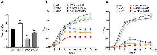
To understand the link between translational fidelity and fitness during exposure in bile salts, we separately introduced mutations I199N and K42N into the ribosomal genes rpsD (uS4) and rpsL (uS12) in the S. enterica Typhimurium strain ATCC 14028. We validated the error rate using our dual-fluorescence reporter readthrough assay [25] in the mutant strains. As expected, the rpsD I199N (rpsD*) and rpsL K42N (rpsL*) strains exhibited increased and decreased rates of UGA readthrough, respectively, when compared with the wild-type (WT) strain (Figure 1A).

Figure 1.
Increased translational fidelity improves growth under bile salt stress. (A) UGA readthrough translational errors were measured using dual-fluorescence reporters. Cells harboring the reporters were grown in Luria-Bertani broth (LB) at 37 °C for 16 h. Fluorescence signals were quantified using a plate reader. (B,C) Growth curve of Salmonella strains in the presence of 15 mg/mL or 40 mg/mL sodium cholate. Overnight cultures were diluted 1:100 in fresh LB with or without sodium cholate and incubated at 37 °C for 16 h with shaking. OD600 was determined using a microplate reader. The results are the average of at least three biological repeats with error bars representing the standard deviations. The p-values are determined using the independent t-test. ***, p < 0.001.
To assess the fitness change of altering translational fidelity in Salmonella under bile salt conditions, we added various concentrations of sodium cholate into the growth medium. The absorbance at 600 nm (optical density) was recorded at different time points. Without bile salts, the rpsD* and rpsL* mutations slightly decreased growth. However, in the presence of 15 mg/mL or 40 mg/mL sodium cholate, the rpsL* high-fidelity strain exhibited a growth advantage and achieved a higher cell density compared to the WT, whereas the rpsD* strain showed poorer growth than the WT (Figure 1B and Figure S1).
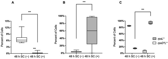
To further test whether bile salts affect the competition between the mutants and WT in co-cultures, we mixed WT with rpsD* or rpsL* carrying different fluorescence reporters at a 1:1 ratio in the presence and absence of sodium cholate. Salmonella cells were grown for a total of 48 h and analyzed using flow cytometry. We found that without sodium cholate, the WT cells outcompeted the rpsD* and rpsL* cells after 48 h, with the fractions of rpsD* and rpsL* cells reduced to 6.8% and 3.6%, respectively (Figure 2). In contrast, in the presence of sodium cholate, the rpsL* strain showed a competitive advantage over the WT (Figure 2A), whereas the rpsD* strain was completely outcompeted by the WT after 48 h (Figure 2B). These results were consistent with the growth curves of individual strains.

Figure 2.
High translational fidelity provides competitive advantage in bile salts. Competition experiments were initiated with a 1:1 mixture of overnight cultures of WT with rpsD*, WT with rpsL*, or rpsL* with rpsD*L* in the presence or absence of 20 mg/mL sodium cholate. Bacteria were grown for a total of 48 h, and bacteria were sorted by flow cytometry. In all, 30,000 gated events were acquired for each sample. (A) The error-prone strain rpsD* was outcompeted by the WT strain after 48 h with the addition of sodium cholate. (B) The error-restrictive strain rpsL* was outcompeted by the WT without sodium cholate but showed an increased advantage over the WT strain in the presence of sodium cholate. (C) Strain rpsD*L* had a higher error rate than rpsL*and was outcompeted by the rpsL* strain in the presence of sodium cholate after 48 h. The data here are representative of results from at least three biological replicates. The p-values were determined using the independent t-test. ***, p < 0.001.
The error-prone rpsD* and high-fidelity rpsL* mutations led to opposite effects in Salmonella fitness in the presence of bile salts. We next combined the rpsD I199N and rpsL K42N mutations in a single strain rpsD*L*. The error rate in the rpsD*L* strain was lower than the WT and rpsD* strains but higher than rpsL* (Figure 1A). Interestingly, competition experiments between rpsL* and rpsD*L* revealed opposite trends with and without sodium cholate: the rpsL* strain was outgrown by rpsD*L* without SC but almost completely outcompeted rpsD*L* in the presence of SC (Figure 2C). Collectively, these results demonstrate that increased ribosomal fidelity provides a growth advantage for Salmonella under bile salt stress.
3.2. Porins, Envelope Stress Response, and Flagella Are Not Major Contributors to Improved Bile Salt Resistance
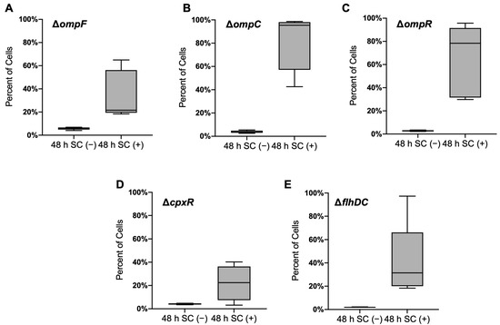
In Salmonella, the outer membrane porins have been implicated as bile transporters and affect virulence [26]. To test whether those porins are involved in mediating the increased bile salt resistance in the rpsL* strain, we deleted ompF and ompC genes that encode the most abundant porins from WT and rpsL* Salmonella. As shown in Figure 3A,B, deleting ompF or ompC did not affect the increase of rpsL* fraction in the presence of sodium cholate. Porin expression is also regulated by the two-component system EnvZ-OmpR in response to changes in osmolality and pH [27,28,29]; we found that deleting ompR did not alter fitness gain by the rpsL* mutation in the presence of sodium cholate either (Figure 3C).

Figure 3.
Fractions of rpsL* cells in competition experiments. Overnight cultures of WT ΔompF with rpsL* ΔompF (A), WT ΔompC with rpsL* ΔompC (B), WT ΔompR with rpsL* ΔompR (C), WT ΔcpxR with rpsL* ΔcpxR (D), and WT ΔflhDC with rpsL* ΔflhDC (E) were 1:1 mixed into the 1 mL LB medium in the presence or absence of 20 mg/mL sodium cholate. Bacteria were grown for a total of 48 h and sorted by flow cytometry. The data are representative of results from at least three biological replicates.
It has been suggested that a bacterial envelope provides a barrier that restricts the entry of bile salts [30,31,32], and bile salts could disrupt the integrity of both the outer and inner membranes leading to envelope stress [33,34]. Translational fidelity may affect protein misfolding and the CpxA-CpxR envelope stress response. Nevertheless, we found that the sodium cholate still enriched rpsL* cells when cpxR was deleted from the WT and rpsL* strains (Figure 3D), suggesting that the envelope stress response is not fully responsible for the increased resistance to bile resistance in high-fidelity cells.
Our previous study demonstrates that the assembly of flagella consumes the proton motive force and decreases the efflux pump activity, leading to antibiotic sensitivity [35]. The rpsL* mutation decreases the expression of flagellar genes and swimming motility [20]. Such trade-off between motility and efflux prompted us to investigate whether the master flagellar regulator flhDC was involved in bile resistance. The results revealed that the deletion of flhDC did not appear to decrease the fitness of rpsL* cells in the presence of sodium cholate (Figure 3E), suggesting that another pathway may lead to bile resistance in high-fidelity cells.
3.3. Improved Fitness in High-Fidelity Strain Depend on Increased Intracellular ATP
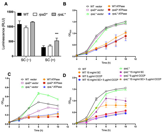
Translational errors affect protein misfolding and aggregation [13,36]. Interestingly, it has also been well established that bile salts cause misfolding and the denaturation of proteins [34,37]. Misfolded proteins harm bacterial cells and rely on ATP-dependent chaperones and proteases for refolding or degradation [38]. We thus examined the ATP levels in WT, rpsD*, and rpsL* cells grown in the presence and absence of sodium cholate. We observed that the cellular ATP level decreased substantially in all three strains after exposure to bile salts, and the rpsL* cells maintained the highest level of ATP among the three strains (Figure 4A). It is likely that the high-fidelity rpsL* cells generate less misfolded proteins in the presence of sodium cholate, therefore consuming less ATP during protein quality control.

Figure 4.
Improved fitness in error-restrictive strain depends on increased ATP levels. (A) Determination of intracellular ATP levels. Overnight cultures were diluted 1:100 in fresh LB with or without 20 mg/mL sodium cholate and incubated at 37 °C with agitation for 6 h. The cellular ATP level (indicated by the luminescence signal) decreased significantly in all three strains after exposure to bile salts, and the rpsL* strain maintained the highest level of ATP. (B,C) Growth curve of Salmonella strains harboring plasmid of IPTG-inducible ATPase. Overnight cultures were diluted 1:100 in fresh LB supplemented without (B) or with (C) sodium cholate and incubated at 37 °C for 10 h with shaking. In total, 1 mM IPTG was added to induce the expression of ATPase for ATP hydrolysis. (D) Growth curve of Salmonella strains with and without CCCP to inhibit ATP synthesis. The data are representative of results from at least three biological replicates. The p-values were determined using the independent t-test. ***, p < 0.001.
To further test whether the cellular ATP level contributes to improved fitness of high-fidelity cells after exposure to bile salts, we used a plasmid encoding the soluble subunit of ATPase to cause IPTG-inducible ATP depletion [24]. In the absence of sodium cholate (Figure 4B and Figure S2), the depletion of ATP caused the retarded growth of all three strains compared to the vector control. In the presence of sodium cholate (Figure 4C), rpsL* cells carrying the vector control exhibited improved growth compared to the WT, but this growth advantage was abolished upon induction of the ATPase. In line with this, we used a protonophore CCCP, which uncouples the proton motive force across the cellular membrane and reduces ATP synthesis to treat the WT and rpsL* strains. As shown in Figure 4D, after CCCP treatment, the rpsL* strain no longer exhibited improved growth when exposed to bile salts. Taken together, these results suggest that a higher cellular ATP level in the rpsL* strain contribute to its improved fitness after exposure to bile salts.
4. Discussion
Bile is a fluid synthesized by hepatocytes with antibacterial capacity due to the presence of bile salts [30,37]. Bile plays an essential role in the digestion and absorption of fat and soluble vitamins in the intestine [39]. It has the ability to dissolve membrane lipids and cause the denaturation of proteins [34,37]. Salmonella is a bacterial pathogen that causes tens of millions of gastrointestinal infections in humans worldwide each year [40,41,42]. During its systemic or chronic infections, Salmonella is exposed to bile salts [21,22], suggesting that they might have bile resistance mechanisms to facilitate their colonization in this environment. The establishment of a novel niche for replication in gallbladder epithelial cells may help Salmonella escape from the extremely high concentrations of bile salts present in the gall bladder lumen [43]. Biofilm formation on the surface of cholesterol gallstones may also protect Salmonella from the bactericidal activities of bile salts [44,45]. However, planktonic Salmonella cells are also found in the gall bladder lumen, implying that some unknown mechanisms are employed to permit their survival and replication. More recent studies show that some degree of translational errors may improve fitness by protecting cells from oxidative damage and antibiotics [18,19,46,47], which prompted us to investigate the role of translational fidelity in bile salt exposure. In the present work, we demonstrate that the error-prone strain rpsD* and error-restrictive strain rpsL* have decreased and increased fitness upon bile salt exposure, respectively, when compared to the WT (Figure 1 and Figure 2).
Genetic and biochemical studies have revealed a number of bile resistance factors in enteric bacteria, including porins [48], efflux pump systems [49], lipopolysaccharide [31], multiple antibiotic resistance (mar) gene [50], and phoPQ [51]. In this study, we show that the major outer membrane proteins OmpF and OmpC are not mainly responsible for the increased bile salt resistance in the rpsL* strain (Figure 3). Instead, we show that high translational fidelity lowers the amount of ATP consumption in the presence of bile salts (Figure 4A), likely through restricting protein misfolding. The resulting higher ATP level in the high-fidelity cells, in turn, benefits growth under bile stress, as suggested by our ATP depletion results (Figure 4C,D). Our work, therefore, reveals a previously unknown impact of translational fidelity on bacterial fitness under stress conditions, with implications in Salmonella–host interactions.
Supplementary Materials
The following are available online at https://www.mdpi.com/article/10.3390/genes13020184/s1, Figure S1: Growth curves of Salmonella strains in the presence of sodium cholate. Figure S2: Growth curves of Salmonella strains. Table S1: Oligonucleotides used in this study.
Author Contributions
Conceptualization, Z.L. and J.L.; methodology, Z.L. and J.L.; validation, Z.L. and J.L.; analysis, Z.L. and J.L.; resources, Z.L. and J.L.; data curation, Z.L. and J.L.; writing, Z.L. and J.L.; supervision, J.L.; funding acquisition, J.L. All authors have read and agreed to the published version of the manuscript.
Funding
This work was funded by NIGMS R35GM136213 (2020-2025) to J.L.
Institutional Review Board Statement
Not applicable.
Informed Consent Statement
Not applicable.
Data Availability Statement
Data supporting the findings are within this paper and supplemental files.
Acknowledgments
We would like to thank Mauricio H. Pontes (Penn State University) for kindly providing the plasmids pUHE-ATPase (pATPase) and pFPV25 (pVector).
Conflicts of Interest
The authors declare no conflict of interest.
References
- Kurland, C.G. Translational Accuracy and the Fitness of Bacteria. Annu. Rev. Genet. 1992, 26, 29–50. [Google Scholar] [CrossRef]
- Kramer, E.B.; Vallabhaneni, H.; Mayer, L.M.; Farabaugh, P.J. A Comprehensive Analysis of Translational Missense Errors in the Yeast Saccharomyces Cerevisiae. RNA 2010, 16, 1797–1808. [Google Scholar] [CrossRef] [PubMed] [Green Version]
- Ling, J.; So, B.R.; Yadavalli, S.S.; Roy, H.; Shoji, S.; Fredrick, K.; Musier-Forsyth, K.; Ibba, M. Resampling and Editing of Mischarged TRNA Prior to Translation Elongation. Mol. Cell 2009, 33, 654–660. [Google Scholar] [CrossRef] [PubMed] [Green Version]
- Traverse, C.C.; Ochman, H. Conserved Rates and Patterns of Transcription Errors across Bacterial Growth States and Lifestyles. Proc. Natl. Acad. Sci. USA 2016, 113, 3311–3316. [Google Scholar] [CrossRef] [Green Version]
- Gordon, A.J.E.; Satory, D.; Halliday, J.A.; Herman, C. Lost in Transcription: Transient Errors in Information Transfer. Curr. Opin. Microbiol. 2015, 24, 80–87. [Google Scholar] [CrossRef] [Green Version]
- Gromadski, K.B.; Rodnina, M.V. Kinetic Determinants of High-Fidelity TRNA Discrimination on the Ribosome. Mol. Cell 2004, 13, 191–200. [Google Scholar] [CrossRef] [Green Version]
- Li, L.; Boniecki, M.T.; Jaffe, J.D.; Imai, B.S.; Yau, P.M.; Luthey-Schulten, Z.A.; Martinis, S.A. Naturally Occurring Aminoacyl-TRNA Synthetases Editing-Domain Mutations That Cause Mistranslation in Mycoplasma Parasites. Proc. Natl. Acad. Sci. USA 2011, 108, 9378–9383. [Google Scholar] [CrossRef] [Green Version]
- Liu, Z.; Vargas-Rodriguez, O.; Goto, Y.; Novoa, E.M.; Ribas de Pouplana, L.; Suga, H.; Musier-Forsyth, K. Homologous Trans-Editing Factors with Broad TRNA Specificity Prevent Mistranslation Caused by Serine/Threonine Misactivation. Proc. Natl. Acad. Sci. USA 2015, 112, 6027–6032. [Google Scholar] [CrossRef] [Green Version]
- Schwartz, M.H.; Pan, T. Temperature Dependent Mistranslation in a Hyperthermophile Adapts Proteins to Lower Temperatures. Nucleic Acids Res. 2016, 44, 294–303. [Google Scholar] [CrossRef] [PubMed]
- Gromadski, K.B.; Rodnina, M.V. Streptomycin Interferes with Conformational Coupling between Codon Recognition and GTPase Activation on the Ribosome. Nat. Struct. Mol. Biol. 2004, 11, 316–322. [Google Scholar] [CrossRef] [PubMed]
- Bullwinkle, T.J.; Reynolds, N.M.; Raina, M.; Moghal, A.; Matsa, E.; Rajkovic, A.; Kayadibi, H.; Fazlollahi, F.; Ryan, C.; Howitz, N.; et al. Oxidation of Cellular Amino Acid Pools Leads to Cytotoxic Mistranslation of the Genetic Code. eLife 2014, 3, e02501. [Google Scholar] [CrossRef]
- Kohanski, M.A.; Dwyer, D.J.; Wierzbowski, J.; Cottarel, G.; Collins, J.J. Mistranslation of Membrane Proteins and Two-Component System Activation Trigger Antibiotic-Mediated Cell Death. Cell 2008, 135, 679–690. [Google Scholar] [CrossRef] [Green Version]
- Lant, J.T.; Kiri, R.; Duennwald, M.L.; O’Donoghue, P. Formation and Persistence of Polyglutamine Aggregates in Mistranslating Cells. Nucleic Acids Res. 2021, 49, 11883–11899. [Google Scholar] [CrossRef] [PubMed]
- Mikkola, R.; Kurland, C.G. Selection of Laboratory Wild-Type Phenotype from Natural Isolates of Escherichia Coli in Chemostats. Mol. Biol. Evol. 1992, 9, 394–402. [Google Scholar] [CrossRef] [PubMed]
- Bezerra, A.R.; Simões, J.; Lee, W.; Rung, J.; Weil, T.; Gut, I.G.; Gut, M.; Bayés, M.; Rizzetto, L.; Cavalieri, D.; et al. Reversion of a Fungal Genetic Code Alteration Links Proteome Instability with Genomic and Phenotypic Diversification. Proc. Natl. Acad. Sci. USA 2013, 110, 11079–11084. [Google Scholar] [CrossRef] [PubMed] [Green Version]
- Pan, T. Adaptive Translation as a Mechanism of Stress Response and Adaptation. Annu. Rev. Genet. 2013, 47, 121–137. [Google Scholar] [CrossRef] [Green Version]
- Ribas de Pouplana, L.; Santos, M.A.S.; Zhu, J.-H.; Farabaugh, P.J.; Javid, B. Protein Mistranslation: Friend or Foe? Trends Biochem. Sci. 2014, 39, 355–362. [Google Scholar] [CrossRef]
- Fan, Y.; Wu, J.; Ung, M.H.; De Lay, N.; Cheng, C.; Ling, J. Protein Mistranslation Protects Bacteria against Oxidative Stress. Nucleic Acids Res. 2015, 43, 1740–1748. [Google Scholar] [CrossRef]
- Mohler, K.; Ibba, M. Translational Fidelity and Mistranslation in the Cellular Response to Stress. Nat. Microbiol. 2017, 2, 1–9. [Google Scholar] [CrossRef] [Green Version]
- Fan, Y.; Thompson, L.; Lyu, Z.; Cameron, T.A.; De Lay, N.R.; Krachler, A.M.; Ling, J. Optimal Translational Fidelity Is Critical for Salmonella Virulence and Host Interactions. Nucleic Acids Res. 2019, 47, 5356–5367. [Google Scholar] [CrossRef] [PubMed]
- Andrews-Polymenis, H.L.; Bäumler, A.J.; McCormick, B.A.; Fang, F.C. Taming the Elephant: Salmonella Biology, Pathogenesis, and Prevention. Infect. Immun. 2010, 78, 2356–2369. [Google Scholar] [CrossRef] [PubMed] [Green Version]
- Gonzalez-Escobedo, G.; Marshall, J.M.; Gunn, J.S. Chronic and Acute Infection of the Gall Bladder by Salmonella Typhi: Understanding the Carrier State. Nat. Rev. Microbiol. 2011, 9, 9–14. [Google Scholar] [CrossRef] [PubMed] [Green Version]
- Datsenko, K.A.; Wanner, B.L. One-Step Inactivation of Chromosomal Genes in Escherichia Coli K-12 Using PCR Products. Proc. Natl. Acad. Sci. USA 2000, 97, 6640–6645. [Google Scholar] [CrossRef] [PubMed] [Green Version]
- Pontes, M.H.; Groisman, E.A. Protein Synthesis Controls Phosphate Homeostasis. Genes Dev. 2018, 32, 79–92. [Google Scholar] [CrossRef] [Green Version]
- Fan, Y.; Evans, C.R.; Barber, K.W.; Banerjee, K.; Weiss, K.J.; Margolin, W.; Igoshin, O.A.; Rinehart, J.; Ling, J. Heterogeneity of Stop Codon Readthrough in Single Bacterial Cells and Implications for Population Fitness. Mol. Cell 2017, 67, 826–836. [Google Scholar] [CrossRef] [PubMed] [Green Version]
- Chatfield, S.N.; Dorman, C.J.; Hayward, C.; Dougan, G. Role of OmpR-Dependent Genes in Salmonella Typhimurium Virulence: Mutants Deficient in Both OmpC and OmpF Are Attenuated in Vivo. Infect. Immun. 1991, 59, 449–452. [Google Scholar] [CrossRef] [PubMed] [Green Version]
- Slauch, J.M.; Silhavy, T.J. Genetic Analysis of the Switch That Controls Porin Gene Expression in Escherichia Coli K-12. J. Mol. Biol. 1989, 210, 281–292. [Google Scholar] [CrossRef]
- Slauch, J.M.; Garrett, S.; Jackson, D.E.; Silhavy, T.J. EnvZ Functions through OmpR to Control Porin Gene Expression in Escherichia Coli K-12. J. Bacteriol. 1988, 170, 439–441. [Google Scholar] [CrossRef] [Green Version]
- Heyde, M.; Portalier, R. Regulation of Major Outer Membrane Porin Proteins of Escherichia Coli K 12 by PH. Mol. Gen. Genet. 1987, 208, 511–517. [Google Scholar] [CrossRef]
- Gunn, J.S. Mechanisms of Bacterial Resistance and Response to Bile. Microbes Infect 2000, 2, 907–913. [Google Scholar] [CrossRef]
- Picken, R.N.; Beacham, I.R. Bacteriophage-Resistant Mutants of Escherichia Coli K12. Location of Receptors within the Lipopolysaccharide. J. Gen. Microbiol. 1977, 102, 305–318. [Google Scholar] [CrossRef] [PubMed] [Green Version]
- Hernández, S.B.; Cava, F.; Pucciarelli, M.G.; García-Del Portillo, F.; de Pedro, M.A.; Casadesús, J. Bile-Induced Peptidoglycan Remodelling in Salmonella Enterica. Environ. Microbiol. 2015, 17, 1081–1089. [Google Scholar] [CrossRef] [PubMed] [Green Version]
- Mitchell, A.M.; Silhavy, T.J. Envelope Stress Responses: Balancing Damage Repair and Toxicity. Nat. Rev. Microbiol. 2019, 17, 417–428. [Google Scholar] [CrossRef]
- Merritt, M.E.; Donaldson, J.R. Effect of Bile Salts on the DNA and Membrane Integrity of Enteric Bacteria. J. Med. Microbiol. 2009, 58, 1533–1541. [Google Scholar] [CrossRef] [Green Version]
- Lyu, Z.; Yang, A.; Villanueva, P.; Singh, A.; Ling, J. Heterogeneous Flagellar Expression in Single Salmonella Cells Promotes Diversity in Antibiotic Tolerance. MBio 2021, 12, e02374-21. [Google Scholar] [CrossRef] [PubMed]
- Ling, J.; Cho, C.; Guo, L.-T.; Aerni, H.R.; Rinehart, J.; Söll, D. Protein Aggregation Caused by Aminoglycoside Action Is Prevented by a Hydrogen Peroxide Scavenger. Mol. Cell 2012, 48, 713–722. [Google Scholar] [CrossRef] [Green Version]
- Begley, M.; Gahan, C.G.M.; Hill, C. The Interaction between Bacteria and Bile. FEMS Microbiol. Rev. 2005, 29, 625–651. [Google Scholar] [CrossRef] [Green Version]
- Baneyx, F.; Mujacic, M. Recombinant Protein Folding and Misfolding in Escherichia Coli. Nat. Biotechnol. 2004, 22, 1399–1408. [Google Scholar] [CrossRef]
- Chen, I.; Cassaro, S. Physiology, Bile salts. In StatPearls; StatPearls Publishing: Treasure Island, FL, USA, 2021. [Google Scholar]
- Gal-Mor, O.; Boyle, E.C.; Grassl, G.A. Same Species, Different Diseases: How and Why Typhoidal and Non-Typhoidal Salmonella Enterica Serovars Differ. Front. Microbiol. 2014, 5, 391. [Google Scholar] [CrossRef] [Green Version]
- Stanaway, J.D.; Parisi, A.; Sarkar, K.; Blacker, B.F.; Reiner, R.C.; Hay, S.I.; Nixon, M.R.; Dolecek, C.; James, S.L.; Mokdad, A.H.; et al. GBD 2017 Non-Typhoidal Salmonella Invasive Disease Collaborators The Global Burden of Non-Typhoidal Salmonella Invasive Disease: A Systematic Analysis for the Global Burden of Disease Study 2017. Lancet Infect. Dis. 2019, 19, 1312–1324. [Google Scholar] [CrossRef] [Green Version]
- LaRock, D.L.; Chaudhary, A.; Miller, S.I. Salmonellae Interactions with Host Processes. Nat. Rev. Microbiol. 2015, 13, 191–205. [Google Scholar] [CrossRef] [PubMed]
- Menendez, A.; Arena, E.T.; Guttman, J.A.; Thorson, L.; Vallance, B.A.; Vogl, W.; Finlay, B.B. Salmonella Infection of Gallbladder Epithelial Cells Drives Local Inflammation and Injury in a Model of Acute Typhoid Fever. J. Infect. Dis. 2009, 200, 1703–1713. [Google Scholar] [CrossRef] [PubMed] [Green Version]
- Biofilm Formation and Interaction with the Surfaces of Gallstones by Salmonella spp. | Infection and Immunity. Available online: https://journals.asm.org/doi/10.1128/IAI.70.5.2640-2649.2002 (accessed on 28 December 2021).
- Gallstones Play a Significant Role in Salmonella spp. Gallbladder Colonization and Carriage | PNAS. Available online: https://www.pnas.org/content/107/9/4353 (accessed on 28 December 2021).
- Lee, J.Y.; Kim, D.G.; Kim, B.-G.; Yang, W.S.; Hong, J.; Kang, T.; Oh, Y.S.; Kim, K.R.; Han, B.W.; Hwang, B.J.; et al. Promiscuous Methionyl-TRNA Synthetase Mediates Adaptive Mistranslation to Protect Cells against Oxidative Stress. J. Cell Sci. 2014, 127, 4234–4245. [Google Scholar] [CrossRef] [PubMed] [Green Version]
- Javid, B.; Sorrentino, F.; Toosky, M.; Zheng, W.; Pinkham, J.T.; Jain, N.; Pan, M.; Deighan, P.; Rubin, E.J. Mycobacterial Mistranslation Is Necessary and Sufficient for Rifampicin Phenotypic Resistance. Proc. Natl. Acad. Sci. USA 2014, 111, 1132–1137. [Google Scholar] [CrossRef] [PubMed] [Green Version]
- Thanassi, D.G.; Cheng, L.W.; Nikaido, H. Active Efflux of Bile Salts by Escherichia Coli. J. Bacteriol. 1997, 179, 2512–2518. [Google Scholar] [CrossRef] [PubMed] [Green Version]
- Rosenberg, E.Y.; Bertenthal, D.; Nilles, M.L.; Bertrand, K.P.; Nikaido, H. Bile Salts and Fatty Acids Induce the Expression of Escherichia Coli AcrAB Multidrug Efflux Pump through Their Interaction with Rob Regulatory Protein. Mol. Microbiol. 2003, 48, 1609–1619. [Google Scholar] [CrossRef]
- Sulavik, M.C.; Dazer, M.; Miller, P.F. The Salmonella Typhimurium Mar Locus: Molecular and Genetic Analyses and Assessment of Its Role in Virulence. J. Bacteriol. 1997, 179, 1857–1866. [Google Scholar] [CrossRef] [Green Version]
- van Velkinburgh, J.C.; Gunn, J.S. PhoP-PhoQ-Regulated Loci Are Required for Enhanced Bile Resistance in Salmonella spp. Infect Immun. 1999, 67, 1614–1622. [Google Scholar] [CrossRef]
Publisher’s Note: MDPI stays neutral with regard to jurisdictional claims in published maps and institutional affiliations. |
© 2022 by the authors. Licensee MDPI, Basel, Switzerland. This article is an open access article distributed under the terms and conditions of the Creative Commons Attribution (CC BY) license (https://creativecommons.org/licenses/by/4.0/).