Article

Identification of Novel Gene Variants for Autism Spectrum Disorders in the Lebanese Population Using Whole-Exome Sequencing

1
Department of Biology, Faculty of Arts and Sciences, Holy Spirit University of Kaslik, Jounieh P.O. Box 446, Lebanon
2
UMR 1253 iBrain, Inserm, Faculté de Médecine, Université de Tours, CEDEX 1, 37032 Tours, France
3
Division of Genomics and Translational Biomedicine, College of Health and Life Sciences, Hamad Bin Khalifa University, Doha P.O. Box 34110, Qatar
4
Laboratoire de Biochimie et Biologie Moléculaire, CHRU de Tours, CEDEX 09, 37044 Tours, France
5
School of Medicine and Medical Sciences, Holy Spirit University of Kaslik, Jounieh P.O. Box 446, Lebanon
*
Authors to whom correspondence should be addressed.
Genes 2022, 13(2), 186; https://doi.org/10.3390/genes13020186
Submission received: 20 December 2021 / Revised: 10 January 2022 / Accepted: 19 January 2022 / Published: 21 January 2022
(This article belongs to the Special Issue Genetics of Neurodevelopmental Disorders)

Abstract

:
In our previous study, in which array CGH was used on 19 Lebanese ASD subjects and their parents, we identified rare copy number variants (CNVs) in 14 subjects. The five remaining subjects did not show any CNVs related to autism spectrum disorders (ASD). In the present complementary study, we applied whole-exome sequencing (WES), which allows the identification of rare genetic variations such as single nucleotide variations and small insertions/deletions, to the five negative CNV subjects. After stringent filtering of initial data on the five families, three novel genes potentially related to neurodevelopment were identified, including a de novo mutation in the MIS18BP1 gene. In addition, genes already known to be related to ASD contained sequence variations. Our findings outline the potential involvement of the novel de novo mutation in the MIS18BP1 gene in the genetic etiology and pathophysiology of ASD and highlights the genetic complexity of these disorders. Further studies with larger cohorts of subjects are needed to confirm these observations, and functional analyses need to be performed to understand the precise pathophysiology in these cases.

1. Introduction

According to the Diagnostic and Statistical Manual of Mental Disorders DSM-5, autism spectrum disorders (ASD) are extremely variable conditions characterized by impairments in reciprocal social communication and the use of restrictive and repetitive routines, typically manifesting before the age of 3 years [1] and affecting boys 4 times more than girls [2]. For 2016, in the United States, the estimated ASD prevalence has gradually risen to affect 1 in every 54 children who are 8 years of age, according to a recently published report by the Centers for Disease Control and Prevention (CDC) [3]. However, the prevalence in Lebanon is estimated to be 1 in 66 children in Beirut and Mount regions [4].
In parallel to the considerable clinical heterogeneity of these disorders, several studies have shown that ASD are multifactorial disorders. Variations in multiple genes provide strong evidence of the involvement of genetic factors that explain most of ASD risk [5]. In fact, ASD is thought to potentially involve more than 1000 genes [6], while 102 genes have been formally associated [7], with variable levels of evidence. An important proportion of these genes encodes proteins implicated in synaptic function, ubiquitination, and chromatin remodeling [8].
Chromosomal abnormalities, rare copy number variations (CNVs) and single nucleotide variants (SNV) have been associated with ASD [9]. Rare, de novo, and inherited CNVs have been entangled in several neurodevelopmental disorders and they are observed in 15–20% of subjects with ASD [10]. Despite the great number of identified ASD susceptibility genes, only a small proportion of them have been strongly validated [10]; as such, identifying specific causative genes is an important challenge.
Recent developments in genomic sequencing have transformed variant discovery. Different approaches have been used to uncover gene variants related to ASD, such as next generation sequencing techniques (NGS) including whole genome sequencing (WGS) and whole-exome sequencing (WES). WES has been used to identify rare and novel genetic variations related to neurodevelopmental disorders [11]. Our previous study aimed to evaluate the presence of rare CNVs in a group of 19 Lebanese ASD subjects and their parents using the high-resolution comparative genomic hybridization technique (array CGH) which is an ultra-high-resolution method of genetic testing that identifies small deletions and duplications. We reported a high percentage of CNVs in 14 subjects. Moreover, this study uncovered several CNVs related to ASD and identified PJA2, SYNPO, APCS, and TAC1 as novel ASD candidate genes [12]. An additional approach to identifying small CNVs, SNVs, and indels in the genome associated with several disorders including ASD [13] could be of interest since array CGH cannot detect balanced structural variations and point mutations. To this end, in this complementary study, whole-exome sequencing (WES) was further implemented among the five families who did not reveal any CNVs related to ASD in the previous study, to identify point mutations in genes and to expand our knowledge of the genetic etiology and the pathophysiology of these disorders. Therefore, the analysis framework was designed to uncover rare de novo and inherited variants in novel or ASD-linked genes that have previously been described. This approach enabled us to detect variants in three novel genes potentially related to neurodevelopment, including one de novo mutation in the MIS18BP1 gene. In addition, variants in genes already known as related to ASD were also detected.

2. Materials and Methods

2.1. Subjects and Clinical Characteristics

The 5 studied subjects were enrolled in the study among the group of 19 [12]. The diagnosis was performed by the psychiatrists of the non-governmental organizations (NGOs). This was based on the Diagnostic and Statistical Manual of Mental Disorders in its 4th edition [1]. As reported by the NGOs, the average Childhood Autism Rating Scale (CARS) score as well as the levels of intellectual disability were moderate in the subjects included in our study [12]. Subjects were recruited from specialized institutions and NGOs distributed all over Lebanon. First, our research team contacted the NGOs to explain the aim of the project. After the approval of the NGOs, a letter summarizing the objectives was sent to the families. Afterwards, the families were invited to a meeting with our team in which we explained the different steps of the study. The families who accepted to participate in our study provided us with a signed informed consent form before the collection of the data and the samples.
The study was complied with the ethical standards and guidelines of the Declaration of Helsinki in 1964 and its later amendments. The Holy Spirit University Ethical Committee reviewed and approved the study protocol (delivered in 2014).

2.2. DNA Extraction

Genomic DNA was extracted from the blood samples of ASD subjects and their 2 parents (except the mother of subject number 64 who was dead and the father of subject 70 who was unknown) using the QIAsymphony robot, and then the concentration and purity of the extracted DNA was assessed by spectrophotometry (Thermo Scientific Nanodrop 2000, Waltham, MA, USA).

2.3. Genetic Studies

Using the V6 SureSelect kits (Agilent, Santa Clara, CA, USA) on an Illumina HiSeq2500 platform at Macrogen (Seoul, Korea), library preparation and subsequent exome capture were performed. Using Novo align, sequences were aligned to the hg19 human genome and variants were called by the Genome Analyses Toolkit (GATK). A Phred scaled quality score (PSQ) of more than 20 was adopted. The Illumina variant studio was used to annotate and analyze the variants. Further analysis of the filtered variants continued using the Varsome platform (www.varsome.com, accessed on 13 September 2020) [14].

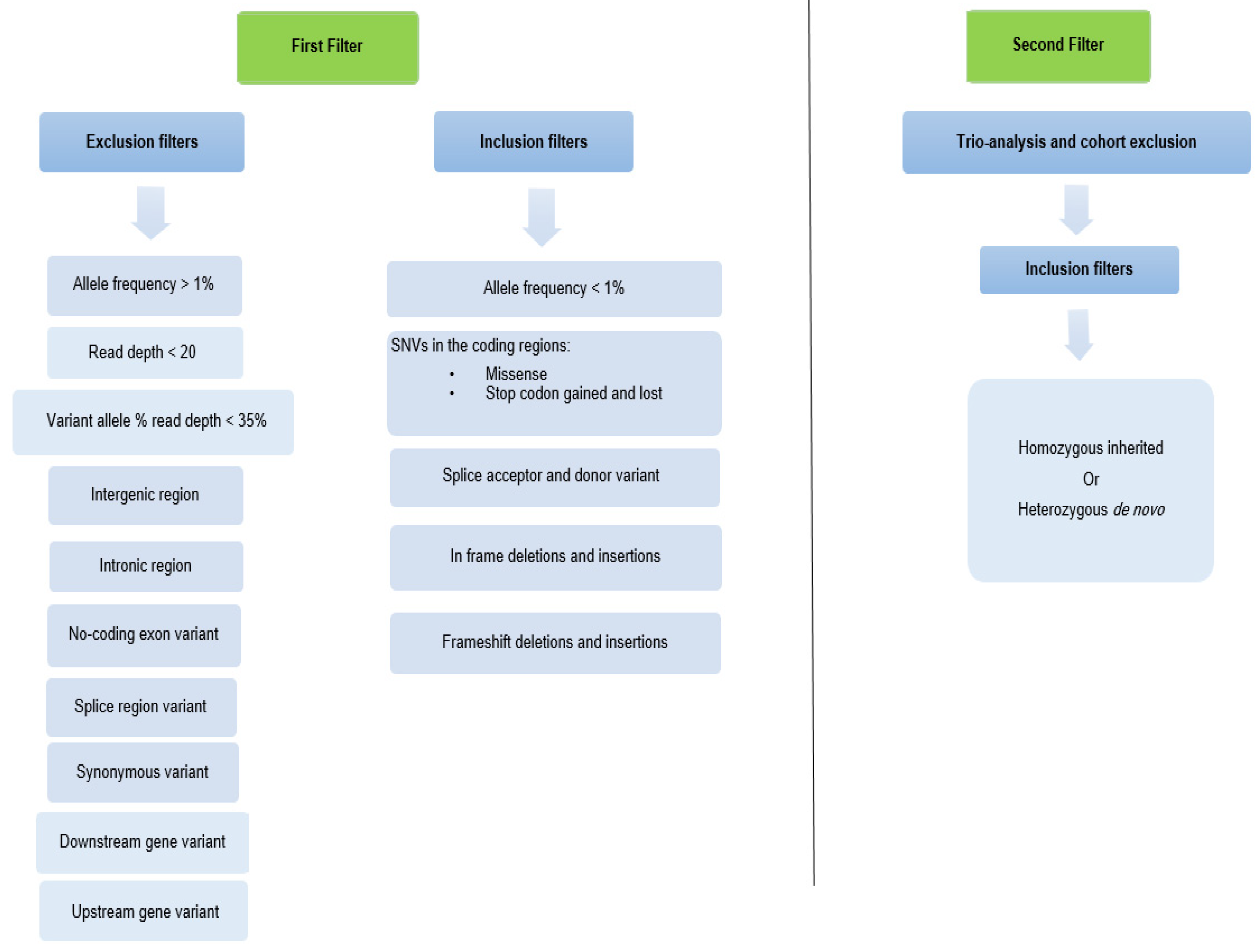
2.4. Variant Analysis

We only kept the SNV and indel gene variants with an allele frequency <1% in the Database of Genomic Variants (DGV) in the UCSC Genome browser. Then, all intergenic, intronic, non-coding, splice region, synonymous, downstream, and upstream variants were excluded. Deleterious variants obtained from exonic frameshift, damaging missense, splice donor and acceptor, stop codon gained and lost, and in-frame deletions and insertions were retained (Figure 1). In addition, the variants with a read depth of less than 20 and a variant allele frequency of less than 35% of the normal allele (proportion of variant reads) were removed. Furthermore, trio analysis and cohort exclusion were performed on the remaining gene variants by assuming a de novo, autosomal recessive, and X-linked mode of inheritance.
The theoretical pathogenicity of the missense variants was evaluated by in silico predictive software (SIFT: https://bii.a.star.edu.sg/, accessed on 11 September 2020 [15], Polyphen2: http://genetics.bwh.harvard.edu/pph2/, accessed on 11 September 2020 [16], CADD: https://cadd.gs.washington.edu/snv, accessed on 19 December 2021) and scored accordingly. Damaging missense SNVs were defined as those having a deleterious prediction in one of the tools. Then, we manually inspected the likely false positive variants using the IGV [17].
In order to verify if the detected variations were in genes already known to be associated with ASD or neurodevelopmental disorders, we checked the following databases: SFARI (https://www.sfari.org/, accessed on 31 December 2020), AutismKB (http://db.cbi.pku.edu.cn/autismkb_v2/, accessed on 31 December 2020) and PubMed (https://pubmed.ncbi.nlm.nih.gov/, accessed on 31 December 2020 ). The gene variants were subsequently classified according to the American College of Medical Genetics and Genomics (ACMG) [18] as pathogenic, likely pathogenic, variants of uncertain clinical significance, likely benign, and benign. We defined the damaging ASD-associated variants as those meeting likely pathogenic or pathogenic criteria according to ACMG standards. The remaining genes were subjected to further screening for biological significance for neural development, known neurological disorders, and function using Online Mendelian Inheritance in Man (OMIM) (https://www.omim.org/, accessed on 15 February 2021), PubMed, Decipher (https://decipher.sanger.ac.uk/, accessed on 15 February 2021), and Gene Cards (https://www.genecards.org/, accessed on 15 February 2021). These genes were classified as novel in ASD related to neurodevelopment and/or neurological disorders. The updated annotation and distribution in the GnomAD database were ascertained using the Varsome portal [14].

2.5. Genetic Validation Using Sanger Sequencing

To genotype the variants, we designed PCR primers for each site using the Primer3Plus software (https://www.bioinformatics.nl/cgi-bin/primer3plus/primer3plus.cgi, accessed on 11 October 2021) and used standard PCR conditions for the available 10 samples (Table S1). Sanger sequencing was performed on an ABI 3130xl Genetic Analyzer (Applied Biosystems, Waltham, MA, USA) following the manufacturer’s instructions to validate the new candidates. Finally, the 10 variants were validated (Figure S2).

3. Results

This study included five eligible subjects with ASD phenotypes. The main characteristics of each subject are displayed in Table 1.
WES was performed for each subject and his/her parents when available to detect SNVs and/or indels in all available genes. The resulting on target-reads of around 40 Mbp showed an ~80X coverage average mean depth of target regions for all samples. We adopted a filtering strategy that associates the genotype to the underlying phenotypes within each family for all variants with less than 1% minor allele frequency (MAF).
We started our analysis with 95,000 variants/subject. After applying the first filter, around 1400 variants/subject were retained. Subsequent filtering based on trio analysis and cohort exclusion reduced the variants to around 3 variants/subject as shown in Figure 2.
The analysis framework of this study was designed to uncover not only de novo variants, but also the ones with autosomal recessive and X-linked mode of inheritance in unknown or ASD linked genes, previously described.

3.1. Detected Variants

3.1.1. De Novo Variants

We detected a missense SNV on chromosome 4 in the USP46 (OMIM #612849) gene in subject number 23 (Table 2). The c.293C>T (NM_022832.3) predicted a p.Pro98Leu variation which was found to be deleterious by SIFT and benign by Polyphen 2.
In subject number 45, a frameshift deletion and a missense SNV were detected on chromosome 14 and 12 in the MIS18BP1 (OMIM #618139) and KRT2 (OMIM #600194) genes, respectively. The c.471delA (NM_018353.4) in the MIS18BP1 gene predicted a p.Lys157AsnfsTer24 variation which was inspected using Integrated Genome Browser Visualization (IGV) (Figure S1). The c.1022G>A (NM_000423.2) on the KRT2 gene predicted a p.Arg341His variation which was found to be deleterious by SIFT and probably damaging by Polyphen 2.
In subject number 73, a missense SNV was detected on chromosome 3 in the LSMEM2 gene. The c.97G>T (NM_153215.1) on this gene predicted a p.Gly33Trp which was considered to be deleterious by SIFT and possibly damaging by Polyphen 2.

3.1.2. Homozygote Variants

In subject number 45, we detected one missense homozygous SNV inherited from both parents on chromosome 2 in the HAAO (OMIM #604521) gene (Table 3). The c.371T>C (NM_012205.2) in this gene led to a p.Met124Thr variation which was predicted to be tolerated by SIFT and benign by Polyphen 2 and was inspected on IGV (Figure S1).
In subject number 70, one homozygous missense SNV on chromosome 18 in the ASXL3 (OMIM #615115) gene was detected and inspected on IGV (Figure S1). Since the father was missing, we cannot assume that it was inherited from both parents. The c.5560G>A (NM_030632.1) on this gene predicted a p.Val1854Ile variation which was considered to be damaging by SIFT and benign by Polyphen 2.
In subject number 73, one homozygous frameshift deletion inherited from the father was detected on chromosome 2 in the KRTAP5-5 gene and inspected using IGV (Figure S1). The c.2773C>T (NM_001001480.2) in KRTAP5-5 gene predicted a p.Arg925Trp variation which was shown to be deleterious by SIFT and possibly damaging by Polyphen 2.

3.1.3. X-Linked Variants

In male subject number 23, a total of 2 missense SNVs inherited from the mother on chromosome X in the SLITRK4 (OMIM #300562) and FLNA (OMIM #300017) genes were detected (Table 4). The c.1860A>C (NM_001184749.1) on the SLITRK4 gene predicted a p.Leu620Phe variation which was considered to be deleterious by SIFT and benign by Polyphen2 and was inspected using IGV (Figure S1). The c.1954G>A (NM_001110556.1) in the FLNA gene predicted a p.Glu652Lys variation which was predicted to be tolerated by SIFT and probably damaging by Polyphen 2.
In male subject number 64, we detected 2 missense SNVs on chromosome X in PTCHD1 (OMIM #300828) and FLNA (OMIM #300017) genes. The c.1804A>G (NM_173495.2) in PTCHD1 gene predicted a p.Thr602Ala variation which was predicted to be tolerated by SIFT and probably damaging by Polyphen 2 and was inspected using IGV (Figure S1). On the FLNA gene, the c.2590G>T (NM_001110556.1) predicted a p.Val864Phe variation which was predicted to be deleterious by SIFT and possibly damaging by Polyphen 2. Since the mother of subject 64 is dead, we cannot assume whether these SNVs are inherited or de novo.
In male subject number 73, we detected a missense SNV in the NHSL2 gene inherited from the mother. The c.2773C>T (NM_001013627.2) led to p.Arg925Trp variations which was predicted to be deleterious by SIFT and possibly damaging by Polyphen 2.

3.2. Oligogenic Model

Our results showed that all the studied subjects had multi-hit SNVs and indels simultaneously. We detected 3 SNVs in subject 23 (Figure 3a), 2 SNVs and 1 deletion in subject 45 (Figure 3b), 2 SNVs in subject 64, whose mother was dead (Figure 3c), 1 SNV in subject 70, whose father was unknown (Figure 3d), and 2 SNVs and 1 insertion in subject 73 (Figure 3e). The homozygous SNV on the HAAO gene was retained since it was inherited from both parents.
Since de novo gene variants have been considered a major cause of early-onset genetic disorders such as ASD [19], Pathway Studio software v12.3 (https://mammal.pathwaystudio.com/, accessed on 24 September 2020) was used to identify the cell processes related to the encoded protein of the novel de novo mutation in the MIS18BP1 gene detected in our study. A biological network was created by integrating MIS18BP1 gene in the software and the encoded protein was connected to its related cell processes (Figure 4).

4. Discussion

This is the first study on Lebanese ASD subjects using the WES approach. Due to the high genetic heterogeneity of ASD and the complexity of inheritance, the genetic factors are not yet fully elucidated. Therefore, it is of interest to search for novel ASD candidate genes in new populations that will lead to a better understanding of the etiology based on a strict familial genotyping–phenotyping correlation approach. In our previous study, in which array CGH was used on 19 ASD subjects, we identified rare CNVs in 14 subjects in the novel candidate genes PJA2, APCS, SYNPO, and TAC1. Thus, in this study, the genomic characterization of the five ASD subjects, who did not reveal any CNVs related to ASD, with that of their parents (except the mother of subject number 64 who was dead and the father of subject 70 who was unknown) also revealed novel gene variants such as SNVs and indels. Among the novel genes, the de novo mutations are considered strong candidates for disease. Ten variants were successfully validated using Sanger sequencing (Figure S3). Furthermore, most of the studied subjects had more than one candidate variant observed, which speaks in favor of a multi-hit genetic model or alternatively on the benignity of some identified variants.
Despite the limited sample size in our study, none of the identified variants had been previously identified in the database of more than 500 exomes of Lebanese subjects with different forms of common and rare diseases [20]. In addition, variants in genes already reported as related to ASD (either in autism databases or in literature) were detected, reinforcing the robustness of our strategy.
Since de novo variants contribute to the genetic etiology of ASD, our study aimed to identify these variants. A novel de novo frameshift deletion on chromosome 14 was identified in the MIS18BP1 gene (MIS18 binding protein1), which is a mitotic regulator. It was confirmed that MIS18BP1 is regulated via SUMO-ubiquitin crosstalk during mitosis [21]. SUMOylation plays an important role in neuronal differentiation, synapse formation control, regulation of synaptic transmission and cell survival [22]. Furthermore, the ubiquitin pathway regulates neurotransmitter release, synaptic vesicle recycling, and changes in post synaptic density and dendritic spines [23]. Using Pathway Studio software, cell processes related to the encoded protein of the MIS18BP1 gene were presented (Figure 4). Chromatin remodeling and DNA methylation, two of the identified processes, are important in human brain development that can be regulated by MIS18BP1, strengthening its potential role in ASD [24]. Subject 45, who is epileptic and has a speech delay, had a deletion in the MIS18BP1 gene. In fact, it has been reported that DNA methylation and chromatin remodeling have been linked to the development of epilepsy [25,26]. Furthermore, variations in mitotic genes which are responsible for kinetochore assembly, chromosome segregation, and condensation can trigger the onset of neurodevelopmental disorders by insufficient cell proliferation and failure in neuronal stem cell replenishment, leading to the underdevelopment of the central nervous system [27]. An important chromatin remodeler, CDC42 (Cell Division Cycle 42), interacts with the encoded protein of MIS18BP1 gene [28]. Furthermore, the encoded protein of CDC42BPB (CDC42 Binding Protein Kinase Beta) gene is a downstream effector of CDC42. CDC42BPB gene was identified as ASD risk gene in a study using WES and subsequent transmission and de novo association (TADA) analysis [8].
In addition, three de novo missense SNVs were detected in the following genes: USP46, KRT2, and LSMEM2. The deubiquitinating enzyme encoded by the USP46 gene is specific for the Alpha-amino-3-hydroxy-5-methyl-4-isoxazolepropionic acid receptor (AMPARs). These receptors are the primary mediators for neuronal development and communication and play a substantial role in learning and memory [29]. A deletion in 4q12 involving the USP46 gene was identified in a female ASD subject in a study on Italian families using oligo array CGH [30].
Two novel missense SNVs were detected in the SLITRK4 (SLIT and NTRK Like Family Member 4) and NHSL2 (NHS-Like Protein 2) genes on chromosome X in male subjects 23 and 73, respectively. SLITRKs family are transmembrane proteins from the leucine-rich repeat (LRR) superfamily. They are expressed in the central nervous system and participate in neurite outgrowth, neuronal survival, and dendritic elaboration [31]. NHSL2 gene is a member of the Nance–Horan syndrome (NHS) gene family. Nance-Horan syndrome is an X-linked developmental disorder characterized by intellectual disability, cataracts, and physical and teeth abnormalities [32]. In a study performed on three NHS families, one affected male had severe mental retardation, epilepsy, and hypotonia [33]. In the same study, NHS expression was detected in fetal brain, lung, kidney, and thymus, and was largely expressed throughout brain development.
Furthermore, some of the inherited gene variants concerned genes that have been already reported in the literature and may represent strong candidates for ASD. On chromosome X, four missense SNVs were detected involving FLNA and PTCHD1 genes in male subjects 23 and 64. In subjects 23 and 64, two SNVs in FLNA (Filamin A) gene were detected. This gene encodes an actin-binding protein which links actin filaments to membrane glycoproteins. In a chromosomal microarray analysis study performed on 195 ASD subjects of Greek origin, a deletion in the FLNA gene was found in a subject with ASD, seizures, and dysmorphic features [34]. In addition, the encoded protein of the PTCHD1 (Patched domain containing 1) gene is a membrane protein with a patched domain. This gene is required for the thalamic reticular nucleus (TRN) development and function. This part of the thalamus is essential for sleep rhythm generation, attention, and sensorimotor processing. In addition, PTCHD1 interacts with the postsynaptic membrane to provide a direct link with the excitatory synaptic network [35]. A genome-wide assessment for structural abnormalities performed on 427 unrelated ASD patients identified novel loci in the PTCHD1 gene, which led to it being an ASD susceptibility gene [36]. Moreover, a study conducted on 23 subjects with PTCHD1 deletions or truncating mutations supported that these gene mutations were the cause of an X-linked non-syndromic neurodevelopmental disorder which has the features of intellectual disability and ASD [37].
On chromosome 2 and 18, two homozygous missense SNVs were detected on the HAAO and ASXL3 genes. Subject 45 was homozygous for the SNV on the HAAO (3-Hydroxyanthranilate 3,4-Dioxygenase) gene (Figure 3b). In this case, the combination of the two inherited alleles led to the phenotype since the parents were unaffected. The expression of the HAAO gene was previously found to be reduced in ASD subjects [38]. This gene encodes a protein belonging to the intramolecular dioxygenases family which exists in low amounts in the central nervous system. The HAAO enzyme catalyzes the synthesis of quinolic acid, which is an excitotoxin. High cerebral levels of this acid may participate in neurologic and inflammatory disorder pathogenesis. Moreover, the ASXL3 (ASXL Transcriptional Regulator 3) gene encodes an important protein for the regulation of gene transcription. Its encoded protein may also inhibit histone de-ubiquitination. In a WES study performed aiming to uncover the susceptibility genes contributing to ASD, a mutation was found in the ASXL3 gene in ASD patients with intellectual disability [39].
The WES approach only sequences the coding regions of the genome. Moreover, like other WES studies, one of the potential limitations is the exclusion of causative variants by a stringent filtering approach to remove false positives. Due to the relatively small sample size in our study and the aim of only investigating rare genetic variations, statistical and functional analyses were not performed. However, our findings need to be validated by further functional studies and more robust genetic findings can be obtained from future studies with enlarged sample sizes.

5. Conclusions

In our study, using the WES technique for the first time in the Lebanese population, we identified one novel de novo mutation in the MIS18BPB1 gene and two other novel inherited mutations in the SLITRK4 and NHSL2 as potential ASD candidate genes. In addition, our results confirmed the presence of other de novo and inherited genetic variations that have been previously described and are shared between Lebanese ASD subjects and other studied populations with ASD. Accordingly, our observations provide a further argument for frequent polygenic models in ASD composed of several inherited and de novo variants. However, further research work (including functional studies) is essential to reinforce and validate our findings and to strengthen their implication in the pathology.

Supplementary Materials

The following supporting information can be downloaded at: www.mdpi.com/article/10.3390/genes13020186/s1. Figure S1: Integrated Genome Browser visualization (IGV) of the variants; Figure S2: Sanger validation of the candidate variants; Table S1: Primer sequences for amplification and sequencing of the candidate genes regions containing the variants.

Author Contributions

W.H., C.R.A., G.N. and T.B. conceived and designed the project. P.G., T.B. and W.H. recruited the subjects. P.G., T.B., G.N., F.L. and S.M. performed the experiments. All the authors analyzed the data and discussed the results. P.G. wrote the paper. All authors have read and agreed to the published version of the manuscript.

Funding

This project was funded by the National Council for Scientific Research in Lebanon (CNRSL) S/3541, the Higher Center for Research of the Holy Spirit University of Kaslik (HCR-USEK) 90010141 and the Institut National de la Santé et de la Recherche Médicale (INSERM).

Institutional Review Board Statement

This study was performed in line with the ethical standards and guidelines declared by the Declaration of Helsinki in 1964 and its later amendments and was approved by the Holy Spirit University Ethical Committee (delivered on 13 August 2014 and renewed on 9 January 2019).

Informed Consent Statement

Informed consent was obtained from all individual participants included in the study. All parents of the participants provided informed consent for publication.

Data Availability Statement

The datasets generated and analyzed during the current study are available in the European Variation Archive (EVA) repository under the accession number PRJEB44446 at https://www.ebi.ac.uk/eva/, accessed on 1 December 2021.

Acknowledgments

We thank all the non-governmental organizations and associations that care for subjects with autism and graciously shared their data with us.

Conflicts of Interest

The authors declare that they have no competing interest.

References

  1. American Psychiatric Association. Diagnostic and Statistical Manual of Mental Disorders, 5th ed.; American Psychiatric Association: Arlington, VA, USA, 2013. [Google Scholar]
  2. Baio, J.; Wiggins, L.; Christensen, D.L.; Maenner, M.J.; Daniels, J.; Warren, Z.; Kurzius-Spencer, M.; Zahorodny, W.; Robinson Rosenberg, C.; White, T.; et al. Prevalence of Autism Spectrum Disorder among Children Aged 8 Years—Autism and Developmental Disabilities Monitoring Network, 11 Sites, United States, 2014. MMWR Surveill. Summ. 2018, 67, 1–23. Available online: https://www.ncbi.nlm.nih.gov/pmc/articles/PMC5919599/ (accessed on 7 March 2020). [CrossRef] [PubMed]
  3. Maenner, M.J. Prevalence of Autism Spectrum Disorder among Children Aged 8 Years—Autism and Developmental Disabilities Monitoring Network, 11 Sites, United States, 2016. MMWR Surveill. Summ. 2020, 69, 1–12. Available online: https://www.cdc.gov/mmwr/volumes/69/ss/ss6904ahtm (accessed on 15 February 2021). [CrossRef] [PubMed]
  4. Chaaya, M.; Saab, D.; Maalouf, F.T.; Boustany, R. Prevalence of Autism Spectrum Disorder in Nurseries in Lebanon: A Cross Sectional Study. J. Autism Dev. Disord. 2016, 46, 514–522. Available online: https://link.springer.com/article/10.1007%2Fs10803-015-2590-7 (accessed on 11 March 2020). [CrossRef] [PubMed]
  5. Sandin, S.; Lichtenstein, P.; Kuja-Halkola, R.; Hultman, C.; Larsson, H.; Reichenberg, A. The Heritability of Autism Spectrum Disorder. JAMA 2017, 318, 1182–1184. [Google Scholar] [CrossRef]
  6. SFARI|Simons Foundation Autism Research Initiative. Available online: https://www.sfari.org/ (accessed on 3 March 2020).
  7. Satterstrom, F.K.; Kosmicki, J.A.; Wang, J.; Breen, M.S.; De Rubeis, S.; An, J.; Peng, M.; Collins, R.; Grove, J.; Klei, L.; et al. Large-Scale Exome Sequencing Study Implicates Both Developmental and Functional Changes in the Neurobiology of Autism. Cell 2020, 180, 568–584. [Google Scholar] [CrossRef]
  8. De Rubeis, S.; He, X.; Goldberg, A.P.; Poultney, C.S.; Samocha, K.; Cicek, A.E.; Kou, Y.; Liu, L.; Fromer, M.; Walker, S.; et al. Synaptic, Transcriptional, and Chromatin Genes Disrupted in Autism. Nature 2014, 515, 209–215. Available online: https://www.ncbi.nlm.nih.gov/pmc/articles/PMC4402723/ (accessed on 12 February 2020). [CrossRef]
  9. Hnoonual, A.; Thammachote, W.; Tim-Aroon, T.; Rojnueangnit, K.; Hansakunachai, T.; Sombuntham, T.; Roongpraiwan, R.; Worachotekamjorn, J.; Chuthapisith, J.; Fucharoen, S.; et al. Chromosomal Microarray Analysis in a Cohort of Underrepresented Population Identifies SERINC2 as a Novel Candidate Gene for Autism Spectrum Disorder. Sci. Rep. 2017, 7, 12096. Available online: https://www.nature.com/articles/s41598-017-12317-3 (accessed on 27 September 2019). [CrossRef] [Green Version]
  10. Huguet, G.; Ey, E.; Bourgeron, T. The Genetic Landscapes of Autism Spectrum Disorders. Annu. Rev. Genom. Hum. Genet. 2013, 14, 191–213. Available online: https://www.annualreviews.org/doi/abs/10.1146/annurev-genom-091212-153431 (accessed on 1 October 2019). [CrossRef]
  11. O’Roak, B.J.; Deriziotis, P.; Lee, C.; Vives, L.; Schwartz, J.J.; Girirajan, S.; Karakoc, E.; Mackenzie, A.P.; Ng, S.B.; Baker, C.; et al. Exome Sequencing in Sporadic Autism Spectrum Disorders Identifies Severe de Novo Mutations. Nat. Genet. 2011, 43, 585–589. Available online: https://www.ncbi.nlm.nih.gov/pubmed/21572417 (accessed on 1 October 2019). [CrossRef]
  12. Bitar, T.; Hleihel, W.; Marouillat, S.; Vonwill, S.; Vuillaume, M.; Soufia, M.; Vourc’h, P.; Laumonnier, F.; Andres, C.R. Identification of Rare Copy Number Variations Reveals PJA2, APCS, SYNPO, and TAC1 as Novel Candidate Genes in Autism Spectrum Disorders. Mol. Genet. Genom. Med. 2019, 7, e786. Available online: https://onlinelibrary.wiley.com/doi/abs/1002/mgg786 (accessed on 1 October 2019). [CrossRef]
  13. Iossifov, I.; Ronemus, M.; Levy, D.; Wang, Z.; Hakker, I.; Rosenbaum, J.; Yamrom, B.; Lee, Y.; Narzisi, G.; Leotta, A.; et al. De novo gene disruptions in children on the autistic spectrum. Neuron 2012, 74, 285–299. [Google Scholar] [CrossRef] [PubMed] [Green Version]
  14. Kopanos, C.; Tsiolkas, V.; Kouris, A.; Chapple, C.E.; Albarca Aguilera, M.; Meyer, R.; Massouras, A. VarSome: The Human Genomic Variant Search Engine. Bioinformatics 2019, 35, 1978–1980. Available online: https://www.ncbi.nlm.nih.gov/pmc/articles/PMC6546127/ (accessed on 13 September 2020). [CrossRef] [PubMed]
  15. Vaser, R.; Adusumalli, S.; Leng, S.N.; Sikic, M.; Ng, P.C. SIFT missense predictions for genomes. Nat. Protoc. 2016, 11, 1–9. [Google Scholar] [CrossRef] [PubMed]
  16. Adzhubei, I.A.; Schmidt, S.; Peshkin, L.; Ramensky, V.E.; Gerasimova, A.; Bork, P.; Kondrashov, A.S.; Sunyaev, S.R. A Method and Server for Predicting Damaging Missense Mutations. Nat. Methods 2010, 7, 248–249. [Google Scholar] [CrossRef] [PubMed] [Green Version]
  17. Robinson, J.T.; Thorvaldsdóttir, H.; Wenger, A.M.; Zehir, A.; Mesirov, J.P. Variant Review with the Integrative Genomics Viewer. Cancer Res. 2017, 77, e31–e34. [Google Scholar] [CrossRef] [PubMed] [Green Version]
  18. Richards, S.; Aziz, N.; Bale, S.; Bick, D.; Das, S.; Gastier-Foster, J.; Grody, W.W.; Hegde, M.; Lyon, E.; Spector, E.; et al. Standards and Guidelines for the Interpretation of Sequence Variants: A Joint Consensus Recommendation of the American College of Medical Genetics and Genomics and the Association for Molecular Pathology. Genet. Med. 2015, 17, 405–424. Available online: https://www.ncbi.nlm.nih.gov/pmc/articles/PMC4544753/ (accessed on 30 May 2020). [CrossRef] [Green Version]
  19. Acuna-Hidalgo, R.; Veltman, J.A.; Hoischen, A. New Insights into the Generation and Role of de Novo Mutations in Health and Disease. Genome Biol. 2016, 17, 241. Available online: https://search.datacite.org/works/1186/s13059-016-1110-1 (accessed on 24 September 2020). [CrossRef] [Green Version]
  20. Refaat, M.M.; Hassanieh, S.; Ballout, J.A.; Zakka, P.; Hotait, M.; Khalil, A.; Bitar, F.; Arabi, M.; Arnaout, S.; Skouri, H.; et al. Non-familial cardiomyopathies in Lebanon: Exome Sequencing Results for Five Idiopathic Cases. BMC Med. Genom. 2019, 12, 33. Available online: https://search.datacite.org/works/1186/s12920-019-0478-7 (accessed on 24 September 2020). [CrossRef] [Green Version]
  21. Cuijpers, S.A.G.; Willemstein, E.; Vertegaal, A.C.O. Converging Small Ubiquitin-like Modifier (SUMO) and Ubiquitin Signaling: Improved Methodology Identifies Co-modified Target Proteins. Mol. Cell. Proteom. 2017, 16, 2281–2295. Available online: https://search.datacite.org/works/1074/mcp.tir000152 (accessed on 19 December 2021). [CrossRef] [Green Version]
  22. Henley, J.M.; Craig, T.J.; Wilkinson, K.A. Neuronal SUMOylation: Mechanisms, Physiology, and Roles in Neuronal Dysfunction. Physiol. Rev. 2014, 94, 1249–1285. Available online: https://www.ncbi.nlm.nih.gov/pmc/articles/PMC4187031/ (accessed on 23 September 2020). [CrossRef]
  23. Glessner, J.T.; Wang, K.; Cai, G.; Korvatska, O.; Kim, C.E.; Wood, S.; Zhang, H.; Estes, A.; Brune, C.W.; Bradfield, J.P.; et al. Autism Genome-Wide Copy Number Variation Reveals Ubiquitin and Neuronal Genes. Nature 2009, 459, 569–573. Available online: https://www.ncbi.nlm.nih.gov/pmc/articles/PMC2925224/ (accessed on 23 September 2020). [CrossRef]
  24. LaSalle, J.M. Autism Genes Keep Turning Up Chromatin. OA Autism 2013, 1, 14. Available online: https://www.ncbi.nlm.nih.gov/pmc/articles/PMC3882126/ (accessed on 23 September 2020). [CrossRef] [PubMed] [Green Version]
  25. Jesus-Ribeiro, J.; Pires, L.M.; Melo, J.D.; Ribeiro, I.P.; Rebelo, O.; Sales, F.; Freire, A.; Melo, J.B. Genomic and Epigenetic Advances in Focal Cortical Dysplasia Types I and II: A Scoping Review. Front. Neurosci. 2020, 14, 580357. [Google Scholar] [CrossRef] [PubMed]
  26. Conboy, K.; Henshall, D.C.; Brennan, G.P. Epigenetic Principles Underlying Epileptogenesis and Epilepsy Syndromes. Neurobiol. Dis. 2021, 148, 105179. Available online: https://www.sciencedirect.com/science/article/pii/S096999612030454X (accessed on 19 December 2021). [CrossRef] [PubMed]
  27. Degrassi, F.; Damizia, M.; Lavia, P. The Mitotic Apparatus and Kinetochores in Microcephaly and Neurodevelopmental Diseases. Cells 2019, 9, 49. Available online: https://www.ncbi.nlm.nih.gov/pmc/articles/PMC7016623/ (accessed on 19 December 2021). [CrossRef] [Green Version]
  28. Lagana, A.; Dorn, J.F.; Rop, V.D.; Ladouceur, A.; Maddox, A.S.; Maddox, P.S. A Small GTPase Molecular Switch Regulates Epigenetic Centromere Maintenance by Stabilizing Newly Incorporated CENP-A. Nat. Cell Biol. 2010, 12, 1186–1193. Available online: https://www.nature.com/articles/ncb2129 (accessed on 26 September 2020). [CrossRef]
  29. Huo, Y.; Khatri, N.; Hou, Q.; Gilbert, J.; Wang, G.; Man, H. The Deubiquitinating Enzyme USP46 Regulates AMPA Receptor Ubiquitination and Trafficking. J. Neurochem. 2015, 134, 1067–1080. Available online: https://onlinelibrary.wiley.com/doi/abs/1111/jnc.13194 (accessed on 25 February 2021). [CrossRef]
  30. Mafalda, M.; Marco, C.D.; Canitano, R.; Buoni, S.; Frullanti, E.; Mencarelli, M.A.; Veronica, B.; Amabile, S.; Radice, L.; Baldassarri, M.; et al. A Genome Wide Copy Number Variations Analysis in Autism Spectrum Disorder (Asd) and Intellectual Disability (Id) in Italian Families. J. Genet. Syndr. Gene Ther. 2016, 7, 2. [Google Scholar] [CrossRef] [Green Version]
  31. Proenca, C.C.; Gao, K.P.; Shmelkov, S.V.; Rafii, S.; Lee, F.S. Slitrks as Emerging Candidate Genes Involved in Neuropsychiatric Disorders. Trends Neurosci. 2011, 34, 143–153. Available online: https://www.ncbi.nlm.nih.gov/pmc/articles/PMC3051006/ (accessed on 23 September 2020). [CrossRef] [Green Version]
  32. Brooks, S.P.; Coccia, M.; Tang, H.R.; Kanuga, N.; Machesky, L.M.; Bailly, M.; Cheetham, M.E.; Hardcastle, A.J. The Nance-Horan Syndrome Protein Encodes a Functional WAVE Homology Domain (WHD) and is Important for Co-Ordinating Actin Remodelling and Maintaining Cell Morphology. Hum. Mol. Genet. 2010, 19, 2421–2432. Available online: https://www.ncbi.nlm.nih.gov/pubmed/20332100 (accessed on 23 September 2020). [CrossRef] [Green Version]
  33. Brooks, S.P.; Ebenezer, N.D.; Poopalasundaram, S.; Lehmann, O.J.; Moore, A.T.; Hardcastle, A.J. Identification of the gene for Nance-Horan syndrome (NHS). J. Med. Genet. 2004, 41, 768. [Google Scholar] [CrossRef] [PubMed] [Green Version]
  34. Oikonomakis, V.; Kosma, K.; Mitrakos, A.; Sofocleous, C.; Pervanidou, P.; Syrmou, A.; Pampanos, A.; Psoni, S.; Fryssira, H.; Kanavakis, E.; et al. Recurrent copy number variations as risk factors for autism spectrum disorders: Analysis of the clinical implications. Clin. Genet. 2016, 89, 708. [Google Scholar] [CrossRef] [PubMed]
  35. Ung, D.C.; Iacono, G.; Meziane, H.; Blanchard, E.; Papon, M.A.; Selten, M.; Rhijn, J.R.v.; Montjean, R.; Rucci, J.; Martin, S.; et al. Ptchd1 Deficiency Induces Excitatory Synaptic and Cognitive Dysfunctions in Mouse. Mol. Psychiatry 2018, 23, 1356–1367. Available online: https://www.narcis.nl/publication/RecordID/oai:repository.ubn.ru.nl:2066%2F193089 (accessed on 23 September 2020). [CrossRef] [Green Version]
  36. Marshall, C.R.; Noor, A.; Vincent, J.B.; Lionel, A.C.; Feuk, L.; Skaug, J.; Shago, M.; Moessner, R.; Pinto, D.; Ren, Y.; et al. Structural Variation of Chromosomes in Autism Spectrum Disorder. Am. J. Hum. Genet. 2008, 82, 477–488. [Google Scholar] [CrossRef] [PubMed] [Green Version]
  37. Chaudhry, A.; Noor, A.; Degagne, B.; Baker, K.; Bok, L.A.; Brady, A.F.; Chitayat, D.; Chung, B.H.; Cytrynbaum, C.; Dyment, D.; et al. Phenotypic Spectrum Associated with PTCHD1 Deletions and Truncating Mutations Includes Intellectual Disability and Autism Spectrum Disorder. Clin. Genet. 2015, 88, 224–233. Available online: https://www.narcis.nl/publication/RecordID/oai:cris.maastrichtuniversity.nl:publications%2F8ce2b8ca-830a-46a3-b59d-1fd464489405 (accessed on 23 September 2020). [CrossRef]
  38. Boccuto, L.; Chen, C.; Pittman, A.R.; Skinner, C.D.; McCartney, H.J.; Jones, K.; Bochner, B.R.; Stevenson, R.E.; Schwartz, C.E. Decreased Tryptophan Metabolism in Patients with Autism Spectrum Disorders. Mol. Autism 2013, 4, 16. Available online: https://search.datacite.org/works/1186/2040-2392-4-16 (accessed on 3 February 2021). [CrossRef] [Green Version]
  39. Husson, T.; Lecoquierre, F.; Cassinari, K.; Charbonnier, C.; Quenez, O.; Goldenberg, A.; Guerrot, A.; Richard, A.; Drouin-Garraud, V.; Brehin, A.; et al. Rare Genetic Susceptibility Variants Assessment in Autism Spectrum Disorder: Detection Rate and Practical Use. Transl. Psychiatry 2020, 10, 77. Available online: https://www.ncbi.nlm.nih.gov/pmc/articles/PMC7039996/ (accessed on 3 February 2021). [CrossRef]
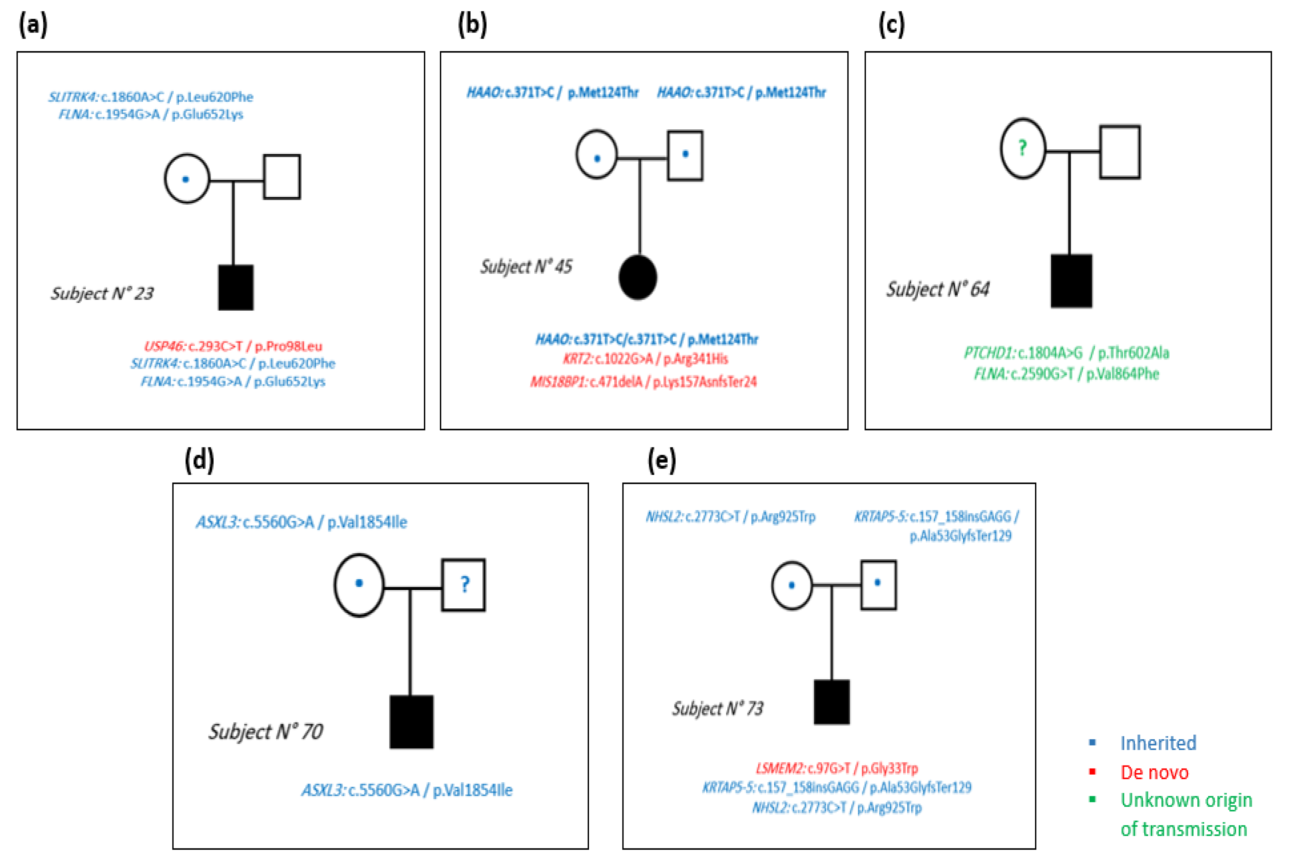
Figure 1. Filtering approach of the variables detected by whole-exome sequencing. In total, 2 filters were applied on the obtained variants from the 5 subjects. The first filter was based on the allele frequency and the protein coding consequences of the variants. The second filter was based on the trio analysis and cohort exclusion.
Genes 13 00186 g001
Figure 2. Number of variants obtained by WES after 2 filtering steps. Around 1400 variants/subject remained after applying the first filter and around 3 variants/subject remained after the second filter.
Genes 13 00186 g002
Figure 3. Pedigrees of the 5 subjects. (a) In subject number 23, 2 SNVs were inherited from the mother and 1 SNV was de novo. (b) In subject number 45, 1 SNV was inherited from both parents and 1 SNV and 1 deletion were de novo. (c) In subject number 64, 2 SNVs were detected on chromosome X. (d) In subject number 70, we detected 1 inherited SNV on chromosome 18 in the ASXL3 gene. (e) In subject number 73, 1 SNV and 1 insertion were inherited and 1 SNV was de novo. Blue color represents the inherited variants, red color represents the de novo variants and green color represents the variants with unknown origin of transmission.
Genes 13 00186 g003
Figure 4. Analysis of the cell processes of the encoded protein of MIS18BP1 gene using Pathway Studio software. A biological network was created connecting the encoded protein of MIS18BP1 gene by the cell processes.
Genes 13 00186 g004
Table 1. Characteristics of ASD subjects included in our study.
Subject NumberGenderAgeRegionParents AvailabilityParents
Consanguinity
Family HistoryCARSAssociated Comorbidities
23Male9BeirutYesNoDiabetes, cancer, and renal disease on both the paternal
and maternal sides
Moderate autismHyperactivity, anxiety
45Female9South LebanonYesNoNAModerate autismEpilepsy, speech delay
64Male10Mount LebanonNoNoHypertension and high cholesterol on the maternal sideModerate autismAnxiety
70Male27BekaaNoFather unknownDiabetes, hypertension, high cholesterol, and triglycerides
on both the maternal and paternal family sides.
Moderate autismAnxiety, depression, hyperactivity, self-injurious behavior
73Male9BekaaYesNoDiabetes, hypertension on both family sides.
Intellectual disability in paternal side
Moderate autismAnxiety, depression, hyperactivity, self-injurious behavior
Table 2. List of de novo gene variants. De novo variants in 4 genes were detected in subjects 23, 45, and 73. AD: autosomal dominant, PS: strong evidence of pathogenicity, PM: moderate evidence of pathogenicity, PVS: very strong evidence of pathogenicity, PP: supporting evidence of pathogenicity, BP: supporting evidence of a benign impact.
Identified Variant
SubjectGeneChromosomeMode of InheritanceTypeBase ChangeProtein ChangeConsequence SIFTPolyphen2CADDACMG Classification
23USP464ADsnvc.293C>Tp.Pro98Leumissensedeleterious (0.03)benign (0.009)23.8Uncertain significance
(PS2, PM2)
45MIS18BP114ADdeletionc.471delAp.Lys157AsnfsTer24frameshift 16.6Uncertain significance
(PVS1, PP3)
KRT212ADsnvc.1022G>Ap.Arg341Hismissensedeleterious (0)probably damaging (1)27.7Likely pathogenic (PM1, PM2, PP2, PP3)
73LSMEM23ADsnvc.97G>Tp.Gly33Trpmissensedeleterious (0.02)possibly damaging (0.688)22.1Uncertain significance
(PM2, BP4)
Table 3. List of autosomal recessive gene variants. In total, 3 homozygous variants were detected in 3 different genes in subjects 45, 70, and 73. AR: autosomal recessive, PM: moderate evidence of pathogenicity, BP: supporting evidence of a benign impact, PVS: very strong evidence of pathogenicity, BS: strong evidence of a benign impact.
Identified Variant
SubjectGeneChromosomeMode of InheritanceTypeBase ChangeProtein ChangeConsequenceSIFTPolyphen2CADDACMG Classification
45HAAO2ARsnvc.371T>Cp.Met124Thrmissensetolerated (1)benign (0)0.05Uncertain significance
(PM1, PM2, BP4)
70ASXL318ARsnvc.5560G>Ap.Val1854IlemissenseDamaging (0.09)Benign (0.05)16.5Uncertain significance
(PM2, BP4)
73KRTAP5-511ARinsertionc.157_158insGAGGp.Ala53GlyfsTer129frameshift 32Uncertain significance
(PVS1, BS1)
Table 4. List of X-linked gene variants. Variants in 4 genes were presented in the X-linked pattern of inheritance in subjects 23, 64, and 73. Two different variants on the FLNA gene were detected in subjects 23 and 64. XR: X-linked recessive, PM: moderate evidence of pathogenicity, PP: supporting evidence of pathogenicity, BP: supporting evidence of a benign impact.
Identified Variant
SubjectGeneChromosomeMode of InheritanceTypeBase ChangeProtein ChangeConsequenceSIFTPolyphen2CADDACMG Classification
23SLITRK4XXRsnvc.1860A>Cp.Leu620Phemissensedeleterious (0)benign (0.441)23.7Uncertain significance
(PM2, PP3)
FLNAXXRsnvc.1954G>Ap.Glu652Lysmissensetolerated (0.09)probably damaging (0.929)25.3Uncertain significance
(PM2, PP3, BP1)
64PTCHD1XXRsnvc.1804A>Gp.Thr602Alamissensetolerated (0.32)probably damaging (0.996)20.9Uncertain significance
(PM2, PP3, BP1)
FLNAXXRsnvc.2590G>Tp.Val864Phemissensedeleterious (0)possibly damaging (0.745)22.7Uncertain significance
(PM2, PP3, BP1)
73NHSL2XXRsnvc.2773C>Tp.Arg925Trpmissensedeleterious (0)possibly damaging (0.847)19.8Uncertain significance
(PM5, PP2, BP1)
Publisher’s Note: MDPI stays neutral with regard to jurisdictional claims in published maps and institutional affiliations.

