Article

Prevalence and Genotype-Phenotype Correlation of Lynch Syndrome in a Selected High-Risk Cohort from Qatar’s Population

by
Mariem Sidenna
1,2,*,
Houssein Khodjet-El-khil
3,*,
Hajar Al Mulla
1,2,
Mashael Al-Shafai
3,
Hind Hassan Habish
2,
Reem AL-Sulaiman
1,2 and
Salha Bujassoum Al-Bader
2
1
Department of Adult and Pediatric Medical Genetics, Hamad Medical Corporation, Doha 3050, Qatar
2
Department of Cancer Genetics, Medical Oncology, National Center of Cancer Care and Research, Hamad Medical Corporation, Doha 3050, Qatar
3
Department of Biomedical Science, College of Health Sciences, QU Health, Qatar University, Doha 2713, Qatar
*
Authors to whom correspondence should be addressed.
Genes 2022, 13(11), 2176; https://doi.org/10.3390/genes13112176
Submission received: 18 September 2022 / Revised: 31 October 2022 / Accepted: 10 November 2022 / Published: 21 November 2022
(This article belongs to the Section Human Genomics and Genetic Diseases)

Abstract

:
Lynch syndrome (LS) is the most common cause of hereditary colorectal cancers (CRC) and is associated with an increased risk for ovarian and endometrial cancers. There is lack of knowledge on the epidemiology of LS in the non-Caucasian populations especially in Qatar. The aim of this retrospective study is to explore the prevalence of LS in a selected high-risk cohort in the State of Qatar in addition to investigating the frequency and genotype-phenotype correlation associated with mismatch repair genes pathogenic variants. Retrospective review of medical records of 31 individuals with LS, 20 affected with colorectal cancer and 11 unaffected with family history of cancers, referred from January 2017 until August 2020. The prevalence of LS among affected and unaffected patients is 22% (20/92) and 2.2% respectively. Among affected individuals, MLH1 and MSH2 genes were highly frequent while for unaffected individuals, a recurrent PMS2 pathogenic variant was reported in several related individuals suggesting a tribal effect. This study highlights the epidemiology of LS in high-risk cohort in Qatar which helps to provide recommendations on genetic testing, and personalize surveillance and management programs

1. Introduction

Lynch syndrome (LS) is the main cause of inherited colorectal cancer, it also accounts for an elevated risk of other extracolonic cancers such as gastric, endometrial and ovarian cancers [1]. Hereditary Non-Polyposis Colorectal Cancer (HNPCC) was the former name of Lynch syndrome which is associated with 1–5% of all colorectal cancer cases [2], about 3% of endometrial cancers (EC) [3], and 2% of ovarian cancers (OC) [4].
LS is inherited in an autosomal dominant manner and results from pathogenic variants in the DNA mismatch repair genes (MMR genes), (MLH1, MSH2, MSH6, and PMS2) [5]. Furthermore, germline deletions in EPCAM gene have been linked to Lynch syndrome [6]. EPCAM gene does not belong to the MMR genes, yet, pathogenic deletions can disturb its 3′ end which results in epigenetic inactivation of its neighboring gene (MSH2) that would be silenced [6].
Individuals who are suspected to have Lynch syndrome are often assessed for meeting revised Amsterdam II [5] or revised Bethesda criteria [7]. These criteria focus on the presence of a strong family history of Lynch syndrome associated cancers, young age at diagnosis and specific histopathological characteristics [8]. However, they have been associated with a limited sensitivity and specificity [9]. Additionally, Microsatellite instability (MSI) and Immunohistochemistry (IHC) tests are used to assess for the eligibility of genetic testing for LS. Defective MMR genes cause a variation in the microsatellites repeat number in the tumor tissue as compared to normal tissues in the same individual which can be detected through MSI testing [10]. Immunohistochemistry (IHC) is a tool to detect the loss of MMR protein expression for colon and endometrial cancers [10]. However, the interaction between MMR proteins could affect the sensitivity of immunohistochemistry in the detection of individual proteins. For instance, MSH2 creates heterodimers with MSH6 [11,12], thus negative staining for both proteins is common in MSH2 pathogenic variant carriers [13]. Likewise, for MLH1 pathogenic variant carriers, IHC often shows an absence of both MLH1 and PMS2 proteins since the two form heterodimers [14].
The results of the international Mismatch Repair Consortium (IMRC) show that MLH1 and MSH2 genes are responsible for 76.5% of all of the mutations identified while MSH6 pathogenic variants account for 15.12% of cases followed by PMS2 and EPCAM genes which accounts for 7.7% and 0.65 respectively of the identified cases [15].
Several studies have investigated genotype-phenotype correlation of MMR genes in Lynch Syndrome [7,16,17,18]. For instance, MSH2 pathogenic variants carriers presented with a higher risk of metachronous and synchronous colon tumors as compared to MLH1 pathogenic variants carriers in according to a Chinese study [19]. Also, a study conducted on one of the largest HNPCC cohorts in Germany found that MSH2 pathogenic carriers had a late-onset endometrial cancer besides a lower percentage of CRC in MLH1 female carriers as compared to male carriers [16].
Nevertheless, the data gained from these studies have been restricted to Caucasian families coming from European descent resulting in a gap in the comprehension of genetic epidemiology and genotype-phenotype correlation in Lynch Syndrome among non-Caucasian families.
The State of Qatar is a Middle Eastern country situated on the northeastern shore of the Arabian Peninsula, encircled by the Arabian/Persian Gulf. The number of inhabitants in Qatar frames a multiethnic local area; around 600,000 (22%) of the 2.7 million population are local Qataris (Ministry of Development Planning and Statistics); expatriates make up the excess part of the population [20]. Lynch Syndrome is the second most common cancer syndrome in Qatar after Hereditary breast and ovarian cancer (HBOC) [21]. There is a lack of studies on the epidemiology and genotype-phenotype correlation in Lynch syndrome in Qatar. While most of Lynch syndrome studies in the literature are population-based studies, our approach is more clinically oriented focusing on patients affected with colorectal cancer or unaffected individuals with family history indicative for Lynch syndrome The aim of our study is to explore the prevalence of Lynch syndrome, frequency of MMR genes pathogenic variants and the genotype-phenotype correlation of Lynch syndrome in colorectal cancer in a selected high risk cohort of ethnically diverse individuals in Qatar.

2. Materials and Methods

2.1. Study Design

The study was conducted by retrospectively reviewing the medical and genetic records of all patients referred to high risk genetic clinic at National Center for Cancer Care and Research hospital (if they meet Amsterdam/Bethesda guidelines, have an extensive family history of cancers or have a known familial mutation) from the period of January 2017 until August 2020.
A total number of 592 individuals were filtered according to our inclusion/exclusion criteria, all of them were tested for MMR genes, including 92 patients affected by colon cancer and have high risk features of LS and 500 unaffected individuals with positive family history of colon cancer and indicative of Lynch syndrome.
Review of data revealed 31 individuals including 20 patients found to be positive for Lynch syndrome among the 92 high-risk patients tested for MMR genes and 11 unaffected individuals positive for lynch syndrome among the 500 unaffected individuals who opted for genetic testing. The remaining 561 individuals were negative for lynch syndrome and were excluded as they did not have pathogenic variants in MMR genes (Figure 1).

2.2. Study Participants

All individuals with MMR/EPCAM genes pathogenic/ likely pathogenic variants were included in the study and were divided into 2 categories affected and unaffected:
Affected individuals are defined as patients with colorectal cancer and harboring pathogenic MMR gene variants who were referred due to meeting Amsterdam/Bethesda criteria.
Unaffected individuals are defined as individuals not affected with cancer and harboring pathogenic MMR genes variants referred due to meeting one or more of Bethesda guidelines, family history of cancers with young ages at onset, and/or a known familial variant. (Figure 1).

2.3. Medical Records and Genetic Testing Reports

Epidemiological information in addition to clinical information including patient’s age, gender, ethnic background, personal cancer history (Including primary site, age at diagnosis, histopathology, immunohistochemistry, grade, stage, lymph node involvement have been collected from the participants medical records.
Genetic tests (using either Comprehensive Cancer Panel (48 genes), Lynch/Colorectal High-risk Panel (7 genes), Colorectal Cancer Panel (21 genes), Breast/Gyni Cancer Panel (24 genes), Common Cancer Management Panel (39 genes) or Targeted Variant Testing) were done at GeneDX lab (https://www.genedx.com/, accessed on 15 May 2021). Genetic testing results were collected from the genetic reports of each individual. These include variants in MMR genes (MLH1, MSH2, MSH6, and PMS2) and EPCAM gene and were used to calculate the frequency of pathogenic variants in these genes among individuals with Lynch Syndrome (affected and unaffected). It was also used to compare their frequencies between affected and unaffected individuals.
The novelty of the variants was reported in the individuals’ genetic test reports and was also assessed using Clinvar (https://www.ncbi.nlm.nih.gov/clinvar/, accessed on 21 February 2021), HGMD (http://www.hgmd.cf.ac.uk/ac/index.php, accessed on 21 February 2021) and Gene Cards (https://www.genecards.org/, accessed on 21 February 2021).

2.4. Prevalence Calculation

Prevalence of Lynch Syndrome in affected individuals was Calculated by dividing the total number of affected individuals (individuals diagnosed with colorectal cancer and harboring MMR pathogenic variants) by the total number of high-risk affected individuals with and without the MMR pathogenic variants.
Prevalence of Lynch Syndrome in unaffected individuals was calculated by dividing the total number of unaffected individuals harboring MMR pathogenic variants by the total number of high-risk unaffected individuals with and without MMR pathogenic variants.

2.5. Genotype-Phenotype Correlation and Statistical Analysis

Genotype-phenotype correlation between the clinicopathological data with the type of mutated gene and with the type of variant were investigated. Correlation between Immunohistochemical staining of MMR proteins versus germline genetic test results was performed. All correlations have been assessed using Fisher’s exact test. They were performed using IBM SPSS statistical software (version 19) for data management. All categorical and binary variables were analyzed by Fischer’s exact test (p = ((a + b) ! (c + d) ! (a + c) ! (b + d) ! )/a ! b ! c ! d ! N !).
A two-sided p value < 0.05 was considered statistically significant. A post hoc test has been performed for significant correlations observed by Fisher’s exact test. Bonferroni correction was used for statistically significant post hoc test values. For the distribution of MMR and EPCAM genes variants in affected and unaffected individuals, a p-value < 0.005 (0.05/10) was considered statistically significant, while for the correlation of CRC side vs the type of variant a p-value <0.006 (0.05/8) was considered to be significant.

2.6. Variants Classification

Pathogenic/ Likely pathogenic variants in MMR/EPCAM genes (in affected and unaffected individuals) have been classified by definition into point mutations (nonsense and missense mutations), Large deletions, Frameshift mutations, and Intronic mutations.

3. Results

The study includes a total of 31 individuals harboring pathogenic variants in MMR genes (20 affected and 11 unaffected). (Figure 1).
Of the 20 affected patients, 14 were males (70%) and 6 were females (30%), with an average age of 48.2 ± 11.2 years. Colon cancer was predominantly left-sided (in 12 out of the 20 patients; 60%). Most of the cancers were in stage 2 (60%) and 14 out of the 20 patients (70%) were diagnosed before the age of 50 years. Positive cancer family history was seen in 15 patients (75%) (Table 1). Regarding the 11 unaffected high-risk individuals, 8 were females (72%) and 3 were males (27.27%) with an average age of 45.2 ± 13.5 years.
The prevalence of Lynch syndrome was calculated in a selected high-risk population of colon cancer patients (selected based on meeting one or more Amsterdam and Bethesda criteria) and in high-risk unaffected individuals (based on meeting one or more Bethesda guidelines, having a family history of cancers and/or a known pathogenic variant in MMR gene in the family). In affected patients, the prevalence of LS was 22% (20/92), while in unaffected individuals it was 2.2%.

3.1. Frequency of MMR Pathogenic Variants

Genetic data was collected for variants in MMR genes (MLH1, MSH2, MSH6, and PMS2) as well as for germline deletions in EPCAM gene. The distribution of MMR genes and EPCAM gene variants was significantly different between LS affected and unaffected individuals (p-value < 0.001 a) (Table 2).
In LS affected individuals, MLH1 and MSH2 were the most commonly reported genes accounting for almost 90% of the variants and they were exclusively reported in affected individuals (p-value = 0.009) which was not statistically significant after Bonferroni correction while MSH6 and PMS2 genes were the least the least reported with 5% each.
For unaffected LS individuals however, the most reported gene was PMS2 accounting for almost 63.6% of the variants (p-value = 0.0001) which is statistically significant after Bonferroni correction, followed by MSH6 and EPCAM genes with each accounting for 18.2% of the variants (Table 2).

3.2. Types of Variants in MMR and EPCAM Genes

In LS affected individuals, point variants and frameshift variants were the most commonly seen variants in MLH1 and MSH2 genes accounting for 66.7% of each variant. For MSH6 gene, one frameshift was reported and for PMS2 gene, only one point mutation was reported (Table 3).
In LS unaffected individuals, most of the pathogenic variants in PMS2 were large deletions (71.4%) followed by point mutations (14.3%) and intronic variants (14.3%) (Table 4). A heterozygous deletion encompassing exons 6 to 11 in PMS2 gene was the most commonly reported pathogenic variant in unaffected individuals and all four carriers (heterozygous) of this pathogenic variant belong to the same tribe. For MSH6 gene, most of the pathogenic variants were intronic (50%) and frameshift variants (50%). However, for EPCAM gene, both pathogenic variants were large deletions (100%) (Table 4).
On the other hand, one large deletion (deletion encompassing exons 6 to 11) was observed to be recurrent in 3 affected members in a homozygous state. The 3 members belong to the same tribe and were diagnosed with Constitutional Mismatch Repair Deficiency Syndrome (CMMRD) (Table S1).
Four novel pathogenic variants have been reported in 7 affected individuals; two in MLH1 gene (p.Thr545ProfsX46 and p.Thr553ProfsX38), one in MSH2 gene (IVS11 + 2T > C) and one in PMS2 gene (deletion encompassing exons 1 to 6) which was also found in unaffected individuals. (Table S1).

3.3. Genotype-Phenotype Correlation

3.3.1. Correlation with Mutated Gene

A Chi-square test was conducted to investigate the correlation of the mutated gene with the clinicopathological parameters. There was no statistically significant correlation between gender, ethnicity, age at diagnosis, tumor location, CRC side, grade of cancer, LV invasion, mucinous component, family history, histopathology, and tumor size with the type of mutated gene (Table 5).

3.3.2. Correlation with Type Pathogenic Variants

A Chi-square test was conducted to investigate the correlation between the type of variant with the clinicopathological parameters A statistically significant correlation between the side of colon cancer and the type of variant has been found, however after Bonferroni correction, the association was not found to be statistically significant. Additionally no statistically significant association was found between any of the clinicopathological parameters and the type of variant (Table 6).

3.3.3. Effect of Mutated MMR Genes on Protein Expression

Since IHC tests were performed for affected patients only, the effect of mutated MMR genes on the protein expression was analyzed in LS affected individuals only. Of the MMR pathogenic variants, 9 (45%) were due to MLH1 germline variants, of these 9 found in our individuals, the majority (6/9) showed correspondence with the loss of the MLH1 protein and its heterodimer PMS2. For the remaining three cases, one showed a loss of expression of PMS2 protein with a frameshift variant in MLH1 gene classified as pathogenic (c.2252_2253delAA, p.Lys571SerfsX3), the second showed a loss of MLH1, PMS2, and MSH6 proteins, however, IHC data were not available for the third case. Regarding MSH2, also 9 (45%) pathogenic and likely pathogenic variants were detected. From these 9 variants, 7 resulted in the loss of MSH2 protein and its heterodimer MSH6, one resulted in the loss of MLH1 and MSH2 proteins, and one resulted in the loss of only MSH2 protein only. For MSH6 gene, one patient had a pathogenic frameshift variant (c.3475delT, p.Tyr1159ThrfsX25) with intact MMR proteins. Finally, there was no data regarding the MMR proteins of patients with germline variants in PMS2 gene (Table 7).

3.4. Assessment of Amsterdam/Bethesda Criteria

Data such as the family history, age at diagnosis, and tumor pathology were used to assess whether affected and unaffected individuals are meeting Amsterdam/Bethesda criteria. All affected patients (100%) strictly met either Bethesda or Amsterdam.
Regarding the eleven unaffected individuals, five (45.45%) were tested due to a family history of a known familial mutation, 3/11 (27.27%) were tested through panel genetic testing due to an extensive family history of multiple cancers, and 2 individuals (18.18%) were tested due to meeting Bethesda guidelines. However Only one individual (U007) had no family history of cancers nor a familial pathogenic variant but was found to be a carrier of a pathogenic variant in PMS2 gene as a secondary finding of her whole-exome sequencing as part of the workup for hereditary glomerulosclerosis she has (Table S2) (Table 8).

4. Discussion

4.1. Prevalence of Lynch Syndrome and Frequency of MMR Pathogenic Variants

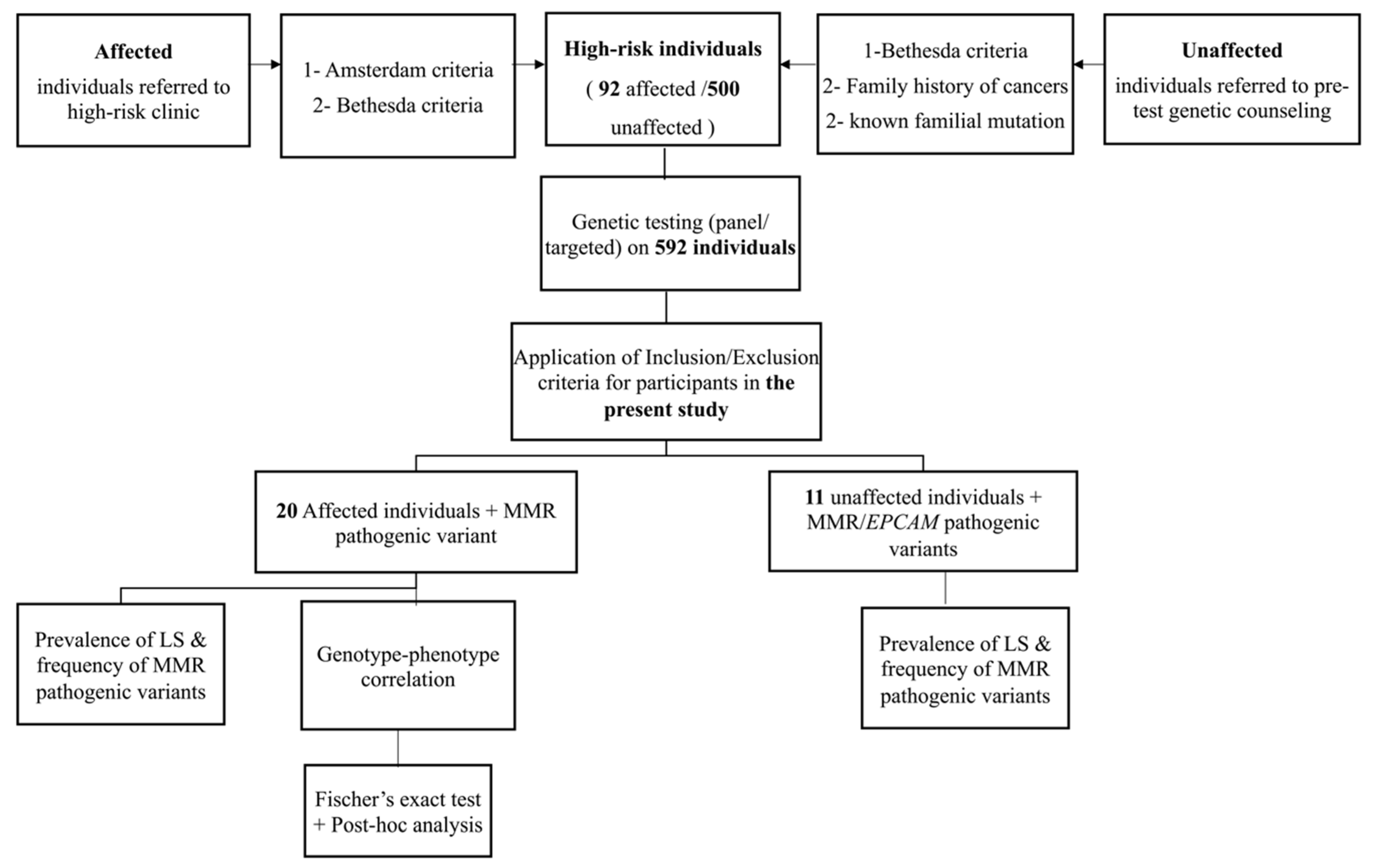
To our knowledge, this is the first study to investigate the prevalence and genotype-phenotype correlation of Lynch syndrome in the state of Qatar. We investigated the prevalence of LS, frequency of pathogenic variants in MMR/EPCAM genes, and genotype-phenotype correlation in 31 individuals; 20 were affected with colon cancer and 11 were unaffected.
The prevalence of Lynch Syndrome among our selected CRC patients was found to be 22% (20/92). Compared to the studies from the MENA (Middle East and North Africa) region, this prevalence is less than what has been found in Pakistani selected patients (34.5%) as they have followed a very stringent criteria of selection (at least three relatives affected with LS associated cancers, at least one of them is a first degree relative of the other two, at least 2 different generations with LS associated cancers, individuals with cancer diagnosed at an age younger than 50 years) [22]. This prevalence of LS found in our patients is however, higher than most of the other reported CRC prevalence in the remaining MENA countries. For instance, in the Kingdom of Saudi Arabia (KSA) the prevalence of LS in CRC patients was 7% [23] and in Iran, it was 5.5% [24]. The prevalence of LS in our CRC patients was also higher compared to the prevalence reported by a study in the United States of America (USA) conducted mainly in the white population of Ohio (72/450) 16% [25].
The prevalence of Lynch syndrome in unaffected high-risk individuals was 2.2% (11/500), this prevalence was lower than that of unaffected high-risk relatives of patients with Lynch syndrome from Columbus, Ohio (USA) (77%; 102/132). Such high prevalence in the Ohio study is due to the fact that it was calculated among mainly first degree relatives of patients with confirmed pathogenic variants in one of the MMR genes and thus the possibility of detecting a pathogenic variant in a first degree relative is 50%. However, this is not the case for the high-risk unaffected individuals in the current study as not all of them had an affected relative confirmed to have pathogenic variants in MMR due to the fact that most didn’t pursue testing which could explain the lower prevalence of Lynch syndrome detected in our study compared to the previous Ohio study. Our approach to evaluate the prevalence of LS in selected high-risk patients/individuals affected or unaffected gives more insights regarding the genetic testing approach mainly for unaffected individuals. In this regard, our findings demonstrate the benefit of panel genetic testing for healthy high-risk individuals based on their family history especially when the types of cancers overlap in multiple genetic syndromes even though no pathogenic variant has been confirmed in relatives. Following, panel genetic testing approach, about 2.2% of unaffected relatives could be positive for any MMR gene variant and their identification could reduce their cancer risk through early surveillance, prophylactic prevention and early detection of cancers [26].
Among the affected individuals, the most reported genes were MLH1 and MSH2 accounting for 90% of the pathogenic variants, which is in agreement with earlier studies from Saudi Arabia [27] the United States [28], Finland [29], Spain [30] and the results of the international Mismatch Repair Consortium (IMRC) [15] This high prevalence of MLH1 and MSH2 genes in our cohort could also be attributed to the fact that affected patients were referred based on Amsterdam and Bethesda criteria, as families fulfilling these 2 criteria are more likely to harbor pathogenic variants in MLH1 and MSH2 genes [31] which might have resulted in missing some patients with pathogenic variants MSH6 and PMS2 genes. For unaffected LS individuals however, the most reported gene was PMS2 accounting for 64% followed by MSH6 and EPCAM genes with each accounting for 18% of the pathogenic variants. This high frequency of PMS2 gene is due to the fact that all carriers were members of the same tribe. This finding suggests the presence of a potential tribal variant in PMS2 gene which has a great clinical benefit as it can facilitate the selection of most of the at-risk individuals belonging to the same tribe for pre-test counseling and early cancer surveillance and pre-implantation genetic diagnosis. Such interventions would prevent further transmission of the pathogenic variant to the future generations. Additionally, the difference in the distribution of the genes between affected and unaffected individuals was statistically significant (p < 0.005) after Bonferroni correction, and the fact that MLH1 and MSH2 genes were not reported in unaffected individuals could be attributed to the high penetrance of these 2 genes and their association with higher cancer risk compared to the remaining MMR and EPCAM genes which makes them less likely to be detected in unaffected individuals [32].
With regards to the type of variants in each gene, for affected patients, point mutations and frameshift variants were commonly reported in MLH1, MSH2, and MSH6 genes which goes in line with what has been reported in the Human Gene Mutation Database regarding the most common variant type in each gene (http://www.hgmd.cf.ac.uk/ac/all.php, accessed on 14 January 2022). For PMS2 gene, however, one point mutation and one novel large deletion (deletion encompassing exons 6 to 11) were reported (Table S1). A large deletion (deletion encompassing exons 6 to 11) was observed to be recurrent in 3 affected members of the same tribe in a homozygous state which is consistent with the diagnosis of Constitutional Mismatch Repair Deficiency Syndrome (CMMRD), a childhood-onset syndrome. Nevertheless, it is interesting to note the delay in onset of disease in these affected individuals (23, 25, and 28 years for patients C0021, C0022, and C0023 respectively) (Table S1). This late-onset could be explained by the fact that CMMRD, PMS2/MSH6 homozygous pathogenic variants are associated with a later onset phenotype compared to homozygous pathogenic variants in MLH1/MSH2 genes which result in more aggressive hematological malignancies during young childhood and are associated with a worse prognosis [33]. A potential explanation for this late age at onset could also be the partial compensation of absent PMS2 by MLH3, which can form a functional heterodimeric protein with MLH1 that has mismatch repair capacity [34]. However, the surveillance for CMMRD is similar for all affected individuals regardless of which MMR gene was involved [35].
For unaffected individuals, the same previous recurrent large deletion (deletion encompassing exons 6 to 11) in PMS2 gene has also been reported in a heterozygous state in 4 unaffected individuals belonging to the same tribe. Generally heterozygous pathogenic variants in PMS2 display an attenuated Lynch syndrome phenotype consisting of lower penetrance and a later age at onset [36]. For EPCAM gene, all pathogenic variants were also large deletions which is in agreement with the nature of the common variants in this gene reported in other studies [6]. The heterozygous deletion of the entire EPCAM gene reported in patient U004 (Table S2) and also previously reported in an individual with Lynch syndrome associated cancer [37], was reported of unknown significance on cancer risk because, it is known that deletions of 3′ region of EPCAM gene are associated with silencing of MSH2 gene through the transcriptional read-through [38]. However, in the deletion we report in this study, the entire EPCAM gene is deleted and as a result transcription and transcriptional read-through might not occur, thus MSH2 gene might not be affected which results in an unknown risk for Lynch Syndrome. However, it is considered pathogenic with respect to Congenital Tufting Enteropathy which is an autosomal recessive condition associated with biallelic pathogenic variants in the EPCAM gene [39]. Thereby, in the Qatari population where the consanguinity rate is high (54%) [40], the chances of serious autosomal recessive childhood-onset conditions such as CMMRD and Congenital Tufting enteropathy is increased especially with the presence of known Tribal variants. This suggests the necessity of implementing testing for targeted tribal variants in pre-marital screening and offering pre-implantation genetic testing for carrier parents to avoid the risk of autosomal recessive conditions associated with being homozygous for pathogenic variants in MMR/EPCAM genes [33,39].

4.2. Genotype-Phenotype Correlation in Affected LS Patients

There was no statistically significant association established between the type of cancer and mutated gene. One previous study found a lower expression of MLH1 protein in right sided colon cancer and a loss of MSH2 protein expression in the left sided colon cancers which might be due to germline mutations in the corresponding genes, however the association was not statistically significant [41].
Additionally, although it was expected that MLH1 and MSH2 genes would be associated with a younger age at onset as compared to MSH6 and PMS2 there was no statistically significant correlation (Table 5)
In line with what has been published in the literature [7,16], we did not find a statistically significant correlation between gender, ethnicity, age, tumor location, type of cancer, grade of cancer, lymph vascular, invasiveness, mucinous component, family history, histopathology, and tumor size with the type of mutated gene which might also be explained by our small sample size (Table 5).
It was expected that carriers of large deletions and frameshift variants would exhibit a more severe phenotype and an earlier age at onset compared to carriers of point mutations [42]. However, there was no statistical correlation between the type of variant and any of the clinicopathological parameters tested especially after Bonferroni correction for the association of CRC side with the type of mutated variant (Table 6). These findings are in agreement with the findings of a previous study on lynch syndrome patients from Spain where their correlation of the type of variant with clinicopathological variables did not yield any statistically significant results [7].
Immunohistochemistry testing was performed on tumors from all affected individuals with colon cancers as a first step (before the referral to the high-risk clinic) to test for the presence of MMR proteins and select individuals at high risk for LS. In our cohort, most of MLH1 pathogenic variants, (6/9) (66.7%) had a corresponding loss of MlH1 protein expression along with the loss of its heterodimer PMS2. The loss of MLH1 and PMS2 proteins on IHC is expected as MLH1/PMS2 tend to form heterodimers. Regarding MSH2, (7 variants resulted in a corresponding loss of MSH2 protein expression and its heterodimer MSH6 protein, the loss of MSH2 and MSH6 proteins on IHC is due to the formation of heterodimers between these 2 proteins. However, the loss of MLH1 protein could be explained by the low sensitivity associated with some antibodies used in the IHC technique [11]. On the other hand, one pathogenic frameshift variant detected in MSH6 gene resulted in intact MMR proteins expression. One of the limitations of IHC technique is that not all pathogenic MMR variants result in loss of immunoreactivity, for instance, frameshift and truncating variants can interfere with the protein function without altering the antigenic site of the protein [43], additionally, the interaction between MMR proteins could affect the sensitivity of immunohistochemistry in the detection of individual proteins [11].
Therefore, regardless of which protein was lost based on IHC, performing Panel testing is recommended for all individuals suspected to have Lynch syndrome [44] and not strictly meeting Bethesda or Amsterdam criteria. Clinicians should attentively take the personal and family history of the patient to be able to assess their eligibility for genetic testing. Evidently, the adoption of an effective screening program is challenging and is a topic of ongoing debate in the literature [27].

4.3. Assessment of Bethesda/Amsterdam Criteria

In the current study, 100% of the affected patients met either Bethesda 18/20 (91.3%) or Amsterdam criteria 2/20 (8.7%).
For unaffected individuals, only 2/11 (18.18%) met Bethesda guidelines, while 3/11 (27.27%) were tested using Panel genetic testing due to an extensive family history of cancers not meeting Amsterdam/Bethesda criteria (Table 8). These findings are suggestive of the high efficacy of Amsterdam/Bethesda criteria for affected high-risk individuals and not for unaffected high-risk individuals, thus Panel genetic testing is important for these unaffected individuals with family histories of multiple cancers even if they do not meet Amsterdam/Bethesda guidelines. Though the sensitivity and specificity of these two criteria could not be confirmed due to the lack of data regarding Lynch Syndrome negative patients. However, the fact that individuals meeting Bethesda guidelines were more than those meeting Amsterdam criteria is due to the flexibility of Bethesda compared to Amsterdam criteria [9].
However, a previous study by Syngal S. et al., (2000) [9] on 70 families with suspected hereditary colorectal cancer, found out that Amsterdam criteria for HNPCC were neither sufficiently sensitive nor specific for use as a sole criterion for determining which families should undergo testing. Additionally, they found out that the application of Bethesda guidelines was a less strict approach and was associated with a higher sensitivity [9].

4.4. Limitations

We believe that the small sample size of both affected and unaffected individuals considered in the present study could have had a significant effect on the statistical analysis and the generalizability of the study findings. Many Lynch syndrome patients could have been missed due to not being correctly identified and referred to genetic testing which would have increased the prevalence of LS. Furthermore, genetic testing for Lynch syndrome is currently not available in Qatar and many non-Qatari “eligible” patients could not afford it, in addition to the fact that some eligible patients who can afford testing but refuse to pursue it due to personal/social reasons especially unaffected patients who often express fear and anxiety of testing. Finally, the lack of clinicopathological data about the Lynch syndrome negative patients have limited a better assessment of the specificity/sensitivity of Amsterdam and Bethesda criteria

5. Conclusions

This study highlights the prevalence and genotype-phenotype correlation of LS in high-risk cohort of patients in the State of Qatar. The prevalence of LS among affected CRC patients and unaffected patients is 22% and 2.2% respectively which indicates an increased prevalence in the high-risk populations and draws recommendations on diagnostic and predictive genetic testing and personalize surveillance and management programs.
Among unaffected individuals, a recurrent PMS2 pathogenic variant (deletion encompassing exons 6 to 11) was reported in several related individuals suggesting a tribal effect. This is indeed a significant finding which impacts our recommendations for at risk families from this high risk tribe, in which the rate of consanguineous marriage is high, who may benefit from preventative measures, risk-reducing strategies and premarital, prenatal and reproductive genetic counseling to reduce the risk of Lynch syndrome and serious autosomal recessive childhood-onset conditions such as CMMRD in these individuals.
Among affected individuals, MLH1 and MSH2 genes were highly frequent, thus drawing conclusions on the importance of establishing germline testing for all MMR’s especially MLH1 and MSH2 genes testing given its high prevalence and which will aide the existing somatic immunohistochemical staining (IHC) testing. Although IHC can aid germline testing, panel germline testing for all MMR’s is recommended to be available for all individuals suspected to have Lynch syndrome regardless of which protein is deficient and not strictly meeting Bethesda or Amsterdam criteria.
This is the first study of its kind in Qatar which serves as the foundation for more studies in the future concerning the epidemiology and genetics of Lynch syndrome in Qatar which can be captured at a bigger scale cohort of patients and at a population level.

Supplementary Materials

The following supporting information can be downloaded at: https://www.mdpi.com/article/10.3390/genes13112176/s1, Table S1: Clinicopathological and genetic test results of affected individuals, Table S2: Genetic test results of Unaffected high-risk individuals with Lynch Syndrome, Figure S1: HMC Institutional Review Board approval letter, Figure S2: Qatar University Institutional Review Board Exemption Letter.

Author Contributions

M.S. performed data analysis, interpretation, drafting and revising the manuscript. H.K.-E.-k. and R.A.-S. contributed to conception and design of the study, as well as in reviewing the manuscript. S.B.A.-B. and M.A.-S. contributed in reviewing the manuscript. S.B.A.-B., R.A.-S., H.H.H. and H.A.M. contributed in data acquisition. All authors have read and agreed to the published version of the manuscript.

Funding

The publication of this article was funded by Qatar University.

Institutional Review Board Statement

The study was approved by the Medical Research Center (MRC) in Hamad Medical Corporation (HMC) (and was granted research ethics exemption by Qatar University with the Research Ethics approval Number (QU-IRB 13831-E/20) (Figure S1) and (Figure S2).

Informed Consent Statement

Not applicable.

Data Availability Statement

Not applicable.

Acknowledgments

We are thankful to [Nawal Bakheet Salem Abdallah], [Sitti Apsa Kusasi] and [Nema Abdou] for their contribution in data collection.

Conflicts of Interest

The authors declare no conflict of interest.

References

  1. Stratton, M.R.; Campbell, P.J.; Futreal, P.A. The cancer genome. Nature 2009, 458, 719–724. [Google Scholar] [CrossRef] [Green Version]
  2. Moreira, L.; Balaguer, F.; Lindor, N.; de la Chapelle, A.; Hampel, H.; Aaltonen, L.A.; Hopper, J.L.; Le Marchand, L.; Gallinger, S.; Newcomb, P.A.; et al. Identification of Lynch syndrome among patients with colorectal cancer. JAMA 2012, 308, 1555–1565. [Google Scholar] [CrossRef]
  3. Ryan, N.A.J.; Glaire, M.A.; Blake, D.; Cabrera-Dandy, M.; Evans, D.G.; Crosbie, E.J. The proportion of endometrial cancers associated with Lynch syndrome: A systematic review of the literature and meta-analysis. Genet. Med. Off. J. Am. Coll. Med. Genet. 2019, 21, 2167–2180. [Google Scholar] [CrossRef] [Green Version]
  4. Nakamura, K.; Banno, K.; Yanokura, M.; Iida, M.; Adachi, M.; Masuda, K.; Ueki, A.; Kobayashi, Y.; Nomura, H.; Hirasawa, A.; et al. Features of ovarian cancer in Lynch syndrome (Review). Mol. Clin. Oncol. 2014, 2, 909–916. [Google Scholar] [CrossRef] [Green Version]
  5. Vasen, H.F.; Blanco, I.; Aktan-Collan, K.; Gopie, J.P.; Alonso, A.; Aretz, S.; Bernstein, I.; Bertario, L.; Burn, J.; Capella, G.; et al. Revised guidelines for the clinical management of Lynch syndrome (HNPCC): Recommendations by a group of European experts. Gut 2013, 62, 812–823. [Google Scholar] [CrossRef]
  6. Tutlewska, K.; Lubinski, J.; Kurzawski, G. Germline deletions in the EPCAM gene as a cause of Lynch syndrome—Literature review. Hered. Cancer Clin. Pract. 2013, 11, 9. [Google Scholar] [CrossRef] [Green Version]
  7. Pérez-Cabornero, L.; Infante, M.; Velasco, E.; Lastra, E.; Miner, C.; Durán, M. Genotype-phenotype correlation in MMR mutation-positive families with Lynch syndrome. Int. J. Color. Dis. 2013, 28, 1195–1201. [Google Scholar] [CrossRef]
  8. Vasen, H.F.; Watson, P.; Mecklin, J.P.; Lynch, H.T. New clinical criteria for hereditary nonpolyposis colorectal cancer (HNPCC, Lynch syndrome) proposed by the International Collaborative group on HNPCC. Gastroenterology 1999, 116, 1453–1456. [Google Scholar] [CrossRef]
  9. Syngal, S.; Fox, E.A.; Eng, C.; Kolodner, R.D.; Garber, J.E. Sensitivity and specificity of clinical criteria for hereditary non-polyposis colorectal cancer associated mutations in MSH2 and MLH1. J. Med. Genet. 2000, 37, 641–645. [Google Scholar] [CrossRef] [Green Version]
  10. Hampel, H.; Frankel, W.L.; Martin, E.; Arnold, M.; Khanduja, K.; Kuebler, P.; Clendenning, M.; Sotamaa, K.; Prior, T.; Westman, J.A.; et al. Feasibility of screening for Lynch syndrome among patients with colorectal cancer. J. Clin. Oncol. Off. J. Am. Soc. Clin. Oncol. 2008, 26, 5783–5788. [Google Scholar] [CrossRef] [Green Version]
  11. Acharya, S.; Wilson, T.; Gradia, S.; Kane, M.F.; Guerrette, S.; Marsischky, G.T.; Kolodner, R.; Fishel, R. hMSH2 forms specific mispair-binding complexes with hMSH3 and hMSH6. Proc. Natl. Acad. Sci. USA 1996, 93, 13629–13634. [Google Scholar] [CrossRef] [Green Version]
  12. Berends, M.J.; Wu, Y.; Sijmons, R.H.; Mensink, R.G.; van der Sluis, T.; Hordijk-Hos, J.M.; de Vries, E.G.; Hollema, H.; Karrenbeld, A.; Buys, C.H.; et al. Molecular and clinical characteristics of MSH6 variants: An analysis of 25 index carriers of a germline variant. Am. J. Hum. Genet. 2002, 70, 26–37. [Google Scholar] [CrossRef] [Green Version]
  13. Hendriks, Y.; Franken, P.; Dierssen, J.W.; De Leeuw, W.; Wijnen, J.; Dreef, E.; Tops, C.; Breuning, M.; Bröcker-Vriends, A.; Vasen, H.; et al. Conventional and tissue microarray immunohistochemical expression analysis of mismatch repair in hereditary colorectal tumors. Am. J. Pathol. 2003, 162, 469–477. [Google Scholar] [CrossRef] [Green Version]
  14. Young, J.; Simms, L.A.; Biden, K.G.; Wynter, C.; Whitehall, V.; Karamatic, R.; George, J.; Goldblatt, J.; Walpole, I.; Robin, S.A.; et al. Features of colorectal cancers with high-level microsatellite instability occurring in familial and sporadic settings: Parallel pathways of tumorigenesis. Am. J. Pathol. 2001, 159, 2107–2116. [Google Scholar] [CrossRef]
  15. Jenkins, M.A.; Reece, J.C.; Win, A.K. The International Mismatch Repair Consortium. In Hereditary Colorectal Cancer: Genetic Basis and Clinical Implications; Valle, L., Gruber, S.B., Capellá, G., Eds.; Springer International Publishing: Cham, Switzerland, 2018; pp. 479–495. [Google Scholar]
  16. Goecke, T.; Schulmann, K.; Engel, C.; Holinski-Feder, E.; Pagenstecher, C.; Schackert, H.K.; Kloor, M.; Kunstmann, E.; Vogelsang, H.; Keller, G.; et al. Genotype-phenotype comparison of German MLH1 and MSH2 mutation carriers clinically affected with Lynch syndrome: A report by the German HNPCC Consortium. J. Clin. Oncol. Off. J. Am. Soc. Clin. Oncol. 2006, 24, 4285–4292. [Google Scholar] [CrossRef] [Green Version]
  17. Talseth-Palmer, B.A.; McPhillips, M.; Groombridge, C.; Spigelman, A.; Scott, R.J. MSH6 and PMS2 mutation positive Australian Lynch syndrome families: Novel mutations, cancer risk and age of diagnosis of colorectal cancer. Hered. Cancer Clin. Pract. 2010, 8, 5. [Google Scholar] [CrossRef] [Green Version]
  18. Lynch, H.T.; Boland, C.R.; Gong, G.; Shaw, T.G.; Lynch, P.M.; Fodde, R.; Lynch, J.F.; de la Chapelle, A. Phenotypic and genotypic heterogeneity in the Lynch syndrome: Diagnostic, surveillance and management implications. Eur. J. Hum. Genet. EJHG 2006, 14, 390–402. [Google Scholar] [CrossRef]
  19. Liu, F.; Yang, L.; Zhou, X.; Sheng, W.; Cai, S.; Liu, L.; Nan, P.; Xu, Y. Clinicopathological and genetic features of Chinese hereditary nonpolyposis colorectal cancer (HNPCC). Med. Oncol. 2014, 31, 223. [Google Scholar] [CrossRef] [Green Version]
  20. Al-Dewik, N.; Al-Mureikhi, M.; Shahbeck, N.; Ali, R.; Al-Mesaifri, F.; Mahmoud, L.; Othman, A.; AlMulla, M.; Sulaiman, R.A.; Musa, S.; et al. Clinical genetics and genomic medicine in Qatar. Mol. Genet. Genom. Med. 2018, 6, 702–712. [Google Scholar] [CrossRef]
  21. Al-Bader, S.B.; Alsulaiman, R.; Bugrein, H.; Ben Omran, T.; Abbaszadeh, F.; Bakheet, N.; Apsa Kusasi, S.; Abdou, N.; Solomon, B.D.; Ghazouani, H. Cancer genetics program: Follow-up on clinical genetics and genomic medicine in Qatar. Mol. Genet. Genom. Med. 2018, 6, 865–872. [Google Scholar] [CrossRef]
  22. Rashid, M.U.; Naeemi, H.; Muhammad, N.; Loya, A.; Lubiński, J.; Jakubowska, A.; Yusuf, M.A. Prevalence and spectrum of MLH1, MSH2, and MSH6 pathogenic germline variants in Pakistani colorectal cancer patients. Hered. Cancer Clin. Pract. 2019, 17, 29. [Google Scholar] [CrossRef] [Green Version]
  23. Alqahtani, M.; Edwards, C.; Buzzacott, N.; Carpenter, K.; Alsaleh, K.; Alsheikh, A.; Abozeed, W.; Mashhour, M.; Almousa, A.; Housawi, Y.; et al. Screening for Lynch syndrome in young Saudi colorectal cancer patients using microsatellite instability testing and next generation sequencing. Fam. Cancer 2018, 17, 197–203. [Google Scholar] [CrossRef]
  24. Esmaeilzadeh, A.; Akhavan Rezayat, K.; Masannen Mozaffari, H.; Bahari, A.; Ghanaei, O.; Ganji, A.; Mokhtarifar, A.; Rahmani Khorram, M.; Goshayeshi, L. Prevalence of hereditary nonpolyposis colorectal cancer in patients with colorectal cancer in Iran: A systematic review. Rev. Clin. Med. 2016, 3, 98–104. [Google Scholar] [CrossRef]
  25. Pearlman, R.; Frankel, W.L.; Swanson, B.; Zhao, W.; Yilmaz, A.; Miller, K.; Bacher, J.; Bigley, C.; Nelsen, L.; Goodfellow, P.J.; et al. Prevalence and Spectrum of Germline Cancer Susceptibility Gene Mutations Among Patients with Early-Onset Colorectal Cancer. JAMA Oncol. 2017, 3, 464–471. [Google Scholar] [CrossRef]
  26. Vasen, H.F.; Möslein, G.; Alonso, A.; Bernstein, I.; Bertario, L.; Blanco, I.; Burn, J.; Capella, G.; Engel, C.; Frayling, I.; et al. Guidelines for the clinical management of Lynch syndrome (hereditary non-polyposis cancer). J. Med. Genet. 2007, 44, 353–362. [Google Scholar] [CrossRef]
  27. Siraj, A.K.; Prabhakaran, S.; Bavi, P.; Bu, R.; Beg, S.; Hazmi, M.A.; Al-Rasheed, M.; Al-Assiri, M.; Sairafi, R.; Al-Dayel, F.; et al. Prevalence of Lynch syndrome in a Middle Eastern population with colorectal cancer. Cancer 2015, 121, 1762–1771. [Google Scholar] [CrossRef]
  28. Hampel, H.; Frankel, W.L.; Martin, E.; Arnold, M.; Khanduja, K.; Kuebler, P.; Nakagawa, H.; Sotamaa, K.; Prior, T.W.; Westman, J.; et al. Screening for the Lynch syndrome (hereditary nonpolyposis colorectal cancer). N. Engl. J. Med. 2005, 352, 1851–1860. [Google Scholar] [CrossRef] [Green Version]
  29. Mecklin, J.-P.; Järvinen, H.J.; Hakkiluoto, A.; Hallikas, H.; Hiltunen, K.-M.; Härkönen, N.; Kellokumpu, I.; Laitinen, S.; Ovaska, J.; Tulikoura, J.; et al. Frequency of hereditary nonpolyposis colorectal cancer. Dis. Colon Rectum 1995, 38, 588–593. [Google Scholar] [CrossRef]
  30. Pérez-Carbonell, L.; Ruiz-Ponte, C.; Guarinos, C.; Alenda, C.; Payá, A.; Brea, A.; Egoavil, C.M.; Castillejo, A.; Barberá, V.M.; Bessa, X.; et al. Comparison between universal molecular screening for Lynch syndrome and revised Bethesda guidelines in a large population-based cohort of patients with colorectal cancer. Gut 2012, 61, 865–872. [Google Scholar] [CrossRef]
  31. Sjursen, W.; Haukanes, B.I.; Grindedal, E.M.; Aarset, H.; Stormorken, A.; Engebretsen, L.F.; Jonsrud, C.; Bjørnevoll, I.; Andresen, P.A.; Ariansen, S.; et al. Current clinical criteria for Lynch syndrome are not sensitive enough to identify MSH6 mutation carriers. J. Med. Genet. 2010, 47, 579–585. [Google Scholar] [CrossRef]
  32. Dominguez-Valentin, M.; Sampson, J.R.; Seppälä, T.T.; Ten Broeke, S.W.; Plazzer, J.P.; Nakken, S.; Engel, C.; Aretz, S.; Jenkins, M.A.; Sunde, L.; et al. Cancer risks by gene, age, and gender in 6350 carriers of pathogenic mismatch repair variants: Findings from the Prospective Lynch Syndrome Database. Genet. Med. Off. J. Am. Coll. Med. Genet. 2020, 22, 15–25. [Google Scholar] [CrossRef] [Green Version]
  33. Ramchander, N.C.; Ryan, N.A.; Crosbie, E.J.; Evans, D.G. Homozygous germ-line mutation of the PMS2 mismatch repair gene: A unique case report of constitutional mismatch repair deficiency (CMMRD). BMC Med. Genet. 2017, 18, 40. [Google Scholar] [CrossRef] [Green Version]
  34. Li, G.M. Mechanisms and functions of DNA mismatch repair. Cell Res. 2008, 18, 85–98. [Google Scholar] [CrossRef] [Green Version]
  35. Durno, C.; Boland, C.R.; Cohen, S.; Dominitz, J.A.; Giardiello, F.M.; Johnson, D.A.; Kaltenbach, T.; Levin, T.R.; Lieberman, D.; Robertson, D.J.; et al. Recommendations on Surveillance and Management of Biallelic Mismatch Repair Deficiency (BMMRD) Syndrome: A Consensus Statement by the US Multi-Society Task Force on Colorectal Cancer. Am. J. Gastroenterol. 2017, 112, 682–690. [Google Scholar] [CrossRef]
  36. Hendriks, Y.M.; Jagmohan-Changur, S.; van der Klift, H.M.; Morreau, H.; van Puijenbroek, M.; Tops, C.; van Os, T.; Wagner, A.; Ausems, M.G.; Gomez, E.; et al. Heterozygous mutations in PMS2 cause hereditary nonpolyposis colorectal carcinoma (Lynch syndrome). Gastroenterology 2006, 130, 312–322. [Google Scholar] [CrossRef]
  37. Yurgelun, M.B.; Allen, B.; Kaldate, R.R.; Bowles, K.R.; Judkins, T.; Kaushik, P.; Roa, B.B.; Wenstrup, R.J.; Hartman, A.R.; Syngal, S. Identification of a Variety of Mutations in Cancer Predisposition Genes in Patients With Suspected Lynch Syndrome. Gastroenterology 2015, 149, 604–613. [Google Scholar] [CrossRef] [Green Version]
  38. Ligtenberg, M.J.; Kuiper, R.P.; Geurts van Kessel, A.; Hoogerbrugge, N. EPCAM deletion carriers constitute a unique subgroup of Lynch syndrome patients. Fam. Cancer 2013, 12, 169–174. [Google Scholar] [CrossRef]
  39. Pathak, S.J.; Mueller, J.L.; Okamoto, K.; Das, B.; Hertecant, J.; Greenhalgh, L.; Cole, T.; Pinsk, V.; Yerushalmi, B.; Gurkan, O.E.; et al. EPCAM mutation update: Variants associated with congenital tufting enteropathy and Lynch syndrome. Hum. Mutat. 2019, 40, 142–161. [Google Scholar] [CrossRef] [Green Version]
  40. Bener, A.; El Ayoubi, H.R.; Chouchane, L.; Ali, A.I.; Al-Kubaisi, A.; Al-Sulaiti, H.; Teebi, A.S. Impact of consanguinity on cancer in a highly endogamous population. Asian Pac. J. Cancer Prev. APJCP 2009, 10, 35–40. [Google Scholar]
  41. Salem, M.E.; Battaglin, F.; Goldberg, R.M.; Puccini, A.; Shields, A.F.; Arguello, D.; Korn, W.M.; Marshall, J.L.; Grothey, A.; Lenz, H.J. Molecular Analyses of Left- and Right-Sided Tumors in Adolescents and Young Adults with Colorectal Cancer. Oncologist 2020, 25, 404–413. [Google Scholar] [CrossRef] [Green Version]
  42. Baudhuin, L.M.; Mai, M.; French, A.J.; Kruckeberg, K.E.; Swanson, R.L.; Winters, J.L.; Courteau, L.K.; Thibodeau, S.N. Analysis of hMLH1 and hMSH2 gene dosage alterations in hereditary nonpolyposis colorectal cancer patients by novel methods. J. Mol. Diagn. JMD 2005, 7, 226–235. [Google Scholar] [CrossRef]
  43. Bai, H.; Wang, R.; Cheng, W.; Shen, Y.; Li, H.; Xia, W.; Ding, Z.; Zhang, Y. Evaluation of Concordance Between Deficient Mismatch Repair and Microsatellite Instability Testing and Their Association with Clinicopathological Features in Colorectal Cancer. Cancer Manag. Res. 2020, 12, 2863–2873. [Google Scholar] [CrossRef]
  44. NCCN. Genetic/Familial High Risk Assessment: Colorectal. Available online: https://www.nccn.org/professionals/physician_gls/pdf/genetics_colon.pdf (accessed on 18 February 2021).
Figure 1. Flow chart of the study design. CRC: Colorectal Cancer. LS: Lynch Syndrome, MMR: Mismatch repair genes.
Genes 13 02176 g001
Table 1. Affected LS patients’ demographics.
Patient CharacteristicsN (%)
Age
Mean48.2
Range(24–67)
Standard Deviation±11.2
Age at diagnosis
<50 y14 (70%)
>50 y6 (30%)
Total20
Family History
Yes15 (75%)
No5 (25%)
Total20
Gender
Male14 (70%)
Female6 (30%)
Total20
Type of Cancer
Right colon cancer8 (40%)
Left colon cancer12 (60%)
Total20
Stage
Stage 16 (30%)
Stage 212 (60%)
Stage 31 (5%)
Stage 41 (5%)
Total20
Table 2. Distribution of MMR genes variants in Affected and Unaffected individuals.
Mutated Genep-Value
MLH1MSH2MSH6PMS2EPCAM<0.001 a
Unaffected individuals002 (18.2%)7 (63.6%)2 (18.2%)
Affected patients9 (45%)9 (45%)1 (5%)1 (5%)0
p-value b0.0090.0090.2300.000 c0.045
Total99382
a = p-value before Bonferroni correction, using Fischer’s exact test. b = p-values after post hoc analysis. c = statistically significant p-values after Bonferroni correction (0.005).
Table 3. Types of variants per MMR gene in Affected individuals.
Type of VariantMutated Gene
MLH1MSH2MSH6PMS2
Frameshift mutations4 (44.4%)2 (22.2%)1 (100%)0
Point mutations1 (11.1%)5 (55.6%)01 (100%)
Large deletions2 (22.2%)000
Intronic mutations2 (22.2%)2 (22.2%)00
Total9911
Table 4. Types of variants per MMR gene in unaffected individuals.
Type of VariantMutated Gene
MSH6PMS2EPCAM
Large deletions05 (71.42%)2 (100%)
Point mutations01 (14.28%)0
Intronic mutations1 (50%)1 (14.28%)0
Frameshift mutations1 (50%)00
Total272
Table 5. Genotype- phenotype correlation with the mutated gene, LV: Lympho-vascular invasion, CRC: colorectal cancer.
MLH1MSH2MSH6PMS2p Value
Gender
Male7 (77.8%)7 (77.8%)000.159
Female2 (22.2%)2 (22.2%)1 (100%)1 (100%)
Total9911
Ethnicity
Qatari001 (100%)1 (100%)0.641
Egyptian2 (22.2%)2 (22.2%)00
Sudanese1 (11.1%)1 (11.1%)00
Bangladeshi2 (22.2%)000
British1 (11.1%)000
Filipino1 (11.1%)1 (11.1%)00
Indian1 (11.1%)1 (11.1%)00
Nepalese1 (11.1%)1 (11.1%)00
Sri lankan 01 (11.1%)00
Pakistani01 (11.1%)00
Palestinian01 (11.1%)00
Total9911
Age at diagnosis
<50 y8 (88.9%)5 (55.6%)01 (100%)0.159
>50 y1 (11.1%)4 (44.4%)1 (100%)0
Total9911
Tumor Location
Ascending Colon3 (33.3%)3 (33.3%)000.883
Descending Colon2 (22.2%)000
Rectum1 (11.1%)2 (22.2%)00
Sigmoid Colon2 (22.2%)2 (22.2%)1 (100%)1 (100%)
Cecum1 (11.1%)1 (11.1%)00
Transverse colon01 (11.1%)00
Total9911
CRC side
Right Colon Cancer4 (44.4%)4 (44.4%)000.687
Left Colon Cancer5 (55.6%)5 (55.6%)1 (100%)1 (100%)
Total9911
Grade
Stage 13 (33.3%)2 (22.2%)1 (100%)00.783
Stage 25 (55.6%)6 (66.7%)01 (100%)
Stage 301 (11.1%)00
Stage 41 (11.1%)000
Total9911
LV invasion (yes/no)
Yes1 (14.3%)0000.712
No6 (85.7%)7 (100%)1 (100%)1 (100%)
Total7711
Mucinous component (yes/no)
yes2 (22.2%)2 (50%)1 (100%)-0.238
No7 (77.8%)2 (50%)0-
Total941-
Family History
Yes6 (66.7%)7 (77.8%)1 (100%)1 (100%)0.792
No3 (33.3%)2 (22.2%)00
Total9911
Histopathology
Well Differentiated adenocarcinoma1 (14.3%)2 (25%)-00.775
Moderately differentiated adenocarcinoma6 (85.7%)5 (62.5%)-1 (100%)
Poorly Differentiated Adenocarcinoma01 (12.5%)-0
Total78-1
Tumor Size
<5 cm4 (57.1%)4 (57.1%)000.542
>5 cm3 (42.9%)3 (42.9%)1 (100%)0
Total7710
Table 6. Genotype- phenotype correlation with the type of the variant, LV: Lympho-vascular invasion, Invasive: Invasive cancer based on pathology.
Large DeletionsIntronic MutationsFrameshiftsPoint Mutationsp-Value
Gender
Male1 (50%)4 (100%)5 (71.4%)4 (57.1%)0.448
Female1 (50%)02 (28.6%)3 (42.9%)
total2477
Age at diagnosis
<50 y2 (100%)3 (75%)5 (71.4%)4 (57.1%)0.691
>50 y01 (25%)2 (28.6%)3 (42.9%)
total2477
Ethnicity
Qatari001 (14.3%)1 (14.3%)0.644
Egyptian01 (25%)3 (42.9%)0
Sudanese1 (50%)001 (14.3%)
Bangladeshi01 (25%)1 (14.3%)0
British001 (14.3%)0
Filipino1 (50%)001 (14.3%)
Indian01 (25%)01 (14.3%)
Nepalese01 (25%)1 (14.3%)0
Sri lankan0001 (14.3%)
Pakistani0001 (14.3%)
Palestinian0001 (14.3%)
Total2477
Tumor Location
Ascending colon04 (100%)1 (14.3%)1 (14.3%)0.110
Descending colon1 (50%)01 (14.3%)0
Rectum002 (28.6%)1 (14.3%)
Sigmoid colon1 (50%)03 (42.9%)2 (28.6%)
Cecum0002 (28.6%)
Transverse colon0001 (14.3%)
Total 2477
CRC Side
Right Colon Cancer04 (100%)1 (14.3%)3 (42.9%)0.026 a
Left Colon Cancer2 (100%)06 (85.7%)4 (57.1%)
p-value b0.230.010.090.84
Total2477
Grade
Stage 101 (25%)5 (71.4%)00.088
Stage 22 (100%)2 (50%)2 (28.6%)6 (85.7%)
Stage 30001 (14.3%)
Stage 401 (25%)00
Total2477
LV invasion (yes/no)
Yes001 (16.7%)00.620
No1 (100%)3 (100%)5 (83.3%)6 (100%)
Total1366
Invasive (yes/no)
Yes1 (50%)1 (33.3%)3 (60%)2 (50%)0.912
No1 (50%)2 (66.7%)2 (40%)2 (50%)
Total2354
Mucinous component (yes/no)
yes002 (40%)3 (75%)0.138
No2 (100%)3 (100%)3 (60%)1 (25%)
Total2354
Family History
Yes2 (100%)2 (50%)6 (85.7%)5 (71.4%)0.480
No02 (50%)1 (14.3%)2 (28.6%)
Total2477
Histopathology
Well Differentiated adenocarcinoma01 (33.3%)1 (25%)1 (14.3%)0.883
Moderately differentiated adenocarcinoma2 (100%)2 (66.7%)3 (75%)5 (71.4%)
Poorly Differentiated Adenocarcinoma0001 (14.3%)
Total2347
Tumor Size
<5 cm02 (66.7%)3 (50%)3 (60%)0.688
>5 cm1 (100%)1 (33.3%)3 (50%)2 (40%)
Total1365
a = p-value before Bonferroni correction, using Fischer’s exact test. b = p-values after post hoc analysis. Significant p-value after Bonferroni correction is <0.006.
Table 7. MMR genes (MLH1, MSH2, and MSH6) with corresponding protein loss by IHC.
MMR Genes/ProteinsMLH1MSH2MSH6
MLH1-PMS2 proteins loss6 (66.7%)00
MLH1-PMS2-MSH6 proteins loss1 (11.1%)00
PMS2 protein loss1 (11.1%)00
MLH1-MSH2 proteins loss01 (11.11%)0
MSH2 protein loss01 (11.11%)0
MSH2-MSH6 Protein loss07 (77.8%)0
Intact MMR proteins 001 (100%)
Unknown status1 (11.1%)00
Total991
Table 8. Summary of criteria of selection of Affected and unaffected individuals.
CriteriaAffectedUnaffected
Amsterdam2 (8.7%)0
Bethesda18 (91.3%)2 (18.18%)
Familial mutation05 (45.45%)
Through Panel testing03 (27.27%)
As a secondary finding (through WES)01 (9.09%)
Total2011
Publisher’s Note: MDPI stays neutral with regard to jurisdictional claims in published maps and institutional affiliations.

