Article

Cloning of Toll-like Receptor 3 Gene from Schizothorax prenanti (SpTLR3), and Expressions of Seven SpTLRs and SpMyD88 after Lipopolysaccharide Induction

1
Shaanxi Key Laboratory of Qinling Ecological Security, Shaanxi Institute of Zoology, Xi’an 710032, China
2
Key Laboratory for Hydrobiology in Liaoning Province, College of Fisheries and Life Science, Dalian Ocean University, Dalian 116023, China
3
Hanzhong Nanzheng District Aquatic Product Workstation, Hanzhong 723100, China
*
Author to whom correspondence should be addressed.
Genes 2022, 13(10), 1862; https://doi.org/10.3390/genes13101862
Submission received: 20 September 2022 / Revised: 9 October 2022 / Accepted: 13 October 2022 / Published: 15 October 2022
(This article belongs to the Section Animal Genetics and Genomics)

Abstract

:
Toll-like receptor 3 (SpTLR3) from Schizothorax prenanti (S. prenanti) was cloned and identified, and the tissue distribution of the SpTLR3 gene was examined in this study. Moreover, the relative mRNA expression levels of myeloid differentiation factor 88 gene (SpMyD88) and seven TLR genes (SpTLR2, SpTLR3, SpTLR4, SpTLR18, SpTLR22-1, SpTLR22-2 and SpTLR22-3) from S. prenanti after lipopolysaccharide (LPS) challenge were analyzed through quantitative real-time polymerase chain reaction (qRT-PCR). The full length of SpTLR3 gene is 3097 bp, and complete coding sequence (CDS) is 2715 bp, which encodes 904 amino acids. The SpTLR3 amino acid sequence shared 43.94–100% identity with TLR3 sequences from other vertebrates; SpTLR3 was expressed in all eight tissues examined; and the highest level appeared in the liver, which was significantly higher than in all other tissues (p < 0.05), followed by the levels in the heart and muscles. LPS significantly up-regulated all eight genes in the S. prenanti tissues at 12 or 24 h (p < 0.05). Compared with the PBS control group, no significant transcripts changes were found in SpTLR2 or SpTLR3 at 12 h after LPS induction, but they were significantly up-regulated at 24 h (p < 0.001). The most abundant transcripts were found in the head kidney SpTLR22 genes after 24 h LPS induction, with high to low levels, which were SpTLR22-1 (564-fold), SpTLR22-3 (508-fold) and SpTLR22-2 (351-fold). Among these eight genes, the expression level of SpTLR4 was the least up-regulated. Overall, SpTLR4 in the head kidney was involved in the antibacterial immune response earlier, and the level was increased at 12 h with extreme significance after LPS stimulation (p < 0.001), while the other seven genes were the most significantly up-regulated at 24 h post injection. Taken together, the results suggest that SpMyD88, SpTLR2, SpTLR3, SpTLR4, SpTLR18, SpTLR22-1, SpTLR22-2 and SpTLR22-3 participate in an innate immune response stimulated by LPS, and the response intensity of the genes was organ-specific, with differing kinetics. Our findings will contribute to a more complete understanding of the roles of these TLR genes in antibacterial immunity.

1. Introduction

The vertebrate immune system includes the innate immune system and the adaptive immune system. In mammals, the adaptive immune system plays a major role in the eventual elimination of pathogens. However, the difference is that fish belong to poikilotherm, and body temperature is not suitable for the development of acquired immune function. Therefore, the innate immune system plays a “leading role” in fish immunity [1]. Similar to mammals, the innate immunity of fish is mainly due to a series of evolutionarily highly conserved pattern recognition receptors (PRRs). The PRRs recognize pathogen-associated molecular patterns (PAMPs), which are conserved on the surface of microorganisms but not present in the host body. The invasion signal is transmitted to the whole host immune system, and the immune signal is transmitted to the cell, thus inducing a disease-resistant immune response [2,3]. Toll-like receptors (TLRs) were first identified in Drosophila melanogaster by Anderson et al. in 1985 [4]. Subsequently, human TLR4 was the first toll-receptor discovered in mammals and was thought to be homologous to TLR1 in D. melanogaster [5,6]. During the same period, TLRs have been gradually discovered in different vertebrates, and the first IL-1R/TLR superfamily member in fish was discovered by Sangrador-Vegas et al. in 2000 [7]. To date, 13 TLRs have been identified in mammals and classified into six TLR subfamilies, namely, TLR1, TLR3, TLR4, TLR5, TLR7 and TLR11, and at least 22 TLRs have been cloned and identified in teleost fish (TLR1-4, 5M, 5S, 7-9, 13-14, and 18-28), which also belong to the six TLR subfamilies [8], some of which are teleost-specific TLRs, such as TLR18-28. As the main PRRs, these TLRs play an important role in identifying microbial pathogens that infect fish. Together with interferon regulatory factors (IRFs), myeloid differentiation factor 88 (MyD88) and other factors in the immune signaling pathway, TLRs participate in the recognition process of bacteria, viruses, parasites and most other pathogenic microorganisms [9,10,11,12]. The fish species are complex, and teleostei experience one whole genome duplication, which greatly increases the diversity of genotypes and the complexity of gene functions [13]. The TLR system for sensing microorganisms has similarities with mammal, and also important and diverse in teleostei, especially for teleost-specific TLRs [12]. The study of fish TLRs is of great significance for understanding the immune system of lower vertebrates.
During TLR signal transduction, adapter molecules are recruited by the Toll/IL-1 receptor (TIR) domain of TLR, leading to the signal entry into different pathways. The TLR signaling pathways can be broadly divided into two categories, including MyD88-dependent and MyD88-independent pathways. In the MyD88-dependent pathway, as an important adaptor protein, MyD88 is recruited by TLRs as the first signal protein and plays a highly important role in TLR signal transduction [14,15,16].
In mammals, TLR3 receives dsRNA stimulatory signals to mediate antiviral immune responses, and fish TLR3 has a similar function [17]. Studies showed that Poly (I:C) up-regulated the expression of TLR3 gene in rainbow trout (Oncorhynchus mykiss) [18] and golden pompano (Trachinotus ovatus) [19], respectively, especially in immune organs. Su et al. [20] reported that TLR3 transcription levels in spleen and skin of grass carp (Ctenopharyngodon idella) were significantly increased after Grass Carp Reovirus (GCRV) infection. Notably, the fish TLR3 subfamily has only one member, TLR3. Similar to mammalian, fish TLR3 mainly recognizes viral nucleic acids, but it has also been shown to be involved in anti-bacterial immunity [17,21]. In addition, SpTLR2 and SpTLR18 involved in this study belong to the TLR1 subfamily, SpTLR4 belongs to the TLR4 subfamily, and SpTLR22s belongs to the TLR11 subfamily [8]. Lipopolysaccharide (LPS) is a major component of the outer membranes of Gram-negative bacteria, which can induce a cascade of immune stimulation and toxic pathophysiological activities in the body [22]. Whether LPS induces changes in S. prenanti TLR3 expression is unknown. Therefore, the SpTLR3 was cloned for the first time in this study, and the specific domains of the TLR3 family were analyzed. The secondary structure composition and the 3D structural models of SpTLR3 protein were predicted. Moreover, the expression patterns of different immune organs in response to the LPS stimulation of the seven SpTLRs and SpMyD88 gene were analyzed using qRT- PCR.
S.prenanti is a unique cold-water fish in China, which belongs to Cyprinidae, the subfamily of schizothorinae, and the genus Schizothorax, and is a rare and high-quality fish in producing areas. In Hanzhong, Shaanxi province, S. prenanti is known locally as “Yang-fish”, whereas in Ya’an, Sichuan province, S. prenanti and the other special economic fish S. davidi are collectively known as “Ya fish” [23,24,25]. Due to intensive feeding, S. prenanti is susceptible to bacterial infection, such as Aeromonas hydrophila [26] and Streptococcus agalactiae [27], which seriously impedes the healthy development of the Ya-fish aquaculture. To date, three SpTLR22 molecules (named SpTLR22-1, SpTLR22-2 and SpTLR22-3) have been cloned and identified [14], as well as two SpTLR5 family members (named SpTLR5-1 and SpTLR5-2) [28] and SpTLR25 [29]. We treated S. prenanti with LPS intraperitoneal injection in this study, and examined the relative mRNA expression of MyD88, TLR2, TLR3, TLR4, TLR18, TLR22-1, TLR22-2 and TLR22-3 genes from S. prenanti in different immune organs (hepatopantras, head kidney, hindgut and spleen) at different infection times, which will help to further clarify the roles of SpTLRs and SpMyD88 genes involved in the antibacterial immunity mechanisms of fish.

2. Materials and Methods

2.1. Animal Treatment

We obtained healthy cultured S. prenanti (109.3 ± 27.1 g) in November 2020 from a professional breeding cooperative (Qunfu Yang-Fish, Hanzhong, China), which is a Chinese national aquatic germplasm resources breeding farm and commercial breeding aqua-farm. The fish were maintained in glass tanks with a size of 60 × 40 × 30 cm3 and with aerated water at a temperature of 19 ± 1 °C; The tank filter was cleaned and about a quarter of the aerated tap water was replaced daily. The fish were fed with a commercial feed (Floating compound feed with crude protein ≥ 32%, crude fat ≥ 3%) at a rate of 2% of their body weight twice daily.
The fish were anesthetized with eugenol at a concentration of 80 mg/L for 3 min before dissected. The heart, liver, head kidney, hindgut, muscle, intraperitoneal fat, spleen and gill were collected from eugenol anaesthetized fish to detect the tissue distribution of SpTLR3 mRNA in unstressed conditions. To understand the expression of SpMyD88, SpTLR2, SpTLR3, SpTLR4, SpTLR18, and SpTLR22s (SpTLR22-1, SpTLR22-2, SpTLR22-3) in response to LPS stimulation, the fish were challenged with an intraperitoneal injection of LPS (10 mg/kg), and the control group was injected with phosphate-buffered saline (PBS) at the same volume. The liver, head kidney, hindgut and spleen were collected from four S.prenanti of PBS–injection and LPS treatment at 12 and 24 h, respectively. The dissected tissues and preserved in liquid nitrogen for RNA extraction.

2.2. RNA Extraction and cDNA Synthesis

Tissue total RNA was extracted using the TaKaRa MiniBEST Universal RNA Extraction Kit (TaKaRa, Dalian, China). The total RNA concentration and purity were determined by RNA electrophoresis and the optical density absorption ratio (A260/280) in the Nanodrop One spectrophotometer (Thermo Fisher Scientific, Wilmington, NC, USA). First-stand cDNA for SpTLR3 partial sequence amplification was synthesized using the RevertAid First Strand cDNA Synthesis Kit (Thermo Scientific, Vilnius, Lithuania), according to the method recommended by the manufacturer. The SpTLR3 5′-rapid amplification of cDNA ends (RACE) cDNA was prepared using the SMARTer® RACE 5′/3′ kit (TaKaRa, Dalian, China).

2.3. Full Length Cloning of the SpTLR3

According to the transcriptome sequencing of S. prenanti, the specific SpTLR3 partial sequence primers were designed (Table 1, TLR3-F and TLR3-R) and synthetized by Tsingke Biotechnology Co., Ltd. (Xi’an, China). Spleen cDNA was used as a template for SpTLR3 amplification, with Primerstar® Max DNA polymerase (TaKaRa, Dalian, China). The PCR program was as follows: 35 cycles at 98 °C for 10 s, 55 °C for 15 s, and 72 °C for 40 s. The PCR products were ligated into a lineareized pRACE vector (TaKaRa, Dalian, China), and transformed into Stellar Competent Cells (TaKaRa, Dalian, China). The positive bacteria clones were sequenced. According to the obtained SpTLR3 partial sequence, the primer for 5′-RACE was designed (Table 1, GSP) and synthetized by Tsingke Biotechnology Co., Ltd. (Xi’an, China). Spleen cDNA was used as a template for SpTLR3 amplification, with SeqAmp DNA polymerase (TaKaRa, Dalian, China). The touchdown PCR program was as follows: 5 cylces at 94 °C for 30 s, 72 °C for 3 min; 5 cycles at 94 °C for 30 s, 70 °C for 30 s, and 72 °C for 3 min; 25 cycles at 94 °C for 30 s, 68 °C for 30 s, and 72 °C for 3 min. The PCR products were ligated into a lineareized pRACE vector (TaKaRa, Dalian, China), and transformed into Stellar Competent Cells (TaKaRa, Dalian, China). The positive bacteria clones were sequenced.

2.4. Sequence Analysis

The SpTLR3 complete CDS was identified using the ORF Finder (http://www.ncbi.nlm.nih.gov/gorf/gorf.html, accessed on 2 September 2022). The isoelectric point and molecular weight were predicted (https://web.expasy.org/compute_pi/, accessed on 2 September 2022). The signal peptide, Leucine-rich repeats (LRRs), transmembrane domain and TIR domain of SpTLR3 was predicted by the SMART programs (http://smart.embl-heidelberg.de/, accessed on 7 September 2022). The secondary structure composition of SpTLR3 protein was predicted (https://www.novopro.cn/tools/secondary-structure-prediction.html, accessed on 2 September 2022). The three-dimensional structure of SpTLR3 protein was predicted (https://swissmodel.expasy.org/, accessed on 2 September 2022). Multiple sequence alignments were performed using the Clustal X2.1 [30]. A phylogenetic tree of different vertebrates TLR3 based on amino acid sequences was constructed by the neighbor-joining method using MEGA 4.0 software [31].

2.5. Tissue Distribution of TLR3 mRNA in Unstressed Conditions

The measured tissues in control group included heart, liver, spleen, head kidney, muscle, intraperitoneal fat, hindgut and gill. Total RNA was extracted and cDNA was synthesized, as described previously (“RNA extraction and cDNA synthesis”). The qRT-PCR analysis was performed using the applied Biosystems Step One Plus (Life Technologies, Foster City, CA, USA). The gene-specific primers are listed in Table 1. S. prenanti-specific β-actin primers were used to normalize the cDNA quantity for each tissue sample. Quantification of SpTLR3 and β-actin was performed in triplicate on all samples using FastStart Essential DNA Green Master (Roche Diagnostics, Risch-Rotkreuz, Switzerland), according to the manufacturer’s instructions. The qRT-PCR data were calculated according to the 2−ΔΔCT method [32].

2.6. Detection of the Expression Patterns Induced by LPS

For the LPS challenge, 8 fish were injected intraperitoneally from the base of the pectoral fin with LPS (Sigma, L2880). The dose of injection was 10 mg/kg LPS per fish. Another 8 fish were injected intraperitoneally PBS at the same amount, as a control. 4 fish samples were taken at 12 and 24 h after LPS injection respectively. In generally, head kidney, liver, and hindgut of fish are regarded as immune organs and central to the immune responses [33], the three tissues plus spleen, another vital immune organ were collected from each fish and used to isolate total RNA. To detect the expression of SpMyD88, SpTLR2, SpTLR 3, SpTLR4, SpTLR18, SpTLR22-1, SpTLR22-2 and SpTLR22-3 changes after LPS induction, total RNA extraction, cDNA synthesis, and qRT-PCR were performed, as described previously (Tissue distribution of SpTLR3 mRNA in unstressed conditions). S. prenanti-specific β-actin primers were used to normalize the cDNA quantity for each tissue sample. The quantitative primers of SpTLR2, SpTLR4, SpMyD88 were designed according to the transcriptome sequencing of S. prenanti. The quantitative primers of SpTLR22-1, SpTLR22-2 and SpTLR22-3 were designed according to the gene sequences unloaded to GenBank (GenBank accesion no. MN082612, MN082613, MN082614). Quantitative primers of SpTLR18 refer to the article published by Li et al. [34]. The quantitative primers used are listed in Table 1.

2.7. Statistical Analysis

SPSS 22.0 and Graphpad Prism 5.0 software was used for data analysis and histogram, respectively. The mRNA expression levels were analyzed by using One-way ANOVA method. All data are presented as the mean ± standard error (n = 4), and the statistically significant differences between PBS control and LPS treatment groups at each time point are expressed with asterisks, * p < 0.05, ** p < 0.01, *** p < 0.001 vs. corresponding control group at the time points.

3. Results

3.1. Identification, Structural, and Phylogenetic Analysis of SpTLR3

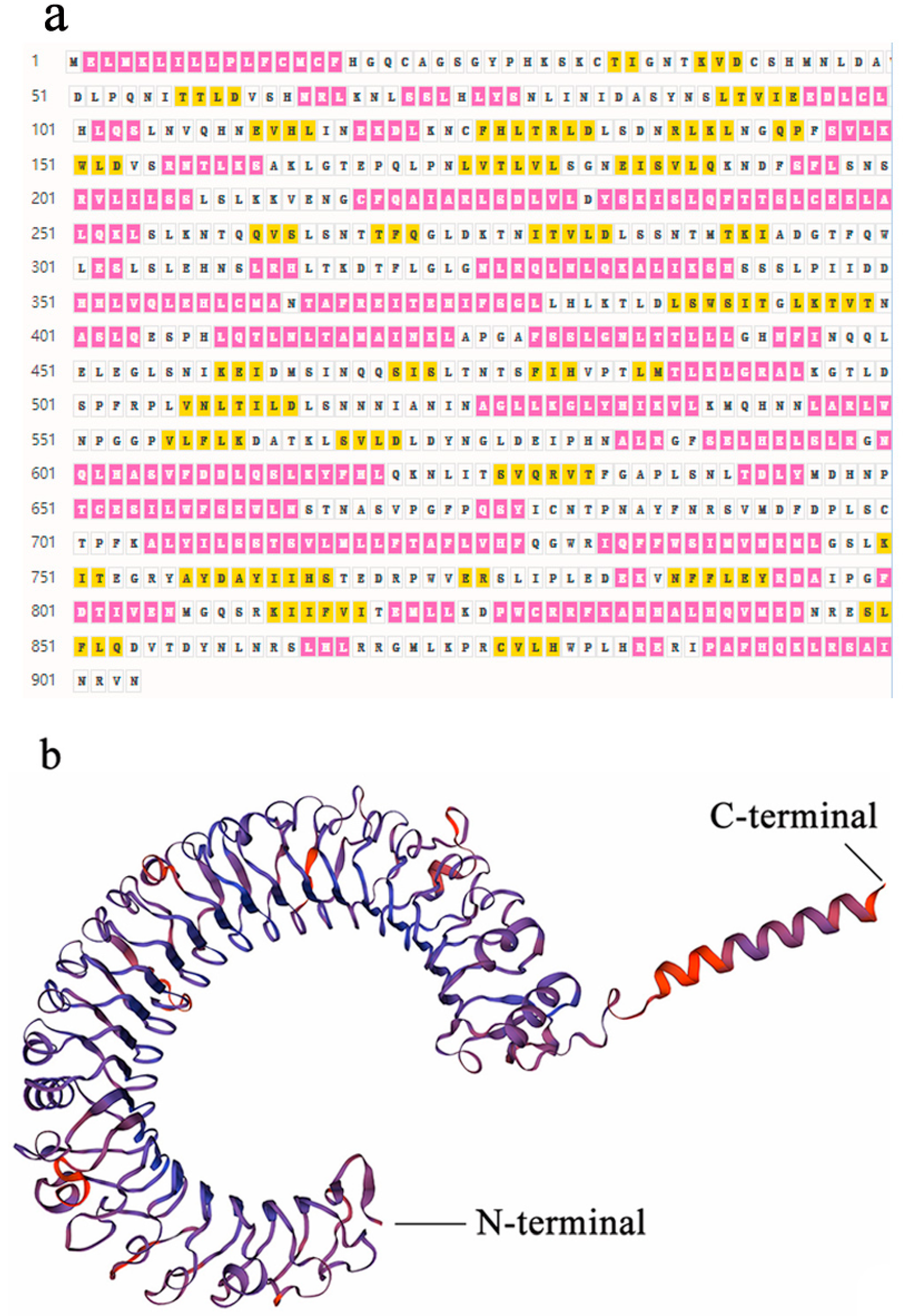
The full length of SpTLR3 gene is 3097 bp (GenBank accesion no. OP589305), including 312 bp of 5’ untranslated region (UTR), 2715 bp of complete CDS (Figure 1) and 70 bp 3’ UTR. The predicted open reading frame (ORF) encoded a protein of 904 amino acids, including one signal peptide, seventeen LRRs, one transmembrane domain and one TIR domain (Figure 2a). The calculated molecular mass and theoretical isoelectric point of the SpTLR3 is 102.29 kDa and 7.08, respectively. Like the TIR domain of other fish TLR3, SpTLR3 has three conserved regions: Box 1, Box 2 and Box 3 (Figure 2b), indicating that the TIR domain is relatively conserved throughout TLR3 in fish. Figure 3a shows the distribution of strands, helixes, and coils, and Figure 3b shows the predicted 3D structure of the SpTLR3 protein. The SpTLR3 amino acid sequence was the most similar to those of other fish and was the closest to snow trout (Schizothorax richardsonii) TLR3, with 100% identity. We analyzed the phylogeny of the SpTLR3 amino acid sequences to determine the evolutionary relationships between SpTLR3 and TLR3 from other vertebrates, based on sequences in the GenBank database (Figure 4). The results revealed a high amino acid sequence identity between SpTLR3 and the TLR3 of Cyprinids. Moreover, SpTLR3 and the TLR3 of S. richardsonii have the same amino acid sequence identity as the two fish belong to the same genus Schizothorax (Teleostei: Cyprinidae).

3.2. Tissue Distribution of SpTLR3 Expression in S. prenanti

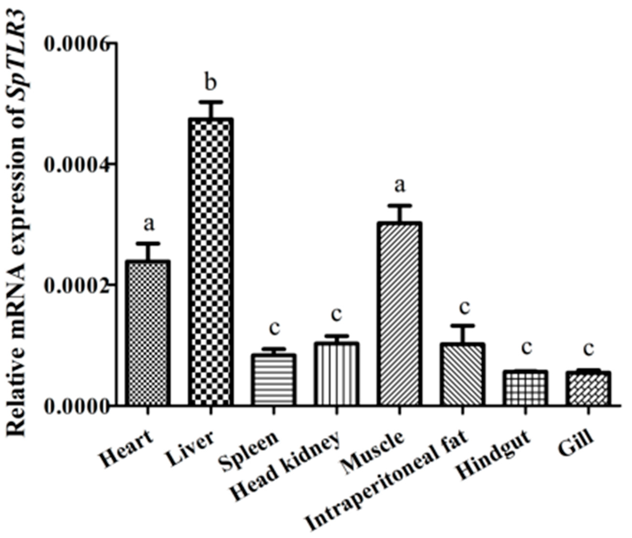
We quantified SpTLR3 mRNA expression in the heart, liver, spleen, head kidney, muscle, intraperitoneal fat, hindgut and gill tissue from four fish using qRT-PCR to determine SpTLR3 transcript expression. The loading control for normalization was β-actin. Figure 5 shows the ubiquitous, but variable expression of SpTLR3 transcripts in all eight tissues. The highest expression level of SpTLR3 appeared in the liver, and the level was significantly higher than in all other tissues (p < 0.05). Furthermore, the expression of SpTLR3 in the heart and muscle was more pronounced than that in the spleen, head kidney, intraperitoneal fat, hindgut and gill (p < 0.05). The expression SpTLR3 did not significantly differ in the spleen, head kidney, intraperitoneal fat, hindgut and gill tissues.

3.3. Expression of SpMyD88 and SpTLRs after Injection of LPS

To determine the change in MyD88, TLR2, TLR3, TLR4, TLR18, TLR22-1, TLR22-2 and TLR22-3 from S. prenanti after LPS infection at 12 and 24 h, the mRNA levels of the genes in the liver, head kidney, spleen and hindgut were quantified using qRT-PCR.

3.3.1. Expression of SpMyD88 after Injection of LPS

The expression of SpMyD88 in the four tissues examined showed no significant change after the treatment of S. prenanti with LPS 12 h later (Figure 6a). However, at 24 h of LPS infection, the gene was significantly up-regulated in all four tissues, especially in the head kidney and spleen tissues, which showed an exponentially up-regulated trend compared with the PBS control group and the 12 h infection group (p < 0.001). Moreover, in the liver and hindgut, SpMyD88 of the 24 h infection group was significantly higher than that in the PBS control group and 12 h infection group (p < 0.01).

3.3.2. Expression of SpTLR2 after Injection with LPS

At 12 h after LPS infection, SpTLR2 expression in all four tissues examined showed no significant change, which was similar to the expression pattern of SpMyD88. However, at 24 h after LPS challenge, the expression level of SpTLR2 in the liver was significantly increased (p < 0.05), whereas the other three tissues were extremely significant up-regulated (p < 0.001). In particular, the expression level of the gene was extremely high in the head kidney, followed by the hindgut and the liver, showing that the expression levels of the SpTLR2 gene in the tissues after infection were extremely different (Figure 6b).

3.3.3. Expression Levels of SpTLR3 after Injection with LPS

After LPS treatment, the expression pattern of SpTLR3 in both the 12 h infection group and the 24 h infection group was similar to the pattern of SpTLR2; that is, compared with the PBS control group, the expression level of SpTLR3 in the LPS 12 h infection group showed no significant change in the four tissues, but the level in the 24 h infection group was significantly higher than that in the PBS and 12 h infection group (p < 0.001), and the highest expression level was found in the head kidney, followed by the spleen and hindgut (Figure 6c).

3.3.4. Expression of SpTLR4 after Injection with LPS

After LPS infection, SpTLR4 expression was different from that of the above genes. Compared with the PBS group, the SpTLR4 of the liver and hindgut in the 12 h infection group showed no significant change, while that of the head kidney tissue was significantly up-regulated (p < 0.001), and the high expression level persisted until 24 h after infection. The levels of the 24 h infection group were as follows: the gene expression of SpTLR4 in the liver was exponentially up-regulated and more strongly significant than that of the PBS group and 12 h infection group (p < 0.001). The level in the head kidney remained high in the 24 h infection group, which was significantly higher than that in the PBS group (p < 0.001). The level in the hindgut was significantly higher than that in the PBS control group and 12 h infection group (p < 0.05). The difference is that the expression level in the spleen was downregulated, with a highly significant difference in both the 12 h and 24 h infection groups compared to in the PBS control group (p < 0.05) (Figure 6d).

3.3.5. Expression of SpTLR18 after Injection with LPS

The temporal expression pattern of SpTLR18 was similar to that of SpTLR2, SpTLR3 and SpMyD88; that is, no significant difference was found between the 12 h infection group and the PBS group. The SpTLR18 gene was overexpressed in the liver, head kidney and hindgut, with significant levels of p < 0.001. The level of SpTLR18 in the spleen was relatively low, but showed a significance level of p < 0.01 (Figure 6e).

3.3.6. Expression of SpTLR22s after Injection with LPS

As shown in Figure 6f–h, the temporal expression patterns of SpTLR22 genes (SpTLR22-1, SpTLR22-2 and SpTLR22-3) in the four organs examined after LPS stimulation were consistent. After 12 h, in the fish challenged with LPS, no significant changes were found compared with the PBS-injection group; however, the levels of SpTLR22-3 in the spleen were significantly up-regulated (p < 0.05). In contrast, the highest transcripts were found in the liver post-24 h LPS injection for all three SpTLR22s, followed by the levels of SpTLR22-1 and SpTLR22-2 in the hindgut, spleen and liver, and then by the levels of SpTLR22-3 in the liver, hindgut and spleen. Furthermore, the expression levels of all SpTLR22s in the four organs spiked after 24 h LPS stimulation, and the level of SpTLR22-1 in liver was significantly higher than that in the PBS control and 12 h groups (p < 0.01), whereas the levels of other SpTLR22s in different organs were much higher than those in the PBS and 12 h groups (p < 0.001).

4. Discussion

In this study, we cloned the SpTLR3 gene for the first time, The predicted SpTLR3 amino acid sequence included the conserved typical structure of TLR protein family: 17 LRR domain, Transmembrane domain and TIR domain. The previous studies have demonstrated that the LRR domain was related to recognizing the pathogen components and the number of LRR domain differs in different animals [17,35] For instance, 16 LRR domains were found in Japanese flounder [17]. In the present study, there were three conserved regions: Box 1, Box 2 and Box 3, indicating that the TIR domain is relatively conserved throughout TLR3 in fish. Box1 and Box2 play an important role in mediating receptor binding to downstream signaling molecules, while Box3 directly controls the expression and localization of receptor molecules [36]. Multiple sequence alignment showed that the SpTLR3 protein is moderately conserved, and that its amino acid sequence is highly similar to that of Cyprinids. The results of the SpTLR3 amino acid sequencing and phylogenetic tree analysis revealed that S. prenanti is more closely related to S. richardsonii. This was consistent with previous findings of SpTLR5-1 [28], whereas SpTLR5-2 [28] and SpTLR22s (SpTLR22-1, SpTLR22-2, and SpTLR22-3) [37] were more closely related to common carp (Cyprinus carpio). Additionally, S. prenanti apelin receptor (APJ) [38] and protein nucleobindin-2A (NUCB2A) [39] were more closely related to zebrafish (Danio rerio) and goldfish (Carassius auratus).
MyD88 is an important transmitter of the TLR signaling pathway, which is highly conserved and plays an indispensable role in both TLR signaling pathway and (Interleukin-1) IL-1 receptor family. It goes through a series of signal transductions after the MyD88-depended pathway is activated by TLRs recognition, that lead to transcription factors such as Nuclear factor-κB (NF-κB), activator protein-1 (AP-1) and interferon regulatory factors (IRFs), eventually triggering inflammatory responses [14,40]. To date, few studies on fish MyD88 have been conducted. Takano et al. [41] found that the number of MyD88-positive cells in the kidney and spleen increased three days after Japanese flounder (Paralichthys olivaceus) was infected with Edwardsiella tarda. The yellow drum (Nibea albiflora) was treated with Pseudomonas plecoglossicida, then, the NaMyD88 in the head kidney was rapidly up-regulated and remained significantly higher than that in the PBS control group from 2 to 24 h after infection, indicating that the gene plays an important role in the innate immunity of the fish [42]. These results are similar to that of SpMyD88 in our study. Moreover, when the yellow croaker (Pseudosciaena crocea) was infected with Vibrio parahaemolyticus, the expression level of spleen PcMyD88 was significantly higher at 24 and 48 h than that of the control group, and the highest expression level appeared at 48 h post-injection [43]. It can be concluded that MyD88 plays a crucial role in the innate immunity of fishes, whereas the time of immune response varies with different pathogens in different species.
To date, a total of 13 TLRs (TLR1-13) have been identified in mammals and classified into 6 TLR subfamilies, named TLR1, TLR3, TLR4, TLR5, TLR7 and TLR11 subfamilies [44], and there are 22 TLRs (TLR1-4, 5M, 5S, 7-9, 13-14, and 18-28) in teleost fish have been identified, which also belong to the 6 TLR subfamilies [8]. The classification of the 6 fish TLR subfamilies are as follows: TLR1 (TLR1, 2, 14, 18, 24, 25, 27 and 28), TLR3 (TLR3), TLR4 (TLR4), TLR5 (TLR5M and TLR5S), TLR7 (TLR7, 8 and 9), and TLR11 (TLR13, 19, 20, 21, 22, 23 and 26) subfamily [8,45,46]. In mammals, TLR2 typically functions as a dimer, recognizing different components of bacteria [45]. Like mammals, the TLR2 of channel catfish (Ictalurus punctatus) can also act as a dimer with TLR1 [47], and the TLR2 of orange-spotted grouper (Epinephelus coioides) participates in the anti-LPS or Poly(I:C) immune response [48]. Our study confirmed that the SpTLR2 participates in antibacterial immunity, and the best monitoring organ is the head kidney. TLR18 is a fish-specific TLR. To date, the TLR18 of zebrafish (D. rerio) [49], channel catfish (I. punctatus) [45], Atlantic salmon (Salmo salar) [50], grass carp [51], yellow catfish (Pelteobagrus fulvidraco) [52] and common carp (Cyprinus carpio L.) [53] have been identified. These reports and the present study have together proved that the TLR18 gene plays an important role in the innate immune responses of teleost fish. However, the TLR18 expression levels in different tissues vary from fish to fish. In this study, the highest expression abundance of SpTLR18 after LPS stimulation was found in the liver.
TLR3 is the only member of the fish TLR3 subfamily [17], mainly involved in mediating antiviral immunity [18,19,20]. However, fish TLR3 can also senses bacterial stimulation. It was reported that TLR3 mRNA levels in the liver, kidney and spleen of channel catfish were significantly up-regulated at different time points after infection with Edwardsiella ictaluri [21]. However, there was no significant change in the TLR3 of zebrafish infected with Mycobacterium marinum [48], although the expression of TLR3 was significantly down-regulated in Rainbow trout infected with Yersinia reuteri [54]. In the current study, the expression level of SpTLR3 increased exponentially in the head kidney, spleen and hindgut after 24 h LPS injection. In conclusion, the response of fish TLR3 to PAMPs may depend on many factors, such as time, host species and pathogens.
Similar to TLR3, TLR4 is also the only member of the TLR4 subfamily of fish, and in mammals, TLR4 is the direct receptor of bacterial LPS. The biggest difference between TLR signaling pathway in fish and that in mammals lies in the TLR4-mediated signaling pathway [54]. Moreover, TLR4 is only present in a few fish [49]. Zebrafish TLR4 has been demonstrated not only to fail to recognize LPS challenge in vitro, but also have no responsive to heat-killed Escherichia coli and Legionella pneumophila [55,56]. Moreover, TLR4 negatively regulated NF-κB activation; therefore, the LPS recognition system in fish is quite different from that in mammals [46,56]. In contrast, it was reported that both LPS and A. hydrophila (a Gram-negative fish pathogen) infection could significantly up-regulate the expression of TLR4 from rohu (Labeo rohita) [57]. Our study confirmed the following three conclusions. First, the TLR4 in S. prenanti is present. Second, the SpTLR4 is involved in anti-LPS immunity, which is consistent with the conclusion of Samanta et al. [57], but different from that of Sullivan et al. [55] and Sepulcre et al. [56]. Finally, the liver is the most sensitive organ for SpTLR4 detection. The expression level of SpTLR4 was the least up-regulated among the eight genes examined, which was in fact the most different SpTLRs in our study. It was the only one that up-regulated at 12 h in head kidney. Those most different results verified the specificity of fish TLR4, and the immune interpretation need further study.
In general, the expression levels of the SpTLR22s were the highest in the seven induced SpTLRs and SpMyD88 examined in the present study. TLR22, which was also one of the TLRs specific to fish, belonging to the TLR11 subfamily, first discovered in goldfish in 2003, The TLR22 of goldfish macrophages was highly up-regulated by LPS, heat-killed Aeromonas salmonicida, and live Mycobacterium chelonei [58]. Subsequently, TLR22 has been cloned and reported in nearly 20 fish species, such as zebrafish [49], Japanese flounder [59], Atlantic salmon [60], yellowtail (Seriola lalandi) [61], and common carp [62]. To date, most of the studies reporting on TLR22 function have focused on bacteria, viruses, parasite infection and PAMP stimulation. It has been found that TLR22 is a multi-functional immune receptor that participates in almost all the defensive immune responses of pathogenic microorganisms. The results of SpTLR22s in this study also confirmed these conclusions. Moreover, two subtypes of TLR22 (namely, TLR22-1 and TLR22-2) were first discovered in Rainbow trout in 2007, which have very similar functions and were called ‘twin’ TLRs, and the expression of the two OmTLR22s in peripheral leukocytes was significantly up-regulated after stimulation by A. salmonicida [63]. Subsequently, three copies of the TLR22 gene (namely, TLR22-1, TLR22-2 and TLR22-3) were found in Atlantic salmon (GenBank accession numbers: AM233509, FM206383 and BT045774), and even up to twelve TLR22 gene copies (namely, TLR22a~l) have been found in Atlantic cod (Gadus morhua) [64]. Du et al. reported that both SpTLR22-1 and SpTLR22-3 in the head kidney and spleen respond to the stimulation of LPS and A. hydrophila, but not SpTLR22-2, in the two immune tissues [14]; this was different from our findings, as we found that the SpTLR22-2 levels in the four organs were significantly up-regulated at 24 h after LPS stimulation. Moreover, SpTLR22-1 in the head kidney was significantly up-regulated at 12 h after LPS challenge, but no pronounced significance was found at the 24 h time point [14], showing a different temporal expression pattern to our findings, in addition to the expression in the spleen. Notably, the temporal expressions of SpTLR22s in our study showed similar kinetics to one another in different immune organs, indicating that the functions of the subtypes of SpTLR22s are highly similar. Our results may indicate that both the SpTLR22s jointly mediate the recognition of LPS and participate in the immune response.

5. Conclusions

In this study, SpTLR3 was cloned and identified for the first time, and the secondary structure composition, the 3D structural models of the SpTLR3 protein, as well as the binding site were predicted. Furthermore, the SpTLR3 gene was expressed in all the tissues examined and mainly expressed in immune-related tissues. Phylogenetic analysis showed that the SpTLR3 protein is most closely related to TLR3 from snow trout. Multiple sequence alignment showed that the SpTLR3 is moderately conserved. Among the seven immune tissues we examined, the distribution of SpTLR3 was the most abundant in the liver, followed by the kidney and spleen. Moreover, the LPS organ specifically induced SpTLR2, SpTLR3, SpTLR4, SpTLR18, SpTLR22s (SpTLR22-1, SpTLR22-2, and SpTLR22-3) and SpMyD88 at 12 or 24 h, which are involved in antibacterial immunity. It is worth noting that the SpTLR22s were the most sensitive to LPS induction.

Author Contributions

Conceptualization, methodology, writing—original draft, J.H. and J.Z.; data curation, software, K.Z.; validation, writing—review & editing J.H. and C.F.; Project administration, Resources, W.L.; project administration, funding acquisition, visualization, Q.W.; All authors have read and agreed to the published version of the manuscript.

Funding

This research was funded by the Department of Science and Technology of Shaanxi Province, China, grant number (2018NY-108).

Institutional Review Board Statement

The animal study protocol was approved by the Animal Ethics Committee of the Shaanxi Institute of Zoology (protocol code: L22D003A54, date of approval: 4 August 2022) for studies involving animals.

Informed Consent Statement

Not applicable.

Data Availability Statement

Publicly available datasets were analyzed in this study. The rest of the data presented in this study are available on request from the corresponding author.

Acknowledgments

Our sincere thanks go to Ling Chen and Xingquan Xiong for their help in providing the experimental fish.

Conflicts of Interest

The authors declare no conflict of interest.

References

  1. Alcorn, S.W.; Murra, A.L.; Pascho, R.J. Effects of rearing temperature on immune functions in sockeye salmon (Oncorhynchus nerka). Fish Shellfish Immunol. 2002, 12, 303–334. [Google Scholar] [CrossRef] [PubMed]
  2. Pietretti, D.; Wiegertjes, G.F. Ligand specificities of Toll-like receptors in fish: Indications from infection studies. Dev. Comp. Immunol. 2014, 43, 205–222. [Google Scholar] [CrossRef] [PubMed]
  3. Hansen, J.D.; Vojtech, L.N.; Laing, K.J. Sensing disease and danger: A survey of vertebrate PRRs and their origins. Dev. Comp. Immunol. 2011, 35, 886–897. [Google Scholar] [CrossRef]
  4. Anderson, K.V.; Jurgens, G.; Nusslein-Volhard, C. Establishment of dorsal-ventral polarity in the Drosophila embryo: Genetic studies on the role of the Toll gene product. Cell 1985, 42, 779–789. [Google Scholar] [CrossRef]
  5. Medzhitov, R.; Preston-Hurlburt, P.; Janeway, C.A. A human homologue of the Drosophila Toll protein signals activation of adaptive immunity. Nature 1997, 388, 394–397. [Google Scholar] [CrossRef]
  6. Rock, F.L.; Hardiman, G.; Timans, J.C.; Kastelein, R.A.; Bazan, J.F. A family of human receptors structurally related to Drosophila Toll. Proc. Natl. Acad. Sci. USA 1998, 95, 588–593. [Google Scholar] [CrossRef] [PubMed] [Green Version]
  7. Sangrador-Vegas, A.; Martin, S.A.; O’Dea, P.G.; Smith, T.J. Cloning and characterization of the rainbow trout (Oncorhynchus mykiss) type II interleukin-1 receptor cDNA. Eur. J. Biochem. 2000, 267, 7031–7037. [Google Scholar] [CrossRef] [PubMed]
  8. Zhang, J.; Kong, X.H.; Zhou, C.J.; Li, L.; Nie, G.X.; Li, X.J. Toll-like receptor recognition of bacteria in fish: Ligand specificity and signal pathways. Fish Shellfish Immunol. 2014, 41, 380–388. [Google Scholar] [CrossRef] [PubMed]
  9. Wang, J.L.; Zhang, Z.; Liu, J.; Li, F.; Chang, F.; Fu, H.; Zhao, J.; Yin, D.L. Structural characterization and evolutionary analysis of fish-specific TLR27. Fish Shellfish Immunol. 2015, 45, 940–945. [Google Scholar] [CrossRef] [PubMed]
  10. Wang, Y.J.; Li, J.R.; Han, J.J.; Shu, C.; Xu, T.J. Identification and characteristic analysis of TLR28: A novel member of the TLR1 family in teleost. Dev. Comp. Immunol. 2016, 62, 102–107. [Google Scholar] [CrossRef] [PubMed]
  11. Aoki, T.; Hikima, J.; Hwang, S.D.; Jung, T.S. Innate immunity of finfish: Primordial conservation and function of viral RNA sensors in teleosts. Fish Shellfish Immunol. 2013, 35, 1689–1702. [Google Scholar] [CrossRef] [PubMed]
  12. Takano, T.; Don, H.S.; Kondo, H.; Hirono, I.; Aoki, T.; Sano, M. Evidence of molecular toll-like receptor mechanisms in teleosts. Fish Pathol. 2010, 45, 1–16. [Google Scholar] [CrossRef] [Green Version]
  13. Venkatesh, B. Evolution and diversity of fish genomes. Curr. Opin. Genet. Dev. 2003, 13, 588–592. [Google Scholar] [CrossRef] [PubMed]
  14. Lin, S.C.; Lo, Y.C.; Wu, H. Helical assembly in the MyD88-IRAK4-IRAK2 complex in TLR/IL-1R signalling. Nature 2010, 465, 885–890. [Google Scholar] [CrossRef] [PubMed] [Green Version]
  15. Wang, C.; Deng, L.; Hong, M.; Akkaraju, G.R.; Inoue, J.; Chen, Z.J. TAK1 is a ubiquitin-dependent kinase of MKK and IKK. Nature 2001, 412, 346–351. [Google Scholar] [CrossRef] [PubMed]
  16. Jiang, X.M.; Chen, Z.J. The role of ubiquitylation in immune defence and pathogen evasion. Nat. Rev. Immunol. 2011, 12, 35–48. [Google Scholar] [CrossRef] [PubMed] [Green Version]
  17. Wang, P.; Zhao, C.; Wang, C.; Fan, S.; Yan, L.; Qiu, L. TLR3 gene in Japanese sea perch (Lateolabrax japonicus): Molecular cloning, characterization and expression analysis after bacterial infection. Fish Shellfish Immunol. 2018, 76, 347–354. [Google Scholar] [CrossRef] [PubMed]
  18. Rodriguez, M.F.; Wiens, G.D.; Purcell, M.K.; Palti, Y. Characterization of Toll-like receptor 3 gene in rainbow trout (Oncorhynchus mykiss). Immunogenetics 2005, 57, 510–519. [Google Scholar] [CrossRef]
  19. Wu, M.; Zhu, K.C.; Guo, H.Y.; Guo, L.; Liu, B.; Jiang, S.G.; Zhang, D.C. Characterization, expression and function analysis of the TLR3 gene in golden pompano (Trachinotus ovatus). Dev. Comp. Immunol. 2021, 117, 103977. [Google Scholar] [CrossRef]
  20. Su, J.G.; Jang, S.H.; Yang, C.R.; Wang, Y.P.; Zhu, Z.Y. Genomic organization and expression analysis of Toll-like receptor 3 in grass carp (Ctenopharyngodon idella). Fish Shellfish Immunol. 2009, 27, 433–439. [Google Scholar] [CrossRef]
  21. Bilodeau, A.L.; Waldbieser, G.C. Activation of TLR3 and TLR5 in channel catfish exposed to virulent Edwardsiella ictaluri. Dev. Comp. Immunol. 2005, 29, 713–721. [Google Scholar] [CrossRef]
  22. Moriwaki, Y.; Begum, N.A.; Kobayashi, M.; Matsumoto, M.; Toyoshima, K.; Seya, T. Mycobacterium bovis Bacillus Calmette-Guerin and its cell wall complex induce a novel lysosomal membrane protein, SIMPLE, that bridges the missing link between lipopolysaccharide and p53-inducible gene, LITAF (PIG7), and estrogen-inducible gene, EET-1. J. Biol. Chem. 2001, 276, 23065. [Google Scholar] [CrossRef] [Green Version]
  23. Wei, R.B.; Yuan, D.Y.; Zhou, C.W.; Wang, T.; Lin, F.J.; Chen, H.; Wu, H.W.; Xin, Z.M.; Yang, S.Y.; Chen, D.F.; et al. Cloning, distribution and effects of fasting status of melanocortin 4 receptor (MC4R) in Schizothorax prenanti. Gene 2013, 532, 100–107. [Google Scholar] [CrossRef]
  24. Song, J.; Song, Z.; Yue, B.; Zheng, W. Assessing genetic diversity of wild populations of Prenant’s schizothoracin, Schizothorax prenanti, using AFLP markers. Environ. Biol. Fish. 2006, 77, 79–86. [Google Scholar] [CrossRef]
  25. Li, J.; Zhang, H.; Zhang, X.; Yang, S.; Yan, T.; Song, Z. Molecular cloning and expression of two heat-shock protein genes (HSC70/HSP70) from Prenant’s schizothoracin (Schizothorax prenanti). Fish Physiol. Biochem. 2015, 41, 573–585. [Google Scholar] [CrossRef]
  26. Zheng, Q.R.; Wu, Y.L.; Xu, H.L.; Wang, H.J.; Tang, H.L.; Xia, X.J.; Feng, J. Immune responses to Aeromonas hydrophila infection in Schizothorax prenanti fed with oxidized konjac glucomannan and its acidolysis products. Fish Shellfish Immunol. 2016, 49, 260–267. [Google Scholar] [CrossRef]
  27. Geng, Y.; Wang, K.Y.; Huang, X.L.; Chen, D.F.; Li, C.W.; Ren, S.Y.; Liao, Y.T.; Zhou, Z.Y.; Liu, Q.F.; Du, Z.; et al. Streptococcus agalactiae, an emerging pathogen for cultured Ya-fish, Schizothorax prenanti, in China. Transbound. Emerg. Dis. 2012, 59, 369–375. [Google Scholar] [CrossRef]
  28. Du, X.; Li, D.; Li, Y.; Wu, J.; Huang, A.; Bu, G.; Meng, F.; Kong, F.; Cao, X.; Han, X.; et al. Clone, identification and functional character of two toll-like receptor 5 molecules in Schizothorax prenanti. Fish Shellfish Immunol. 2019, 95, 81–92. [Google Scholar] [CrossRef] [PubMed]
  29. Li, Y.; Wu, J.; Li, D.; Huang, A.; Bu, G.; Meng, F.; Kong, F.; Cao, X.; Han, X.; Pan, X.; et al. Teleost-specific TLR25 identified from Schizothorax prenanti may recognize bacterial/viral components and activate NF-κB and type I IFNs signaling pathways. Fish Shellfish Immunol. 2018, 82, 361–370. [Google Scholar] [CrossRef]
  30. Thompson, J.D.; Gibson, T.J.; Plewniak, F.; Jeanmougin, F.; Higgins, D.G. The Clustal_X windows interface: Flexible strategies for multiple sequence alignment aided by quality analysis tools. Nuclc. Acids. Res. 1997, 25, 4876–4882. [Google Scholar] [CrossRef] [PubMed]
  31. Koichiro, T.; Joel, D.; Masatoshi, N.; Sudhir, K. MEGA4: Molecular Evolutionary Genetics Analysis (MEGA) software version 4.0. Mol. Biol. Evol. 2007, 24, 1596–1599. [Google Scholar] [CrossRef]
  32. Livak, K.J.; Schmittgen, T.D. Analysis of relative gene expression data using realtime quantitative PCR and the 2(-Delta Delta C(T)) Method. Methods 2001, 25, 402–408. [Google Scholar] [CrossRef] [PubMed]
  33. Alvarez-Pellitero, P. Fish immunity and parasite infections: From innate immunity to immunoprophylactic prospects. Vet. Immunol. Immunopathol. 2008, 126, 171–198. [Google Scholar] [CrossRef] [PubMed]
  34. Li, Y.; Wu, J.; Li, D.; Huang, A.; Bu, G.; Meng, F.; Kong, F.; Cao, X.; Han, X.; Pan, X.; et al. Transcriptome analysis of spleen reveals the signal transduction of toll-like receptors after Aeromonas hydrophila infection in Schizothorax prenanti. Fish Shellfish Immunol. 2019, 84, 816–824. [Google Scholar] [CrossRef]
  35. Hwang, S.D.; Asahi, T.; Kondo, H.; Hirono, I.; Aoki, T. Molecular cloning and expression study on Toll-like receptor 5 paralogs in Japanese flounder, Paralichthys olivaceus. Fish Shellfish Immunol. 2010, 29, 630–638. [Google Scholar] [CrossRef] [PubMed]
  36. O’Neill, L.A.; Fitzgerald, K.A.; Bowie, A.G. The Toll-IL-1 receptor adaptor family grows to five members. Trends Immunol. 2003, 24, 286–290. [Google Scholar] [CrossRef]
  37. Slack, J.L.; Schooley, K.; Bonnert, T.P.; Mitcham, J.L.; Qwarnstrom, E.E.; Sims, J.E.; Dower, S.K. Identification of two major sites in the type I interleukin-1 receptor cytoplasmic region responsible for coupling to pro-inflammatory signaling pathways. J. Biol. Chem. 2000, 275, 4670–4678. [Google Scholar] [CrossRef] [Green Version]
  38. Du, X.; Wu, J.; Li, Y.; Xia, P.; Li, D.; Yang, X.; Yu, G.; Bu, G.; Huang, A.; Meng, F.; et al. Multiple subtypes of TLR22 molecule from Schizothorax prenanti present the functional diversity in ligand recognition and signal activation. Fish Shellfish Immunol. 2019, 93, 986–996. [Google Scholar] [CrossRef]
  39. Lin, F.; Wu, H.; Chen, H.; Xin, Z.; Yuan, D.; Wang, T.; Liu, J.; Gao, Y.; Zhang, X.; Zhou, C.; et al. Molecular and physiological evidences for the role in appetite regulation of apelin and its receptor APJ in Ya-fish (Schizothorax prenanti). Mol. Cell Endocrinol. 2014, 396, 46–57. [Google Scholar] [CrossRef]
  40. Kawasaki, T.; Kawai, T. Toll-like receptor signaling pathways. Front. Immunol. 2014, 5, 461. [Google Scholar] [CrossRef]
  41. Lin, F.; Zhou, C.; Chen, H.; Wu, H.; Xin, Z.; Liu, J.; Gao, Y.; Yuan, D.; Wang, T.; Wei, R.; et al. Molecular characterization, tissue distribution and feeding related changes of NUCB2A/nesfatin-1 in Ya-fish (Schizothorax prenanti). Gene 2014, 536, 238–246. [Google Scholar] [CrossRef] [PubMed]
  42. Takano, T.; Kondo, H.; Hirono, I.; Saito-Taki, T.; Endo, M.; Aoki, T. Identification and characterization of a myeloid differentiation factor 88 (MyD88) cDNA and gene in Japanese flounder, Paralichthys olivaceus. Dev. Comp. Immunol. 2006, 30, 807–816. [Google Scholar] [CrossRef] [PubMed]
  43. Tang, X.; Yang, M.; Liu, J.; Zheng, L.; Xu, D.; Chi, C.; Lv, Z.; Liu, H. Identification, functional characterization and expression pattern of myeloid differentiation factor 88 (MyD88) in Nibea albiflora. Fish Shellfish Immunol. 2022, 124, 380–390. [Google Scholar] [CrossRef] [PubMed]
  44. Yao, C.L.; Kong, P.; Wang, Z.Y.; Ji, P.F.; Liu, X.D.; Cai, M.Y.; Han, X.Z. Molecular cloning and expression of MyD88 in large yellow croaker, Pseudosciaena crocea. Fish Shellfish Immunol. 2009, 26, 249–255. [Google Scholar] [CrossRef] [PubMed]
  45. Takeda, K.; Akira, S. Toll-like receptors in innate immunity. Int. Immunol. 2005, 17, 1–14. [Google Scholar] [CrossRef] [PubMed] [Green Version]
  46. Palti, Y. Toll-like receptors in bony fish: From genomics to function. Dev. Comp. Immunol. 2011, 35, 1263–1272. [Google Scholar] [CrossRef] [PubMed]
  47. Quiniou, S.M.; Boudinot, P.; Bengtén, E. Comprehensive survey and genomic characterization of Toll-like receptors (TLRs) in channel catfish, Ictalurus punctatus: Identification of novel fish TLRs. Immunogenetics 2013, 65, 511–530. [Google Scholar] [CrossRef]
  48. Wei, Y.C.; Pan, T.S.; Chang, M.X.; Huang, B.; Xu, Z.; Luo, T.R.; Nie, P. Cloning and expression of Toll-like receptors 1 and 2 from a teleost fish, the orange-spotted grouper Epinephelus coioides. Vet. Immunol. Immunop. 2011, 141, 173–182. [Google Scholar] [CrossRef]
  49. Meijer, A.H.; Gabby, K.S.F.; Medina, R.I.A.; He, S.; Bitter, W.; Ewa, S.J.B.; Spaink, H.P. Expression analysis of the Toll-like receptor and TIR domain adaptor families of zebrafish. Mol. Immunol. 2004, 40, 773–783. [Google Scholar] [CrossRef]
  50. Lee, P.T.; Zou, J.; Holland, J.W.; Martin, S.A.M.; Collet, B.; Kanellos, T.; Secombes, C.J. Identification and characterisation of TLR18-21 genes in Atlantic salmon (Salmo salar). Fish Shellfish Immunol. 2014, 41, 549–559. [Google Scholar] [CrossRef]
  51. Huang, W.J.; Shen, Y.b.; Xu, X.Y.; Hu, M.Y.; Li, J.L. Identification and characterization of the TLR18 gene in grass carp (Ctenopharyngodon idella). Fish Shellfish Immunol. 2015, 47, 681–688. [Google Scholar] [CrossRef] [PubMed]
  52. Wang, K.L.; Ji, W.; Zhang, G.R.; Wei, K.J.; Shi, Z.C.; Zhang, X.T.; Zheng, H.; Fan, Q.X. Molecular characterization and expression analysis of three TLR genes in yellow catfish (Pelteobagrus fulvidraco): Responses to stimulation of Aeromonas hydrophila and TLR ligands. Fish and Shellfish Immunol. 2017, 66, 466–479. [Google Scholar] [CrossRef] [PubMed]
  53. Shan, S.; Liu, D.; Liu, R.; Zhu, Y.; Li, T.; Zhang, F.; An, L.; Yang, G.; Li, H. Non-mammalian Toll-like receptor 18 (Tlr18) recognizes bacterial pathogens in common carp (Cyprinus carpio L.): Indications for a role of participation in the NF-κB signaling pathway. Fish Shellfish Immunol. 2018, 72, 187–198. [Google Scholar] [CrossRef] [PubMed]
  54. Osamu, T.; Shizuo, A. Pattern recognition receptors and inflammation. Cell 2010, 140, 805–820. [Google Scholar] [CrossRef] [Green Version]
  55. Sullivan, C.; Charette, J.; Catchen, J.; Lage, C.R.; Giasson, G.; Postlethwait, J.H.; Millard, P.J.; Kim, C.H. The gene history of zebrafish tlr4a and tlr4b is predictive of their divergent functions. J. Immunol. 2009, 183, 5896–5908. [Google Scholar] [CrossRef] [Green Version]
  56. Sepulcre, M.P.; Alcaraz-Perez, F.; Lopez-Munoz, A.; Roca, F.J.; Meseguer, J.; Cayuela, M.L.; Mulero, V. Evolution of lipopolysaccharide (LPS) recognition and signaling: Fish TLR4 does not recognize LPS and negatively regulates NF-κB activation. J. Immunol. 2009, 182, 1836–1845. [Google Scholar] [CrossRef] [Green Version]
  57. Samanta, M.; Basu, M.; Swain, B.; Paichha, M.; Lenka, S.S.; Das, S.; Jayasankar, P.; Maiti, N.K. Molecular cloning and characterization of LrTLR4, analysis of its inductive expression and associated down-stream signaling molecules following lipopolysaccharide stimulation and Gram-negative bacterial infection. Fish Shellfish Immunol. 2017, 60, 164–176. [Google Scholar] [CrossRef]
  58. Stafford, J.L.; Ellestad, K.K.; Magor, K.E.; Belosevic, M.; Magor, B.G. A toll-like receptor (tlr) gene is up-regulated in activated goldfish macrophages. Dev. Comp. Immunol. 2003, 27, 685–698. [Google Scholar] [CrossRef]
  59. Hirono, I.; Takami, M.; Miyata, M.; Miyazaki, T.; Han, H.J.; Takano, T.; Endo, M.; Aoki, T. Characterization of gene structure and expression of two toll-like receptors from Japanese flounder, Paralichthys olivaceus. Immunogenetics 2004, 56, 38–46. [Google Scholar] [CrossRef]
  60. Salazar, C.; Haussmann, D.; Kausel, G.; Figueroa, J. Molecular cloning of Salmo salar Toll-like receptors (TLR1, TLR22, TLR5M and TLR5S) and expression analysis in SHK-1 cells during Piscirickettsia salmonis infection. J. Fish Dis. 2016, 39, 239–248. [Google Scholar] [CrossRef]
  61. Reyes-Becerril, M.; Ascencio-Valle, F.; Alamillo, E.; Hirono, I.; Kondo, H.; Jirapongpairoj, W.; Angulo, C. Molecular cloning and comparative responses of Toll-like receptor 22 following ligands stimulation and parasitic infection in yellowtail (Seriola lalandi). Fish Shellfish Immunol. 2015, 46, 323–333. [Google Scholar] [CrossRef] [PubMed]
  62. Li, H.; Yang, G.; Ma, F.; Li, T.; Yang, H.; Rombout, J.H.; An, L. Molecular characterization of a fish-specific toll-like receptor 22 (TLR22) gene from common carp (Cyprinus carpio L.): Evolutionary relationship and induced expression upon immune stimulants. Fish Shellfish Immunol. 2017, 63, 74–86. [Google Scholar] [CrossRef] [PubMed]
  63. Rebl, A.; Siegl, E.; Köllner, B.; Fischer, U.; Seyfert, H.M. Characterization of twin toll-like receptors from rainbow trout (Oncorhynchus mykiss): Evolutionary relationship and induced expression by Aeromonas salmonicida salmonicida. Dev. Comp. Immunol. 2007, 31, 499–510. [Google Scholar] [CrossRef] [PubMed]
  64. Sundaram, A.Y.M.; Kiron, V.; Dopazo, J.; Fernandes, J.M.O. Diversification of the expanded teleost-specific toll-like receptor family in Atlantic cod, Gadus morhua. BMC Evol. Biol. 2012, 12, 256. [Google Scholar] [CrossRef] [PubMed]
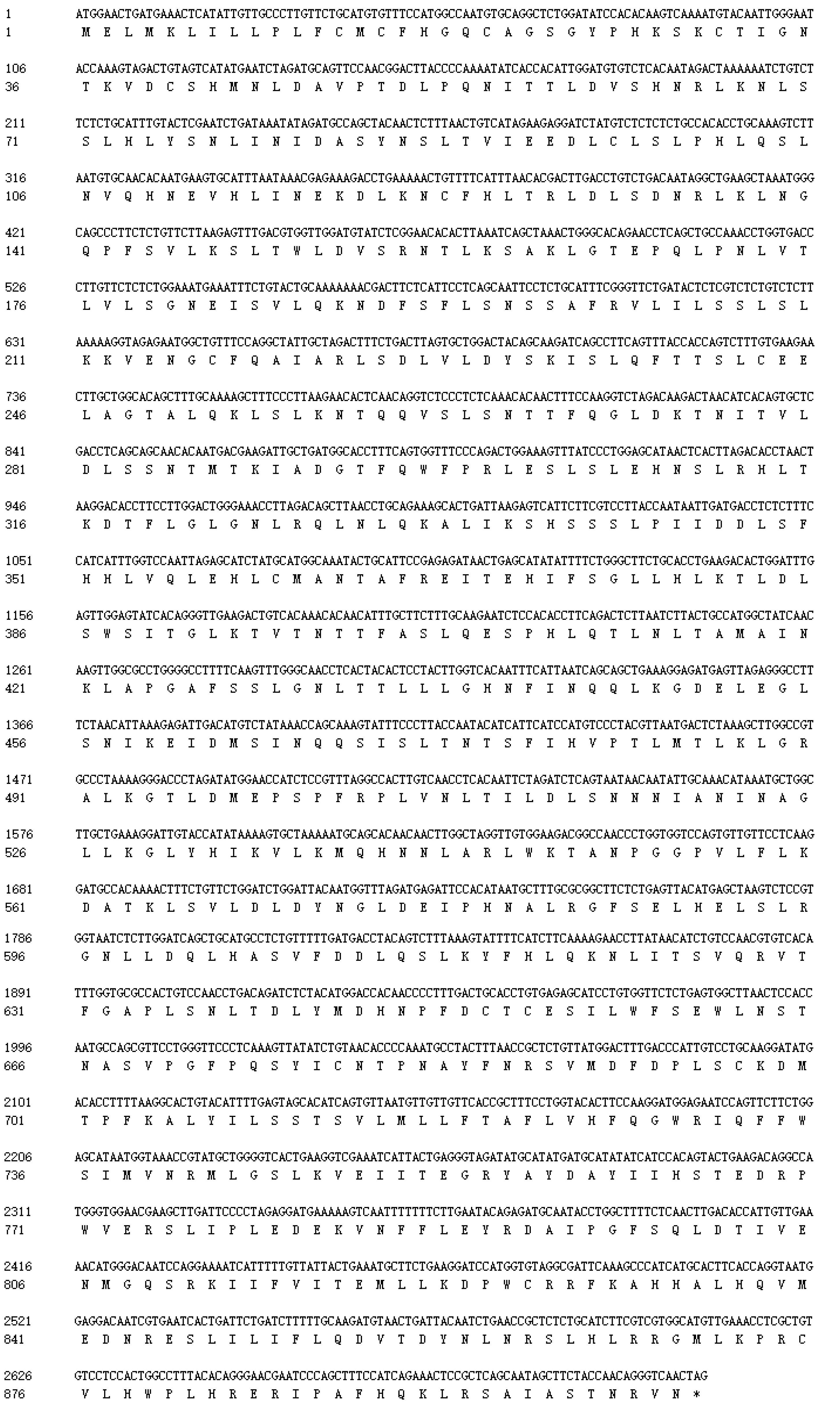
Figure 1. Nucleotide and deduced amino acid sequences of SpTLR3. * Stop codon.
Genes 13 01862 g001
Figure 2. The predicted domain of SpTLR3 protein by SMART programs. (a) Schematic representation of TLR3 domains predicted; (b) Multiple alignment of TLR3-TIR domains from different species. Snow trout (GenBank accession no. AGJ74274), Crucian carp (GenBank accession no. AGO57935), Silver crucian carp (GenBank accession no. AGR53439), Carp (GenBank accession no. AHE74142), Goldfish (GenBank accession no. ABC86865), Grass carp (GenBank accession no. ABI64155), Barbel chub (GenBank accession no.ALO75529), Bluntnose black bream (GenBank accession no. ABI83673) and Zebrafish (GenBank accession no. AAI07956). Asterisk (*), identical; colon (:), conserved; and dot (.), semi-conserved residues. The conserved box1 is shown in blue, the conserved box2 is shown in yellow and the conserved box3 is shown in green.
Genes 13 01862 g002
Figure 3. Predicted secondary structure and 3D structural models of SpTLR3 protein. (a) Predicted secondary structure of SpTLR3 protein. ■ indicates helix, ☐ indicates coil, indicates strand. (b) Three-dimensional model structures of predicted SpTLR3 protein.
Genes 13 01862 g003
Figure 4. Phylogenetic tree of relationships between S. prenanti TLR3 and other vertebrates. Tree was constructed by neighbor-joining method using MEGA 4.0 software. Numbers at nodes indicate proportions of bootstrapping after 1,000 replications. ● S. prenanti TLR3.
Genes 13 01862 g004
Figure 5. Abundance of SpTLR3 transcripts in heart, liver, spleen, head kidney, muscle, intraperitoneal fat, and intestine of S. prenanti was determined by qRT-PCR. The loading control for normalization was β-actin. The a, b, c means with different letters are significantly different from each other (p < 0.05). Values are shown as the means ± standard error (n = 4). Error bars, standard error of the means (n = 4 fish per group).
Genes 13 01862 g005
Figure 6. The relative expression levels of SpMyD88 (a), SpTLR2 (b), SpTLR3 (c), SpTLR4 (d), SpTLR18 (e), SpTLR22-1 (f), SpTLR22-2 (g) and SpTLR22-3 (h) transcripts in liver, head kidney, hindgut, and spleen of S. prenanti at 12 and 24 h after LPS injection determined by qRT-PCR. Values were normalized using β-actin. Data are expressed as the means ± standard error (n = 4). Statistically significant differences between the groups are indicated by asterisks (* p < 0.05, ** p < 0.01, *** p < 0.001). PBS, phosphate-buffered saline.
Genes 13 01862 g006
Table 1. The primers used for SpTLR3 clone and qRT-PCR.
PrimerSequence (5′-3′)Annealing Temperature (°C)Size (bp)
Primers for partial sequence cloning
TLR3-FCAGACTCTTAATCTTACTG551558
TLR3-RGACATACCAAATAAGGAC
Primers for 5′RACE
GSPCGACCTTCAGTGACCCCAGCATACGGTouchdown PCR 2543
UPM
(kit provided)
CTAATACGACTCACTATAGGGCAAG
CAGTGGTATCAACGCAGAGT
Primers for qRT-PCR
TLR2-FGATCAACGGCACAGTGTTTG62170
TLR2-RCAGGTCTGAAAGGAGGTTCTG
TLR3-FGCTGAAAGGAGATGAGTTAGAG62110
TLR3-RACGTAGGGACATGGATGAA
TLR4-FCTTGGTGTCGCTTTGAGTTTG62107
TLR4-RGTCTCTGCTCCACTTTAGGTATG
TLR18-FACAGACTAAATGGCCAGGGAAG62118
TLR18-RAACCACAAGCAAGGGCAAAG
TLR22-1-FCCTCTTCTTAGCCTTCCTTTAC6294
TLR22-1-RCTCGTCTTTGGTGTTGTAGG
TLR22-2-FTTCCAGGGACTGTGGTATTTG6298
TLR22-2-RGCCCACAGATAAGGAGTGTAAG
TLR22-3-FCCATCGGCATTCTTTGGTTT62169
TLR22-3-RCTGTGTTCAGGAATGCCTTG
MyD88-FGAGTTTCCCACTCCGTTAAGA6292
MyD88-RCGCCGAGATGATGGACTTTA
β-actin-FGACCACCTTCAACTCCATCAT62126
β-actin -RGTGATCTCCTTCTGCATCCTATC
Publisher’s Note: MDPI stays neutral with regard to jurisdictional claims in published maps and institutional affiliations.