Article

BRCA2 Haploinsufficiency in Telomere Maintenance

by
Soffía R. Gunnarsdottir
1,
Hördur Bjarnason
1,
Birna Thorvaldsdottir
1,
Felice Paland
1,2,
Margrét Steinarsdottir
3,
Jórunn E. Eyfjörd
1 and
Sigrídur K. Bödvarsdottir
1,*
1
BioMedical Center, School of Health Sciences, Faculty of Medicine, University of Iceland, 101 Reykjavík, Iceland
2
Faculty of Medicine, Eberhard Karls University, 72074 Tübingen, Germany
3
Department of Genetics and Molecular Medicine, Landspitali the National University Hospital of Iceland, 101 Reykjavík, Iceland
*
Author to whom correspondence should be addressed.
Genes 2022, 13(1), 83; https://doi.org/10.3390/genes13010083
Submission received: 17 September 2021 / Revised: 13 December 2021 / Accepted: 23 December 2021 / Published: 28 December 2021
(This article belongs to the Special Issue BRCA1 and BRCA2: Genome Instability and Tumorigenesis)

Abstract

:
Our previous studies showed an association between monoallelic BRCA2 germline mutations and dysfunctional telomeres in epithelial mammary cell lines and increased risk of breast cancer diagnosis for women with BRCA2 999del5 germline mutation and short telomeres in blood cells. In the current study, we analyzed telomere dysfunction in lymphoid cell lines from five BRCA2 999del5 mutation carriers and three Fanconi Anemia D1 patients by fluorescence in situ hybridization (FISH). Metaphase chromosomes were harvested from ten lymphoid cell lines of different BRCA2 genotype origin and analyzed for telomere loss (TL), multitelomeric signals (MTS), interstitial telomere signals (ITS) and extra chromosomal telomere signals (ECTS). TL, ITS and ECTS were separately found to be significantly increased gradually between the BRCA2+/+, BRCA2+/- and BRCA2-/- lymphoid cell lines. MTS were found to be significantly increased between the BRCA2+/+ and the BRCA2+/- heterozygous (p < 0.0001) and the BRCA2-/- lymphoid cell lines (p < 0.0001) but not between the BRCA2 mutated genotypes. Dysfunctional telomeres were found to be significantly increased in a stepwise manner between the BRCA2 genotypes indicating an effect of BRCA2 haploinsufficiency on telomere maintenance.

1. Introduction

The breast cancer susceptibility 2 (BRCA2) gene was identified in 1995 and has been shown to play an essential role in DNA double strand break (DSB) repair by homologous recombination (HR) and DNA crosslink repair by the Fanconi anemia (FA) pathway [1,2]. Germline mutations in the BRCA2 gene are known to be highly associated with the risk of diagnosis with breast, ovarian, prostate, and pancreatic cancer [3,4,5,6]. Breast cancer risk is not only associated with women but also men. In Iceland, a BRCA2 999del5 truncation mutation in exon 9 was isolated in a family with several cases of male and female breast cancer [3]. This mutation has been shown to have a high frequency (0.7%) in the Icelandic population [5,7]. Cloning of the 999del5 mutant and expression studies showed that even though the short mutant RNA is produced there is no detectable corresponding protein, indicating BRCA2 heterozygosity [8].
Multiple chromosomal abnormalities have been found in breast tumors with the BRCA2 999del5 mutation [9,10] and in murine cells deficient in the BRCA2 homolog [11]. The abnormalities include broken chromosomes and chromatids, markers of defective mitotic recombination, and tri- and quadriradial chromosomes that are known to be related to defects in the FA pathway [12]. Indeed, BRCA2 has been shown to be one of the genes playing a key role in the FA pathway, subtype D1 [13]. Patients with the FA-D1 subtype have biallelic BRCA2 germline mutations, whereas at least one of the mutations is mild, and has high susceptibility for diagnosis with leukemia, solid tumor, brain tumor and Wilm’s tumor in childhood [14].
Studies on the role of the BRCA2 protein have shown the requirement of HR-dependent RAD51 filament formation plus stabilization by BRCA2 and the core FA pathway for efficient protection in replication forks from degradation following short-term replication arrest. FA and BRCA2 defective cells are defective in fork protection and show an increase in genome instability, manifesting chromatid breaks and radial structures on mitotic chromosome spreads [15,16]. DNA replication of repetitive sequences like telomeres is known to take place in the late S-phase of the cell cycle. It has been shown that BRCA2 is required to limit replication stress (RS) at telomeres by RAD51 filament loading [17,18]. Our study on BRCA2 heterozygous mammary epithelial cell lines showed telomere dysfunction such as chromosome end-to-end fusions, interstitial telomere sequences (ITS), telomere loss (TL), extrachromosomal telomere sequences (ECTS) and frequent telomere sister chromatid exchanges (T-SCE) as seen in cells that rely on the alternative lengthening of telomeres (ALT) in absence of telomerase. These results indicated the important role of BRCA2 in telomere stabilization and protection [9]. A recent study on our cohort showed that women with the BRCA2 999del5 germline mutation and relative short telomere length measured in blood cells were at significantly higher risk of being diagnosed with breast cancer [19]. These results indicate that monoallelic BRCA2 gene expression may not be enough to fulfill telomere maintenance, indicating BRCA2 haploinsufficiency. Anticipation effects have also been reported from this study cohort where daughters of mother-daughter pairs with the BRCA2 999del5 germline mutation have been shown to be diagnosed with breast cancer on average 10 years younger than the mothers [7].
The 50 years old Knudson’s two-hit hypothesis suggests that both alleles of a tumor suppressor gene require to be inactivated to cause tumor formation, as was first described in heritable cases with retinoblastoma [20]. We have, however, shown that only about half of the BRCA2 999del5 breast cancer cases [21,22] and pancreatic cancer cases [23] had lost their BRCA2 wild-type allele in the tumor tissue. Similar results have been reported from another study cohort where the absence of BRCA2 locus-specific loss of heterozygosity was observed in 46% of breast tumors [24]. Of note is that BRCA2 999del5 breast cancer cases with wild-type allelic loss had significantly worse breast cancer specific survival compared to cases that remained the wild-type allele [22]. A recent study showed the BRCA2 haploinsufficient phenotype in vivo in breast tissues of BRCA2 mutation carriers exhibiting DNA damage that resulted from failed RS- and DNA damage responses and consequently aneuploidy [25].
Mechanisms shown to respond to RS that could counteract karyotypic diversity and contribute to tumor progression include polymerase theta-mediated end joining (TMEJ) alternative repair pathway at resected DSBs when HR is deficient to repair broken forks [26,27]. Other recent studies have shown that genic RS induced by the absence of Brca2 led to delays in replication and mitotic DNA synthesis (MiDAS) [25,28]. RAD52 has been shown to promote MiDAS following RS that occurs independently of RAD51 [29]. Abrogation of Brca2 has been shown to reinforce MiDAS related break-induced replication (BIR) and engagement with the ALT pathway. For this Brca2-deficient ALT induction, Rad51 filament loading was dispensable, but Mre11 dependent DSB resection and Rad52 were required [30]. RS at telomeres has as well been linked to MiDAS [31,32].
In the present study, we compared telomere dysfunction in lymphoid cell lines with different BRCA2 genotypes including two BRCA2 wild-type cell lines, five BRCA2 heterozygous cell lines with the 999del5 germline mutation and three FA-D1 cell lines with biallelic BRCA2 mutations. The originality of this study was to compare all three BRCA2 genotypes in a cell type not associated with BRCA2 hereditary cancer. Results showed increased telomere abnormalities between the BRCA2 genotypes in a stepwise manner, including TL, ITS and ECTS.

2. Materials and Methods

2.1. Lymphoid Cell Lines

Viable lymphoid cells from women with and without the heritable BRCA2 999del5 mutation were Epstein-Barr virus transformed (Table 1). This was carried out according to permits from the Icelandic Data Protection Commission (2006050307) and Bioethics Committee (VSNb2006050001/03-16). Three FA-D1 lymphoid cell lines were generously provided by Professor Helmut Hanenberg, University of Duisburg-Essen, Germany. The FA-D1 cell lines were with biallelic mutations in the BRCA2 gene (Table 1). All cell lines were cultured in RPMI 1640 medium (GIBCO BRL) with penicillin and streptomycin. 10% heat-inactivated fetal bovine serum (FBS, GIBCO BRL) was used for the BRCA2 999del5 cell lines and the wild-type cell line whereas 10–20% non-heat inactivated FBS was used for the FA-D1 cell lines.

2.2. Chromosomal Analysis

Chromosomes were harvested after 3–4 h short-term culture with colcemid (0.016 μg/mL; KaryoMAX-colcemid, GIBCO, Patsley, UK). Telomere fluorescence in situ hybridization (FISH) was performed by ready-to-use Cy3-conjugated PNA pan-telomere probe (Telomere PNA FISH kit/Cy3, Cat. K5326, Dako, Glostrup, Denmark) in mixture with FITC-conjugated PNA pan-centromere probe (Dako) and counterstained with 4′,6-diamidino-2-phenylindole (DAPI) as previously described [9]. The microscope used for image capture was Leica DMRA2 connected to a Leica DC350F CCD camera and the Leica CW4000 image acquisition software. The image acquisition was performed through a ×100 oil objective by constant intensity and exposure times for different fluorophores. From each lymphoid cell line about 60 FISH labeled metaphases were analyzed. Events were counted on each metaphase and correction was made according to 46 chromosomes each metaphase if metaphases were aneuploidy. TL was detected either as a single telomere end (STE) on a single chromatid or as telomere free ends (TFE) on both sister chromatids. When results from STE and TFE analysis were combined the number of TFE was duplicated for both sister chromatids. Multitelomeric signals (MTS) were detected either as a single MTS (SMTS) on a sister chromatid or as double MTS (DMTS) on both sister chromatids. When results from SMTE and DMTS analysis were combined the number of DMTS was duplicated for both sister chromatids. ITS were detected as telomere FISH signals within a chromatid and ECTS as telomere FISH signals scattered around chromosomes without a centromere signal. The non-specific background noise of autofluorescence combined with red and green spots or cytoplasmic background were excluded and treated as unspecific background. Telomere FISH metaphase analysis was performed independently by four researchers in a blinded way for the genotypes of the EB lymphoid cell lines. Each BRCA2+/- and BRCA2-/- lymphoid cell line was analyzed by a single researcher. All researchers measured the same metaphase images of the EB0392 BRCA2+/+ lymphoid cell line that was used as one of the controls and for correction of individual differences. The ECTS analysis was, however, performed by one researcher for all the lymphoid cell lines except one control line.

2.3. Statistical Analysis

Statistical analyses were performed on chromosomal abnormalities as per metaphase and results presented as an average mean with standard error of mean (SEM) bars. Results from each of the BRCA2 genotypes were combined and performed as one genotype. A two-tailed Student´s t-test was used for statistical analysis and p < 0.05 was considered statistically significant. The R (CRAN) software was used for graphical representation.

3. Results

At least 60 metaphases were analyzed from each lymphoid cell line with one exception of the HSC62N cell line (Table 2). Most of the metaphases were diploid or near diploid with few exceptions of tetraploid metaphases or metaphases with incomplete chromosome number (Table 2, Supplementary Tables S1–S8).
In addition to analysis of TL, MTS, ITS and ECTS other chromosomal aberrations were measured including sister chromatid and end-to-end chromosome fusions, radial chromosomal configurations and chromosome fragments without telomere signals. These chromosomal aberrations were, however, not included in the results since average events per metaphase were below one which may affect the significance of the results.

3.1. Telomere Loss Was Found to Increase in a Stepwise Manner between the BRCA2 Genotypes

TL was analyzed either as STE or TFE (Figure 1a). STE was found to be significantly increased in a stepwise manner between the BRCA2+/+, BRCA2+/- and BRCA2-/- lymphoid cell lines, and a significant difference was found between the BRCA2+/- and BRCA2-/- genotypes (Figure 1b; Supplementary Table S1). TFE was also found to be significantly increased in a stepwise manner between the BRCA2 genotypes (Figure 1c; Supplementary Table S2). Note the strong difference found in TFE between the BRCA2+/- and BRCA2-/- genotypes, whereas a lower difference was found in STE between the two genotypes. Total TL was found to be significantly increased in a stepwise manner between the three BRCA2 genotypes of lymphoid cell lines (Figure 1d; Supplementary Table S3).

3.2. Multitelomeric Signals Increase between BRCA2 Wild-Type and Mutated Genotypes

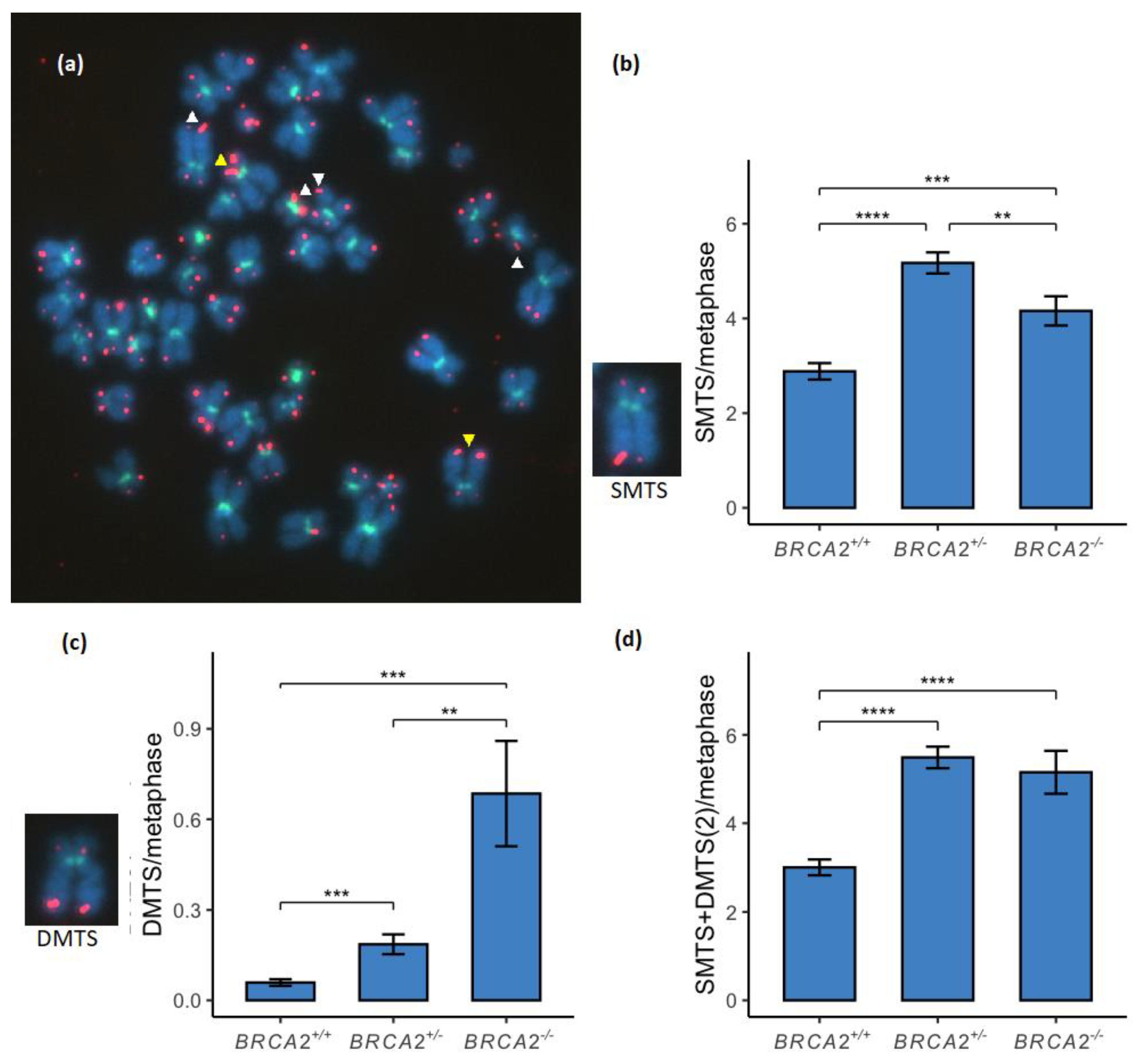
MTS were analyzed either as SMTS or DMTS (Figure 2a). SMTS was found to be significantly increased between the BRCA2+/+ and the BRCA2+/- lymphoid cell lines and between the BRCA2+/+ and the BRCA2-/- lymphoid cell lines (Figure 2b; Supplementary Table S4). SMTS was also found to be significantly higher among the BRCA2+/- lymphoid cell lines compared to the BRCA2-/- genotype. DMTS were, however, found to be significantly increased in a stepwise manner between the three BRCA2 genotypes (Figure 2c; Supplementary Table S5). Total MTS was measured by a combination of SMTS and duplicated DMTS. Total MTS was found to be significantly increased between the BRCA2 wild-type and the BRCA2+/- lymphoid cell lines and between the BRCA2 wild-type and the BRCA2-/- lymphoid cell lines (Figure 2d; Supplementary Table S6). No difference of total MTS was, however, found between the two BRCA2+/- and BRCA2-/- genotypes.

3.3. Interstitial Telomere Sequences Increase in a Stepwise Manner between the BRCA2 Genotypes

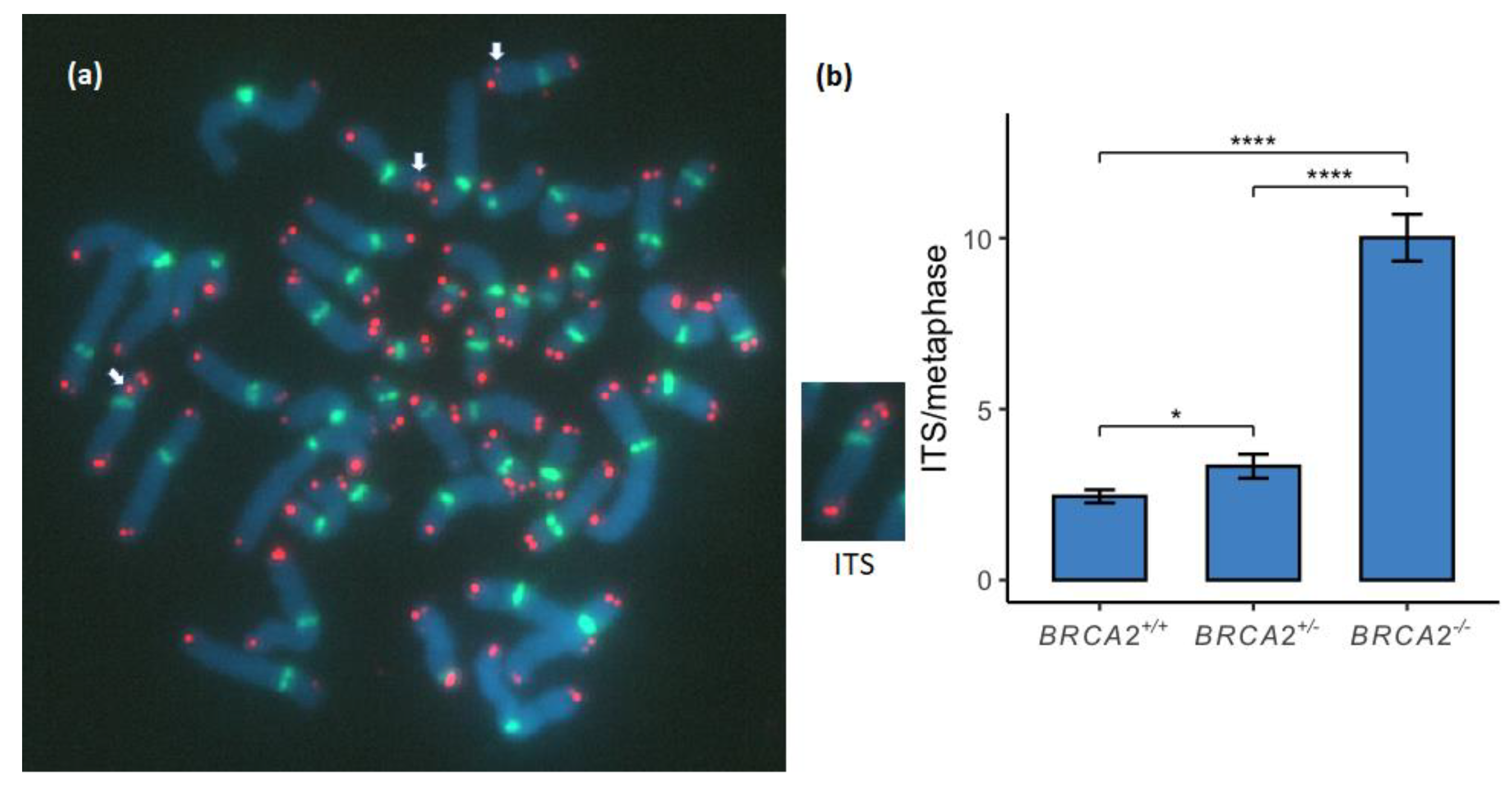
ITS were measured as telomere signals within chromosome arms (Figure 3a). ITS was found to be significantly increased between the BRCA2 genotypes of the lymphoid cell lines in a stepwise manner (Figure 3b; Supplementary Table S7).

3.4. Extrachromosomal Telomere Sequences Increase in a Stepwise Manner between the BRCA2 Genotypes

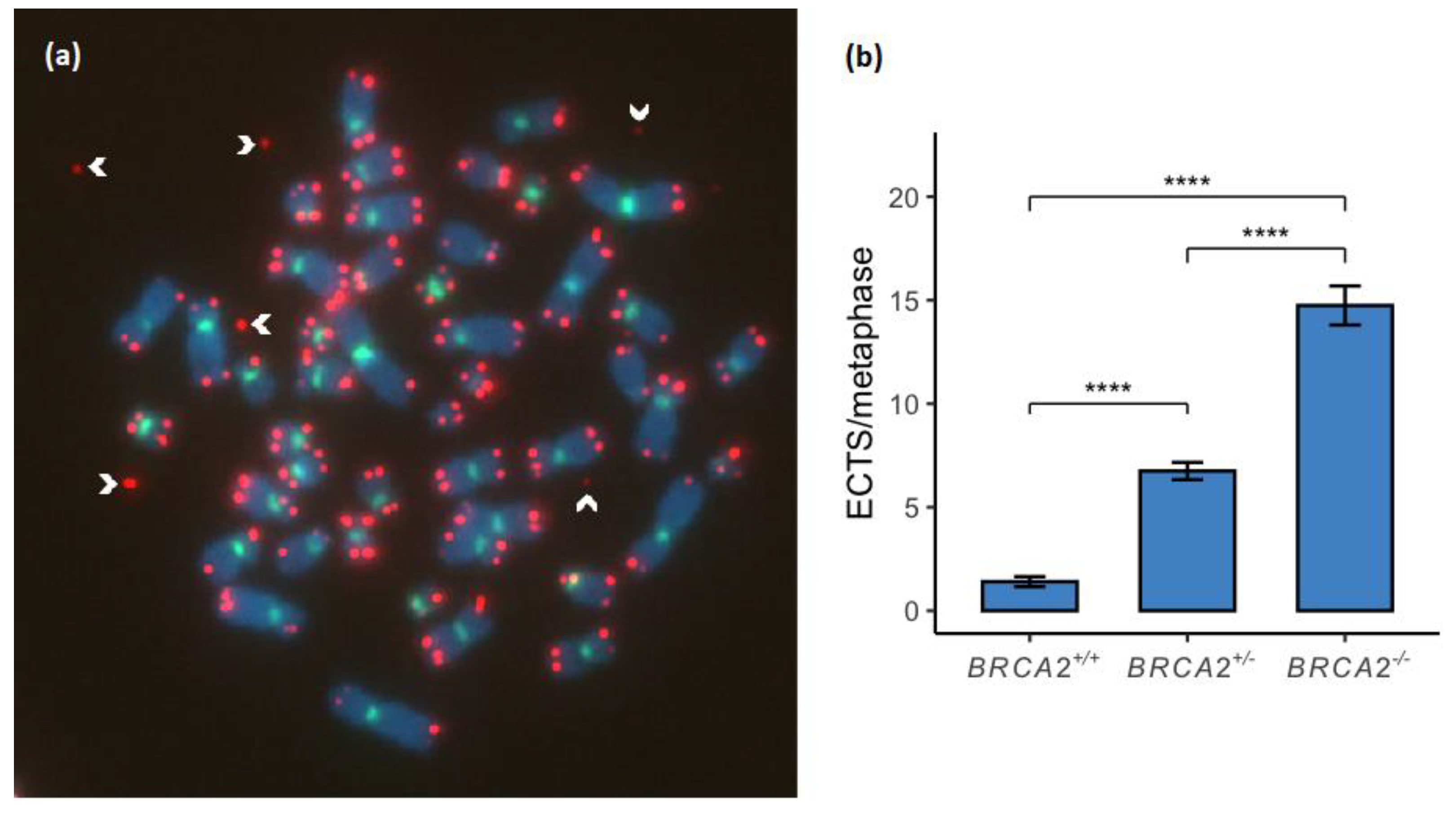
ECTS were measured as DNA fragments with a telomere signal and without a centromere signal scattered around metaphases (Figure 4a). ECTS were found to be significantly increased between the BRCA2 genotypes of the lymphoid cell lines in a stepwise manner (Figure 4b; Supplementary Table S8).

4. Discussion

TL is likely to result from RS that leads to terminal fork collapse, either at a single sister chromatid strand or at both sister chromatids on chromosome ends. TL was found to be increased among BRCA2 heterozygous mammary epithelial cell lines in our previous study [9]. In the current study we found a significant stepwise increase in TL between the BRCA2+/+, BRCA2+/- and BRCA2-/- genotypes with a high difference between the BRCA2+/+ and BRCA2+/- genotypes of the STE subgroup and between the BRCA2+/- and BRCA2-/- genotypes of the TFE subgroup (Figure 1). This indicates that events of TL caused by RS and collapsed terminal forks are BRCA2 haploinsufficiency dependent, showing an intermediate increase in TL with the STE subgroup and the most increase within the BRCA2+/- genotype whereas the FA-D1 cell lines show most increase within the TFE subgroup. Terminal fork collapse can also appear as inter-chromatid discrepancies of telomere FISH signals [35]. Frequent telomere discrepancies were reported as unequal telomere signals on sister chromatids in our previous study on BRCA2+/- mammary epithelial cell lines [9]. Telomere discrepancies were also frequently noted in the BRCA2+/- and FA-D1 lymphoid cell lines in the present study although not reported. To further analyze telomere discrepancies in BRCA2+/- cells and find out if they are consequences from terminal fork collapse or an ALT-related repair mechanism at the telomere termini or both, needs to be further analyzed by chromosome orientation FISH (CO-FISH) [36].
MTS is often referred to as fragile telomeres. They have been suggested to represent a recombination event among inter-/intra-chromatid telomeric sequences or ITS in the proximal regions of telomeres [37] or restarted collapsed terminal forks but their formation is still unknown. MTS has been shown to be suppressed by the key factors of HR repair, BRCA2 and RAD51 [18]. BRCA2 is required for telomere replication of the G-rich strand that has a high propensity to adopt G-quadruplex secondary structures leading to MTS formation in BRCA2 deficient cells [38]. In the current study, we found a significant increase of MTS between the BRCA2+/+ and the BRCA2+/- and BRCA2-/- lymphoid cell lines, respectively (Figure 2). A significant increase of DMTS was also found between the BRCA2+/- and BRCA2-/- lymphoid cell lines but no difference was, however, found of the total MTS between the two genotypes. Lack of significance in MTS formation between the BRCA2+/- and BRCA2-/- genotypes may be explained by >50% higher TL of the BRCA2-/- genotype (Figure 1c). The significant increase of DMTS found between the BRCA2+/- and BRCA2-/- genotypes is, however, a strong indication of BRCA2 deficiency related telomere RS due to G-quadruplex formation.
Most ITS probably result from the formation of DSB during RS that is mediated by targeted telomere insertions [35], although they have also been suggested to result from subsequent healing involving telomerase [39]. Some ITS may be the result of chromosome end-to-end fusions or chromatid fusions which can be detected as antiparallel orientated telomeres by CO-FISH. These ITS have been shown to be alternative TMEJ and include random nucleotides at the telomere junction [26]. Previously we reported significantly higher ITS frequency among BRCA2+/- mammary epithelial cell lines compared to a commercial BRCA2+/+ mammary epithelial cell line and cell lines that rely on the classical ALT-positive mechanism for telomere maintenance [9]. In the present study we found a significant stepwise increase of ITS between the BRCA2+/+, BRCA2+/- and BRCA2-/- lymphoid cell lines (Figure 3) supporting our previous findings.
ECTS found scattered around chromosome spreads presumably result from terminal fork collapse caused by telomere RS. In this study we found a significant stepwise increase of ECTS between the BRCA2+/+, BRCA2+/- and BRCA2-/- lymphoid cell lines (Figure 4) probably directly correlated with increased TL between the three BRCA2 genotypes. Sometimes these ECTS have strong fluorescence signals indicating amplification of telomeric c-circles that have been associated with cancer cells that rely on ALT rather than telomerase for telomere maintenance [40]. Recent studies have shown that abrogation of BRCA2 strongly increases c-circle amplification indicating the presence of ALT activity in BRCA2 deficient cells [30,41].
The results clearly show a gradual increase in telomere defects between the BRCA2 genotypes. This was shown in lymphoid cells that are not known to be prone to BRCA2 related cancer risk. It needs to be kept in mind, however, that apart from the BRCA2 genotype the cell lines have generic individual genomic differences. For this same reason, the two BRCA2+/+ control cell lines used in this study may affect the interpretation. The most ideal setup would be to use a homogenous genetic background of a single cell line edited with the CRISPR/Cas9 gene editing technique for insertion of the BRCA2 9999del5 mutation. This has, however, not yet been successful due to the very low viability of cells with the homozygous mutation. This correlates with the fact that no homozygous individuals have been known to exist although expected under the Hardy–Weinberg equilibrium and allele frequency in the Icelandic population [5].
Our and other previous findings have shown unequal T-SCE in BRCA2 deficient cells [9,18,30]. Collapsed terminal forks can be recovered by T-SCE with two possible mechanisms, HR or BIR that is mediated by MiDAS [37]. HR dependent T-SCE is detected by DNA synthesis at both chromatid ends (semi-conservative DNA synthesis), whereas BIR dependent T-SCE is detected by DNA synthesis at single chromatid end (conservative DNA synthesis) by CO-FISH. Analysis by CO-FISH has shown that the ALT pathway in human cells can be a conservative DNA synthesis process potentially via the BIR pathway [42]. A more recent study showed that BRCA2 depletion reinforces RAD52 mediated BIR and engages with the ALT pathway by conservative telomeric DNA synthesis [30]. ALT has, however, been shown to be a bifurcated pathway involving both RAD52-dependent and RAD52-independent BIR. The RAD52-independent BIR pathway has been shown to be responsible for c-circle amplification which is reciprocally suppressed by RAD51 [43]. The HR-dependent semi-conservative DNA synthesis result as a response to Holliday junction resolution of the collapsed replication fork [37]. A recent study on the fruit fly showed that TMEJ compensates for the BRCA2 dependent loss of HR on Holliday junction resolvases by using HR-intermediates that suppress mitotic crossing over and preserve the genomic stability [44]. BRCA2 deficiency has been shown to be synthetic lethal with disruption of either RAD52 or polymerase theta (POLQ) [26,27,45,46,47,48] or both [49]. Both RAD52 and POLQ are, therefore, important backup pathways for DSB repair and RS responses in BRCA2 deficient cells. RAD52 and POLQ are of high interest as therapeutic targets leading to synthetic lethal interaction of HR deficient tumors for future therapies especially due to acquired resistance of the currently used Poly (ADP-ribose) polymerase inhibitors [50]. However, the involvement of RAD52 and POLQ backup pathways in telomere maintenance of BRCA2 deficient cells is not fully understood and needs to be further investigated.
Telomere length homeostasis in unaffected BRCA2 999del5 mutation carriers does not differ from non-carriers in blood cells [19]. After a diagnosis of breast cancer, however, the measured relative telomere length in blood cells of BRCA2 999del5 mutation carriers was found to be significantly shorter than among non-carriers. Telomere length was shown to be a modifier of breast cancer risk in BRCA2 999del5 mutation carriers in the same study. Another aspect is that BRCA2 999del5 mutation carriers with breast cancer have been associated with a significantly worse prognosis than non-carriers [51]. How defects in telomere length homeostasis of BRCA2 mutation carriers might be related to consequences of possible telomere RS before and after breast cancer diagnosis needs to be answered in future studies. Such knowledge could have an impact on the prediction of early cancer development, and therefore, be used to advise the timing of preventive therapies, and targeted therapies of BRCA2 related breast cancer in the future as discussed here above.

5. Conclusions

Taken together, we found a stepwise increase in telomere abnormalities between the BRCA2+/+, BRCA2+/- and BRCA2-/- genotypes showing a strong indication of BRCA2 haploinsufficiency in telomere maintenance. Our results from telomere FISH show a clear increase in TL, MTS, ITS and ECTS between the genotypes that are a sign of telomere RS in BRCA2 deficient cells. More research is needed on the possible involvement of the RAD52 related BIR and the POLQ related TMEJ backup pathways in telomere maintenance when BRCA2 is not fully expressed and the distinct roles of the two pathways on telomere maintenance and genomic stability.

Supplementary Materials

The following supporting information can be downloaded at: https://www.mdpi.com/article/10.3390/genes13010083/s1, Table S1: Single telomere end (STE) analysis, Table S2: Analysis of telomere free ends (TFE), Table S3: Telomere loss (TL) as a combination of STE and TFE, Table S4: Analysis of multitelomere signals on single sister chromatids (SMTS), Table S5: Analysis of multitelomere signals on both (double) sister chromatids (DMTS), Table S6: Total multitelomere signals (MTS) by a combination of SMTS and DMTS, Table S7: Analysis of interstitial telomere sequences (ITS), Table S8: Analysis of extrachromosomal telomere sequences (ECTS).

Author Contributions

Conceptualization, J.E.E. and S.K.B.; methodology, H.B., B.T., M.S., F.P. and S.K.B.; software, B.T.; validation, S.R.G., H.B., B.T., F.P. and S.K.B.; formal analysis, S.R.G. and S.K.B.; investigation, S.R.G., H.B., B.T. and F.P.; resources, M.S. and J.E.E.; data curation, J.E.E. and S.K.B.; writing—original draft preparation, S.K.B.; writing—review and editing, S.R.G., B.T., M.S. and J.E.E.; visualization, S.R.G., B.T. and S.K.B.; supervision, M.S., J.E.E. and S.K.B.; project administration, S.K.B.; funding acquisition, S.K.B. All authors have read and agreed to the published version of the manuscript.

Funding

This research was funded by the Icelandic Research Fund, grants number 100455021 and 207301051.

Institutional Review Board Statement

The study was conducted according to the guidelines of the Declaration of Helsinki and approved by the Icelandic Data Protection Commission (2006050307) and Bioethics Committee (VSNb2006050001/03-16).

Informed Consent Statement

Informed consent was obtained from all subjects involved in the study.

Data Availability Statement

The data presented in this study are available in Supplementary Tables S1–S8.

Acknowledgments

The authors thank Helmut Hanenberg, University of Duisburg-Essen, Germany for generously providing FA-D1 lymphoid cell lines used in this study.

Conflicts of Interest

The authors declare no conflict of interest. The funders had no role in the design of the study; in the collection, analyses, or interpretation of data; in the writing of the manuscript, or in the decision to publish the results.

References

  1. Fradet-Turcotte, A.; Sitz, J.; Grapton, D.; Orthwein, A. BRCA2 functions: From DNA repair to replication fork stabilization. Endocr. Relat. Cancer 2016, 23, T1–T17. [Google Scholar] [CrossRef] [PubMed] [Green Version]
  2. Le, H.P.; Heyer, W.-D.; Liu, J. Guardians of the Genome: BRCA2 and Its Partners. Genes 2021, 12, 1229. [Google Scholar] [CrossRef] [PubMed]
  3. Thorlacius, S.; Olafsdottir, G.; Tryggvadottir, L.; Neuhausen, S.; Jonasson, J.G.; Tavtigian, S.V.; Tulinius, H.; Ögmundsdottir, H.M.; Eyfjörd, J.E. A single BRCA2 mutation in male and female breast cancer families from Iceland with varied cancer phenotypes. Nat. Genet. 1996, 13, 117–119. [Google Scholar] [CrossRef] [PubMed]
  4. Tulinius, H.; Olafsdottir, G.H.; Sigvaldason, H.; Arason, A.; Barkardottir, R.B.; Egilsson, V.; Ogmundsdottir, H.M.; Tryggvadottir, L.; Gudlaugsdottir, S.; Eyfjord, J.E. The effect of a single BRCA2 mutation on cancer in Iceland. J. Med. Genet. 2002, 39, 457–462. [Google Scholar] [CrossRef] [Green Version]
  5. Rafnar, T.; Sigurjonsdottir, G.R.; Stacey, S.N.; Halldorsson, G.; Sulem, P.; Pardo, L.M.; Helgason, H.; Sigurdsson, S.T.; Gudjonsson, T.; Tryggvadottir, L.; et al. Association of BRCA2 K3326* With Small Cell Lung Cancer and Squamous Cell Cancer of the Skin. J. Natl. Cancer Inst. 2018, 110, 967–974. [Google Scholar] [CrossRef] [Green Version]
  6. Dullens, B.; de Putter, R.; Lambertini, M.; Toss, A.; Han, S.; Van Nieuwenhuysen, E.; Van Gorp, T.; Vanderstichele, A.; Van Ongeval, C.; Keupers, M.; et al. Cancer Surveillance in Healthy Carriers of Germline Pathogenic Variants in BRCA1/2: A Review of Secondary Prevention Guidelines. J. Oncol. 2020, 2020, 9873954. [Google Scholar] [CrossRef]
  7. Thorlacius, S.; Sigurdsson, S.; Bjarnadottir, H.; Olafsdottir, G.; Jonasson, J.G.; Tryggvadottir, L.; Tulinius, H.; Eyfjörd, J.E. Study of a single BRCA2 mutation with high carrier frequency in a small population. Am. J. Hum. Genet. 1997, 60, 1079–1084. [Google Scholar]
  8. Mikaelsdottir, E.K.; Valgeirsdottir, S.; Eyfjord, J.E.; Rafnar, T. The Icelandic founder mutation BRCA2 999del5: Analysis of expression. Breast Cancer Res. 2004, 6, R284–R290. [Google Scholar] [CrossRef] [Green Version]
  9. Bodvarsdottir, S.K.; Steinarsdottir, M.; Bjarnason, H.; Eyfjord, J.E. Dysfunctional telomeres in human BRCA2 mutated breast tumors and cell lines. Mutat. Res. 2012, 729, 90–99. [Google Scholar] [CrossRef]
  10. Gretarsdottir, S.; Thorlacius, S.; Valgardsdottir, R.; Gudlaugsdottir, S.; Sigurdsson, S.; Steinarsdottir, M.; Jonasson, J.G.; Anamthawat-Jonsson, K.; Eyfjörd, J.E. BRCA2 and p53 mutations in primary breast cancer in relation to genetic instability. Cancer Res. 1998, 58, 859–862. [Google Scholar]
  11. Patel, K.J.; Yu, V.P.; Lee, H.; Corcoran, A.; Thistlethwaite, F.C.; Evans, M.J.; Colledge, W.H.; Friedman, L.S.; Ponder, B.A.; Venkitaraman, A.R. Involvement of Brca2 in DNA Repair. Mol. Cell 1998, 1, 347–357. [Google Scholar] [CrossRef]
  12. Eyfjord, J.E.; Bodvarsdottir, S.K. Genomic instability and cancer: Networks involved in response to DNA damage. Mutat. Res. 2005, 592, 18–28. [Google Scholar] [CrossRef] [PubMed]
  13. Howlett, N.G.; Taniguchi, T.; Olson, S.; Cox, B.; Waisfisz, Q.; De Die-Smulders, C.; Persky, N.; Grompe, M.; Joenje, H.; Pals, G.; et al. Biallelic Inactivation of BRCA2 in Fanconi Anemia. Science 2002, 297, 606–609. [Google Scholar] [CrossRef]
  14. Alter, B.P.; Rosenberg, P.S.; Brody, L.C. Clinical and molecular features associated with biallelic mutations in FANCD1/BRCA2. J. Med. Genet. 2007, 44, 1–9. [Google Scholar] [CrossRef] [Green Version]
  15. Schlacher, K.; Christ, N.; Siaud, N.; Egashira, A.; Wu, H.; Jasin, M. Double-strand break repair-independent role for BRCA2 in blocking stalled replication fork degradation by MRE11. Cell 2011, 145, 529–542. [Google Scholar] [CrossRef] [Green Version]
  16. Schlacher, K.; Wu, H.; Jasin, M. A distinct replication fork protection pathway connects Fanconi anemia tumor suppressors to RAD51-BRCA1/2. Cancer Cell 2012, 22, 106–116. [Google Scholar] [CrossRef] [Green Version]
  17. Badie, S.; Escandell, J.M.; Bouwman, P.; Carlos, A.R.; Thanasoula, M.; Gallardo, M.M.; Suram, A.; Jaco, I.; Benitez, J.; Herbig, U.; et al. BRCA2 acts as a RAD51 loader to facilitate telomere replication and capping. Nat. Struct. Mol. Biol. 2010, 17, 1461–1469. [Google Scholar] [CrossRef] [Green Version]
  18. Min, J.; Choi, E.S.; Hwang, K.; Kim, J.; Sampath, S.; Venkitaraman, A.R.; Lee, H. The Breast Cancer Susceptibility Gene BRCA2 Is Required for the Maintenance of Telomere Homeostasis. J. Biol. Chem. 2012, 287, 5091–5101. [Google Scholar] [CrossRef] [Green Version]
  19. Thorvaldsdottir, B.; Aradottir, M.; Stefansson, O.A.; Bodvarsdottir, S.K.; Eyfjörd, J.E. Telomere length is predictive of breast cancer risk in BRCA2 mutation carriers. Cancer Epidemiol. Biomark. Prev. 2017, 26, 1248–1254. [Google Scholar] [CrossRef] [PubMed] [Green Version]
  20. Knudson, A.G., Jr. Mutation and Cancer: Statistical Study of Retinoblastoma. Proc. Natl. Acad. Sci. USA 1971, 68, 820–823. [Google Scholar] [CrossRef] [PubMed] [Green Version]
  21. Stefansson, O.A.; Jonasson, J.G.; Olafsdottir, K.; Bjarnason, H.; Th Johannsson, O.; Bodvarsdottir, S.K.; Valgeirsdottir, S.; Eyfjord, J.E. Genomic and phenotypic analysis of BRCA2mutated breast cancers reveals co-occurring changes linked to progression. Breast Cancer Res. 2011, 13, R95. [Google Scholar] [CrossRef] [PubMed] [Green Version]
  22. Aradottir, M.; Reynisdottir, S.T.; Stefansson, O.A.; Jonasson, J.G.; Sverrisdottir, A.; Tryggvadottir, L.; Eyfjord, J.E.; Bodvarsdottir, S.K. Aurora A is a prognostic marker for breast cancer arising inBRCA2mutation carriers. J. Pathol. Clin. Res. 2015, 1, 33–40. [Google Scholar] [CrossRef] [Green Version]
  23. Skoulidis, F.; Cassidy, L.D.; Pisupati, V.; Jonasson, J.G.; Bjarnason, H.; Eyfjord, J.E.; Karreth, F.A.; Lim, M.; Barber, L.M.; Clatworthy, S.A.; et al. Germline Brca2 Heterozygosity Promotes KrasG12D -Driven Carcinogenesis in a Murine Model of Familial Pancreatic Cancer. Cancer Cell 2010, 18, 499–509. [Google Scholar] [CrossRef] [PubMed] [Green Version]
  24. Maxwell, K.N.; Wubbenhorst, B.; Wenz, B.M.; De Sloover, D.; Pluta, J.; Emery, L.; Barrett, A.; Kraya, A.A.; Anastopoulos, I.N.; Yu, S.; et al. BRCA locus-specific loss of heterozygosity in germline BRCA1 and BRCA2 carriers. Nat. Commun. 2017, 8, 319. [Google Scholar] [CrossRef] [PubMed]
  25. Karaayvaz-Yildirim, M.; Silberman, R.E.; Langenbucher, A.; Saladi, S.V.; Ross, K.N.; Zarcaro, E.; Desmond, A.; Yildirim, M.; Vivekanandan, V.; Ravichandran, H.; et al. Aneuploidy and a deregulated DNA damage response suggest haploinsufficiency in breast tissues of BRCA2 mutation carriers. Sci. Adv. 2020, 6, eaay2611. [Google Scholar] [CrossRef] [Green Version]
  26. Mateos-Gomez, P.A.; Gong, F.; Nair, N.; Miller, K.M.; Lazzerini-Denchi, E.; Sfeir, A. Mammalian polymerase theta promotes alternative NHEJ and suppresses recombination. Nature 2015, 518, 254–257. [Google Scholar] [CrossRef] [Green Version]
  27. Ceccaldi, R.; Liu, J.C.; Amunugama, R.; Hajdu, I.; Primack, B.; Petalcorin, M.I.; O’Connor, K.W.; Konstantinopoulos, P.A.; Elledge, S.J.; Boulton, S.J.; et al. Homologous-recombination-deficient tumours are dependent on Poltheta-mediated repair. Nature 2015, 518, 258–262. [Google Scholar] [CrossRef] [Green Version]
  28. Lai, X.; Broderick, R.; Bergoglio, V.; Zimmer, J.; Badie, S.; Niedzwiedz, W.; Hoffmann, J.S.; Tarsounas, M. MUS81 nuclease activity is essential for replication stress tolerance and chromosome segregation in BRCA2-deficient cells. Nat. Commun. 2017, 8, 15983. [Google Scholar] [CrossRef] [Green Version]
  29. Bhowmick, R.; Minocherhomji, S.; Hickson, I.D. RAD52 Facilitates Mitotic DNA Synthesis Following Replication Stress. Mol. Cell 2016, 64, 1117–1126. [Google Scholar] [CrossRef] [Green Version]
  30. Kwon, M.S.; Lee, J.J.; Min, J.; Hwang, K.; Park, S.G.; Kim, E.H.; Kim, B.C.; Bhak, J.; Lee, H. Brca2 abrogation engages with the alternative lengthening of telomeres via break-induced replication. FEBS J. 2019, 286, 1841–1858. [Google Scholar] [CrossRef]
  31. Min, K.B.; Min, J.Y. Association between leukocyte telomere length and serum carotenoid in US adults. Eur. J. Nutr. 2017, 56, 1045–1052. [Google Scholar] [CrossRef]
  32. Ozer, O.; Bhowmick, R.; Liu, Y.; Hickson, I.D. Human cancer cells utilize mitotic DNA synthesis to resist replication stress at telomeres regardless of their telomere maintenance mechanism. Oncotarget 2018, 9, 15836–15846. [Google Scholar] [CrossRef] [PubMed]
  33. Popp, H.; Kalb, R.; Fischer, A.; Lobitz, S.; Kokemohr, I.; Hanenberg, H.; Schindler, D. Screening Fanconi anemia lymphoid cell lines of non-A, C, D2, E, F, G subtypes for defects in BRCA2/FANCD1. Cytogenet. Genome Res. 2003, 103, 54–57. [Google Scholar] [CrossRef]
  34. Castells-Roca, L.; Gutiérrez-Enríquez, S.; Bonache, S.; Bogliolo, M.; Carrasco, E.; Aza-Carmona, M.; Montalban, G.; Muñoz-Subirana, N.; Pujol, R.; Cruz, C.; et al. Clinical consequences of BRCA2 hypomorphism. NPJ Breast Cancer 2021, 7, 117. [Google Scholar] [CrossRef] [PubMed]
  35. Cherdyntseva, V.; Gagos, S. Chromosome extremities under the microscopy lens: Molecular cytogenetics in telomere research. Curr. Opin. Genet. Dev. 2020, 60, 69–76. [Google Scholar] [CrossRef] [PubMed]
  36. Bailey, S.M.; Williams, E.S.; Cornforth, M.N.; Goodwin, E.H. Chromosome Orientation Fluorescence In Situ Hybridization or Strand-Specific FISH. Methods Mol. Biol. 2010, 659, 173–183. [Google Scholar] [CrossRef] [PubMed]
  37. Min, J.; Wright, W.E.; Shay, J.W. Alternative Lengthening of Telomeres Mediated by Mitotic DNA Synthesis Engages Break-Induced Replication Processes. Mol. Cell. Biol. 2017, 37, e00226-17. [Google Scholar] [CrossRef] [Green Version]
  38. Zimmer, J.; Tacconi, E.M.C.; Folio, C.; Badie, S.; Porru, M.; Klare, K.; Tumiati, M.; Markkanen, E.; Halder, S.; Ryan, A.; et al. Targeting BRCA1 and BRCA2 Deficiencies with G-Quadruplex-Interacting Compounds. Mol. Cell. 2016, 61, 449–460. [Google Scholar] [CrossRef] [Green Version]
  39. Aksenova, A.Y.; Mirkin, S.M. At the Beginning of the End and in the Middle of the Beginning: Structure and Maintenance of Telomeric DNA Repeats and Interstitial Telomeric Sequences. Genes 2019, 10, 118. [Google Scholar] [CrossRef] [Green Version]
  40. Henson, J.D.; Cao, Y.; Huschtscha, L.I.; Chang, A.C.; Au, A.Y.; Pickett, H.A.; Reddel, R.R. DNA C-circles are specific and quantifiable markers of alternative-lengthening-of-telomeres activity. Nat. Biotechnol. 2009, 27, 1181–1185. [Google Scholar] [CrossRef]
  41. Pompili, L.; Maresca, C.; Dello Stritto, A.; Biroccio, A.; Salvati, E. BRCA2 Deletion Induces Alternative Lengthening of Telomeres in Telomerase Positive Colon Cancer Cells. Genes 2019, 10, 697. [Google Scholar] [CrossRef] [Green Version]
  42. Roumelioti, F.M.; Sotiriou, S.K.; Katsini, V.; Chiourea, M.; Halazonetis, T.D.; Gagos, S. Alternative lengthening of human telomeres is a conservative DNA replication process with features of break-induced replication. EMBO Rep. 2016, 17, 1731–1737. [Google Scholar] [CrossRef] [PubMed]
  43. Zhang, J.-M.; Yadav, T.; Ouyang, J.; Lan, L.; Zou, L. Alternative Lengthening of Telomeres through Two Distinct Break-Induced Replication Pathways. Cell Rep. 2019, 26, 955–968.e3. [Google Scholar] [CrossRef] [PubMed] [Green Version]
  44. Carvajal-Garcia, J.; Crown, K.N.; Ramsden, D.A.; Sekelsky, J. DNA polymerase theta suppresses mitotic crossing over. PLoS Genet. 2021, 17, e1009267. [Google Scholar] [CrossRef] [PubMed]
  45. Hengel, S.R.; Spies, M.A.; Spies, M. Small-Molecule Inhibitors Targeting DNA Repair and DNA Repair Deficiency in Research and Cancer Therapy. Cell Chem. Biol. 2017, 24, 1101–1119. [Google Scholar] [CrossRef] [PubMed] [Green Version]
  46. Lok, B.H.; Carley, A.C.; Tchang, B.; Powell, S.N. RAD52 inactivation is synthetically lethal with deficiencies in BRCA1 and PALB2 in addition to BRCA2 through RAD51-mediated homologous recombination. Oncogene 2013, 32, 3552–3558. [Google Scholar] [CrossRef] [Green Version]
  47. Chen, X.S.; Pomerantz, R.T. DNA Polymerase theta: A Cancer Drug Target with Reverse Transcriptase Activity. Genes 2021, 12, 1146. [Google Scholar] [CrossRef]
  48. Zatreanu, D.; Robinson, H.M.R.; Alkhatib, O.; Boursier, M.; Finch, H.; Geo, L.; Grande, D.; Grinkevich, V.; Heald, R.A.; Langdon, S.; et al. Poltheta inhibitors elicit BRCA-gene synthetic lethality and target PARP inhibitor resistance. Nat. Commun. 2021, 12, 3636. [Google Scholar] [CrossRef]
  49. Kelso, A.A.; Lopezcolorado, F.W.; Bhargava, R.; Stark, J.M. Distinct roles of RAD52 and POLQ in chromosomal break repair and replication stress response. PLoS Genet. 2019, 15, e1008319. [Google Scholar] [CrossRef] [Green Version]
  50. Patel, P.S.; Algouneh, A.; Hakem, R. Exploiting synthetic lethality to target BRCA1/2-deficient tumors: Where we stand. Oncogene 2021, 40, 3001–3014. [Google Scholar] [CrossRef]
  51. Jonasson, J.G.; Stefansson, O.A.; Johannsson, O.T.; Sigurdsson, H.; Agnarsson, B.A.; Olafsdottir, G.H.; Alexiusdottir, K.K.; Stefansdottir, H.; Mitev, R.M.; Olafsdottir, K.; et al. Oestrogen receptor status, treatment and breast cancer prognosis in Icelandic BRCA2 mutation carriers. Br. J. Cancer 2016, 115, 776–783. [Google Scholar] [CrossRef] [PubMed]
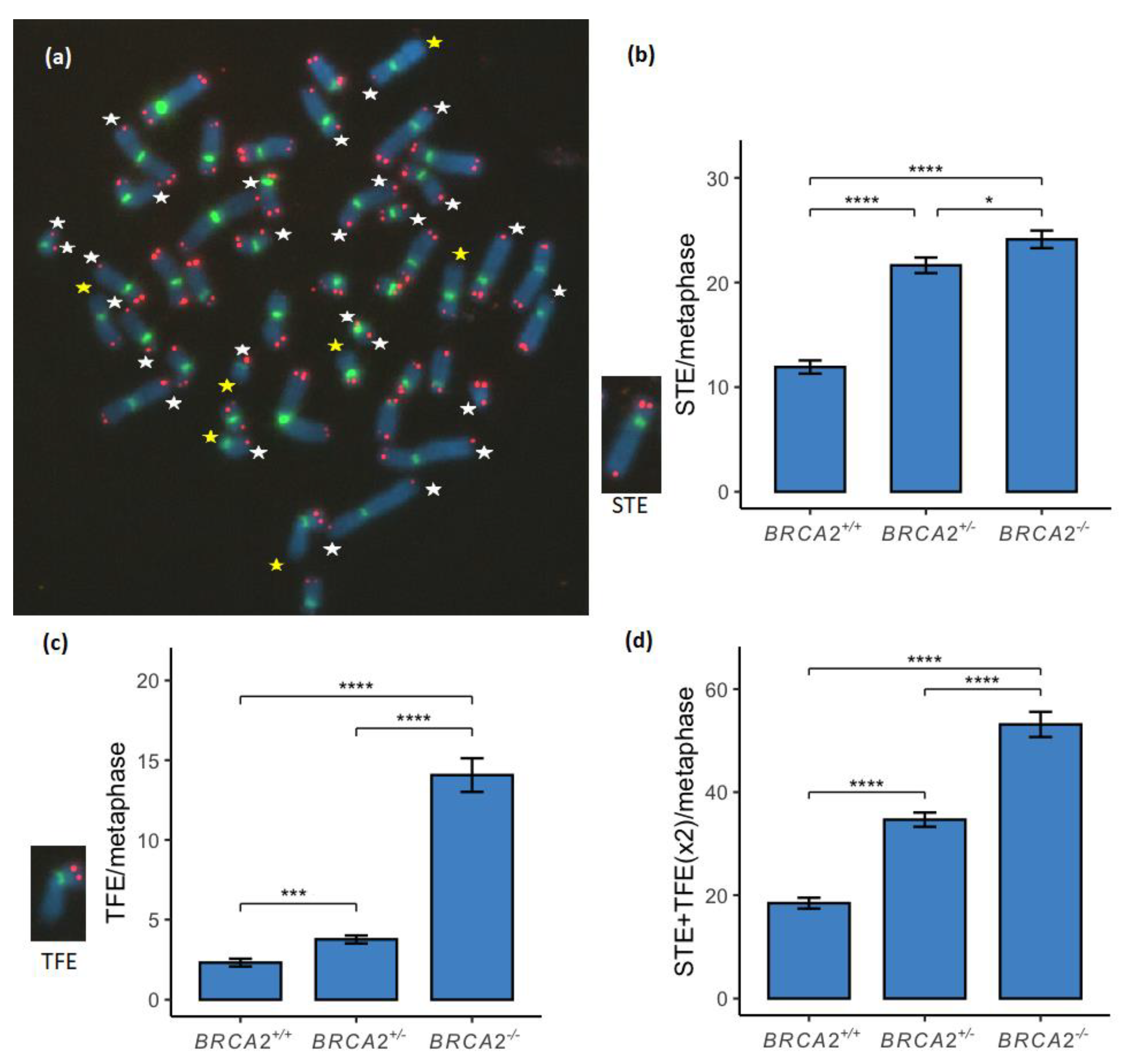
Figure 1. Telomere loss (TL) compared between the three BRCA2 genotypes of lymphoid cell lines. (a) In each metaphase single telomere end (STE) was measured as a loss of a telomere signal (red) from a single sister chromatid (white stars) and telomere free ends (TFE) on chromosomes were measured when both telomere signals from a chromosome end were lost (yellow stars). (b) STE was significantly increased between the wild-type and the BRCA2+/- genotypes and between the wild-type and the BRCA2-/- genotypes (p < 0.0001), and significance was also found between the BRCA2+/- and BRCA2-/- genotypes (p < 0.05). (c) TFE was significantly increased between the three genotypes of BRCA2+/+, BRCA2+/- and BRCA2-/- in a stepwise manner (p < 0.001 and p < 0.0001, respectively). (d) TL was measured by a combination of numbers of STE and TFE duplicated. TL was significantly increased between the three genotypes of BRCA2+/+, BRCA2+/- and BRCA2-/- in a stepwise manner (p < 0.0001). * corresponds to p < 0.05, *** corresponds to p < 0.001 and **** corresponds to p < 0.0001. Metaphase chromosomes were analyzed with FISH by Cy3-conjugated telomere probe and FITC-conjugated centromere probe and counterstained with DAPI. The metaphase shown is from the EB1690 BRCA2+/- lymphoid cell line.
Genes 13 00083 g001
Figure 2. Multitelomeric signals (MTS) on chromosome ends compared between the three BRCA2 genotypes of lymphoid cell lines. (a) Single-MTS (SMTS) on chromosome ends were measured as an MTS on a single sister chromatid (white triangles) and double-MTS (DMTS) on chromosome ends when both sister chromatids had MTS on a chromosome end (yellow triangles). (b) SMTS was significantly increased between the BRCA2 wild-type and the BRCA2+/- lymphoid cell lines (p < 0.0001) and the BRCA2-/- lymphoid cell lines (p < 0.001). A significant difference was found between the BRCA2+/- and BRCA2-/- genotypes (p < 0.01). (c) DMTS were significantly increased between the three genotypes of BRCA2+/+, BRCA2+/- and BRCA2-/- in a stepwise manner (p < 0.001 and p < 0.01, respectively) and a significant difference was also found between the BRCA2+/+ and BRCA2-/- genotypes (p < 0.001). (d) Total MTS were measured by a combination of numbers of SMTS and DMTS duplicated. MTS was significantly increased between the BRCA2 wild-type, BRCA2+/- and BRCA2-/- genotypes (p < 0.0001). No difference was, however, found between the BRCA2+/- and BRCA2-/- genotypes in total MTS. ** corresponds to p < 0.01, *** corresponds to p < 0.001 and **** corresponds to p < 0.0001. Metaphase chromosomes were analyzed with FISH by Cy3-conjugated telomere probe and FITC-conjugated centromere probe and counterstained with DAPI. The metaphase shown is from the EB2302 BRCA2+/- lymphoid cell line.
Genes 13 00083 g002
Figure 3. Interstitial telomere sequences (ITS) were compared between the BRCA2 genotypes. (a) ITS were measured as signals within a chromosome arm (white arrows). (b) ITS were found to be significantly increased between the genotypes of BRCA2+/+, BRCA2+/- and BRCA2-/- in a stepwise manner (p < 0.05 and p < 0.0001, respectively). * corresponds to p < 0.05 and **** corresponds to p < 0.0001. Metaphase chromosomes were analyzed with FISH by Cy3-conjugated telomere probe and FITC-conjugated centromere probe and counterstained with DAPI. The metaphase shown is from the EB6085 BRCA2+/- lymphoid cell line.
Genes 13 00083 g003
Figure 4. Extrachromosomal telomere sequences (ECTS) were compared between the BRCA2 genotypes. (a) ECTS were measured as telomeric signals (red) scattered around the chromosomes (white arrowheads). (b) ECTS were found to be significantly increased between the genotypes of BRCA2+/+, BRCA2+/- and BRCA2-/- in a stepwise manner (p < 0.0001). **** corresponds to p < 0.0001. Metaphase chromosomes were analyzed with FISH by Cy3-conjugated telomere probe and FITC-conjugated centromere probe and counterstained with DAPI. The metaphase shown is from the EB2302 BRCA2+/- lymphoid cell line.
Genes 13 00083 g004
Table 1. List of lymphoid cell lines used in the study including information of the BRCA2 genotype and mutation(s).
Lymphoid Cell LinesBRCA2 Genotype 1BRCA2 Mutation(s)
EB0392+/+None
EB6457+/+None
EB1482+/-999del5 2
EB1690+/-999del5
EB1830+/-999del5
EB2302+/-999del5
EB6085+/-999del5
HSC62N 3-/-IVS 19-1 G > AIVS 19-1 G > A
NORD 4-/-886delGT8447T > A
SPAN 5-/-15-16 exons del1597del
1BRCA2 genotypes: +/+ BRCA2 wild-type, +/- BRCA2 monoallelic mutation and -/- BRCA2 biallelic mutations. 2 The official mutation name is rs80359671, NM_000059.3:c.767_771delCAAAT. 3 [13]. 4 Lymphoid cell line IFAR 772/1 from patient FA19 [33]. 5 Lymphoid cell line from patient FA62 [34].
Table 2. Metaphase number analyzed in each lymphoid cell line including information about mean chromosome number and the chromosome number range.
Lymphoid Cell LinesBRCA2 GenotypeMetaphases AnalyzedMean Chromosome NumberChromosome Number Range
EB0392+/+6146.531–92
EB6457+/+6545.431–83
EB1482+/-6448.038–92
EB1690+/-6047.641–92
EB1830+/-6347.626–92
EB2302+/-6246.645–92
EB6085+/-6046.644–93
HSC62N-/-4346.336–87
NORD-/-6344.735–46
SPAN-/-6645.039–47
Publisher’s Note: MDPI stays neutral with regard to jurisdictional claims in published maps and institutional affiliations.