
A Precise Method to Evaluate 360 Degree Measures of Optic Cup and Disc Morphology in an African American Cohort and Its Genetic Applications


Abstract
:1. Introduction
2. Materials and Methods
2.1. Study Population
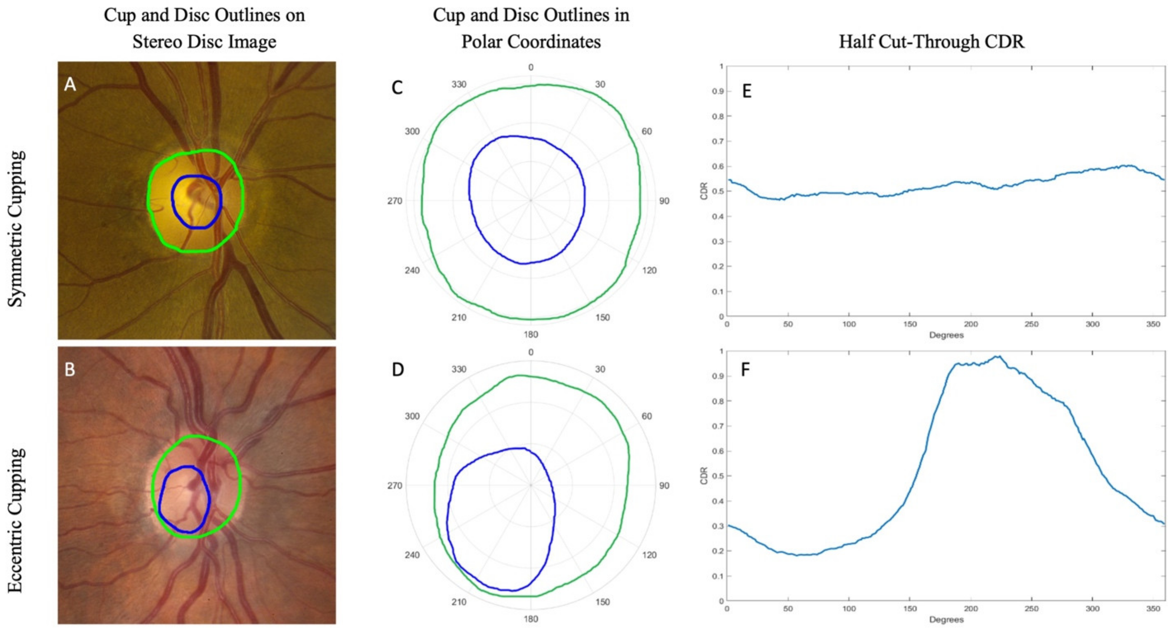
2.2. Outlining of Cup and Disc Boundaries
- (1)
- The optic cup, using only contour and vascular cues (“contour cup”);
- (2)
- The optic disc, defined as the outer border of the nerve rim and the inner border of the scleral ring, if a scleral ring was present.
2.3. Adjudication of Images
2.4. Polar Representation of Cup and Disc Boundaries
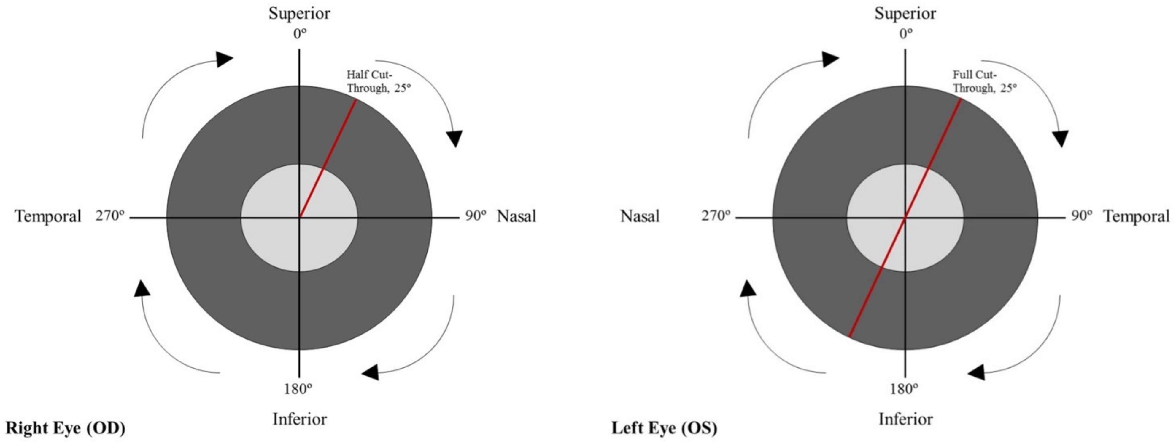
2.5. Half Cut-Throughs and Full Cut-Throughs
2.6. Notching
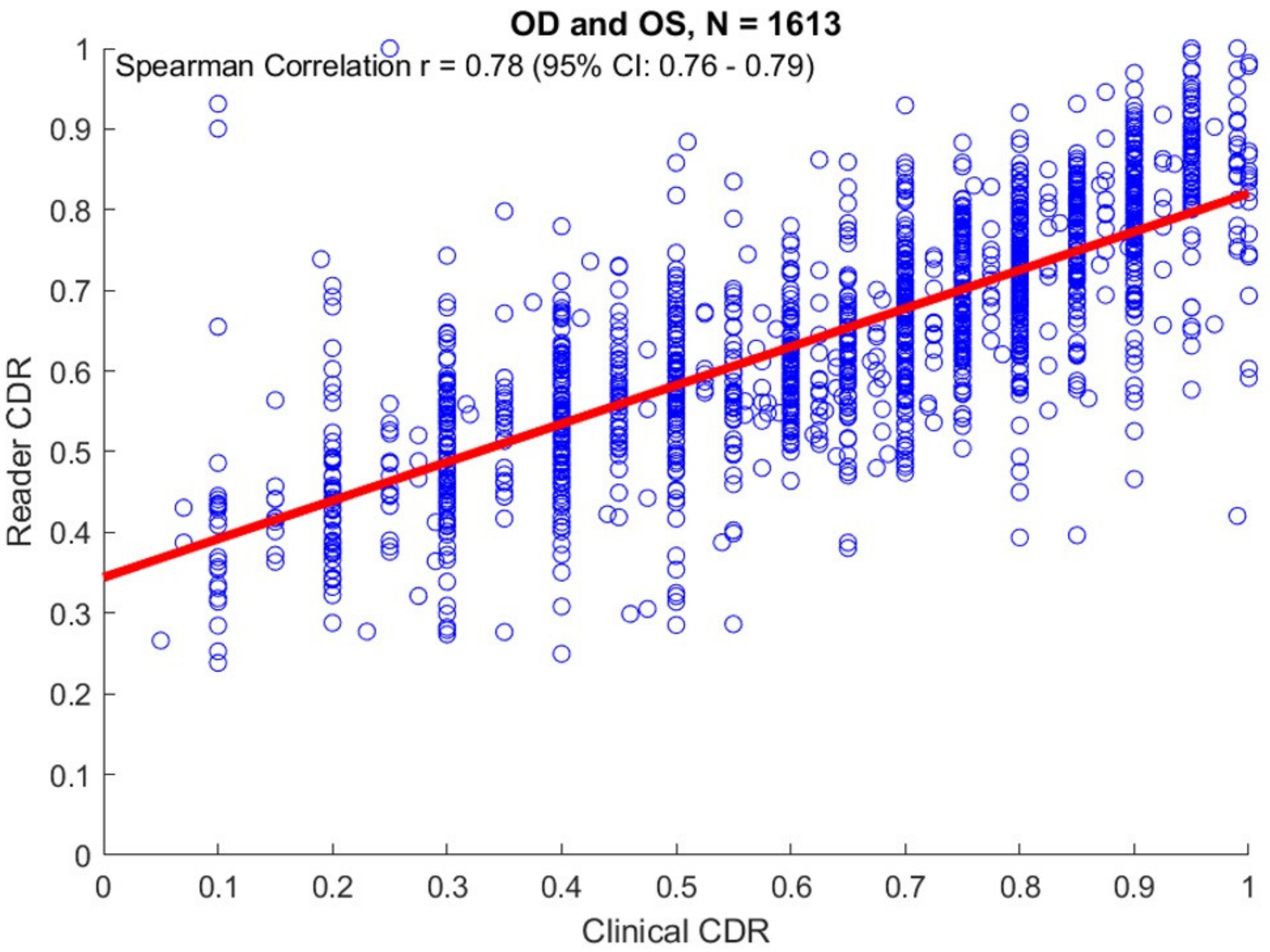
2.7. Correlation with Clinical VCDR Values
2.8. Correlation with Genetic Findings
3. Results
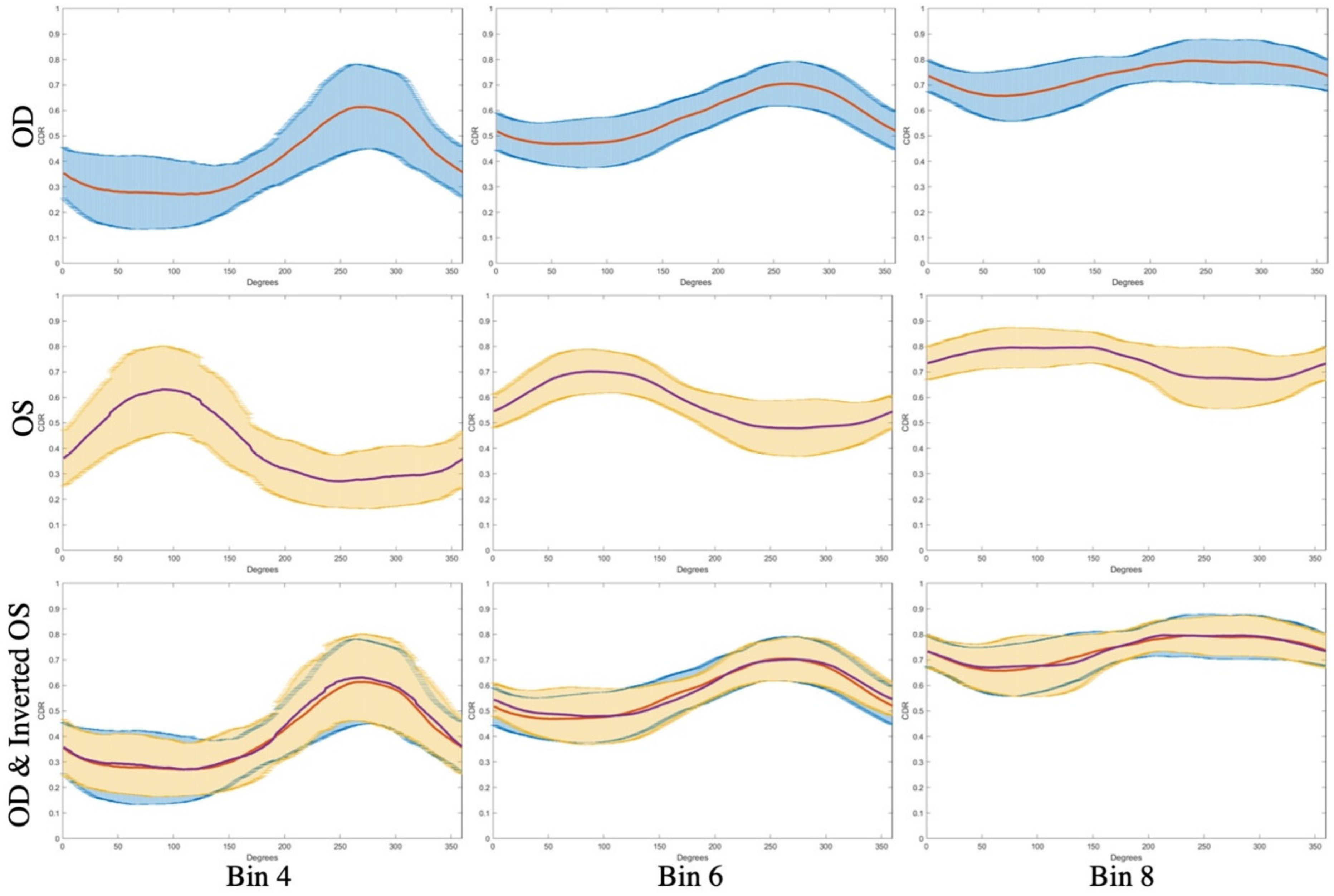
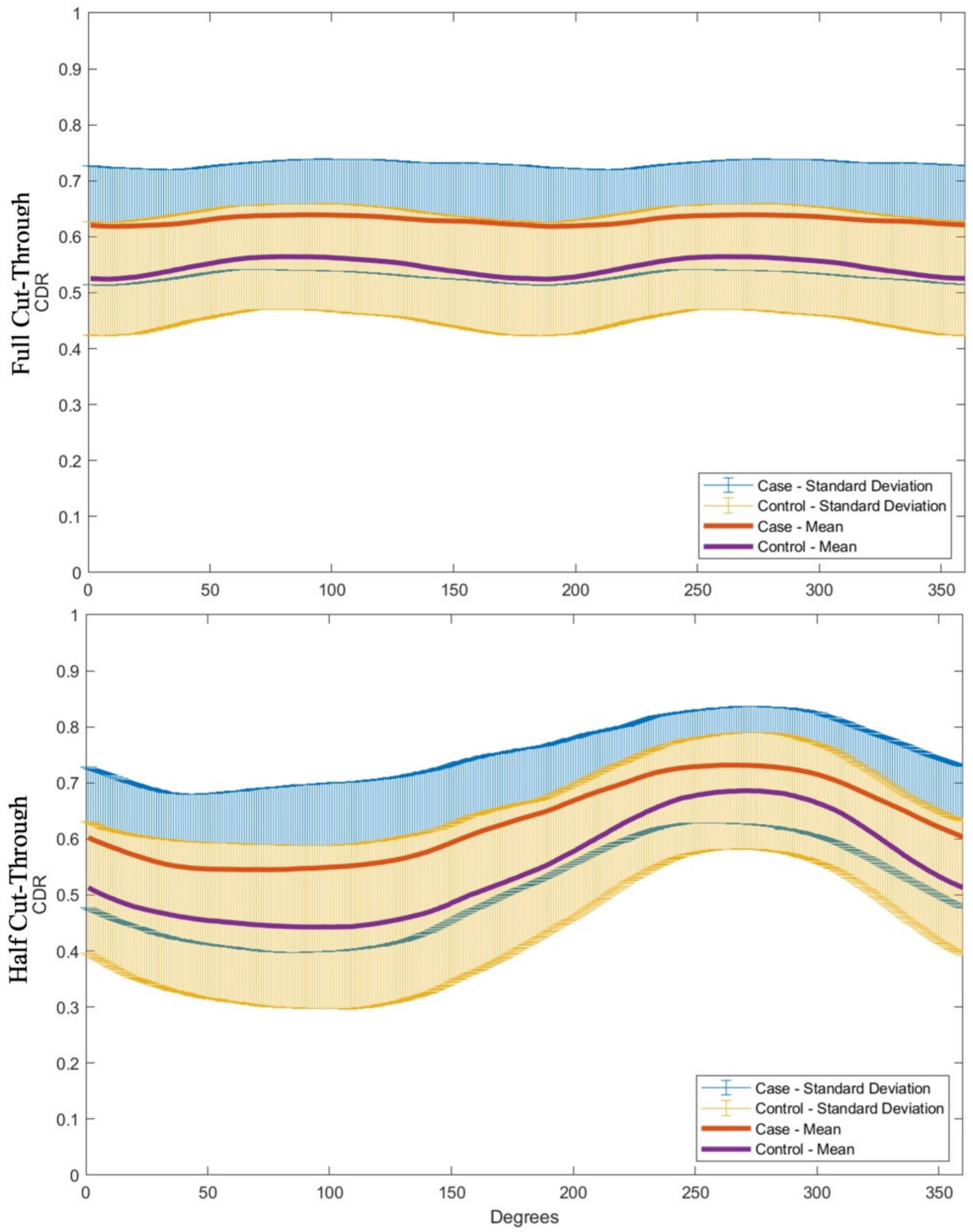
3.1. CDR Analysis
3.2. Genetic Associations
4. Discussion
Supplementary Materials
Author Contributions
Funding
Institutional Review Board Statement
Informed Consent Statement
Data Availability Statement
Conflicts of Interest
References
- Weinreb, R.N.; Leung, C.K.; Crowston, J.G. Primary open-angle glaucoma. Nat. Rev. Dis. Primers 2016, 2, 16067. [Google Scholar] [CrossRef] [PubMed]
- Quigley, H.A. Glaucoma. Lancet 2011, 377, 1367–1377. [Google Scholar] [CrossRef]
- Quigley, H.A.; Addicks, E.M.; Green, W.R. Optic nerve damage in human glaucoma. III. Quantitative correlation of nerve fiber loss and visual field defect in glaucoma, ischemic neuropathy, papilledema, and toxic neuropathy. Arch. Ophthalmol. 1982, 100, 135–146. [Google Scholar] [CrossRef] [PubMed]
- Sommer, A.; Katz, J.; Quigley, H.A. Clinically detectable nerve fiber atrophy precedes the onset of glaucomatous field loss. Arch. Ophthalmol. 1991, 109, 77–83. [Google Scholar] [CrossRef]
- Kass, M.A.; Heuer, D.K.; Higginbotham, E.J. The Ocular Hypertension Treatment Study: A randomized trial determines that topical ocular hypotensive medication delays or prevents the onset of primary open-angle glaucoma. Arch. Ophthalmol. 2002, 120, 701–713. [Google Scholar] [CrossRef] [PubMed]
- Hirooka, K.; Manabe, S.; Tenkumo, K.; Nitta, E.; Sato, S.; Tsujikawa, A. Use of the structure-function relationship in detecting glaucoma progression in early glaucoma. BMC Ophthalmol. 2014, 14, 118–2415. [Google Scholar] [CrossRef] [Green Version]
- Surgucheva, I.; McMahan, B.; Ahmed, F.; Tomarev, S.; Wax, M.B.; Surguchov, A. Synucleins in glaucoma: Implication of γ-synuclein in glaucomatous alterations in the optic nerve. J. Neurosci. Res. 2002, 68, 97–106. [Google Scholar] [CrossRef]
- Cheng, J.; Liu, J.; Xu, Y. Superpixel classification based optic disc and optic cup segmentation for glaucoma screening. IEEE Trans. Med. Imaging 2013, 32, 1019–1032. [Google Scholar] [CrossRef]
- Montgomery, D.M.; Craig, J.P. Optic disc interpretation in glaucoma: Is confidence misplaced? Ophthalmic Physiol. Opt. 1993, 13, 383–386. [Google Scholar] [CrossRef]
- Jindra, L.F.; Kubena, T.; Gaudino, R.N. Analytic methods in assessment of optic nerve cupping. Cesk. Slov. Oftalmol. 2014, 70, 79–88. [Google Scholar] [PubMed]
- Wolfs, R.C.; Borger, P.H.; Ramrattan, R.S. Changing views on open-angle glaucoma: Definitions and prevalences--The Rotterdam Study. Invest. Ophthalmol. Vis. Sci. 2000, 41, 3309–3321. [Google Scholar] [PubMed]
- Kahn, H.A.; Milton, R.C. Alternative definitions of open-angle glaucoma. Effect on prevalence and associations in the Framingham eye study. Arch. Ophthalmol. 1980, 98, 2172–2177. [Google Scholar] [CrossRef] [PubMed]
- Lichter, P.R. Variability of expert observers in evaluating the optic disc. Trans. Am. Ophthalmol. Soc. 1976, 74, 532–572. [Google Scholar]
- Abrams, L.S.; Scott, I.U.; Spaeth, G.L.; Quigley, H.A.; Varma, R. Agreement among optometrists, ophthalmologists, and residents in evaluating the optic disc for glaucoma. Ophthalmology 1994, 101, 1662–1667. [Google Scholar] [CrossRef]
- Harper, R.; Reeves, B.; Smith, G. Observer variability in optic disc assessment, implications for glaucoma shared care. Ophthalmic. Physiol. Opt. 2000, 20, 265–273. [Google Scholar] [CrossRef] [PubMed]
- Nicolela, M.T.; Drance, S.M.; Broadway, D.C.; Chauhan, B.C.; McCormick, T.A.; LeBlanc, R.P. Agreement among clinicians in the recognition of patterns of optic disk damage in glaucoma. Am. J. Ophthalmol. 2001, 132, 836–844. [Google Scholar] [CrossRef]
- Reus, N.J.; Lemij, H.G.; Garway-Heath, D.F. Clinical assessment of stereoscopic optic disc photographs for glaucoma, the European Optic Disc Assessment Trial. Ophthalmology 2010, 117, 717–723. [Google Scholar] [CrossRef] [PubMed]
- Varma, R.; Steinmann, W.C.; Scott, I.U. Expert agreement in evaluating the optic disc for glaucoma. Ophthalmology 1992, 99, 215–221. [Google Scholar] [CrossRef]
- Tielsch, J.M.; Katz, J.; Quigley, H.A.; Miller, N.R.; Sommer, A. Intraobserver and interobserver agreement in measurement of optic disc characteristics. Ophthalmology 1988, 95, 350–356. [Google Scholar] [CrossRef]
- Gaasterland, D.E.; Blackwell, B.; Dally, L.G. The Advanced Glaucoma Intervention Study (AGIS): 10. Variability among academic glaucoma subspecialists in assessing optic disc notching. Trans. Am. Ophthalmol. Soc. 2001, 99, 177–184, discussion 184. [Google Scholar]
- Breusegem, C.; Fieuws, S.; Stalmans, I.; Zeyen, T. Agreement and accuracy of non-expert ophthalmologists in assessing glaucomatous changes in serial stereo optic disc photographs. Ophthalmology 2011, 118, 742–746. [Google Scholar] [CrossRef] [PubMed]
- Addis, V.; Oyeniran, E.; Daniel, E. Non-physician grader reliability in measuring morphological features of the optic nerve head in stereo digital images. Eye 2019, 33, 838–844. [Google Scholar] [CrossRef] [PubMed]
- Stein, J.D.; Talwar, N.; Laverne, A.M.; Nan, B.; Lichter, P.R. Trends in use of ancillary glaucoma tests for patients with open-angle glaucoma from 2001 to 2009. Ophthalmology 2012, 119, 748–758. [Google Scholar] [CrossRef] [PubMed] [Green Version]
- Banister, K.; Boachie, C.; Bourne, R. Can Automated Imaging for Optic Disc and Retinal Nerve Fiber Layer Analysis Aid Glaucoma Detection? Ophthalmology 2016, 123, 930–938. [Google Scholar] [CrossRef] [PubMed] [Green Version]
- Wong, D.K.; Liu, J.; Lim, J.H. Level-set based automatic cup-to-disc ratio determination using retinal fundus images in ARGALI. Conf. Proc. IEEE Eng. Med. Biol. Soc. 2008, 2008, 2266–2269. [Google Scholar]
- Coops, A.; Henson, D.B.; Kwartz, A.J.; Artes, P.H. Automated analysis of heidelberg retina tomograph optic disc images by glaucoma probability score. Invest. Ophthalmol. Vis. Sci. 2006, 47, 5348–5355. [Google Scholar] [CrossRef]
- Wollstein, G.; Garway-Heath, D.F.; Hitchings, R.A. Identification of early glaucoma cases with the scanning laser ophthalmoscope. Ophthalmology 1998, 105, 1557–1563. [Google Scholar] [CrossRef]
- Perera, S.A.; Foo, L.L.; Cheung, C.Y. Cup-to-Disc Ratio From Heidelberg Retina Tomograph 3 and High-Definition Optical Coherence Tomography Agrees Poorly With Clinical Assessment. J. Glaucoma 2016, 25, 198–202. [Google Scholar] [CrossRef]
- Abramoff, M.D.; Alward, W.L.; Greenlee, E.C. Automated segmentation of the optic disc from stereo color photographs using physiologically plausible features. Invest. Ophthalmol. Vis. Sci. 2007, 48, 1665–1673. [Google Scholar] [CrossRef]
- Muramatsu, C.; Nakagawa, T.; Sawada, A. Automated segmentation of optic disc region on retinal fundus photographs: Comparison of contour modeling and pixel classification methods. Comput. Methods Programs Biomed. 2011, 101, 23–32. [Google Scholar] [CrossRef]
- Aquino, A.; Gegundez-Arias, M.E.; Marin, D. Detecting the optic disc boundary in digital fundus images using morphological, edge detection, and feature extraction techniques. IEEE Trans. Med. Imaging. 2010, 29, 1860–1869. [Google Scholar] [CrossRef] [Green Version]
- Yin, F.; Liu, J.; Ong, S.H. Model-based optic nerve head segmentation on retinal fundus images. Conf. Proc. IEEE Eng. Med. Biol. Soc. 2011, 2011, 2626–2629. [Google Scholar]
- Zheng, Y.; Stambolian, D.; O’Brien, J.; Gee, J.C. Optic disc and cup segmentation from color fundus photograph using graph cut with priors. Med. Image. Comput. Comput. Assist. Interv. 2013, 16, 75–82. [Google Scholar] [PubMed] [Green Version]
- Charlson, E.S.; Sankar, P.S.; Miller-Ellis, E. The primary open-angle african american glaucoma genetics study: Baseline demographics. Ophthalmology 2015, 122, 711–720. [Google Scholar] [CrossRef] [Green Version]
- Healey, P.R.; Mitchell, P. Presence of an optic disc notch and glaucoma. J Glaucoma. 2015, 24, 262–266. [Google Scholar] [CrossRef] [PubMed] [Green Version]
- Gudiseva, H.V.; Verma, S.S.; Chavali, V.R.M. Genome wide-association study identifies novel loci in the Primary Open-Angle African American Glaucoma Genetics (POAAGG) study. bioRxiv 2020, 968156. [Preprint]. [Google Scholar]
- Cornes, B.K.; Khor, C.C.; Nongpiur, M.E. Identification of four novel variants that influence central corneal thickness in multi-ethnic Asian populations. Hum. Mol. Genet. 2012, 21, 437–445. [Google Scholar] [CrossRef] [Green Version]
- Ying, G.S.; Maguire, M.G.; Glynn, R.; Rosner, B. Tutorial on Biostatistics: Statistical Analysis for Correlated Binary Eye Data. Ophthalmic. Epidemiol. 2018, 25, 1–12. [Google Scholar] [CrossRef]
- Ying, G.S.; Maguire, M.G.; Glynn, R.; Rosner, B. Tutorial on Biostatistics: Linear Regression Analysis of Continuous Correlated Eye Data. Ophthalmic. Epidemiol. 2017, 24, 130–140. [Google Scholar] [CrossRef] [Green Version]
- Bengtsson, B. The variation and covariation of cup and disc diameters. Acta Ophthalmol 1976, 54, 804–818. [Google Scholar] [CrossRef] [PubMed]
- Jonas, J.B.; Gusek, G.C.; Naumann, G.O. Optic disc, cup and neuroretinal rim size, configuration and correlations in normal eyes. Invest Ophthalmol. Vis. Sci. 1988, 29, 1151–1158. [Google Scholar] [PubMed]
- Tielsch, J.M.; Sommer, A.; Katz, J.; Royall, R.M.; Quigley, H.A.; Javitt, J. Racial variations in the prevalence of primary open-angle glaucoma. The Baltimore Eye Survey. JAMA. 1991, 266, 369–374. [Google Scholar] [CrossRef] [PubMed]
- Broman, A.T.; Quigley, H.A.; West, S.K. Estimating the rate of progressive visual field damage in those with open-angle glaucoma, from cross-sectional data. Invest Ophthalmol. Vis. Sci. 2008, 49, 66–76. [Google Scholar] [CrossRef] [PubMed]
- Harizman, N.; Oliveira, C.; Chiang, A. The ISNT rule and differentiation of normal from glaucomatous eyes. Arch. Ophthalmol. 2006, 124, 1579–1583. [Google Scholar] [CrossRef] [Green Version]
- MacCormick, I.J.C.; Williams, B.M.; Zheng, Y. Accurate, fast, data efficient and interpretable glaucoma diagnosis with automated spatial analysis of the whole cup to disc profile. PLoS ONE 2019, 14, e0209409. [Google Scholar]

| Grader Cup-to-Disc Area Ratio | Grader Vertical Cup-to-Disc Ratio | |||
|---|---|---|---|---|
| SNP (rs ID) | Number of Risk Alleles | # of Eyes | Adjusted Mean (SE) * | Adjusted Mean (SE) * |
| 2:237653539_G (rs12328841) | 0 | 458 | 0.50 (0.01) | 0.69 (0.01) |
| 1 | 584 | 0.50 (0.01) | 0.69 (0.01) | |
| 2 | 180 | 0.47 (0.01) | 0.66 (0.01) | |
| p-value § | 0.17 | 0.08 | ||
| 9:13173885_G (rs4740546) | 0 | 1169 | 0.49 (0.01) | 0.68 (0.00) |
| 1 | 53 | 0.55 (0.02) | 0.73 (0.02) | |
| p-value § | 0.02 | 0.01 | ||
| 10:127738557_G (rs ID-N/A) | 0 | 406 | 0.49 (0.01) | 0.68 (0.01) |
| 1 | 582 | 0.49 (0.01) | 0.68 (0.01) | |
| 2 | 234 | 0.49 (0.01) | 0.68 (0.01) | |
| p-value § | 0.65 | 0.82 | ||
Publisher’s Note: MDPI stays neutral with regard to jurisdictional claims in published maps and institutional affiliations. |
© 2021 by the authors. Licensee MDPI, Basel, Switzerland. This article is an open access article distributed under the terms and conditions of the Creative Commons Attribution (CC BY) license (https://creativecommons.org/licenses/by/4.0/).
Share and Cite
Addis, V.; Chen, M.; Zorger, R.; Salowe, R.; Daniel, E.; Lee, R.; Pistilli, M.; Gao, J.; Maguire, M.G.; Chan, L.; et al. A Precise Method to Evaluate 360 Degree Measures of Optic Cup and Disc Morphology in an African American Cohort and Its Genetic Applications. Genes 2021, 12, 1961. https://doi.org/10.3390/genes12121961
Addis V, Chen M, Zorger R, Salowe R, Daniel E, Lee R, Pistilli M, Gao J, Maguire MG, Chan L, et al. A Precise Method to Evaluate 360 Degree Measures of Optic Cup and Disc Morphology in an African American Cohort and Its Genetic Applications. Genes. 2021; 12(12):1961. https://doi.org/10.3390/genes12121961
Chicago/Turabian StyleAddis, Victoria, Min Chen, Richard Zorger, Rebecca Salowe, Ebenezer Daniel, Roy Lee, Maxwell Pistilli, Jinpeng Gao, Maureen G. Maguire, Lilian Chan, and et al. 2021. "A Precise Method to Evaluate 360 Degree Measures of Optic Cup and Disc Morphology in an African American Cohort and Its Genetic Applications" Genes 12, no. 12: 1961. https://doi.org/10.3390/genes12121961
APA StyleAddis, V., Chen, M., Zorger, R., Salowe, R., Daniel, E., Lee, R., Pistilli, M., Gao, J., Maguire, M. G., Chan, L., Gudiseva, H. V., Zenebe-Gete, S., Merriam, S., Smith, E. J., Martin, R., Parker Ostroff, C., Gee, J. C., Cui, Q. N., Miller-Ellis, E., ... Sankar, P. S. (2021). A Precise Method to Evaluate 360 Degree Measures of Optic Cup and Disc Morphology in an African American Cohort and Its Genetic Applications. Genes, 12(12), 1961. https://doi.org/10.3390/genes12121961