Case Report

A Case of Inherited t(4;10)(q26;q26.2) Chromosomal Translocation Elucidated by Multiple Chromosomal and Molecular Analyses. Case Report and Review of the Literature

1
Medical Genetics Department, “Grigore T. Popa” University of Medicine and Pharmacy, 16 University Street, 700115 Iasi, Romania
2
“C. I. Parhon” National Institute of Endocrinology, 34-35 Aviatorilor Avenue, 011853 Bucharest, Romania
3
Personal Genetics Laboratory Bucharest, 4 Strada Frumoasa Street, 010987 Bucharest, Romania
4
Medical Genetics Department, “Carol Davila” University of Medicine and Pharmacy, 8 Eroii Sanitari Avenue, 050474 Bucharest, Romania
5
Medical Genetics Department, “Cuza-Vodă” Obstetrics and Gynecology Hospital, 34 Cuza Voda Street, 700038 Iasi, Romania
6
Microscopic Morphology Department, “Victor Babes” University of Medicine and Pharmacy, 2 Piata Eftimie Murgu, 300041 Timișoara, Romania
7
Neonatology Department, “Grigore T. Popa” University of Medicine and Pharmacy, 16 University Street, 700115 Iasi, Romania
8
Stefan S. Nicolau Institute of Virology, Romanian Academy, 285 Mihai Bravu, 030304 Bucharest, Romania
*
Authors to whom correspondence should be addressed.
Genes 2021, 12(12), 1957; https://doi.org/10.3390/genes12121957
Submission received: 4 November 2021 / Revised: 2 December 2021 / Accepted: 6 December 2021 / Published: 7 December 2021
(This article belongs to the Section Genetic Diagnosis)

Abstract

:
We present a complex chromosomal anomaly identified using cytogenetic and molecular methods. The child was diagnosed during the neonatal period with a multiple congenital anomalies syndrome characterized by: flattened occipital region; slight turricephaly; tall and broad forehead; hypertelorism; deep-set eyes; down slanting and short palpebral fissures; epicanthic folds; prominent nose with wide root and bulbous tip; microstomia; micro-retrognathia, large, short philtrum with prominent reliefs; low set, prominent ears; and congenital heart disease. The GTG banding karyotype showed a 46,XY,der(10)(10pter→10q26.2::4q26→4qter) chromosomal formula and his mother presented an apparently balanced reciprocal translocation: 46,XX,t(4;10)(q26;q26.2). The chromosomal anomalies of the child were confirmed by MLPA, and supplementary investigation discovered a quadruplication of the 4q35.2 region. The mother has a triplication of the same chromosomal fragment (4q35.2). Using array-CGH, we described the anomalies completely. Thus, the boy has a 71,057 kb triplication of the 4q26–q35.2 region, a 562 kb microdeletion in the 10q26.3 region, and a 795 kb quadruplication of the 4q35.2 region, while the mother presents a 795 kb triplication of the 4q35.2 region. Analyzing these data, we consider that the boy’s phenotype is influenced only by the 4q partial trisomy. We compare our case with similar cases, and we review the literature data.

1. Introduction

Congenital anomalies are structural defects produced during the prenatal period and are present at birth [1]. Usually, the severe syndromes with congenital anomalies (SCAs) are generated by an unbalanced chromosomal rearrangement that disrupts large numbers of developmentally important genes, which results in specific and complex phenotypes. The confirmation of a chromosomal anomaly requires GTG-banded chromosomal analysis in association with supplementary analyses, such as FISH, MLPA, or array-CGH [2,3].
Partial trisomy 4q is a rare unbalanced structural chromosomal anomaly that was first described in the early 1970s [4,5,6]. Since then, less than 100 cases have been described. Some anomalies were de novo, but the majority are derived from the malsegregation of a parental balanced chromosomal anomaly. In such cases, the phenotype is induced by the association of 4q partial trisomy with partial monosomy of a different chromosome [7]. The delineation of genotype–phenotype correlation in 4q trisomy is difficult because the described cases had different breakpoints and presented different associated monosomies and the ages of reported patients were variable [8,9].
Partial 10q monosomy was first described by Lewandowski et al. (1978), and since then, about 110 cases have been reported in the literature [10]. The chromosomal anomalies are: terminal 10q monosomy, terminal 10q monosomy associated with a partial trisomy of a different chromosome, and 10q26.1 microdeletion. The symptomatology is variable and depends on the type of anomaly, the fragment size, and coexistence with other anomalies. The phenotypic spectrum is heterogeneous, and it is characterized by some common clinical features, such as craniofacial anomalies, developmental delay, intellectual disability, urinary tract abnormalities, and heart defects [11,12,13,14,15,16].
We present a case with a multiple congenital anomalies syndrome, identified during the neonatal period, that has an association between partial 4q trisomy and partial 10q monosomy. These anomalies were generated by the malsegregation of the maternal chromosomes 4 and 10, which are involved in a balanced reciprocal translocation. The 4q fragment located on the derivative chromosome 10 also presents a small 4q35.2 duplication, also inherited from the mother.

2. Materials and Methods

2.1. Clinical Evaluation

The patient underwent a multidisciplinary evaluation, including the following specialties: genetics, pediatrics, cardiology, and neonatology.
The patient’s parents gave written informed consent (including for publication of images) considering the Declaration of Helsinki. This study was approved by the Ethics Committee for Scientific Research of the “Grigore T. Popa” University of Medicine and Pharmacy, Iasi, Romania.

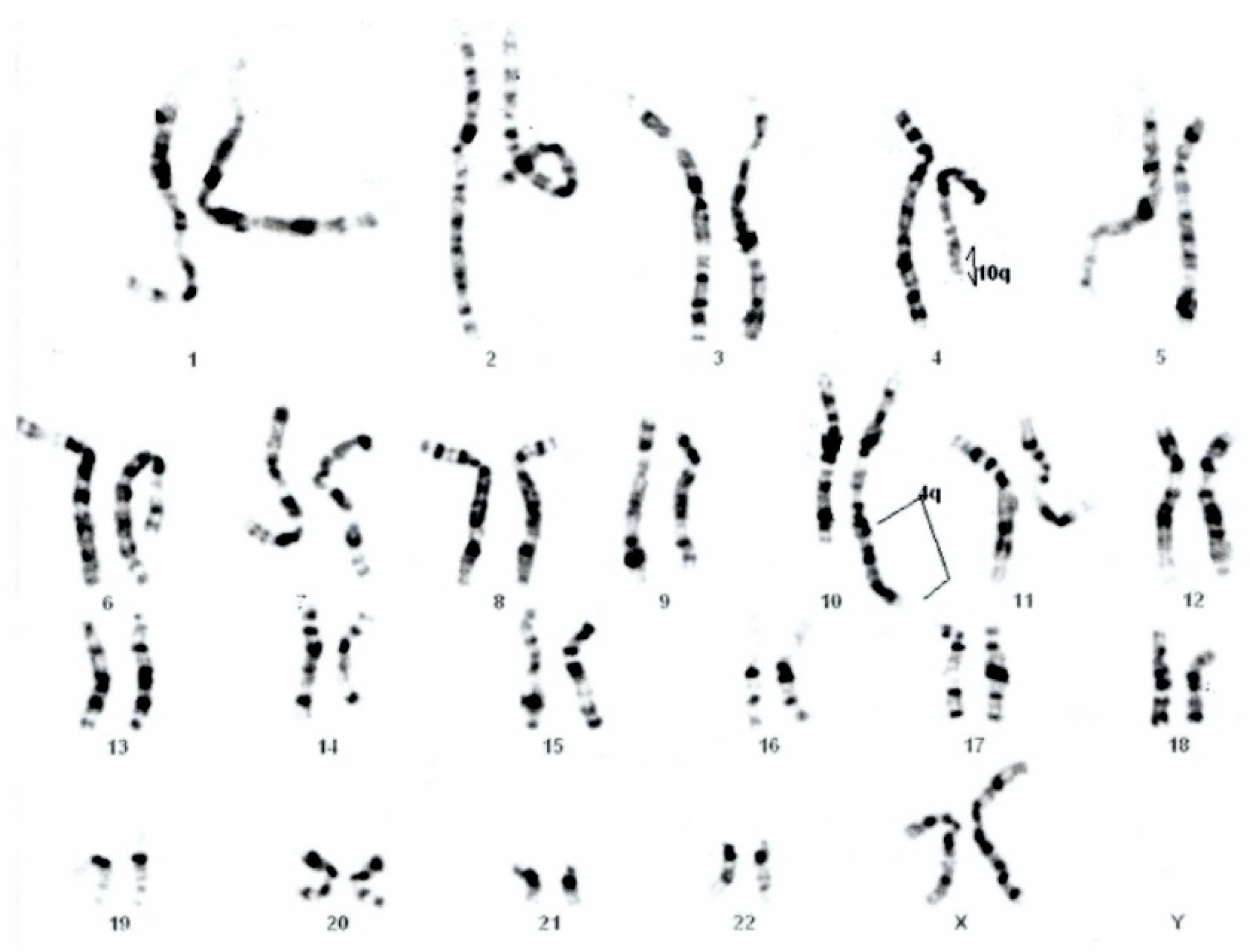
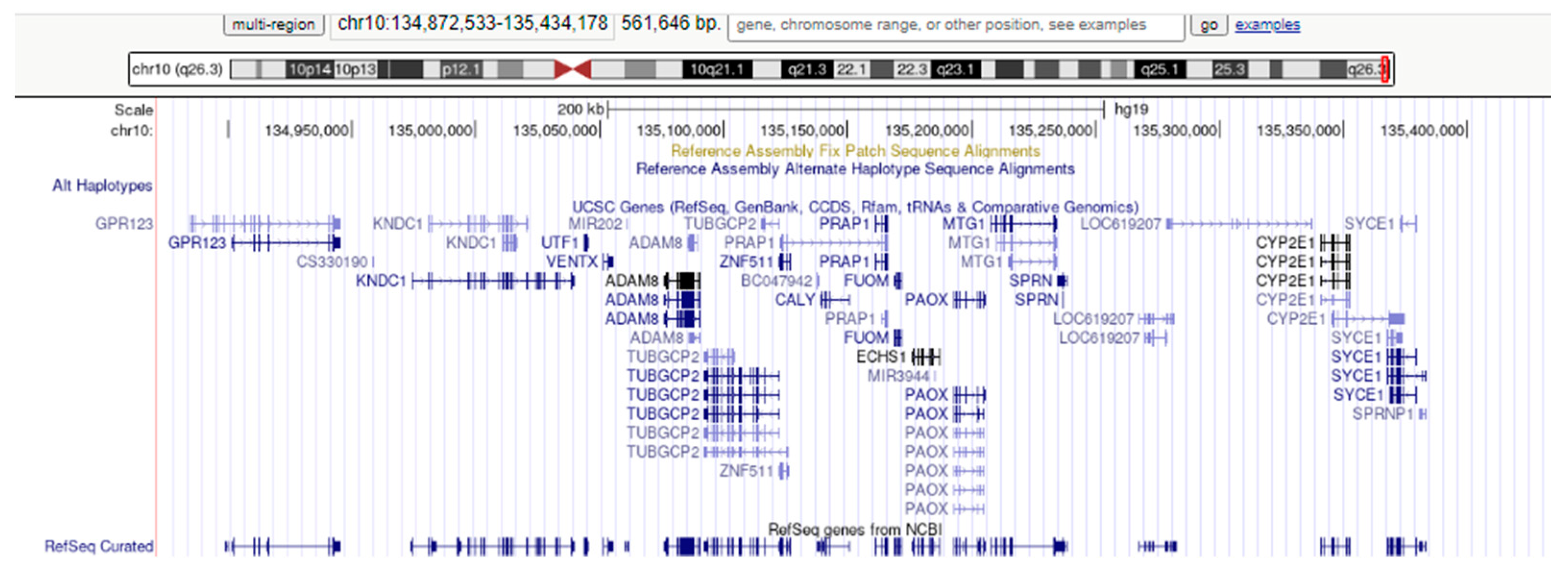
2.2. Cytogenetic Analyses

Peripheral blood lymphocytes were cultured in a growth medium (RPMI 1640, Gibco, Thermo Fisher Scientific Inc, Waltham, MA, USA). Metaphase chromosomes were harvested, and slides were made for analysis. GTG banding was used for staining (at the 550-band level). Chromosomal analysis was performed using CytoVision software 2.81 (Applied Imaging, Grand Rapids, MI, USA), and the aberrations and karyotypes were classified according to the International System for Human Cytogenomic Nomenclature (ISCN 2016).

2.3. MLPA Technique

We used commercially available P036 and P070 SALSA MLPA kits (MRC-Holland, Amsterdam, The Netherlands). These kits screen for subtelomeric copy number variations and contain one MLPA probe for each subtelomeric region except for the short arms of acrocentric chromosomes (13p, 14p, 15p, 21p, and 22p), for which a probe on the q arm, close to the centromere, is included instead. To confirm the abnormalities detected, we applied kits P264 (for subtelomeric regions of chromosomes 1q, 2q, 3q, and 4q) and P286 (for subtelomeric regions of chromosomes 9q, 10q, 11q, and 12q) (MRC-Holland, Amsterdam, The Netherlands).
The DNA extraction from peripheral blood was performed using the Wizard Genomic DNA Purification Kit (Promega Corp., Madison, WI, USA). The standard MLPA analysis was performed according to the manufacturer’s instructions. Briefly, 300 nanograms of genomic DNA were denatured and hybridized with SALSA probes at 60 °C for 16 h. After 15 min ligation at 54 °C, PCR was performed in a Gradient Palm-Cycler (Corbett Research, Mortlake, NSW, Australia), available in a 96-well format, using Cy5 universally labeled primers. Fluorescent amplification products were subsequently separated through capillary electrophoresis in a CEQ 8000 GeXP Genetic Analysis System (Beckman Coulter, Brea, CA, USA) sequencer and were analyzed using the default software. The number of DNA copies was estimated using the Coffalyser.NET V9 software (MRC-Holland, Amsterdam, The Netherlands), which calculates the ratio of peak areas in test samples over those of normal controls for each target sequence. Reference DNA samples were collected from healthy individuals (without developmental delay, intellectual disabilities, or congenital abnormalities), prepared using the same DNA extraction method, and previously evaluated for copy number variation of target areas. There was a minimum of 3 reference samples per run, randomly distributed, with one more for every 7 test samples.

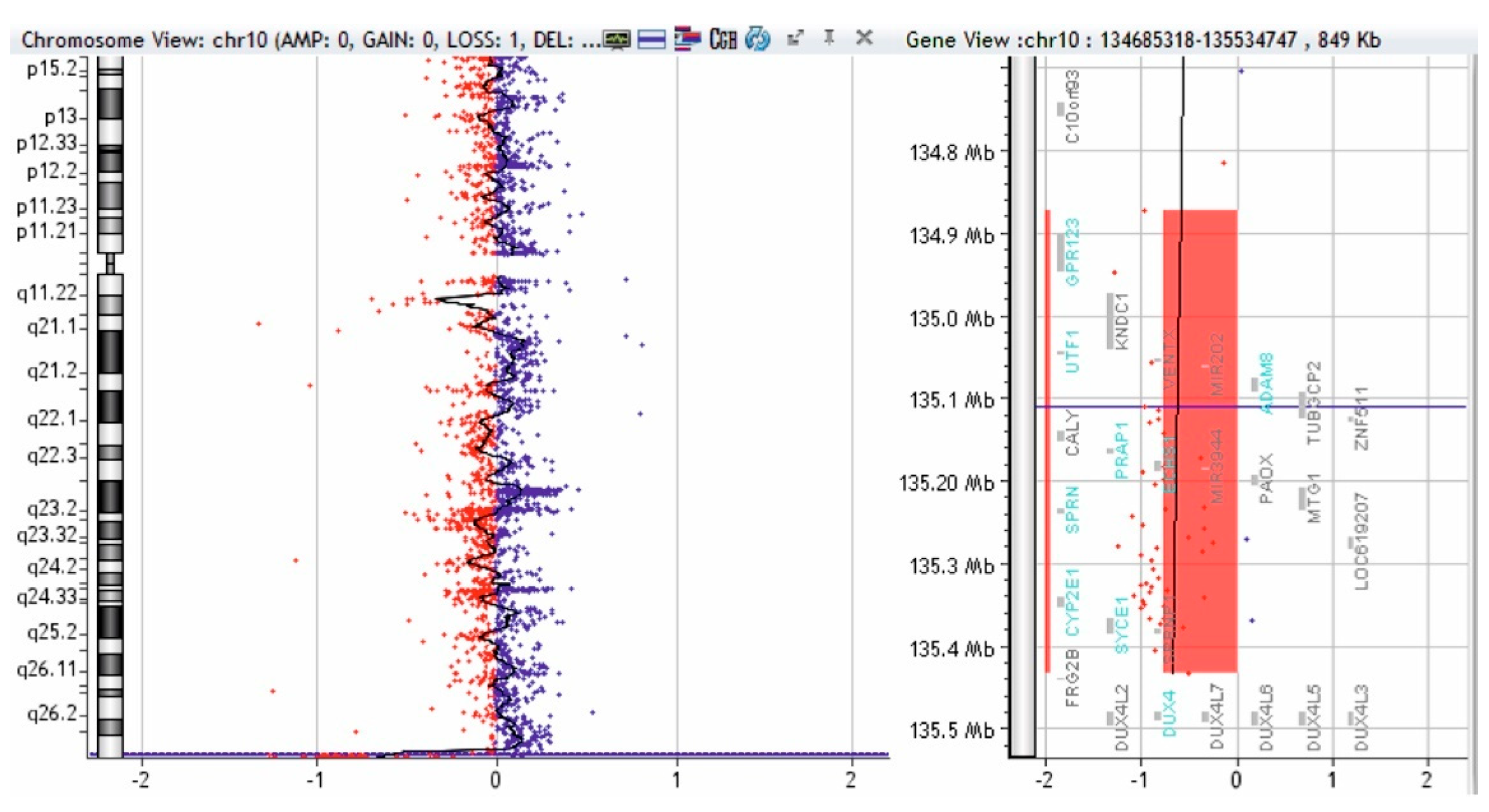
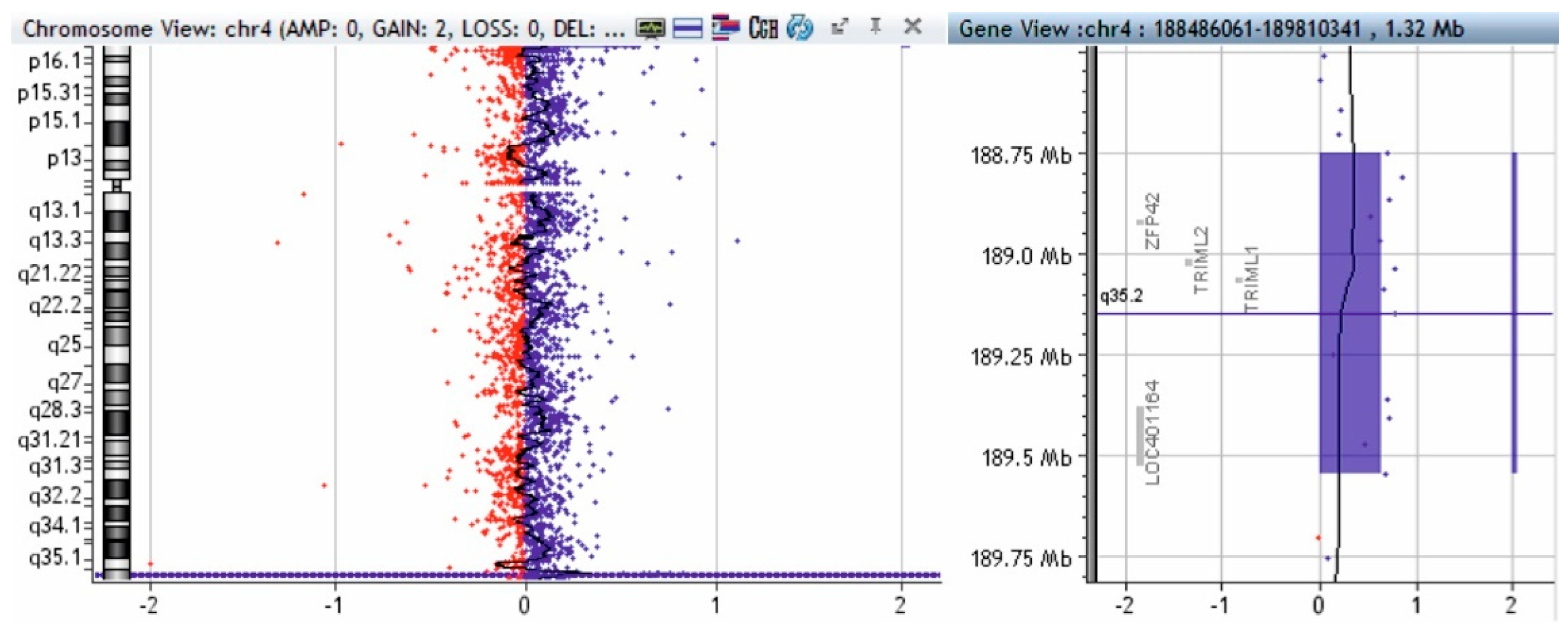
2.4. Array-CGH Technique

Array Comparative Genomic Hybridization (aCGH) analysis was performed using the Sure Print G3 ISCA V2 CGH 8x60K Array Kit (Agilent Technologies®, Santa Clara, CA, USA), the NimbleGen MS 200 Microarray Scanner (Roche Applied Science, Basel, Switzerland), and NimbleGen MS 200 Software v1.1.

3. Results

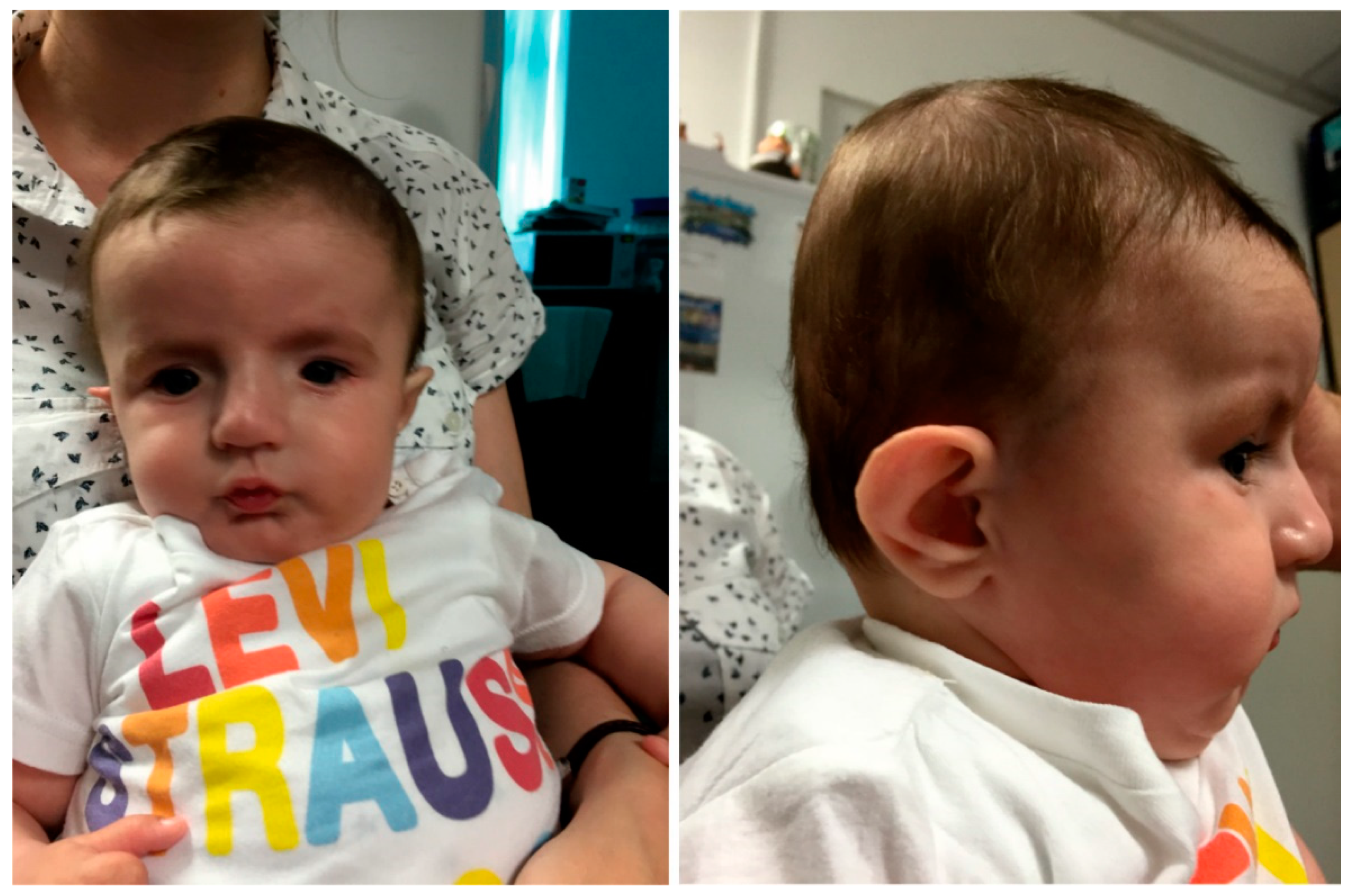
Clinical Presentation

We present a boy with a mild development delay and multiple congenital anomalies syndrome, the result of a pregnancy supervised clinically and by prenatal screening. Prenatal ultrasound scan (second trimester) was normal and biochemical screening (maternal serum markers—alpha-fetoprotein, human chorionic gonadotropin, and unconjugated estriol at 16th weeks of pregnancy) showed values without risk for chromosomal abnormalities. Parents are both healthy, young (father—32 years, mother—28 years), unrelated, with no “known” family history of genetic disorders or birth defects. The child is the first offspring of the couple, and he was born at 38 WA, with a weight of 2350 gr, height 50 cm, head circumference 35 cm, and an Apgar score of 8 at 1 min. and 8 at 5 min. The patient was first examined during the neonatal period and was re-evaluated at 6 months. He presented some facial dysmorphic features: flattened skull in the occipital region, with slight turricephaly; tall and broad forehead; hypertelorism—inner canthal distance 2.7 cm (+2SD), outer canthal distance 6.3 cm (+2SD); interpupillary distance 4.6 cm (+2SD); deep-set eyes, down slanting and short palpebral fissures; epicanthic folds; prominent nose, with wide root and bulbous tip; microstomia and micro-retrognathia; large, short philtrum with prominent reliefs,; low set, prominent ears, with big scaphoid fossa and big cavum concha (Figure 1). The following skills were developed: follows moving objects with eyes; listens to and follows simple directions; cries, babbles, and coos; reaches for objects and grasps; puts objects in mouth; lifts head and sits with back straight; smiles spontaneously. The ultrasound scan revealed a persistent ductus arteriosus (ΔP = 5 mm, velocity = 1 m/s, with left-to-right shunting) and atrial septal defect ostium secundum (3 mm, with left-to-right shunting), but without changes in kidneys. The cardiologist decided that no surgery was needed.
GTG banding showed a 46,XY,der(10)(10pter→10q26.2::4q26→4qter) chromosomal formula (Figure 2) that imposed parental karyotype. The father was normal, but the mother have a balanced reciprocal translocation between chromosomes 4 and 10: 46,XX,t(4;10)(q26;q26.2) (Figure 3).
Using the MLPA technique with telomere probes (P036 and P070, MRC-Holland, Amsterdam, The Netherlands), we discovered a 1.5 fold amplification of the 4q35.2-4q35.1 segment (concordant with a 4q partial trisomy) and a 0.5 fold amplification of the 10qter segment (concordant with a partial 10q monosomy); we also discovered a 2-fold amplification of the 4q35.2 segment (concordant with a 4q partial tetrasomy—a segment that includes the ZPF42 and TRIML2 genes). To validate these changes, we used MLPA probe P264 (for subtelomeric regions of chromosomes 1q, 2q, 3q, and 4q) and probe P286 (for subtelomeric regions of chromosomes 9q, 10q, 11q, and 12q).
To complete the genetic investigation, we applied an array-CGH that confirmed a 71,057 kb triplication of the 4q26-q35.2 region (genomic coordinates on chromosome 4: 119839900—190896674) (Figure 4), a 562 kb microdeletion of the 10q26.3 region (genomic coordinates on chromosome 10: 134872533—135434178) (Figure 5), and a 795 kb quadruplication of the 4q35.2 region (Figure 6).
The triplication of 71,057 kb of the 4q26–q35.2 region was pathogenic. The microdeletion of 562 kb in the 10q26.3 region is probably not pathogenic. To verify if the 4q35.2 quadruplication is pathogenic, we performed array-CGH on both parents, and the mother presented a triplication of 795 kb in the 4q35.2 region (Figure 7).

4. Discussion

We present a case that associates an important 4q partial trisomy (4q26–q35.2) and a small 10q partial monosomy (10q26.3). These chromosomal anomalies originate from the mother, who carries a balanced reciprocal translocation t(4q;10q)(q26;q26.3). Moreover, the son presents a 795 kb quadruplication in the 4q35.2 region, inherited from the maternal line. Due to the small size of the 10q partial monosomy, we could presume that the phenotype of our patient is determined mainly by the 4q partial trisomy.
Our case is the second cited in the literature. Zhang et al. (2009) presented a patient that associated a 4q partial trisomy (4q26–q35.2) and a 10q microdeletion (10q26.3) [7]. The breakpoints on chromosomes 4 and 10 are similar in these two cases. The breakpoints on chromosome 4q are located at 119,839,900 bp from the 4p telomere in our case and at 118,785,802 bp from the 4p telomere in the case presented by Zhang et al. (2009). The breakpoints on chromosome 10q are located at 134,872,533 bp from the 10p telomere in our case and at 134,750,859 bp from the 10 p telomere in the case presented by Zhang et al. (2009). Thus, in both cases, the size of the 4q duplication is approximately 71 Mb, while the 10q microdeletion’s size is approximately 0.55 Mb.
Trisomy 4q syndrome is a heterogeneous disorder because the reported patients have different breakpoints located on the long arm of chromosome 4; some patients associate different monosomies, and the age of reported patients vary between newborn and adulthood [17]. The breakpoints in trisomy 4q syndrome are distributed throughout the long arm of chromosome 4, except for the 4q11 band. The distribution of different types of 4q partial trisomy is presented in Figure 8.
Trisomy 4q syndrome shows a complex phenotype that includes growth retardation, intellectual disability, a specific craniofacial dysmorphism (microcephaly, epicanthic folds, high nasal bridge, short philtrum, microretrognathia, and low set and malformed ears), congenital heart defects (atrial or ventricular septal defect and patent ductus arteriosus), renal defects, and/or thumb anomalies (Table 2).
Celle et al. (2000) analyzed the duplications of the long arm of chromosome 4 and concluded that 4q partial trisomy could be generated by multiple genetic mechanisms, such as unbalanced translocation (the result of malsegregation of parental derivative chromosomes involved in a balanced reciprocal translocation) de novo duplication, de novo unbalanced translocation, and, more rarely, a chromosomal insertion [17]. Table 1 illustrates the heterogeneity of chromosomal anomalies in 4q partial trisomy. Thus, in 22 cases, the anomaly was de novo (16 cases with de novo duplication and 6 cases with de novo translocation). The other cases were inherited anomalies/unknown status of inheritance, and most of them (28 cases) are represented by different translocations between chromosome 4 and other chromosomes. Out of the translocations (de novo or inherited), the most frequent are: t(4;18) and t(4;21)—4 cases; t(4;13), t (2;4) and t(4;10)—3 cases; and t(4;8), t(4;9), and t(4;20), each of them with two cases.
Figure 8 shows the heterogeneity of the breakpoints’ position on the long arm of chromosome 4. Thus, we can divide 4q partial trisomy into four categories: proximal (between 4q12 and 4q23), intermediate (between 4q22–23 and 4q31), terminal (between 4q31 and 4qter), and large 4q partial trisomy (between 4q21–25 and 4qter). Phenotypic changes generated by these anomalies are different, and we will analyze them separately.
The proximal 4q partial trisomies are represented by the cases 1 [18], 2 [19], 11 [28], 19 [35], and 30 [43] from Table 1 and Figure 8. In all these cases, the phenotype is milder and the main characteristics are growth and psychomotor retardation, microcephaly, and low set malformed ears. For example, Shashi et al. (1999) reported a boy with a 4q12–q13 duplication that presented microcephaly and intellectual disability as main features [28]. Additionally, minimal phenotypic changes were reported by Bonnet et al. (2006), who presented a child with a 4q11–4q13.2 partial trisomy that had only a mild psychomotor delay [61]. There were similar findings by Matoso et al. (2013), who presented two siblings that have a 4q13.1–q13.3 partial trisomy that presented only delayed developmental milestones and attention-deficit hyperactivity disorder [62].
Additionally, a mild phenotype seems to be associated with a 4q partial trisomy of the 4q23–q27 region. Thus, Case 3 from Table 2 and Figure 8, reported by Halal et al. (1991), presented growth and psychomotor delay and nonspecific dysmorphic features such as epicanthic folds and a high nasal bridge [20].
Cases 4 [21], 5 [22], 6 [23], 7 [24], 8 [25], 12 [29], and 17 [33] from Table 2 and Figure 8 represent the intermediate 4q partial trisomy of the 4q23–q31 region. All these cases presented a severe phenotype characterized by: growth retardation (7/7), intellectual disability (7/7), high nasal bridge (7/7), epicanthic folds (6/7), low set malformed ears (6/7), micrognathia (5/7), thumb anomalies (5/7), and short philtrum (5/7). However, the genes located in this region seem to have a lesser impact on the development of the heart because only one of these patients presented a congenital heart defect. Lundin et al. (2002) presumed that the 4q27.4–q31 partial trisomy induced the most severe clinical effects, including growth retardation, intellectual disability, microcephaly, facial asymmetry, thumb anomalies, hearing impairment, epilepsy, and congenital heart defects [31].
Terminal 4q partial trisomies are represented by Cases 13 [30], 14 [30], 20 [36], 21 [37], 23 [38], 26 [39], 28 [41], 29 [42], 31 [44], 37 [49], 41 [53], 42 [54], 43 [55], 44 [56], 45 [57], 47 [59], 48 [59], and 49 [60] (Table 2, Figure 8). The majority of these encompassed the 4q31-q35(qter) region. The main phenotypic changes were represented by low set malformed ears (16/18), intellectual disability (17/18), high nasal bridge (13/18), growth retardation (14/18), microcephaly (11/18) epicanthic folds (7/18), and micrognathia (8/18). Thumb anomalies (6/18) and short philtrum (5/18) are rare, while cardiac heart defects were found in only 4 cases. Renal anomalies could also be considered less frequent, being found in 6/18 cases.
The majority of the cases presented in Table 2 and Figure 8 are represented by large 4q partial trisomies that encompassed at least the 4q27–qter region (Cases 15 [31], 16 [32], 22 [6], 24 [7], 25 [9], 27 [40], 32 [45], 33 [46], 34 [47], 35 ([48], case A), 36 ([48], case B), 38 [50], 39 [51], 40 [52], 46 [58], and our case). The phenotype of these patients is severe, being correlated with the large amount of genetic material present in the three copies. The major anomalies reported are: growth retardation (15/16), low set malformed ears (13/16), intellectual disability (13/16), microcephaly (14/16), high nasal bridge (13/16), and micrognathia (12/16). Epicanthic folds (7/16) and short philtrum (6/16) are not very frequent. Visceral anomalies are relatively frequent, congenital heart disease having been documented in 7 cases and renal anomalies in 6 cases.
Rinaldi et al. (2005) discussed the correlation between 4q partial trisomy and the phenotype and observed an important clinical heterogeneity correlated with the variability of the duplicated region. They considered that psychomotor and growth retardation, retromicrognathia, and low set and/or malformed ears are non-specific clinical findings associated with chromosomal unbalances [40].
The relationship between 4q partial trisomy and renal anomalies has been discussed by different authors but still lacks certitude. Zollino et al. (1995) suggested that the 4q22–q23 region is involved in the development of the acrorenal field [19]. On the other hand, Battaglia et al. (2005) considered that the development of the acrorenal field is more likely associated with genes from the 4q25–q28 region [8]. Otsuka et al. (2005) suggested that renal hypoplasia may have a close relationship with the duplication of 4q33–q34 [30]. This fact is also suggested by the elements presented in Table 2. Thus, all the cases with renal anomalies presented a minimum of a 4q33–qter partial trisomy.
Congenital cardiac defects were present in only 14 of 50 patients with 4q partial trisomy (see Table 2). In seven of those cases, the presence of congenital heart disease could be considered a consequence of supplementary genetic material from chromosome 4 because they had either a 4q duplication [9,20,25,26,50] or a translocation, but the monosomy of the other chromosome did not alter the phenotype [35,40]. In the rest of the seven cases, an association between 4q partial trisomy and monosomy of chromosomes 13 [38], 18 [39,48], 20 [51], 21 [58], 5 [56], and 10 (our case) was present, and thus, the delineation of a chromosomal region involved in the development of congenital heart disease is difficult. However, Rinaldi et al. (2005) presumed that the 4q26-q27 region could be the critical region that contains genes for congenital development; this segment is duplicated in 9 of the 12 cases discussed above [40]. The HAND2 gene (4q34.1) is implicated in cardiac morphogenesis. The effects on the phenotype are different for haploinsufficiency and for duplication of the HAND2 gene because only haploinsufficiency seems to be associated with congenital heart defects [57].
Thumb anomalies are considered a particularity of 4q partial trisomy, but the data from Table 2 indicate such an anomaly in only 17 out of 50 cases. The analysis of these data enables us to presume that the 4q27–q28 segment could be the region that contains genes involved in thumb development because all cases (except for the siblings reported by Otsuka et al., 2005 and Lin, 2018, Thapa case B) presented 4q duplication that encompasses this segment [30,56,59]. A similar hypothesis formulated by Rinaldi et al. (2005) indicates that the 4q25-q28 region is a minimal commonly duplicated segment [40].
The 10q subtelomeric deletion is a rare anomaly. The symptomatology of this syndrome includes: variable dysmorphic facial features (microcephaly, dolichocephaly, triangular asymmetric face, prominent nasal root with beaked nose and flared nostrils, long philtrum, thin upper lip, small pointed jaw, malformed and low set ears, low posterior hairline), delayed psychomotor development, and poor speech and language development [11,14,63,64,65]. In all these cases, the chromosomal deletion was of minimum 5 Mb and encompassed a region located between the 10q26 band and the 10q telomere. Yatsenko et al. (2009) considered that the minimum critical region is approximately 600 kb, and it is located in the 10q26.2 region [15]. In this segment are present genes DOCK1 and C10ORF90. The DOCK1 gene influences phagocytosis, cell migration, apoptosis and tumorigenesis, and cardiovascular development. Lin et al. (2016) suggested that the distal 10q26 terminal deletion with a breakpoint at ~130.0 Mb from the 10p telomere may be involved in the phenotype of the 10q26 deletion syndrome, but this could be influenced by other pathogenic genes: FGFR2, CTBP2, CALY, WDR11, HMX2, and HMX3 [16]. A similar hypothesis was proposed by Sangu et al. (2016), who presented a patient with a de novo microdeletion of 10q26.11-q26.13, where genes FGFR2, HMX2, and HMX3 are located [66].
However, in our case, the 10q26.3 microdeletion is located much closer to the 10q telomere, and the dimension is only 562 kb. The 10q26.3 region contains 27 genes, 18 of which encode proteins (Figure 9). Three of these, the genes ECHS1 (enoyl-CoA hydratase, short chain 1), SYCE1 (synaptonemal complex central element protein 1), and TUBGCP2 (tubulin gamma complex associated protein 2) have been associated with autosomal recessive disorders [67]. Therefore, the absence of a gene copy in our patient is not involved in phenotypic changes. This is concordant with other reports. Martin et al. (2002) presented two relatives (mother and child) that had a 10q telomere deletion with minimum phenotypic changes (mother healthy; child with some unspecified neurological features) [68]. A similar case was discussed by Ravnan et al. (2006), who identified a mother–child transmission of a 10q telomeric deletion and considered this anomaly to be a chromosomal variant [69]. Zhang et al. (2009), who presented an anomaly similar to that reported by us, considered that the 10q26.3 microdeletion was without phenotypic consequences and all phenotypic changes in their patient resulted from the 4q26–q35.2 partial trisomy [7]. Riegel et al. (2001) reported two siblings with a der(10)t(4;10)(q35.2;q26.3) anomaly that presented moderate intellectual disability, dysmorphic features (triangular face; hypertelorism; downward slanting palpebral fissures; short, upturned nose with broad and prominent root and hypoplastic alae; thin upper lip; downturned corners of the mouth; bilateral preauricular tags), smooth palmar creases, and short fingers [70]. Yan et al. (2014) presented a 10q26.3 microdeletion (arr10q26.3(131,585,685–134,832,720)x1) identified prenatally in a child with a ventricular septal defect, overriding aorta, and right ventricular hypertrophy [71]. The absent segment, in this case, does not encompass the 10q chromosomal region identified by us. Considering all this information, we presume that the 10q26.3 microdeletion had minimal influence on the phenotype of our patient. This is concordant with the theory postulated by Kowalczyk et al. (2007), who explained the absence of phenotypic changes in microdeletion by several facts: the lost material is non-essential; the genes located in the deleted region are haplosufficient; some deleted genes have additional copies, which can compensate for the loss; the chromosomal region is imprinted and the remaining allele on the other chromosome of the pair is active [72].
The fragment of 795 kb located in the 4q35.2 region, present in three copies in the mother and in four copies in the child, can be considered non-pathogenic, although it contains the genes ZFP42, TRIML1, and TRIML2. All these genes encode proteins that are members of the superfamily of zinc-finger. The ZFP42 gene is not involved in human pathology, while mice homologous gene Zfp42 intervenes in the reprogramming of X chromosome inactivation [73]. Genes TRIML1 and TRIML2 are involved in early embryogenesis [74].
Human duplication is favored by the existence of low copy repeat sequences involved in homologous recombination events mediated by high sequence identity between the low copy repeats [75]. Abnormal recombination induces chromosomal rearrangements involved in genomic disorders such as Williams-Beuren syndrome [76], Smith-Magenis syndrome and the corresponding 17p11.2 duplication [77], and the DiGeorge/velo-cardio-facial syndrome and cat-eye syndrome produced by rearrangements in the 22q11 region [78].
In other cases, such chromosomal changes do not modify the phenotype and generate only a copy number variation (CNV) [79]. Some duplications (or even triplication/quadruplication) without phenotype changes were identified on the long arm of chromosome 4. For example, Chen et al. (2014) reported a 4.5 Mb benign 4q12–q13.1 triplication transmitted from father to son, and Chen et al. (2012) presented a 4.4 Mb benign 4q12-q13.1 quadruplication transmitted from parent to child [80,81].
Bertelsen et al. (2014) reported a patient with Tourette syndrome that associated a 1.7 Mb benign duplication in the 4q35.2 region (chr4.hg19:g.(?_188104036)_(189795272_?)), transmitted from father to child [82]. The duplicated segment includes ZFP42, TRIML1, and TRIML2 genes. This duplication is similar to the anomaly discovered by us, and, thus, we can conclude that the duplication in the 4q35.2 region is without phenotypic changes. The 4q35.2 duplication could be favored by the genomic architecture of this chromosomal region. Close to the region that contains the ZFP42, TRIML1, and TRIML2 genes is located an array of 3.3 kb repeats (D4Z4). The contraction of these repeats from 11–100 copies to ≤10 generates facio-scapulo-humeral muscular dystrophy and can modify the organization of chromatin in this segment of chromosome 4. In addition, the D4Z4 repeats of chromosome 4 have a similar macrosatellite homologous segment in the 10q26.3 region [83].
Matsumura et al. (2002) described a high rate of translocation between the 4q35.2 region and the 10q26.3 region in both Asiatic and European populations, and these rearrangements are favored by genomic homology between these chromosomal segments [84]. Starting from this, we can presume that this homology could favor the chromosomal rearrangements between chromosomes 4 and 10, including the complex rearrangements described by us.
In these conditions, prenatal diagnosis of the future pregnancies of the mother becomes mandatory due to the theoretical 2/3 risk of forming fetuses with unbalanced chromosomal abnormalities. However, the practical risk is only around 5% because viable unbalanced embryos can result from an adjacent-1 segregation of maternal derivative chromosomes and the embryos carrying the association between 4q trisomy and 10q monosomy. The embryos with an association between 10q trisomy and 4q monosomy (generated by an adiacent-1 segregation) and those that result from an adjacent-2 segregation of maternal derivative chromosomes are unviable [85,86,87,88].

5. Conclusions

We present a rare complex chromosomal anomaly identified in one patient with multiple congenital anomalies syndrome. The boy has a chromosomal formula of 46,XY,der(10)(10pter→10q26.2::4q26→4qter) and his mother presents an apparently balanced reciprocal translocation - 46,XX,t(4;10)(q26;q26.2). Using molecular analyses (MLPA and array-CGH), we confirmed the presence in our patient of an association between a large triplication of 71,057 kb in the 4q26–q35.2 region, a 562 kb microdeletion in the 10q26.3 region, and a 795 kb quadruplication of the 4q35.2 region. In these conditions, we consider that phenotype was modified mainly by the 4q partial trisomy, while the 10q26.3 microdeletion allowed a minimal influence on the phenotype; thus, we consider our case as a “pure” 4q partial trisomy. Because of the mother–son transmission of the 4q35.2 triplication, we consider that this genomic change is only a CNV without pathogenic importance. This report highlights the importance of the use of different cytogenetic and molecular methods to identify specific chromosomal anomalies present in some multiple congenital anomalies syndromes.

Author Contributions

Conceptualization, L.C., C.C.D. and E.V.G.; methodology, L.C., C.G. and E.V.G.; software, R.P. and M.Z.; validation, L.C., C.C.D. and E.V.G.; formal analysis, E.V.G.; investigation, L.P., M.S., M.-C.P., L.B., E.B., C.R., E.V.G., M.G., S.P., V.M., R.P., G.C. and M.Z.; resources, L.C.; data curation, E.V.G.; writing—original draft preparation, E.V.G.; writing—review and editing, E.V.G., L.C., C.C.D. and C.G.; visualization, S.P.; supervision, E.V.G.; project administration, C.C.D. and E.V.G.; funding acquisition, M.G. and R.P. All authors have read and agreed to the published version of the manuscript.

Funding

This study was supported by funding from the PN-II-PT-PCCA-2013-4-133 Program of UEFISCDI (National Romanian Organization of Research).

Institutional Review Board Statement

The study was conducted according to the guidelines of the Declaration of Helsinki and approved by the Ethics Committee of “Stefan S. Nicolau” National Institute of Virology, Bucharest, Romania (Approval no. 1130/25.08.2014).

Informed Consent Statement

Informed consent was obtained from all subjects involved in the study. Written informed consent has been obtained from the patient(s) to publish this paper.

Conflicts of Interest

The authors declare no conflict of interest.

References

  1. World Health Organization. Congenital Anomalies. Available online: https://www.who.int/health-topics/congenital-anomalies#tab=tab (accessed on 12 February 2021).
  2. Covic, M.; Ştefănescu, D.; Sandovici, I.; Gorduza, E.V. Genetică Medicală, 3rd ed.; Polirom: Iaşi, Romania, 2017. [Google Scholar]
  3. Shaffer, L.; Theisen, A. Disorders caused by chromosome abnormalities. Appl. Clin. Genet. 2010, 3, 159–174. [Google Scholar] [CrossRef] [Green Version]
  4. Francke, U. Quinacrine mustard fluorescence of human chromosomes: Characterization of unusual translocations. Am. J. Hum. Genet. 1972, 24, 189–213. [Google Scholar]
  5. Surana, R.B.; Conen, P.E. Partial trisomy 4 resulting from a 4-18 reciprocal translocation. Ann. Génét. 1972, 15, 191–194. [Google Scholar]
  6. Schrott, H.G.; Sakaguchi, S.; Francke, U.; Luzzatti, L.; Fialkow, P.J. Translocation, t(4q-;13q+), in three generations resulting in partial trisomy of the long arm of chromosome 4 in the fourth generation. J. Med. Genet. 1974, 11, 201–205. [Google Scholar] [CrossRef] [Green Version]
  7. Zhang, Y.-L.; Dai, Y.; Tu, Z.-G.; Li, Q.-Y. ‘Pure’ Partial Trisomy 4q26?q35.2 Resulting from a Familial Unbalanced Translocation t(4;10)(q26;q26.3). Cytogenet. Genome Res. 2009, 127, 67–72. [Google Scholar] [CrossRef]
  8. Battaglia, A.; Chen, Z.; Brothman, A.; Morelli, S.; Palumbos, J.; Carey, J.; Hudgins, L.; Disteche, C. Karyotype/phenotype correlations in duplication 4q: Evidence for a critical region within 4q27-28 for preaxial defects. Am. J. Med. Genet. Part A 2005, 134A, 334–337. [Google Scholar] [CrossRef]
  9. Cernakova, I.; Kvasnicova, M.; Lovasova, Z.; Badova, N.; Drabek, J.; Bouchalova, K.; Trojanec, R.; Hajduch, M. A DUPLICATION dup(4)(q28q35.2) DE NOVO IN A NEWBORN. Biomed. Pap. 2006, 150, 113–116. [Google Scholar] [CrossRef] [Green Version]
  10. Lewandowski, R.C.; Kukolich, M.K.; Sears, J.W.; Mankinen, C.B. Partial deletion 10q. Qual. Life Res. 1978, 42, 339–343. [Google Scholar] [CrossRef]
  11. Irving, M.; Hanson, H.; Turnpenny, P.; Brewer, C.; Ogilvie, C.M.; Davies, A.; Berg, J. Deletion of the distal long arm of chromosome 10; is there a characteristic phenotype? A report of 15 de novo and familial cases. Am. J. Med. Genet. 2003, 123A, 153–163. [Google Scholar] [CrossRef]
  12. Scigliano, S.; Grégoire, M.; Schmitt, M.; Jonveaux, P.; LeHeup, B. Terminal deletion of the long arm of chromosome. Clin. Genet. 2004, 65, 294–298. [Google Scholar] [CrossRef] [PubMed]
  13. Kehrer-Sawatzki, H.; Daumiller, E.; Müller-Navia, J.; Kendziorra, H.; Rossier, E.; Du Bois, G.; Barbi, G. Interstitial deletion del(10)(q25.2q25.3∼26.11)—Case report and review of the literature. Prenat. Diagn. 2005, 25, 954–959. [Google Scholar] [CrossRef]
  14. Courtens, W.; Wuyts, W.; Rooms, L.; Pera, S.B.; Wauters, J. A subterminal deletion of the long arm of chromosome 10: A clinical report and review. Am. J. Med. Genet. Part A 2006, 140A, 402–409. [Google Scholar] [CrossRef] [PubMed]
  15. Yatsenko, S.; Kruer, M.; Bader, P.; Corzo, D.; Schuette, J.; Keegan, C.; Nowakowska, B.; Peacock, S.; Cai, W.; Peiffer, D.; et al. Identification of critical regions for clinical features of distal 10q deletion syndrome. Clin. Genet. 2009, 76, 54–62. [Google Scholar] [CrossRef]
  16. Lin, S.; Zhou, Y.; Fang, Q.; Wu, J.; Zhang, Z.; Ji, Y.; Luo, Y. Chromosome 10q26 deletion syndrome: Two new cases and a review of the literature. Mol. Med. Rep. 2016, 14, 5134–5140. [Google Scholar] [CrossRef] [Green Version]
  17. Celle, L.; Lee, L.; Rintoul, N.; Savani, R.C.; Long, W.; Mennuti, M.T.; Krantz, I.D. Duplication of chromosome region 4q28.3-qter in monozygotic twins with discordant phenotypes. Am. J. Med. Genet. 2000, 94, 125–140. [Google Scholar] [CrossRef]
  18. Mattei, M.-G.; Mattei, J.-F.; Bernard, R.; Giraud, F. Partial trisomy 4 resulting from a complex maternal rearrangement of chromosomes 2, 4, and 18 with interstitial translocation. Qual. Life Res. 1979, 51, 55–61. [Google Scholar] [CrossRef]
  19. Zollino, M.; Zampino, G.; Torrioli, G.; Pomponi, M.G.; Neri, G. Further contribution to the description of phenotypes associated with partial 4q duplication. Am. J. Med. Genet. 1995, 57, 69–73. [Google Scholar] [CrossRef] [PubMed]
  20. Halal, F.; Vekemans, M.; Chitayat, D. Interstitial tandem direct duplication of the long arm of chromosome 4 (q23–q27) and possible assignment of the structural gene encoding human aspartylglucosaminidase to this segment. Am. J. Med. Genet. 1991, 39, 418–421. [Google Scholar] [CrossRef]
  21. Jeziorowska, A.; Ciesla, W.; Houck, G.E., Jr.; Yao, X.L.; Harris, M.S.; Truszczak, B.; Skorski, M.; Jakubowski, L.; Jenkins, E.C.; Kaluzewski, B. Cytogenetic and molecular identification of a de novo direct duplication of the long arm of chromosome 4(q21.3→q31.3). Am. J. Med. Genet. 1993, 46, 83–87. [Google Scholar] [CrossRef] [PubMed]
  22. Fryns, J.P.; Berghe, H.V.D. Partial duplication of the long arm of chromosome. Ann. Génét. 1980, 23, 52–53. [Google Scholar]
  23. Vogel, W.; Siebers, J.W.; Gunkel, J.; Haas, B.; Knörr-Gärtner, H.; Niethammer, D.G.; Noel, B. Uneinheitlicher Phänotyp bei Partialtrisomie 4q. Qual. Life Res. 1975, 28, 103–112. [Google Scholar] [CrossRef]
  24. Dutrillaux, B.; Laurent, C.; Forabosco, A.; Noel, B.; Suerinc, E.; Biemont, M.C.; Cotton, J.B. La trisomie 4q partielle. Apropos de trois observations. Ann. Genet. 1975, 18, 21–27. [Google Scholar]
  25. Taylor, K.M.; Francke, U.; Brown, M.G.; George, D.L.; Kaufhold, M.; Opitz, J.M. Inverted tandem (“mirror”) duplications in human chromosomes: Inv dup 8p, 4q, 22q. Am. J. Med. Genet. 1977, 1, 3–19. [Google Scholar] [CrossRef]
  26. Goodman, B.K.; Capone, G.T.; Hennessey, J.; Thomas, G.H. Familial tandem duplication of band q31.1 to q32.3 on chromo-some 4 with mild phenotypic effect. Am. J. Med. Genet. 1997, 73, 119–124. [Google Scholar] [CrossRef]
  27. Muraki, K.; Katano, R.; Hiraki, Y.; Ueda, K.; Fujita, H. A case of an interstitial tandem direct duplication of long arm of chromosome 4: 46, XY, dup (4) (q25q31.3) de novo. Hiroshima J. Med. Sci. 1997, 46, 105–108. [Google Scholar]
  28. Shashi, V.; Berry, M.N.; Santos, C.; Pettenati, M.J. Partial duplication of 4q12q13 leads to a mild phenotype. Am. J. Med. Genet. 1999, 86, 51–53. [Google Scholar] [CrossRef]
  29. Navarro, E.G.; Romero, M.M.; Expósito, I.L.; Velasco, C.M.; Llamas, J.G.; Ramón, F.H.; Jimenez, R.D. De novo interstitial tandem dupli-cation of chromosome 4(q21–q28). Am. J. Med. Genet. 1996, 62, 297–299. [Google Scholar] [CrossRef]
  30. Otsuka, T.; Fujinaka, H.; Imamura, M.; Tanaka, Y.; Hayakawa, H.; Tomizawa, S. Duplication of chromosome 4q: Renal pathology of two siblings. Am. J. Med. Genet. Part A 2005, 134A, 330–333. [Google Scholar] [CrossRef]
  31. Lundin, C.; Zech, L.; Sjörs, K.; Wadelius, C.; Annerén, G. Trisomy 4q syndrome: Presentation of a new case and review of the literature. Ann. Génét. 2002, 45, 53–57. [Google Scholar] [CrossRef]
  32. Mikelsaar, R.V.; Lurie, I.W.; EIlus, T. “Pure” partial trisomy 4q25-qter owing to a de novo 4;22 translocation. J. Med. Genet. 1996, 33, 344–345. [Google Scholar] [CrossRef] [Green Version]
  33. Cui, Y.-X.; Wang, Y.-H.; Hao, L.-J.; Hou, L.; Li, W.; Huang, Y.-F. Partial trisomy 4q: A case report. Chin. Med. J. 2006, 119, 1136–1139. [Google Scholar] [CrossRef]
  34. Maltby, E.L.; Barnes, I.C.; Bennett, C.P. Duplication involving band 4q32 with minimal clinical effect. Am. J. Med. Genet. 1999, 83, 431. [Google Scholar] [CrossRef]
  35. Assawamakin, A.; Wattanasirichaigoon, D.; Tocharoentanaphol, C.; Waeteekul, S.; Tansatit, M.; Thongnoppakhun, W.; Limwongse, C. A novel maternally-derived insertional translocation resulting in partial trisomy 4q13.2-q22.1 with complex translocation t(8;20) in a family with intellectual disability. Am. J. Med. Genet. Part A 2012, 158, 901–908. [Google Scholar] [CrossRef]
  36. Angulo, M.A.; Castro-Magana, M.; Sherman, J.; Collipp, P.J.; Milson, J.; Trunca, C.; Derenoncourt, A.N. Endocrine abnormalities in a patient with partial trisomy 4q. J. Med. Genet. 1984, 21, 303–307. [Google Scholar] [CrossRef] [PubMed] [Green Version]
  37. Watanabe, G.; Kiyoi, Y.; Takeyama, I.; Kawana, S.; Yamamoto, M. Inherited chromosomal translocation in two families (t(4q-;13q+) and t(5q-;13q+)). Tohoku J. Exp. Med. 1977, 121, 179–184. [Google Scholar] [CrossRef] [Green Version]
  38. Jenkins, E.C.; Curcuru-Giordano, F.M.; Krishna, S.G.; Cantarella, J. De novo occurrence of 46,XX,t(4;13) (q31;q14) in a mentally retarded girl. Ann. de Génétique 1975, 18, 117–120. [Google Scholar]
  39. Horbinski, C.; Carter, E.M.; Heard, P.L.; Sathanoori, M.; Hu, J.; Vockley, J.; Gunn, S.; Hale, D.E.; Surti, U.; Cody, J.D. Molecular and clinical characterization of a recurrent cryptic unbalanced t(4q;18q) resulting in an 18q deletion and 4q duplication. Am. J. Med. Genet. Part A 2008, 146A, 2898–2904. [Google Scholar] [CrossRef] [Green Version]
  40. Rinaldi, R.; De Bernardo, C.; Assumma, M.; Grammatico, B.; Buffone, E.; Poscente, M.; Grammatico, P. Cytogenetic and molecular characterization of a de novo 4q24qter duplication and correlation to the associated phenotype. Am. J. Med. Genet. 2003, 118A, 122–126. [Google Scholar] [CrossRef]
  41. Kadotani, T.; Watanabe, Y.; Kiyuna, T.; Takeuchi, S. A case of partial trisomy 4q resulting from familial (4;9)(q23;p24) translocation. Proc. Jpn. Acad. Ser. B 1981, 57, 374–377. [Google Scholar] [CrossRef] [Green Version]
  42. Issa, M.; Potter, A.M.; Blank, C.E. Multiple congenital defects associated with trisomy for long arm of No. J. Med. Genet. 1976, 13, 326–329. [Google Scholar] [CrossRef] [Green Version]
  43. Colla, A. Duplicación parcial del cromosoma 4 asociada con coloboma ocular bilateral. Arch. Argent. Pediatr. 2012, 110, 59–62. [Google Scholar] [CrossRef] [PubMed]
  44. Baccichetti, C.; Tenconi, R.; Anglani, F.; Zacchello, F. Trisomy 4q32 leads to 4qter due to a maternal 4/21 translocation. J. Med. Genet. 1975, 12, 425–427. [Google Scholar] [CrossRef]
  45. Patel, V.; Raghuveer, T.; Persons, D.; Myers, S. Trisomy 4q Syndrome: A rare Syndrome Phenotypically Similar to Trisomy. Internet J. Pediatr. Neonatol. 2006, 7, 133–172. [Google Scholar]
  46. Cervenka, J.; Djavadi, G.R.; Gorlin, R.J. Partial trisomy 4q syndrome: Case report and review. Hum. Genet. 1976, 34, 1–7. [Google Scholar] [CrossRef]
  47. Biederman, B.; Bowen, P. Partial trisomy 4q due to familial 2/4 translocation. Qual. Life Res. 1976, 33, 147–153. [Google Scholar] [CrossRef]
  48. Bonfante, A.; Stella, M.; Rossi, G. Partial trisomy 4q: Two cases resulting from a familial translocation t(4;18)(q27;p11). Qual. Life Res. 1979, 52, 85–90. [Google Scholar] [CrossRef]
  49. Skrlec, I.; Wagner, J.; Puseljić, S.; Heffer, M.; Stipoljev, F. Partial monosomy 2p and partial trisomy 4q due to paternal trans-location t(2;4)(p25.1;q31.3). Coll. Antropol. 2014, 38, 759–762. [Google Scholar]
  50. Sag, S.O.; Gorukmez, O.; Ture, M.; Gulten, T.; Yakut, T. De novo partial trisomy distal 4q: A case report. Genet. Couns. 2014, 25, 423–428. [Google Scholar]
  51. Machuca, S.T.V. Duplicación parcial 4q en un neonato originado por reordenamiento der (20), t(4;20) (q21;q13.1)mat. Rev. Peru. Investig. Matern. Perinat. 2016, 5, 75–81. [Google Scholar] [CrossRef] [Green Version]
  52. El-Ruby, M.; Hemly, N.A.; Zaki, M.S. Maternal balanced translocation (4;21) leading to an offspring with partial duplica-tion of 4q and 21q without phenotypic manifestations of Down syndrome. Genet. Couns. 2007, 18, 217–226. [Google Scholar]
  53. Annerén, G.; Lübeck, P.-O. Trisomy 4q31 →qter due to a maternal 4/8 translocation. Hereditas 2008, 100, 45–49. [Google Scholar] [CrossRef]
  54. Romero, M.C.; Mialdea, O.G.; Company, A.V.; Tapia, M.C.; Piñera, J.G. Duplicación parcial del cromosoma 4q (q31, q35): Síndrome aurículo-acro-renal. In Anales de Pediatría; Elsevier: Amsterdam, The Netherlands, 2008; Volume 68, pp. 361–364. [Google Scholar] [CrossRef]
  55. Bellucco, F.T.; Fock, R.A.; de Oliveira-Júnior, H.R.; Perez, A.B.; Melaragno, M.I. Complex Small Supernumerary Marker Chromosome Leading to Partial 4q/21q Duplications: Clinical Implication and Review of the Literature. Cytogenet Genome Res 2018, 156, 173–178. [Google Scholar] [CrossRef]
  56. Lin, S.; Rawlins, L.; Turner, C.; Doyle, E.; Sleep, T. Novel ocular findings with 5p deletion and partial trisomy of distal 4q. Can. J. Ophthalmol. 2017, 53, e89–e90. [Google Scholar] [CrossRef]
  57. Mohamed, A.M.; El-Bassyouni, H.T.; El-Gerzawy, A.M.; Hammad, S.A.; Helmy, N.A.; Kamel, A.K.; Ismail, S.I.; Issa, M.Y.; Eid, O.; Zaki, M.S. Cytogenomic characterization of 1q43q44 deletion associated with 4q32.1q35.2 duplication and phenotype correlation. Mol. Cytogenet. 2018, 11, 57. [Google Scholar] [CrossRef]
  58. Shenoy, R.D.; Shenoy, V.; Shetty, V. Chromosomal Abnormalities in Syndromic Orofacial Clefts: Report of Three Children. Case Rep. Genet. 2018, 2018, 1928918. [Google Scholar] [CrossRef] [Green Version]
  59. Thapa, M.; Asamoah, A.; Gowans, G.C.; Platky, K.C.; Barch, M.J.; Mouchrani, P.; Rajakaruna, C.; Hersh, J.H. Molecular characterization of distal 4q duplication in two patients using oligonucleotide array-based comparative genomic hybridization (oaCGH) analysis. Am. J. Med. Genet. Part A 2014, 164, 1069–1074. [Google Scholar] [CrossRef] [PubMed]
  60. Zaki, M.S.; Eid, O.M.; Eid, M.M.; Mohamed, A.M.; Sayed, I.S.; Abdel-Hamid, M.S.; Abdel-Salam, G.M. Bilateral Calcification of Basal Ganglia in a Patient with Duplication of Both 11q13.1q22.1 and 4q35.2 with New Phenotypic Features. Cytogenet. Genome Res. 2019, 159, 130–136. [Google Scholar] [CrossRef]
  61. Bonnet, C.; Zix, C.; Grégoire, M.-J.; Brochet, K.; Duc, M.; Rousselet, F.; Philippe, C.; Jonveaux, P. Characterization of mosaic supernumerary ring chromosomes by array-CGH: Segmental aneusomy for proximal 4q in a child with tall stature and obesity. Am. J. Med. Genet. Part A 2006, 140, 233–237. [Google Scholar] [CrossRef]
  62. Matoso, E.; Melo, J.B.; Ferreira, S.I.; Jardim, A.; Castelo, T.M.; Weise, A.; Carreira, I.M. Insertional translocation leading to a 4q13 duplication including theEPHA5gene in two siblings with attention-deficit hyperactivity disorder. Am. J. Med. Genet. Part A 2013, 161, 1923–1928. [Google Scholar] [CrossRef]
  63. Mehta, L.; Duckett, D.P.; Young, I.D. Behaviour disorder in monosomy 10qter. J. Med. Genet. 1987, 24, 185–186. [Google Scholar] [CrossRef] [PubMed] [Green Version]
  64. Tanabe, S.; Akiba, T.; Katoh, M.; Satoh, T. Terminal deletion of chromosome 10q: Clinical features and literature review. Pediatr. Int. 1999, 41, 565–567. [Google Scholar] [CrossRef]
  65. Miller, N.D.; Nance, M.A.; Wohler, E.S.; Hoover-Fong, J.E.; Lisi, E.; Thomas, G.H.; Pevsner, J. Molecular (SNP) analyses of overlapping hemizygous deletions of 10q25.3 to 10qter in four patients: Evidence for HMX2 and HMX3 as candidate genes in hearing and vestibular function. Am. J. Med. Genet. Part A 2009, 149A, 669–680. [Google Scholar] [CrossRef] [Green Version]
  66. Sangu, N.; Okamoto, N.; Shimojima, K.; Ondo, Y.; Nishikawa, M.; Yamamoto, T. A de novo microdeletion in a patient with inner ear abnormalities suggests that the 10q26.13 region contains the responsible gene. Hum. Genome Var. 2016, 3, 16008. [Google Scholar] [CrossRef] [PubMed] [Green Version]
  67. Online Mendelian Inheritance in Man (OMIMTM). Available online: http://www.ncbi.nlm.nih.gov/omim/ (accessed on 29 November 2021).
  68. Martin, C.L.; Waggoner, D.J.; Wong, A.; Uhrig, S.; A Roseberry, J.; Hedrick, J.F.; Pack, S.; Russell, K.L.; Zackai, E.H.; Dobyns, W.; et al. “Molecular rulers” for calibrating phenotypic effects of telomere imbalance. J. Med. Genet. 2002, 39, 734–740. [Google Scholar] [CrossRef]
  69. Ravnan, J.B.; Tepperberg, J.H.; Papenhausen, P.; Lamb, A.N.; Hedrick, J.; Eash, D.; Ledbetter, D.H.; Martin, C.L. Subtelomere FISH analysis of 11 688 cases: An evaluation of the frequency and pattern of subtelomere rearrangements in individuals with developmental disabilities. J. Med. Genet. 2006, 43, 478–489. [Google Scholar] [CrossRef] [Green Version]
  70. Riegel, M.; Baumer, A.; Jamar, M.; Delbecque, K.; Herens, C.; Verloes, A.; Schinzel, A. Submicroscopic terminal deletions and duplications in retarded patients with unclassified malformation syndromes. Qual. Life Res. 2001, 109, 286–294. [Google Scholar] [CrossRef] [PubMed]
  71. Yan, Y.; Wu, Q.; Zhang, L.; Wang, X.; Dan, S.; Deng, D.; Sun, L.; Yao, L.; Ma, Y.; Wang, L. Detection of submicroscopic chromosomal aberrations by array-based comparative genomic hybridization in fetuses with congenital heart disease. Ultrasound Obstet. Gynecol. 2013, 43, 404–412. [Google Scholar] [CrossRef] [PubMed] [Green Version]
  72. Kowalczyk, M.; Srebniak, M.; Tomaszewska, A. Chromosome abnormalities without phenotypic consequences. J. Appl. Genet. 2007, 48, 157–166. [Google Scholar] [CrossRef] [PubMed]
  73. Navarro, P.; Oldfield, A.; Legoupi, J.; Festuccia, N.; Dubois, A.; Attia, M.; Schoorlemmer, J.; Rougeulle, C.; Chambers, I.; Avner, P. Molecular coupling of Tsix regulation and pluripotency. Nat. Cell Biol. 2010, 468, 457–460. [Google Scholar] [CrossRef]
  74. Tian, L.; Wu, X.; Lin, Y.; Liu, Z.; Xiong, F.; Han, Z.; Zhou, Y.; Zeng, Q.; Wang, Y.; Deng, J.; et al. Characterization and potential function of a novel pre-implantation embryo-specific RING finger protein: TRIML. Mol. Reprod. Dev. 2009, 76, 656–664. [Google Scholar] [CrossRef]
  75. Ji, Y.; Eichler, E.E.; Schwartz, S.; Nicholls, R.D. Structure of Chromosomal Duplicons and their Role in Mediating Human Genomic Disorders. Genome Res. 2000, 10, 597–610. [Google Scholar] [CrossRef] [PubMed] [Green Version]
  76. Peoples, R.; Franke, Y.; Wang, Y.-K.; Pérez-Jurado, L.; Paperna, T.; Cisco, M.; Francke, U. A Physical Map, Including a BAC/PAC Clone Contig, of the Williams-Beuren Syndrome–Deletion Region at 7q11. Am. J. Hum. Genet. 2000, 66, 47–68. [Google Scholar] [CrossRef] [PubMed] [Green Version]
  77. Potocki, L.; Chen, K.-S.; Park, S.S.; Osterholm, D.E.; Withers, M.A.; Kimonis, V.; Summers, A.M.; Meschino, W.S.; Anyane-Yeboa, K.; Kashork, C.D.; et al. Molecular mechanism for duplication 17p11.2—The homologous recombination reciprocal of the Smith-Magenis microdeletion. Nat. Genet. 2000, 24, 84–87. [Google Scholar] [CrossRef] [PubMed]
  78. Yobb, T.M.; Somerville, M.J.; Willatt, L.; Firth, H.V.; Harrison, K.; MacKenzie, J.; Gallo, N.; Morrow, B.E.; Shaffer, L.G.; Babcock, M.; et al. Microduplication and Triplication of 22q11.2: A Highly Variable Syndrome. Am. J. Hum. Genet. 2005, 76, 865–876. [Google Scholar] [CrossRef] [PubMed] [Green Version]
  79. Weckselblatt, B.; Rudd, M.K. Human Structural Variation: Mechanisms of Chromosome Rearrangements. Trends Genet. 2015, 31, 587–599. [Google Scholar] [CrossRef] [PubMed] [Green Version]
  80. Chen, C.-P.; Lin, M.-H.; Chern, S.-R.; Chen, Y.-T.; Wu, P.-S.; Kuo, Y.-L.; Lee, M.-S.; Wang, W. Directly transmitted 4.5-Mb triplication of 4q12-q13.1: Prenatal diagnosis and molecular cytogenetic characterization. Taiwan. J. Obstet. Gynecol. 2014, 53, 123–125. [Google Scholar] [CrossRef] [PubMed] [Green Version]
  81. Chen, C.-P.; Su, Y.-N.; Chen, Y.-T.; Chen, W.-L.; Hsu, L.J.; Wang, W. Prenatal diagnosis of directly transmitted benign 4q12-q13.1 quadruplication associated with tandem segmental amplifications of the LPHN3 gene. Taiwan. J. Obstet. Gynecol. 2011, 50, 401–404. [Google Scholar] [CrossRef] [PubMed] [Green Version]
  82. Bertelsen, B.; Melchior, L.; Jensen, L.R.; Groth, C.; Glenthøj, B.; Rizzo, R.; Debes, N.M.; Skov, L.; Brøndum-Nielsen, K.; Paschou, P.; et al. Intragenic deletions affecting two alternative transcripts of the IMMP2L gene in patients with Tourette syndrome. Eur. J. Hum. Genet. 2014, 22, 1283–1289. [Google Scholar] [CrossRef] [PubMed] [Green Version]
  83. Pope, B.; Tsumagari, K.; Battaglia, D.; Ryba, T.; Hiratani, I.; Ehrlich, M.; Gilbert, D.M. DNA Replication Timing Is Maintained Genome-Wide in Primary Human Myoblasts Independent of D4Z4 Contraction in FSH Muscular Dystrophy. PLoS ONE 2011, 6, e27413. [Google Scholar] [CrossRef] [PubMed] [Green Version]
  84. Matsumura, T.; Goto, K.; Yamanaka, G.; Lee, J.H.; Zhang, C.; Hayashi, Y.K.; Arahata, K. Chromosome 4q;10q translocations; Comparison with different ethnic populations and FSHD patients. BMC Neurol. 2002, 2, 7. [Google Scholar] [CrossRef] [PubMed] [Green Version]
  85. Gug, C.; Rațiu, A.; Navolan, D.; DrĂgan, I.; Groza, I.-M.; Păpurică, M.; Vaida, M.-A.; Mozoș, I.; Jurcă, M.C. Incidence and Spectrum of Chromosome Abnormalities in Miscarriage Samples: A Retrospective Study of 330 Cases. Cytogenet. Genome Res. 2019, 158, 171–183. [Google Scholar] [CrossRef] [PubMed]
  86. Gug, C.; Stoicanescu, D.; Mozos, I.; Nussbaum, L.; Cevei, M.; Stambouli, D.; Pavel, A.G.; Doros, G. De novo 8p21.3→p23.3 Duplication With t(4;8)(q35;p21.3) Translocation Associated With Mental Retardation, Autism Spectrum Disorder, and Congenital Heart Defects: Case Report With Literature Review. Front. Pediatr. 2020, 8, 375. [Google Scholar] [CrossRef] [PubMed]
  87. Gug, C.; Huțanu, D.; Vaida, M.; Doroş, G.; Popa, C.; Stroescu, R.; Furău, G.; Furău, C.; Grigoriță, L.; Mozos, I. De novo unbalanced translocation t(15;22)(q26.2;q12) with velo-cardio-facial syndrome: A case report and review of the literature. Exp. Ther. Med. 2018, 16, 3589–3595. [Google Scholar] [CrossRef] [PubMed] [Green Version]
  88. Stene, J.; Stengel-Rutkowski, S. Genetic risks of familial reciprocal and Robertsonian translocation carriers. In The Cytogenetics of Mammalian Autosomal Rearran-Gements; Daniel, A., Ed.; Alan R Liss, Inc.: New York, NY, USA, 1988; pp. 1–54. [Google Scholar]
Figure 1. Cranio-facial aspect of the index case.
Genes 12 01957 g001
Figure 2. Child’s karyotype 46,XY,der(10)(10pter→10q26.2::4q26→4qter).
Genes 12 01957 g002
Figure 3. Mother’s karyotype 46,XX,t(4; 10)(q26; q26.2).
Genes 12 01957 g003
Figure 4. Array-CGH test of patient—triplication of 71,057 kb in the 4q26-q35.2 region (shaded in blue). Red and blue dots represent the log2 ratio of fluorescence (Cy5/Cy3), calculated by analytical software; log2 ratio at 1 indicates a duplication of the DNA of the region (3 copies) in the test sample versus the control (in blue color), and a log2 ratio at -1 indicates a deletion of the DNA of that region in the test sample versus the control (red deviation).
Genes 12 01957 g004
Figure 5. Array-CGH test of patient—microdeletion of 562 kb in the 10q26.3 region (shaded in red). Red and blue dots represent the log2 ratio of fluorescence (Cy5/Cy3), calculated by analytical software; log2 ratio at 1 indicates a duplication of the DNA of the region (3 copies) in the test sample versus the control (in blue color) and a log2 ratio at -1 indicates a deletion of the DNA of that region in the test sample versus the control (red deviation).
Genes 12 01957 g005
Figure 6. Array-CGH test of patient—quadruplication of 795 kb in the 4q35.2 region (shaded in blue). Red and blue dots represent the log2 ratio of fluorescence (Cy5/Cy3), calculated by analytical software; log2 ratio at 1 indicates a duplication of the DNA of the region (3 copies) in the test sample versus the control (in blue color), and a log2 ratio at -1 indicates a deletion of the DNA of that region in the test sample versus the control (red deviation).
Genes 12 01957 g006
Figure 7. Array-CGH test of patient’s mother—triplication of 795 kb in the 4q35.2 region (shaded in blue). Red and blue dots represent the log2 ratio of fluorescence (Cy5/Cy3), calculated by analytical software; log2 ratio at 1 indicates a duplication of the DNA of the region (3 copies) in the test sample versus the control (in blue color) and a log2 ratio at -1 indicates a deletion of the DNA of that region in the test sample versus the control (red deviation).
Genes 12 01957 g007
Figure 8. Ideogram of chromosome 4 and distribution of breakpoints in 4q partial trisomy. Red lines represent different 4q duplications identified by different authors; blue lines represent the cases with a translocation between chromosome 4 and other chromosomes; the numbers 1–50 represent the references presented in Table 1.
Genes 12 01957 g008
Figure 9. Genome Browser image of genes in the 10q26.3 region.
Genes 12 01957 g009
Table 1. Different types of 4q partial trisomy.
NoReferenceChromosome 4 RegionType of Anomaly
1Mattei et al., 1979 [18]q12–q13IH, t(2;4) mat
2Zollino et al., 1995 [19]q13.3–q22.2DN, dup(4)(q13.3q22.2)
3Halal et al., 1991 [20]q23–q27DN, dup(4)(q23q27)
4Jeziorowska et al., 1993 [21]q21.3–q31.3DN, dup(4)(q21.3q31.3)
5Fryns, 1980 [22]q25–q31DN, dup(4)(q25q31)
6Vogel et al., 1975 [23]q22–q34DN, dup(4)(q22q34)
7Dutrillaux et al., 1975 [24]q22–q34DN, dup(4)(q22q34)
8Taylor et al., 1977 [25]q26–q35DN, dup(4)(q26q35)
9Goodman et al., 1997 [26]q31.1–q32.3IH, dup(4)(q31.1q32.3)
10Muraki et al., 1997 [27]q25–q31.3DN, dup(4)(q25q31.3)
11Shashi et al., 1999 [28]q12–q13DN, dup(4)(q12q13)
12Guillen Navarro et al., 1996 [29]q21–q28DN, dup(4)(q21q28)
13Otsuka et al., Case 1, 2005 [30]q31.22–q35.2IH, dup(4)(q31.22q35.2)
14Otsuka et al., Case 2, 2005 [30]q31.22–q35.2IH, dup(4)(q31.22q35.2)
15Lundin et al., 2002 [31]q27–q35DN, t(4;7)(q27;p22)
16Mikelsaar et al., 1996 [32]q25–qterDN, t(4;22)(q25;p11)
17Cui et al., 2006 [33]q27–q35DN, t(4;5)(q27;q35)
18Maltby et al., 1999 [34]q31–q33IH, dup(4)(q31q33)
19Assawamakin et al., 2012 [35]q13.2–q22.1IH, der(8)(20qter–>20q12::4q22.1–>q21.21::4q13.3–>4q13.2::8q22.1–>8p11.12::8q22.3–>qter),der(20)(20pter –>20q12::4q13.3–>q21.21::8q22.3–>q22.1::8p11.12–>pter)
20Angulo et al., 1984 [36]q31–qterIH, t(4;12)(q31;q24)
21Watanabe et al., 1977 [37]q33–qterIH, t(4;13)(q33;q33)
22Schrott et al., 1974 [6]q26–qterIH, t(4;13)(q26;q34)
23Jenkins et al., 1975 [38]q31–qterDN, t(4;13)(q31;q14)
24Zhang et al., 2009 [7]q25–qterIH, t(4;10)(q26;q26.3)
25Cernakova et al., 2006 [9]q28–q35.2DN, dup(4)(q28q35.2)
26Horbinski et al., 2008 [39]q35–qterDN, t(4;18)(q35;q23)
27Rinaldi et al., 2005 [40]q24–q35DN, t(4;14)(q24;p12)
28Kadotani et al., 1981 [41]q32–qterIH, t(4;9)(q23;p24)
29Issa et al., 1976 [42]q31–qterIH, t(4;9)(q31;q34)
30Collia et al., 2012 [43]q12–q22DN, dup(4)(q11q22)
31Baccichetti et al., 1975 [44]q32–qterIH, t(4;21)(q32;q22)
32Patel et al., 2006 [45]q25–qterIH, t(4;18)(q25;q22)
33Cervenka et al., 1976 [46]q25–qterIH, t(X;4)(q27;q25)
34Biederman and Bowen 1976 [47]q21–qterIH, t(2;4)(p25;q21)
35Bonfante et al., case A, 1979 [48]q27–qterIH, t(4;18)(q27;p11)
36Bonfante et al., case B, 1979 [48]q27–qterIH, t(4;18)(q27;p11)
37Škrlec et al., 2014 [49]q31.3–qterIH, t(2;4)(p25.1;q31.3)
38Gorukmez et al., 2014 [50]q21–q35DN, dup(4)(q21q35)
39Vargas Machuca et al., 2016 [51]q21–qterIH, t(4;20)(q21;q13.1)
40El-Ruby et al., 2007 [52]q25–qterIH, t(4;21)(q25;q22)
41Anneren et al., 1984 [53]q31–qterIH, t(4;8)(q31;p23)
42Carrascosa Romero et al., 2008 [54]q31–q35DN, dup(4)( q31q35)
43Bellucco et al., 2018 [55]q32.1–q35.247,XX,+der(21)t(4; 21)(q32.1;q21.2)mat. arr[GRCh37/hg19] 4q32.
1q35.2 (158907036_190957460)x3,21q11.2q21.2(15016486_25605895)x3
44Lin et al., 2018 [56]q32.3–qterND, t(4;5)(q32.3;p14.2)
45Mohamed et al., 2018 [57]q32.1–q35.2DN 46,XX,add1(q44)
46Shenoy et al., 2018 [58]q27q35.2IH, t(4;21)(q27;q22)
47Thapa et al., case A, 2014 [59]q32.1–q35.2ND, dup(4)(q32.1q35.2)
48Thapa et al., case B, 2014 [59]q32.2–q34.3ND,dup(4)(q32.2q34.3)
49Zaki et al., 2019 [60]q35.2DN, dup(4)(q35.2)
50This caseq26–qterIH, t(4;10)(q26;q26.3)
IH—inherited; DN—de novo; ND—not discussed.
Table 2. Phenotype changes in different types of 4q partial trisomy.
NoReferenceChromosome 4 Regionabcdefghijk
1Mattei et al., 1979 [18]q12–q13+++++
2Zollino et al., 1995 [19]q13.3–q22.2++
3Halal et al., 1991 [20]q23–q27+++++
4Jeziorowska et al., 1993 [21]q21.3–q31.3++++++++++
5Fryns, 1980 [22]q25–q31+++++
6Vogel et al., 1975 [23]q22–q34+++++++++
7Dutrillaux et al., 1975 [24]q22–q34++++++++++
8Taylor et al., 1977 [25]q26–q35+++++++++ND
9Goodman et al., 1997 [26]q31.1–q32.3+++NDND+
10Muraki et al., 1997 [27]q25–q31.3+++
11Shashi et al., 1999 [28]q12–q13++++++
12Guillen Navarro et al., 1996 [29]q21–q28++++ND+++
13Otsuka et al., Case 1, 2005 [30]q31.22–q35.2++++++++++
14Otsuka et al., Case 2, 2005 [30]q31.22–q35.2++++++++++
15Lundin et al., 2002 [31]q27–q35+++++ND+
16Mikelsaar et al., 1996 [32]q25–qter+++++++
17Cui et al., 2006 [33]q27–q35++++ND+
18Maltby et al., 1999 [34]q31–q33ND
19Assawamakin et al., 2012 [35]q13.2–q22.1++ND+
20Angulo et al., 1984 [36]q31–qter+++++ND++
21Watanabe et al., 1977 [37]q33–qter++++ND+ND+
22Schrott et al., 1974 [6]q26–qter+++ND+ND+
23Jenkins et al., 1975 [38]q31–qter++++ND+ND+
24Zhang et al., 2009 [7]q25–qter++++++++
25Cernakova et al., 2006 [9]q28–q35.2++++++++
26Horbinski et al., 2008 [39]q35–qter+++++++
27Rinaldi et al., 2005 [40]q24–q35+++ND+ND+++++
28Kadotani et al., 1981 [41]q32–qter+ND+ND+ND+++NDND
29Issa et al., 1976 [42]q31–qter++ND+ND+ND
30Collia et al., 2012 [43]q12–q22++ND+
31Baccichetti et al., 1975 [44]q32–qter++++++ND
32Patel et al., 2006 [45]q25–qter+++ND+ND++++
33Cervenka et al., 1976 [46]q25–qter++++++++
34Biederman and Bowen, 1976 [47]q21–qter+++ND+++++ND
35Bonfante et al., case A, 1979 [48]q27–qter+++++
36Bonfante et al., case B, 1979 [48]q27–qter+++++
37Škrlec et al., 2014 [49]q31.3–qter++NDND+
38Gorukmez et al., 2014 [50]q21–q35++++++++++
39Vargas Machuca et al., 2016 [51]q21–qter+ND+ND+ND+++
40El-Ruby et al., 2007 [52]q25–qter++++++
41Anneren et al., 1984 [53]q31–qter+++++ND+
42Carrascosa Romero et al., 2008 [54]q31–q35++ND+++++
43Bellucco et al., 2018 [55]q32.1–q35.2++++
44Lin et al., 2018 [56]q32.3–qterND+NDNDNDNDNDND+++
45Mohamed et al., 2018 [57]q32.1–q35.2+++ND++
46Shenoy et al., 2018 [58]q27q35.2+ND+NDNDND++ND++
47Thapa et al., case A, 2014 [59]q32.1–q35.2++ND++++ND
48Thapa et al., case B, 2014 [59]q32.2–q34.3++NDNDND++ND
49Zaki et al., 2019 [60]q35.2++NDNDNDND+
50This caseq26–qter+++++++++
ND—not discussed; a—growth retardation; b—psychomotor retardation; c—microcephaly; d—epicanthic folds; e—high nasal bridge; f—short philtrum; g—micrognathia; h—low set and malformed ears; i—thumb anomalies; j—congenital heart diseases; k—renal anomalies.
Publisher’s Note: MDPI stays neutral with regard to jurisdictional claims in published maps and institutional affiliations.

