Article

Identification of miR-199-5p and miR-199-3p Target Genes: Paxillin Facilities Cancer Cell Aggressiveness in Head and Neck Squamous Cell Carcinoma

1
Department of Oral Science, Graduate School of Medicine, Chiba University, Chiba 260-8670, Japan
2
Department of Functional Genomics, Graduate School of Medicine, Chiba University, Chiba 260-8670, Japan
3
Department of Otorhinolaryngology, Head and Neck Surgery, Graduate School of Medicine, Chiba University, Chiba 260-8670, Japan
*
Author to whom correspondence should be addressed.
These authors contributed equally to this work.
Genes 2021, 12(12), 1910; https://doi.org/10.3390/genes12121910
Submission received: 11 October 2021 / Revised: 21 November 2021 / Accepted: 24 November 2021 / Published: 27 November 2021
(This article belongs to the Special Issue The Role of MicroRNA in Cancer)

Abstract

:
Our previous study revealed that the miR-199 family (miR-199a-5p/-3p and miR-199b-5p/-3p) acts as tumor-suppressive miRNAs in head and neck squamous cell carcinoma (HNSCC). Furthermore, recent studies have indicated that the passenger strands of miRNAs are involved in cancer pathogenesis. The aim of this study was to identify cancer-promoting genes commonly regulated by miR-199-5p and miR-199-3p in HNSCC cells. Our in silico analysis and luciferase reporter assay identified paxillin (PXN) as a direct target of both miR-199-5p and miR-199-3p in HNSCC cells. Analysis of the cancer genome atlas (TCGA) database showed that expression of PXN significantly predicted a worse prognosis (5-year overall survival rate; p = 0.0283). PXN expression was identified as an independent factor predicting patient survival according to multivariate Cox regression analyses (p = 0.0452). Overexpression of PXN was detected in HNSCC clinical specimens by immunostaining. Functional assays in HNSCC cells showed that knockdown of PXN expression attenuated cancer cell migration and invasion, suggesting that aberrant expression of PXN contributed to HNSCC cell aggressiveness. Our miRNA-based approach will provide new insights into the molecular pathogenesis of HNSCC.

1. Introduction

Global cancer statistics in 2018 stated that head and neck squamous cell carcinoma (HNSCC) was the eighth most common human malignancy worldwide [1]. Every year, there are approximately more than 800,000 new HNSCC cases diagnosed and 430,000 deaths from HNSCC [2]. HNSCC arises from the oral cavity, hypopharynx, nasopharynx, and larynx, and the most common subtype of HNSCC is oral squamous cell carcinoma (OSCC) [3]. There are regional differences in the frequency of HNSCC occurrence [4], and India and Sri Lanka are high-risk countries for HNSCC, accounting for more than 20% of all cancers [5].
Approximately 60% of patients with HNSCC are at an advanced stage at the time of diagnosis [6]. Aggressive progression of HNSCC is characterized by high rates of recurrence, distant metastasis, and drug resistance acquired by cancer cells during treatment [7]. To date, a number of treatment strategies, such as radiotherapy, chemoradiotherapy, molecular targeted agents and immune checkpoint inhibitors have been devised and implemented for HNSCC [8]. However, the treatment outcomes of HNSCC have not improved significantly in recent decades, and the molecular mechanisms of malignant transformation of HNSCC are not completely understood.
A vast number of studies indicate that non-coding RNAs are involved in several biological processes, e.g., cell proliferation, apoptosis, development, epithelial-to-mesenchymal transition (EMT), and chromatin remodeling [9,10]. In human cancers, aberrant expression of non-coding RNAs closely contributes to malignant transformation, metastasis, and drug resistance [11]. However, the biological role and functions of non-coding RNAs are still mostly uncharacterized.
MicroRNA (miRNA) is small non-coding RNA that regulates the expression of RNA transcripts in normal and disease cells in a sequence-dependent manner [12]. A unique property of miRNAs is that a single miRNA can control a vast number of genes in a cell. In addition, the genes regulated by miRNAs vary from cell to cell. Therefore, aberrant expression of miRNAs disrupts intracellular transcriptional networks, which, in turn, causes human diseases, including cancers [13]. A growing body of evidence suggests that large non-coding RNAs act as miRNA sponges, reducing their regulatory effect on mRNAs [14]. It is likely that complex transcriptional regulatory networks involving long non-coding RNAs, miRNAs and protein-coding RNAs exist in cells. Elucidation of these complex RNA networks will lead to an understanding of the molecular pathogenesis of human cancers.
In the human genome, there are multiple miRNAs with the same mature miRNA sequence located at different positions on chromosomes; these constitute an miRNA family. Analysis of our miRNA signatures, determined by RNA sequencing, showed that the miR-199 family is frequently downregulated in a wide range of cancer tissues [15]. The miR-199 family is composed of three members, miR-199a-1, miR-199a-2, and miR-199b, located on human chromosomes 19p13.2, 1q24.3, and 9q34.11, respectively. In addition, two mature miRNAs (the guide strand and passenger strand) are derived from each pre-miRNA; thus, there are six miRNAs in the miR-199 family. The guide strands of the miR-199 family share the same seed sequence (CCAGUGU), and the passenger strands share the seed sequence (CAGUAGU). Analysis of our miRNA expression signatures of HNSCC and OSCC, determined by RNA sequencing, showed that the miR-199 family is downregulated in cancer tissues [15,16,17]. Our previous study revealed that the miR-199 family members are downregulated in HNSCC tissues, and ectopic expression of these miRNAs markedly attenuated cancer cell migration and invasive abilities [16]. It is important to identify the genes regulated by miRNAs in different cancer types. The aim of this study was to identify the miR-199 gene targets closely involved in HNSCC oncogenesis. Our in silico analysis revealed that a total of 12 genes (ABCA1, ADRBK2, ANKRD52, DEPDC1B, FXR1, ITGA3, KLF12, NLK, PCDH17, PDE7A, PXN, and SLC24A2) are regulated by miR-199-5p and miR-199-3p in HNSCC cells. Among these targets, high expression of paxillin (PXN) and FMR1 autosomal homolog 1 (FXR1) significantly predicted the 5-year overall survival rate of patients with HNSCC. Furthermore, we demonstrated that aberrant expression of PXN facilitated cancer cell migration and invasion.

2. Materials and Methods

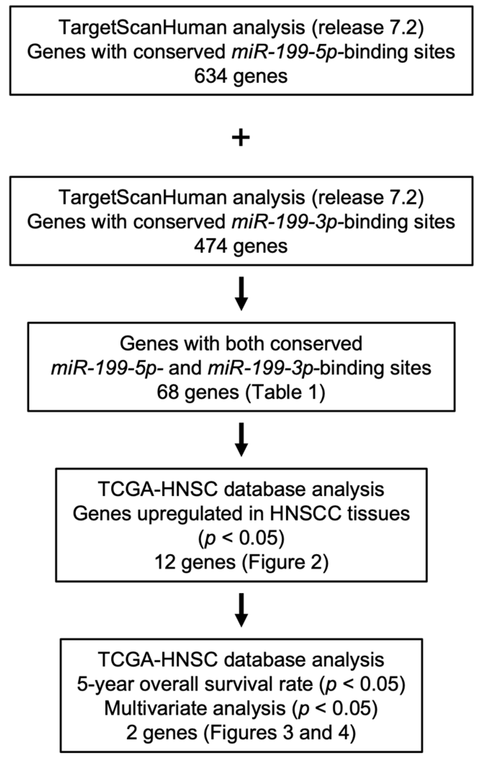
2.1. Identification of Putative Targets Controlled by miR-199-5p and miR-199-3p in HNSCC Cells

The sequences of members of the miR-199 family were confirmed using miRbase ver. 22.1 (https://www.mirbase.org, accessed on 10 April 2020) [18]. A flowchart of the procedure used to identify common target genes of miR-199-5p and miR-199-3p in this study is shown in Figure 1. We selected putative target genes that had both miR-199-5p and miR-199-3p-binding sites using TargetScanHuman ver. 7.2 (http://www.targetscan.org/vert_72/; data downloaded on 10 July 2020) [19]. The candidate genes were narrowed down by analyzing HNSCC clinical information obtained from The Cancer Genome Atlas (TCGA-HNSC).
For the Kaplan–Meier survival analysis and multivariate Cox regression analysis, we downloaded TCGA-HNSC clinical data (TCGA, Firehose Legacy) from cBioportal (https://www.cbioportal.org, accessed on 10 April 2020). Expression data for each gene were collected from OncoLnc (http://www.oncolnc.org; accessed on 20 April 2021) [20]. Patients from TCGA-HNSC were divided into low and high FXR1/PXN expression groups based on the median gene expression level, and the log-rank test was performed to compare the 5-year overall survival rate between the groups. In addition, Cox proportional hazards regression was conducted, using gene expression levels, tumor stage, pathological grade, and age as covariates.
We used JMP Pro 15 (SAS Institute Inc., Cary, NC, USA) to perform the log-rank test and Cox proportional hazards regression.

2.2. Gene set Enrichment Analysis (GSEA)

To analyze the molecular pathways related to FXR1 and PXN (regulated by miR-199a-5p and 3p), we performed GSEA. Using TCGA-HNSC data, we divided HNSCC patients into high and low expression groups according to the Z-score of the FXR1 or PXN expression level. A ranked list of genes was generated by log2 ratio comparing the expression levels of each gene between the two groups. The genes upregulated in the high expression groups were ranked higher. The obtained gene lists were uploaded into GSEA software [21,22]. We used KEGG subset of canonical pathways in The Molecular Signatures Database [21,23].

2.3. HNSCC Cell Lines

The HNSCC cell lines (SAS and Sa3) used in this study were obtained from the RIKEN BioResource Center (Tsukuba, Ibaraki, Japan). The characteristics of the cell lines are shown in Supplementary Table S1.

2.4. Transfection of Mature miRNAs and Small-Interfering RNAs (siRNAs) into HNSCC Cells

The miRNA precursors, negative control miRNA, and siRNAs were obtained from Invitrogen/Thermo Fisher Scientific (Waltham, MA, USA). The procedures used for transient transfection of miRNAs, siRNAs, and plasmid vectors have been described in our previous studies [16,17,24]. The reagents used are listed in Supplementary Table S2. All miRNAs and siRNA were transfected into HNSCC cell lines at 10 nM using RNAiMAX.

2.5. RNA Extraction and Quantitative Reverse-Transcription PCR (qRT-PCR)

RNA extraction from HNSCC cell lines and qRT-PCR were performed as described in our previous studies [16,17,24]. Briefly, total RNA was harvested using Trizol reagent and the PureLink™ RNA Mini Kit (Invitrogen/Thermo Fisher Scientific, Waltham, MA, USA). Reverse transcription was performed using the High-Capacity cDNA Reverse Transcription Kit (Applied Biosystems, Waltham, MA, USA). qRT-PCR was performed using the StepOnePlus™ Real-Time PCR System (Applied Biosystems). GAPDH was used as the internal control.

2.6. Immunohistochemistry

The immunohistochemistry procedures have been described in our previous studies [16,17,24]. Briefly, paraffin sections of tumors were obtained from HNSCC patients who underwent surgical treatment at Chiba University hospital. The clinical features of the HNSCC patients are summarized in Table S3. Our study was approved by the Ethics Committee of Chiba University (approval number: 28–65, 10 February 2015). Specimens were incubated with primary anti-PXN antibody overnight at 4 °C, after which they were incubated with DAKO Env+ secondary antibody for 30 min, washed, and counterstained with hematoxylin.

2.7. Western Blotting

The Western blotting procedures have been described in our previous studies [16,17,24]. Briefly, cell lysates were loaded onto 4–15% polyacrylamide gels at 18 μg/well. The proteins were transferred to polyvinylidene fluoride membranes and incubated with the primary antibody overnight at 4 °C and with the secondary antibody for 1 h. GAPDH was used as internal control.

2.8. Dual Luciferase Reporter Assays

A PXN DNA sequence with or without the miRNA-binding sequence of interest in its 3′ untranslated region (UTR) was inserted into the psiCHECK-2 vector (C8021; Promega, Madison, WI, USA). The plasmid vectors were then transfected into cells using Lipofectamine 2000 (Invitrogen, Waltham, MA, USA) at a final concentration of 50 ng/well. After 48 h of transfection, dual luciferase reporter assays were performed using the Dual Luciferase Reporter Assay System (Promega, Madison, WI, USA). Normalized data are expressed as the Renilla/Firefly luciferase activity ratio.

2.9. Cell Proliferation, Migration, and Invasion Assays in HNSCC Cells

The XTT assay to assess cell proliferation, and the Matrigel chamber assay to assess invasion, were performed in HNSCC cells as described previously [16,17,24]. In the wound healing assay, to assess migration a wound was created using a micropipette tip in the middle of each plate of cells transfected with siRNAs for 48 h. We incubated the plates at 37 °C and captured live cell migration after 6 and 12 h.

2.10. Statistical Analysis

Statistical analyses were performed using JMP Pro 15 (SAS Institute Inc., Cary, NC, USA). Differences between two groups were evaluated by Welch’s t-test. Dunnett’s test was used for multiple group comparisons. A p-value < 0.05 was considered statistically significant. The bar graphs present means and standard errors.

3. Results

3.1. Identification of miR-199-5p and miR-199-3p Coordinately Regulated Genes in HNSCC Cells

Chromosomal locations and mature sequences of the miR-199 family are shown in Supplementary Figure S1. In humans, the miR-199 family is composed of three members, miR-199a-1, miR-199a-2, and miR-199b, located on human chromosomes 19p13.2, 1q24.3, and 9q34.11, respectively. The guide strands of the miR-199 family share the same seed sequence (CCAGUGU); the passenger strands share same sequence (CAGUAGU).
TCGA database analysis showed the downregulation of all members of the miR-199 family in HNSCC clinical tissues (Supplementary Figure S2). At first, we confirmed tumor-suppressive functions of miR-199-5p and miR-199-3p in HNSCC cells by ectopic expression assays. Expression of miR-199-5p and miR-199-3p did not affect cell proliferation in SAS and Sa3 cells (Supplementary Figure S3A). Cell invasion and migration features were markedly suppressed by miR-199-5p and miR-199-3p expression in SAS and Sa3 cells (Supplementary Figure S3B,C). Typical images of cells from the invasion and migration assays are shown in Supplementary Figures S4 and S5. These data clearly show that both miR-199-5p and miR-199-3p acted as tumor-suppressive miRNAs in HNSCC cells.
The flowchart in Figure 1 shows the procedure we used to identify target genes of miR-199-5p and miR-199-3p. First, we searched for putative miR-199-5p- and miR-199-3p-binding genes in the TargetScan database (release 7.2). We found 634 and 474 transcripts that contained conserved miR-199-5p- and miR-199-3p-binding sites in their 3′UTRs, respectively. Of these transcripts, 68 transcripts possessed putative binding sites for both miR-199-5p and miR-199-3p (Table 1).

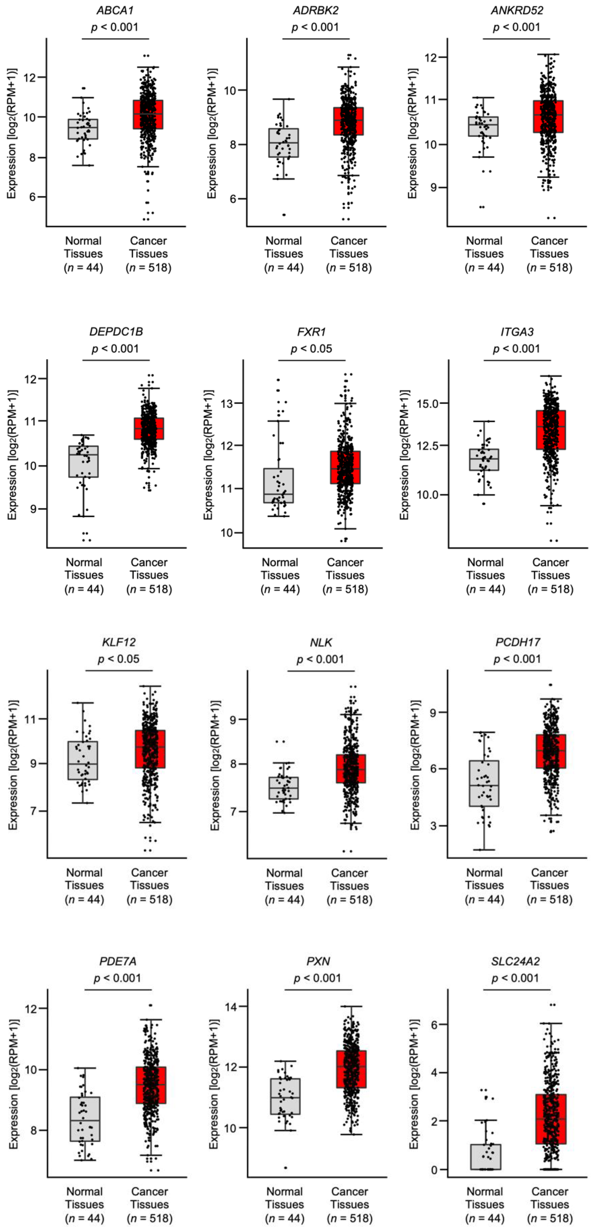
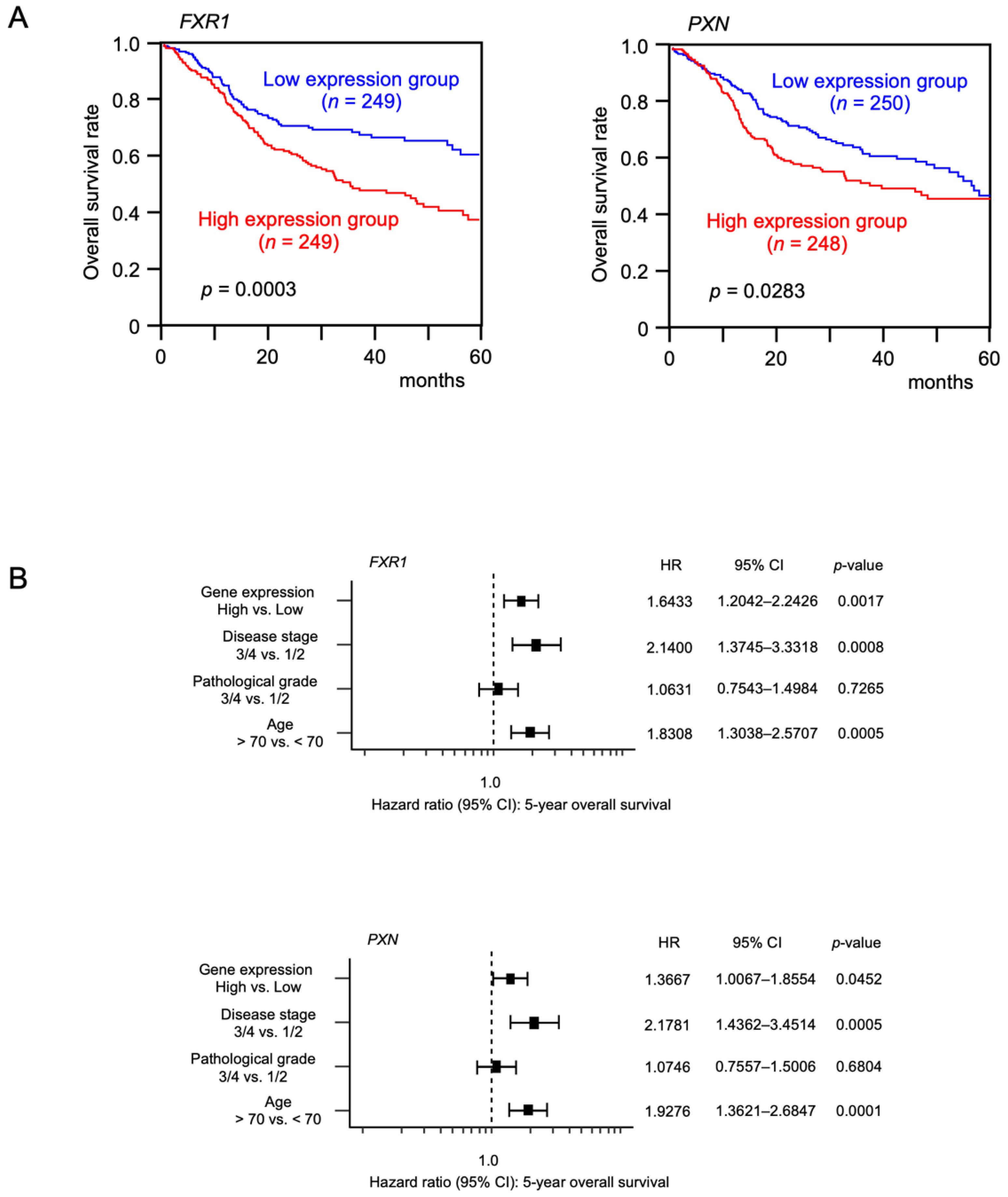
3.2. Expression and Clinical Significance of the Putative Target Genes in Patients with HNSCC according to TCGA Analysis

The expression levels of the putative targets (68 genes) of miR-199-5p and miR-199-3p were evaluated using the TCGA database (TCGA-HNSC). Among these genes, the expression levels of 12 genes (ABCA1, ADRBK2, ANKRD52, DEPDC1B, FXR1, ITGA3, KLF12, NLK, PCDH17, PDE7A, PXN, and SLC24A2) were significantly upregulated in HNSCC tissues (n = 518) compared with normal tissues (n = 44) (Figure 2). Among these 12 genes, DEPDC1B had a negative correlation with the miR-199 family in cancer tissues according to Spearman’s rank test (Supplementary Figure S6).
To determine clinical relevance, the 5-year overall survival rates of HNSCC patients according to the expression levels of these 12 genes were determined using TCGA-HNSC data. Patients with high expression of either FXR1 (p = 0.0003) or PXN (p = 0.0283) had a significantly worse prognosis compared with those with low expression (Figure 3). Moreover, multivariate Cox regression analysis revealed that the expression levels of FXR1 (p = 0.0017) and PXN (p = 0.0452) were independent prognostic factors in patients with HNSCC (Figure 3).

3.3. FXR1- and PXN-Mediated Pathways in HNSCC Cells

We investigated the genes differentially expressed between the high and low FXR1 or PXN expression groups in the HNSCC cohort from TCGA using GSEA. A total of four and fourteen gene sets were significantly enriched (FDR q-value < 0.05) in the high FXR1 and PXN expression groups, respectively (Table 2B). The most enriched gene set in the high FXR1 expression group was the cardiac muscle contraction KEGG pathway, whereas that in the high PXN expression group was the focal adhesion KEGG pathway. Although both FXR1 and PXN were involved in the prognosis of HNSCC, based on our in silico analyses, the results of GSEA analysis suggested that FXR1 was mainly involved in the pathogenesis of myocardial function. HNSCC is characterized by high rates of local recurrence and distant metastasis. Therefore, we focused on the roles of PXN in the KEGG pathways of focal adhesion and ECM receptor interaction, which are closely associated with local recurrence and distant metastasis, by performing functional analyses.

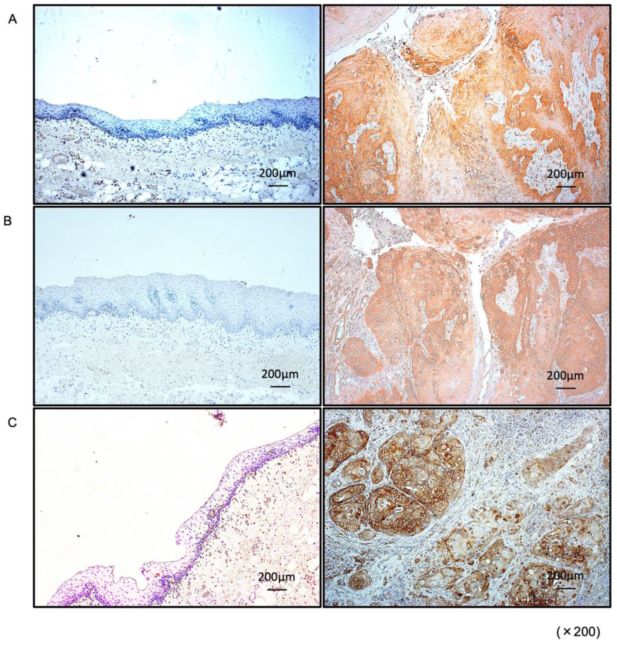
3.4. Expression of PXN in HNSCC Clinical Specimens

Expression of PXN protein was investigated by immunostaining in HNSCC clinical specimens. Aberrant expression of PXN was detected in HNSCC lesions (Figure 4). In contrast, there was almost no PXN expression in the normal epithelium (Figure 4).

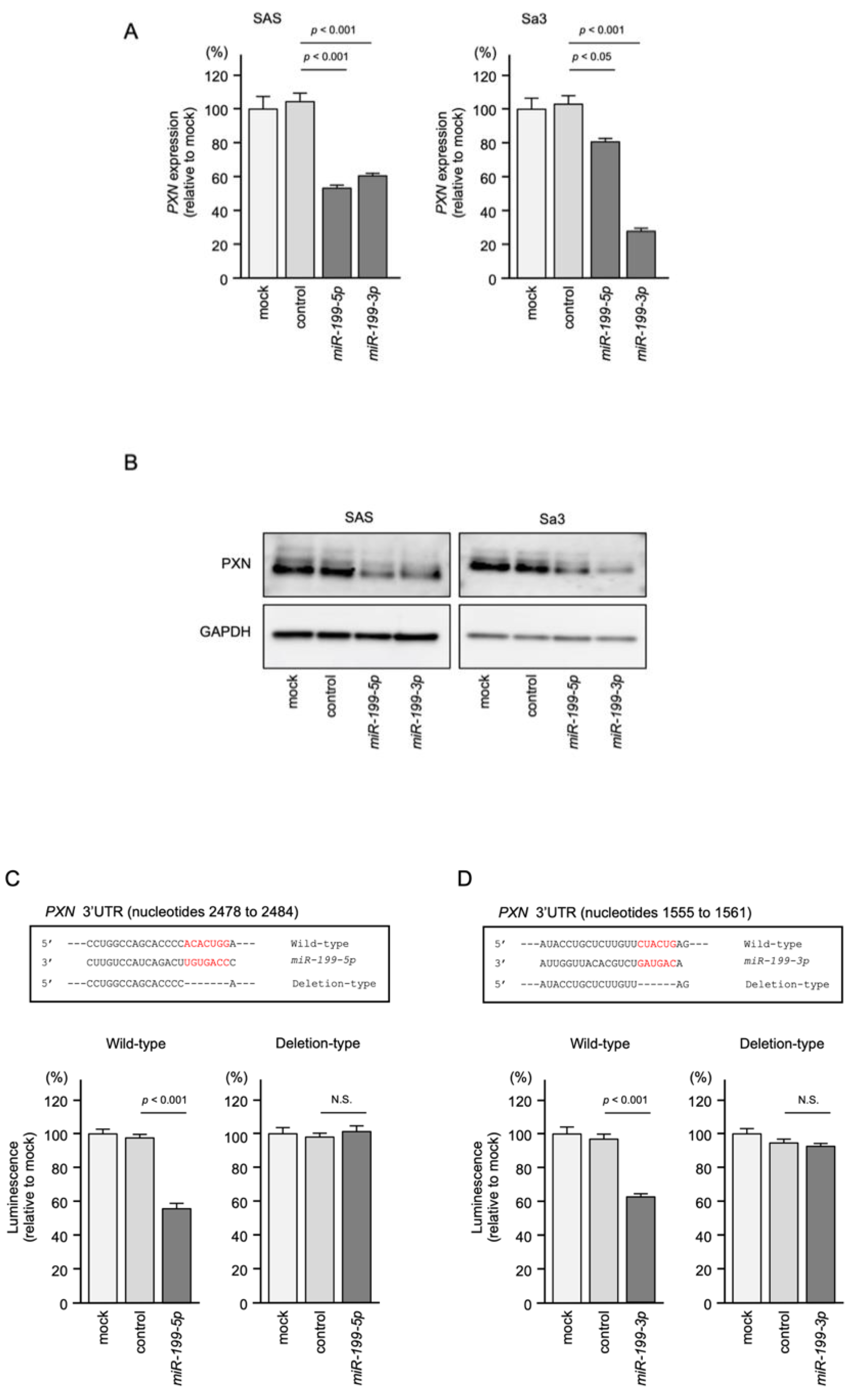
3.5. Regulation of PXN Expression by miR-199-5p and miR-199-3p in HNSCC Cells

Both the mRNA and protein levels of PXN were reduced after miR-199-5p and miR-199-3p transfection in SAS and Sa3 cells (Figure 5A,B). Full-size Western blots are shown in Supplementary Figure S7.
To investigate whether miR-199-5p and miR-199-3p bind directly to PXN in HNSCC cells, we conducted a dual-luciferase reporter assay. Luciferase activity was significantly reduced following cotransfection with miR-199-5p and a vector containing the miR-199-5p-binding site in the 3′UTR of PXN. On the other hand, cotransfection with a vector containing the PXN 3′UTR in which the miR-199-5p-binding site was deleted resulted in no change in luciferase activity (Figure 5C).
Similar to miR-199-5p, luciferase activity was significantly reduced following cotransfection with miR-199-3p and a vector containing the miR-199-3p-binding site in the 3′UTR of PXN, but not with a vector lacking the miR-199-3p-binding site in the PXN 3′UTR (Figure 5D).
These findings suggest that miR-199-5p and miR-199-3p bind directly to the 3′UTR of PXN and regulate the expression of PXN in HNSCC cells.

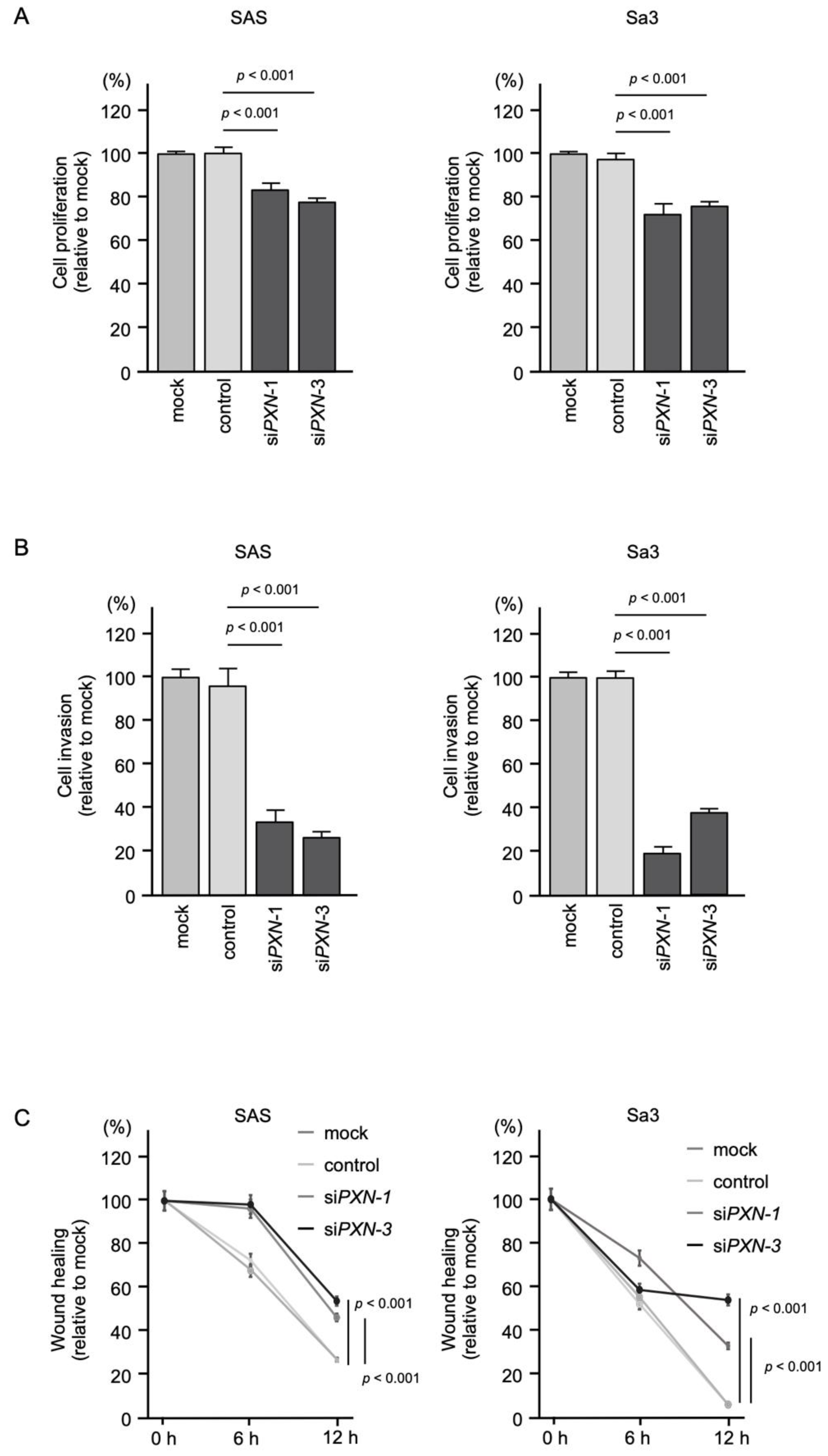
3.6. Effects of PXN Knockdown on the Proliferation, Invasion and Migration of HNSCC Cells

To assess the tumor-promoting effect of PXN in HNSCC cells, we performed knockdown assays using siRNAs. First, the inhibitory effects of three different siRNAs targeting PXN (siPXN-1, siPXN-2 and siPXN-3) on PXN expression were examined. The PXN mRNA and protein levels were effectively suppressed after transfection of each siRNA into SAS and Sa3 cells (Supplementary Figure S8A,B). For the subsequent functional assays in HNSCC cells, we used siPXN-1 and siPXN-3.
Knockdown of PXN had a slight inhibitory effect on cell proliferation in SAS and Sa3 cells (Figure 6A), and cell invasion and migration were significantly inhibited after siPXN-1 and siPXN-3 transfection in SAS and Sa3 cells (Figure 6B,C). Experimental photographs of typical results from the invasion and wound healing assays are shown in Supplementary Figures S9 and S10.
These findings indicate that overexpression of PXN is involved in promoting cancer cell migration and invasion.

4. Discussion

Currently available RNA sequencing technologies are suitable for creating miRNA expression signatures in various types of human cancers [25]. Analysis of our miRNA expression signatures in cancers revealed that some passenger strands of miRNAs derived from pre-miRNAs (e.g., miR-31-3p, miR-145-3p, miR-99a-3p, and miR-139-3p) are dysregulated in HNSCC tissues. Moreover, our functional assays showed that these passenger strands contribute to HNSCC oncogenesis, and that several of their target genes are closely associated with HNSCC pathogenesis [17,24,26,27].
Recently, a large number of TCGA cohort studies showed that certain miRNAs (e.g., both the 5p and 3p strands of miR-29, miR-30a, miR-143, miR-145, and miR-139) coordinately regulate oncogenic pathways in cancer cells [28]. In opposition to the traditional concept of miRNA biogenesis, the involvement of miRNA passenger strands in cancer pathogenesis is a newly proposed concept. Therefore, searching for target genes of miRNA passenger strands, as well as guide strands, is an important issue in cancer research.
Previous studies have reported that miR-199 family members are closely implicated in a variety of cancers as either tumor-suppressors or oncogenes [29]. Expression of miR-199a-5p was significantly reduced in OSCC tissues, and its overexpression blocked the EMT cascade by targeting SOX4 [30]. Another study showed that transient transfection of miR-199a-5p suppressed the malignant phenotypes of OSCC cells, for example, blocking cell proliferation and inducing G0/G1 arrest and apoptosis. This study indicated that IKKb, a pivotal activator of NF-κB, is a direct target of miR-199a-5p [31].
Our next focus was to identify oncogenic targets regulated by both miR-199-5p and miR-199-3p in HNSCC cells. Our in silico analysis revealed that FXR1 and PXN are candidate targets of miR-199-5p and miR-199-3p in HNSCC cells. Importantly, expression of FXR1 and PXN was closely associated with the molecular pathogenesis of HNSCC.
Overexpression of FXR1, an RNA-binding protein, has been reported in a wide range of cancers, including HNSCC and oral cancer [32,33,34,35]. Previous studies showed that overexpressed FXR1 promoted cancer cell aggressiveness by binding to and disrupting p21 mRNA [36]. Interestingly, in oral cancer cells, FXR1 stabilized miR-301a-3p, and these events reduced expression of p21 [37]. How aberrantly expressed FXR1 is involved in miRNA networks in HNSCC cells is an important question.
PXN is an intercellular adaptor protein that interacts with multiple proteins involved in cell adhesion [38]. Notably, PXN can connect integrins to focal adhesion kinase (FAK) and is a pivotal player in the assembly and disassembly of focal adhesions [39]. FAK is an important molecule that acts as a relay station for signals mediated by integrins or receptor tyrosine kinase, such as cytoskeletal changes, lamellipodia formation, cell proliferation and motility [40]. Aberrant activation of FAK-mediated signaling enhances cell proliferation, invasion, and metastasis [41]. Previous studies showed that aberrant expression of PXN was detected in a wide range of cancers, and its overexpression was usually correlated with worse prognosis of the patients [42]. However, in some types of cancers, downregulation of PXN was reported, e.g., breast cancer, colorectal cancer, leukemia, and low-grade glioma [43]. Here, we clearly showed that overexpression of PXN closely contributes to malignant transformation of HNSCC cells and a worse patient prognosis. It is an interesting finding that the expression of PXN differs depending on the type of cancer. Analysis of the epigenetic modification that regulates PXN expression is required for each cancer type. In HNSCC cells, analysis of PXN and PXN-mediated downstream signals will aid the search for therapeutic target molecules for this disease.
Recent reports have shown that several dysregulated non-coding RNAs (long non-coding RNAs [lncRNAs] and miRNAs) are involved in aberrant expression of PXN in cancer cells [44,45,46]. Overexpressed lncRNAs were found to adsorb tumor-suppressor miRNAs and suppress their antitumor effects in cancer cells [47,48,49,50]. In gastric cancer, the lncRNA XIST, when overexpressed, adsorbed miR-132 and caused upregulation of PXN in cancer cells. This study found that XIST acts as an oncogenic lncRNA in gastric cancer [51]. In triple-negative breast cancer, the lncRNA DLX6 antisense RNA 1 was upregulated in cancer tissues, and its expression promoted cell proliferation, EMT, and cisplatin resistance by regulating the miR-199b-5p/PXN axis [52]. These data support our current results. It is expected that many lncRNAs are involved in suppression of miR-199 family expression in HNSCC cells.
A previous study showed that expression of miR-218 was reduced by human papillomavirus-16 (HPV-16) in cervical cancer [53]. In HPV-16-infected OSCC cells, downregulation of miR-218 and upregulation of PXN were detected, and these events contributed to colony formation and invasion of OSCC cells [54]. Moreover, low expression of miR-218, or high expression of PXN, was closely associated with overall survival and relapse-free survival [55]. Our previous studies demonstrated that miR-218 acts as a tumor-suppressive miRNA in HNSCC by markedly suppressing cancer cell migration and invasive abilities [56]. Suppression of miR-218 expression might be linked to aberrant expression of PXN in HNSCC cells.
Many non-coding RNA molecules are involved in the regulation of PXN expression, and the complexity of the molecular mechanisms of cancer cells has been clarified. To clarify the malignant transformation of HNSCC, genome-wide analysis including non-coding RNA molecules will be indispensable.

5. Conclusions

PXN was discovered by searching for oncogenic target genes of miR-199-5p and miR-199-3p, tumor suppressive miRNAs in HNSCC. Aberrant expression of PXN facilitated cancer cell migration and invasion, and its expression was closely associated with HNSCC molecular pathogenesis. Searching for target genes of tumor-suppressive miRNAs is an attractive strategy for exploring the molecular mechanism of HNSCC.

Supplementary Materials

The following are available online at https://www.mdpi.com/article/10.3390/genes12121910/s1. Figure S1: Chromosomal locations and mature sequences of the miR-199 family. Figure S2: The expression levels of miR-199 family. Figure S3: Functional assays following ectopic expression of miR-199-5p and miR-199-3p, Figure S4: Photomicrographs of cells subjected to invasion assays. Figure S5: Photomicrographs of cells subjected to wound healing assays. Figure S6: Correlations between miR-199 family and putative target genes. Figure S7: Full-size images of the Western blots shown in Figure 5. Figure S8: Efficiency of siRNA-mediated PXN knockdown in HNSCC cell lines (SAS and Sa3 cells). Figure S9: Photomicrographs of cells subjected to invasion assays. Figure S10: Photomicrographs of cells subjected to wound healing assays. Table S1: Features of HNSCC cell lines. Table S2: Reagents used in this study. Table S3: Clinical features of three HNSCC cases used for immunohistochemical staining.

Author Contributions

Data curation, N.T., C.M., T.K. and N.K.; formal analysis, N.T., C.M. and S.A.; investigation, N.T., C.M., S.O., A.K. (Ayaka Koma), T.K. and N.K.; project administration, N.S.; supervision, T.H. and K.U.; writing—original draft, N.T. and N.S.; writing—review & editing, A.K. (Atsushi Kasamatsu). All authors have read and agreed to the published version of the manuscript.

Funding

This study was supported by JSPS KAKENHI grant numbers 19H03847, 19K09863, 19K18759, 20H03883, and 21K09577.

Institutional Review Board Statement

The study was conducted according to the Declaration of Helsinki, and approved by the Bioethics Committee of Chiba University (approval number: 28–65, 10 February 2015).

Informed Consent Statement

Informed consent was obtained from all subjects invsolved in the study.

Data Availability Statement

The results shown here are, in part, based upon data generated by the TCGA Research Network: https://www.cancer.gov/tcga (accessed on 10 April 2020). The data presented in this study are available on request from the corresponding author.

Conflicts of Interest

The authors declare no conflict of interest.

References

  1. Bray, F.; Ferlay, J.; Soerjomataram, I.; Siegel, R.L.; Torre, L.A.; Jemal, A. Global cancer statistics 2018: GLOBOCAN estimates of incidence and mortality worldwide for 36 cancers in 185 countries. CA Cancer J. Clin. 2018, 68, 394–424. [Google Scholar] [CrossRef] [PubMed] [Green Version]
  2. Johnson, D.E.; Burtness, B.; Leemans, C.R.; Lui, V.W.Y.; Bauman, J.E.; Grandis, J.R. Head and neck squamous cell carcinoma. Nat. Rev. Dis. Primers 2020, 6, 92. [Google Scholar] [CrossRef]
  3. Cramer, J.D.; Burtness, B.; Le, Q.T.; Ferris, R.L. The changing therapeutic landscape of head and neck cancer. Nat. Rev. Clin. Oncol. 2019, 16, 669–683. [Google Scholar] [CrossRef]
  4. Mehanna, H.; Paleri, V.; West, C.M.; Nutting, C. Head and neck cancer—Part 1: Epidemiology, presentation, and preservation. Clin. Otolaryngol. 2011, 36, 65–68. [Google Scholar] [CrossRef] [PubMed]
  5. Sha, J.; Bai, Y.; Ngo, H.X.; Okui, T.; Kanno, T. Overview of Evidence-Based Chemotherapy for Oral Cancer: Focus on Drug Resistance Related to the Epithelial-Mesenchymal Transition. Biomolecules 2021, 11, 893. [Google Scholar] [CrossRef]
  6. Chow, L.Q.M. Head and Neck Cancer. N. Engl. J. Med. 2020, 382, 60–72. [Google Scholar] [CrossRef] [PubMed]
  7. Leemans, C.R.; Braakhuis, B.J.; Brakenhoff, R.H. The molecular biology of head and neck cancer. Nat. Rev. Cancer 2011, 11, 9–22. [Google Scholar] [CrossRef]
  8. Cohen, E.E.W.; Soulières, D.; Le Tourneau, C.; Dinis, J.; Licitra, L.; Ahn, M.J.; Soria, A.; Machiels, J.P.; Mach, N.; Mehra, R.; et al. Pembrolizumab versus methotrexate, docetaxel, or cetuximab for recurrent or metastatic head-and-neck squamous cell carcinoma (KEYNOTE-040): A randomised, open-label, phase 3 study. Lancet 2019, 393, 156–167. [Google Scholar] [CrossRef]
  9. Betel, D.; Wilson, M.; Gabow, A.; Marks, D.S.; Sander, C. The microRNA.org resource: Targets and expression. Nucleic Acids Res. 2008, 36, D149–D153. [Google Scholar] [CrossRef] [Green Version]
  10. Anfossi, S.; Babayan, A.; Pantel, K.; Calin, G.A. Clinical utility of circulating non-coding RNAs—An update. Nat. Rev. Clin. Oncol. 2018, 15, 541–563. [Google Scholar] [CrossRef]
  11. Nelson, K.M.; Weiss, G.J. MicroRNAs and cancer: Past, present, and potential future. Mol. Cancer Ther. 2008, 7, 3655–3660. [Google Scholar] [CrossRef] [PubMed] [Green Version]
  12. Krek, A.; Grun, D.; Poy, M.N.; Wolf, R.; Rosenberg, L.; Epstein, E.J.; MacMenamin, P.; da Piedade, I.; Gunsalus, K.C.; Stoffel, M.; et al. Combinatorial microRNA target predictions. Nat. Genet. 2005, 37, 495–500. [Google Scholar] [CrossRef]
  13. Rupaimoole, R.; Slack, F.J. MicroRNA therapeutics: Towards a new era for the management of cancer and other diseases. Nat. Rev. Drug Discov 2017, 16, 203–222. [Google Scholar] [CrossRef] [PubMed]
  14. Tay, Y.; Rinn, J.; Pandolfi, P.P. The multilayered complexity of ceRNA crosstalk and competition. Nature 2014, 505, 344–352. [Google Scholar] [CrossRef] [Green Version]
  15. Koshizuka, K.; Hanazawa, T.; Arai, T.; Okato, A.; Kikkawa, N.; Seki, N. Involvement of aberrantly expressed microRNAs in the pathogenesis of head and neck squamous cell carcinoma. Cancer Metastasis Rev. 2017, 36, 525–545. [Google Scholar] [CrossRef]
  16. Koshizuka, K.; Hanazawa, T.; Kikkawa, N.; Arai, T.; Okato, A.; Kurozumi, A.; Kato, M.; Katada, K.; Okamoto, Y.; Seki, N. Regulation of ITGA3 by the anti-tumor miR-199 family inhibits cancer cell migration and invasion in head and neck cancer. Cancer Sci. 2017, 108, 1681–1692. [Google Scholar] [CrossRef] [Green Version]
  17. Koma, A.; Asai, S.; Minemura, C.; Oshima, S.; Kinoshita, T.; Kikkawa, N.; Koshizuka, K.; Moriya, S.; Kasamatsu, A.; Hanazawa, T.; et al. Impact of Oncogenic Targets by Tumor-Suppressive miR-139-5p and miR-139-3p Regulation in Head and Neck Squamous Cell Carcinoma. Int. J. Mol. Sci. 2021, 22, 9947. [Google Scholar] [CrossRef]
  18. Kozomara, A.; Birgaoanu, M.; Griffiths-Jones, S. miRBase: From microRNA sequences to function. Nucleic Acids Res. 2019, 47, D155–D162. [Google Scholar] [CrossRef]
  19. Agarwal, V.; Bell, G.W.; Nam, J.W.; Bartel, D.P. Predicting effective microRNA target sites in mammalian mRNAs. eLife 2015, 4. [Google Scholar] [CrossRef] [PubMed]
  20. Anaya, J. Linking TCGA survival data to mRNAs, miRNAs, and lncRNAs. Peer J. Comput. Sci. 2016, 2, e67. [Google Scholar] [CrossRef] [Green Version]
  21. Subramanian, A.; Tamayo, P.; Mootha, V.K.; Mukherjee, S.; Ebert, B.L.; Gillette, M.A.; Paulovich, A.; Pomeroy, S.L.; Golub, T.R.; Lander, E.S.; et al. Gene set enrichment analysis: A knowledge-based approach for interpreting genome-wide expression profiles. Proc. Natl. Acad. Sci. USA 2005, 102, 15545–15550. [Google Scholar] [CrossRef] [Green Version]
  22. Mootha, V.K.; Lindgren, C.M.; Eriksson, K.F.; Subramanian, A.; Sihag, S.; Lehar, J.; Puigserver, P.; Carlsson, E.; Ridderstråle, M.; Laurila, E.; et al. PGC-1alpha-responsive genes involved in oxidative phosphorylation are coordinately downregulated in human diabetes. Nat. Genet. 2003, 34, 267–273. [Google Scholar] [CrossRef] [PubMed]
  23. Liberzon, A.; Subramanian, A.; Pinchback, R.; Thorvaldsdóttir, H.; Tamayo, P.; Mesirov, J.P. Molecular signatures database (MSigDB) 3.0. Bioinformatics 2011, 27, 1739–1740. [Google Scholar] [CrossRef] [PubMed]
  24. Oshima, S.; Asai, S.; Seki, N.; Minemura, C.; Kinoshita, T.; Goto, Y.; Kikkawa, N.; Moriya, S.; Kasamatsu, A.; Hanazawa, T.; et al. Identification of Tumor Suppressive Genes Regulated by miR-31-5p and miR-31-3p in Head and Neck Squamous Cell Carcinoma. Int. J. Mol. Sci. 2021, 22, 6199. [Google Scholar] [CrossRef]
  25. Hong, M.; Tao, S.; Zhang, L.; Diao, L.T.; Huang, X.; Huang, S.; Xie, S.J.; Xiao, Z.D.; Zhang, H. RNA sequencing: New technologies and applications in cancer research. J. Hematol. Oncol. 2020, 13, 166. [Google Scholar] [CrossRef] [PubMed]
  26. Yamada, Y.; Koshizuka, K.; Hanazawa, T.; Kikkawa, N.; Okato, A.; Idichi, T.; Arai, T.; Sugawara, S.; Katada, K.; Okamoto, Y.; et al. Passenger strand of miR-145-3p acts as a tumor-suppressor by targeting MYO1B in head and neck squamous cell carcinoma. Int. J. Oncol. 2018, 52, 166–178. [Google Scholar] [CrossRef] [PubMed]
  27. Okada, R.; Koshizuka, K.; Yamada, Y.; Moriya, S.; Kikkawa, N.; Kinoshita, T.; Hanazawa, T.; Seki, N. Regulation of Oncogenic Targets by miR-99a-3p (Passenger Strand of miR-99a-Duplex) in Head and Neck Squamous Cell Carcinoma. Cells 2019, 8, 1535. [Google Scholar] [CrossRef] [PubMed] [Green Version]
  28. Mitra, R.; Adams, C.M.; Jiang, W.; Greenawalt, E.; Eischen, C.M. Pan-cancer analysis reveals cooperativity of both strands of microRNA that regulate tumorigenesis and patient survival. Nat. Commun. 2020, 11, 968. [Google Scholar] [CrossRef] [PubMed] [Green Version]
  29. Wang, Q.; Ye, B.; Wang, P.; Yao, F.; Zhang, C.; Yu, G. Overview of microRNA-199a Regulation in Cancer. Cancer Manag. Res. 2019, 11, 10327–10335. [Google Scholar] [CrossRef] [Green Version]
  30. Wei, D.; Wang, W.; Shen, B.; Zhou, Y.; Yang, X.; Lu, G.; Yang, J.; Shao, Y. MicroRNA-199a-5p suppresses migration and invasion in oral squamous cell carcinoma through inhibiting the EMT-related transcription factor SOX4. Int. J. Mol. Med. 2019, 44, 185–195. [Google Scholar] [CrossRef] [Green Version]
  31. Wei, D.; Shen, B.; Wang, W.; Zhou, Y.; Yang, X.; Lu, G.; Yang, J.; Shao, Y. MicroRNA-199a-5p functions as a tumor suppressor in oral squamous cell carcinoma via targeting the IKKβ/NF-κB signaling pathway. Int. J. Mol. Med. 2019, 43, 1585–1596. [Google Scholar] [CrossRef] [Green Version]
  32. Jin, X.; Zhai, B.; Fang, T.; Guo, X.; Xu, L. FXR1 is elevated in colorectal cancer and acts as an oncogene. Tumour Biol. 2016, 37, 2683–2690. [Google Scholar] [CrossRef]
  33. Qian, J.; Chen, H.; Ji, X.; Eisenberg, R.; Chakravarthy, A.B.; Mayer, I.A.; Massion, P.P. A 3q gene signature associated with triple negative breast cancer organ specific metastasis and response to neoadjuvant chemotherapy. Sci. Rep. 2017, 7, 45828. [Google Scholar] [CrossRef] [PubMed] [Green Version]
  34. Comtesse, N.; Keller, A.; Diesinger, I.; Bauer, C.; Kayser, K.; Huwer, H.; Lenhof, H.P.; Meese, E. Frequent overexpression of the genes FXR1, CLAPM1 and EIF4G located on amplicon 3q26-27 in squamous cell carcinoma of the lung. Int. J. Cancer 2007, 120, 2538–2544. [Google Scholar] [CrossRef]
  35. Qie, S.; Majumder, M.; Mackiewicz, K.; Howley, B.V.; Peterson, Y.K.; Howe, P.H.; Palanisamy, V.; Diehl, J.A. Fbxo4-mediated degradation of Fxr1 suppresses tumorigenesis in head and neck squamous cell carcinoma. Nat. Commun. 2017, 8, 1534. [Google Scholar] [CrossRef] [PubMed] [Green Version]
  36. Majumder, M.; House, R.; Palanisamy, N.; Qie, S.; Day, T.A.; Neskey, D.; Diehl, J.A.; Palanisamy, V. RNA-Binding Protein FXR1 Regulates p21 and TERC RNA to Bypass p53-Mediated Cellular Senescence in OSCC. PLoS Genet. 2016, 12, e1006306. [Google Scholar] [CrossRef] [Green Version]
  37. Majumder, M.; Palanisamy, V. RNA binding protein FXR1-miR301a-3p axis contributes to p21WAF1 degradation in oral cancer. PLoS Genet. 2020, 16, e1008580. [Google Scholar] [CrossRef] [Green Version]
  38. Brown, M.C.; Turner, C.E. Paxillin: Adapting to change. Physiol. Rev. 2004, 84, 1315–1339. [Google Scholar] [CrossRef] [PubMed]
  39. Mitra, S.K.; Hanson, D.A.; Schlaepfer, D.D. Focal adhesion kinase: In command and control of cell motility. Nat. Rev. Mol. Cell Biol. 2005, 6, 56–68. [Google Scholar] [CrossRef]
  40. Wozniak, M.A.; Modzelewska, K.; Kwong, L.; Keely, P.J. Focal adhesion regulation of cell behavior. Biochim. Biophys. Acta 2004, 1692, 103–119. [Google Scholar] [CrossRef]
  41. Zhou, J.; Yi, Q.; Tang, L. The roles of nuclear focal adhesion kinase (FAK) on Cancer: A focused review. J. Exp. Clin. Cancer Res. 2019, 38, 250. [Google Scholar] [CrossRef] [Green Version]
  42. Chen, Y.; Zhao, H.; Xiao, Y.; Shen, P.; Tan, L.; Zhang, S.; Liu, Q.; Gao, Z.; Zhao, J.; Zhao, Y.; et al. Pan-cancer analysis reveals an immunological role and prognostic potential of PXN in human cancer. Aging 2021, 13, 16248–16266. [Google Scholar] [CrossRef] [PubMed]
  43. Deakin, N.O.; Pignatelli, J.; Turner, C.E. Diverse roles for the paxillin family of proteins in cancer. Genes Cancer 2012, 3, 362–370. [Google Scholar] [CrossRef] [PubMed] [Green Version]
  44. Jiang, H.; Zhang, H.; Hu, X.; Li, W. Knockdown of long non-coding RNA XIST inhibits cell viability and invasion by regulating miR-137/PXN axis in non-small cell lung cancer. Int. J. Biol. Macromol. 2018, 111, 623–631. [Google Scholar] [CrossRef] [PubMed]
  45. Yan, J.; Jia, Y.; Chen, H.; Chen, W.; Zhou, X. Long non-coding RNA PXN-AS1 suppresses pancreatic cancer progression by acting as a competing endogenous RNA of miR-3064 to upregulate PIP4K2B expression. J. Exp. Clin. Cancer Res. 2019, 38, 390. [Google Scholar] [CrossRef] [PubMed] [Green Version]
  46. Yuan, J.H.; Liu, X.N.; Wang, T.T.; Pan, W.; Tao, Q.F.; Zhou, W.P.; Wang, F.; Sun, S.H. The MBNL3 splicing factor promotes hepatocellular carcinoma by increasing PXN expression through the alternative splicing of lncRNA-PXN-AS1. Nat. Cell Biol. 2017, 19, 820–832. [Google Scholar] [CrossRef] [PubMed]
  47. Xu, J.; Zhang, P.; Sun, H.; Liu, Y. LINC01094/miR-577 axis regulates the progression of ovarian cancer. J. Ovarian Res. 2020, 13, 122. [Google Scholar] [CrossRef] [PubMed]
  48. Xiao, Z.S.; Long, H.; Zhao, L.; Li, H.X.; Zhang, X.N. LncRNA HOTTIP promotes proliferation and inhibits apoptosis of gastric carcinoma cells via adsorbing miR-615-3p. Eur. Rev. Med. Pharmacol. Sci. 2020, 24, 6692–6698. [Google Scholar] [CrossRef] [PubMed]
  49. Cui, Y.; Zhang, C.; Lian, H.; Xie, L.; Xue, J.; Yin, N.; Guan, F. LncRNA linc00460 sponges miR-1224-5p to promote esophageal cancer metastatic potential and epithelial-mesenchymal transition. Pathol. Res. Pract. 2020, 216, 153026. [Google Scholar] [CrossRef] [PubMed]
  50. Chen, A.Y.; Zhang, K.; Liu, G.Q. LncRNA LINP1 promotes malignant progression of pancreatic cancer by adsorbing microRNA-491-3p. Eur. Rev. Med. Pharmacol. Sci. 2020, 24, 9315–9324. [Google Scholar] [CrossRef]
  51. Li, P.; Wang, L.; Li, P.; Hu, F.; Cao, Y.; Tang, D.; Ye, G.; Li, H.; Wang, D. Silencing lncRNA XIST exhibits antiproliferative and proapoptotic effects on gastric cancer cells by up-regulating microRNA-132 and down-regulating PXN. Aging 2020, 13, 14469–14481. [Google Scholar] [CrossRef] [PubMed]
  52. Du, C.; Wang, Y.; Zhang, Y.; Zhang, J.; Zhang, L.; Li, J. LncRNA DLX6-AS1 Contributes to Epithelial-Mesenchymal Transition and Cisplatin Resistance in Triple-negative Breast Cancer via Modulating Mir-199b-5p/Paxillin Axis. Cell Transplant. 2020, 29, 963689720929983. [Google Scholar] [CrossRef]
  53. Liu, Z.; Mao, L.; Wang, L.; Zhang, H.; Hu, X. miR-218 functions as a tumor suppressor gene in cervical cancer. Mol. Med. Rep. 2020, 21, 209–219. [Google Scholar] [CrossRef] [PubMed] [Green Version]
  54. Li, X.; He, J.; Shao, M.; Cui, B.; Peng, F.; Li, J.; Ran, Y.; Jin, D.; Kong, J.; Chang, J.; et al. Downregulation of miR-218-5p promotes invasion of oral squamous cell carcinoma cells via activation of CD44-ROCK signaling. Biomed. Pharmacother. 2018, 106, 646–654. [Google Scholar] [CrossRef] [PubMed]
  55. Wu, D.W.; Chuang, C.Y.; Lin, W.L.; Sung, W.W.; Cheng, Y.W.; Lee, H. Paxillin promotes tumor progression and predicts survival and relapse in oral cavity squamous cell carcinoma by microRNA-218 targeting. Carcinogenesis 2014, 35, 1823–1829. [Google Scholar] [CrossRef] [PubMed]
  56. Kinoshita, T.; Hanazawa, T.; Nohata, N.; Kikkawa, N.; Enokida, H.; Yoshino, H.; Yamasaki, T.; Hidaka, H.; Nakagawa, M.; Okamoto, Y.; et al. Tumor suppressive microRNA-218 inhibits cancer cell migration and invasion through targeting laminin-332 in head and neck squamous cell carcinoma. Oncotarget 2012, 3, 1386–1400. [Google Scholar] [CrossRef] [Green Version]
Figure 1. Flowchart of the strategy used to identify candidate miR-199-5p and miR-199-3p target genes in HNSCC cells.
Genes 12 01910 g001
Figure 2. HNSCC tissue expression of 12 target genes with miR-199-5p- and miR-199-3p-binding sites in their 3’UTRs using TCGA-HNSC data. The expression levels of 12 genes (ABCA1, ADRBK2, ANKRD52, DEPDC1B, FXR1, ITGA3, KLF12, NLK, PCDH17, PDE7A, PXN, and SLC24A2) were analyzed using TCGA-HNSC database. A total of 518 HNSCC tissues and 44 normal epithelial tissues were evaluated.
Genes 12 01910 g002
Figure 3. Clinical significance of FXR1 and PXN using TCGA-HNSC data. (A) Kaplan–Meier survival analyses of HNSC patients using data from TCGA database. Patients were divided into high and low expression groups according to the median FXR1 and PXN expression levels. The red and blue lines indicate the high and low expression groups, respectively. (B) Forest plot showing the multivariate analysis results for two genes (FXR1 and PXN), which were identified as independent prognostic factors for overall survival after adjustment for patient age, disease stage, and pathological grade.
Genes 12 01910 g003
Figure 4. Overexpression of PXN in HNSCC clinical specimens. (AC) Immunohistochemical staining of PXN in HNSCC clinical specimens. PXN expression was high in the nuclei and/or cytoplasm of cancer cells (right panels) but weak in normal mucosa (left panels).
Genes 12 01910 g004
Figure 5. Direct regulation of PXN expression by both miR-199-5p and miR-199-3p in HNSCC cells. (A) qRT-PCR showing significantly reduced expression of PXN mRNA at 72 h after miR-199-5p or miR-199-3p transfection in SAS and Sa3 cells. Expression of GAPDH was used as an internal control. (B) Western blot showing reduced expression of PXN protein at 72 h after miR-199-5p or miR-199-3p transfection in SAS and Sa3 cells. Expression of GAPDH was used as an internal control. (C) TargetScan database analysis predicting a single putative miR-199-5p-binding site in the 3′-UTR of PXN (upper panel). Dual luciferase reporter assays showed reduced luminescence activity after cotransfection of the wild-type vector and miR-199-5p in SAS cells (lower panel). Normalized data are expressed as the Renilla/Firefly luciferase activity ratio (N.S., not significant). (D) TargetScan database analysis predicting a single putative miR-199-3p-binding site in the 3′-UTR of PXN (upper panel). Dual luciferase reporter assays showed reduced luminescence activity after cotransfection of the wild-type vector and miR-199-3p in SAS cells (lower panel).
Genes 12 01910 g005
Figure 6. Functional assays of cell proliferation, invasion and migration following transient transfection of siRNAs targeting PXN in HNSCC cell lines (SAS and Sa3 cells). (A) Cell proliferation assessed by XTT assay at 72 h after siRNA transfection. (B) Cell invasion assessed by Matrigel invasion assays at 48 h after seeding siRNA-transfected cells into chambers. (C) Cell migration assessed by wound healing assay at 0, 6, and 12 h after cell scratch formation.
Genes 12 01910 g006
Table 1. miR-199-5p/3p common target genes.
Entrez
Gene ID
Gene
Symbol
Gene NamemiR-199-5p
Total Conserved Sites
miR-199-3p
Total Conserved Sites
19ABCA1ATP-binding cassette, sub-family A (ABC1), member 111
340485ACER2alkaline ceramidase 211
92ACVR2Aactivin A receptor, type IIA12
93ACVR2Bactivin A receptor, type IIB31
57188ADAMTSL3ADAMTS-like 313
120ADD3adducin 3 (γ)11
157ADRBK2adrenergic, beta, receptor kinase 211
81573ANKRD13Cankyrin repeat domain 13C11
283373ANKRD52ankyrin repeat domain 5211
57569ARHGAP20Rho GTPase activating protein 2011
23365ARHGEF12Rho guanine nucleotide exchange factor (GEF) 1211
222255ATXN7L1ataxin 7-like 111
27443CECR2cat eye syndrome chromosome region, candidate 221
10659CELF2CUGBP, Elav-like family member 212
387119CEP85Lcentrosomal protein 85kDa-like11
153222CREBRFCREB3 regulatory factor11
51232CRIM1cysteine rich transmembrane BMP regulator 1 (chordin-like)11
1496CTNNA2catenin (cadherin-associated protein), α211
84301DDI2DNA-damage inducible 1 homolog 2 (S. cerevisiae)11
55789DEPDC1BDEP domain containing 1B11
2066ERBB4v-erb-b2 avian erythroblastic leukemia viral oncogene homolog 412
55137FIGNfidgetin11
23767FLRT3fibronectin leucine rich transmembrane protein 311
10690FUT9fucosyltransferase 9 (α (1,3) fucosyltransferase)11
8087FXR1fragile X mental retardation, autosomal homolog 121
2651GCNT2glucosaminyl (N-acetyl) transferase 2, I-branching enzyme (I blood group)11
54891INO80DINO80 complex subunit D11
3675ITGA3integrin, α3 (antigen CD49C, α 3 subunit of VLA-3 receptor)11
8516ITGA8integrin, α811
11278KLF12Kruppel-like factor 1211
26249KLHL3kelch-like family member 313
84458LCORligand dependent nuclear receptor corepressor22
10960LMAN2lectin, mannose-binding 211
84061MAGT1magnesium transporter 111
4217MAP3K5mitogen-activated protein kinase 511
5599MAPK8mitogen-activated protein kinase 811
90411MCFD2multiple coagulation factor deficiency 211
54842MFSD6major facilitator superfamily domain containing 611
51701NLKnemo-like kinase31
57532NUFIP2nuclear fragile X mental retardation protein interacting protein 211
10298PAK4p21 protein (Cdc42/Rac)-activated kinase 412
27253PCDH17protocadherin 1711
5150PDE7Aphosphodiesterase 7A11
57475PLEKHH1pleckstrin homology domain containing,
family H (with MyTH4 domain) member 1
11
5495PPM1Bprotein phosphatase, Mg2+/Mn2+ dependent, 1B11
55607PPP1R9Aprotein phosphatase 1, regulatory subunit 9A11
63976PRDM16PR domain containing 1611
5813PURApurine-rich element binding protein A11
5829PXNpaxillin11
5925RB1retinoblastoma 111
54502RBM47RNA binding motif protein 4711
5991RFX3regulatory factor X, 3 (influences HLA class II expression)11
6096RORBRAR-related orphan receptor B12
9644SH3PXD2ASH3 and PX domains 2A11
25769SLC24A2solute carrier family 24 (sodium/potassium/calcium exchanger), member 211
8303SNNstannin11
6667SP1Sp1 transcription factor11
257397TAB3TGF-β activated kinase 1/MAP3K7 binding protein 311
57551TAOK1TAO kinase 112
10099TSPAN3tetraspanin 311
57695USP37ubiquitin specific peptidase 3711
23063WAPALwings apart-like homolog (Drosophila)12
10472ZBTB18zinc finger and BTB domain containing 1812
26137ZBTB20zinc finger and BTB domain containing 2022
6935ZEB1zinc finger E-box binding homeobox 111
80139ZNF703zinc finger protein 70311
374655ZNF710zinc finger protein 71011
283337ZNF740zinc finger protein 74011
Table 2. Gene set enrichment analysis.
A. Significantly Enriched Gene Sets in the High FXR1 Expression Group
NameNormalized Enrichment ScoreFDR q-Value
KEGG_Cardiac muscle contraction2.0090.001
KEGG_Dilated cardiomyopathy1.9680.001
KEGG_Hypertrophic cardiomyopathy HCM1.9290.003
KEGG_Maturity onset diabetes of the young1.9180.002
B. Significantly enriched gene sets in the high PXN expression group
Namenormalized enrichment scoreFDR q-value
KEGG_Focal adhesion2.458q < 0.001
KEGG_ECM receptor interaction2.316q < 0.001
KEGG_Cytosolic DNA sensing pathway2.1170.001
KEGG_Proteosome2.0290.003
KEGG_Hypetrophic cardiomyopathy HCM1.9950.005
KEGG_NOD-like receptor signaling pathway1.8960.009
KEGG_Viral myocarditis1.8940.008
KEGG_Dilated Cardiomyopathy1.8040.014
KEGG_Bladder cancer1.7840.016
KEGG_Cytokine cytokine receptor interaction1.7760.016
KEGG_JAK/STAT signaling pathway1.7120.026
KEGG_RIG-I-like receptor signaling pathway1.6730.033
KEGG_Small cell lung cancer1.6440.038
KEGG_Arryhythmogenic right ventricular cardiomyopathy ARVC1.6390.037
Publisher’s Note: MDPI stays neutral with regard to jurisdictional claims in published maps and institutional affiliations.

