Abstract
High-quality chicken meat is an important source of animal protein for humans. Gene expression profiles in breast muscle tissue were determined, aiming to explore the common regulatory genes relevant to muscle and intramuscular fat (IMF) during the developmental stage in chickens. Results show that breast muscle weight (BMW), breast meat percentage (BMP, %), and IMF (%) continuously increased with development. A total of 256 common differentially expressed genes (DEGs) during the developmental stage were screened. Among them, some genes related to muscle fiber hypertrophy were upregulated (e.g., CSRP3, LMOD2, MUSTN1, MYBPC1), but others (e.g., ACTC1, MYL1, MYL4) were downregulated from Week 3 to Week 18. During this period, expression of some DEGs related to the cells cycle (e.g., CCNB3, CCNE2, CDC20, MCM2) changed in a way that genetically suggests possible inhibitory regulation on cells number. In addition, DEGs associated with energy metabolism (e.g., ACOT9, CETP, LPIN1, DGAT2, RBP7, FBP1, PHKA1) were found to regulate IMF deposition. Our data identified and provide new insights into the common regulatory genes related to muscle growth, cell proliferation, and energy metabolism at the developmental stage in chickens.
1. Introduction
Chickens are an important source of high-quality animal protein for humans. The broiler industry is one of the most active industry sectors today, aiming to obtain the highest meat tissue, the optimal ratio of carcass muscle to fat, acceptable physicochemical characteristics, flavor, health, and safety for consumers. Among them, muscle and intramuscular fat (IMF) are the two main characteristics, respectively, representing the yield and quality of meat [1,2]. Essentially, this depends on the development of muscle and adipose tissue or cells, and their relationship.
Muscle fiber is the basic unit of skeletal muscle. It is formed by the fusion of several muscle cells [3]. Myoblasts originate from the mesoderm of the embryo. They proliferate in large numbers, migrate, and fuse into polynucleated cells to form muscle tubes, and then differentiate to form muscle fibers by regulation of Wnt, Shh, MyoD, and Myf5 [4,5,6]. The number of skeletal muscle fibers is mainly determined at the embryonic stage, and the increase of meat produced is mainly due to the increase in muscle cells volume [7,8]. Under the regulation of extracellular factors, new skeletal muscle originates after birth primarily due to the activation, proliferation, and differentiation of satellite cells [9,10].
Energy metabolism plays an important role in muscle growth and development. Skeletal muscle could absorb and utilize glucose and fatty acids, whereas a lack of energy will trigger muscle atrophy, governed by specific signaling pathways. Protein synthesis consumes energy, and a key sensor for cellular energy levels (AMPK) is sensitive to the body’s nutritional status [11]. When the body is deficient in energy and nutrition, skeletal muscle protein synthesis will decrease [12].
Although there are a few studies on the regulatory mechanisms of muscle development and lipid metabolism in chickens [13], knowledge on the common key genes regulating these processes at the developmental stage is scarce. In this study, we focused on exploring the genetic regulation of muscle development and energy metabolism at the developmental stage in chickens. Gene expression profiling was used to identify candidate genes that potentially govern muscle development and lipid metabolism during development. Our findings constitute a theoretical basis for producing higher quality chicken meat.
2. Materials and Methods
2.1. Animals and Ethics Statement
The study was conducted in accordance with the guidelines for experimental animals developed by the Ministry of Science and Technology of China. The protocols of animal experiments were approved by the Science Research Department (in charge of animal welfare issues) of Nanjing Agricultural University (Nanjing, China; No. NJAU20181102).
The Beijing-You (BJ-Y) chicken, a unique Chinese commercial breed with a high meat quality, was used in this study. One hundred and twenty male BJ-Y chickens with a similar weight at Day 1 came from the same half-sib family and were randomly distributed into four groups. Birds were maintained in 24 floor pens (each 4.55 m2) in an environmentally controlled room, at a temperature range of 20–25 °C and relative humidity (RH) between 40% and 70%, throughout the feeding process. Feed and water were provided ad libitum during the experiment. Diets were formulated based on the National Research Council (1994) requirements and the Feeding Standards of Chickens established by the Ministry of Agriculture, Beijing, China (2004). Composition of the diet is shown in Table 1.

Table 1.
Composition and nutrient levels of experimental diets (% as fed-basis, 22 day–126 day).
2.2. Tissue Samples and Measurements
Under carbon dioxide anesthesia, chickens with a similar weight were euthanized by severing the carotid artery at Weeks 3, 8, 13, and 18, respectively (n = 20 per time point, five from each group). After slaughter, the pectoral muscles were dissected in the same area for all chickens, snap-frozen in liquid nitrogen, and stored at −80 °C until use for RNA-sequencing. Samples from the pectoral muscle on the other side were stored at −20 °C for biochemical analysis. In addition, the breast meat weight (BMW) and eviscerated weight (EW) were recorded, and breast meat percentage (BMP, %) was calculated (BMW as a percentage of EW).
Two grams of each sample were thawed, obvious fat was removed, and the samples were minced thoroughly. Minced samples were dried in two 10–12-h stages (at 65 °C and 105 °C, respectively), followed by cooling and drying in a desiccator for at least 30 min. The IMF contents in the pectoralis major were measured by the Soxhlet method [14,15], using anhydrous ether as the solvent. Results are expressed as percentages, on the basis of dry tissue weight.
Samples (~2 cm3) of 3 randomly selected birds were removed from the same locations on the breast muscle. The samples were oriented for transverse fiber sectioning and mounted on cork disks using OCT Tissue-Tek (Sakura Finetechnical Co., Tokyo, Japan). Serial cryostat sections (10-μm; −20 °C) were cut, mounted, and stained with hematoxylin and eosin [16]. For each bird, muscle fiber size was estimated by measuring the minimum fiber diameter of 100 fibers using image analysis software, and the density of muscle fibers (fibers/mm2) was estimated by point-counting stereology, counting 500 points.
2.3. RNA Extraction and Gene Expression Profiling
Total RNA was extracted from the pectoral tissue of the chickens at different time points (Weeks 3, 8, 13, and 18), using the TRIzol reagent ((Invitrogen, Carlsbad, CA, USA) according to the manufacturer’s protocol. The quality of the RNA was assessed by 1% gel electrophoresis, and the RNA concentration was determined by a NanoDrop 2000 spectrophotometer (Thermo Fisher Scientific, Hudson, DE, USA). The optical density (OD) 260/280 values of all samples were limited to a range of 1.8 to 2.0. RNA samples were subsequently used for gene expression profiling.
RNA from three representative chickens per week of sampling were selected for transcript detection. Based on ultra-high-throughput sequencing (HiSeq2500; Illumina, San Diego, CA, USA), gene expression profiling was performed at Berry Genomics (Beijing, China). Raw data were converted to FASTQ files using bcl2fastq (Illumina). Clean reads were generated by removing reads containing adapter and low-quality sequences. The results were mapped to the reference chicken genome and genes (Gallus gallus, Galgal5; available at https://www.ncbi.nlm.nih.gov/assembly/GCF_000002315.3) using TopHat 1.3.2 (https://ccb.jhu.edu/software/tophat). Gene expression levels were calculated using the RPKM method, as described by Mortazavi et al. [17]. Differentially expressed genes (DEGs) in different time-points comparisons (3w vs. 8w, 3w vs. 13w, and 3w vs. 18w) were analyzed using the R package edgeR. These genes were screened by the following criteria: |log2 FC| ≥ 0.58, with p < 0.05.
2.4. Data Analysis and qRT-PCR Detection
Based on the DEGs, Gene Ontology (GO) enrichment analysis was performed to identify the gene function classes and categories corresponding to the DEGs, using the ClueGO and CluePedia plugins of Cytoscape (https://cytoscape.org/). The significance level of GO terms enrichment was set at p < 0.05 as indicated in the Yekutieli method [18]. According to the results of the GO enrichment analysis, the related DEGs were screened. Significantly enriched signaling pathways of DEGs were analyzed by the KEGG (Kyoto Encyclopedia of Genes and Genomes), using Kobas 3.0 [19]. A p < 0.05 was considered to be indicative of statistical significance.
Using nine RNA samples from every groups, quantitative real-time polymerase chain reaction (qRT-PCR) was performed to confirm the results of the gene expression profiling. RNA samples were reverse transcribed using a TIANGEN® FastQuant RT Kit (TIANGEN, Beijing, China), and specific primers were designed and shown in Table 2, placing them at or just outside of the exon/exon junctions. Samples were amplified using the real-time PCR Detection System ABI 7500 (Applied Biosystems, Shanghai, China). The PCR mixture contained 10 μL of 2× iQ™ SYBR Green Supermix, 0.5 µL (10 µmol/L) of primers, and 1 μL of cDNA, along with ddH2O for a total volume of 20 μL. After initial denaturation for 30 s at 95 °C, amplification was performed for 40 cycles (95 °C for 5 s and 60 °C for 32 s). PCR efficiency for these genes and β-actin was consistent. The comparative cycle threshold (CT) method was used to determine fold-changes in gene expression [20], with fold-changes being calculated as 2−ΔΔCT. The results are expressed as the mean fold-change in gene expression from triplicate analyses, using samples of 3-week-old chickens as the calibrators (arbitrarily assigned an expression level of 1 for each gene). Correlations between relative abundance from qRT-PCR and gene expression profiling data were also calculated.

Table 2.
The specific primers for Q-PCR in this study.
2.5. Statistical Analysis
Statistical differences between pairs of groups (3w vs. 8w, 3w vs. 13w, and 3w vs. 18w) were evaluated using the Student’s t-test. All computations were performed, using SPSS Version 20.0 (IBM Corporation, Armonk, NY, USA). The Spearman rank correlation analysis was performed to assess the association between data from gene expression profiling and qRT-PCR. A p < 0.05 was considered significant, and data are presented as mean ± SEM.
3. Results
3.1. Changes in Live Weight, Pectoral Muscle, and IMF
Data on the BMW, BMP, and IMF in breast muscle tissue of the chickens at 3, 8, 13, and 18 weeks are presented in Figure 1a. Both of the BMW and BMP (%) have continuously increased (p < 0.01) through development from 3 weeks to 18 weeks. Similar observations were recorded for IMF (%), which also continuously and significantly increased (p < 0.05 or p < 0.01) throughout the development period. In addition, the density and diameter of the muscle fibers were also analyzed, showing that the diameters of the breast muscle fibers continuously and significantly increased, while the density of the fibers accordingly decreased through development from 3 weeks to 18 weeks (p < 0.05 for both; Table 3 and Figure 1b).

Figure 1.
Breast muscle tissue characteristics in male chickens through development from 3 to 18 weeks. (a) Breast muscle weight (BMW), breast meat percentage (BMP, %), and intramuscular fat (IMF, %) continuously increased. n = 20. (b) Continuous hypertrophy of muscle fibers through development. Shown micrographs are at magnification of 40×. n = 3.

Table 3.
The diameter and density of muscle fibers in breast tissue at different stages of development.
3.2. Identification of DEGs
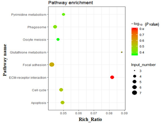
Using gene expression profiling, a total of 256 commonly known DEGs from three comparisons (3w vs. 8w, 3w vs. 13w, and 3w vs. 18w) were screened. Of these, 86 were downregulated and 170 were upregulated (Table S1). Gene Ontology (GO) analysis was performed on these 256 DEGs, with the main GO terms being positive regulation of angiogenesis, positive regulation of DNA binding, cell division, kinetochore organization, negative regulation of transcription, cell wall macromolecule catabolic process, regulation of cardiac muscle contraction, muscle contraction, and more (Table S2). Similarly, using KEGG pathway analysis on these 256 DEGs, eight significantly enriched pathways were found (Table S3 and Figure 2), including pathways related to cell number (apoptosis, cell cycle, and oocyte meiosis). In addition, the focal adhesion, ECM-receptor interaction, glutathione metabolism, phagosome, and pyrimidine metabolism were also screened out.

Figure 2.
Enriched pathways based on the 256 DEGs. KEGG (Kyoto Encyclopedia of Genes and Genomes) pathway analysis of DEGs showed that various fat metabolism pathways (glycerolipid metabolism, steroid biosynthesis, etc.) were enriched (p < 0.05).
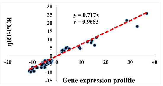
According to the GO terms and KEGG pathways analyses results, a total of 24 DEGs related to muscle development (n = 7), cell number (n = 10, including apoptosis, n = 4, and cell cycle, n = 6), and energy metabolism (n = 7, including lipid metabolism, n = 5, and glycol-metabolism, n = 2) were respectively indicated (Table 4). Moreover, qRT-PCR was performed for these 24 genes to validate the accuracy of the gene expression profiling data, and the association between data from gene expression profiling and qRT-PCR was analyzed by Spearman rank correlation. Results showed that the fold-change of gene expression between the two methods was significantly correlated (r = 0.9683, p < 0.01; Figure 3).

Table 4.
The screened 24 DEGs related to muscle development, cell number, and energy metabolism from data of gene expression profiling.

Figure 3.
Spearman rank correlation analysis of gene expression profiling and qRT-PCR results of 24 genes in three comparisons. A high correlation coefficient (r = 0.9683, p < 0.01) was present, indicating that the gene expression profiling data were reliable. n = 72.
3.3. Differentially Expressed Genes Related to Muscle Development, Cell Number, and Energy Metabolism
As shown in Table 4, the seven common DEGs related to muscle development, at the timeframe studied, were screened and the gene expression fold-change ranged between 2 and 32. Samples from Week 3 were used as control. Results from the qRT-PCR analysis showed that the expression levels of CSRP3, LMOD2, MUSTN1, and MYBPC1 were significantly higher (p < 0.05 or p < 0.01) at all other time points when compared to those at Week 3, but the expression levels of ACTC1, MYL1, and MYL4 were significantly lower (p < 0.01) in the same comparisons (Figure 4). These seven genes were only enriched in the GO terms, such as regulation of cardiac muscle contraction and muscle contraction, but not in the related signaling pathway.

Figure 4.
Verification of DEGs related to muscle development by quantitative real-time polymerase chain reaction (qRT-PCR). These DEGs were significantly upregulated or downregulated (*p < 0.05 or **p < 0.01) in breast muscle tissue at 8, 13, and 18 weeks when compared to those at 3 weeks. n = 9.
Similarly, seven common DEGs related to glycol-metabolism or lipid metabolism at the developmental stage were screened. Again, samples from Week 3 were used as control. The fold-change found ranged between 2 and 5. qRT-PCR results showed that the expression levels of ACOT9, CETP, and LPIN1 were significantly higher and those of DGAT2, RBP7, FBP1, and PHKA1 were significantly lower at all evaluation time points when compared to those at Week 3 (p < 0.01; Figure 5). These genes were found to be mainly involved in the corresponding signaling pathways, but not significantly enriched.

Figure 5.
Verification of DEGs related to cell number by quantitative real-time polymerase chain reaction (qRT-PCR). These DEGs were significantly downregulated (**p < 0.01) in breast muscle tissue at 8, 13, and 18 weeks when compared to those at 3 weeks. n = 9.
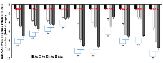
In addition, 10 common DEGs related to cell cycle or apoptosis at the developmental stage were screened. All were shown to have been downregulated when compared to samples from Week 3 that acted as the control. Fold-change ranged between 2 and 11. Using qRT-PCR, we found that the expression levels of these 10 genes, which were enriched in the cell cycle and oocyte meiosis pathways (BUB1, CCNB3, CCNE2, CDC20, MCM2, PLK1) or the apoptosis pathway (BIRC5, CTSK, LMNB1, LMNB2) (Figure 6), were significantly lower (p < 0.05 or p < 0.01) at other stages of development when compared to those at Week 3.

Figure 6.
Verification of DEGs related to energy metabolism by quantitative real-time polymerase chain reaction (qRT-PCR). These DEGs were significantly upregulated or downregulated or (*p < 0.05 or **p < 0.01) in breast muscle tissue at 8, 13, and 18 weeks when compared to those at 3 weeks. n = 9.
4. Discussion
Muscle development of broilers closely affects the quantity of chicken products available for human consumption. Intramuscular fat is an important factor affecting meat quality [21,22,23]. Local Chinese chickens have a high meat quality as they have a high IMF content [24]. The developmental stage is the main muscle and IMF formation period. At the same time, myocytes and adipocytes mutually influence each-other due to their close adjacent relationship in the muscle tissues. Identification of co-expressed genes in the muscle tissues during the developmental stage has great significance for controlling muscle production and regulating meat quality. Such analysis could reveal the molecular regulation relationship between them. The remaining energy from the yolk sac can supply the body’s needs for yellow-feather broilers during the first two weeks after hatching [25]. Therefore, four different stages of development were assessed: Week 3 (starting time, acted as baseline control), Week 8 (rapid development), Week 13 (development peak), and Week 18 (market time). Comparisons between Week 3 and the other three time points was performed to screen for common DEGs, which might be the key functional genes affecting muscle or IMF development during the entire developmental period. In addition, the RPKM method was used in the calculation of gene expression levels to obtain more information on the related genes, though the TPM will be more reliable. Meanwhile, the verification of the screened candidate genes was also strengthened by Q-PCR to ensure the reliability of data.
For the 256 DEGs screened out in breast muscle tissue samples at different developmental stages, we further identified the related functional genes by GO and KEGG analyses. Both final muscle and IMF production are the joint result of cell proliferation and differentiation, and energy metabolism plays an important role in this process [26]. Consequently, the functional genes related to muscle development (n = 7), cell number (n = 10, cell proliferation and apoptosis combined), and energy metabolism (n = 7, glucose and lipid metabolism combined) were combed out. Subsequently, these 24 genes were assessed by qRT-PCR in breast muscle tissue, at the different time points, to verify the gene expression profiles data. The verification ratio reached 7.8%, and the correlation analysis showed a high degree consistency between the two methods (r = 0.9683, p < 0.01), which supports the accuracy of the data from the gene expression profiles.
The number of skeletal muscle fibers is mainly determined at the embryonic stage, and the occurrence of changes in the skeletal muscle after birth is mainly due to the fusion of the muscle satellite cells with the muscle fibers, resulting in hypertrophy of the skeletal muscle fibers [9,10]. In this study, seven common DEGs related to muscle development during the developmental stage were screened out. Among them, CSRP3, LMOD2, MUSTN1, and MYBPC1 levels were significantly higher, and levels of ACTC1, MYL1, and MYL4 were significantly lower at all time points, when compared to Week 3. In a similar study, MYBPC1 had been shown to have a significantly higher expression level during development in breast muscle tissue [13]. It had been reported that CSRP3, LMOD2, MUSTN1, and MYBPC1 have important positive regulation on myofibril assembly and hypertrophy of muscle fibers [27,28,29], while MYL1 and MYL4 have a negative regulatory effect on myogenesis by inhibiting myoblast proliferation [30]. Phenotypic results showed that BMW, BMP (%), and muscle fiber diameter have all continuously and significantly increased, indicating that muscle growth mainly depends on regulation of the differentiation (hypertrophy) of muscle fiber from 3 weeks to 18 weeks. Comprehensively considering these results, these seven genes were identified as key regulatory genes related to muscle growth at the developmental stage in chickens.
Energy metabolism plays an important role in muscle development. A lack of energy will trigger muscle atrophy by various signaling pathways. It was found that FBP1 and PHKA1 mRNA levels have significantly decreased during development. According to published information, FBP1 and PHKA1 play an important regulatory role in gluconeogenesis or glycogen synthesis [31,32]. These results point to the possibility that positive regulation of glucose utilization might be enhanced, and regulation of gluconeogenesis or glycogen synthesis would consequently be reduced in muscle tissue at the developmental stage. For genes related to lipid metabolism, it is known that DGAT2 and RBP7 have an important positive regulatory role in lipid deposition [33,34], ACOT9 and LPIN1 promote lipolysis [35,36], and CETP is involved in reversed cholesterol transport and reduced fat accumulation in chickens [37]. In this study, mRNA expression levels of DGAT2 and RBP7 were significantly lower, and those of ACOT9, CETP, and LPIN1 were significantly higher in breast muscle tissue throughout the development period. Combined with the continuous increase in IMF over time, although the rate of increase slowed down gradually, our results suggest that these lipid metabolism-related genes have a regulatory function on IMF deposition at the developmental stage in chickens.
Tissue development is the combined result of cell proliferation and differentiation. Thus, regulation of cell number was also analyzed. It is known that both cell proliferation and apoptosis could affect cell number. These were therefore also a focus of this study. Ten common DEGs related to cell proliferation (BUB1, CCNB3, CCNE2, CDC20, MCM2, and PLK1, which were mainly enriched in the cell cycle and oocyte meiosis pathways) and apoptosis (BIRC5, CTSK, LMNB1, and LMNB2) were screened out, and mRNA expression levels of all were significantly lower in breast muscle tissue throughout the development stage. As is widely known, BUB1, CCNB3, CCNE2, CDC20, MCM2, and PLK1 genes have a positive regulatory effect on the cell cycle [38,39,40,41,42], and BIRC5, CTSK, LMNB1, and LMNB2 genes promote cell apoptosis [43,44,45]. The number of skeletal muscle fibers is mainly determined at the embryonic stage, while occurrence of new skeletal muscle after birth is mainly due to activation, proliferation, and differentiation of satellite cells [9,10]. Results on the density and diameter of muscle fibers showed no increase in the number of muscle fibers. Genetically, it was made clear that these 10 key genes regulate cell number in breast muscle tissue at the developmental stage in chickens.
The approach of the present study was to use gene expression profiling to identify the common functional genes that regulate muscle development, IMF accumulation, and cell number in muscle tissue of chickens during development. Possible regulation by translational mechanisms and post-translational modifications may have also contributed. Because of tissue complexity, additional experiments on the expression, localization, and function of the regulatory genes should be further performed to reveal the clear function and regulatory mechanism of these candidate genes on muscle development in chickens.
5. Conclusions
In this study, we screened for common regulatory genes related to muscle development, cell number, or energy metabolism at the developmental stage in chickens. Upregulation of CSRP3, LMOD2, MUSTN1, MYBPC1, and MYCBP2, and downregulation of ACTC1, MYL1, and MYL4, are associated with muscle development, primarily hypertrophy of muscle fibers, during development from 3 weeks to 18 weeks. Expression change for genes related to lipid metabolism (ACOT9, CETP, LPIN1, DGAT2, and RBP7) and glycol-metabolism (FBP1 and PHKA1) may have contributed to the continuous increase in IMF deposition. Meanwhile, the screened-out cell-cycle- and apoptosis-related genes reflect on the negative regulation of cell number at the developmental stage. These findings provide new insights into the regulation of muscle development in chickens.
Supplementary Materials
The following are available online at https://www.mdpi.com/2073-4425/11/3/244/s1: Table S1: The screened 256 DEGs in the study (XLSX). Table S2: The involved GO terms based on 256 DEGs (XLSX). Table S3: The involved KEGG pathways based on 256 DEGs (XLSX).
Author Contributions
W.D. and K.Z. contributed to the design of the study; D.L., Z.P. and J.Z. performed of the study and wrote the manuscript; D.L., K.Z., M.Y., D.Y., and Y.L. contributed to interpretation of data; D.L., and J.W. contributed to the design of the study and modifying the manuscript. All authors have read and agreed to the published version of the manuscript.
Funding
The research was supported by National Natural Science Foundation of China (31702110), Fundamental Research Funds for the Central Universities (KJQN201830), Science and Technology Major Project of Guangxi (Guike AA17204027).
Conflicts of Interest
The authors declare no conflict of interest.
Abbreviations
ACOT9: Acyl-CoA thioesterase 9; ACTC1: Actin alpha cardiac muscle 1; AMPK, Adenosine 5′-monophosphate (AMP)-activated protein kinase; BIRC5: Baculoviral IAP repeat containing 5; BJ-Y: Beijing-Youchicken; BMP: breast meat percentage; BMW: Breast muscle weight; CCNB3: Cyclin B3; CCNE2: Cyclin E2; CDC20: Cell division cycle 20; CETP: Cholesteryl ester transfer protein; CSRP3: Cysteine and glycine rich protein 3; CT: cycle threshold; CTSK: Cathepsin K; DGAT2: Diacylglycerol O-acyltransferase 2; DEGs: Differentially expressed genes; EW: eviscerated weight; FBP1: Fructose-bisphosphatase 1; GO: Gene Ontology; IMF: Intramuscular fat; KEGG: Kyoto Encyclopedia of Genes and Genomes; LMNB1: Lamin B1; LMNB2: Lamin B2; LMOD2: Leiomodin 2; LPIN1: Lipin 1; MCM2: Minichromosome maintenance complex component 2; MYBPC1: Myosin binding protein C1; MYL1: Myosin light chain 1; MYL4: Myosin light chain 4; MUSTN1: Musculoskeletal, embryonic nuclear protein 1; PHKA1: Phosphorylase kinase regulatory subunit alpha 1; PLK1: Polo like kinase 1; qRT-PCR: Quantitative real-time polymerase chain reaction; RBP7, Retinol binding protein 7.
References
- Hocquette, J.F.; Gondret, F.; Baeza, E.; Medale, F.; Jurie, C.; Pethick, D.W. Intramuscular fat content in meat-producing animals: Development, genetic and nutritional control, and identifification of putative markers. Animal 2010, 4, 303–319. [Google Scholar] [CrossRef] [PubMed]
- Warner, R.D.; Greenwood, P.L.; Pethick, D.W.; Ferguson, D.M. Genetic and environmental effects on meat quality. Meat Sci. 2010, 86, 171–183. [Google Scholar] [CrossRef] [PubMed]
- Deng, S.; Azevedo, M.; Baylies, M. Acting on identity: Myoblast fusion and the formation of the syncytial muscle fiber. Semin. Cell Dev. Biol. 2017, 72, 45–55. [Google Scholar] [CrossRef] [PubMed]
- Chal, J.; Pourquié, O. Making muscle: Skeletal myogenesis in vivo and in vitro. Development 2017, 144, 2104–2122. [Google Scholar] [CrossRef] [PubMed]
- Hu, L.; Han, F.; Chen, L.; Peng, X.; Chen, D.; Wu, D.; Che, L.; Zhang, K. High nutrient intake during the early postnatal period accelerates skeletal muscle fiver growth and maturity in intrauterine growth-restricted pigs. Genes Nutr. 2018, 13, 23. [Google Scholar] [CrossRef]
- Böl, M.; Leichsenring, K.; Siebert, T. Effects of growth on muscle, tendon, and aponeutosis tissues in rabbit shank musculature. Anat. Rec. (Hoboken) 2017, 300, 1123–1136. [Google Scholar] [CrossRef]
- Smith, J.H. Relation of body size to muscle cell size and number in the chicken. Poult. Sci. 1963, 42, 283–290. [Google Scholar] [CrossRef]
- Egner, I.M.; Bruusgaard, J.C.; Gundersen, K. Satellite cell depletion prevents fiber hypertrophy in skeletal muscle. Development 2016, 143, 2898–2906. [Google Scholar] [CrossRef]
- Morgan, J.E.; Partridge, T.A. Muscle satellite cells. Int. J. Biochem. Cell Biol. 2003, 35, 1151–1156. [Google Scholar] [CrossRef]
- Dumont, N.A.; Bentzinger, C.F.; Sincennes, M.C.; Rudnicki, M.A. Satellite cells and skeletal muscle regeneration. Compr. Physiol. 2015, 5, 1027–1059. [Google Scholar]
- Kimura, N.; Tokunaga, C.; Dalal, S.; Richardson, C.; Yoshino, K.; Hara, K.; Kemp, B.E.; Witters, L.A.; Mimura, O.; Yonezawa, K. A possible linkage between AMP-activated Protein kinase (AMPK) and mammalian targe to frapamycin (m TOR) signaling pathway. Genes Cells 2003, 8, 65–79. [Google Scholar] [CrossRef]
- Zhu, M.J.; Ford, S.P.; Nathanielsz, P.W.; Du, M. Effect of maternal nutrient restriction in sheep on the development of fetal skeletal muscle. Biol. Reprod. 2004, 71, 1968–1973. [Google Scholar] [CrossRef] [PubMed]
- Cui, H.X.; Liu, R.R.; Zhao, G.P.; Zheng, M.Q.; Chen, J.L.; Wen, J. Identification of differentially expressed genes and pathways for intramuscular fat deposition in pectoralis major tissues of fast- and slow-growing chickens. BMC Genom. 2012, 13, 213. [Google Scholar] [CrossRef] [PubMed]
- Arlington, V.A. Official Methods of Analysis, 15th ed.; Association of Official Analytical Chemists: Washington, WA, USA, 1990. [Google Scholar]
- Zerehdaran, S.; Vereijken, A.L.J.; van Arendonk, J.A.M.; van der Waaij, E.H. Estimation of genetic parameters for fat deposition and carcass traits in broilers. Poult. Sci. 2004, 83, 521–525. [Google Scholar] [CrossRef] [PubMed]
- Cumming, W.J.K.; Fulthorpe, J.; Hudgson, P.; Mahon, M. Colour Atlas of Muscle Pathology; Mosby-Wolfe: London, UK, 1994. [Google Scholar]
- Mortazavi, A.; Williams, B.A.; McCue, K.; Schaeffer, L.; Wold, B. Mapping and quantifying mammalian transcriptomes by RNA-Seq. Nat. Methods 2008, 5, 621. [Google Scholar] [CrossRef] [PubMed]
- Benjamini, Y.; Yekutieli, D. The control of the false discovery rate in multiple testing under dependency. Ann. Stat. 2001, 29, 1165–1188. [Google Scholar]
- Wu, J.; Mao, X.; Tao, C.; Luo, J.; Wei, L. KOBAS server: A web-based platform for automated annotation and pathway identification. Nucleic Acids Res. 2006, 34, W720–W724. [Google Scholar] [CrossRef]
- Livak, K.J.; Schmittgen, T.D. Analysis of relative gene expression data using real-time quantitative PCR and the 2−ΔΔCT method. Methods A Companion Methods Enzymol. 2001, 25, 402–408. [Google Scholar] [CrossRef]
- Eikelenboom, G.; Hoving-Bolink, A.H.; Wal, P.G. The eating quality of pork: 2. The influence of intramuscular fat. Fleischwirtschaft 1996, 76, 517–518. [Google Scholar]
- Fernandez, X.; Monin, G.; Talmant, A.; Mourot, J.; Lebret, B. Influence of intramuscular fat content on the quality of pig meat-1. Composition of the lipid fraction and sensory characteristics of m. longissimus lumborum. Meat Sci. 1999, 53, 59–65. [Google Scholar] [CrossRef]
- Lambe, N.R.; McLean, K.A.; Gordon, J.; Evans, D.; Clelland, N.; Bunger, L. Prediction of intramuscular fat content using CT scanning of packaged lamb cuts and relationships with meat eating quality. Meat Sci. 2017, 123, 112–119. [Google Scholar] [CrossRef]
- Cui, X.Y.; Liu, R.R.; Cui, H.X.; Zhao, G.P.; Zheng, M.Q.; Li, Q.H.; Liu, J.; Liu, Z.; Wen, J. Effects of caponization and ovariectomy on objective indices related to meat quality in chickens. Poult Sci. 2017, 96, 770–777. [Google Scholar] [CrossRef]
- Liu, L.; Cui, H.X.; Fu, R.Q.; Zheng, M.Q.; Liu, R.R.; Zhao, G.P.; Wen, J. The regulation of IMF deposition in pectoralis major of fast- and slow- growing chickens at hatching. J. Anim. Sci. Biotechnol. 2017, 8, 77. [Google Scholar] [CrossRef] [PubMed]
- Shan, T.; Xu, Z.; Liu, J.; Wu, W.; Wang, Y. Lkb1 regulation of skeletal muscle development, metabolism muscle progenitor cell homeostasis. J. Cell Physiol. 2017, 232, 2653–2656. [Google Scholar] [CrossRef] [PubMed]
- Tsukada, T.; Pappas, C.T.; Moroz, N.; Antin, P.B.; Kostyukova, A.S.; Gregorio, C.C. Leiomodin-2 is an antagonist of tropomodulin-1 at the pointed end of the thin filaments in cardiac muscle. J. Cell Sci. 2010, 123, 3136–3145. [Google Scholar] [CrossRef]
- Xu, T.S.; Gu, L.H.; Sun, Y.; Zhang, X.H.; Ye, B.G.; Liu, X.L.; Hou, S.S. Characterization of MUSTN1 gene ofand its relationship with skeletal muscle development at postnatal stages in Pekin ducks. Genet. Mol. Res. 2015, 14, 4448–4460. [Google Scholar] [CrossRef] [PubMed]
- Han, S.; Cui, C.; Wang, Y.; He, H.; Liu, Z.; Shen, X.; Chen, Y.; Li, D.; Zhu, Q.; Yin, H. Knockdown of CSRP3 inhibits differentiation of chicken satellite cells by promoting TGF-β/Smad3 signaling. Gene 2019, 707, 36–43. [Google Scholar] [CrossRef]
- Zhang, S.; Xie, H.; Xu, Y.; Li, X.; Deng, L.; Chen, X.; Qiu, L.; Yang, Z. An experimental study of the role of myosin light chain in myogenesis in vitro. Zhongguo Xiu Fu Chong Jian Wai Ke Za Zhi 2008, 22, 753–758. [Google Scholar]
- Ghanem, A.; Kitanovic, A.; Holzwarth, J.; Wölfl, S. Mutational analysis of fructose-1,6-bis-phosphatase FEP1 indicates partially independent functions in gluconeogenesis and sensitivity to genotoxic stress. Microb. Cell 2017, 4, 52–63. [Google Scholar] [CrossRef]
- Uruno, A.; Yagishita, Y.; Katsuoka, F.; Kitajima, Y.; Nunomiya, A.; Nagatomi, R.; Pi, J.; Biswal, S.S.; Yamamoto, M. Nrf2-Mediated regulation of skeletal muscle glycogen metabolism. Mol. Cell. Biol. 2016, 36, 1655–1672. [Google Scholar] [CrossRef]
- Hung, Y.H.; Carreiro, A.L.; Buhman, K.K. Dgat1 and Dgat2 regulate enterocyte triacylglycerol distribution and alter proteins associated with cytoplasmic lipid droplets in response to dietary fat. Biochim. Biophys. Acta Mol. Cell Biol. Lipids 2017, 1862, 600–614. [Google Scholar] [CrossRef] [PubMed]
- Ahn, J.; Shin, S.; Suh, Y.; Park, J.Y.; Hwang, S.; Lee, K. Identification of the avian RBP7 gene as a new adipose-specific gene and RBP7 promoter-driven GFP expression in adipose tissue of transgenic quail. PLoS ONE 2015, 10, e0124768. [Google Scholar] [CrossRef] [PubMed]
- Tillander, V.; Arvidsson Nordström, E.; Reilly, J.; Strozyk, M.; Van Veldhoven, P.P.; Hunt, M.C.; Alexson, S.E. Acyl-CoA thioesterase 9 (ACOT9) in mouse may provide a novel link between fatty acid and amino acid metabolism in mitochondria. Cell. Mol. Life Sci. 2014, 71, 933–948. [Google Scholar] [CrossRef] [PubMed]
- Kim, J.; Lee, Y.J.; Kim, J.M.; Lee, S.Y.; Bae, M.A.; Ahn, J.H.; Han, D.C.; Kwon, B.M. PPARγ agonists induce adipocyte differentiation by modulating the expression of Lipin-1, which acts as a PPARγ phosphatase. Int. J. Biochem. Cell Biol. 2016, 81, 57–66. [Google Scholar] [CrossRef]
- Greaves, K.A.; Going, S.B.; Fernandez, M.L.; Milliken, L.A.; Lohman, T.G.; Bassford, T.; McNamara, D.J. Cholesteryl ester transfer protein and lecithin:cholesterol acyltransferase activities in hispanic and anglo postmenopausal women: Associations with total and regional body fat. Metabolism 2003, 52, 282–289. [Google Scholar] [CrossRef]
- Kajiura-Kobayashi, H.; Kobayashi, T.; Nagahama, Y. The cloning of cyclin B3 and its gene expression during hormonally induced spermatogenesis in the teleost, Anguilla japonica. Biochem. Biophys. Res. Commun. 2004, 323, 288–292. [Google Scholar] [CrossRef]
- Xu, B.; Xu, T.; Liu, H.; Min, Q.; Wang, S.; Song, Q. MiR-490-5p suppresses cell proliferation and invasion by targeting bub1 in hepatocellular carcinoma cells. Pharmacology 2017, 100, 269–282. [Google Scholar] [CrossRef]
- Zhu, C.; Huang, Q.; Zhu, H. MiR-383 inhibited the cell cycle progression of gastric cancer cells via targeting cyclin E2. DNA Cell Biol. 2019, 38, 849–856. [Google Scholar] [CrossRef]
- Kashkin, K.; Chernov, I.; Stukacheva, E.; Monastyrskaya, G.; Uspenskaya, N.; Kopantzev, E.; Sverdlov, E. Cancer specificity of promoters of the genes controlling cell proliferation. J. Cell. Biochem. 2015, 116, 299–309. [Google Scholar] [CrossRef]
- Shang, G.; Ma, X.; Lv, G. Cell division cycle 20 promotes cell proliferation and invasion and inhibits apoptosis in osteosarcoma cells. Cell Cycle 2018, 17, 43–52. [Google Scholar] [CrossRef]
- Chen, W.; Yang, S.; Abe, Y.; Li, M.; Wang, Y.; Shao, J.; Li, E.; Li, Y.P. Novel pycnodysostosis mouse model uncovers cathepsin K function as a potential regulator of osteoclast apoptosis and senescence. Hum. Mol. Genet. 2007, 16, 410–423. [Google Scholar] [CrossRef] [PubMed]
- Yang, S.H.; Chang, S.Y.; Yin, L.; Tu, Y.; Hu, Y.; Yoshinaga, Y.; de Jong, P.J.; Fong, L.G.; Young, S.G. An absence of both lamin B1 and lamin B2 in keratinocytes has no effect on cell proliferation or the development of skin and hair. Hum. Mol. Genet. 2011, 20, 3537–3544. [Google Scholar] [CrossRef] [PubMed]
- Li, P.L.; Zhang, X.; Wang, L.L.; Du, L.T.; Yang, Y.M.; Li, J.; Wang, C.X. MicroRNA-218 is a prognostic indicator in colorectal cancer and enhances 5-fluorouracil-induced apoptosis by targeting BIRC5. Carcinogenesis 2015, 36, 1484–1493. [Google Scholar] [PubMed]
© 2020 by the authors. Licensee MDPI, Basel, Switzerland. This article is an open access article distributed under the terms and conditions of the Creative Commons Attribution (CC BY) license (http://creativecommons.org/licenses/by/4.0/).