Article

Neutrophil Extracellular Traps (NETs) in the Cerebrospinal Fluid Samples from Children and Adults with Central Nervous System Infections

1
Division of Drug Research, Department of Medical and Health Sciences, Linköping University, SE-581 85 Linköping, Sweden
2
Division of Neuro and Inflammation Sciences, Department of Clinical and Experimental Medicine, Linköping University, SE-581 85 Linköping, Sweden
3
Center for Clinical Research Dalarna-Uppsala University, Region Dalarna and Faculty of Medicine and Health Sciences, Örebro University, SE-702 81 Örebro, Sweden
4
Åland Central Hospital, Department of Infectious Diseases, AX-22 100 Mariehamn, Åland, Finland
5
Bimelix AB, AX-22 100 Mariehamn, Åland, Finland
6
Department of Internal Medicine 3-Rheumatology and Immunology, Universitätsklinikum Erlangen, Friedrich-Alexander-University Erlangen-Nürnberg (FAU), DE-91 054 Erlangen, Germany
7
Clinic of Infectious Diseases, Linköping University Hospital, SE-581 85 Linköping, Sweden
8
Department of Clinical and Experimental Medicine, Linköping University, SE-581 85 Linköping, Sweden
*
Author to whom correspondence should be addressed.
Cells 2020, 9(1), 43; https://doi.org/10.3390/cells9010043
Submission received: 28 October 2019 / Revised: 5 December 2019 / Accepted: 17 December 2019 / Published: 23 December 2019

Abstract

:
Neutrophils operate as part of the innate defence in the skin and may eliminate the Borrelia spirochaete via phagocytosis, oxidative bursts, and hydrolytic enzymes. However, their importance in Lyme neuroborreliosis (LNB) is unclear. Neutrophil extracellular trap (NET) formation, which is associated with the production of reactive oxygen species, involves the extrusion of the neutrophil DNA to form traps that incapacitate bacteria and immobilise viruses. Meanwhile, NET formation has recently been studied in pneumococcal meningitis, the role of NETs in other central nervous system (CNS) infections has previously not been studied. Here, cerebrospinal fluid (CSF) samples from clinically well-characterised children (N = 111) and adults (N = 64) with LNB and other CNS infections were analysed for NETs (DNA/myeloperoxidase complexes) and elastase activity. NETs were detected more frequently in the children than the adults (p = 0.01). NET presence was associated with higher CSF levels of CXCL1 (p < 0.001), CXCL6 (p = 0.007), CXCL8 (p = 0.003), CXCL10 (p < 0.001), MMP-9 (p = 0.002), TNF (p = 0.02), IL-6 (p < 0.001), and IL-17A (p = 0.03). NETs were associated with fever (p = 0.002) and correlated with polynuclear pleocytosis (rs = 0.53, p < 0.0001). We show that neutrophil activation and active NET formation occur in the CSF samples of children and adults with CNS infections, mainly caused by Borrelia and neurotropic viruses. The role of NETs in the early phase of viral/bacterial CNS infections warrants further investigation.

1. Introduction

Lyme neuroborreliosis (LNB), which is caused by a complex of Gram-negative spirochaetes called Borrelia burgdorferi sensu lato and transmitted to humans by the Ixodes ricinus tick, is the most serious manifestation of Lyme disease (LD) [1]. Borrelia constitutes a common cause of bacterial infection of the central nervous system (CNS) among children and adults in Sweden [2,3,4,5] and Finland [6]. According to the diagnostic criteria for LNB [7], mononuclear pleocytosis must be present in the cerebrospinal fluid (CSF), and this is an indicator of an active infection. Despite being a bacterial CNS infection, LNB is characterised by a distinct mononuclear predominance in the CSF. The reason for the relatively low proportion of polynuclear cells, such as neutrophils, not only in the CSF [8] but also in the skin of patients with erythema migrans [9]—the earliest manifestation of a borrelia infection—remains unclear. However, as the neutrophils may be present at an early stage prior to lumbar puncture or skin biopsy, they might be overlooked.
Neutrophils are an essential component of the innate immune system, in that they limit locally the pathogen load through phagocytosis and the production of hydrolytic enzymes and chemokines, thereby orchestrating the subsequent adaptive immune response against the bacteria [10]. Beyond this, neutrophils create extracellular traps (NETs), which consist of the neutrophil’s own DNA in the form of de-condensed chromatin, histones, peptides including neutrophil elastase (NE), and myeloperoxidase (MPO). Thus, they entrap extracellular pathogens, causing bacterial death, in a process that was first described in 2004 and termed ‘NETosis’ [11,12,13]. NETs are capable of binding both Gram-negative and Gram-positive bacteria, and their activities not only result in bacterial death but also prevent further spread of the infection and maintain homeostasis at the point of entry, even in unexpected body compartments [14,15,16]. NETs can also ensnare and immobilise viruses, thereby preventing viral entry into cells and spreading [17].
The activities and roles of NETs are mostly unknown in the context of LD and human CNS infections overall. Nevertheless, NETs have been detected in the skin samples of Borrelia-infected, tick-bitten mice and have been shown to entrap and kill the spirochaetes despite a tick saliva-mediated decrease in the production of reactive oxygen species by the neutrophils [18]. In addition, NETs were detected in the CSF in a porcine model of Streptococcus suis meningitis [19] and in a human study of acute bacterial meningitis caused by pneumococci, in which the NETs were shown to play a harmful role in hinder the clearance of bacteria from the rat brain. However, in the same study, NETs were not detected in the CSF samples of patients with LNB (N = 3) and acute viral meningitis (N = 4) [20].
Neutrophils and their activation products are important early during infection, and they most likely affect the course and outcome of many diseases. With LNB, children tend to seek medical care earlier than adults owing to their having higher frequencies of meningeal symptoms and facial nerve palsy [3,4,21]. Therefore, we systematically evaluated whether NETs could be detected (using two different assays) in the CSF samples of children and adults with LNB, as well as in CSF samples from other infections and disorders affecting the CNS. NETs play a complex role in inflammation, although in general they are more stable than cytokines/chemokines and are thus more suitable for analyses of innate immune activation, especially in the sensitive environment of the CNS [22].

2. Patients and Methods

2.1. Paediatric Patients

The clinical characteristics of the children are shown in Table 1. CSF samples from 111 well-characterised children (64 girls, 47 boys; median age, 10 years; interquartile range [IQR], 5–15 years) with definitive or possible LNB (N = 28), tick-borne encephalitis (TBE; N = 3), enteroviral meningitis (EVM; N = 7), other viral infection (OVI, N = 4), and other non-infectious disorders with neurological or CNS symptoms without pleocytosis (N = 69; as described in Table 1) were obtained by lumbar puncture prospectively during the period 2010–2014 as part of a multi-centre study evaluating children with suspected LNB in Sweden, as previously reported [23]. LNB was diagnosed according to European guidelines [7]. Definite LNB (N = 20) was defined as the presence of: (i) symptoms attributable to LNB, (ii) mononuclear pleocytosis in the CSF, and (iii) intrathecally produced anti-Borrelia antibodies. All the children in the ‘possible LNB’ group (N = 8) had symptoms that were attributable to LNB and pleocytosis in the CSF but had neither detectable intrathecally produced anti-Borrelia antibodies nor clinical signs or laboratory evidence of other infections. These children all responded well to antibiotic treatment. Definite and possible LNB (N = 28) were referred to collectively as ‘LNB patients’. All samples were drawn before the initiation of antibiotic treatment, and the CSF samples for evaluation of NETs were frozen at −70 °C until analysis. All the children were evaluated for intrathecal anti-Borrelia antibody production (Borrelia burgdorferi-specific IgG and/or IgM), using the flagella antigen-based, enzyme-linked immunosorbent assay (ELISA, for serum and CSF) in the IDEIA Lyme Neuroborreliosis Kit (Oxoid Ltd., Hampshire, UK) [24]. An index >0.3 was considered as positive, indicating intrathecal production of anti-Borrelia antibodies according to the manufacturer’s instructions. Pleocytosis in the CSF was defined as ≥5 × 10⁶ cells/L. The children were followed for assessment of recovery 2 months after admittance to hospital.

2.2. Adult Patients

The clinical characteristics of the adult patients are shown in Table 2. CSF samples from 64 well-characterised adults (36 women, 28 men; median age, 55 years; IQR, 43–63 years) with LNB (N = 32), unspecified LD (N = 6), TBE (N = 3), other viral meningitides (OVM, N = 2), and other non-infectious disorders with neurological or CNS symptoms (N = 21; as described in Table 2) were prospectively collected by lumbar puncture as part of the clinical routine for patients who were being evaluated for suspected tick-borne CNS infection at Åland Central Hospital (Mariehamn, Finland) [7]. Definite LNB (N = 29) was defined as the presence of: (i) symptoms attributable to LNB; (ii) mononuclear pleocytosis in the CSF; and (iii) intrathecally produced anti-Borrelia antibodies. In addition, the level of CSF-CXCL13 was high (>100 pg/mL) in all the patients, supporting the diagnosis [25]. Patients in the possible LNB group (N = 3) had symptoms attributable to LNB and mononuclear pleocytosis in the CSF, and they showed neither clinical signs nor laboratory evidence of other infections. Increased levels of CSF-CXCL13 supported the diagnosis. The patients with definite and possible LNB ( N = 31) were collectively referred to as the ‘LNB patients’. Unspecified LD is a common manifestation in the highly Borrelia-endemic Åland Islands (personal communication, the Åland Borrelia Research Group, Åland Islands), where a large proportion of the population has anti-Borrelia antibodies in the serum and in the CSF due to previous exposure. Unspecified LD is defined as the presence of: (i) new symptoms suggestive of LD (headache, radiating pain, myalgia, arthralgia and fatigue); (ii) significantly increased anti-Borrelia antibody levels in the serum; (iii) a clear effect of antibiotic treatment for B. burgdorferi on current symptoms; (iv) a lack of pleocytosis and no increase in CSF-CXCL13; and (v) neither clinical signs nor laboratory evidence of other infections. Pleocytosis in the CSF was defined as ≥5 × 10⁶ cells/L. The CSF samples from adults were handled in the same manner as those from the children, i.e., frozen at −70 °C until analysis. CSF samples were analysed for anti-Borrelia IgG antibodies using the recomBead Luminex-based assay (Mikrogen, Germany). An index >0.3 was considered as positive, indicating intrathecal production of anti-Borrelia antibodies, in line with the manufacturer’s instructions. The adult patients were followed for the assessment of recovery 3 weeks and 6 months after admittance to hospital.

2.3. NET Remnant Assay (NETs)

Phorbol-12-myristate-13-acetate (PMA; Sigma-Aldrich, St Louis, MO, USA)-induced NETs were used to create the standard curve for the ELISA. To induce NETs, neutrophils were isolated through Percoll density centrifugation, as described previously [26]. The neutrophils were re-suspended in culture media [RPMI 1640 plus 2% foetal bovine serum (FBS)], seeded onto a 12-well plate at 1.5 × 10⁶ cells per well, and stimulated with 20 nM PMA for 3 h. The wells were then washed twice with culture medium before incubation with 20 U/mL of the restriction enzyme AluI (New England Biolabs Inc., Ipswich, MA, USA) at 37 °C for 20 min to cleave the NETs. The samples were centrifuged at 300× g for 5 min and NET-rich supernatants were pooled, aliquoted, and stored at −80 °C.
The NET remnant ELISA was performed as previously described [27], although with PBS plus 0.05% Tween 20 used as the blocking and washing solution. A 96-well Nunc MediSorp immunoplate was coated with a monoclonal mouse anti-human MPO antibody (DAKO, Carpinteria, CA, USA) at 4 °C overnight. Blocking solution was added for 1 h at room temperature before incubation with standards (PMA-induced NETs), CSF samples, and a peroxidase-labelled anti-DNA antibody (detection antibody of the Human Cell Death Detection ELISAPLUS; Roche Diagnostics GmbH, Mannheim, Germany) for 2 h. Standards and samples were run in duplicate. For the standard curve, an 8-point dilution series with 2-fold dilutions of the PMA-induced NETs was used, and two plasma samples (high and low values) served as positive controls. After incubation, a substrate for peroxidase (ABTS; Roche Diagnostics) was added for 40 min before the plate was read at 405 nm in a VersaMax ELISA microplate reader (Molecular Devices, Sunnyvale, CA, USA). All the samples were interpolated from the standard curve using the sigmoidal 4-parameter logistic regression equation and reported as arbitrary units (a.u.). Non-detectable samples were given the value of 0 a.u. Analysis of NETs in the 175 CSF samples, which originated from the 111 children and 64 adults, was performed at the Division of Drug Research, Linköping University (Linköping, Sweden) [28].

2.4. Neutrophil Elastase (NE) Activity Assay

To confirm NET formation, 28 representative CSF samples (from 26 children and 2 adults) were also analysed for NE activity at the Friedrich-Alexander-Universität Erlangen-Nürnberg (Erlangen, Germany). For measurement of NE activity in the CSF, 100 µL of CSF were added to 100 µL PBS plus 25 µL of 1 M fluorogenic substrate MeOSuc-AAPV-AMC (sc-201163; Santa Cruz Biotechnology) plus 25 µL of 3.3 mM sivelestat (S7198; Sigma-Aldrich) or PBS in black 96-well plates (137101; ThermoFischer Scientific), as previously described [28]. Fluorescence readings were acquired on a TECAN Infinite 200 Pro using the filter set (excitation 360 nm, emission 465 nm) after 51 h of incubation at 37 °C. Assays were performed with technical duplicates and the results are expressed as mean fluorescence intensity (m.f.i.).

2.5. Cytokine and Chemokine Assays

CSF samples from 13 NET remnant-positive children (54% girls; mean age, 9 years; IQR, 5–13 years) and 25 age- and sex-matched NET remnant-negative children (52% girls; mean age, 9 years; IQR, 4.5–12 years) were analysed for chemokines and cytokines using two different magnetic Luminex® assays (both from R&D Systems, Abingdon, UK). The Human Magnetic Luminex® assay was used to measure the levels of CXCL1, CXCL6, CXCL8 (IL-8), CXCL10, and MMP-9, whereas a high-sensitivity assay (Luminex® Performance Assay) was used for the quantification of TNF, IL-6, and IL-17A. The m.f.i. value of the respective analyte was obtained using FLEXMAP 3D (Luminex Inc., Austin, TX, USA), and the Bio-Plex Manager ver. 6.2 software (Bio-Rad Laboratories, Hercules, CA, USA) was used for data analysis. One of the 11 NET-positive samples was not available for the high-sensitivity assay. The samples were run in two different dilutions at a minimum, and the concordance between dilutions was checked carefully when concentrations had to be determined from different dilutions. Concentrations above the range of quantification were assigned the value of the expected concentration of the highest standard. An m.f.i. value below the lowest standard concentration was assigned a value of 0. The cytokines and chemokines were selected based on their associations with neutrophils and/or NETosis [29,30,31,32].

2.6. Statistics

Data were analysed using the GraphPad Prism ver. 8.01 software (GraphPad Software Inc., San Diego, CA, USA). Chi-square or Fisher’s exact test was used to compare discrete variables in 2 × 2 contingency tables. D’Agostino and Pearson omnibus normality tests were used to determine whether or not the data for continuous variables had a Gaussian distribution. For a non-Gaussian distribution of the data, the Mann–Whitney U-test was applied in comparisons between two groups with independent observations. The Kruskal–Wallis test was used for comparisons of more than two groups and if the Kruskal–Wallis test result was significant, Dunn’s post hoc test was performed. For correlation analyses, Spearman’s rank correlation coefficient (rs) was employed. In all the analyses, a p-value < 0.05 was considered statistically significant.

2.7. Ethics

The study protocol was approved by the Regional Ethics Review Board in Uppsala (Dnr 2010/106, children) and the local Ethics Committee of the Åland Health Care, Finland (2/2010, 1/2011, adults).

3. Results

3.1. CSF-NETs in Relation to Patient Diagnoses

NETs were detected in the CSF samples of significantly more children than adults (p = 0.01); in 14 (13%) children, whereof 7 (6%) with LNB, 4 (4%) with EVM, 2 (2%) with TBE, and 1 (1%) with idiopathic peripheral facial nerve palsy (IPFP) without pleocytosis (belonged to the OVI group). Only one adult patient, with meningitis and mononuclear pleocytosis caused by Varicella zoster virus (VZV), had detectable NETs (5 a.u.). The range of NETs levels were as follows: NET-positive patients with LNB, 2–18 a.u. (IQR, 3.2–12.7); NET-positive patients with EVM, 38–2643 a.u. (IQR, 56–2023); in the two NET-positive patients with TBE, 39–74 a.u.; and in the single NET-positive patient with IPFP, the level was 17 a.u. The proportions of NET-positive patients in the LNB (25%), TBE (67%), EVM (57%), and OVI (25%) groups did not differ significantly. Higher levels of NETs were observed among patients with fever (p = 0.002). The NETs levels correlated significantly with the total white blood cell counts in the CSF samples (rs = 0.43, p < 0.001) (Figure 1A), and with polynuclear pleocytosis (rs = 0.53, p < 0.0001) (Figure 1B) as well as mononuclear pleocytosis (rs = 0.41, p < 0.0001) (Figure 1C), although not with age, gender or duration of symptoms. However, when the 14 NET-positive children were analysed separately, the levels of NETs were shown to correlate with polynuclear but not with mononuclear pleocytosis (rs = 0.58, p = 0.03 and rs = −0.10, p = 0.72, respectively) (Figure 1D,E). Children with EVM displayed a significantly shorter disease duration than the children with LNB (p = 0.03); and the number of polynuclear cells in CSF among paediatric patients showed an inverse significant correlation with the duration of neurological symptoms (data available for 77 of the 111 cases; rs = −0.45, p < 0.0001).

3.2. CSF-NE Activity

NE activity was detected in 13/13 NET-positive children but was not detected in the NET-positive adult patient (overall concordance between methods: 93%). The levels of NETs and NE activity correlated significantly (rs = 0.81, p < 0.0001) (Figure 1F).

3.3. NETs in Relation to Clinical Outcome in Children

As we only found one NET-positive adult patient, we focused on evaluating NETs in relation to the disease prognosis for children (Table 1). The vast majority of the LNB cases (25/28; 89%) had recovered at the two-month follow-up. Two of the non-recovered children were NET-negative and one had low levels of NETs (2 a.u.). Accordingly, the occurrence of NETs in the CSF was not associated with recovery among the children with LNB (p = 0.72). All seven children with EVM were fully recovered at the two-month follow-up, four of whom were NET-positive. Among the three patients with TBE, one of the two patients who recovered was NET-positive, as was the patient who did not recover. Two of the four OVI cases had recovered at follow-up, whereas none had detectable NETs in the CSF. Only two patients in the group with other neurological and non-neurological disorders with CNS symptoms were NET-positive, and they had not recovered at follow-up.

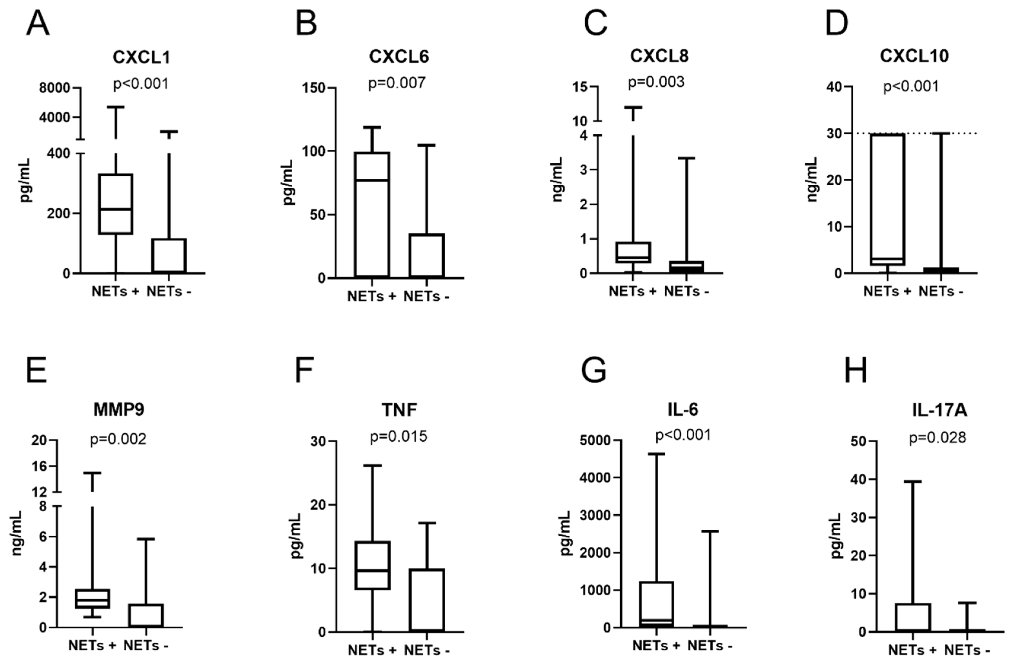
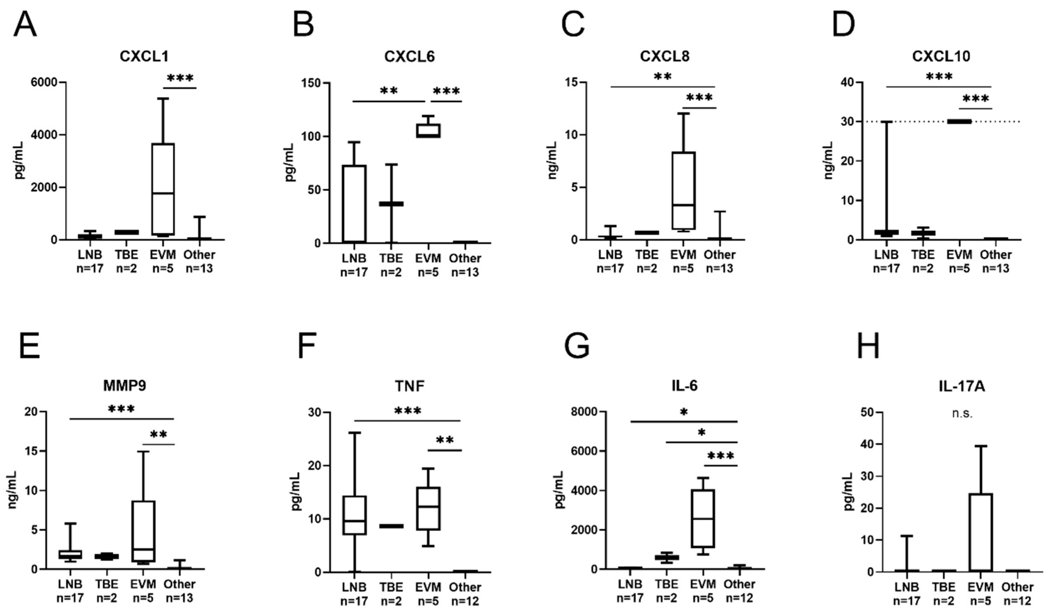
3.4. Cytokines and Chemokines

All eight cytokines/chemokines were detected in at least one sample among the NET-positive (N = 13) and NET-negative (N = 24 or 25) CSF samples, respectively. The total number of samples within the range of quantification is shown in Table 3. The levels of all the cytokines/chemokines were significantly higher among children with NETs: IL-6 (p < 0.001), CXCL8 (p = 0.003), IL-17A (p = 0.03), TNF (p = 0.02), MMP-9 (p = 0.002), CXCL1 (p < 0.001), CXCL6 (p = 0.007) and CXCL10 (p < 0.001) (Figure 2A–H) and fever (N = 16); IL-6 (p < 0.001), CXCL8 (p = 0.001), IL-17A (p = 0.028), TNF (p < 0.001), MMP-9 (p = 0.009), CXCL1 (p = 0.002), CXCL6 (p = 0.002), and CXCL10 (p = 0.001). Furthermore, all the cytokines/chemokines, with the exception of IL-17A, were detected at significantly higher levels in the CSF samples from children with pleocytosis (N = 24) (p < 0.001). A strong and significant correlation was noted between the CSF levels of IL-6, CXCL8, TNF, MMP-9, CXCL1, CXCL6, CXCL10 and NETs, NE, and total CSF cell count, as well as mononuclear and polynuclear pleocytosis, respectively (Table 3). The levels of IL-6, CXCL8, TNF, MMP-9, CXCL1 and CXCL10 in the CSF samples obtained from the different patient groups, respectively, are shown in Figure 3A–H. All the cytokines, with the exception of IL-17A, were present at significantly higher levels in the patients with EVM compared to the patients with “other neurological and non-neurological disorders”. Furthermore, all the cytokines/chemokines, with the exceptions of CXCL1, CXCL6, and IL-17A, were detected at higher levels in the patients with LNB, as compared to the patients with “other neurological and non-neurological disorders”. CXCL6 was the only cytokine that was detected at significantly different levels when comparing the LNB group with the EVM patient group.

4. Discussion

In recent years, interest in NETs has shifted from their roles in innate immune defence to their pathogenic importance in several clinical conditions, such as autoimmune diseases, cancer, cardio-vascular morbidity, and most recently, in infections [14,33]. In the present study, we present, for the first time, evidence of NETs in the CSF of patients with LNB with meningeal inflammation, consisting of both mononuclear and polynuclear pleocytosis, as well as in patients with viral infections of the CNS. NETs are defined as complexes of DNA and MPO, although NE constitutes an additional NET component [34]. All but two of the CSF samples with detectable NETs displayed NE activity also, supporting the actual presence of NETs in the samples (concordance between methods: 93%).
The levels of NETs showed strong variability between the different patient groups, ranging from 2 to 2000 a.u. Interestingly, the highest levels of NETs were detected in children with EVM and TBE, who also had the highest median levels of polynuclear cells. Children with EVM also had the highest median levels of NETs/neutrophil-associated cytokines and chemokines in their CSF samples (among the 38 patients in whom cytokines and chemokines were analysed). In addition, the levels of NETs correlated significantly with the numbers of polynuclear cells, i.e., including neutrophils, which were absent in the paediatric controls (Other disorders) and were overall present at lower levels in the adult patients with longer disease duration, which might explain the absence of NETs in these samples. The only adult who possessed detectable NETs had VZV meningitis with a symptom duration of several weeks and few polynuclear cells in the CSF.
The median pleocytosis values were comparable among children with LNB and EVM, although the proportion of polynuclear cells was higher in the latter group. This may be due to the fact that all the patients with EVM had meningeal signs and, consequently, a significantly shorter disease duration before admittance to hospital, as compared to the patients with LNB. In line with this, the numbers of polynuclear cells in the CSF were found to be inversely correlated with the duration of symptoms.
The finding of neutrophil activation in both LNB and EVM is intriguing, since these infections have similarities with respect to both clinical presentation and CSF cellular profile, which may include neutrophil predominance in the early phase [35]. Several studies have de facto shown the presence of neutrophil-activating cytokines and chemokines in the CSF samples of patients with LNB [36,37,38,39] and EVM [40,41], and the production of neutrophil chemo-attractants, such as CXCL1, CXCL8 and CXCL10 after in vitro stimulation of neuronal cells in response to B. burgdorferi spirochaetes [42]. Thus, there appears to be a prerequisite for neutrophil recruitment into the CNS in infections caused by B. burgdorferi and enterovirus. To clarify further the presence of neutrophils in these CNS infections, we analysed several potent neutrophil-activating cytokines and chemokines in the NET-positive samples. The results showed increased levels of almost all the measured cytokines/chemokines, and these levels strongly correlated with both poly- and mononuclear pleocytosis.
The chemokine CXCL10, also called interferon (IFN) γ-inducible protein 10 (IP-10), is produced by innate immune cells, such as neutrophils, and it stimulates chemotaxis of mononuclear cells into the CNS in response to meningitis/encephalitis caused by, for instance, enterovirus [43], B. burgdorferi [44], and TBE virus [45]. Furthermore, IP-10 has been proposed as a potential discriminator between bacterial and viral CNS infections [41]. Interestingly, CXCL10 was particularly increased in the EVM group, in which the proportion of polynuclear cells was highest. We also found especially increased levels of the fever-inducing cytokines TNF and IL-6 [46] in the EVM group, in which all the patients reported having fever. The chemokines CXCL1, CXCL6 and CXCL8 bind to the CXC-receptor 2 on neutrophils [47], activating them and enabling their entry into the CNS during infections [48]. The highest levels of these cytokines were, as expected, detected in the EVM patients. IL-17A, which is produced by Th17 cells, is a potent activator of neutrophils through stimulation of CXCL1 and CXCL8. Its pathogenic role in the CNS has been studied in LNB [36] and in other bacterial as well as viral CNS infections [49].
The paucity of NETs in the CSF of the adult patients in general, and in the adult LNB patients in particular, might be attributable to a delay between innate immune activation and CSF analysis through lumbar puncture, since the adult patients had longer duration of symptoms at admittance. While the reason for the modest neutrophil activation in LNB is unknown, it might involve an inhibitory effect of the spirochaetes on early pathogen recognition and elimination. Indeed, outer surface proteins (Osp), which are major virulence factors expressed on the surface of B. burgdorferi in the early phase of LD, are required for the spirochaetes to establish infection. OspB have been shown to inhibit the phagocytosis and oxidative burst of human neutrophils and may interfere with complement activation [50]. OspC is known to protect Borrelia sp. from phagocytosis by mononuclear phagocytes, and also to some extent by neutrophils [51].
Since extracellular traps are not produced exclusively by neutrophils, being also produced by mononuclear cells, termed monocyte or macrophage extracellular traps (METs) [52,53], we calculated the proportions of polynuclear and mononuclear cells in the NET-positive samples. It proved to be specifically the polynuclear cells, rather than the cause of the pleocytosis (i.e., the diagnosis), that correlated with the levels of NETs. Moreover, the NET levels and NE activity showed a high degree of correlation. Therefore, we conclude that the NET structures detected were primarily of neutrophil origin. Despite measuring the NET components with two different methods, we cannot affirm unambiguously that the presence of these components is exclusively the result of NET formation, which is always a limitation when one is not studying the actual cellular processes. To investigate this aspect further, cells from the CSF need to be monitored in detail.
We are unable to draw firm conclusions regarding the levels of NETs in relation to disease recovery in any of the patient groups, except in the paediatric LNB group, in which the occurrence of NETs at admission was shown not to be associated with poor clinical outcome at the two-month follow-up. The strengths of the present study are that it is hypothesis driven and has a systematic design, in addition to having well-characterised patients and concordant results. However, the uneven distribution of patients in the different groups and the heterogeneity within the group of other disorders are limitations that made comparisons of the results between patients more difficult.

5. Conclusions

We demonstrate the presence of NETs in the CSF samples collected from patients with LNB and viral CNS infections and show that NETs strongly correlate with the presence of pleocytosis and neutrophil-stimulating cytokines/chemokines in the CSF. Further studies of human neutrophils and their activation products are warranted, so as to elucidate their pathogenic and prognostic roles in acute bacterial and viral infections of the CNS.

Author Contributions

All authors have read and agree to the published version of the manuscript. Conceptualization, J.S., C.S. and D.A.; methodology, D.A., H.E., J.S., J.K. and L.E.M.; software, D.A. and H.E.; validation, D.A., H.E., M.N., L.P., D.N., C.S. and J.S.; formal analysis, D.A., H.E., C.S. and J.S.; investigation, D.A., H.E., B.H.S., M.N., L.P., D.N., C.N., J.K., L.E.M. and J.S.; resources, D.A., H.E., B.H.S., M.N., L.P., D.N., C.N., J.K., L.E.M., C.S. and J.S.; data curation, D.A., H.E. and L.E.M., writing—original draft preparation, D.A., H.E., J.K., L.E.M. and J.S.; writing—review and editing, D.A., H.E., B.H.S., M.N., L.P., D.N., C.N., J.K., L.E.M., C.S. and J.S.; visualization, D.A., H.E., C.S. and J.S.; supervision, C.S., D.N. and L.E.M.; project administration, J.S.; funding, D.A., B.H.S., D.N., L.E.M. and C.S.; acquisition, D.A., L.E.M., C.S. and J.S. All authors have read and agreed to the published version of the manuscript.

Funding

This study was funded by grants from Region Östergötland (ALF grants), the Swedish Rheumatism Association, the Ingrid Asp Foundation, the Center for Clinical Research Dalarna, the King Gustaf V and Queen Victoria’s Freemasons Foundations, the Åland Cultural Foundation, the Wilhelm & Else Stockmann Foundation, the German Research Foundation (project GRK1660), the European Commission (H2020-MSCA-RISE-2015; project 690836 PANG) and the VolkswagenStiftung (grant 90361).

Acknowledgments

We thank Susanne Olausson at Bimelix, Mariehamn, the Åland Islands, and Karin Elving at Clinical Microbiology, Region Dalarna, Sweden for invaluable assistance with the CSF samples. The authors thank all the patients and their parents/guardians for participating in the study, as well as the staff at the paediatric units in Linköping, Norrköping, Jönköping, Skövde, Lidköping, Västerås and Falun for patient recruitment.

Conflicts of Interest

The authors declare no conflict of interest.

References

  1. Halperin, J.J. Lyme neuroborreliosis. Curr. Opin. Infect. Dis. 2019, 32, 259–264. [Google Scholar] [CrossRef] [PubMed]
  2. Henningsson, A.J.; Malmvall, B.E.; Ernerudh, J.; Matussek, A.; Forsberg, P. Neuroborreliosis-An epidemiological, clinical and healthcare cost study from an endemic area in the south-east of Sweden. Clin. Microbiol. Infect. 2010, 16, 1245–1251. [Google Scholar] [CrossRef] [PubMed] [Green Version]
  3. Skogman, B.H.; Croner, S.; Nordwall, M.; Eknefelt, M.; Ernerudh, J.; Forsberg, P. Lyme neuroborreliosis in children: A prospective study of clinical features, prognosis, and outcome. Pediatr. Infect. Dis. J. 2008, 27, 1089–1094. [Google Scholar] [CrossRef] [PubMed]
  4. Sodermark, L.; Sigurdsson, V.; Nas, W.; Wall, P.; Trollfors, B. Neuroborreliosis in Swedish Children: A Population-based Study on Incidence and Clinical Characteristics. Pediatr. Infect. Dis. J. 2017, 36, 1052–1056. [Google Scholar] [CrossRef] [PubMed]
  5. Dahl, V.; Wisell, K.T.; Giske, C.G.; Tegnell, A.; Wallensten, A. Lyme neuroborreliosis epidemiology in Sweden 2010 to 2014: Clinical microbiology laboratories are a better data source than the hospital discharge diagnosis register. Euro Surveill. 2019, 24, 20. [Google Scholar] [CrossRef] [Green Version]
  6. Pietikainen, A.; Oksi, J.; Hytonen, J. Point-of-care testing for CXCL13 in Lyme neuroborreliosis. Diagn Microbiol. Infect. Dis. 2018, 91, 226–228. [Google Scholar] [CrossRef]
  7. Mygland, A.; Ljostad, U.; Fingerle, V.; Rupprecht, T.; Schmutzhard, E.; Steiner, I.; European Federation of Neurological Societies. EFNS guidelines on the diagnosis and management of European Lyme neuroborreliosis. Eur. J. Neurol. 2010, 17, 8–16. [Google Scholar] [CrossRef]
  8. Shah, S.S.; Zaoutism, T.E.; Turnquist, J.; Hodinka, R.L.; Coffin, S.E. Early differentiation of Lyme from enteroviral meningitis. Pediatr. Infect. Dis. J. 2005, 24, 542–545. [Google Scholar] [CrossRef]
  9. Salazar, J.C.; Pope, C.D.; Sellati, T.J.; Feder, H.M.; Jr Kiely, T.G.; Dardick, K.R.; Buckman, R.L.; Moore, M.W.; Caimano, M.J.; Pope, J.G.; et al. Coevolution of markers of innate and adaptive immunity in skin and peripheral blood of patients with erythema migrans. J. Immunol. 2003, 171, 2660–2670. [Google Scholar] [CrossRef] [Green Version]
  10. Liew, P.X.; Kubes, P. The Neutrophil’s Role During Health and Disease. Physiol. Rev. 2019, 99, 1223–1248. [Google Scholar] [CrossRef]
  11. Kruger, P.; Saffarzadeh, M.; Weber, A.N.; Rieber, N.; Radsak, M.; von Bernuth, H.; Benarafa, C.; Roos, D.; Skokowa, J. Hartl DNeutrophils: Between host defence immune modulation,, tissue, injury. PLoS Pathog. 2015, 11, e1004651. [Google Scholar] [CrossRef] [PubMed] [Green Version]
  12. Papayannopoulos, V. Neutrophil extracellular traps in immunity and disease. Nat. Rev. Immunol. 2018, 18, 134–147. [Google Scholar] [CrossRef]
  13. Brinkmann, V.; Reichard, U.; Goosmann, C.; Fauler, B.; Uhlemann, Y.; Weiss, D.S.; Weinrauch, Y.; Zychlinsky, A. Neutrophil extracellular traps kill bacteria. Science 2004, 303, 1532–1535. [Google Scholar] [CrossRef] [PubMed]
  14. Brinkmann, V. Neutrophil Extracellular Traps in the Second Decade. J. Innate Immun. 2018, 10, 414–421. [Google Scholar] [CrossRef] [PubMed]
  15. Mahajan, A.; Gruneboom, A.; Petru, L.; Podolska, M.J.; Kling, L.; Maueroder, C.; Dahms, F.; Christiansen, S.; Günter, L.; Krenn, V.; et al. Frontline Science: Aggregated neutrophil extracellular traps prevent inflammation on the neutrophil-rich ocular surface. J. Leukoc. Biol. 2019, 105, 1087–1098. [Google Scholar] [CrossRef]
  16. Munoz, L.E.; Boeltz, S.; Bilyy, R.; Schauer, C.; Mahajan, A.; Widulin, N.; Grüneboom, A.; Herrmann, I.; Boada, E.; Rauh, M.; et al. Neutrophil Extracellular Traps Initiate Gallstone Formation. Immunity 2019, 51, 443–450. [Google Scholar] [CrossRef] [Green Version]
  17. Schonrich, G.; Raftery, M.J. Neutrophil Extracellular Traps Go Viral. Front. Immunol. 2016, 7, 366. [Google Scholar] [CrossRef] [Green Version]
  18. Menten-Dedoyart, C.; Faccinetto, C.; Golovchenko, M.; Dupiereux, I.; Van Lerberghe, P.B.; Dubois, S.; Desmet, C.; Elmoualij, B.; Baron, F.; Rudenko, N.; et al. Neutrophil extracellular traps entrap and kill Borrelia burgdorferi sensu stricto spirochetes and are not affected by Ixodes ricinus tick saliva. J. Immunol. 2012, 189, 5393–5401. [Google Scholar] [CrossRef] [Green Version]
  19. De Buhr, N.; Reuner, F.; Neumann, A.; Stump-Guthier, C.; Tenenbaum, T.; Schroten, H.; Ishikawa, H.; Müller, K.; Beineke, A.; Hennig-Pauka, I.; et al. Neutrophil extracellular trap formation in the Streptococcus suis-infected cerebrospinal fluid compartment. Cell Microbiol. 2017, 19, 12649. [Google Scholar] [CrossRef] [Green Version]
  20. Mohanty, T.; Fisher, J.; Bakochi, A.; Neumann, A.; Cardoso, J.F.P.; Karlsson, C.A.Q.; Pavan, C.; Lundgaard, I.; Nilson, B.; Peter, R.; et al. Neutrophil extracellular traps in the central nervous system hinder bacterial clearance during pneumococcal meningitis. Nat. Commun. 2019, 10, 1667. [Google Scholar] [CrossRef]
  21. Knudtzen, F.C.; Andersen, N.S.; Jensen, T.G.; Skarphedinsson, S. Characteristics and Clinical Outcome of Lyme Neuroborreliosis in a High Endemic Area, 1995–2014: A Retrospective Cohort Study in Denmark. Clin. Infect. Dis. 2017, 65, 1489–1495. [Google Scholar] [CrossRef] [PubMed]
  22. Schauer, C.; Janko, C.; Munoz, L.E.; Zhao, Y.; Kienhofer, D.; Frey, B. Aggregated neutrophil extracellular traps limit inflammation by degrading cytokines and chemokines. Nat. Med. 2014, 20, 511–517. [Google Scholar] [CrossRef] [PubMed]
  23. Backman, K.; Skogman, B.H. Occurrence of erythema migrans in children with Lyme neuroborreliosis and the association with clinical characteristics and outcome—A prospective cohort study. BMC Pediatr. 2018, 18, 189. [Google Scholar] [CrossRef] [PubMed] [Green Version]
  24. Hansen, K.; Lebech, A.M. Lyme neuroborreliosis: A new sensitive diagnostic assay for intrathecal synthesis of Borrelia burgdorferi-specific immunoglobulin GA. and M. Ann. Neurol. 1991, 30, 197–205. [Google Scholar] [CrossRef]
  25. Rupprecht, T.A.; Manz, K.M.; Fingerle, V.; Lechner, C.; Klein, M.; Pfirrmann, M.; Koedel, U. Diagnostic value of cerebrospinal fluid CXCL13 for acute Lyme neuroborreliosis. A systematic review and meta-analysis. Clin. Microbiol. Infect. 2018, 24, 1234–1240. [Google Scholar] [CrossRef] [Green Version]
  26. Soderberg, D.; Kurz, T.; Motamedi, A.; Hellmark, T.; Eriksson, P.; Segelmark, M. Increased levels of neutrophil extracellular trap remnants in the circulation of patients with small vessel vasculitis, but an inverse correlation to anti-neutrophil cytoplasmic antibodies during remission. Rheumatolog (Oxf.) 2015, 54, 2085–2094. [Google Scholar] [CrossRef] [Green Version]
  27. Kessenbrock, K.; Krumbholz, M.; Schonermarck, U.; Back, W.; Gross, W.L.; Werb, Z.; Gröne, H.J.; Brinkmann, V.; Jenne, D.E. Netting neutrophils in autoimmune small-vessel vasculitis. Nat. Med. 2009, 15, 623–625. [Google Scholar] [CrossRef]
  28. Appelgren, D.; Dahle, C.; Knopf, J.; Bilyy, R.; Vovk, V.; Sundgren, P.C.; Bengtsson, A.A.; Wetterö, J.; Muñoz, L.E.; Herrmann, M.; et al. Active NET formation in Libman-Sacks endocarditis without antiphospholipid antibodies: A dramatic onset of systemic lupus erythematosus. Autoimmunity 2018, 51, 310–318. [Google Scholar] [CrossRef] [Green Version]
  29. Rumble, J.M.; Huber, A.K.; Krishnamoorthy, G.; Srinivasan, A.; Giles, D.A.; Zhang, X.; Wang, L.; Segal, B.M. Neutrophil-related factors as biomarkers in EAE and MS. J. Exp. Med. 2015, 212, 23–35. [Google Scholar] [CrossRef] [Green Version]
  30. Michael, M.; Vermeren, S. A neutrophil-centric view of chemotaxis. Essays Biochem. 2019, 63, 607–618. [Google Scholar]
  31. Isailovic, N.; Daigo, K.; Mantovani, A.; Selmi, C. Interleukin-17 and innate immunity in infections and chronic inflammation. J. Autoimmun. 2015, 60, 1–11. [Google Scholar] [CrossRef]
  32. Hellberg, S.; Eklund, D.; Gawel, D.R.; Kopsen, M.; Zhang, H.; Nestor, C.E.; Kockum, I.; Olsson, T.; Skogh, T.; Kastbom, A.; et al. Dynamic Response Genes in CD4+ T Cells Reveal a Network of Interactive Proteins that Classifies Disease Activity in Multiple Sclerosis. Cell Rep. 2016, 16, 2928–2939. [Google Scholar] [CrossRef] [PubMed] [Green Version]
  33. O’Neil, L.J.; Kaplan, M.J.; Carmona-Rivera, C. The Role of Neutrophils and Neutrophil Extracellular Traps in Vascular Damage in Systemic Lupus Erythematosus. J. Clin. Med. 2019, 8, 1325. [Google Scholar] [CrossRef] [PubMed] [Green Version]
  34. Papayannopoulos, V.; Metzler, K.D.; Hakkim, A.; Zychlinsky, A. Neutrophil elastase and myeloperoxidase regulate the formation of neutrophil extracellular traps. J. Cell Biol. 2010, 191, 677–691. [Google Scholar] [CrossRef] [Green Version]
  35. Makis, A.; Shipway, D.; Hatzimichael, E.; Galanakis, E.; Pshezhetskiy, D.; Chaliasos, N.; Stebbing, J.; Siamopoulou, A. Cytokine and adhesion molecule expression evolves between the neutrophilic and lymphocytic phases of viral meningitis. J. Interferon Cytokine Res. 2010, 30, 661–665. [Google Scholar] [CrossRef] [PubMed]
  36. Gyllemark, P.; Forsberg, P.; Ernerudh, J.; Henningsson, A.J. Intrathecal Th17- and B cell-associated cytokine and chemokine responses in relation to clinical outcome in Lyme neuroborreliosis: A large retrospective study. J. Neuroinflamm. 2017, 14, 27. [Google Scholar] [CrossRef] [PubMed] [Green Version]
  37. Pietikainen, A.; Maksimow, M.; Kauko, T.; Hurme, S.; Salmi, M.; Hytonen, J. Cerebrospinal fluid cytokines in Lyme neuroborreliosis. J. Neuroinflamm. 2016, 13, 273. [Google Scholar] [CrossRef] [Green Version]
  38. Cerar, T.; Ogrinc, K.; Lotric-Furlan, S.; Kobal, J.; Levicnik-Stezinar, S.; Strle, F.; Ruzić-Sabljic, E. Diagnostic value of cytokines and chemokines in lyme neuroborreliosis. Clin. Vaccine Immunol. 2013, 20, 1578–1584. [Google Scholar] [CrossRef]
  39. Eckman, E.A.; Pacheco-Quinto, J.; Herdt, A.R.; Halperin, J.J. Neuroimmunomodulators in Neuroborreliosis and Lyme Encephalopathy. Clin. Infect. Dis. 2018, 67, 80–88. [Google Scholar] [CrossRef]
  40. Park, S.E.; Shin, K.; Song, D.; Nam, S.O.; Kim, K.M.; Lyu, S.Y. Comparison of Cerebrospinal Fluid Cytokine Levels in Children of Enteroviral Meningitis With Versus Without Pleocytosis. J. Interferon Cytokine Res. 2018, 38, 348–355. [Google Scholar] [CrossRef] [Green Version]
  41. Maric, L.S.; Lepej, S.Z.; Gorenec, L.; Grgic, I.; Trkulja, V.; Rode, O.D.; Roglic, S.; Grmoja, T.; Barisic, N.; Tesovic, G. Chemokines CXCL10, CXCL11, and CXCL13 in acute disseminated encephalomyelitis, non-polio enterovirus aseptic meningitis, and neuroborreliosis: CXCL10 as initial discriminator in diagnostic algorithm? Neurol Sci. 2018, 39, 471–479. [Google Scholar] [CrossRef] [PubMed]
  42. Brissette, C.A.; Kees, E.D.; Burke, M.M.; Gaultney, R.A.; Floden, A.M.; Watt, J.A. The multifaceted responses of primary human astrocytes and brain microvascular endothelial cells to the Lyme disease spirochete, Borrelia burgdorferi. ASN Neuro 2013, 5, 221–229. [Google Scholar] [CrossRef] [PubMed] [Green Version]
  43. Cavcic, A.; Tesovic, G.; Gorenec, L.; Grgic, I.; Benic, B.; Lepej, S.Z. Concentration gradient of CXCL10 and CXCL11 between the cerebrospinal fluid and plasma in children with enteroviral aseptic meningitis. Eur. J. Paediatr. Neurol. 2011, 15, 502–507. [Google Scholar] [CrossRef] [PubMed] [Green Version]
  44. Henningsson, A.J.; Tjernberg, I.; Malmvall, B.E.; Forsberg, P.; Ernerudh, J. Indications of Th1 and Th17 responses in cerebrospinal fluid from patients with Lyme neuroborreliosis: A large retrospective study. J. Neuroinflamm. 2011, 8, 36. [Google Scholar] [CrossRef] [Green Version]
  45. Zajkowska, J.; Moniuszko-Malinowska, A.; Pancewicz, S.A.; Muszynska-Mazur, A.; Kondrusik, M.; Grygorczuk, S.; Swierzbińska-Pijanowska, R.; Dunaj, J.; Czupryna, P. Evaluation of CXCL10, CXCL11, CXCL12 and CXCL13 chemokines in serum and cerebrospinal fluid in patients with tick borne encephalitis (TBE). Adv. Med. Sci. 2011, 56, 311–317. [Google Scholar] [CrossRef]
  46. Evans, S.S.; Repasky, E.A.; Fisher, D.T. Fever and the thermal regulation of immunity: The immune system feels the heat. Nat. Rev. Immunol. 2015, 15, 335–349. [Google Scholar] [CrossRef]
  47. Sadik, C.D.; Kim, N.D.; Luster, A.D. Neutrophils cascading their way to inflammation. Trends Immunol. 2011, 32, 452–460. [Google Scholar] [CrossRef] [Green Version]
  48. Simmons, S.B.; Liggitt, D.; Goverman, J.M. Cytokine-regulated neutrophil recruitment is required for brain but not spinal cord inflammation during experimental autoimmune encephalomyelitis. J. Immunol. 2014, 193, 555–563. [Google Scholar] [CrossRef] [Green Version]
  49. Morichi, S.; Urabe, T.; Morishita, N.; Takeshita, M.; Ishida, Y.; Oana, S.; Yamanaka, G.; Kashiwagi, Y.; Kawashima, H. Pathological analysis of children with childhood central nervous system infection based on changes in chemokines and interleukin-17 family cytokines in cerebrospinal fluid. J. Clin. Lab Anal. 2018, 32, 521784686. [Google Scholar] [CrossRef] [Green Version]
  50. Hartiala, P.; Hytonen, J.; Suhonen, J.; Lepparanta, O.; Tuominen-Gustafsson, H.; Viljanen, M.K. Borrelia burgdorferi inhibits human neutrophil functions. Microbes Infect. 2008, 10, 60–68. [Google Scholar] [CrossRef]
  51. Carrasco, S.E.; Troxell, B.; Yang, Y.; Brandt, S.L.; Li, H.; Sandusky, G.E.; Condon, K.W.; Serezani, C.H.; Yang, X.F. Outer surface protein OspC is an antiphagocytic factor that protects Borrelia burgdorferi from phagocytosis by macrophages. Infect. Immun. 2015, 83, 4848–4860. [Google Scholar] [CrossRef] [PubMed] [Green Version]
  52. Doster, R.S.; Rogers, L.M.; Gaddy, J.A.; Aronoff, D.M. Macrophage Extracellular Traps: A Scoping Review. J. Innate Immun. 2018, 10, 3–13. [Google Scholar] [CrossRef] [PubMed]
  53. Granger, V.; Faille, D.; Marani, V.; Noel, B.; Gallais, Y.; Szely, N.; Flament, H.; Pallardy, M.; Chollet-Martin, S.; de Chaisemartin, L. Human blood monocytes are able to form extracellular traps. J. Leukoc. Biol. 2017, 102, 775–781. [Google Scholar] [CrossRef] [PubMed] [Green Version]
Figure 1. Correlations between cell counts in the CSF and NET remnants: (AC) (N = 111 children) and (DE) (N = 14 children). Correlation between NET remnants and elastase activity: (F) (N = 28; 13 NET-positive and 15 NET-negative cases).
Cells 09 00043 g001
Figure 2. Concentrations of chemokines (AD), MMP9 (E) and cytokines (FH) in the CSF samples from the NET remnant-positive and NET remnant-negative children. Whiskers indicate the min-max values. The p-values are derived from the Mann–Whitney U-test. CXCL10 concentrations above the range of quantification (>30 ng/mL, N = 5) were assigned a value of 30 ng/mL (upper range of quantification is indicated by a dashed line). NETs: neutrophil extracellular traps.
Cells 09 00043 g002
Figure 3. Concentrations of chemokines (AD), MMP9 (E) and cytokines (FH) in the CSF samples from the different patient groups. Whiskers indicate the min-max values. CXCL10 concentrations above the range of quantification (>30 ng/mL, N = 5) were assigned a value of 30 ng/mL (upper range of quantification is indicated by a dashed line). Only one child (rotavirus infection, no pleocytosis) belonged to the OVI group and is not included in the figure. LNB. Lyme neuroborreliosis; TBE, tick-borne encephalitis; EVM, enteroviral meningitis; Other, other non-infectious neurological and non-neurological disorders with CNS symptoms. * p < 0.05, ** p < 0.01, *** p < 0.001
Cells 09 00043 g003
Table 1. Characteristics of paediatric patients.
On Admission LNB
(N = 28)
TBE
(N = 3)
EVM
(N = 7)
OVI
(N = 4)
Other Disorder
(N = 69)
AgeMedian (range)10 (3–15)10 (3–15)10 (4–15)4 (1–14)13 (0–19)
SexFemale, N (%)14 (50)02 (30)3 (75)44 (64)
Duration of symptoms<1 week, N (%)16 (59)1 (50)5 (83)1 (100)9 (22)
1–4 weeks, N (%)9 (33)1 (50)1 (17)011 (27)
1–2 months, N (%)1 (4)0005 (12)
>2 months, N (%)1 (4)00016 (39)
Clinical features Facial nerve palsy, N (%)22 (79)0 (0)0 (0)1 (25)17 (25)
Meningeal
symptoms ¤, N (%)
22 (79)2 (67)7 (100)1(25)50 (72)
Fever >38 °C, N (%)13 (46)2 (67)7 (100)2 (50)7 (10)
Fatigue, N (%)23 (82)3 (100)6 (86)4 (100)38 (55)
Laboratory findingsPleocytosis *, median (range)157 (20–486)100 (0–130)156 (20–634)0 (0–6)0 (0–74)
CSF mono, median (range)149 (8–484)77 (0–116)86 (16–610)60 (0–40)
CSF poly, median (range)4 (0–30)14 (0–28)24 (4–164)00 (0–34)
Recovery at follow-up:
2 monthsYes, N (%)25 (89)2 (67)7 (100)2 (50)45 (65)
CSF, cerebrospinal fluid; LNB, Lyme neuroborreliosis; LD, Lyme disease; TBE, tick-borne encephalitis; EVM, enteroviral meningitis; OVI, other viral infection; Other disorders (Demyelinating polyneuropathy, Idiopathic peripheral facial nerve palsy, Idiopathic intracranial hypertension, Epilepsy, Head trauma, Myasthenia, Infantile spasm, Autoimmune encephalitis, Guillan-Barré syndrome, Narcolepsy, Optic neuritis, Fatigue, Microcephaly, Mycoplasma infection, Voice hallucinations, Periodic fever aphthous stomatitis pharyngitis cervical adenitis (PFAPA), Papillary oedema, Multiple sclerosis). * ≥5 × 10⁶ cells /L; ¤ Meningeal symptoms: Headache, Neck pain, Neck stiffness.
Table 2. Characteristics of adult patients.
On Admission LNB
(N = 32)
LD Unspec.
(N = 6)
TBE
(N = 3)
OVM
(N = 2)
Other Disorders
(N = 21)
AgeMedian (range)58 (18–82)52 (30–80)50 (46–57)60 (50–69)51 (24–88)
SexFemale, N (%)15 (47)6 (100)1 (33)1 (50)13 (62)
Duration of symptoms<1 week, N (%)4 (13)0 (0)001 (5)
1–4 weeks, N (%)7 (23)1 (20)3 (100)1 (50)1 (5)
1–2 months, N (%)10 (31)1 (20)001 (5)
>2 months, N (%)11 (34)3 (60)01 (50)18 (86)
Clinical featuresFacial nerve palsy, N (%)5 (16)1 (17)000
Meningeal
symptoms ¤, N (%)
15 (47)3 (50)2 (67)2 (100)10 (48)
Fever >38 °C, N (%)3 (9)03 (100)1 (50)3 (14)
Fatigue, N (%)17 (53)5 (83)3 (100)1 (50)9 (43)
Radiating pain, N (%)22 (69)2 (33)01 (50)9 (43)
Laboratory findingsPleocytosis *, median (range)43 (6–390)5 (4–5)82 (51–131)33 (18–47)6 (0–292)
CSF mono, median (range)67 (5–355)NA62 (49–118)26 (6–45)54 (17–91) &
CSF poly, median (range)3 (0–45)NA13 (2–20)7 (2–12)106 (10–202) &
Recovery at follow-up:
3 weeksYes, N (%)12 (38)3 (50)01 (50)4 (20)
6 monthsYes, N (%)24 (77)6 (100)2 (67)1 (50)8 (42)
CSF, cerebrospinal fluid; LNB, Lyme neuroborreliosis; LD, Lyme disease; TBE, tick-borne encephalitis; OVM, other viral meningitis; Other disorders (Guillan-Barré syndrome, Spinal disk hernia, Trigeminal neuralgia, Sinusitis, Hypermobility syndrome, Depression, Chronic fatigue syndrome, Benign intracranial hypertension, Dementia, Recurrent iridocyclitis, Cerebral ischemia, Subarachnoidal haemorrhage, Chronic musculoskeletal pain, Meningeal inflammation of unknown origin). NA, not analysed. * ≥5 × 10⁶/L; & Based on 2 samples (cell count <10 × 106 not diff in mono/poly); ¤ Meningeal symptoms: Headache, Neck pain, Neck stiffness.
Table 3. Spearman’s correlation between cytokines, chemokines, cells and NETs in CSF.
NETs (Remnants)NETs (Elastase) Cells (Total)Polynuclear CellsMononuclear Cells
AnalyteQuantified *rsp-Valuersp-Valuersp-Valuersp-Valuersp-Value
CXCL121/380.61<0.0010.76<0.0010.520.0060.69<0.0010.480.02
CXCL614/380.520.0070.650.0020.72<0.0010.74<0.0010.67<0.001
CXCL838/380.530.0060.640.0030.60<0.0010.65<0.0010.570.002
CXCL1032/380.560.0020.660.0020.79<0.0010.81<0.0010.76<0.001
MMP925/380.530.006N/An.s.0.70<0.0010.65<0.0010.68<0.001
TNF22/37N/An.s.N/An.s.0.81<0.0010.65<0.0010.80<0.001
IL-628/370.70<0.0010.81<0.0010.63<0.0010.73<0.0010.59<0.001
IL-17A4/37N/An.s.N/An.s.N/An.s.0.460.03N/An.s.
* Samples within range of quantification (N)/total number of samples analysed (N). All samples outside the range of quantification were below the range of quantification, except for CXCL10; Data on NETs by elastase assay was only available in 26 samples (CXCL1, CXCL6, CXCL8, CXCL10, MMP9) or 25 samples (TNF, IL-6 and IL-17A); p-values have been multiplied by the number of analytes (i.e. 8) to adjust for multiple testing. Grey shading indicates rs ≥ 0.70. n.s. = non-significant, N/A = Not applicable.

