Article

Effect of Adenomatous Polyposis Coli Loss on Tumorigenic Potential in Pancreatic Ductal Adenocarcinoma

by
Jennifer M. Cole
1,2,
Kaitlyn Simmons
2,3 and
Jenifer R. Prosperi
1,2,3,*
*
Author to whom correspondence should be addressed.
1.
Department of Biochemistry and Molecular Biology, Indiana University School of Medicine, South Bend, IN 46617, USA
2.
Mike and Josie Harper Cancer Research Institute, South Bend, IN 46617, USA
3.
Department of Biological Sciences at University of Notre Dame, South Bend, IN 46617, USA
Cells 2019, 8(9), 1084; https://doi.org/10.3390/cells8091084
Submission received: 27 August 2019 / Revised: 10 September 2019 / Accepted: 11 September 2019 / Published: 14 September 2019

Abstract

:
Loss of the Adenomatous Polyposis Coli (APC) tumor suppressor in colorectal cancer elicits rapid signaling through the Wnt/β-catenin signaling pathway. In contrast to this well-established role of APC, recent studies from our laboratory demonstrated that APC functions through Wnt-independent pathways to mediate in vitro and in vivo models of breast tumorigenesis. Pancreatic ductal adenocarcinoma (PDAC) has an overall median survival of less than one year with a 5-year survival rate of 7.2%. APC is lost in a subset of pancreatic cancers, but the impact on Wnt signaling or tumor development is unclear. Given the lack of effective treatment strategies for pancreatic cancer, it is important to understand the functional implications of APC loss in pancreatic cancer cell lines. Therefore, the goal of this project is to study how APC loss affects Wnt pathway activation and in vitro tumor phenotypes. Using lentiviral shRNA, we successfully knocked down APC expression in six pancreatic cancer cell lines (AsPC-1, BxPC3, L3.6pl, HPAF-II, Hs 766T, MIA PaCa-2). No changes were observed in localization of β-catenin or reporter assays to assess β-catenin/TCF interaction. Despite this lack of Wnt/β-catenin pathway activation, the majority of APC knockdown cell lines exhibit an increase in cell proliferation. Cell migration assays showed that the BxPC-3 and L3.6pl cells were impacted by APC knockdown, showing faster wound healing in scratch wound assays. Interestingly, APC knockdown had no effect on gemcitabine treatment, which is the standard care for pancreatic cancer. It is important to understand the functional implications of APC loss in pancreatic cancer cells lines, which could be used as a target for therapeutics.

1. Introduction

Pancreatic ductal adenocarcinoma (PDAC) has an overall median survival of less than one year and is the third leading cause of cancer-related deaths for both men and women in the United States [1]. Patients are often asymptomatic early on, which results in approximately 53% of patients being diagnosed in advanced stages. The five-year survival rate for advanced stage PDAC is 7.2% [2]. Although researchers are actively investigating early detection methods, there are currently no reliable methods for detecting pancreatic cancer (Cancer Facts and Figures 2015). In contrast to the increased survival of patients with many other cancers, advancements in pancreatic cancer treatment have been slow [1]. In fact, death rates due to pancreatic cancer are rising for both men and women [1]. Treatments for patients with PDAC include surgery or chemotherapy treatments using a DNA nucleoside, gemcitabine [3]. Although these treatment options may relieve symptoms or extend survival, they seldom produce a cure. Because pancreatic cancer is typically detected after metastasis has occurred, less than 20% of patients are candidates for surgery.
The APC tumor suppressor is a potent negative regulator of the Wnt/β-catenin signaling pathway. Mutations in APC were first studied in colorectal cancer [4,5,6,7]. APC inactivation has been found in approximately 35% to 88% of colorectal tumors, making it the most common genetic alteration observed in colorectal cancers [8]. Recent studies have also identified APC mutations in many epithelial cancers, including breast and lung cancer (reviewed in [9]). In some extracolonic tumors, including pancreatic [10,11], inactivation of APC occurs through promoter methylation and/or results in Wnt-independent signaling mechanisms [9], suggesting a tissue-specific effect of APC on tumor development. The importance of APC in pancreatic cancer is not yet fully understood, and appears complicated depending on the type of pancreatic cancer being assessed [11,12]. APC was methylated in 58.6% of PDAC, with prevalence of APC methylation increasing with tumor progression [13]. In another study, somatic mutations in APC were observed in 4 of 10 pancreatic tumors examined [14]. Of these, two tumors contained mutations in the mutation cluster region (MCR), which includes the β-catenin binding domain. These frameshift mutations were caused by single base pair deletions, leading to a truncated protein and loss of function [14]. Familial adenomatous polyposis (FAP) is caused by a mutation in the tumor suppressor, APC, and has been linked to patients with pancreatic cancer [15,16,17]. One study collected data from The Johns Hopkins Polyposis Registry, and found 4/1391 patients with FAP who developed extraintestinal cancer in the pancreas, with a relative risk (observed/expected) of 4.5 when compared to the general population [15]. Patients with FAP have demonstrated intraductal papillary and mucinous pancreatic tumors, and high-grade pancreatic intraepithelial neoplasia, a precursor to invasive ductal carcinoma [17,18].
Given that not much is known about APC in PDAC, the impact of APC loss on Wnt/β-catenin signaling and tumor development in PDAC is unclear. It is important to understand the functional implications of APC loss in pancreatic cancer cells lines. Our research investigates whether APC loss in pancreatic cancer mediates in vitro tumorigenic potential. The studies herein describe the effect of APC loss on PDAC cell proliferation, migration, and response to gemcitabine.

2. Materials and Methods

2.1. Cells and Lentiviral Transductions

Six pancreatic cancer cell lines (MIA PaCa-2, BxPC-3, L3.6pl, Hs 766T, AsPC-1, and HPAF-II) were received from Dr. Reginald Hill (previously at University of Notre Dame; now at USC), and were used for these studies. MIA PaCa-2, BxPC-3 and L3.6pl pancreatic cancer cell lines, and control SW480 and MCF-7 cells were maintained in DMEM supplemented with 10% fetal bovine serum, 1% penicillin/streptomycin and 5 μg/mL plasmocin (InvivoGen, San Diego, CA, USA). Hs 766T, AsPC-1, and HPAF-II cells were maintained in RPMI 1640 media supplemented with 10% fetal bovine serum, 1% penicillin/streptomycin and 5 μg/mL plasmocin. The BxPC-3 cells have been previously shown to have moderate APC expression [19]. While APC expression has not been investigated in all cell lines, a previous investigation showed a lack of Wnt pathway activation in the AsPC-1, BxPC-3, Hs 766T, and MIA-PaCa-2 cells, suggesting intact APC expression [20]. All cells were routinely passaged using 0.25% trypsin/EDTA and maintained at 37 °C with 5% CO2. Lentiviral mediated shRNA knockdown of APC was obtained using two different MISSION shRNA APC constructs (Sigma-Aldrich, St Louis, MO, USA), with pLKO.1 or the SHC002 scrambled vector (Sigma-Aldrich) as the control. APC knockdown was maintained in each cell line using puromycin (1 μg/mL for BxPC-3, L3.6pl, HPAF-II, and AsPC-1, 0.5 μg/mL for MIA PaCa-2, and 3 μg/mL for Hs 766T) (Sigma-Aldrich).

2.2. Real-Time PCR

RNA was isolated using TriReagent (Molecular Research Center, Cincinnati, OH, USA). cDNA synthesis was performed with iScript from 1 μg RNA (BioRad Laboratories, Hercules, CA, USA). The knockdown of APC was quantified using RT-PCR using Power SYBR Green Master Mix (Applied Biosystems, Foster City, CA, USA), 1 μg of cDNA, and 7.5 μM of each primer (5′ to 3′ forward primer of APC TGTCCCGTTCTTATGGAA and 5′ to 3′ reverse primer of APC TCTTGGAAATGAACCCATAGG) and CFX Connect 96 thermal cycler (Bio-Rad Laboratories). Cycling conditions were 50 °C for 2 min, 95 °C for 10 min, 40 cycles of 95 °C for 15 s, and 60 °C for 1 min. Glyceraldehyde 3-phosphate dehydrogenase (GAPDH) was used as a reference gene for these experiments. Samples were analyzed in duplicate and values were averaged.

2.3. Immunofluorescence

Cells were plated on coverslips in duplicate at a density of 1 × 10⁵ cells per well in 6-well plates and grown to 50%–70% confluence. Cells were then fixed with 3.7% paraformaldehyde and permeabilized with 0.3% Triton X-100. Staining was performed with β-catenin mouse primary antibody (BD Laboratories, San Diego, CA, USA) diluted 1:400 in blocking buffer. Expression was detected using Alexa-conjugated β-catenin goat, anti-mouse 488 secondary antibodies (1:1000 each; Life Technologies, Carlsbad, CA, USA). For visualization of F-actin, cells were co-stained with Alexa-conjugated Phalloidin 594 (1:200; Life Technologies). All slides were mounted with Fluoromount G (SouthernBiotech, Birmingham, AL) with Hoechst and 63× oil images were obtained.

2.4. β-Catenin/TCF Reporter Assays

Cells were plated in triplicate at a density of 1 × 10⁵ cells per well in 24-well plates and transfected with pTOPflash or pFOPflash, and co-transfected with pRL-TK for transfection efficiency control (Promega, Madison, WI, USA). SW480, MCF7, and L3.6pl cells were transfected with Lipofectamine 2000 (Invitrogen, Carlsbad, CA, USA) as per the manufacturer’s instructions. MIA PaCa-2, BxPC-3, and AsPC-1 cells were transfected using Trans-IT 2020 (Mirus Bio, Madison, WI, USA) as per the manufacturer’s instructions. Hs 766T cells were transfected with Optifect (Thermo Fisher Scientific, Rockford, IL) and HPAF-II cells were transfected with FuGENE HD (Promega) as per the manufacturer’s instructions. Lysates were harvested after 48 h and analyzed using the Dual Luciferase Assay System kit (Promega). Luciferase activity was normalized for transfection efficiency and FOPflash activity as previously described [9].

2.5. Cell Growth Assay

Cells were plated in triplicate at a density of 5 × 10⁴ cells/well in 12-well plates and counted at 24, 48, and 72 h.

2.6. Cell Migration Assay

Cells were plated at a density of 1 × 10⁵ cells/well in a 6-well plate and grown to confluence. Upon reaching confluence, cells were treated with 2.5 μg/mL mitomycin C (Sigma, St Louis, MI, USA) to inhibit proliferation, and three scratch marks were made per well with a 0.1–10 μL pipet tip. Images were taken at 0, 12, 24, and 48 h post-wounding and analyzed with T-Scratch software version 1.0 (CSElab at ETH Zurich, [21]).

2.7. Gemcitabine Assay

Cells were plated in duplicate per condition at a density of 1 × 10⁵ cells/well in 6-well plates and grown to 50% confluence. Cells were then treated with 1 μM gemcitabine (Sun Pharmaceutical Ind. Ltd., Gujarat, India) or vehicle control and counted 48 h post-treatment.

2.8. Statistics

All experiments were performed three independent times, and analyzed for significance using a standard t-test. All data with a p-value of less than 0.05 are considered statistically significant.

3. Results and Discussion

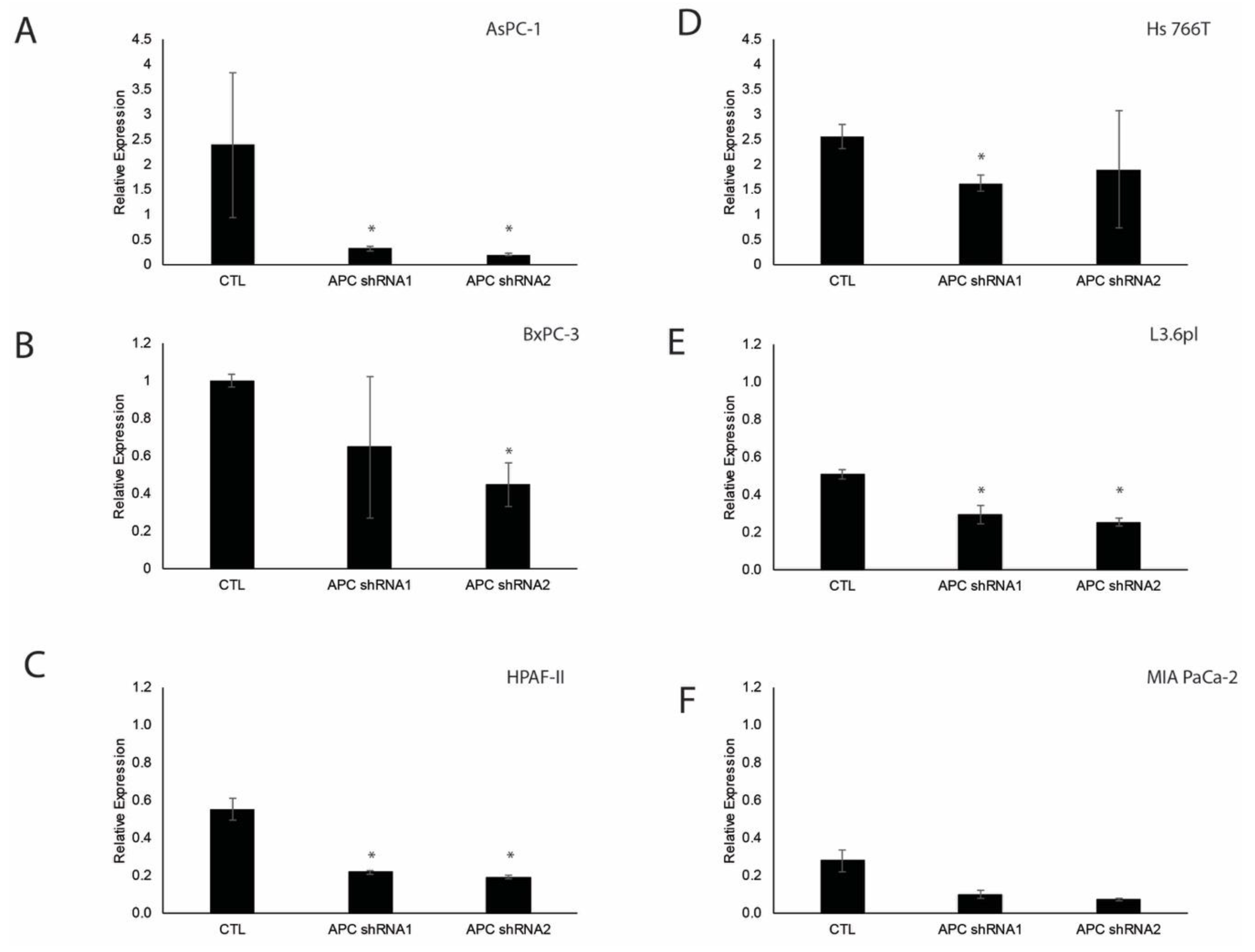
3.1. Verification of APC Knockdown in Pancreatic Ductal Adenocarcinoma Cell Lines

Using lentiviral-mediated MISSION shRNA (Sigma), we knocked down APC in six pancreatic ductal adenocarcinoma cell lines (AsPC-1, BxPC-3, HPAF-II, Hs 766T, L3.6pl, and MIA PaCa-2). AsPC-1 pancreatic cancer cells were derived from nude mouse xenografts initiated with cells from ascites of a patient with cancer of the pancreas. The BxPC-3 adenocarcinoma cells were derived from a primary pancreatic tumor. HPAF-II are human adenocarcinoma cells derived from peritoneal ascites fluid from a male with primary pancreas adenocarcinoma with metastasis to the liver, diaphragm and lymph nodes. Hs 766T are pancreatic carcinoma cells derived from lymph nodes. L3.6pl are a metastatic pancreatic cancer line. MIA PaCa-2 are carcinoma cells derived from tumor tissue of the pancreas. We chose these cell lines because they provided the primary and metastatic backgrounds of PDAC and have varying response to gemcitabine. To verify knockdown, we used quantitative RT-PCR to determine the relative expression of APC, which revealed knockdown in most of the cell lines compared to their respective controls (Figure 1A–F).

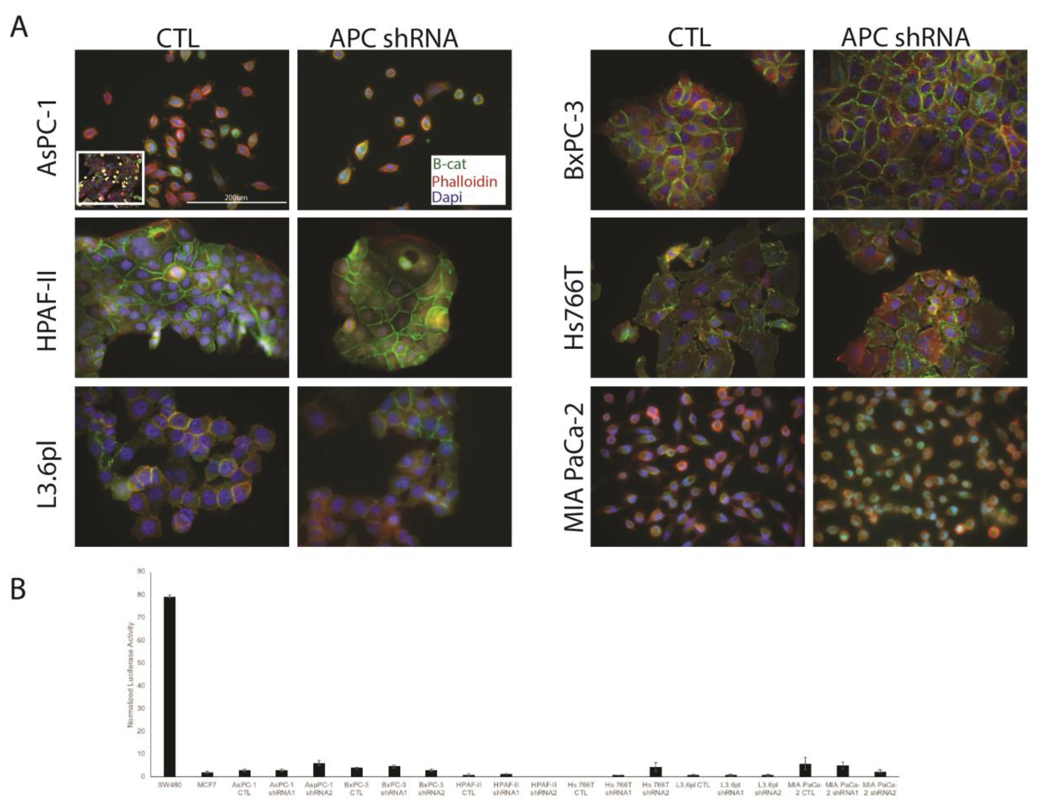
3.2. APC Knockdown in PDAC Cells Does Not Elicit Signaling through Wnt/Β-Catenin

In the APC knockdown PDAC cell lines, we performed immunofluorescence (IF) for β-catenin and showed no changes in localization after APC knockdown (Figure 2A). In addition, we observed no alterations in Wnt/β-catenin signaling using β-catenin/TCF reporter assays (Figure 2B). In both assays, the SW480 human colorectal cancer cells were used as a positive control given their enhanced Wnt/β-catenin signaling. MCF-7 human breast cancer cells were used as a negative control for the reporter assay, as they show minimal Wnt signaling. We have also investigated common Wnt target genes (BIRC5, c-myc, and cyclin D1), and showed no changes after APC knockdown (data not shown). These results, showing lack of activation of the Wnt/β-catenin signaling pathway, are similar to our studies in breast cancer cells and MDCK cells with APC knockdown [9,22,23]. While inhibitors of Wnt/β-catenin have shown promise in in vitro studies [24], our results suggest that APC may have Wnt-independent functions in pancreatic cancer regulation.

3.3. APC Loss Enhances Proliferation in PDAC Cells

APC loss has been shown to enhance proliferation in breast and colon cancer, amongst others [9,25,26]. Therefore, we assessed whether proliferation was altered in APC knockdown PDAC cells. Cell growth was measured via total cell counts at 24, 48 and 72 h post-plating for each cell line. We demonstrated that APC knockdown significantly increased proliferation in BxPC-3, Hs 766T and L3.6pl cell lines compared to their individual controls (Figure 3B,D,E). The AsPC1, HPAF-II and MIA PaCa-2 cell lines showed no significant differences in cell proliferation of the APC shRNA cells lines compared to the control (Figure 3A,C,F). These three cell lines could be proliferating quickly already and therefore see no differences in APC knockdown compared to the control.
Our data show similar doubling time as previously published [27]. AsPC-1 is a pancreatic adenocarcinoma cell line derived from a metastatic site (ascites) with a doubling time of 38–40 h [28]. BxPC-3 are primary pancreatic adenocarcinomas with a doubling time of 48–60 h [29]. HPAF-II is a human pancreatic adenocarcinoma cell line derived from a metastatic site (ascites) with a doubling time of 42 h [30]. Hs 766T are pancreatic carcinoma cells derived from a metastatic site (lymph node) with a doubling time of 6–7 days [31]. L3.6pl is a pancreas cell line derived from a liver metastasis. Cell division genes have been shown to be upregulated significantly in gene expression profiles generated from microarrays of the highly metastatic cell line L3.6pl [32]. MIA Paca-2 is a primary tumor with a doubling time of 40 h [33].

3.4. Effect of APC Knockdown on PDAC Cell Migration

Given that tumor metastasis is associated with a poor prognosis, we next sought to investigate cell migration using a wound-healing assay. Cells were grown to confluence, scratched, imaged, and analyzed at 0, 24, and 48 h. Using the T-scratch software, we determined the area filled in to represent cell migration [21]. No significant differences were observed in HPAF-II, Hs766T, and MIA PaCa-2 cells, in percent filled area of APC knockdown cells versus the control (Figure 4C,D,F). While the AsPC-1 APC shRNA1 cells showed no changes, the APC shRNA2 cells migrated slower than the CTL cells (Figure 4A). APC knockdown in the BxPC-3 and L3.6pl cells resulted in faster wound healing (Figure 4B,E), suggesting that these cells become more aggressive with APC knockdown. Specifically, 48 h post-wounding of the BxPC-3 cells, the APC shRNA1 and APC shRNA2 cell lines were significantly more closed than the CTL cells (Figure 4B). In L3.6pl, there were significant differences in percent filled area in APC shRNA1 and APC shRNA2 at 24 h and APC shRNA1 at 48 h compared to CTL cells (Figure 4E). In our studies, we found that the BxPC-3 cells with APC knockdown had filled the wound area more at 24 and 48 h compared to the control. BxPC-3 cells are a primary cell line derived from a patient that had no signs of metastasis. Very few studies have looked at the migration patterns of multiple PDAC cell lines; however, one study showed BxPC-3 had a relative wound density of around 80% at 48 h and reached complete closure at 60 h post-wounding [34].

3.5. APC Knockdown Does Not Impact PDAC Cell Response to Gemcitabine

Treatments for patients with PDAC include chemotherapy using the DNA nucleoside, gemcitabine [3]. We previously demonstrated that APC loss results in resistance to chemotherapeutic agents in mouse and human breast cancer cells [25]. Therefore, we sought to investigate the effect of APC knockdown in pancreatic cancer cells on response to gemcitabine. Cells were treated with 1 µM gemcitabine, and overall cell number was assessed at 24 (data not shown) and 48 h. While gemcitabine decreased cell number in most cell lines, there was no impact of APC knockdown on response to treatment. In AsPC-1 cells, we saw significant decreases in total cell numbers in controls and APC shRNA1 when treated with 1 µM gemcitabine for 48 h (Figure 5A). In BxPC-3 cells, 1 µM gemcitabine decreased total cell numbers in all cell lines (Figure 5B). In HPAF-II cells, 1 µM gemcitabine decreased total cell numbers in pLKO.1, APC shRNA1, and APC shRNA2, but not in CTL cells (Figure 5C). In Hs 766T cells, 48 h treatment with 1 µM gemcitabine resulted in significant decreases in total cell numbers in controls and APC shRNA1 (Figure 5D). In L3.6pl and MIA PaCa-2 cells, 1 µM gemcitabine decreased total cell numbers in all cell lines (Figure 5E,F). Our studies showed that all cell line controls and their APC knockdown cell lines were sensitive to 1 µM gemcitabine except for the HPAF-II control and two APC shRNA lines in the AsPC-1 and Hs 766T cell lines. L3.6pl and BxPC-3 are sensitive to, and Hs 766T, AsPC-1 and MIA PaCa-2 have been shown to be resistant to, 10 µmol/L gemcitabine [35]. This suggests that APC status would not serve as a marker for response to gemcitabine. A range of gemcitabine doses has been tested on multiple cell lines, including BxPC-3 and MIA-Paca-2, and it was observed that MIA PaCA-2 cells were more sensitive than BxPC-3 with an IC50 of 61 ± 3 mM versus 128 ± 16 mM, respectively [36].

4. Conclusions

In the current study, we successfully knocked down APC in six pancreatic ductal adenocarcinoma cell lines; AsPC-1, BxPC-3, HPAF-II, Hs 766T, L3.6pl, and MIA PaCa-2. β-catenin localization was at the cell–cell junctions and not nuclear, suggesting that the Wnt pathway is not active, which is similar to previously published data [20]. This was confirmed by reporter assays to assess β-catenin/TCF interaction. We saw only one of the AsPC-1 knockdown lines that was elevated in the TCF reporter assay. Proliferation was significantly increased after APC knockdown in BxPC-3 and L3.6pl at 48 h and in Hs 766T at 48 and 72 h compared to CTL. Wound healing was decreased after APC knockdown in one of the AsPC-1 knockdown lines at 24 and 48 h. In the BxPC-3 and L3.6pl knockdown lines, we saw faster wound healing, as expected with APC knockdown. Responses to gemcitabine, a standard pancreatic cancer chemotherapeutic agent, showed sensitivity in most CTL and APC shRNA lines, but was not dependent on APC status. These results are summarized in Table 1. Of the three cell lines showing phenotypic changes with APC knockdown (BxPC-3, Hs 766T, L3.6pl), there are no overt similarities in known oncogenic mutations. A search of the COSMIC database and literature showed no pattern of mutations in KRAS, SMAD4, CDKN2A, or p53. The Hs 766T and L3.6pl cells both have mutant KRAS and WT p53, while the Bx-PC3 cells have WT KRAS and mutant p53. Of the three cell lines, only the Hs 766T cells have mutant CDKN2A. This suggests that further investigation into the cell lines is required to uncover the mechanism by which APC status controls proliferation and migration in pancreatic cancer cells. Previous studies have shown a link between p53 or BRCA status and APC in pancreatic cancer [37,38], suggesting that APC works in concert with other oncogenic mutations to drive pancreatic tumorigenesis. Given the variable results observed in different cell lines, future studies would be necessary to understand the crosstalk between APC loss and other drivers of pancreatic cancer. In addition, the possibility of using APC status as either a prediction marker for pancreatic cancer or a tool to determine response to therapy should be explored [39,40].

Author Contributions

Conceptualization, J.R.P.; methodology, J.R.P.; formal analysis, J.R.P. and J.M.C.; investigation, J.M.C. and K.S.; resources, J.R.P.; writing—original draft preparation, J.M.C.; writing—review and editing, J.R.P.; supervision, J.R.P.; project administration, J.R.P.; funding acquisition, J.R.P.”

Funding

This research was supported by the American Cancer Society – Institutional Research Grant (JRP), the Indiana Clinical and Translational Sciences Institute, funded in part by grant #UL1 TR001108 from the National Institutes of Health, National Center for Advancing Translational Sciences, Clinical and Translational Sciences Award (JRP) (A. Shekhar, PI), Start-up funds from Indiana University School of Medicine – South Bend, and the Navari Family Foundation (JRP). Kaitlyn Simmons was supported by a College of Science – Summer Undergraduate Research Fellowship (University of Notre Dame).

Acknowledgments

The authors thank Reginald Hill for review of the manuscript, consultation in the early stages of this study, and for providing the cell lines. We also thank Monica VanKlompenberg and Claire Bedalov for technical assistance.

Conflicts of Interest

The authors declare no conflict of interest.

References

  1. Siegel, R.L.; Miller, K.D.; Jemal, A. Cancer statistics. CA Cancer J. Clin. 2015, 65, 5–29. [Google Scholar] [CrossRef]
  2. Stark, A.P.; Sacks, G.D.; Rochefort, M.M.; Donahue, T.R.; Reber, H.A.; Tomlinson, J.S.; Dawson, D.W.; Eibl, G.; Hines, O.J. Long-term Survival in Patients with Pancreatic Ductal Adenocarcinoma. Surgery 2016, 159, 1520–1527. [Google Scholar] [CrossRef] [PubMed]
  3. Burris III, H.A.; Moore, M.J.; Andersen, J.; Green, M.R.; Rothenberg, M.L.; Modiano, M.R.; Cripps, M.C.; Portenoy, R.K.; Storniolo, A.M.; Tarassoff, P.; et al. Improvements in survival and clinical benefit with gemcitabine as first-line therapy for patients with advanced pancreas cancer: A randomized trial. J. Clin. Oncol. 1997, 15, 2403–2413. [Google Scholar] [CrossRef] [PubMed]
  4. Herrera, L.; Kakati, S.; Gibas, L.; Pietrzak, E.; Sandberg, A.A.; Reynolds, J.F. Gardner syndrome in a man with an interstitial deletion of 5q. Am. J. Med Genet. 1986, 25, 473–476. [Google Scholar] [CrossRef] [PubMed]
  5. Bodmer, W.F.; Bailey, C.J.; Bodmer, J.; Bussey, H.J.; Ellis, A.; Gorman, P.; Lucibello, F.C.; Murday, V.A.; Rider, S.H.; Scambler, P.; et al. Localization of the gene for familial adenomatous polyposis on chromosome 5. Nature 1987, 328, 614–616. [Google Scholar] [CrossRef] [PubMed] [Green Version]
  6. Leppert, M.; Burt, R.; Hughes, J.P.; Samowitz, W.; Nakamura, Y.; Woodward, S.; Gardner, E.; Lalouel, J.M.; White, R. Genetic analysis of an inherited predisposition to colon cancer in a family with a variable number of adenomatous polyps. New England J. Med. 1990, 322, 904–908. [Google Scholar] [CrossRef] [PubMed]
  7. Tanaka, K.; Oshimura, M.; Kikuchi, R.; Seki, M.; Hayashi, T.; Miyaki, M. Suppression of tumorigenicity in human colon carcinoma cells by introduction of normal chromosome 5 or 18. Nature 1991, 349, 340–342. [Google Scholar] [CrossRef] [PubMed]
  8. Segditsas, S.; Tomlinson, I. Colorectal cancer and genetic alterations in the Wnt pathway. Oncogene 2006, 25, 7531–7537. [Google Scholar] [CrossRef] [PubMed] [Green Version]
  9. Prosperi, J.R.; Khramtsov, A.I.; Khramtsova, G.F.; Goss, K.H. Apc mutation enhances PyMT-induced mammary tumorigenesis. PLoS ONE 2011, 6, e29339. [Google Scholar] [CrossRef]
  10. Ginestà, M.M.; Diaz-Riascos, Z.V.; Busquets, J.; Peláez, N.; Serrano, T.; Peinado, M.A.; Jorba, R.; García-Borobia, F.J.; Capellá, G.; Fabregat, J. APC promoter is frequently methylated in pancreatic juice of patients with pancreatic carcinomas or periampullary tumors. Oncol. Lett. 2016, 12, 2210–2216. [Google Scholar] [CrossRef]
  11. Jäkel, C.; Bergmann, F.; Toth, R.; Assenov, Y.; Van Der Duin, D.; Strobel, O.; Hank, T.; Klöppel, G.; Dorrell, C.; Grompe, M.; et al. Genome-wide genetic and epigenetic analyses of pancreatic acinar cell carcinomas reveal aberrations in genome stability. Nat. Commun. 2017, 8, 1323. [Google Scholar] [CrossRef] [PubMed]
  12. Wang, J.; Gerrard, G.; Poskitt, B.; Dawson, K.; Trivedi, P.; Foroni, L.; El-Bahrawy, M. Targeted next generation sequencing of pancreatic solid pseudopapillary neoplasms show mutations in Wnt signaling pathway genes. Pathol. Int. 2019, 69, 193–201. [Google Scholar] [CrossRef] [PubMed] [Green Version]
  13. Peng, D.-F.; Kanai, Y.; Sawada, M.; Ushijima, S.; Hiraoka, N.; Kitazawa, S.; Hirohashi, S. DNA methylation of multiple tumor-related genes in association with overexpression of DNA methyltransferase 1 (DNMT1) during multistage carcinogenesis of the pancreas. Carcinogenesis 2006, 27, 1160–1168. [Google Scholar] [CrossRef] [PubMed] [Green Version]
  14. Horii, A.; Nakatsuru, S.; Miyoshi, Y.; Ichii, S.; Nagase, H.; Ando, H.; Yanagisawa, A.; Tsuchiya, E.; Kato, Y.; Nakamura, Y. Frequent somatic mutations of the APC gene in human pancreatic cancer. Cancer Res. 1992, 52. [Google Scholar]
  15. Giardiello, F.M.; Offerhaus, G.J.; Lee, D.H.; Krush, A.J.; Tersmette, A.C.; Booker, S.V.; Kelley, N.C.; Hamilton, S.R. Increased risk of thyroid and pancreatic carcinoma in familial adenomatous polyposis. Gut 1993, 34, 1394–1396. [Google Scholar] [CrossRef] [PubMed]
  16. Maire, F.; Hammel, P.; Terris, B.; Olschwang, S.; O’Toole, D.; Sauvanet, A.; Palazzo, L.; Ponsot, P.; Laplane, B.; Levy, P.; et al. Intraductal papillary and mucinous pancreatic tumour: A new extracolonic tumour in familial adenomatous polyposis. Gut 2002, 51, 446–449. [Google Scholar] [CrossRef] [PubMed]
  17. Arnau, G.; Nemorin, A.; Maledon, E.; Abraham, K. Revision of ploidy status of Dioscorea alata L. (Dioscoreaceae) by cytogenetic and microsatellite segregation analysis. Theor. Appl. Genet. 2009, 118, 1239–1249. [Google Scholar] [CrossRef]
  18. Chetty, R.; Salahshor, S.; Bapat, B.; Berk, T.; Croitoru, M.; Gallinger, S. Intraductal papillary mucinous neoplasm of the pancreas in a patient with attenuated familial adenomatous polyposis. J. Clin. Pathol. 2005, 58, 97–101. [Google Scholar] [CrossRef]
  19. Qin, R.-F.; Zhang, J.; Huo, H.-R.; Yuan, Z.-J.; Xue, J.-D. MiR-205 mediated APC regulation contributes to pancreatic cancer cell proliferation. World J. Gastroenterol. 2019, 25, 3775–3786. [Google Scholar] [CrossRef]
  20. Ikenoue, T.; Ijichi, H.; Kato, N.; Kanai, F.; Masaki, T.; Rengifo, W.; Okamoto, M.; Matsumura, M.; Kawabe, T.; Shiratori, Y.; et al. Analysis of the beta-catenin/T cell factor signaling pathway in 36 gastrointestinal and liver cancer cells. Jpn. J. Cancer Res. 2002, 93, 1213–1220. [Google Scholar] [CrossRef]
  21. Gebäck, T.; Schulz, M.M.P.; Koumoutsakos, P.; Detmar, M. TScratch: A novel and simple software tool for automated analysis of monolayer wound healing assays. Biotechniques 2009, 46, 265–274. [Google Scholar] [CrossRef] [PubMed]
  22. Lesko, A.C.; Goss, K.H.; Yang, F.F.; Schwertner, A.; Hulur, I.; Onel, K.; Prosperi, J.R. The APC tumor suppressor is required for epithelial cell polarization and three-dimensional morphogenesis. Biochim. et Biophys. Acta (BBA) - Bioenerg. 2015, 1853, 711–723. [Google Scholar] [CrossRef] [PubMed] [Green Version]
  23. Odenwald, M.A.; Prosperi, J.R.; Goss, K.H. APC/beta-catenin-rich complexes at membrane protrusions regulate mammary tumor cell migration and mesenchymal morphology. BMC Cancer 2013, 13, 12. [Google Scholar] [CrossRef] [PubMed]
  24. Chartier, C.; Raval, J.; Axelrod, F.; Bond, C.; Cain, J.; Dee-Hoskins, C.; Ma, S.; Fischer, M.M.; Shah, J.; Wei, J.; et al. Therapeutic Targeting of Tumor-Derived R-Spondin Attenuates beta-Catenin Signaling and Tumorigenesis in Multiple Cancer Types. Cancer Res. 2016, 76, 713–723. [Google Scholar] [CrossRef] [PubMed]
  25. VanKlompenberg, M.K.; Bedalov, C.O.; Soto, K.F.; Prosperi, J.R. APC selectively mediates response to chemotherapeutic agents in breast cancer. BMC Cancer 2015, 15, 9. [Google Scholar] [CrossRef]
  26. Sansom, O.J.; Reed, K.R.; Hayes, A.J.; Ireland, H.; Brinkmann, H.; Newton, I.P.; Batlle, E.; Simon-Assmann, P.; Clevers, H.; Näthke, I.S.; et al. Loss of Apc in vivo immediately perturbs Wnt signaling, differentiation, and migration. Genome Res. 2004, 18, 1385–1390. [Google Scholar] [CrossRef] [PubMed] [Green Version]
  27. Deer, E.L.; Gonzalez-Hernandez, J.; Coursen, J.D.; Shea, J.E.; Ngatia, J.; Scaife, C.L.; Firpo, M.A.; Mulvihill, S.J. Phenotype and Genotype of Pancreatic Cancer Cell Lines. Pancreas 2010, 39, 425–435. [Google Scholar] [CrossRef] [Green Version]
  28. Chen, W.H.; Horoszewicz, J.S.; Leong, S.S.; Shimano, T.; Penetrante, R.; Sanders, W.H.; Berjian, R.; Douglass, H.O.; Martin, E.W.; Chu, T.M. Human pancreatic adenocarcinoma: In vitro and in vivo morphology of a new tumor line established from ascites. Vitr. Cell. Dev. Biol. Plant 1982, 18, 24–34. [Google Scholar] [CrossRef]
  29. Tan, M.H.; Nowak, N.J.; Loor, R.; Ochi, H.; Sandberg, A.A.; Lopez, C.; Pickren, J.W.; Berjian, R.; Douglass, H.O.; Chu, T.M. Characterization of a new primary human pancreatic tumor line. Cancer Investig. 1986, 4, 15–23. [Google Scholar] [CrossRef]
  30. Metzgar, R.S.; Gaillard, M.T.; Levine, S.J.; Tuck, F.L.; Bossen, E.H.; Borowitz, M.J. Antigens of human pancreatic adenocarcinoma cells defined by murine monoclonal antibodies. Cancer Res. 1982, 42, 601–608. [Google Scholar]
  31. Smith, H.S. In Vitro Properties of Epithelial Cell Lines Established from Human Carcinomas and Nonmalignant Tissue2. J. Natl. Cancer Inst. 1979, 62, 225–230. [Google Scholar]
  32. Nakamura, T.; Fidler, I.J.; Coombes, K.R. Gene expression profile of metastatic human pancreatic cancer cells depends on the organ microenvironment. Cancer Res. 2007, 67, 139–148. [Google Scholar] [CrossRef]
  33. Yunis, A.; Arimura, G.K.; Russin, D.J. Human pancreatic carcinoma (MIA PaCa-2) in continuous culture: Sensitivity to asparaginase. Int. J. Cancer 1977, 19, 128–135. [Google Scholar] [CrossRef]
  34. Sauter, D.R.P.; Novak, I.; Pedersen, S.F.; Larsen, E.H.; Hoffmann, E.K. ANO1 (TMEM16A) in pancreatic ductal adenocarcinoma (PDAC). Pflugers Arch. 2015, 467, 1495–1508. [Google Scholar] [CrossRef]
  35. Arumugam, T.; Ramachandran, V.; Fournier, K.F.; Wang, H.; Marquis, L.; Abbruzzese, J.L.; Gallick, G.E.; Logsdon, C.D.; McConkey, D.J.; Choi, W. Epithelial to Mesenchymal Transition Contributes to Drug Resistance in Pancreatic Cancer. Cancer Res. 2009, 69, 5820–5828. [Google Scholar] [CrossRef]
  36. Fryer, R.; Barlett, B.; Galustian, C.; Dalgleish, A.G. Mechanisms underlying gemcitabine resistance in pancreatic cancer and sensitisation by the iMiDTM lenalidomide. Anticancer Res. 2011, 31, 3747–3756. [Google Scholar]
  37. Kuo, T.L.; Weng, C.C.; Kuo, K.K.; Chen, C.Y.; Wu, D.C.; Hung, W.C.; Cheng, K.H. APC haploinsufficiency coupled with p53 loss sufficiently induces mucinous cystic neoplasms and invasive pancreatic carcinoma in mice. Oncogene 2016, 35, 2223–2234. [Google Scholar] [CrossRef]
  38. Goehringer, C.; Sutter, C.; Kloor, M.; Gebert, J.; Slater, E.P.; Keller, M.; Treiber, I.; Ganschow, P.; Kadmon, M.; Moog, U. Double germline mutations in APC and BRCA2 in an individual with a pancreatic tumor. Fam. Cancer 2017, 16, 303–309. [Google Scholar] [CrossRef]
  39. Yurgelun, M.B.; Chittenden, A.B.; Morales-Oyarvide, V.; Rubinson, D.A.; Dunne, R.F.; Kozak, M.M.; Qian, Z.R.; Welch, M.W.; Brais, L.K.; Da Silva, A.; et al. Germline cancer susceptibility gene variants, somatic second hits, and survival outcomes in patients with resected pancreatic cancer. Genet. Med. 2019, 21, 213–223. [Google Scholar] [CrossRef]
  40. Zhan, W.; Shelton, C.A.; Greer, P.J.; Brand, R.E.; Whitcomb, D.C. Germline Variants and Risk for Pancreatic Cancer: A Systematic Review and Emerging Concepts. Pancreas 2018, 47, 924–936. [Google Scholar] [CrossRef]
Figure 1. Quantitative PCR demonstrates that APC is knocked down in the cell lines transfected with APC shRNA constructs. Using lentiviral-mediated MISSION shRNA we verified APC knock down in six pancreatic ductal adenocarcinoma cell lines using quantitative PCR and relative expression of APC revealed gene knockdown in AsPC-1, BxPC-3, HPAF-II, Hs 766T, L3.6pl, and MIA PaCa-2 cell lines compared to CTL (Figure 1AF). *p < 0.05.
Cells 08 01084 g001
Figure 2. APC knockdown did not increase nuclear β-catenin. (A) In AsPC-1, BxPC-3, HPAF-II, Hs 766T, L3.6pl and MIA PaCa-2 CTL cells, β-catenin is localized at the cell–cell junctions. After the knockdown of APC in each of those cell lines: APC shRNA (representative), there were no changes in beta catenin localization. (B) In the APC knockdown PDAC cell lines, we observed no alterations in Wnt/β-catenin signaling using β-catenin/TCF reporter assays.
Cells 08 01084 g002
Figure 3. Proliferation changes are observed in some cell lines that have been transfected with APC shRNA constructs compared to controls. Proliferation was measured in total cell counts at 24, 48 and 72 h post-plating for each cell line. (B and E) In the BxPC-3 and L3.6pl cells, knockdown of APC resulted in a significant increase in cell proliferation at 48 h. (D) In the Hs 766T cells, knockdown of APC resulted in a significant increase in cell proliferation at 48 and 72 h. (A, C, and F) The AsPC-1, HPAF-II and MIA PaCa-2 cell lines showed no significant differences in cell proliferation of the APC shRNA cells lines compared to CTL. *p < 0.05.
Cells 08 01084 g003
Figure 4. Migration of cells in wound healing assays revealed faster migration with APC knockdown in some pancreatic cancer cell lines. (A) The AsPC-1 APC shRNA2 knockdown cells migrated slower than the control at 24 and 48 h post wounding. (B and E) Only the BxPC-3 and L3.6pl cells were impacted by APC knockdown, showing faster wound healing. Specifically, at 48 h post-wounding, the APC shRNA1 and APC shRNA2 cell lines were significantly more closed than the parent BxPC-3 cells. (E) In L3.6pl, there were significant differences in percent filled area in APC shRNA1 and APC shRNA2 at 24 h and APC shRNA2 at 48 h compared to CTL. (A, C, D, and F) In AsPC-1, HPAF-II, Hs766T, and MIA PaCa-2 cells, there were no significant differences in percent filled area in APC shRNA cells versus CTL. *p < 0.05.
Cells 08 01084 g004
Figure 5. Effect of gemcitabine on PDAC cell lines. (A) In AsPC-1 cells, we saw significant decreases in total cell numbers in controls and APC shRNA1, but not in APC shRNA2 when treated with 1 µM gemcitabine for 48 h. (B) In BxPC-3 cells, 1 µM gemcitabine decreased total cell numbers in all cell lines. (C) In HPAF-II cells, 1 µM gemcitabine decreased total cell numbers in APC shRNA1, and APC shRNA2, but not in the CTL. (D) In Hs 766T cells, 48 h treatment with 1 µM gemcitabine resulted in significant decreases in total cell numbers in controls and APC shRNA1, but not in APC shRNA2. (E) In L3.6pl cells, 1 µM gemcitabine decreased total cell numbers in CTL APC shRNA1, and APC shRNA2. (F) In MIA PaCa-2 cells, 1 µM gemcitabine decreased total cell numbers in CTL, APC shRNA1, and APC shRNA2. *p < 0.05.
Cells 08 01084 g005
Table 1. Summary of data.
β-Catenin LocalizationProliferationWound HealingResponse to Gemcitabine
AsPC-1 CTLJunctional Doubling time of approximately 24–48 h.Wound closed approximately 73% at 48 h.Significantly decreased total cell number compared to untreated.
APC shRNA1NCNCNCNC
APC shRNA2NCNCWound closed significantly slower compared to CTL at 24 and 48 h.No significance differences compared to untreated.
BxPC-3 CTLJunctional Doubling time of approximately 24 h.Wound closed approximately 81% at 48 h.Significantly decreased total cell number compared to untreated.
APC shRNA1NCNCWound closed significantly faster compared to CTL at 48 h.NC
APC shRNA2NCSignificantly different from CTL at 48 h.Wound closed significantly faster compared to CTL at 24 and 48 h.NC
HPAF-II CTLJunctional Doubling time of approximately 48 h.Wound closed approximately 92% at 48 h.No significance differences compared to untreated.
APC shRNA1NCNCNCSignificantly decreased total cell number compared to untreated.
APC shRNA2NCNCNCSignificantly decreased total cell number compared to untreated.
Hs 766T CTLJunctional Doubling time greater than our 72 h experimental timeframe.Wound closed approximately 27% at 48 h.Significantly decreased total cell number compared to CTL.
APC shRNA1NCNCNCNC
APC shRNA2NCSignificantly different from CTL at 48 and 72 h.NCNo significance differences compared to untreated.
L3.6pl CTLJunctional Doubling time of approximately 24 h.Wound closed approximately 95% at 48 h.Significantly decreased total cell number compared to untreated.
APC shRNA1NCSignificantly different from CTL at 48 h.Wound closed significantly faster compared to CTL at 24 and 48 h.NC
APC shRNA2NCNCWound closed significantly faster compared to CTL at 24 h.NC
MIA-PaCa CTLJunctional Doubling time of approximately 30 h.Wound closed approximately 93% at 48 h.Significantly decreased total cell number compared to untreated.
APC shRNA1NCNCNCNC
APC shRNA2NCNCNCNC

