Article

Lipocalin-2, Matrix Metalloproteinase-9, and MMP-9/NGAL Complex in Upper Aerodigestive Tract Carcinomas: A Pilot Study

1
Department of Sensory Organs, Sapienza University of Rome, 00161 Roma, Italy
2
Department of Experimental Medicine, Sapienza University of Rome, 00161 Roma, Italy
3
PhD School of Applied Medical-Surgical Sciences, University of Rome “Tor Vergata”, Via Montpellier 1, 00133 Roma, Italy
4
Department of Biology, University of Rome “Tor Vergata”, 00133 Roma, Italy
5
Institute of Biochemistry and Cell Biology (IBBC-CNR), 00161 Roma, Italy
6
Division of Otolaryngology-Head and Neck Surgery, San Camillo de Lellis Hospital, ASL Rieti-Sapienza University, Viale Kennedy, 02100 Rieti, Italy
*
Author to whom correspondence should be addressed.
These authors contributed equally to this work.
Cells 2025, 14(7), 506; https://doi.org/10.3390/cells14070506
Submission received: 27 January 2025 / Revised: 17 March 2025 / Accepted: 26 March 2025 / Published: 29 March 2025

Abstract

Upper aerodigestive tract (UADT) carcinomas have a high and rapidly increasing incidence, particularly in industrialized countries. The identification of diagnostic and prognostic biomarkers remains a key objective in oncological research. However, conflicting data have been reported regarding Lipocalin-2 (LCN-2 or NGAL), Matrix Metalloproteinase-9 (MMP-9), and the MMP-9/NGAL complex in UADT carcinomas. For this reason, the primary aim of this study was to investigate the involvement and modulation of the LCN-2 system in UADT cancer by selecting patients at first diagnosis and excluding any pharmacological or interventional treatments that could act as confounding factors. In this clinical retrospective pilot study, we investigated LCN-2 and MMP-9 tissue gene expression, as well as circulating levels of LCN-2, MMP-9, and the MMP-9/NGAL complex. Our findings revealed a downregulation of LCN-2 and an upregulation of MMP-9 gene expression in tumor tissues compared to healthy counterparts. A similar trend was observed in circulating levels, with decreased LCN-2 and increased MMP-9 in cancer patients compared to healthy controls. Additionally, serum levels of the MMP-9/NGAL complex were significantly elevated in UADT cancer patients relative to controls. Our study suggests a potentially distinct role for the free form of LCN-2 and its conjugated form (MMP-9/NGAL complex) in UADT tumors. These findings not only provide new insights into the molecular mechanisms underlying tumor progression but also highlight the potential clinical relevance of these biomarkers. The differential expression patterns observed suggest that the LCN-2 and MMP-9/NGAL complex could serve as valuable tools for improving early diagnosis, monitoring disease progression, and potentially guiding therapeutic strategies. Further research is needed to validate their utility in clinical settings and to explore their prognostic and predictive value in personalized treatment approaches.

Graphical Abstract

1. Introduction

Cancers of the upper aerodigestive tract (UADT) involve the oral cavity (lips, tongue, floor of the mouth, gingival mucosa, inner surface of the cheeks and palate), the nasopharynx, the oropharynx, the hypopharynx, and the larynx [1]. UADT cancers rank fourth in terms of cancer incidence and second in terms of cancer mortality worldwide [2,3]. Advanced forms are more difficult to cure even after therapeutic care, although localized forms can usually be managed in the long term with surgery or treatments (chemotherapy, radiotherapy, and/or immunotherapy). The primary risk factor for UADT cancer is prolonged exposure to alcohol and tobacco [4,5,6], often associated with an unfavorable prognosis. The identification of diagnostic and prognostic biomarkers remains a crucial objective for oncological research to early identify patients who respond or not to targeted therapies or who may or may not develop metastases or recurrences [7,8,9,10,11,12].
Lipocalin-2 (LCN-2), also referred to as oncogene 24p3 or neutrophil gelatinase-associated lipocalin (NGAL), is a small glycoprotein weighing 25 kDa, and it belongs to the lipocalin family. Initially, LCN-2 was isolated from activated human neutrophils and found to be associated with gelatinase, an enzyme crucial for neutrophil diapedesis [13,14,15]. It has subsequently been shown that NGAL was stored in specific neutrophil granules, not only as a heterodimer with gelatinase but also as a monomer or homodimer [16]. LCN-2 plays a role in various biological functions, particularly in pathological conditions [13,17,18]. Specifically, LCN-2 regulates iron trafficking during bacterial infections, mediating antibiotic activity by sequestering siderophore-iron delivery, which is vital for the survival of microorganisms. Additionally, LCN-2 can influence iron homeostasis in malignant cells, impacting cell proliferation, angiogenesis, migration, and invasion across different cancer types [17,19]. Elevated tissue levels of LCN-2 have been found in several types of cancers [18], linked to increased cell proliferation and metastasis [18]. However, recent research indicates that LCN-2 is significantly down-regulated in primary and metastatic malignant tumors of the oral cavity compared to healthy tissues [20]. Moreover, the downregulation of LCN-2 appears to correlate with the degree of differentiation and the stage of the disease, suggesting a prognostic role for oral cancer [20].
Matrix metalloprotease 9 (MMP-9), also known as gelatinase B, is a zinc metalloproteinase that plays a role in breaking down extracellular matrix components and the basement membrane; high levels of MMP-9 are associated with a greater capacity for neoplastic invasion [21,22].
MMP-9 and LCN-2 can form a protein complex (MMP-9/NGAL complex) that protects MMP-9 from degradation, thereby extending its enzymatic activity [23,24]. Therefore, this interaction plays a fundamental role in the process of progression and metastasis in many neoplasms [25,26].
In the present clinical retrospective observational study, we analyzed LCN-2 and MMP-9 tissue gene expression and LCN-2, MMP-9, and MMP-9/NGAL complex serum levels in patients with a first diagnosis of UADT cancer and avoided any kind of treatment (pharmacological and/or interventional) and correlated the results with the observed hematological profile.

2. Materials and Methods

2.1. Participants’ Selection and Sampling

In this pilot study, we recruited 30 adult individuals (24 men and 6 women) suffering from UADT carcinomas over a period of 2 years (2020–2021) who attended the Otolaryngology Clinic of the “Policlinico Umberto I”, Sapienza University Hospital. As inclusion criteria, we recruited adult male and female patients entering the hospital for the first diagnosis of UADT carcinomas. Based on the power analysis calculator for the sample size in a pilot study, we obtained that with at least 15 samples per group (30 total sample size), we could guarantee an actual power of 0.91. Greater test power has been obtained with the addition of just a few more samples (0.95 for a 35 total sample size), which indeed occurred during the recruitment of both patients and healthy individuals (for additional details, see Supplementary Materials).
Regarding the exclusion criteria, we did not include patients with cancer relapse, those undergoing drug treatments (such as chemotherapy, anti-inflammatory drugs, or immunosuppressants), or those receiving radiotherapy and/or immunotherapy. Additionally, patients with severe infectious diseases (HIV, HBV, or HCV) or other ongoing inflammatory, cardiovascular, endocrine, or autoimmune disorders were excluded. Patients were enrolled at the time of diagnosis, and all samples were collected during pre-hospitalization, prior to the initiation of any treatment. Moreover, 20 adult healthy controls, age- and sex-matched, were selected among the blood donors from the Policlinico Umberto I Transfusion Center. Moreover, based on questionnaires administered during the recruitment phase, both controls and patients reported being smokers and occasional alcohol consumers.
Peripheral blood samples (5 mL) were collected in BD Vacutainer™ Serum Separation Tubes and centrifuged at 3000 rpm for 15 min to separate the serum from blood cells. The serum was then stored at −80 °C until the day of analysis.
Tumor tissues (n = 9) and non-tumor tissues (n = 19), collected from the resection margins during surgery, were obtained from patients eligible for tumor removal. These tissues were used for RNA extraction to analyze the gene expression of interest (see above).
The study was approved by the Ethics Committee of the “Policlinico Umberto I” Hospital (Rif. 6462); informed consent was signed by each patient, and all the study procedures were in accordance with the Helsinki Declaration of 1975, as revised in 1983, for human experimentation.

2.2. Hematologic Parameters

Hematologic parameters were evaluated in the laboratory of the Experimental Medicine Department of the Policlinico Umberto I hospital. Serum glycemia (70.3–100.9 mg/dL), albumin (35–55 g/L), PCR (100–6000 μg/L), transferrin (2.15–3.65 g/L), protein (60–82 g/L), urea (10.2–49.8 mg/dL), phosphate (2.80–4.60 mg/dL), magnesium (0.66–1.03 mmol/L), calcium (2.1–2.5 mmol/L), chloride (98–109 mmol/L), potassium (3.4–5.5 mmol/L), sodium (136–145 mmol/L), iron (64.8–174.9 µg/dL), ferritin (30–400 μg/L), lactate dehydrogenase LDH (males 135–225 UI/L; females 135–214 UI/L), aspartate aminotransferase AST (males 9–45 UI/L; females 9–35 UI/L), alanine aminotransferase ALT (males 10–40 UI/L; females 7–35 UI/L), gamma glutamyl transpeptidase gGT (males 8–61 UI/L; females 5–36 UI/L), direct bilirubin (<0.20 mg/dL), total bilirubin (0.3–1.2 mg/dL), creatinine (males 0.7–1.2 mg/dL; females 0.5–0.9 mg/dL) and creatinine kinase CPK (males 20–200 UI/L; females 20–180 UI/L) were analyzed using Cobas C 501 platform (Roche Diagnostics, Mannheim, Germany).

2.3. RNA Extraction

Approximately 10 mg of tumor and non-tumor tissues were obtained for each patient, the latter taken from the margins of the resection. Tissue samples were immediately immersed in 1 mL of Trizol reagent and stored at −80 °C until the moment they were processed. Tissues were homogenized before extraction; RNA was obtained using Trizol Reagent (Invitrogen, Waltham, MA, USA) protocol, following manufacturer instructions. RNA from each available tissue was obtained in a volume of 20 μL. Extracted RNA was then quantified using the Qubit fluorimetric assay (Invitrogen, Waltham, MA, USA).

2.4. Reverse Transcription

A DNase treatment was performed to remove any contaminating genomic DNA: 4 μg of total RNA were incubated with 2.2 units (0.7 μL) of DNase I (New England Biolabs, Ipswich, MA, USA) and 2 μL of 10x DNase1 reaction buffer at 37 °C for 10′ in a final volume of 20 μL. Next, 2.2 μL of 1x EDTA (5 mM, pH = 8) (Sigma-Aldrich, St. Louis, MO, USA) were added to the samples; a 75 °C incubation for 10′ was then performed for enzyme inactivation. From the digested sample, 5.5 μL (corresponding to 1 mg of RNA) was added to 1 μL of iScript Reverse Transcriptase, 4 μL of 5x iScript Reaction mix, and 9.5 μL of distilled water (all reagents from iScript cDNA Synthesis Kit—Bio-Rad, Hercules, CA, USA) in a final volume of 20 μL. The reaction was placed in a PTC100 thermal cycler (Bio-Rad, Hercules, CA, USA) and followed the protocol: 25 °C for 5′; 42 °C for 30′; 85 °C for 5′. As quality control, an amplification of the GAPDH housekeeping gene was performed: 2 μL of cDNA, 5.7 μL of distilled water, 2.1 μL of dNTPs 1.25 mM (Thermo Fisher Scientific, Waltham, MA, USA), 3 μL of Buffer Mix Reaction 5x (Promega, Madison, WI, USA), 0.9 μL MgCl2 25 mM (Promega, Madison, WI, USA), 0.1 μL di Gotaq DNA polymerase 5 U/mL (Promega, Madison, WI, USA), 0.6 μL of the GAPDH forward primer (5′-CCCTTCATTGACCTCAACTACATG-3′) and 0.6 μL of the reverse primer (5′-TGGGATTTCCATTGATGACAAGC-3′) in a total reaction volume of 15 μL were assembled. Thermal cycling was set as follows: 95 °C for 2′, (94 °C for 45″; 60 °C for 1.5′; 70 °C for 2.5′) for 28 cycles; 72 °C for 7′; 10 °C ∞. Electrophoresis (3.3 μL of each sample) was performed in a 2% agarose gel (Bio-Rad, Hercules, CA, USA); PCR amplicons were distinguished by their size using a 50 bp molecular weight standard (Thermo Fisher, Waltham, MA, USA).

2.5. Digital Droplet PCR

Digital droplet PCR (ddPCR) assays were performed by assembling 1 μL of cDNA mix with 10 μL of 2X ddPCR Probe Supermix (no dUTP) (Bio-Rad, Hercules, CA, USA, 1863024), 1 μL of TaqMan LCN-2 gene expression probe (code 4331182, ID Hs01008571_m1; ThermoFisher Scientific) or MMP-9 (code 4453320, ID Hs00957562_m1; ThermoFisher Scientific), and 8 μL of H2O in a final reaction volume of 20 μL [27]. For details on the procedure, see the Supplementary Materials.

2.6. Serum LCN-2, MMP-9, and MMP-9/NGAL Complex Analysis

Human serum LCN-2 (Cat. No. DY8556), MMP-9 (Cat. No. DY911), and MMP-9/NGAL complex (Cat. No. DY1757) were measured using sandwich enzyme-linked immunosorbent assay (ELISA) kits (DuoSet ELISA, R&D Systems, Minneapolis, MN, USA), according to the protocols provided by the manufacturer. For details on the procedure, see the Supplementary Materials.

2.7. Statistical Analysis

The statistical analysis was performed using Prism 9 for MacOS, Version 9.3.1. Data were presented as mean ± standard deviation (SD) or standard error mean (SEM), where appropriate. Statistical significance was determined using one-way ANOVA, two-way ANOVA, and Student’s t-test where appropriate. Receiver operating characteristics (ROC) analyses were performed with the standard parameters in Prism 9, using the Wilson/Brown method for confidence interval calculation. For all tests, the threshold of statistical significance was 0.05. The Spearman correlation test was performed to investigate the association between serum LCN-2, MMP-9, and MMP-9/NGAL complex levels with the hematological parameters measured.

3. Results

3.1. General Description of the Enrolled Individuals

The cohort of cancer patients enrolled in the study (n. 30 with a middle age of 62.5 ± 1.7 years old) and of healthy controls (n. 20 with a middle age of 58.7 ± 1.0 years old) showed a higher prevalence of men compared to women in both groups (80% vs. 20%). This distribution aligns with global epidemiological data, which indicate that UADT cancers are more common in men than in women [28]. The general characteristics of the enrolled patients have been summarized in Table 1. The clinical (c) or pathological (p) tumor-node-metastasis TNM staging system followed the AJCC/UICC—TNM staging system—8th Edition [29,30].

3.2. Hematologic Parameters

Table 2 illustrates the hematologic parameters detected in recruited patients, which resulted in significantly different results compared to healthy controls. Overall, oncologic patients showed a very similar hematologic profile, and some alterations were in line with the pathological condition, such as neoplastic cachexia, inadequate caloric-protein consumption, and chronic inflammation state. In particular, they displayed glucose, urea, and phosphate increases as well as augmented ferritin, direct bilirubin, gGT, and PCR. Conversely, a decrease in albumin, total protein, iron, transferrin, creatinine, LDH, and CPK was found compared to healthy controls.

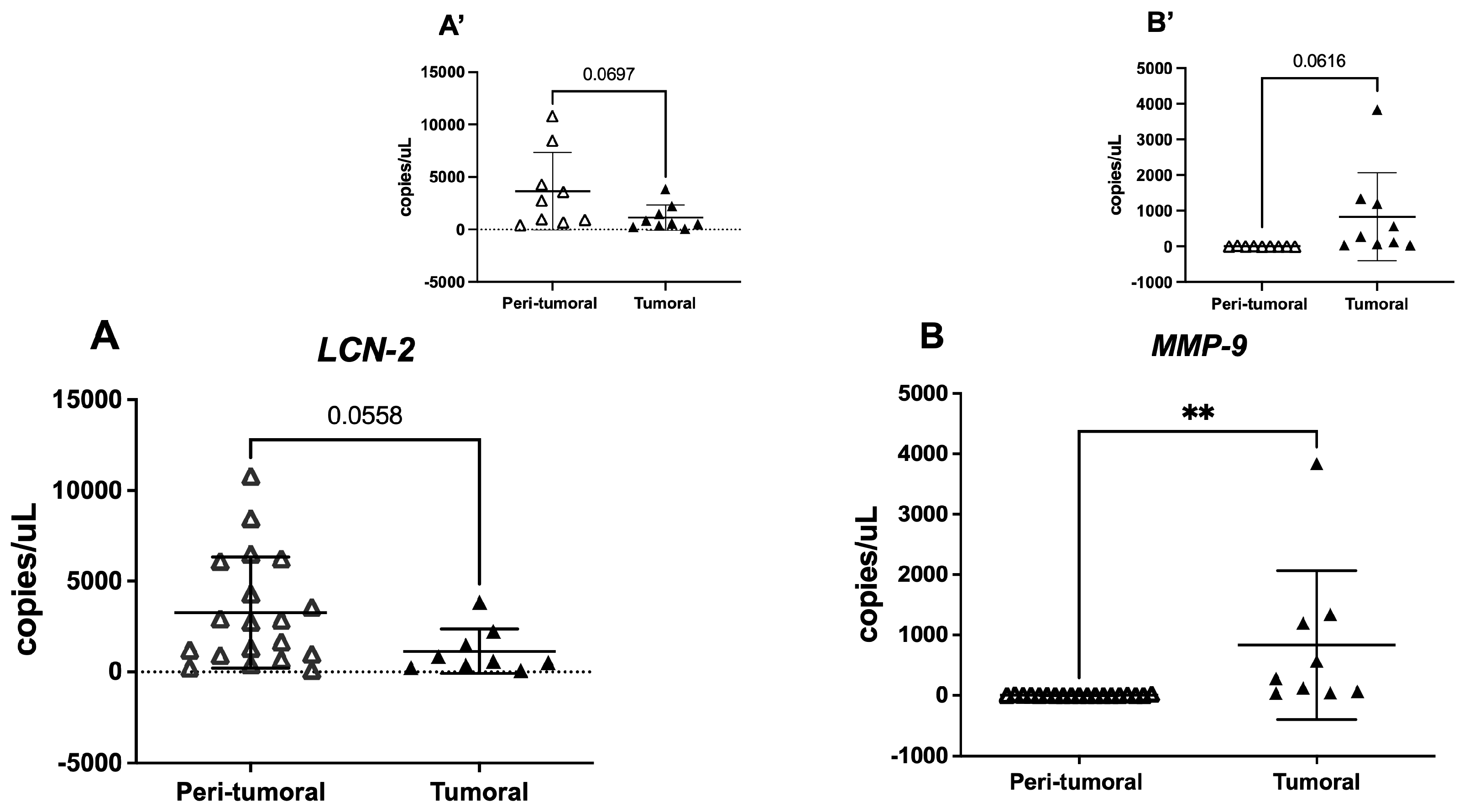
3.3. Tissue LCN-2 and MMP-9 Gene Expression

The available tissue analysis revealed that in tumoral samples, LCN-2 had a lower (t = 2.002, df = 26, p = 0.056) copy number with respect to normal tissues. On the contrary, MMP-9 expression in tumoral portions was significantly higher with respect to peri-tumoral tissues (t = 2.996, df = 26, p = 0.0059) (Figure 1A,B). When analyzing peri-tumoral and tumoral tissues from the same UADT cancer patients (n = 9), we observed a similar, though not significant, trend in the expression of both LCN-2 and MMP-9 (Figure 1A’,B’).

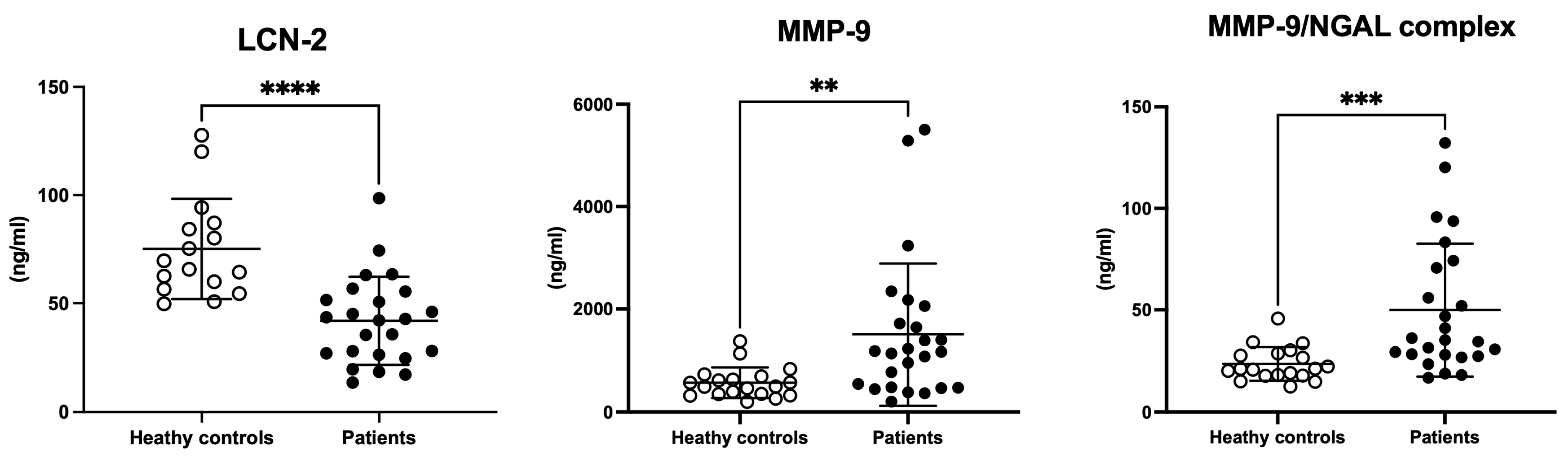
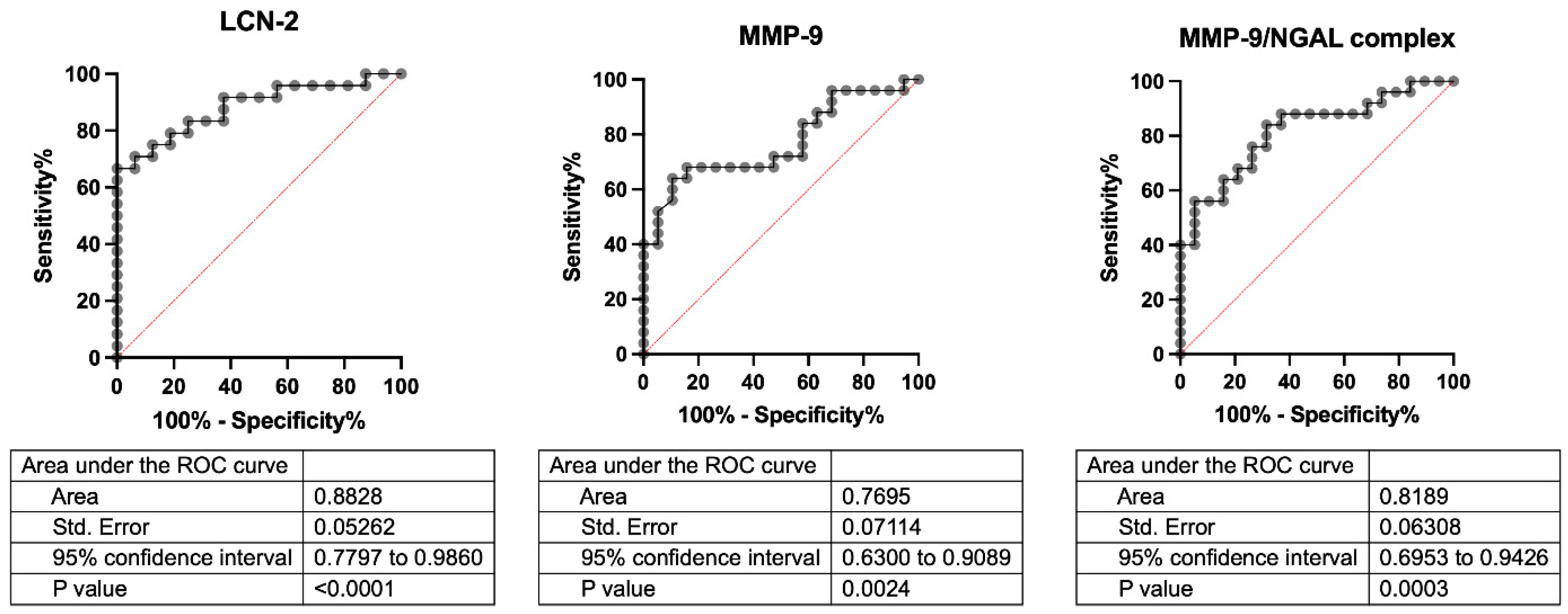
3.4. Serum LCN-2, MMP-9, and MMP-9/NGAL Complex

As shown in Figure 2, LCN-2 serum levels were decreased in UADT cancer patients in comparison to healthy controls (t = 4.796, df = 38, p < 0.0001). Conversely, MMP-9 and MMP-9/NGAL complex circulating levels were up-regulated in the same groups (MMP-9: t = 2.897, df = 42, p = 0.006; MMP-9/NGAL complex: t = 3.447, df = 42, p = 0.001).
In the present pilot study, no significant differences were found in any of the three biomarkers analyzed when UADT patients were stratified by distinct disease stages or lymph node involvement (see Figure S1 in Supplementary Results).
ROC curve analysis (Figure 3) revealed the ability of LCN-2 to discriminate between healthy controls and UADT cancer patients, surpassing the other biological markers tested (MMP-9 and MMP-9/NGAL complex).

3.5. Spearman’s Correlation

Table 3 summarized only the statistically significant correlations between serum LCN-2, MMP-9, and MMP-9/NGAL complex and hematologic parameters in healthy controls and UADT cancer patients. Interestingly, in physiological conditions, LCN-2 is positively correlated with transferrin and negatively with ferritin, both proteins involved in iron trafficking. The conjugated form of LCN-2 (MMP-9/NGAL complex) is directly correlated to MMP-9 serum levels. In cancer patients, only the inverse relation between LCN-2 and ferritin is conserved, even if weaker than in healthy conditions, whilst a strong direct association with MMP-9/NGAL was observed. Finally, MMP-9/NGAL and ferritin were found to be negatively correlated under pathological conditions. Scatter plot graphs of significant correlations have been presented in Supplementary Results (Figures S2 and S3).

4. Discussion

In the present retrospective observational study, we analyzed tissue gene expression of LCN-2 and MMP-9 and the circulating levels of LCN-2, MMP-9, and the MMP-9/NGAL complex in a selected cohort of patients with a first diagnosis of UADT carcinomas. The strict criteria limited patients to those who had never received any kind of treatment (interventional and/or pharmacological) before the recruitment, reducing the risk of potential confounding factors.
Tissue LCN-2 and MMP-9 gene analysis showed a decrease in LCN-2 and an increase in MMP-9 expression in tumoral tissues with respect to peri-tumoral portions. Interestingly, serum levels of LCN-2 were significantly lower than those of controls, whilst its conjugated form with MMP-9 (MMP-9/NGAL complex) was higher when compared to healthy people. As expected, MMP-9 circulating levels were elevated in cancer patients. Finally, ROC curve analysis disclosed an interesting potential value of serum LCN-2 to discriminate between healthy and unhealthy people, compared to the two other proteins analyzed.
UADT cancers represent a major global health challenge, with strong epidemiological differences related to behavioral risk factors, viral infections, and geographic variables [1,2]. A healthy lifestyle (reducing tobacco and alcohol consumption and adopting a healthy diet) and Human Papillomavirus vaccination are crucial and controllable preventive measures to reduce the incidence and improve the prognosis of these tumors [28]. Nevertheless, the identification of biomarkers for the diagnosis and/or progression monitoring of the disease or to evaluate the efficacy of a treatment is a growing field of interest in oncology.
LCN-2 has been studied as a biomarker in tumors due to its overexpression in various types of cancer, making it useful for diagnosis, prognosis, and treatment monitoring [18,31,32]. LCN-2 levels are elevated in several types of cancer, including breast cancer [33], colorectal cancer [34], and lung cancer [35]. Overexpression of LCN-2 has been associated with a worse prognosis in various tumors, particularly in association with metastatic invasion [36,37,38,39,40]. The presence of LCN-2 in plasma or tumor fluid may indicate a functional inflammation in response to tumor proliferation. In breast cancer, for example, LCN-2 has been found elevated in patients with advanced tumors and lymph node metastases [33,36].
Concerning UADT carcinomas, some research evidenced contrasting findings. Up-regulation of LCN-2 was found in oral squamous cell carcinoma (OSCC) tissues (tongue) and oral carcinoma cell lines in relation to cancer progression (higher levels in well-differentiated tissues with respect to poorly/moderately differentiated cells), suggesting that LCN-2 could be a useful marker of tumor-cell differentiation [41,42,43,44]. The only study that took into consideration circulating (plasmatic) levels of LCN-2, MMP-9, and the MMP-9/NGAL complex disclosed increased levels of the three molecules in patients with OSCC compared to those in healthy controls [42]. Both these studies presented results partially in contrast with ours (tissue and circulating LCN-2 increased levels). We hypothesize that these discrepancies may be due, at least in part, to the inclusion/exclusion criteria for patient enrollment. In the cited studies [41,42], no information was available on potential pharmacological and/or interventional treatments, at the time of sampling, that could significantly affect the results.
On the other hand, some research studies were in line with our findings. Monisha and collaborators [20] showed that, in tissue microarray slides for head and neck cancers and OSCC, LCN-2, detected in all samples analyzed, was weakly expressed compared to healthy tissues. Authors also demonstrated that LCN-2 knockdown cells possessed higher invasive ability than control cells, as shown in in vitro invasion and migration assays. Moreover, in the same cells, MMP-9 was found to be overexpressed (in line with our findings) and presumably responsible for the increase in cell motility [20].
In another study [45], Lin et al. demonstrated that LCN-2 expression decreased in patients with OSCC with lymph node metastasis compared with that in patients without metastasis. A higher LCN-2 expression correlated with the survival of patients with OSCC. Furthermore, LCN-2 overexpression in OSCC cells reduced in vitro migration and invasion and in vivo metastasis, whereas its silencing induced an increase in cell motility [45]. This effect was also confirmed in nasopharyngeal carcinoma cell lines, where downregulation of LCN-2 levels via siRNA targeting LCN-2 enhanced cell migration and invasion [46].
The intriguing observation that both upregulation and downregulation of LCN-2 have been associated with a malignant phenotype in different cancer types powerfully suggests distinctive transduction pathways that converge on an overall pro-carcinogenic role of the LCN-2 system. In support of this hypothesis, our study demonstrated a dual trend of the LCN-2 system in the serum of UADT cancer patients. Whilst the free form of LCN-2 decreased when compared to healthy controls, the form complexed with MMP-9 (MMP-9/NGAL complex) strongly increased in oncological patients, as well as the unconjugated MMP-9. Moreover, tissue analysis carried out through droplet digital PCR, technology that provides ultrasensitive nucleic acid detection and absolute quantification, disclosed that both LCN-2 and MMP-9 gene expression levels were comparable to that of the circulating levels.
The LCN-2 reduction in serum and tissue samples of oncological patients was in line with its role in promoting invasion and migration in UADT carcinomas [47], as well as in colorectal cancer and breast cancer when upregulated [32,34]. Moreover, as demonstrated by Monisha’s study, the knockdown of LCN-2 in tumoral cells activated mTOR signaling via the LKB1-AMPK-p53-Redd1 pathway, inducing resistance to autophagy and providing a survival advantage to OSCC tumoral cells [20].
Regarding the mechanism of action of LCN-2 in its conjugated form with MMP-9 (MMP-9/NGAL complex), overexpressed in the serum of patients with UADT cancer, the role of the two proteins in the dynamics of metastasis and tumor progression comes into play. MMP-9 is a metalloproteinase present in the extracellular matrix capable of degrading some components of the matrix, such as type IV collagen, gelatin, and other structural proteins [47,48]. MMP-9 is also expressed, in addition to neutrophils, by other cells, such as tumor cells. In this context, MMP-9 plays a critical role in tumor invasion and metastasis by facilitating, through the degradation of the extracellular matrix, infiltration, and diffusion into surrounding tissues [48,49,50]. NGAL and MMP-9 are also associated with the establishment of an inflammatory response, a favorable factor for tumor progression [50]. It has been shown that NGAL protein binds to MMP-9, forming the MMP-9/NGAL complex that improves the stability of the enzyme, promoting its extracellular matrix remodeling activity and ultimately preparing the tissue for the spread of tumor cells [51,52]. A recent study demonstrated that the MMP-9/NGAL complex associated with lysyl oxidase-like 2 (LOXL2) facilitated tumor metastasis in esophageal cancer [51].
Another interesting finding observed in our study derived from Spearman’s correlation analysis of serum values and, in particular, the relationship between LCN-2 (free and conjugated forms) and the circulating proteins (ferritin and transferrin) involved in iron trafficking. Iron, although present in modest quantities, plays an important role in the organism [53,54]. Maintaining the balance of its levels is of fundamental importance since both a decrease and an increase can be the cause of numerous pathological conditions, including the promotion of tumor initiation, growth, and metastasis [55,56,57]. LCN-2 plays an important role in iron binding and sequestration [58,59]. This process helps to limit the availability of iron to pathogens, which are dependent on iron for their growth and survival [60]. The formation of the complex with iron promotes its transport to the appropriate sites, such as immune system cells that require it for their functioning [54]. Moreover, LCN-2, by limiting the availability of free iron, reduces the possibility of triggering lipid peroxidation, a process capable of causing cellular and tissue damage, especially in tissues involved in chronic inflammatory processes [61,62]. These findings are consistent with what was observed in our study. LCN-2 levels in healthy individuals are negatively correlated with ferritin (a protein that mainly promotes iron accumulation in specific reserve tissues) and positively with transferrin (which transports iron to cells that need it). In UADT cancer patients, the drastic reduction in LCN-2/ferritin correlation is followed by the loss of relationship with transferrin, supporting the idea that the modification in LCN-2 levels also impacts the processes involved in the endogenous regulation of circulating iron. The strong reduction of serum LCN-2 levels in cancer patients could contribute to making available circulating levels of iron useful to tumor cells for their active proliferation. The concomitant increase in ferritin observed could contribute to the accumulation of intracellular iron used for tumor growth.

5. Conclusions and Perspectives

The limited number of samples analyzed may represent a weakness of our study, although the stringent selection of patients only with the first diagnosis of UADT carcinoma constitutes a solid starting point for further investigations. Our study confirms and extends the knowledge on the involvement of the LCN-2 system in upper aerodigestive tract tumors, revealing a potentially different role of the free form (LCN-2) and the one conjugated with MMP-9 (MMP-9/NGAL complex) in this pathological condition. These findings stimulate further investigations on the potential value of the two forms as markers of different stages in the carcinogenesis process. On the one hand, the conjugated form of LCN-2 could be an index of tumor invasiveness, whilst the free form could be of support in monitoring therapeutic efficacy or detecting tumor recurrence. In line with this consideration, ROC curve analysis for LCN-2, showing the significant ability to discriminate between healthy controls and cancer patients, offers an interesting food for thought in this direction. Understanding these mechanisms would offer potential innovative therapeutic approaches for the treatment of cancer.

Supplementary Materials

The following supporting information can be downloaded at: https://www.mdpi.com/article/10.3390/cells14070506/s1, Figure S1: LCN-2, MMP-9 and MMP-9/NGAL complex circulating levels UADT cancer patients, stratified following number of lymph node involved (N0-1-2-3-4) (upper panels) and tumor stage (T0-1-2-3-4). Figure S2: Scatter plot graphs reporting significant Spearman’s Correlations in healthy controls; Figure S3: Scatter plot graphs reporting significant Spearman’s Correlations in UADT cancer patients.

Author Contributions

Conceptualization: L.C., C.P. and G.F.; methodology: L.C., S.F., G.F., F.F., A.M. and C.P.; validation: L.C., S.F., G.F., F.F., C.B., A.G., A.M. and C.P.; formal analysis: S.F., G.F., F.F., D.P. and C.P.; investigation: L.C., S.F., G.F., F.F., C.B., A.G., A.M. and C.P.; data curation: L.C., S.F., G.F., D.P., F.F. and C.P.; writing—original draft preparation: L.C., S.F., G.F., F.F. and C.P.; writing—review and editing, L.C., S.F., G.F., D.P., F.F., C.B., A.G., A.M. and C.P.; visualization: L.C., S.F., G.F., F.F., D.P., C.B., A.G., A.M. and C.P. All authors have read and agreed to the published version of the manuscript.

Funding

This research was supported by the grant Bandi Ateneo Sapienza Progetti Avvio alla Ricerca, s.a. 263/2023 del 10.10.2023 (Dr. Luca Cavalcanti).

Institutional Review Board Statement

The study was conducted in accordance with the Declaration of Helsinki and approved by the Ethics Committee of Policlinico Umberto I Hospital (protocol number Rif. 6462, 15 September 2021).

Informed Consent Statement

Informed consent was obtained from the patient involved in the study.

Data Availability Statement

Data are available on request due to ethical reasons.

Acknowledgments

The authors thank the Sapienza University of Rome and IBBC-CNR.

Conflicts of Interest

The authors declare no conflicts of interest.

References

  1. Gupta, B.; Lalloo, R.; Johnson, N.W. Life course models for upper aero-digestive tract cancer. Int. Dent. J. 2015, 65, 111–119. [Google Scholar] [CrossRef] [PubMed]
  2. Barnes, L.; Eveson, J.W.; Reichart, P.; Sidransky, D. Pathology and Genetics of Head and Neck Tumours; IARC: Lyon, France, 2005; Volume 9. [Google Scholar]
  3. Warnakulasuriya, S. Global epidemiology of oral and oropharyngeal cancer. Oral Oncol. 2009, 45, 309–316. [Google Scholar] [CrossRef]
  4. Sawabe, M.; Ito, H.; Oze, I.; Hosono, S.; Kawakita, D.; Tanaka, H.; Hasegawa, Y.; Murakami, S.; Matsuo, K. Heterogeneous impact of alcohol consumption according to treatment method on survival in head and neck cancer: A prospective study. Cancer Sci. 2017, 108, 91–100. [Google Scholar] [CrossRef] [PubMed]
  5. Cohen, N.; Fedewa, S.; Chen, A.Y. Epidemiology and Demographics of the Head and Neck Cancer Population. Oral Maxillofac. Surg. Clin. N. Am. 2018, 30, 381–395. [Google Scholar] [CrossRef]
  6. Warren, G.W.; Arnold, S.M.; Valentino, J.P.; Gal, T.J.; Hyland, A.J.; Singh, A.K.; Rangnekar, V.M.; Cummings, K.M.; Marshall, J.R.; Kudrimoti, M.R. Accuracy of self-reported tobacco assessments in a head and neck cancer treatment population. Radiother. Oncol. 2012, 103, 45–48. [Google Scholar] [CrossRef] [PubMed]
  7. Pandruvada, S.; Kessler, R.; Thai, A. Head and neck cancer treatment in the era of molecular medicine. Adv. Cancer Res. 2023, 160, 205–252. [Google Scholar] [CrossRef]
  8. Mesia, R.; Iglesias, L.; Lambea, J.; Martínez-Trufero, J.; Soria, A.; Taberna, M.; Trigo, J.; Chaves, M.; García-Castaño, A.; Cruz, J. SEOM clinical guidelines for the treatment of head and neck cancer (2020). Clin. Transl. Oncol. 2021, 23, 913–921. [Google Scholar] [CrossRef]
  9. Marur, S.; Forastiere, A.A. Head and Neck Squamous Cell Carcinoma: Update on Epidemiology, Diagnosis, and Treatment. In Mayo Clinic Proceedings; Elsevier: Amsterdam, The Netherlands, 2016; Volume 91, pp. 386–396. [Google Scholar] [CrossRef]
  10. Meliante, P.G.; Petrella, C.; Fiore, M.; Minni, A.; Barbato, C. Antioxidant Use after Diagnosis of Head and Neck Squamous Cell Carcinoma (HNSCC): A Systematic Review of Application during Radiotherapy and in Second Primary Cancer Prevention. Antioxidants 2023, 12, 1753. [Google Scholar] [CrossRef]
  11. Riccardi, G.; Bellizzi, M.G.; Fatuzzo, I.; Zoccali, F.; Cavalcanti, L.; Greco, A.; de Vincentiis, M.; Ralli, M.; Fiore, M.; Petrella, C.; et al. Salivary Biomarkers in Oral Squamous Cell Carcinoma: A Proteomic Overview. Proteomes 2022, 10, 37. [Google Scholar] [CrossRef]
  12. Meliante, P.G.; Zoccali, F.; de Vincentiis, M.; Ralli, M.; Petrella, C.; Fiore, M.; Minni, A.; Barbato, C. Diagnostic Predictors of Immunotherapy Response in Head and Neck Squamous Cell Carcinoma. Diagnostics 2023, 13, 862. [Google Scholar] [CrossRef]
  13. Al Jaberi, S.; Cohen, A.; D’souza, C.; Abdulrazzaq, Y.M.; Ojha, S.; Bastaki, S.; Adeghate, E.A. Lipocalin-2: Structure, function, distribution and role in metabolic disorders. Biomed. Pharmacother. 2021, 142, 112002. [Google Scholar] [CrossRef]
  14. Romejko, K.; Markowska, M.; Niemczyk, S. The Review of Current Knowledge on Neutrophil Gelatinase-Associated Lipocalin (NGAL). Int. J. Mol. Sci. 2023, 24, 10470. [Google Scholar] [CrossRef] [PubMed]
  15. Kjeldsen, L.; Johnsen, A.; Sengeløv, H.; Borregaard, N. Isolation and primary structure of NGAL, a novel protein associated with human neutrophil gelatinase. J. Biol. Chem. 1993, 268, 10425–10432. [Google Scholar] [CrossRef] [PubMed]
  16. Kjeldsen, L.; Bainton, D.F.; Sengeløv, H.; Borregaard, N. Identification of neutrophil gelatinase-associated lipocalin as a novel matrix protein of specific granules in human neutrophils. Blood 1994, 83, 799–807. [Google Scholar]
  17. Wang, Y.; Lam, K.S.L.; Kraegen, E.W.; Sweeney, G.; Zhang, J.; Tso, A.W.; Chow, W.-S.; Wat, N.M.; Xu, J.Y.; Hoo, R.L.; et al. Lipocalin-2 is an inflammatory marker closely associated with obesity, insulin resistance, and hyperglycemia in humans. Clin. Chem. 2007, 53, 34–41. [Google Scholar] [CrossRef]
  18. Santiago-Sánchez, G.S.; Pita-Grisanti, V.; Quiñones-Díaz, B.; Gumpper, K.; Cruz-Monserrate, Z.; Vivas-Mejía, P.E. Biological Functions and Therapeutic Potential of Lipocalin 2 in Cancer. Int. J. Mol. Sci. 2020, 21, 4365. [Google Scholar] [CrossRef] [PubMed]
  19. Meyers, K.; López, M.; Ho, J.; Wills, S.; Rayalam, S.; Taval, S. Lipocalin-2 deficiency may predispose to the progression of spontaneous age-related adiposity in mice. Sci. Rep. 2020, 10, 14589. [Google Scholar] [CrossRef]
  20. Monisha, J.; Roy, N.K.; Padmavathi, G.; Banik, K.; Bordoloi, D.; Khwairakpam, A.D.; Arfuso, F.; Chinnathambi, A.; Alahmadi, T.A.; Alharbi, S.A.; et al. NGAL is Downregulated in Oral Squamous Cell Carcinoma and Leads to Increased Survival, Proliferation, Migration and Chemoresistance. Cancers 2018, 10, 228. [Google Scholar] [CrossRef]
  21. Rashid, Z.A.; Bardaweel, S.K. Novel Matrix Metalloproteinase-9 (MMP-9) Inhibitors in Cancer Treatment. Int. J. Mol. Sci. 2023, 24, 12133. [Google Scholar] [CrossRef]
  22. Mondal, S.; Adhikari, N.; Banerjee, S.; Amin, S.A.; Jha, T. Matrix metalloproteinase-9 (MMP-9) and its inhibitors in cancer: A minireview. Eur. J. Med. Chem. 2020, 194, 112260. [Google Scholar] [CrossRef]
  23. Eilenberg, W.; Stojkovic, S.; Kaider, A.; Kozakowski, N.; Domenig, C.M.; Burghuber, C.; Nanobachvili, J.; Huber, K.; Klinger, M.; Neumayer, C.; et al. NGAL and MMP-9/NGAL as biomarkers of plaque vulnerability and targets of statins in patients with carotid atherosclerosis. Clin. Chem. Lab. Med. (CCLM) 2017, 56, 147–156. [Google Scholar] [CrossRef]
  24. D’Amico, F.; Candido, S.; Libra, M. Interaction between matrix metalloproteinase-9 (MMP-9) and neutrophil gelatinase-associated lipocalin (NGAL): A recent evolutionary event in primates. Dev. Comp. Immunol. 2021, 116, 103933. [Google Scholar] [CrossRef] [PubMed]
  25. Candido, S.; Abrams, S.L.; Steelman, L.S.; Lertpiriyapong, K.; Fitzgerald, T.L.; Martelli, A.M.; Cocco, L.; Montalto, G.; Cervello, M.; Polesel, J.; et al. Roles of NGAL and MMP-9 in the tumor microenvironment and sensitivity to targeted therapy. Biochim. Biophys. Acta (BBA)-Mol. Cell Res. 2016, 1863, 438–448. [Google Scholar] [CrossRef]
  26. Tsakogiannis, D.; Kalogera, E.; Zagouri, F.; Zografos, E.; Balalis, D.; Bletsa, G. Determination of FABP4, RBP4 and the MMP-9/NGAL complex in the serum of women with breast cancer. Oncol. Lett. 2021, 21, 85. [Google Scholar] [CrossRef] [PubMed]
  27. Bruno, S.M.; Blaconà, G.; Cicero, S.L.; Castelli, G.; Virgulti, M.; Testino, G.; Pierandrei, S.; Fuso, A.; Cimino, G.; Ferraguti, G.; et al. Quantitative Evaluation of CFTR Gene Expression: A Comparison between Relative Quantification by Real-Time PCR and Absolute Quantification by Droplet Digital PCR. Genes 2023, 14, 1781. [Google Scholar] [CrossRef] [PubMed]
  28. Barsouk, A.; Aluru, J.S.; Rawla, P.; Saginala, K.; Barsouk, A. Epidemiology, Risk Factors, and Prevention of Head and Neck Squamous Cell Carcinoma. Med Sci. 2023, 11, 42. [Google Scholar] [CrossRef] [PubMed] [PubMed Central]
  29. Glastonbury, C.M. Head and Neck Squamous Cell Cancer: Approach to Staging and Surveillance. In Diseases of the Brain, Head and Neck, Spine 2020–2023: Diagnostic Imaging; Springer: Berlin/Heidelberg, Germany, 2020; pp. 215–222. [Google Scholar] [CrossRef]
  30. Sano, D.; Yabuki, K.; Arai, Y.; Tanabe, T.; Chiba, Y.; Nishimura, G.; Takahashi, H.; Yamanaka, S.; Oridate, N. The applicability of new TNM classification for humanpapilloma virus-related oropharyngeal cancer in the 8th edition of the AJCC/UICC TNM staging system in Japan: A single-centre study. Auris Nasus Larynx 2018, 45, 558–565. [Google Scholar] [CrossRef]
  31. Asaf, S.; Maqsood, F.; Jalil, J.; Sarfraz, Z.; Sarfraz, A.; Mustafa, S.; Ojeda, I.C. Lipocalin 2—not only a biomarker: A study of current literature and systematic findings of ongoing clinical trials. Immunol. Res. 2023, 71, 287–313. [Google Scholar] [CrossRef]
  32. Živalj, M.; Van Ginderachter, J.A.; Stijlemans, B. Lipocalin-2: A Nurturer of Tumor Progression and a Novel Candidate for Targeted Cancer Therapy. Cancers 2023, 15, 5159. [Google Scholar] [CrossRef]
  33. Bao, Y.; Yan, Z.; Shi, N.; Tian, X.; Li, J.; Li, T.; Cheng, X.; Lv, J. LCN2: Versatile players in breast cancer. Biomed. Pharmacother. 2024, 171, 116091. [Google Scholar] [CrossRef]
  34. Kim, S.; Lee, S.T.; Min, I.S.; Park, Y.R.; Lee, J.H.; Kim, D.; Kim, S. Lipocalin 2 negatively regulates cell proliferation and epithelial to mesenchymal transition through changing metabolic gene expression in colorectal cancer. Cancer Sci. 2017, 108, 2176–2186. [Google Scholar] [CrossRef] [PubMed]
  35. Li, A.; Zhang, K.; Zhou, J.; Li, M.; Fan, M.; Gao, H.; Ma, R.; Gao, L.; Chen, M. Bioinformatics and experimental approach identify lipocalin 2 as a diagnostic and prognostic indicator for lung adenocarcinoma. Int. J. Biol. Macromol. 2024, 272, 132797. [Google Scholar] [CrossRef]
  36. Provatopoulou, X.; Gounaris, A.; Kalogera, E.; Zagouri, F.; Flessas, I.; Goussetis, E.; Nonni, A.; Papassotiriou, I.; Zografos, G. Circulating levels of matrix metalloproteinase-9 (MMP-9), neutrophil gelatinase-associated lipocalin (NGAL) and their complex MMP-9/NGAL in breast cancer disease. BMC Cancer 2009, 9, 390. [Google Scholar] [CrossRef] [PubMed]
  37. Chiang, K.C.; Yeh, T.S.; Wu, R.C.; Pang, J.H.S.; Cheng, C.T.; Wang, S.Y.; Yeh, C.N. Lipocalin 2 (LCN2) is a promising target for cholangiocarcinoma treatment and bile LCN2 level is a potential cholangiocarcinoma diagnostic marker. Sci. Rep. 2016, 6, 36138. [Google Scholar] [CrossRef] [PubMed]
  38. Bauer, M.; Eickhoff, J.C.; Gould, M.N.; Mundhenke, C.; Maass, N.; Friedl, A. Neutrophil gelatinase-associated lipocalin (NGAL) is a predictor of poor prognosis in human primary breast cancer. Breast Cancer Res. Treat. 2008, 108, 389–397. [Google Scholar] [CrossRef]
  39. Iannetti, A.; Pacifico, F.; Acquaviva, R.; Lavorgna, A.; Crescenzi, E.; Vascotto, C.; Tell, G.; Salzano, A.M.; Scaloni, A.; Vuttariello, E.; et al. The neutrophil gelatinase-associated lipocalin (NGAL), a NF-κB-regulated gene, is a survival factor for thyroid neoplastic cells. Proc. Natl. Acad. Sci. USA 2008, 105, 14058–14063. [Google Scholar] [CrossRef]
  40. Gomez-Chou, S.B.; Swidnicka-Siergiejko, A.K.; Badi, N.; Chavez-Tomar, M.; Lesinski, G.B.; Bekaii-Saab, T.; Farren, M.R.; Mace, T.A.; Schmidt, C.; Liu, Y.; et al. Lipocalin-2 Promotes Pancreatic Ductal Adenocarcinoma by Regulating Inflammation in the Tumor Microenvironment. Cancer Res. 2017, 77, 2647–2660. [Google Scholar] [CrossRef]
  41. Noguchi, K.; Hiromoto, T.; Yamamura, M.; Zushi, Y.; Segawa, E.; Takaoka, K.; Moridera, K.; Kishimoto, H.; Urade, M. Up-regulation of neutrophil gelatinase-associated lipocalin in oral squamous cell carcinoma: Relation to cell differentiation. Oncol. Rep. 2011, 26, 1415–1421. [Google Scholar] [CrossRef]
  42. Lin, C.W.; Tseng, S.W.; Yang, S.F.; Ko, C.P.; Lin, C.H.; Wei, L.H.; Hsieh, Y.S. Role of lipocalin 2 and its complex with matrix metalloproteinase-9 in oral cancer. Oral Dis. 2012, 18, 734–740. [Google Scholar] [CrossRef]
  43. Huang, Z.; Rui, X.; Yi, C.; Chen, Y.; Chen, R.; Liang, Y.; Wang, Y.; Yao, W.; Xu, X.; Huang, Z. Silencing LCN2 suppresses oral squamous cell carcinoma progression by reducing EGFR signal activation and recycling. J. Exp. Clin. Cancer Res. 2023, 42, 60, Erratum in J. Exp. Clin. Cancer Res. 2023, 42, 104. https://doi.org/10.1186/s13046-023-02679-0. [Google Scholar] [CrossRef]
  44. Moghaddam, S.J.K.; Roushandeh, A.M.; Hamidi, M.; Nemati, S.; Jahanian-Najafabadi, A.; Roudkenar, M.H. Lipocalin-2 Upregulation in Nasopharyngeal Carcinoma: A Novel Potential Diagnostic Biomarker. Iran. J. Med. Sci. 2023, 48, 268–276. [Google Scholar] [CrossRef]
  45. Lin, C.-W.; Yang, W.-E.; Lee, W.-J.; Hua, K.-T.; Hsieh, F.-K.; Hsiao, M.; Chen, C.-C.; Chow, J.-M.; Chen, M.-K.; Yang, S.-F.; et al. Lipocalin 2 prevents oral cancer metastasis through carbonic anhydrase IX inhibition and is associated with favourable prognosis. Carcinog. 2016, 37, 712–722. [Google Scholar] [CrossRef]
  46. Li, J.-P.; Lin, C.-W.; Huang, C.-C.; Lu, Y.-T.; Ho, Y.-T.; Yang, S.-F.; Hsin, C.-H. Lipocalin 2 Reduces MET Levels by Inhibiting MEK/ERK Signaling to Inhibit Nasopharyngeal Carcinoma Cell Migration. Cancers 2022, 14, 5707. [Google Scholar] [CrossRef]
  47. Vafadari, B.; Salamian, A.; Kaczmarek, L. MMP-9 in translation: From molecule to brain physiology, pathology, and therapy. J. Neurochem. 2016, 139, 91–114. [Google Scholar] [CrossRef]
  48. Mittal, R.; Patel, A.P.; Debs, L.H.; Nguyen, D.; Patel, K.; Grati, M.; Mittal, J.; Yan, D.; Chapagain, P.; Liu, X.Z. Intricate Functions of Matrix Metalloproteinases in Physiological and Pathological Conditions. J. Cell. Physiol. 2016, 231, 2599–2621. [Google Scholar] [CrossRef] [PubMed]
  49. Lin, M.B.; Ashraf, N.S.; Mahjabeen, I. Deregulation of MMP-2 and MMP-9 in laryngeal cancer: A retrospective observational study. Medicine 2024, 103, e38362. [Google Scholar] [CrossRef]
  50. Song, Z.; Wang, J.; Su, Q.; Luan, M.; Chen, X.; Xu, X. The role of MMP-2 and MMP-9 in the metastasis and development of hypopharyngeal carcinoma. Braz. J. Otorhinolaryngol. 2021, 87, 521–528. [Google Scholar] [CrossRef]
  51. Xia, Q.; Du, Z.; Chen, M.; Zhou, X.; Bai, W.; Zheng, X.; Lin, L.; Zhao, Y.; Ding, J.; Wu, Z.; et al. A protein complex of LCN2, LOXL2 and MMP9 facilitates tumour metastasis in oesophageal cancer. Mol. Oncol. 2023, 17, 2451–2471. [Google Scholar] [CrossRef]
  52. Liu, M.-F.; Hu, Y.-Y.; Jin, T.; Xu, K.; Wang, S.-H.; Du, G.-Z.; Wu, B.-L.; Li, L.-Y.; Xu, L.-Y.; Li, E.-M.; et al. Matrix Metalloproteinase-9/Neutrophil Gelatinase-Associated Lipocalin Complex Activity in Human Glioma Samples Predicts Tumor Presence and Clinical Prognosis. Dis. Markers 2015, 2015, 138974. [Google Scholar] [CrossRef]
  53. Gao, G.; Li, J.; Zhang, Y.; Chang, Y.-Z. Cellular Iron Metabolism and Regulation. In Brain Iron Metabolism and CNS Diseases; Springer: Berlin/Heidelberg, Germany, 2019; pp. 21–32. [Google Scholar] [CrossRef]
  54. Ni, S.; Yuan, Y.; Kuang, Y.; Li, X. Iron Metabolism and Immune Regulation. Front. Immunol. 2022, 13, 816282. [Google Scholar] [CrossRef]
  55. Gamage, S.M.; Lee, K.T.; Dissabandara, D.L.O.; Lam, A.K.-Y.; Gopalan, V. Dual role of heme iron in cancer; promotor of carcinogenesis and an inducer of tumour suppression. Exp. Mol. Pathol. 2021, 120, 104642. [Google Scholar] [CrossRef]
  56. Dev, S.; Babitt, J.L. Overview of iron metabolism in health and disease. Hemodial. Int. 2017, 21 (Suppl. S1), S6–S20. [Google Scholar] [CrossRef] [PubMed]
  57. Salnikow, K. Role of iron in cancer. Semin. Cancer Biol. 2021, 76, 189–194. [Google Scholar] [CrossRef] [PubMed]
  58. Jung, M.; Mertens, C.; Bauer, R.; Rehwald, C.; Brüne, B. Lipocalin-2 and iron trafficking in the tumor microenvironment. Pharmacol. Res. 2017, 120, 146–156. [Google Scholar] [CrossRef]
  59. Xiao, X.; Yeoh, B.S.; Vijay-Kumar, M. Lipocalin 2: An Emerging Player in Iron Homeostasis and Inflammation. Annu. Rev. Nutr. 2017, 37, 103–130. [Google Scholar] [CrossRef] [PubMed]
  60. Smith, K.D. Iron metabolism at the host pathogen interface: Lipocalin 2 and the pathogen-associated iroA gene cluster. Int. J. Biochem. Cell Biol. 2007, 39, 1776–1780. [Google Scholar] [CrossRef]
  61. Rochette, L.; Dogon, G.; Rigal, E.; Zeller, M.; Cottin, Y.; Vergely, C. Lipid Peroxidation and Iron Metabolism: Two Corner Stones in the Homeostasis Control of Ferroptosis. Int. J. Mol. Sci. 2022, 24, 449. [Google Scholar] [CrossRef]
  62. Deng, L.; He, S.; Guo, N.; Tian, W.; Zhang, W.; Luo, L. Molecular mechanisms of ferroptosis and relevance to inflammation. Inflamm. Res. 2023, 72, 281–299. [Google Scholar] [CrossRef]
Figure 1. LCN-2 and MMP-9 gene expression in the available peri-tumoral and tumoral tissues in cancer patients. The figure represents the mean ± SD of LCN-2 and MMP-9 expression, measured in RNA copy number/μL, as determined by droplet PCR. Panels (A,B) compare total peri-tumoral vs. tumoral tissues, while panels (A’,B’) present data from matched patient samples. Student’s t-test ** p < 0.01.
Cells 14 00506 g001
Figure 2. LCN-2, MMP-9, and MMP-9/NGAL complex circulating levels in healthy controls vs. UADT cancer patients. Scatter dot plots represent the mean ± SD of serum levels from controls and patients. Student’s t-test ** p < 0.01; *** p < 0.001; **** p < 0.0001.
Cells 14 00506 g002
Figure 3. ROC curves for circulating levels of LCN-2, MMP-9, and MMP-9/NGAL complex in healthy controls vs. UADT cancer patients. Below each graph, the following have been shown: area under the curve, standard error, 90% confidence interval, and p values.
Cells 14 00506 g003
Table 1. General characteristics of the enrolled patients: gender, age, cancer district, clinical (c) or pathological radiation (TNM) (p), availability for serum and tissue sampling.
Patient ID#SexAge (Years)Cancer
District
p/cTNMSerum Sampling
(Yes/No)
Tissue Sampling
(Yes/No)
Peri-
Tumoral
Tumoral
1M60HypopharynxcT4bN3bM0YesNoNo
2F57HypopharynxpT4aN0M0YesNoNo
3M72LarynxpT4aN0M0YesNoNo
4M58HypopharynxpT3N3bM0YesNoNo
5M54LarynxpT3N3bM0YesNoNo
6M63LarynxpT4aN0M0YesYesNo
7M57TonguepT3N1M0YesYesNo
8M49LarynxpT3N0M0YesYesNo
9M66OropharynxcT4N2bM0YesNoNo
10F58TonguepT3N3bM0YesYesNo
11M59Upper
Gingiva
cT4bN2cM1YesNoNo
12M60TonguecT4aN2bM0YesNoNo
13M53OropharynxcT4aN3bM0YesNoNo
14M46TonguepT1N0M0YesNoNo
15M65LarynxpT3N0M0YesNoNo
16M70LarynxpT4aN3bM0YesYesNo
17F75LarynxpT3N0M0YesYesNo
18M56LarynxpT3N0M0YesYesNo
19M71LarynxpT3N3bM0YesYesNo
20F60LarynxpT2N0M0YesYesNo
21M67LarynxpT4aN0M0YesYesNo
22M78LarynxpT3N0M0YesYesYes
23M56LarynxpT4aN1M0YesYesYes
24F73LarynxpT3N3bM0YesYesYes
25M77LarynxpT3N0M0YesYesYes
26F62LarynxpT2N0M0NoYesYes
27M69LarynxpT4aN0M0NoYesYes
28M76LarynxpT2N0M0NoYesYes
29M65LarynxpT4aN2bM0NoYesYes
30M60LarynxpT4aN3bM0NoYesYes
Table 2. Hematologic parameters in healthy controls and UADT cancer patients.
Parameter (Range)SexHealthy Controls
(n. 20; M16/F4)
Cancer Patients
(n. 25; M20/F5)
p-Value(df), F
Glucose
(70.3–100.9 mg/dL)
tot88.31 ± 2.23113.32 ± 6.460.002(1,42), 10.591
M87.6 ± 2.51111 ± 19.4p(disease) 0.007
p(gender) 0.464
(1,40)
F(disease) 7.99
F(gender) 0.646
F91 ± 5.35122 ± 6.71
UREA
(10.20–49.80 mg/dL)
tot16.94 ± 1.1531.8 ± 3.51<0.001(1,42), 12.749
M17.45 ± 1.2930.47 ± 3.85p(disease) 0.002
p(gender) 0.686
(1,40)
F(disease) 11.41
F(gender) 0.16
F15 ± 2.6437.18 ± 8.92
Phosphate
(2.80–4.60 mg/dL)
tot3.12 ± 0.103.52 ± 0.100.01(1,42), 7.364
M2.98 ± 0.093.46 ± 0.103p(disease) 0.081
p(gender) 0.006
(1,40)
F(disease) 3.215
F(gender) 8.468
F3.65 ± 0.173.78 ± 0.32
Protein
(60–82 g/L)
tot72.05 ± 0.8366.6 ± 1.580.007(1,42), 7.911
M71.2 ± 0.8766.35 ± 1.86p(disease) 0.013
p(gender) 0.301
(1,40)
F(disease) 6.806
F(gender) 1.097
F72.25 ± 1.5567.4 ± 2.89
Albumin
(35–55 g/L)
tot44.16 ± 0.5740.64 ± 1.190.03(1,42), 5.072
M43.9 ± 0.7140.1 ± 1.44p(disease) 0.130
p(gender) 0.340
(1,40)
F(disease) 2.393
F(gender) 0.933
F45 ± 0.4142.8 ± 3.0
Bilirubin–Direct
(<0.20 mg/dL)
tot0.15 ± 0.020.31 ± 0.040.002(1,42), 10.481
M0.15 ± 0.030.32 ± 0.05p(disease) 0.013
p(gender) 0.832
(1,40)
F(disease) 6.729
F(gender) 0.05
F0.13 ± 0.020.31 ± 0.05
Iron
(64.8–174.90 ug/dL)
tot97.89 ± 5.8475.14 ± 9.170.059(1,42), 3.775
M99.73 ± 7.2478.28 ± 10.70p(disease) 0.09
p(gender) 0.410
(1,40)
F(disease) 2.883
F(gender) 0.694
F91 ± 6.1078.28 ± 10.70
PCR
(100–6000 ug/L)
tot1392 ± 31929212± 113870.04(1,42), 4.372
M1673 ± 27432955 ± 13990p(disease) 0.197
p(gender) 0.587
(1,40)
F(disease) 1.721
F(gender) 0.300
F2350 ± 121114240 ± 10081
Transferrin
(2.15–3.65 g/L)
tot2.76 ± 0.102.23 ± 0.110.001(1,42), 2.999
M2.70 ± 0.112.24 ± 0.13p(disease) 0.002
p(gender) 0.617
(1,40)
F(disease) 10.808
F(gender) 0.254
F2.97 ± 0.192.17 ± 0.21
Creatinine
(M: 0.70–1.20 mg/Dl
F: 0.50–0.9 mg/dL)
M1.04 ± 0.020.85 ± 0.060.012(1,33), 7.112
F0.78 ± 0.050.64 ± 0.080.189(1,7), 2.117
gGT
(M: 10.00–40.00 U/L
F: 7.00–35.00 U/L)
M17.88 ± 1.5455 ± 18.240.01(1,33), 3.073
F16.25 ± 4.2526.6 ± 3.720.109(1,7), 3.374
LDH
(M: 135.00–225.00 U/L
F: 135.00–214.00 U/L)
M161.26 ± 8.14129.95 ± 9.280.02(1,33), 5.950
F173.5 ± 12.63138 ± 28.530.334(1,7), 1.077
CPK
(M: 20–200 U/L
F:20–180 U/L)
M139.27 ± 27.6851.90 ± 12.910.004(1,33), 9.627
F89.75 ± 13.7030.20 ± 5.400.003(1,7), 19.454
Ferritin
(M: 30–400 ug/L
F: 15–150 ug/L)
M67 ± 10.89402.95 ± 87.450.002(1,33), 10.891
F61.50 ± 18.40313 ± 76.350.02(1,7), 8.154
Table 3. Spearman’s correlation between LCN-2, MMP-9, and NGAL/MMP-9 complex vs. hematologic parameters, in healthy controls and UADT cancer patients.
Spearman’s Correlations in Healthy Controls
Variable LCN-2MMP-9/NGALMMP-9TransferrinFerritin
LCN-2Spearman’s rho
p-value
MMP-9/NGALSpearman’s rho0.236
p-value0.397
MMP-9Spearman’s rho−0.4430.751 ***
p-value0.100<0.001
TransferrinSpearman’s rho0.524 *0.203−0.106
p-value0.0400.4180.674
FerritinSpearman’s rho−0.680 **−0.338−0.137−0.688 **
p-value0.0040.1710.5870.001
Spearman’s Correlations in UADT Cancer Patients
Variable LCN-2MMP-9/NGALMMP-9TransferrinFerritin
LCN-2Spearman’s rho
p-value
MMP-9/NGALSpearman’s rho0.598 **
p-value0.002
MMP-9Spearman’s rho0.2420.674 ***
p-value0.242<0.001
TransferrinSpearman’s rho0.1550.1300.053
p-value0.4590.5340.800
FerritinSpearman’s rho−0.400 *−0.479 *−0.254−0.425 *
p-value0.0490.0160.2200.034
* p < 0.05, ** p < 0.01, *** p < 0.001.
Disclaimer/Publisher’s Note: The statements, opinions and data contained in all publications are solely those of the individual author(s) and contributor(s) and not of MDPI and/or the editor(s). MDPI and/or the editor(s) disclaim responsibility for any injury to people or property resulting from any ideas, methods, instructions or products referred to in the content.

