Review

Platelet Reactive Oxygen Species, Oxidised Lipid Stress, Current Perspectives, and an Update on Future Directions

1
Discovery and Translational Science Department, Leeds Institute of Cardiovascular & Metabolic Medicine, University of Leeds, Leeds LS2 9JT, UK
2
Centre for Biomedical Science Research, School of Health, Leeds Beckett University, Leeds LS1 3HE, UK
*
Author to whom correspondence should be addressed.
Cells 2025, 14(7), 500; https://doi.org/10.3390/cells14070500
Submission received: 19 November 2024 / Revised: 20 March 2025 / Accepted: 24 March 2025 / Published: 27 March 2025
(This article belongs to the Special Issue Molecular and Cellular Insights into Platelet Function)

Abstract

:
Blood platelets are anucleate cells that play a vital role in haemostasis, innate immunity, angiogenesis, and wound healing. However, the inappropriate activation of platelets also contributes to vascular inflammation, atherogenesis, and thrombosis. Platelet activation is a highly complex receptor-mediated process that involves a multitude of signalling intermediates in which Reactive Oxygen Species (ROS) are proposed to play an important role. However, like for many cells, changes in the balance of ROS generation and/or scavenging in disease states may lead to the adoption of maladaptive platelet phenotypes. Here, we review the diverse roles of ROS in platelet function and how ROS are linked to specific platelet activation pathways. We also examine how changes in disease, particularly the plasma oxidised low-density lipoprotein (oxLDL), affect platelet ROS generation and platelet function.

1. Introduction

Blood platelets play a critical role in the prevention of blood loss and wound healing upon vascular injury. In circulation, the combination of shear stress and their size ensures that platelets are marginalised to the periphery of vessel where they scan for the integrity of the vessel wall. Under normal conditions, platelets are in a constant state of inhibition as a consequence of continual exposure to endothelial-derived inhibitors, prostacyclin [1], and nitric oxide [2], which prevent platelet activation through cyclic adenosine monophosphate (cAMP) and cyclic guanosine monophosphate (cGMP) signalling, respectively. Upon vascular injury, tonic inhibition of platelets is overcome to promote rapid activation and thrombus formation. The precise mechanism of platelet activation at areas of vascular damage is complex and involves a number of adhesive and soluble ligands, working through both tyrosine kinase and G-protein coupled receptor (GPCR) signalling cascades [3].
Vascular damage exposes collagen and tissue factor to the circulation, tissue factor induces the extrinsic coagulation cascade, resulting in the localised generation of the potent platelet agonist thrombin. The combination of collagen and thrombin drives platelet exocytosis of dense granules containing adenosine diphosphate (ADP), and generation of thromboxane A2 (TxA2), that act to further enhance recruitment of platelets into the growing thrombus [4]. Activated platelets also release α-granules, which translocate membrane-bound receptors to the platelet surface and assist in propagation of activation and adhesion through receptors such as P-selectin and CD40L, or soluble factors such as fibrinogen. These two receptors promote platelet-endothelial cell and platelet-leukocyte adhesion via interaction with P-selectin glycoprotein ligand 1 and CD40, enabling vascular adhesion and bridging of immune complexes to the site of injury [5].
The adhesion of platelets to the vessel wall is mediated by the interaction of previously inactive or cryptic adhesive ligands including von Willebrand factor (vWF), collagen, laminin and fibronectin [6]. Interaction between these factors and numerous glycoprotein and integrin receptors facilitates platelet accumulation at the vessel wall. Of particular interest are GPIbα, part of the larger GPIb-V-IX complex, and GPVI, which act to tether and activate platelets through vWF and collagen, respectively. Activated platelets within a thrombus differ in their morphology and function based primarily on an activation gradient [7]. Canonically, thrombi possess a sustained and potent phenotype of platelet activation in the core, with the appearance of procoagulant platelets, characterised by the redistribution of phosphatidylserine (PS) from the inner leaflet of the platelet plasma membrane to the outer leaflet [8].
In this review, we summarise the diverse roles of Reactive Oxygen Species (ROS) in platelet function and how it is linked to specific platelet activation pathways. We also examine how changes in disease, particularly hyperlipidaemia, affect ROS production and platelet function.

2. Reactive Oxygen Species and Platelet Activation

ROS consist of a range of oxidant species containing oxygen free radicals, such as superoxide anion (O2), hydroxyl radical (OH), peroxyl radicals (ROO), and non-radical oxidants such hydrogen peroxide (H2O2), lipid peroxides (LOO), and singlet oxygen (O). The production of these ROS occurs during the physiological process of platelet activation. Early studies from Marcus and colleagues demonstrated the capacity of platelets to synthesise O2 [9]. Subsequently, it was established that platelet activation led to the endogenous generation of both O2 and H2O2, potentially OH, and that quenching of ROS diminished platelet activation [10]. Stimulation of platelets with physiological agonists, such as collagen and thrombin, produces ROS, which are required for effective platelet activation [11]. In addition, in vitro inhibition of ROS generation blocks collagen- and thrombin-mediated platelet aggregation [12]. It is widely accepted that ROS play a critical role in the signal transduction mechanisms that drive platelet activation.

2.1. ROS Generation in Platelets

Several sources of ROS have been identified in platelets, these include enzymatic processes such as nicotinamide adenine dinucleotide phosphate (NADPH) oxidase (NOX), cyclooxygenase-1 (COX-1), xanthine oxidase (XO), and the mitochondrial respiratory chain.

2.1.1. NADPH Oxidases (NOX)

NOXs are a family of enzymes responsible for the majority of the non-mitochondrial ROS production in platelets [13]. While the NOX family is composed of seven different isoforms, including NOX1–5 and dual oxidase 1–2; to date only NOX1 and NOX2 have been shown to be expressed in both human and murine platelets [14]. Both NOX1 and NOX2 are composed of subunits that come together to form complexes upon platelet activation. The activated form of NOX1 includes the assembly of the catalytic subunit NOX1 with regulatory subunits of p22phox, NOX organiser 1 (NOXO1, a homologue of p47phox), NOX activator 1 (NOXA1), and Rac. Similarly, the activated NOX2 is composed of the catalytic subunit gp91phox, and the regulatory subunits of p22phox, p40phox, NOXO1, p67phox (NOXA1 homologue), and Rac [15]. Activation of these enzymes is driven by protein kinase C (PKC)-mediated phosphorylation of cytosolic p47phox (or its homologue NOXO1), which facilitates its interaction with membrane bound p22phox and the formation of holoenzymes [16]. The binding of GTP-Rac-p67phox or GTP-Rac-NOXA1 completes the active enzyme. The activated NOX complexes then transfer electrons from NADPH to molecular oxygen, producing O2.
NOX1/2/4 triple knockout exhibit impaired platelet aggregation, adhesion, activation, and thrombus formation, and diminished O2 production in response to collagen and thrombin [17]. These mice were protected against FeCl3-induced carotid artery thrombosis and collagen/epinephrine-induced pulmonary embolism; however, tail bleeding time and procoagulant PS exposure were unaffected. Elevated ROS production reduces the threshold for platelet activation and modulates endogenous inhibitory pathways [18]. Platelets from the triple knockout mice had significantly higher levels of intracellular cGMP, suggesting a link between NOX-induced ROS and platelet inhibitory signalling [17].
Many of the studies examining the role of ROS in platelets have focused on the individual NOX isoforms, which are now proposed to be activated in an agonist-specific manner and play functionally discrete roles in platelet activation, although contradictory findings have been reported (summarised in Table 1). The role of GPVI-mediated platelet activation and ROS generation is likely via NOX, although the precise NOX isoform involved remains unknown. Early studies with platelets isolated from X-linked chronic granulomatous disease (CGD) patients with genetic NOX2 (gp91phox) deficiency show significantly less ROS generation after collagen activation [19]. More recently GPVI-mediated activation has led to the generation of extracellular vesicles that contain NOX1, which are able to bind to and activate platelets [20]. Using NOX1- and NOX2-deficient mice, Delaney et al., demonstrated NOX1 involvement in thrombin/GPCR-induced signalling, while NOX2 was responsive to cross-linked collagen-related peptide (CRP-XL)/GPVI-induced signalling [21]. In contrast, pharmacological studies using NOX1 inhibitor ML171, combined with NOX2-deficient mice, suggest that NOX2 is not required for GPVI-mediated platelet activation [22]. To add further conflicting evidence, the simultaneous measurement of platelet aggregation with intracellular or extracellular oxygen radicals using electron paramagnetic resonance (EPR), combined with NOX-specific inhibitory peptides, suggested that NOX1 and NOX2 were linked to collagen- and thrombin-mediated activation, respectively [23].
Pharmacologically, in vivo administration of NOX1 inhibitors diminished murine platelet aggregation, oxygen radical production, thrombus formation, and carotid artery occlusion, without impacting haemostasis. NOX1 inhibition could, therefore, be a viable strategy to control collagen-induced platelet activation and reduce thrombosis without deleterious effects on haemostasis [22,23].
As with many platelet studies, the use of different pharmacological inhibitors, reliance on animal models, and the limited capacity to study long-term effects in in vitro models can limit study conclusions. Whilst discrepancies exist (summarised in Table 1) and further studies are needed to better understand the role of NOX isoforms downstream of specific agonists, it is clear that NOX enzymes play key roles in platelet activation and have subsequent targeting potential for the prevention of thrombosis.

2.1.2. Cyclooxygenase-1 (COX1)

Generation of TxA2 from membrane arachidonic acid (AA) is a critical element of platelet activation. The AA liberated from membrane phospholipids, by the action of phospholipase A2 (PLA2), is metabolised through COX-1 [24]. COX is found in human platelets with 10,000 copy number per platelet [25]. The conversion of AA to the intermediate prostaglandin H2 leads to the generation of O2 [26]. Platelet O2 production by AA was almost completely suppressed in patients with inherited deficiency of gp91phox, the catalytic core of NOX2 [27].

2.1.3. Xanthine Oxidase (XO)

XO is involved in the purine metabolic pathway, which catalyses the oxidation of hypoxanthine, a breakdown product of ATP, to xanthine, and subsequent oxidation of xanthine to uric acid producing O2 and H2O2, respectively [28]. Studies using allopurinol, an XO inhibitor, demonstrate inhibition of platelet activation and modulation of thrombosis in dogs [29]. Although XO is not detected in the human platelet proteome studies performed by Burkhart et al. [25], XO activity ranging from 0.65 to 25.2 mU/mL was detected in platelets from healthy volunteers [30]. Additionally, increased levels of XO have been observed in platelets isolated from unstable angina patients, compared to healthy individuals, indicating that cardiac ischaemia is linked to ROS production by platelets [31]. Further studies with XO-deficient mice would be required to confirm a role in vivo.

2.1.4. Mitochondrial Respiratory Chain

Mitochondria are a major source of O2 in all cells utilising oxidative phosphorylation (OXPHOS). Mitochondrial ROS production is important for redox signalling, which links mitochondrial function to overall cell biology, but can also induce cell dysfunction in situations of mitochondrial stress. Mitochondrial ROS serve as signalling molecules in response to evolving microenvironmental changes, including coordinating the resistance and adaptation to hypoxia, activating cell survival mechanisms and regulating cellular differentiation [32]. However, excessive mitochondrial ROS production can lead to cell damage and contribute to disease pathology. Complexes I and III of the electron transport chain (ETC) are the two major loci of production of O2 [33], which is then dismutated to H2O2 by mitochondrial manganese superoxide dismutase (MnSOD or SOD2) and subsequently to H2O by glutathione peroxidase (GPx). However, mitochondrial O2 can also be released into the cytosol through the opening of the inner membrane anion channel [34], triggering elevated ROS production in neighbouring mitochondria, termed ROS-induced ROS release [35].
Platelets require high levels of ATP, and these are sourced from both glycolysis and mitochondrial aerobic respiration [36]. Several research groups have demonstrated that platelet activation by collagen and thrombin induces a rapid and transient increase in the mitochondrial membrane potential (ΔΨm) and OXPHOS, likely via Ca2+ mobilisation [37]. The increase in ΔΨm is associated with increased ROS generation in mitochondria, and hyperpolarisation of the membrane reduces the ETC, resulting in leakage of electrons from the chain, followed by upregulation of O2 production [38]. The loss of ΔΨm causes the opening of the mitochondrial permeability transition pore, and release of cytochrome c from the mitochondrial matrix to the cytosol, which may play a role in apoptosis-dependent PS exposure [39].
Numerous studies have demonstrated that platelet ROS produced by the mitochondria have a significant impact on platelet function. Increased mitochondrial ROS production correlates with increased platelet activation in sickle cell disease patients, where elevated P-selectin and integrin αIIbβ3 activation were attenuated with mitochondrial uncoupling and mitochondrial ROS scavenging [40]. In addition, hyperglycemia-induced ROS generation in diabetic patients was prevented in the presence of thenoyltrifluoroacetone, an inhibitor of mitochondrial ETC complex II and carbonyl cyanide m-chlorophenylhydrazone, an uncoupler of OXPHOS, suggesting that in this condition, ROS arise from the mitochondrial ETC [41]. Recently, mitoquinone, a mitochondria-targeted antioxidant, has been shown to significantly decrease mitochondrial ROS generation, subsequently inhibiting platelet expression of P-selectin and CD63 (α- and dense granule secretion, respectively), platelet aggregation induced by collagen, convulxin, PAR1, and phorbol 12-myristate 13-acetate, and platelet adhesion and spreading on collagen [42]. This new study suggests that mitochondrial ROS generation is important to many basic platelet functions.

2.2. Platelet Antioxidant Systems

Platelets possess a range of antioxidant systems that act to control passive and excessive ROS production, preventing potential ROS-induced platelet dysfunction. The most prominent of these endogenous antioxidant systems are superoxide dismutase (SOD), catalase, and glutathione peroxidases (GPx).

2.2.1. Superoxide Dismutase (SOD)

SOD is a class of enzymes that catalyses the dismutation of the highly reactive O2 into molecular oxygen and hydrogen peroxide. Platelets express two intracellular SOD enzymes: SOD1 and SOD2, with 13,300 and 29,500 copy numbers per platelet in human, respectively [25]. SOD1, which is found in the cytosol, is a homodimeric metalloprotein, which has a copper-zinc active site for dismutation of O2 [43]. In contrast, SOD2, which is present in the mitochondrial matrix, has a manganese-iron active site [44], and, hence, it is also known as MnSOD. Human platelets contain approximately 1 fg of SOD/platelet, and of this approximately 77% is SOD1 [45]. Thrombus formation on collagen is significantly attenuated in the presence of SOD [46]. Several studies have assessed the potential role of SOD isoforms in regulating platelet activity in mice. Deficiency in SOD1 enhances the susceptibility to both arterial and venous thrombosis in mice, but it does not enhance platelet activation in response to thrombin, suggesting that platelet SOD1 does not confer protection against platelet hyperactivation. Instead, absence of SOD1 impairs thrombomodulin-dependent protein c activation, hence anti-coagulant activity is affected [47]. Studies using mice deficient in platelet SOD2 demonstrated increased platelet mitochondrial ROS, but overall cellular ROS was unchanged. Interestingly, platelet specific SOD2 deletion did not alter arterial thrombosis, haemostasis, or outcomes in immune disorders (sepsis and inflammatory arthritis) in which platelets play a role [48], suggesting that SOD2 is dispensable for platelet redox balance. The age of the mice used in this study was 7–15 weeks old, suggesting that low basal mitochondrial superoxide produced in platelets at young age is insufficient to cause pathologic changes even with concomitant SOD2 deficiency. During ageing, calcium elevation, mitochondrial hyperpolarisation, PS exposure, and platelet-dependent thrombin generation were exacerbated in SOD2 knockout platelets compared with control mice [49]. When a mitochondrially targeted SOD2 mimetic was applied, age-associated platelet pro-oxidant generation, procoagulant platelet formation, and in vivo arterial thrombosis were prevented [49].

2.2.2. Catalase

Catalase is a tetrameric porphyrin-containing enzyme that catalyses the conversion of H2O2 to water and molecular oxygen. Catalase is present in human platelets with a copy number of 12,000 per platelet [25]. In a platelet study using rats as a model, catalase activity is detected at approximately 120 U/mg protein [50]. In human platelets, catalase inhibits collagen-stimulated TxA2 production, and release of AA from platelet membranes in a dose-dependent manner [51], corroborated by another study where collagen-induced whole blood aggregation was found to be associated with the production of H2O2, a process dose-dependently inhibited by catalase [52].

2.2.3. Glutathione Peroxidases (GPx)

There are four isoforms of GPx detected in the human platelet proteome: GPx1, GPx3, GPx4, and GPx7, with the copy numbers of 34,100, 1800, 4600, and 1000 per platelet, respectively [25]. GPx are a family of selenocysteine-containing enzymes tightly coupled to the pentose phosphate pathway via reduced NADPH, which restores reduced glutathione (GSH) concentrations via GSH reductase [53]. GPx reduces hydrogen peroxide to water and lipid peroxides to their corresponding alcohols using GSH as a co-substrate [54]. GSH depletion in platelets leads to attenuated GPx activity and increased lipid peroxidation [45]. Therefore, GPx plays a crucial role in protecting cells from oxidative damage caused by lipid hydroperoxides accumulation. In platelets, GPx has been functionally linked with 12-lipoxygenase, where it oxygenates AA into 12-hydroperoxy-eicosatetraenoic acid (12-HpETE), which is then reduced into 12-hydroxy derivative (12-HETE) by a cytosolic GPx [53]. By keeping the hydroperoxides at a low level, GPx lowers the peroxide tone of platelets, preventing accelerated oxygenation of AA. It has been reported that lower GPx activity can lead to a relative accumulation of 12-HpETE [55], where such an increase may activate signal transduction pathways leading to AA release, amplifying platelet activation [56].
It has been reported that members of a family with a cerebral thrombotic disorder, exhibited reduced levels of GPx3 activity, increased circulating H2O2, and decreased NO levels in plasma [57]. Subsequent development of a GPx3-deficient mice to assess platelet function and thrombosis, demonstrated lack of GPx3 leads to enhanced ROS flux and platelet-dependent thrombosis in vivo, in part owing to the decreased NO bioavailability in the plasma [58]. GPx potentiates the inhibition of platelet function via inactivation of NO by reducing lipid hydroperoxides [59]. Suppression of NO was associated with greater platelet aggregation due to impairment of platelet inhibitory mechanisms, predisposing those affected to thrombotic complications.

2.3. The Role of ROS in Platelet Activation

Most ROS generation studies in platelets have focussed on activation by thrombin and collagen, although ADP [60,61] and AA [27,60] were found to also affect platelet ROS generation. Depending on the agonist, and, therefore, the stimulated receptor and signalling pathway, the ROS produced is spatially distinct within the cell [62]. Investigations using extracellular antioxidants have enabled the contribution of intracellular and extracellular platelet ROS to platelet activation to be distinguished. Stimulation of platelets with the GPVI agonist convulxin induces intraplatelet ROS production, whereas thrombin, a GPIbα and protease-activated receptor (PAR) agonist, induces mainly extracellular ROS formation [62]. There is evidence to suggest that ROS generation in response to these agonists may be linked to distinct platelet functions, and pharmacological antioxidants do not inhibit all measures of platelet activation induced by agonists. For example, a study investigated the spatial regulation of ROS, surface expression of P-selectin, and CD40L, and activated integrin αIIbβ3 induced by convulxin and thrombin was found to be abolished by the GPx mimetics Ebselen and NAC, whilst the externalisation of PS was unaffected [62]. However, in the presence of both extracellular antioxidants polyethylene glycol-SOD (PEG-SOD) and catalase, only the P-selectin expression and αIIbβ3 activation upon thrombin stimulation, but not convulxin, were significantly reduced [62]. Therefore, these data suggest that both intra- and extra-cellular ROS have a role in regulating the biochemical steps in platelet activation, and spatially resolved, site-specific ROS, may be targeted to control platelet activation via either thrombin or collagen. However, more studies are required to validate this.
ROS production downstream of GPVI is biphasic with the initial phase being spleen-tyrosine kinase (Syk) independent, while the second phase is Syk dependent. The initial burst of ROS occurs within 2 min of GPVI-mediated activation, followed by additional ROS production reaching a plateau after 15–20 min. In the presence of a Syk inhibitor, BAY61-3606, there was no effect on the initial ROS burst, but a complete inhibition of the second phase of ROS production [63]. These pathways may involve an interaction of GPVI with TNF adapter receptor 4 (TRAF4) [64], which acts to link PKC-δ-regulated p47phox phosphorylation, translocation, and NOX2 activation, resulting in ROS generation. On the other hand, thrombin-induced ROS generation is reported to require both GPIbα- and PAR4-mediated signalling through focal adhesion kinase (FAK) and NOX1 activation [65]. This may involve cleavage of the thrombin-binding site on GPIbα, and be linked specifically to PAR4, as well as involving cyclophilin A (CyPA). CyPA has been identified as a vital component in the generation of ROS under conditions of thrombin stimulation [66]. In vascular smooth muscle cells, CyPA has been shown to interact with the p47phox subunit to modulate the assembly of the cytoplasmic membrane NADPH complex, which generates ROS [67]. In CyPA-deficient mice, there is reduced thrombin-induced ROS formation and platelet activation via the integrin αIIbβ3 [66].
Whilst the link between agonist stimulation and ROS generation in platelets is well established, the association between ROS and its downstream effects is relatively under-examined, although some mechanisms are known. Protein tyrosine phosphatase (PTP) is one of the proteins that is oxidised by ROS, where its active site cysteine residue, in the PTP domain, loses its catalytic activity following ROS oxidation [68]. PTPs are crucial regulators of GPVI-mediated signalling pathways in platelets [69,70]. It has been demonstrated by Jang et al. [71] that Src homology region 2 domain-containing PTP-2 (SHP-2) is oxidised by ROS generated in collagen-activated platelets, inactivating SHP-2 and promoting collagen-induced phosphorylation of downstream signalling molecules, subsequently enhancing granule secretion, αIIbβ3 activation, aggregation, and thrombosis. In addition, O2 can react with GSH, producing oxidised glutathione GSSG and more O2 [72]. Such reaction decreases the GSH/GSSG ratio and affects the redox regulation of protein thiol groups, increasing platelet sensitivity towards agonists activation via calcium mobilisation [73]. A previous study has demonstrated that O2 contributes to thrombus formation by disrupting the redox potential-dependent regulation (GSH/GSSG ratio) of the platelet αIIbβ3 integrin [74,75], owing to the fact that the extracellular domains of αIIbβ3 contain disulphide bonds, which on reduction activates the receptor and induces aggregation [76]. However, more work is needed to further our understanding of the redox-dependent changes in the biochemistry of the signalling proteins involved in platelet activation.

2.4. ROS and Antioxidants in Disease and Ageing

The imbalance between ROS generation and antioxidant mechanisms leads to increased oxidative stress, which is a common pathophysiological mechanism associated with various cardiovascular diseases (CVD) and platelet dysfunction.
Changes in antioxidant protection has been associated with platelet hyperactivity in several disease states including unstable angina and myocardial infarction [31], type 1 and 2 diabetes (T1D, T2D) [77], coronary artery stenosis [78], and autoimmune thrombocytopenia (ITP) [79]. Basal platelet thromboxane B2 (TxB2), the stable catabolite of TxA2, has been shown to be higher in T1D and T2D patients compared to controls, whereas platelet malondialdehyde (MDA) level, which assesses the overall lipid peroxide level, was only higher in T2D platelets, but cytosolic GPx activities were lower in platelets from both patient groups [77]. These data indicate that increased ROS and impaired antioxidant capacity may contribute to the increased risk of thrombotic occurrence of vascular diseases, and particularly in T2D patients. In murine models of streptozotocin-induced T1D, GSH levels, and the expression of antioxidant enzymes GPx-1 and SOD1, were lowered in platelets [80]. Treatment with the ROS scavenger n-acetylcysteine (NAC), a precursor of GSH, restored the antioxidant reserve of the platelets and protected T1D mice against the risk for stroke. However, a contradictory study has reported that the activities of SOD and catalase in platelets from T2D patients showed no difference in comparison with healthy controls [81]. In coronary artery stenosis patients, reduced mean platelet GPx activity and increased MDA production have been reported, which could be a contributing factor for the development of coronary artery disease [78]. Alongside this, in patients with unstable angina and myocardial infarction, a remarkable increase in platelet XO activity and MDA levels, with concomitant reductions in the activities of SOD, catalase and GPx, was found, clearly indicating the critical role of oxidants and antioxidants in ischaemic heart conditions [31]. Interestingly, a study of platelets from ITP patients, in the active phase of disease, demonstrated increased platelet oxidative stress, reduced antioxidant capacity, and increased platelet activity, which returned to normal during remission [79]. These data suggests that oxidative stress and platelet activation are implicated in the active phase of ITP.
In addition to disease states, ageing (40–79 years old) has also been shown to be associated with increased platelet ROS, decreased antioxidant activity (catalase, SOD), and enhanced activation (sCD62p, sCD40L) [82]. Intriguingly, beyond 79 years old, there is an improved platelet phenotype and reduction in hyperactivity, due to enhanced levels of platelet antioxidant enzymes and improved redox homeostasis [82], suggesting an age-associated adaptive mechanism. However, it is important to note that, in this oldest age group (>79 years old), the elevation in activated integrin αIIbβ3 levels was the highest in comparison to other age groups [82], indicating that this element of platelet activation is not correlated to the antioxidant status.

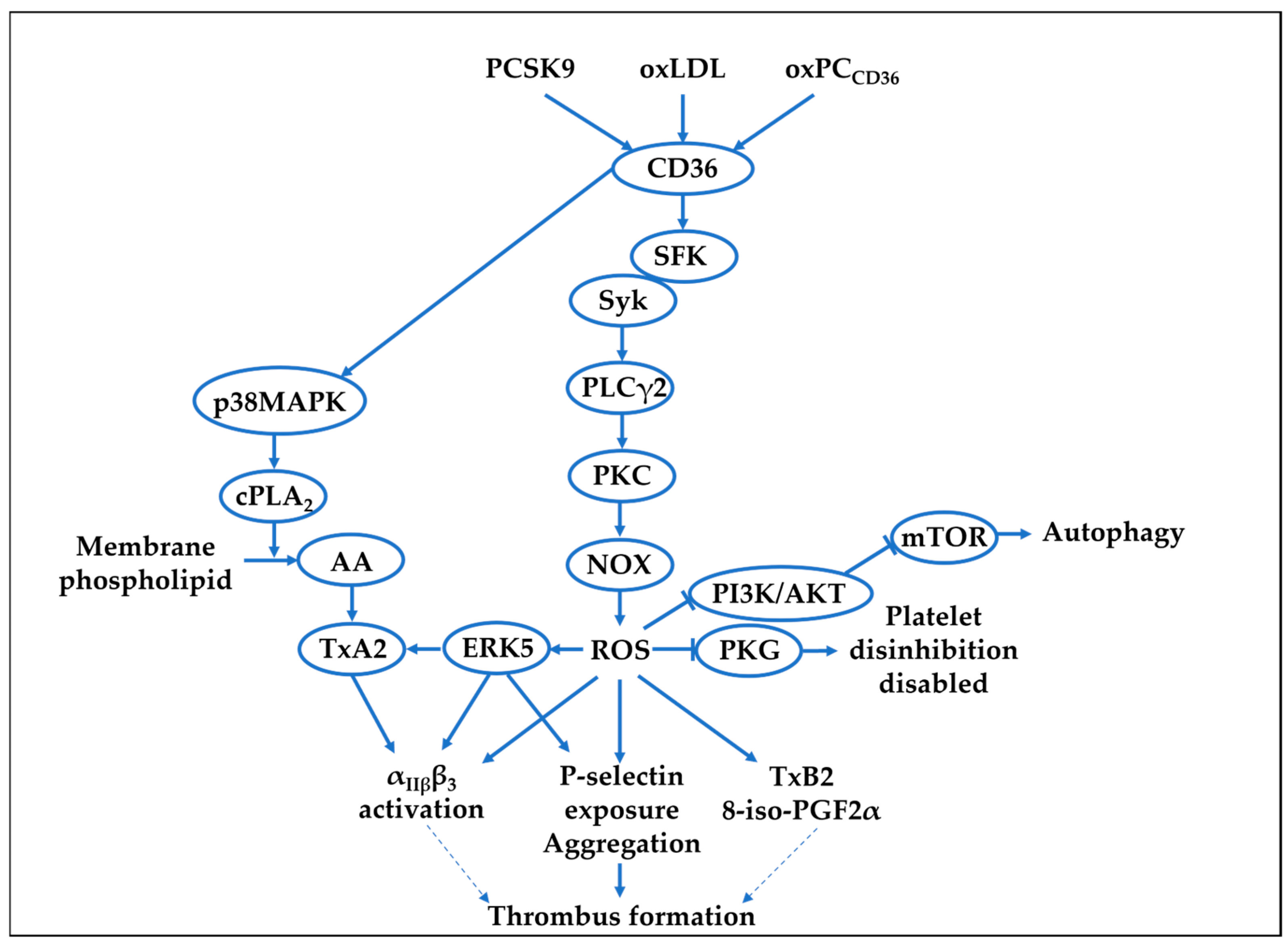
3. Platelet Reactive Oxygen Species and Hyperlipidaemia

The inflammatory milieu associated with atherothrombosis shifts platelet towards a maladaptive phenotype characterised by hyperactivity, and this is associated with a systemic pro-thrombotic phenotype and increased ROS [83,84]. Patients with T2D and coronary artery disease exhibit increased platelet reactivity, and have increased prospective risk for coronary events and death [85]. Hyperlipidaemia, a key driver of atherothrombosis, causes a change in platelet biology in which aberrant ROS production plays a critical role. The appearance of pathological ligands in the blood, such as oxidised lipids, acting as endogenous damage associated molecular patterns (DAMPs), interact with pattern recognition scavenger receptors including CD36, scavenger receptor A1, and lectin-like oxLDL receptor-1 (LOX-1), and promote unregulated platelet activity exacerbated by ROS.

3.1. oxLDL and Platelet ROS

Hyperlipidaemia, which is characterised by significantly elevated plasma lipids, including oxidised low-density lipoprotein (oxLDL), a circulating DAMP known to activate human and murine platelets [86,87,88], increases platelet–endothelial interactions [89]. Platelets recognise oxLDL via the cell surface receptors CD36 and LOX-1, which lead to ROS generation. Table 2 and Figure 1 summarise the mechanisms of platelet ROS generation in response to hyperlipidaemia and its downstream platelet functions.
OxLDL propagates platelet aggregation and enhances platelet-dependent thrombosis, and these effects have been shown to be diminished by peptides targeting oxLDL receptors CD36 or LOX1, acting through NOX2 inhibition [90]. Our work, and that of others, has dissected the downstream signalling mechanisms of oxLDL-CD36 ligation leading to ROS production (Figure 1). A report by Assinger et al., identified a role for oxLDL-CD36 in the induction of ROS production alongside calcium flux, platelet activation, and expression of CD40L, and this activation was shown to be sensitive to the non-specific NOX inhibitor apocynin, suggesting ROS production may be via NOX1, NOX2, or both [95]. OxLDL treatment of platelets also leads to increased autophagy, a highly conserved pathway mediated by lysosomes that degrades cytosolic components, by modulating the PI3K/AKT/mTOR signalling pathway (Figure 1), which was reversed by the ROS scavenger NAC, suggesting that ROS acts as a critical signalling node in this pathway [91]. Our group has identified NOX2 as the primary ROS-generating system in platelets linked to hyperlipidaemia, which influences both activatory and inhibitory pathways. Ligation of CD36 by oxLDL led to PLCγ2-mediated ROS production (Figure 1). This ROS production was required for P-selectin expression, which was ablated by genetic deletion of CD36 or PLCγ2, inhibition of NOX2, but not NOX1, and the ROS scavenger NAC [92]. As NOX1 has been previously implicated in thrombin-mediated signalling, and NOX2 in collagen-mediated signalling, the observation that NOX2 is critical to oxLDL-ROS is supported, as collagen-GPVI signals via a tyrosine phosphorylation cascade, which shares many similarities with the signalling of oxLDL-CD36.
In addition to driving platelet activation, there is strong evidence that the CD36-NOX2 pathway can influence procoagulant function in platelets. OxLDL causes the production of catalase-sensitive H2O2, which in turn activates the MAP kinase ERK5, which leads to platelet activation and increased PS exposure required for the binding of coagulation factors [93]. Furthermore, our group found that NOX2-dependent ROS generation is driven by oxLDL-CD36-PKC signalling (Figure 1), and this promotes platelet activation through inhibition of the inhibitory NO-cGMP signalling pathway, preventing downstream activation of the protein kinase G (PKG) substrate vasodilator-stimulated phosphoprotein (VASP) [18]. Phosphorylation of VASP by PKG at serine 239 has been demonstrated to inhibit cytoskeletal rearrangement and prevent platelet integrin activation [96]. Platelets from hyperlipidaemic apolipoproteinE-deficient (ApoE−/−) mice have reduced sensitivity to cGMP when tested ex vivo [18]. When these mice were infused with the NOX2-inhibitor peptide gp91 ds-tat for 4 weeks, platelet hyposensitivity to cGMP was corrected, showing that platelet NOX2 may be a target for controlling platelet hyperactivity under hyperlipidaemic conditions (Table 2). Overall, current data indicate that ROS downstream of oxLDL-CD36 drives platelet degranulation and procoagulant function and disinhibits key inhibitory pathways (Figure 1). The kinetics of ROS production by oxLDL sustained over 3 h (as longest time point tested) [92] are in stark difference to those induced by haemostatic agonist such as a collagen and thrombin, which peak rapidly and then wane. It is attractive to speculate that as oxLDL concentration increases in the circulation, it drives maladaptive platelet functions through chronic and low-grade ROS production, eventually overcoming intrinsic antioxidant capacity leading to a persistent hyperactive platelet phenotype.

3.2. PCSK9 and Platelet ROS

Another DAMP, proprotein convertase subtilisin/kexin type 9 (PCSK9), a serine protease mainly synthesised by the liver [97], is also elevated in hyperlipidaemia, and it has been shown to have direct effects on platelet ROS production. There is evidence showing that plasma PCSK9 levels are associated with future risk of CVD events [98]. In healthy volunteers, PCSK9 binds to CD36 in platelets, activates Src, ERK5, and JNK, enhancing ROS production and further activating the p38MAPK/cPLA2/COX-1 signalling downstream of CD36 [94]. Aspirin, which is a COX1 inhibitor and attenuates ROS-enhanced platelet activation [99], abolishes the effects of PCSK9 on platelet activation and in vivo thrombosis [94]. Similar effects were found in atrial fibrillation patients where the investigators observed that PCSK9 levels over 1.2 ng/mL exhibit higher H2O2 production, urinary 8-iso-PGF2α biosynthesis, and serum sNOX2-dp, compared to those lesser than 1.2 ng/mL PCSK9 [100]. This study also reported that PCSK9 forms an immune complex with CD36 and initiates the subsequent cascade, including ROS generation by NOX2 activation (Figure 1).

3.3. Mitochondrial ROS and Hyperlipidaemia

As previously mentioned, mitochondrial ROS play a key role in many aspects of cell signalling [101]; however, its role in platelets activated by oxLDL has not yet been fully explored. Chatterjee et al. have demonstrated that oxLDL triggers intracellular rise in ROS and intraplatelet mitochondrial O2production [102]. Interestingly, both oxidative conversion and intraplatelet lipid peroxidation were significantly reduced by MnTMPyP, a cell-permeable SOD2-mimetic O2scavenger, suggesting that mitochondrial O2could drive lipid peroxidation in platelets. OxLDL-induced αIIbβ3 integrin activation and P-selectin expression were also shown to be inhibited by MnTMPyP. Furthermore, thrombogenic PS exposure and mitochondrial membrane depolarisation were observed, corroborating the mitochondrial O2 generation observations. Other than platelets, endothelial cells treated with oxLDL show an increase in mitochondrial O2, and a functional shift in mitochondrial phenotype towards impaired enzyme activity in mitochondrial respiratory chain complexes [103]. Studies of oxLDL-treated macrophages in vitro and hyperlipidaemic diet-induced macrophages in vivo identified a significant shift towards an increased rate of oxidative phosphorylation and elevated mitochondrial ROS, which is dependent on CD36 [104]. There is also evidence linking LPS-stimulated LOX1-mediated ROS production to mitochondrial function in macrophages [105]; therefore, it is possible to hypothesise that oxLDL-LOX1 interactions may lead to similar outcomes in platelets. Whether platelets also show a shift in mitochondrial function and bioenergetics in the context of hyperlipidaemia is yet to be explored; however, as the techniques required for these studies are now established in the field of platelet biology [36], these research questions should be pursued. In summary, mitochondrial dysfunction underpinned by excess O2 and hyperpolarisation may drive changes in platelet function in various diseases, which are typically associated with a hyperactive platelet phenotype.

4. Conclusions and Future Perspectives

There is an ever-expanding field of research into the role of disease-mediated ROS in platelet hyperactivity, which is underpinned by molecular studies identifying both specific activators of ROS production and molecular generators of ROS, followed by pre-clinical translational studies. While multiple mechanisms have been suggested to mediate the dysfunction of platelet ROS in diseases, what remains consistently clear is that the failure of ROS homeostasis within various diseases is central to platelet hyperactivity. In many of these cases, targeting the activators that drive ROS production is unfeasible, and in such situations the development and application of ROS scavengers may provide an alternative approach, which can blunt platelet hyperactivity without losing platelet function. For instance, the antioxidant NAC, a synthetic derivative of L-cysteine, is typically safe and well tolerated at high doses in a range of CVD clinical trials [106,107,108]. Clinical studies have shown the effectiveness of NAC in improving cardiovascular functions, including reduced infarct size [107] and preserved left ventricular function in MI patients [106,108], yet its effect in platelet functions was underexamined. In vitro, NAC inhibits platelet function, aggregation, adhesion to collagen matrix, ROS generation, and intracellular calcium mobilisation [109]. In addition, the low bioavailability of NAC [110,111], due to poor membrane permeability [112], could potentially limit its clinical potential. N-acetylcysteine amide (AD4/NACA) with improved lipophilicity and hydrophobicity as well as thioredoxin mimetic peptides offer anti-aggregatory activity induced by collagen [113]. Therefore, these peptides, which display higher redox potency over NAC, could be tested in clinical settings as potential antioxidant approaches for treatment of CVD against platelet hyperactivity.

Author Contributions

L.T.C. writing—original draft preparation, review, and editing; M.S.H. writing—original draft preparation, review, and editing; J.S.K. writing—review and editing; C.D. writing—review and editing; A.J.U. writing—review and editing; K.M.N. writing—review and editing, and funding acquisition. All authors have read and agreed to the published version of the manuscript.

Funding

This research was funded by British Heart Foundation, grant number RG/16/5/32250 and RG/F/22/110067.

Data Availability Statement

Not applicable.

Conflicts of Interest

The authors declare no conflicts of interest.

References

  1. Schwarz, U.R.; Walter, U.; Eigenthaler, M. Taming platelets with cyclic nucleotides. Biochem. Pharmacol. 2001, 62, 1153–1161. [Google Scholar] [CrossRef] [PubMed]
  2. Radomski, M.W.; Palmer, R.M.; Moncada, S. An L-arginine/nitric oxide pathway present in human platelets regulates aggregation. Proc. Natl. Acad. Sci. USA 1990, 87, 5193–5197. [Google Scholar] [CrossRef] [PubMed]
  3. Stalker, T.J.; Newman, D.K.; Ma, P.; Wannemacher, K.M.; Brass, L.F. Platelet signaling. In Handbook of Experimental Pharmacology; Springer: Berlin/Heidelberg, Germany, 2012; pp. 59–85. [Google Scholar] [CrossRef]
  4. Gibbins, J.M. Platelet adhesion signalling and the regulation of thrombus formation. J. Cell Sci. 2004, 117, 3415–3425. [Google Scholar] [CrossRef]
  5. André, P.; Nannizzi-Alaimo, L.; Prasad, S.K.; Phillips, D.R. Platelet-derived CD40L: The switch-hitting player of cardiovascular disease. Circulation 2002, 106, 896–899. [Google Scholar] [CrossRef] [PubMed]
  6. Bergmeier, W.; Hynes, R.O. Extracellular matrix proteins in hemostasis and thrombosis. Cold Spring Harb. Perspect. Biol. 2012, 4, a005132. [Google Scholar] [CrossRef]
  7. Stalker, T.J.; Traxler, E.A.; Wu, J.; Wannemacher, K.M.; Cermignano, S.L.; Voronov, R.; Diamond, S.L.; Brass, L.F. Hierarchical organization in the hemostatic response and its relationship to the platelet-signaling network. Blood 2013, 121, 1875–1885. [Google Scholar] [CrossRef]
  8. Heemskerk, J.W.M.; Mattheij, N.J.A.; Cosemans, J. Platelet-based coagulation: Different populations, different functions. J. Thromb. Haemost. 2013, 11, 2–16. [Google Scholar] [CrossRef]
  9. Marcus, A.J.; Silk, S.T.; Safier, L.B.; Ullman, H.L. Superoxide production and reducing activity in human platelets. J. Clin. Investig. 1977, 59, 149–158. [Google Scholar] [CrossRef]
  10. Iuliano, L.; Colavita, A.R.; Leo, R.; Praticò, D.; Violi, F. Oxygen free radicals and platelet activation. Free Radic. Biol. Med. 1997, 22, 999–1006. [Google Scholar] [CrossRef]
  11. Liao, R.; Wang, L.; Zeng, J.; Tang, X.; Huang, M.; Kantawong, F.; Huang, Q.; Mei, Q.; Huang, F.; Yang, Y.; et al. Reactive oxygen species: Orchestrating the delicate dance of platelet life and death. Redox Biol. 2025, 80, 103489. [Google Scholar] [CrossRef]
  12. Chlopicki, S.; Olszanecki, R.; Janiszewski, M.; Laurindo, F.R.; Panz, T.; Miedzobrodzki, J. Functional role of NADPH oxidase in activation of platelets. Antioxid. Redox Signal. 2004, 6, 691–698. [Google Scholar] [CrossRef] [PubMed]
  13. Zhang, Y.; Wong, H.S. Are mitochondria the main contributor of reactive oxygen species in cells? J. Exp. Biol. 2021, 224 Pt 5, jeb221606. [Google Scholar] [CrossRef]
  14. Rowley, J.W.; Oler, A.J.; Tolley, N.D.; Hunter, B.N.; Low, E.N.; Nix, D.A.; Yost, C.C.; Zimmerman, G.A.; Weyrich, A.S. Genome-wide RNA-seq analysis of human and mouse platelet transcriptomes. Blood 2011, 118, e101–e111. [Google Scholar] [CrossRef] [PubMed]
  15. Bokoch, G.M.; Diebold, B.A. Current molecular models for NADPH oxidase regulation by Rac GTPase. Blood 2002, 100, 2692–2696. [Google Scholar] [CrossRef]
  16. Streeter, J.; Schickling, B.M.; Jiang, S.; Stanic, B.; Thiel, W.H.; Gakhar, L.; Houtman, J.C.; Miller, F.J., Jr. Phosphorylation of Nox1 regulates association with NoxA1 activation domain. Circ. Res. 2014, 115, 911–918. [Google Scholar] [CrossRef]
  17. Vara, D.; Mailer, R.K.; Tarafdar, A.; Wolska, N.; Heestermans, M.; Konrath, S.; Spaeth, M.; Renné, T.; Schröder, K.; Pula, G. NADPH Oxidases Are Required for Full Platelet Activation In Vitro and Thrombosis In Vivo but Dispensable for Plasma Coagulation and Hemostasis. Arterioscler. Thromb. Vasc. Biol. 2020, 41, Atvbaha120315565. [Google Scholar] [CrossRef] [PubMed]
  18. Magwenzi, S.; Woodward, C.; Wraith, K.S.; Aburima, A.; Raslan, Z.; Jones, H.; McNeil, C.; Wheatcroft, S.; Yuldasheva, N.; Febbriao, M.; et al. Oxidized LDL activates blood platelets through CD36/NOX2-mediated inhibition of the cGMP/protein kinase G signaling cascade. Blood 2015, 125, 2693–2703. [Google Scholar] [CrossRef]
  19. Carnevale, R.; Loffredo, L.; Nocella, C.; Bartimoccia, S.; Sanguigni, V.; Soresina, A.; Plebani, A.; Azzari, C.; Martire, B.; Pignata, C.; et al. Impaired platelet activation in patients with hereditary deficiency of p47(phox). Br. J. Haematol. 2018, 180, 454–456. [Google Scholar] [CrossRef]
  20. Gaspar, R.S.; Ferreira, P.M.; Mitchell, J.L.; Pula, G.; Gibbins, J.M. Platelet-derived extracellular vesicles express NADPH oxidase-1 (Nox-1), generate superoxide and modulate platelet function. Free Radic. Biol. Med. 2021, 165, 395–400. [Google Scholar] [CrossRef]
  21. Delaney, M.K.; Kim, K.; Estevez, B.; Xu, Z.; Stojanovic-Terpo, A.; Shen, B.; Ushio-Fukai, M.; Cho, J.; Du, X. Differential Roles of the NADPH-Oxidase 1 and 2 in Platelet Activation and Thrombosis. Arterioscler. Thromb. Vasc. Biol. 2016, 36, 846–854. [Google Scholar] [CrossRef]
  22. Walsh, T.G.; Berndt, M.C.; Carrim, N.; Cowman, J.; Kenny, D.; Metharom, P. The role of Nox1 and Nox2 in GPVI-dependent platelet activation and thrombus formation. Redox Biol. 2014, 2, 178–186. [Google Scholar] [CrossRef] [PubMed]
  23. Vara, D.; Tarafdar, A.; Celikag, M.; Patinha, D.; Gulacsy, C.E.; Hounslea, E.; Warren, Z.; Ferreira, B.; Koeners, M.P.; Caggiano, L.; et al. NADPH oxidase 1 is a novel pharmacological target for the development of an antiplatelet drug without bleeding side effects. FASEB J. 2020, 34, 13959–13977. [Google Scholar] [CrossRef] [PubMed]
  24. Korbecki, J.; Baranowska-Bosiacka, I.; Gutowska, I.; Chlubek, D. The effect of reactive oxygen species on the synthesis of prostanoids from arachidonic acid. J. Physiol. Pharmacol. 2013, 64, 409–421. [Google Scholar] [PubMed]
  25. Burkhart, J.M.; Vaudel, M.; Gambaryan, S.; Radau, S.; Walter, U.; Martens, L.; Geiger, J.; Sickmann, A.; Zahedi, R.P. The first comprehensive and quantitative analysis of human platelet protein composition allows the comparative analysis of structural and functional pathways. Blood 2012, 120, e73–e82. [Google Scholar] [CrossRef]
  26. Morel, A.; Miller, E.; Bijak, M.; Saluk, J. The increased level of COX-dependent arachidonic acid metabolism in blood platelets from secondary progressive multiple sclerosis patients. Mol. Cell. Biochem. 2016, 420, 85–94. [Google Scholar] [CrossRef]
  27. Pignatelli, P.; Sanguigni, V.; Lenti, L.; Ferro, D.; Finocchi, A.; Rossi, P.; Violi, F. gp91phox-dependent expression of platelet CD40 ligand. Circulation 2004, 110, 1326–1329. [Google Scholar] [CrossRef]
  28. Harrison, R. Structure and function of xanthine oxidoreductase: Where are we now? Free Radic. Biol. Med. 2002, 33, 774–797. [Google Scholar] [CrossRef]
  29. Kuwano, K.; Ikeda, H.; Oda, T.; Nakayama, H.; Koga, Y.; Toshima, H.; Imaizumi, T. Xanthine oxidase mediates cyclic flow variations in a canine model of coronary arterial thrombosis. Am. J. Physiol.-Heart Circ. Physiol. 1996, 270, H1993–H1999. [Google Scholar] [CrossRef]
  30. Cecerska-Heryć, E.; Jesionowska, A.; Klaudyna, S.; Katarzyna, S.; Dominika, M.; Dominika, P.; Marta, U.; Dołęgowska, B. Xanthine oxidoreductase reference values in platelet-poor plasma and platelets in healthy volunteers. Oxid. Med. Cell. Longev. 2015, 2015, 341926. [Google Scholar] [CrossRef]
  31. Pandey, N.R.; Kaur, G.; Chandra, M.; Sanwal, G.G.; Misra, M.K. Enzymatic oxidant and antioxidants of human blood platelets in unstable angina and myocardial infarction. Int. J. Cardiol. 2000, 76, 33–38. [Google Scholar] [CrossRef]
  32. Palma, F.R.; Gantner, B.N.; Sakiyama, M.J.; Kayzuka, C.; Shukla, S.; Lacchini, R.; Cunniff, B.; Bonini, M.G. ROS production by mitochondria: Function or dysfunction? Oncogene 2024, 43, 295–303. [Google Scholar] [CrossRef]
  33. Schulz, E.; Wenzel, P.; Münzel, T.; Daiber, A. Mitochondrial redox signaling: Interaction of mitochondrial reactive oxygen species with other sources of oxidative stress. Antioxid. Redox Signal. 2014, 20, 308–324. [Google Scholar] [CrossRef]
  34. Vanden Hoek, T.L.; Becker, L.B.; Shao, Z.H.; Li, C.Q.; Schumacker, P.T. Reactive oxygen species released from mitochondria during brief hypoxia induce preconditioning in cardiomyocytes. J. Biol. Chem. 1998, 273, 18092–18098. [Google Scholar] [CrossRef]
  35. Zorov, D.B.; Juhaszova, M.; Sollott, S.J. Mitochondrial reactive oxygen species (ROS) and ROS-induced ROS release. Physiol. Rev. 2014, 94, 909–950. [Google Scholar] [CrossRef]
  36. Aibibula, M.; Naseem, K.M.; Sturmey, R.G. Glucose metabolism and metabolic flexibility in blood platelets. J. Thromb. Haemost. 2018, 16, 2300–2314. [Google Scholar] [CrossRef]
  37. Lopez, J.J.; Salido, G.M.; Gómez-Arteta, E.; Rosado, J.A.; Pariente, J.A. Thrombin induces apoptotic events through the generation of reactive oxygen species in human platelets. J. Thromb. Haemost. 2007, 5, 1283–1291. [Google Scholar] [CrossRef]
  38. Girish, K.S.; Paul, M.; Thushara, R.M.; Hemshekhar, M.; Shanmuga Sundaram, M.; Rangappa, K.S.; Kemparaju, K. Melatonin elevates apoptosis in human platelets via ROS mediated mitochondrial damage. Biochem. Biophys. Res. Commun. 2013, 438, 198–204. [Google Scholar] [CrossRef]
  39. Leytin, V.; Gyulkhandanyan, A.V.; Freedman, J. Role of mitochondrial membrane permeabilization and depolarization in platelet apoptosis. Br. J. Haematol. 2018, 181, 281–285. [Google Scholar] [CrossRef]
  40. Cardenes, N.; Corey, C.; Geary, L.; Jain, S.; Zharikov, S.; Barge, S.; Novelli, E.M.; Shiva, S. Platelet bioenergetic screen in sickle cell patients reveals mitochondrial complex V inhibition, which contributes to platelet activation. Blood 2014, 123, 2864–2872. [Google Scholar] [CrossRef]
  41. Yamagishi, S.; Edelstein, D.; Du, X.L.; Brownlee, M. Hyperglycemia potentiates collagen-induced platelet activation through mitochondrial superoxide overproduction. Diabetes 2001, 50, 1491–1494. [Google Scholar] [CrossRef] [PubMed]
  42. Méndez, D.; Arauna, D.; Fuentes, F.; Araya-Maturana, R.; Palomo, I.; Alarcón, M.; Sebastián, D.; Zorzano, A.; Fuentes, E. Mitoquinone (MitoQ) Inhibits Platelet Activation Steps by Reducing ROS Levels. Int. J. Mol. Sci. 2020, 21, 6192. [Google Scholar] [CrossRef]
  43. McCord, J.M.; Fridovich, I. Superoxide dismutase. An enzymic function for erythrocuprein (hemocuprein). J. Biol. Chem. 1969, 244, 6049–6055. [Google Scholar] [CrossRef]
  44. Weisiger, R.A.; Fridovich, I. Superoxide dismutase. Organelle specificity. J. Biol. Chem. 1973, 248, 3582–3592. [Google Scholar] [CrossRef]
  45. Freedman, J.E. Oxidative stress and platelets. Arterioscler. Thromb. Vasc. Biol. 2008, 28, s11–s16. [Google Scholar] [CrossRef]
  46. Krötz, F.; Sohn, H.Y.; Gloe, T.; Zahler, S.; Riexinger, T.; Schiele, T.M.; Becker, B.F.; Theisen, K.; Klauss, V.; Pohl, U. NAD(P)H oxidase-dependent platelet superoxide anion release increases platelet recruitment. Blood 2002, 100, 917–924. [Google Scholar] [CrossRef]
  47. Dayal, S.; Gu, S.X.; Hutchins, R.D.; Wilson, K.M.; Wang, Y.; Fu, X.; Lentz, S.R. Deficiency of superoxide dismutase impairs protein C activation and enhances susceptibility to experimental thrombosis. Arterioscler. Thromb. Vasc. Biol. 2015, 35, 1798–1804. [Google Scholar] [CrossRef]
  48. Fidler, T.P.; Rowley, J.W.; Araujo, C.; Boudreau, L.H.; Marti, A.; Souvenir, R.; Dale, K.; Boilard, E.; Weyrich, A.S.; Abel, E.D. Superoxide Dismutase 2 is dispensable for platelet function. Thromb. Haemost. 2017, 117, 1859–1867. [Google Scholar] [CrossRef]
  49. Sonkar, V.K.; Eustes, A.S.; Ahmed, A.; Jensen, M.; Solanki, M.V.; Swamy, J.; Kumar, R.; Fidler, T.P.; Houtman, J.C.D.; Allen, B.G.; et al. Endogenous SOD2 (Superoxide Dismutase) Regulates Platelet-Dependent Thrombin Generation and Thrombosis During Aging. Arterioscler. Thromb. Vasc. Biol. 2023, 43, 79–91. [Google Scholar] [CrossRef]
  50. Manasa, K.; Vani, R. Influence of Oxidative Stress on Stored Platelets. Adv. Hematol. 2016, 2016, 4091461. [Google Scholar] [CrossRef]
  51. Pignatelli, P.; Pulcinelli, F.M.; Lenti, L.; Gazzaniga, P.P.; Violi, F. Hydrogen peroxide is involved in collagen-induced platelet activation. Blood 1998, 91, 484–490. [Google Scholar] [CrossRef] [PubMed]
  52. Praticó, D.; Pasin, M.; Barry, O.P.; Ghiselli, A.; Sabatino, G.; Iuliano, L.; FitzGerald, G.A.; Violi, F. Iron-dependent human platelet activation and hydroxyl radical formation: Involvement of protein kinase C. Circulation 1999, 99, 3118–3124. [Google Scholar] [CrossRef]
  53. Bryant, R.W.; Simon, T.C.; Bailey, J.M. Role of glutathione peroxidase and hexose monophosphate shunt in the platelet lipoxygenase pathway. J. Biol. Chem. 1982, 257, 14937–14943. [Google Scholar] [CrossRef]
  54. Lubos, E.; Loscalzo, J.; Handy, D.E. Glutathione peroxidase-1 in health and disease: From molecular mechanisms to therapeutic opportunities. Antioxid. Redox Signal. 2011, 15, 1957–1997. [Google Scholar] [CrossRef]
  55. Coulon, L.; Calzada, C.; Moulin, P.; Véricel, E.; Lagarde, M. Activation of p38 mitogen-activated protein kinase/cytosolic phospholipase A2 cascade in hydroperoxide-stressed platelets. Free Radic. Biol. Med. 2003, 35, 616–625. [Google Scholar] [CrossRef] [PubMed]
  56. Calzada, C.; Véricel, E.; Mitel, B.; Coulon, L.; Lagarde, M. 12(S)-Hydroperoxy-eicosatetraenoic acid increases arachidonic acid availability in collagen-primed platelets. J. Lipid Res. 2001, 42, 1467–1473. [Google Scholar] [CrossRef]
  57. Freedman, J.E.; Loscalzo, J.; Benoit, S.E.; Valeri, C.R.; Barnard, M.R.; Michelson, A.D. Decreased platelet inhibition by nitric oxide in two brothers with a history of arterial thrombosis. J. Clin. Investig. 1996, 97, 979–987. [Google Scholar] [CrossRef]
  58. Jin, R.C.; Mahoney, C.E.; Coleman Anderson, L.; Ottaviano, F.; Croce, K.; Leopold, J.A.; Zhang, Y.Y.; Tang, S.S.; Handy, D.E.; Loscalzo, J. Glutathione peroxidase-3 deficiency promotes platelet-dependent thrombosis in vivo. Circulation 2011, 123, 1963–1973. [Google Scholar] [CrossRef]
  59. Freedman, J.E.; Frei, B.; Welch, G.N.; Loscalzo, J. Glutathione peroxidase potentiates the inhibition of platelet function by S-nitrosothiols. J. Clin. Investig. 1995, 96, 394–400. [Google Scholar] [CrossRef]
  60. Becatti, M.; Fiorillo, C.; Gori, A.M.; Marcucci, R.; Paniccia, R.; Giusti, B.; Violi, F.; Pignatelli, P.; Gensini, G.F.; Abbate, R. Platelet and leukocyte ROS production and lipoperoxidation are associated with high platelet reactivity in Non-ST elevation myocardial infarction (NSTEMI) patients on dual antiplatelet treatment. Atherosclerosis 2013, 231, 392–400. [Google Scholar] [CrossRef]
  61. Marwali, M.R.; Hu, C.P.; Mohandas, B.; Dandapat, A.; Deonikar, P.; Chen, J.; Cawich, I.; Sawamura, T.; Kavdia, M.; Mehta, J.L. Modulation of ADP-induced platelet activation by aspirin and pravastatin: Role of lectin-like oxidized low-density lipoprotein receptor-1, nitric oxide, oxidative stress, and inside-out integrin signaling. J. Pharmacol. Exp. Ther. 2007, 322, 1324–1332. [Google Scholar] [CrossRef]
  62. Bakdash, N.; Williams, M.S. Spatially distinct production of reactive oxygen species regulates platelet activation. Free Radic. Biol. Med. 2008, 45, 158–166. [Google Scholar] [CrossRef] [PubMed]
  63. Arthur, J.F.; Qiao, J.; Shen, Y.; Davis, A.K.; Dunne, E.; Berndt, M.C.; Gardiner, E.E.; Andrews, R.K. ITAM receptor-mediated generation of reactive oxygen species in human platelets occurs via Syk-dependent and Syk-independent pathways. J. Thromb. Haemost. 2012, 10, 1133–1141. [Google Scholar] [CrossRef]
  64. Arthur, J.F.; Shen, Y.; Gardiner, E.E.; Coleman, L.; Murphy, D.; Kenny, D.; Andrews, R.K.; Berndt, M.C. TNF receptor-associated factor 4 (TRAF4) is a novel binding partner of glycoprotein Ib and glycoprotein VI in human platelets. J. Thromb. Haemost. 2011, 9, 163–172. [Google Scholar] [CrossRef] [PubMed]
  65. Carrim, N.; Arthur, J.F.; Hamilton, J.R.; Gardiner, E.E.; Andrews, R.K.; Moran, N.; Berndt, M.C.; Metharom, P. Thrombin-induced reactive oxygen species generation in platelets: A novel role for protease-activated receptor 4 and GPIbα. Redox Biol. 2015, 6, 640–647. [Google Scholar] [CrossRef]
  66. Wang, L.; Soe, N.N.; Sowden, M.; Xu, Y.; Modjeski, K.; Baskaran, P.; Kim, Y.; Smolock, E.M.; Morrell, C.N.; Berk, B.C. Cyclophilin A is an important mediator of platelet function by regulating integrin αIIbβ3 bidirectional signalling. Thromb. Haemost. 2014, 111, 873–882. [Google Scholar] [CrossRef] [PubMed]
  67. Soe, N.N.; Sowden, M.; Baskaran, P.; Smolock, E.M.; Kim, Y.; Nigro, P.; Berk, B.C. Cyclophilin A is required for angiotensin II-induced p47phox translocation to caveolae in vascular smooth muscle cells. Arterioscler. Thromb. Vasc. Biol. 2013, 33, 2147–2153. [Google Scholar] [CrossRef]
  68. Salmeen, A.; Barford, D. Functions and mechanisms of redox regulation of cysteine-based phosphatases. Antioxid. Redox Signal. 2005, 7, 560–577. [Google Scholar] [CrossRef]
  69. Nieswandt, B.; Watson, S.P. Platelet-collagen interaction: Is GPVI the central receptor? Blood 2003, 102, 449–461. [Google Scholar] [CrossRef]
  70. Watson, S.P.; Auger, J.M.; McCarty, O.J.; Pearce, A.C. GPVI and integrin alphaIIb beta3 signaling in platelets. J. Thromb. Haemost. 2005, 3, 1752–1762. [Google Scholar] [CrossRef]
  71. Jang, J.Y.; Min, J.H.; Chae, Y.H.; Baek, J.Y.; Wang, S.B.; Park, S.J.; Oh, G.T.; Lee, S.H.; Ho, Y.S.; Chang, T.S. Reactive oxygen species play a critical role in collagen-induced platelet activation via SHP-2 oxidation. Antioxid. Redox Signal. 2014, 20, 2528–2540. [Google Scholar] [CrossRef]
  72. Winterbourn, C.C. Revisiting the reactions of superoxide with glutathione and other thiols. Arch. Biochem. Biophys. 2016, 595, 68–71. [Google Scholar] [CrossRef] [PubMed]
  73. van Gorp, R.M.; van Dam-Mieras, M.C.; Hornstra, G.; Heemskerk, J.W. Effect of membrane-permeable sulfhydryl reagents and depletion of glutathione on calcium mobilisation in human platelets. Biochem. Pharmacol. 1997, 53, 1533–1542. [Google Scholar] [CrossRef]
  74. Essex, D.W.; Li, M. Redox control of platelet aggregation. Biochemistry 2003, 42, 129–136. [Google Scholar] [CrossRef] [PubMed]
  75. Ball, C.; Vijayan, K.V.; Nguyen, T.; Anthony, K.; Bray, P.F.; Essex, D.W.; Dong, J.F. Glutathione regulates integrin alpha(IIb)beta(3)-mediated cell adhesion under flow conditions. Thromb. Haemost. 2008, 100, 857–863. [Google Scholar] [CrossRef]
  76. Yan, B.; Smith, J.W. Mechanism of integrin activation by disulfide bond reduction. Biochemistry 2001, 40, 8861–8867. [Google Scholar] [CrossRef] [PubMed]
  77. Véricel, E.; Januel, C.; Carreras, M.; Moulin, P.; Lagarde, M. Diabetic patients without vascular complications display enhanced basal platelet activation and decreased antioxidant status. Diabetes 2004, 53, 1046–1051. [Google Scholar] [CrossRef]
  78. Guidi, G.; Schiavon, R.; Sheiban, I.; Perona, G. Platelet glutathione peroxidase activity is impaired in patients with coronary heart disease. Scand. J. Clin. Lab. Investig. 1986, 46, 549–551. [Google Scholar] [CrossRef]
  79. Kamhieh-Milz, J.; Bal, G.; Sterzer, V.; Kamhieh-Milz, S.; Arbach, O.; Salama, A. Reduced antioxidant capacities in platelets from patients with autoimmune thrombocytopenia purpura (ITP). Platelets 2012, 23, 184–194. [Google Scholar] [CrossRef]
  80. Wang, B.; Yee Aw, T.; Stokes, K.Y. N-acetylcysteine attenuates systemic platelet activation and cerebral vessel thrombosis in diabetes. Redox Biol. 2018, 14, 218–228. [Google Scholar] [CrossRef]
  81. De Bona, K.S.; Bellé, L.P.; Sari, M.H.; Thomé, G.; Schetinger, M.R.; Morsch, V.M.; Boligon, A.; Athayde, M.L.; Pigatto, A.S.; Moretto, M.B. Syzygium cumini extract decrease adenosine deaminase, 5′nucleotidase activities and oxidative damage in platelets of diabetic patients. Cell. Physiol. Biochem. 2010, 26, 729–738. [Google Scholar] [CrossRef]
  82. Jain, K.; Tyagi, T.; Patell, K.; Xie, Y.; Kadado, A.J.; Lee, S.H.; Yarovinsky, T.; Du, J.; Hwang, J.; Martin, K.A.; et al. Age associated non-linear regulation of redox homeostasis in the anucleate platelet: Implications for CVD risk patients. EBioMedicine 2019, 44, 28–40. [Google Scholar] [CrossRef]
  83. Barrachina, M.N.; Hermida-Nogueira, L.; Moran, L.A.; Casas, V.; Hicks, S.M.; Sueiro, A.M.; Di, Y.; Andrews, R.K.; Watson, S.P.; Gardiner, E.E.; et al. Phosphoproteomic Analysis of Platelets in Severe Obesity Uncovers Platelet Reactivity and Signaling Pathways Alterations. Arterioscler. Thromb. Vasc. Biol. 2021, 41, 478–490. [Google Scholar] [CrossRef]
  84. Barrachina, M.N.; Sueiro, A.M.; Izquierdo, I.; Hermida-Nogueira, L.; Guitián, E.; Casanueva, F.F.; Farndale, R.W.; Moroi, M.; Jung, S.M.; Pardo, M.; et al. GPVI surface expression and signalling pathway activation are increased in platelets from obese patients: Elucidating potential anti-atherothrombotic targets in obesity. Atherosclerosis 2019, 281, 62–70. [Google Scholar] [CrossRef] [PubMed]
  85. Angiolillo, D.J.; Bernardo, E.; Sabaté, M.; Jimenez-Quevedo, P.; Costa, M.A.; Palazuelos, J.; Hernández-Antolin, R.; Moreno, R.; Escaned, J.; Alfonso, F.; et al. Impact of platelet reactivity on cardiovascular outcomes in patients with type 2 diabetes mellitus and coronary artery disease. J. Am. Coll. Cardiol. 2007, 50, 1541–1547. [Google Scholar] [CrossRef]
  86. Naseem, K.M.; Goodall, A.H.; Bruckdorfer, K.R. Differential effects of native and oxidatively modified low-density lipoproteins on platelet function. Platelets 1997, 8, 163–173. [Google Scholar] [CrossRef] [PubMed]
  87. Wraith, K.S.; Magwenzi, S.; Aburima, A.; Wen, Y.C.; Leake, D.; Naseem, K.M. Oxidized low-density lipoproteins induce rapid platelet activation and shape change through tyrosine kinase and Rho kinase-signaling pathways. Blood 2013, 122, 580–589. [Google Scholar] [CrossRef] [PubMed]
  88. Chen, K.; Febbraio, M.; Li, W.; Silverstein, R.L. A specific CD36-dependent signaling pathway is required for platelet activation by oxidized low-density lipoprotein. Circ. Res. 2008, 102, 1512–1519. [Google Scholar] [CrossRef]
  89. Chan, H.C.; Ke, L.Y.; Chu, C.S.; Lee, A.S.; Shen, M.Y.; Cruz, M.A.; Hsu, J.F.; Cheng, K.H.; Chan, H.C.B.; Lu, J.; et al. Highly electronegative LDL from patients with ST-elevation myocardial infarction triggers platelet activation and aggregation. Blood 2013, 122, 3632–3641. [Google Scholar] [CrossRef]
  90. Carnevale, R.; Bartimoccia, S.; Nocella, C.; Di Santo, S.; Loffredo, L.; Illuminati, G.; Lombardi, E.; Boz, V.; Del Ben, M.; De Marco, L.; et al. LDL oxidation by platelets propagates platelet activation via an oxidative stress-mediated mechanism. Atherosclerosis 2014, 237, 108–116. [Google Scholar] [CrossRef]
  91. Wang, X.; Fu, Y.F.; Liu, X.; Feng, G.; Xiong, D.; Mu, G.F.; Chen, F.P. ROS Promote Ox-LDL-Induced Platelet Activation by Up-Regulating Autophagy Through the Inhibition of the PI3K/AKT/mTOR Pathway. Cell. Physiol. Biochem. 2018, 50, 1779–1793. [Google Scholar] [CrossRef]
  92. Berger, M.; Wraith, K.; Woodward, C.; Aburima, A.; Raslan, Z.; Hindle, M.S.; Moellmann, J.; Febbraio, M.; Naseem, K.M. Dyslipidemia-associated atherogenic oxidized lipids induce platelet hyperactivity through phospholipase C gamma 2-dependent reactive oxygen species generation. Platelets 2019, 30, 467–472. [Google Scholar] [CrossRef] [PubMed]
  93. Yang, M.; Cooley, B.C.; Li, W.; Chen, Y.L.; Vasquez-Vivar, J.; Scoggins, N.O.; Cameron, S.J.; Morrell, C.N.; Silverstein, R.L. Platelet CD36 promotes thrombosis by activating redox sensor ERK5 in hyperlipidemic conditions. Blood 2017, 129, 2917–2927. [Google Scholar] [CrossRef]
  94. Qi, Z.; Hu, L.; Zhang, J.; Yang, W.; Liu, X.; Jia, D.; Yao, Z.; Chang, L.; Pan, G.; Zhong, H.; et al. PCSK9 (Proprotein Convertase Subtilisin/Kexin 9) Enhances Platelet Activation, Thrombosis, and Myocardial Infarct Expansion by Binding to Platelet CD36. Circulation 2021, 143, 45–61. [Google Scholar] [CrossRef]
  95. Assinger, A.; Koller, F.; Schmid, W.; Zellner, M.; Koller, E.; Volf, I. Hypochlorite-oxidized LDL induces intraplatelet ROS formation and surface exposure of CD40L-A prominent role of CD36. Atherosclerosis 2010, 213, 129–134. [Google Scholar] [CrossRef]
  96. Krause, M.; Dent, E.W.; Bear, J.E.; Loureiro, J.J.; Gertler, F.B. Ena/VASP proteins: Regulators of the actin cytoskeleton and cell migration. Annu. Rev. Cell Dev. Biol. 2003, 19, 541–564. [Google Scholar] [CrossRef] [PubMed]
  97. Urban, D.; Pöss, J.; Böhm, M.; Laufs, U. Targeting the proprotein convertase subtilisin/kexin type 9 for the treatment of dyslipidemia and atherosclerosis. J. Am. Coll. Cardiol. 2013, 62, 1401–1408. [Google Scholar] [CrossRef]
  98. Leander, K.; Mälarstig, A.; Van’t Hooft, F.M.; Hyde, C.; Hellénius, M.L.; Troutt, J.S.; Konrad, R.J.; Öhrvik, J.; Hamsten, A.; de Faire, U. Circulating Proprotein Convertase Subtilisin/Kexin Type 9 (PCSK9) Predicts Future Risk of Cardiovascular Events Independently of Established Risk Factors. Circulation 2016, 133, 1230–1239. [Google Scholar] [CrossRef]
  99. Shi, P.; Zhang, L.; Zhang, M.; Yang, W.; Wang, K.; Zhang, J.; Otsu, K.; Huang, G.; Fan, X.; Liu, J. Platelet-Specific p38α Deficiency Improved Cardiac Function After Myocardial Infarction in Mice. Arterioscler. Thromb. Vasc. Biol. 2017, 37, e185–e196. [Google Scholar] [CrossRef]
  100. Cammisotto, V.; Pastori, D.; Nocella, C.; Bartimoccia, S.; Castellani, V.; Marchese, C.; Scavalli, A.S.; Ettorre, E.; Viceconte, N.; Violi, F.; et al. PCSK9 Regulates Nox2-Mediated Platelet Activation via CD36 Receptor in Patients with Atrial Fibrillation. Antioxidants 2020, 9, 296. [Google Scholar] [CrossRef]
  101. Zharikov, S.; Shiva, S. Platelet mitochondrial function: From regulation of thrombosis to biomarker of disease. Biochem. Soc. Trans. 2013, 41, 118–123. [Google Scholar] [CrossRef]
  102. Chatterjee, M.; Rath, D.; Schlotterbeck, J.; Rheinlaender, J.; Walker-Allgaier, B.; Alnaggar, N.; Zdanyte, M.; Müller, I.; Borst, O.; Geisler, T.; et al. Regulation of oxidized platelet lipidome: Implications for coronary artery disease. Eur. Heart J. 2017, 38, 1993–2005. [Google Scholar] [CrossRef] [PubMed]
  103. Chowdhury, S.K.R.; Sangle, G.V.; Xie, X.P.; Stelmack, G.L.; Halayko, A.J.; Shen, G.X. Effects of extensively oxidized low-density lipoprotein on mitochondrial function and reactive oxygen species in porcine aortic endothelial cells. Am. J. Physiol.-Endocrinol. Metab. 2010, 298, E89–E98. [Google Scholar] [CrossRef] [PubMed]
  104. Chen, Y.L.; Yang, M.; Huang, W.X.; Chen, W.J.; Zhao, Y.Q.; Schulte, M.L.; Volberding, P.; Gerbec, Z.; Zimmermann, M.T.; Zeighami, A.; et al. Mitochondrial Metabolic Reprogramming by CD36 Signaling Drives Macrophage Inflammatory Responses. Circ. Res. 2019, 125, 1087–1102. [Google Scholar] [CrossRef]
  105. Ding, Z.F.; Liu, S.J.; Wang, X.W.; Dai, Y.; Khaidakov, M.; Deng, X.Y.; Fan, Y.B.; Xiang, D.; Mehta, J.L. LOX-1, mtDNA damage, and NLRP3 inflammasome activation in macrophages: Implications in atherogenesis. Cardiovasc. Res. 2014, 103, 619–628. [Google Scholar] [CrossRef]
  106. Yesilbursa, D.; Serdar, A.; Senturk, T.; Serdar, Z.; Sağ, S.; Cordan, J. Effect of N-acetylcysteine on oxidative stress and ventricular function in patients with myocardial infarction. Heart Vessel. 2006, 21, 33–37. [Google Scholar] [CrossRef]
  107. Pasupathy, S.; Tavella, R.; Grover, S.; Raman, B.; Procter, N.E.K.; Du, Y.T.; Mahadavan, G.; Stafford, I.; Heresztyn, T.; Holmes, A.; et al. Early Use of N-acetylcysteine With Nitrate Therapy in Patients Undergoing Primary Percutaneous Coronary Intervention for ST-Segment-Elevation Myocardial Infarction Reduces Myocardial Infarct Size (the NACIAM Trial [N-acetylcysteine in Acute Myocardial Infarction]). Circulation 2017, 136, 894–903. [Google Scholar] [CrossRef]
  108. Arstall, M.A.; Yang, J.; Stafford, I.; Betts, W.H.; Horowitz, J.D. N-acetylcysteine in combination with nitroglycerin and streptokinase for the treatment of evolving acute myocardial infarction. Safety and biochemical effects. Circulation 1995, 92, 2855–2862. [Google Scholar] [CrossRef] [PubMed]
  109. Eligini, S.; Porro, B.; Aldini, G.; Colli, S.; Banfi, C. N-Acetylcysteine Inhibits Platelet Function through the Regeneration of the Non-Oxidative Form of Albumin. Antioxidants 2022, 11, 445. [Google Scholar] [CrossRef]
  110. Borgström, L.; Kågedal, B.; Paulsen, O. Pharmacokinetics of N-acetylcysteine in man. Eur. J. Clin. Pharmacol. 1986, 31, 217–222. [Google Scholar] [CrossRef]
  111. Olsson, B.; Johansson, M.; Gabrielsson, J.; Bolme, P. Pharmacokinetics and bioavailability of reduced and oxidized N-acetylcysteine. Eur. J. Clin. Pharmacol. 1988, 34, 77–82. [Google Scholar] [CrossRef]
  112. Offen, D.; Gilgun-Sherki, Y.; Barhum, Y.; Benhar, M.; Grinberg, L.; Reich, R.; Melamed, E.; Atlas, D. A low molecular weight copper chelator crosses the blood-brain barrier and attenuates experimental autoimmune encephalomyelitis. J. Neurochem. 2004, 89, 1241–1251. [Google Scholar] [CrossRef] [PubMed]
  113. Eligini, S.; Munno, M.; Atlas, D.; Banfi, C. N-acetylcysteine Amide AD4/NACA and Thioredoxin Mimetic Peptides Inhibit Platelet Aggregation and Protect against Oxidative Stress. Antioxidants 2023, 12, 1395. [Google Scholar] [CrossRef] [PubMed]
Figure 1. Signalling mechanisms involved in hyperlipidaemic-induced ROS production 399 and the downstream effects [23,79,80,81,82,83]. 8-iso-PGF2α, 8-iso-prostaglandin F2α; AA, arachidonic acid; cPLA2, cytosolic phospholipase A2; ERK5, extracellular signal-regulated kinase 5; mTOR, mechanistic target of rapamycin; NOX, NADPH oxidase; oxLDL, oxidised low-density lipoprotein; oxPCCD36, oxidised phospholipid specific to CD36; p38MAPK, p38 mitogen-activated protein kinases; PCSK9, proprotein convertase subtilisin/kexin 9; PI3K/AKT, phosphoinositide-3-kinase/protein kinase B; PKC, protein kinase c; PKG, protein kinase G; PLCγ2, phospholipase C gamma 2; ROS, reactive oxygen species; SFK, Src family kinases; Syk, spleen-associated tyrosine kinase; TXA2, thromboxane A2; TXB2, thromboxane B2.
Cells 14 00500 g001
Table 1. Discrepancies in the roles of NOX downstream GPCR and GPVI signalling pathways.
AgonistsMice/
Inhibitor
ObservationsConclusionsRef.
CRP (0.5 µg/mL)

Thrombin (0.018 or 0.025 U/mL)
NOX1−/−ROS
Aggregation
ATP secretion
Ca2+ mobilisation
Thrombin ↓
CRP ↔
NOX1 is involved in GPCR-induced platelet activation [21]
NOX2−/−ROS
Aggregation
ATP secretion
Ca2+ mobilisation
Thrombin partial CRP ↓
NOX2 is involved in GPCR- and GPVI-induced platelet activation
NOX1−/− and NOX2−/− Carotid artery occlusion

Tail bleeding

NOX1−/−
NOX2−/−

NOX1−/−
NOX2−/−
NOX2 is involved in thrombosis

NOX1 and NOX2 are dispensable for haemostasis
Collagen (3 µg/mL)

Thrombin (0.1 U/mL)
NOX1−/− Aggregation
Thrombin ↔
Collagen ↓
NOX1 is involved in GPVI-induced platelet activation[23]
NOX2−/− Aggregation
Thrombin ↓
Collagen ↔
NOX2 is involved in GPCR-induced platelet activation
NOX1−/− and NOX2−/− Carotid artery occlusion

Tail bleeding
NOX1−/−
NOX2−/−

NOX1−/−
NOX2−/−
NOX1 is involved in thrombosis

NOX1 and NOX2 are dispensable for haemostasis
2-APT (NOX1 inhibitor)Superoxide anion


Aggregation


Static adhesion over collagen

Thrombus formation under flow

Carotid artery occlusion

Tail bleeding
Thrombin ↓
Collagen ↓

Thrombin marginally↓
Collagen ↓


NOX1 inhibition impairs GPVI-induced platelet activation without affecting GPCR responses
Collagen (10 µg/mL)

CRP (5 µg/mL)

Thrombin (0.25U or 1U/mL)
NOX1/2/4−/− Superoxide anion
Aggregation
αIIbβ3

P-selectin
PS exposure


Carotid artery occlusion

Tail bleeding
Thrombin ↓
Collagen
/CRP ↓

Thrombin ↔
CRP ↓


NOXs are critical for GPVI-induced platelet activation.
GPCR-associated integrin activation and platelet aggregation are NOX-dependent, whilst P-selectin and PS exposure are NOXs independent
[17]
NOX1−/− Aggregation


Thrombus formation over collagen

Thrombin ↔
Collagen ↓


NOX1 is involved in GPVI- and GPCR-induced platelet activation
NOX2−/− Aggregation


Thrombus formation over collagen
Thrombin ↓
Collagen ↔


NOX2 is involved in GPCR-induced platelet activation
NOX4−/− Aggregation

Thrombus formation over collagen
Thrombin ↔
Collagen ↔

NOX4 has negligible role in platelet regulation
↓, decreased upon agonist stimulation; ↔, unaffected; 2-APT, 2-Acetylphenothiazine; ATP, adenosine triphosphate; CRP, collagen-related protein; GPCR, G protein-coupled receptor; GPVI, glycoprotein VI; NOX, NADPH oxidase, PS, phosphatidylserine; ROS, reactive oxygen species.
Table 2. Effect of ROS in platelet hyperlipidaemia.
Hyperlipidaemic InsultPlatelet Function Downstream of ROS ProductionROS Related Inhibitors UsedPathway InvolvedRef.
oxLDLαIIbβ3 activation, TxB2 production, thrombosis under shear stress-Gp91ds-tatCD36/LOX1-p38MAPK/PKC-NOX2[90]
oxLDLaggregation, P-selectin, adhesion under shear stressNACPI3K-AKT-mTOR[91]
oxPCCD36P-selectinNAC
Gp91ds-tat
ML171
CD36-Src-PLCγ2-NOX2[92]
oxLDLaggregationVAS2870
DPTA-NONOate
PEG-catalase
CD36-Src-NOX-ERK5[93]
oxLDL
oxPCCD36
diminished sensitivity to inhibitory NO-cGMP signallingTEMPOL
MnTMPyP
Gp91ds-tat
CD36-Src-Syk-PLCγ2-PKC-NOX2[18]
PCSK9thrombin-induced platelet aggregationVAS2870CD36-Src-ERK5-JNK-ROS-p38MAPK/cPLA2/COX-1/TxA2[94]
AKT, protein kinase B; cGMP, cyclic guanosine monophosphate; COX-1, cyclooxygenase-1; cPLA2, cytosolic phospholipase A2; DPTA-NONOate, dipropylenetriamine diazeniumdiolate; ERK5, extracellular signal-regulated kinase 5; Gp91ds-tat, peptide inhibitor for NOX; JNK, c-Jun N-terminal kinases; LOX1, lectin-like oxLDL receptor-1; ML171, 2-acetylphenothiazine; MnTMPyP, superoxide dismutase mimetic; NAC, N-acetylcysteine; NO, nitric oxide; NOX, NADPH oxidase, oxLDL, oxidised low-density lipoprotein; oxPCCD36, oxidised phospholipid specific to CD36; p38MAPK, p38 mitogen-activated protein kinases; PCSK9, proprotein convertase subtilisin/kexin 9; PEG-catalase, polyethylene glycol-catalase; PI3K, phosphoinositide-3-kinase; PKC, protein kinase C; PLCγ2, phospholipase C gamma 2; mTOR, mechanistic target of rapamycin; ROS, reactive oxygen species; Src, proto-oncogene tyrosine-protein kinase; Syk, spleen-associated tyrosine kinase; TEMPOL, 4-hydroxy-2,2,6,6-tetramethylpiperidine-1-oxyl; TxA2, thromboxane A2; TxB2, thromboxane B2; VAS2870, NOX inhibitor.
Disclaimer/Publisher’s Note: The statements, opinions and data contained in all publications are solely those of the individual author(s) and contributor(s) and not of MDPI and/or the editor(s). MDPI and/or the editor(s) disclaim responsibility for any injury to people or property resulting from any ideas, methods, instructions or products referred to in the content.

