Review

Ayurvedic Phytochemicals in Oncology: ADP-Ribosylation as a Molecular Nexus

by
Gali Sri Venkata Sai Rishma Reddy
1,
Suman Kumar Nandy
2,
Pitchaiah Cherukuri
1,
Krishna Samanta
3,* and
Pulak Kar
1,*
1
Department of Biological Sciences, SRM University-AP, Amaravati 522240, India
2
BIRAC’s BioNEST Bio-Incubator (B3I) Facility, North-Eastern Hill University, Tura Campus, Tura 794002, India
3
Department of Biotechnology, Koneru Lakshmaiah Education Foundation, Vaddeswaram, Guntur 522302, India
*
Authors to whom correspondence should be addressed.
Cells 2025, 14(22), 1753; https://doi.org/10.3390/cells14221753
Submission received: 30 September 2025 / Revised: 30 October 2025 / Accepted: 4 November 2025 / Published: 10 November 2025
(This article belongs to the Section Cell Signaling)

Abstract

Cancer remains one of the most pressing health challenges of the 21st century, with rising global incidence underscoring the need for innovative therapeutic strategies. Despite significant advances in biotechnology, curative outcomes remain limited, prompting interest in integrative approaches. Ayurveda, the traditional Indian system of medicine, suggests a holistic therapeutic framework that is now gaining molecular validation in oncology. In this review, the literature was systematically collected and analyzed from major databases, including PubMed, Scopus, and Web of Science, encompassing studies across ethnopharmacology, biochemistry, and cancer biology. The analysis focused on Ayurvedic phytochemicals that modulate ADP-ribosylation (ADPr), a dynamic post-translational modification central to DNA repair, chromatin organization, and cellular stress responses, with particular emphasis on poly (ADP-ribose) polymerase (PARP)-mediated pathways and their oncological relevance. We have also explored the role of p53, a key stress-response regulator intricately linked to ADPr dynamics, which acts as a downstream effector integrating these molecular events with cell fate decisions. Evidence indicates that several Ayurvedic compounds, including curcumin, resveratrol, and withaferin A, influence PARP–p53 signaling networks, thereby modulating DNA repair fidelity, apoptosis, and tumor adaptation. The review further addresses challenges related to the poor solubility of these phytochemicals and highlights recent advances in Phyto-nanomedicine-based delivery systems that enhance their stability and therapeutic efficacy. Overall, the synthesis of Ayurvedic pharmacology with molecular oncology reveals mechanistic insights that may inform the rational development of novel, mechanism-driven cancer therapeutics.

1. Introduction

Cancer arises through a gradual accumulation of genetic and epigenetic alterations that disrupt normal cellular control, leading to malignant transformation and tumour heterogeneity across different cancer types [1,2]. Landmark discoveries such as oncogene activation—through viral mechanisms like the Rous sarcoma virus or via mutations in proto-oncogenes, have laid the foundation of modern cancer biology [3]. Although both viral and non-viral origins of cancer are well recognized, a unified mechanistic understanding of tumour progression remains elusive. Recent studies highlight the interplay among driver mutations, epigenetic regulators, signalling proteins, and metabolic pathways, alongside the critical influence of the tumour microenvironment (TME), immune system, and microbiome in shaping cancer initiation and therapy response [4]. Within this context, tumour metabolism, particularly involving the TME and infiltrating immune cells, underscores the dynamic adaptability of cancer and its resistance to therapy [4,5]. While complete prevention may be unrealistic, early detection remains key to improving survival outcomes [5]. Despite advances in targeted and immune-based therapies, challenges such as toxicity, financial burden, and reduced quality of life, especially from oral oncolytics—persist [6,7,8].
In response, there has been a growing global shift toward integrative cancer care that emphasizes early screening, preventive measures, supportive therapies, and palliative management. Complementary and alternative medicine (CAM) has gained increasing attention for its ability to enhance patient well-being and reduce treatment-associated complications [9,10]. However, treatment responses are often accompanied by challenges such as therapy resistance, tumour recurrence, and cancer stem cell regeneration, along with potential off-target effects on intracellular organelles, all of which collectively limit therapeutic efficacy [10,11]. Among the various CAM modalities, Ayurveda, the ancient Indian medical system, has emerged as a promising adjunctive approach, particularly for its role in strengthening immunity and supporting post-treatment recovery [4]. With a history of more than 5000 years and foundations in classical texts such as Charaka Samhita, Sushruta Samhita, and Ashtanga Hridaya, Ayurveda offers a holistic framework for health and disease management [4]. Contemporary studies have reported its potential to alleviate symptoms and improve the quality of life in cancer patients [12,13,14]. Nevertheless, issues such as lack of standardization, limited reproducibility, and inadequate clinical validation continue to hinder its broader integration into global oncology frameworks [15].
In recent years, advances in nanoscience and molecular medicine have opened new avenues to modernize Ayurvedic formulations, particularly through green nanotechnology, offering a scientific bridge between traditional wisdom and contemporary biomedical innovation [16,17]. For example, Nano Swarna Bhasma (NSB), a gold nanoparticle formulation developed through green nanotechnology, has shown promise in metastatic breast cancer by enabling targeted delivery of bioactive phytochemicals such as γ-terpinene, α-pinene, cuminaldehyde, quercetin, kaempferol, and apigenin [15,18,19]. These strategies improve bioavailability and reduce systemic toxicity, creating a foundation for more standardized and reproducible therapeutic platforms [15,18,20,21,22]. Ayurvedic cancer interventions include polyherbal formulations, dietary modifications, detoxification procedures, and lifestyle practices aimed at reducing inflammation and oxidative stress while strengthening immune function [4,15,23,24].
Earlier Ayurvedic studies often focused on single compounds like curcumin or withanolides, but the multifactorial nature of cancer demands multi-targeted approaches [25]. Ayurveda’s holistic perspective may provide an advantage by influencing complex cellular pathways such as ADPr [26,27]. This post-translational modification regulates DNA repair, chromatin remodelling, inflammation, and apoptosis, making it highly relevant to cancer progression and therapy resistance [26,27]. Although direct evidence connecting Ayurvedic formulations to ADPr is limited, herbs such as Withania somnifera and Curcuma longa, known for their antioxidant and anti-inflammatory properties [28,29], may affect upstream processes like oxidative stress and DNA damage response that converge with ADPr signaling [30,31]. Rasayana therapies, traditionally used to promote vitality and immune resilience, may also influence these mechanisms, offering a promising yet underexplored direction for integrative oncology [32]. Despite centuries of empirical use, strong clinical validation remains limited [4]. This review consolidates current evidence on the molecular basis of Ayurvedic interventions, highlighting their effects on inflammation, immune modulation, oxidative stress, tumor signaling, and microenvironment [15,18,20,21,22], and explores their potential role in biomarker-driven cancer diagnostics and therapeutics. It presents a comprehensive analysis of the emerging interplay between Ayurvedic phytomedicine and cellular ADPr, a post-translational modification mediated by PARPs, which are tightly coupled to the DNA damage response and implicated in various diseases, including cancer. By bridging traditional medicine with molecular oncology, this work underscores the untapped potential of Ayurvedic principles in elucidating cancer mechanisms, advancing preventive strategies, and shaping next-generation therapeutic paradigms. Ultimately, it envisions a future where ancient wisdom and molecular precision converge to redefine the model of personalized cancer care.

2. Selected Ayurvedic Phytochemicals in Cancer-Mechanistic Insights into Molecular Targets and ADP-Ribosylation

Ayurvedic plants and their bioactive constituents modulate key signaling pathways involved in cancer progression, including inflammation, apoptosis, cell cycle regulation, and angiogenesis. Elucidating their molecular targets offers valuable insights into their therapeutic potential in cancer management [33]. Several compounds illustrated in Figure 1 exhibit anticancer effects through direct interactions with specific molecular targets. While the established activities of Ayurvedic phytochemicals largely stem from their ability to regulate oxidative stress, inflammation, apoptotic signaling, and cell cycle checkpoints, emerging evidence suggests their possible involvement in ADPr signaling, particularly within the DNA damage response and programmed cell death pathways. ADPr, catalyzed by enzymes such as PARPs and mono (ADP-ribosyl) transferases, represents a crucial post-translational modification that governs DNA damage sensing and repair, chromatin remodeling, transcriptional regulation, and cell death mechanisms including apoptosis, necroptosis, and parthanatos [34,35].
Although the present study does not include extensive experimental validation directly linking PARP mediated ADPr with specific phytochemicals, the conceptual framework has been strengthened by integrating recent computational insights. Several investigations have demonstrated the mechanistic feasibility of phytochemical interactions with PARP enzymes through molecular docking and simulation-based analyses. A recent study applied 3D QSAR, docking, and molecular dynamics simulations to curcumin analogues, revealing strong binding affinity and modulation of PARP1 activity [36]. Another work employed a multistage virtual screening approach combining 3D pharmacophore modelling, docking, and molecular dynamics simulations to identify selective PARP1 inhibitors [37]. The development of CDK1 PARP1 dual inhibitors further highlights the translational relevance of Ayurvedic phytochemicals as promising anticancer scaffolds [38]. Computational studies have also examined PARP7 modulation and its association with androgen receptor signalling in prostate cancer [39,40]. Moreover, recent evidence suggests that machine learning approaches can enhance virtual screening accuracy for PARP1 inhibitors [41]. These studies provide molecular level evidence supporting phytochemical PARP ADP ribosylation crosstalk and underline the need for experimental validation within the Ayurvedic phytochemical context.
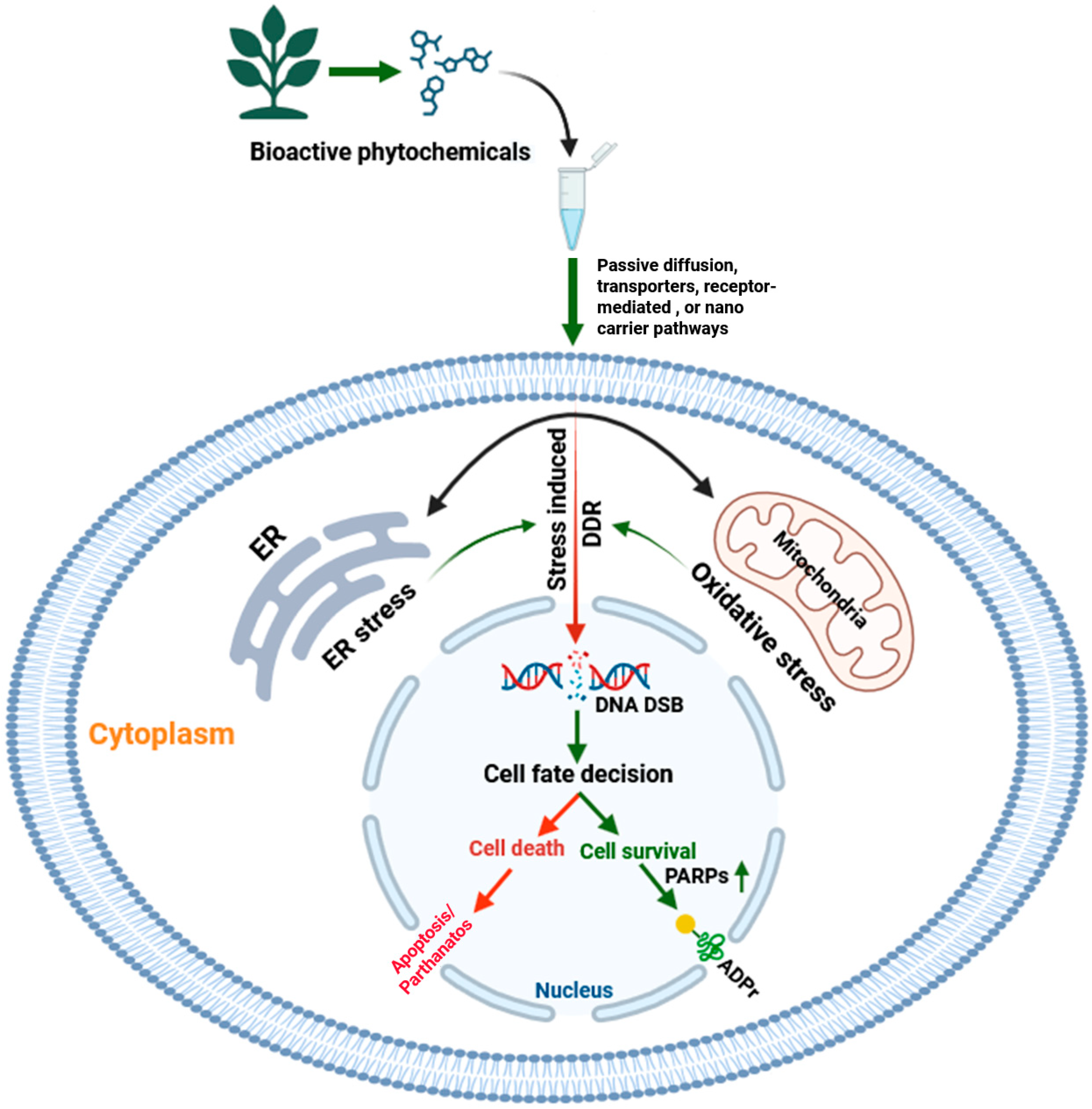
Direct experimental evidence linking Ayurvedic compounds to the modulation of PARPs or other ADP ribosyl transferases remains limited; however, their indirect cellular actions such as triggering DNA damage, influencing NAD+ metabolism, and promoting oxidative stress suggest a potential regulatory impact on ADPr signaling pathways. As illustrated in Figure 2, bioactive phytochemicals may enter cells through passive diffusion, transporter-mediated uptake, or receptor-triggered signaling. Once internalized, they can influence endoplasmic reticulum and mitochondrial oxidative stress, leading to activation of the DNA damage response (DDR), particularly DNA double strand breaks. In this context, PARP1 becomes activated and catalyzes ADPR to facilitate DNA repair and promote cell survival. However, excessive or prolonged activation can deplete cellular NAD+ and trigger programmed cell death.
This conceptual model suggests that Ayurvedic phytochemicals may fine-tune these processes, sensitizing damaged cells toward apoptosis or enhancing repair and survival mechanisms depending on the stress level. Such modulation of DDR and ADPr pathways could have significant therapeutic implications, especially when combined with PARP inhibitors in DNA repair deficient cancers such as BRCA mutated breast and ovarian tumors [42,43]. Further experimental validation of these interactions may provide new directions for integrating Ayurvedic phytochemicals into modern oncotherapy. This underexplored mechanistic interface underscores the potential of Ayurvedic phytochemicals as adjunctive agents in PARP-targeted therapies. The subsequent sections therefore provide an overview of key Ayurvedic plants, their active phytoconstituents, and the molecular mechanisms underlying their anticancer effects, with emphasis on emerging links to genome stability, oxidative stress regulation, and ADPr signaling pathways.
Quercetin (3,3′,4′,5,7-pentahydroxyflavone): Quercetin is a naturally occurring polyphenolic flavanol abundantly found in onions, grapes, berries, cherries, broccoli, and citrus fruits [44]. It is also widely distributed across plant species such as Santalum album, Mangifera indica, Emblica officinalis, Curcuma domestica, Withania somnifera, Foeniculum vulgare, and Cuscuta reflexa [45]. Exhibiting antioxidants, anti-inflammatory, antifungal, antiviral, anti-allergic, and anticancer activities, quercetin protects against oxidative stress, tissue injury, and drug-induced toxicities [44]. Mechanistically, by inhibiting PARP-mediated single-strand break repair, quercetin increases unrepaired lesions in homologous recombination-defective (BRCA2-deficient) cells, leading to double-strand break accumulation and selective cytotoxicity, supporting its role as a small-molecule modulator of ADPr-linked DNA repair pathways [46]. It broadly modulates oncogenic signalling by inhibiting PI3K/AKT/mTOR and MAPK/ERK pathways, elevating Bax/Bcl-2 ratios, activating caspase-3/-9 and cytochrome c release, and reducing matrix metalloproteinase expression [47]. Although direct evidence linking quercetin to PARP family proteins remains limited, its induction of DNA damage and redox perturbation likely enhances PARP/ADPr activity, while inhibition of survival signalling may impair repair efficiency. From an Ayurvedic and nanomedicine standpoint, its poor bioavailability has led to nano-formulation strategies offering translational potential in ADPr-modulated oncology. Moreover, quercetin modulates ADPr signalling through Sirtuin 6 (SIRT6), a nuclear NAD+-dependent deacetylase and mono-ADP-ribosyl transferase that regulates DNA repair and chromatin remodeling via ADPr of PARP1 (Lys521) and BAF170 (Lys312), thereby promoting double-strand break repair and NRF2-dependent transcription [48,49]. Quercetin regulates SIRT6 activity in a dose dependent manner, acting as an inhibitor at low concentrations and an activator at higher doses, as supported by mass spectrometry, molecular docking, and molecular dynamics analyses [49,50]. This dual modulatory behaviour underscores its potential to fine-tune ADPr signalling controlling genomic stability, metabolism, and tumour suppression. Although direct experimental evidence of quercetin-mediated PARP modulation remains scarce, future studies dissecting its influence on PARP/SIRT crosstalk in the DNA damage response may reveal crucial insights into its anticancer potential.
Rosmarinic acid (RA) [Rosmarinus officinalis L. (Lamiaceae)] is a natural polyphenolic ester of caffeic acid and 3,4-dihydroxyphenyllactic acid, first isolated from Rosmarinus officinalis (rosemary) [51,52]. Abundant in Lamiaceae herbs like rosemary, lemon balm, mint, and sage, RA exhibits antioxidant, anti-inflammatory, antimicrobial, and anticancer activities [51]. Its anticancer effects are linked to suppression of neoplastic transformation and bone metastasis in breast cancer via downregulation of receptor activator of NF-kappa B ligand (RANKL) and interleukin-8 (IL-8) [51,53], reversal of multidrug resistance by targeting multidrug resistance by downregulating MDR1 and P-glycoprotein (P-gp) in gastric cancer [51], and modulation of MAPK/ERK and ERK/PKA pathways in several cancer models [51,53]. Notably, Su et al. (2017) reported weak but measurable inhibition of PARP-1 (~20% at 100 µM) by RA, suggesting a potential role in ADPr-linked DNA repair, especially under BRCA2-deficient conditions [54]. Further investigation into RA’s impact on cellular ADPr may support its development as an anticancer agent. However, its clinical use remains limited due to poor bioavailability, which may be improved through nanotechnology-based delivery strategies [55].
Turmeric (Curcuma longa): It has long been a cornerstone of Ayurvedic medicine, valued particularly for its potent anti-inflammatory and antioxidant properties. Its major bioactive constituents-curcuminoids, including curcumin, demethoxycurcumin, and bisdemethoxycurcumin-have been extensively studied for their therapeutic potential in cancer [56]. Curcumin, the most prominent curcuminoid, exerts broad-spectrum anticancer effects by suppressing multiple cellular processes involved in tumour initiation and progression, such as cellular transformation, uncontrolled proliferation, invasion, angiogenesis, and metastasis. These effects are largely mediated through the inhibition of TNF-α-induced NF-κB activation and subsequent downregulation of NF-κB-dependent genes implicated in cell survival, anti-apoptotic signalling, and metastatic behaviour [56,57]. In addition to these well-characterized mechanisms, curcumin has also been reported to induce oxidative stress and DNA damage in cancer cells, accompanied by the downregulation of critical DNA repair proteins such as BRCA1 and RAD51. This disruption of homologous recombination repair potentially sensitizes tumour cells to PARP inhibitors, providing a rationale for combinatorial strategies in cancers with compromised DNA repair capacity [43,54]. Furthermore, preclinical studies have demonstrated that curcumin can enhance the cytotoxic effects of PARP inhibitors, suggesting a functional connection to ADPr signalling, a key regulatory pathway in DNA repair and cell death [43].
Kalmegh (Andrographis paniculata): It is a well-known Ayurvedic herb with documented anticancer properties. Its principal bioactive compound, andrographolide, exhibits strong anti-inflammatory, antioxidant, and immunomodulatory activities, contributing to its ability to inhibit cancer cell proliferation, induce apoptosis, and suppress tumour progression in breast, prostate, lung, and colorectal cancers [58]. Kalmegh extracts have also been shown to enhance conventional therapies and reduce chemotherapy-associated toxicity [58]. Emerging evidence indicates that andrographolide induces DNA damage and oxidative stress [59], both of which activate PARP1, a key enzyme in ADPr signalling and DNA repair [34]. Though direct effects on ADP-ribosyl transferases are yet to be clarified, the modulation of upstream pathways such as PI3K/AKT suggests a potential influence on ADPr-mediated cell death and repair mechanisms [60].
Ashwagandha (Withania somnifera): The plant Withania somnifera, commonly known as Ashwagandha or Indian ginseng, holds a prominent place in Ayurvedic medicine for its anti-inflammatory and antitumor properties. Its bioactive constituents, particularly withanolides derived from roots and leaves, exhibit strong anticancer effects by suppressing NF-κB activation, inhibiting cell proliferation, enhancing apoptosis, and reducing invasion in various cancer models [61]. Among these, withaferin A has shown notable antitumor activity through the inhibition of Notch-1 signalling and downregulation of pro-survival pathways such as Akt/NF-κB/Bcl-2, highlighting its therapeutic potential [62]. In addition to these effects, withaferin A has been reported to induce endoplasmic reticulum (ER) stress, activate caspases, and downregulate survival signals like NF-κB-mechanisms that intersect with PARP1 activation and ADPr-dependent cell death pathways [63,64]. Furthermore, withaferin A can cause DNA fragmentation and oxidative damage, potentially leading to PARP1 overactivation and NAD+ depletion-hallmarks of parthanatos, a regulated necrotic cell death driven by excessive ADPr [35].
Giloy (Tinospora cordifolia): It is an herbaceous vine native to the Indian subcontinent, is widely revered in Ayurvedic medicine for its broad therapeutic potential, including its role in cancer management [65,66]. Giloy is believed to exert anticancer effects through its immunomodulatory, antioxidant, and anti-inflammatory properties. Studies have demonstrated that extracts from Giloy can inhibit the proliferation of cancer cells and induce apoptosis in various cancer types, including breast, colon, and liver cancers [65]. Moreover, Giloy has been shown to enhance the efficacy of chemotherapy and radiotherapy, while reducing their toxicity, highlighting its value as a complementary therapy in oncology [65,66]. Recent findings also suggest that oxidative stress modulation by Giloy may intersect with ADPr signalling pathways. Since ROS-induced DNA damage is a known activator of PARP1, key to initiating ADPr-mediated DNA repair and cell death mechanisms, the antioxidant and pro-apoptotic effects of Giloy raise the possibility of indirect regulation of this pathway [34]. Although direct evidence linking T. cordifolia constituents to PARP or ADP-ribosyl transferase activity is currently lacking, its influence on upstream events such as DNA damage response and immune signalling suggests potential crosstalk with ADPr-dependent processes, warranting further investigation.
Neem (Azadirachta indica): It is commonly known as neem, has long been used in traditional medicine for its broad therapeutic potential. Phytochemicals isolated from neem leaves, such as azadirachtin and nimbolide, exhibit significant anticancer properties. Azadirachtin has been shown to act as an anti-tumor agent by inhibiting NF-κB, a central regulator of cancer cell survival and progression [67]. Nimbolide, a limonoid found in neem leaves and flowers, has demonstrated potent effects against prostate cancer, primarily through targeting androgen receptor signalling, generating reactive oxygen species (ROS), and inducing cell cycle arrest and DNA damage [68]. These actions suggest a potential interface with PARP-mediated DNA damage responses, as ROS and DNA damage are upstream activators of ADPr signalling. While the direct modulation of PARP enzymes by nimbolide has yet to be fully elucidated, its ability to induce genotoxic stress supports a plausible link to ADPr-dependent cancer cell death mechanisms.
Amalaki (Phyllanthus emblica): It is also known as Indian gooseberry or Amla, has gained attention for its potential anticancer properties. Research suggests that compounds found in Amalaki-such as polyphenols, flavonoids, and tannins-exhibit antioxidant and anti-inflammatory effects that may contribute to its anticancer activity [33,69]. Additionally, Amalaki extracts have shown cytotoxic effects on cancer cells, inhibiting their growth and proliferation, and are believed to enhance the immune system, which can aid in combating cancer [33,69]. Although direct evidence is limited, Amalaki may influence ADPr-associated pathways via its effects on oxidative stress, DNA damage, and NAD+ metabolism.
Tulsi (Ocimum sanctum): It demonstrates notable anticancer potential primarily through its antioxidant properties. Ursolic acid, a key bioactive compound in tulsi, inhibits NF-κB activation induced by various carcinogens, thereby downregulating genes such as cyclin D1, COX-2, and MMP-9 involved in cancer progression [33,70]. Additionally, it suppresses STAT3 activation, which is crucial for cancer cell survival and proliferation, and promotes apoptosis and cell cycle arrest, thereby enhancing the effects of other anticancer agents [70,71]. Tulsi’s ability to modulate redox balance, inflammation, and transcriptional responses suggests a possible indirect influence on ADPr-dependent pathways relevant to cancer and immunity [71,72]. The dualistic nature of its action-either suppressive or promotive-warrants further investigation to clarify its role in cancer biology [73]. Overall, its broad pharmacological profile makes tulsi a promising candidate for cancer prevention and therapy [33,70].
Punarnava (Boerhaavia diffusa): It is commonly known as Punarnava, has long been valued in traditional medicine for its wide range of therapeutic properties [74,75]. Recent studies have highlighted its anti-cancer potential. The aqueous methanolic extract of B. diffusa significantly reduced metastasis formation in B16F-10 melanoma models, while punarnavine, an alkaloid isolated from the plant, enhanced immune responses against metastatic progression in mice [33,76]. In addition, Punarnava contains bioactive phytochemicals such as boeravinone B and punarnavine, which exhibit strong antioxidant and anti-inflammatory activities [77]. Given that oxidative stress and DNA damage are key triggers for PARP activation, these compounds may indirectly modulate ADPr by mitigating ROS levels. Furthermore, B. diffusa has shown genoprotective effects, including a reduction in cyclophosphamide-induced chromosomal aberrations, suggesting a possible role in DNA repair processes—though direct evidence linking it to ADPr remains to be established [78].
Vasicinone (Adhatoda vasica): Vasicinone, derived from Adhatoda vasica Nees, shows bronchodilator activity and potential anti-proliferative effects on cancer cells. Research on A549 lung carcinoma cells demonstrated reduced viability, DNA fragmentation, LDH leakage, disrupted mitochondrial potential, and impaired wound healing upon vasicinone treatment [79]. Annexin V/PI staining suggested apoptosis induction via compromised membrane integrity. Mechanistically, vasicinone downregulated Bcl-2 and Fas death receptor while upregulating PARP, BAD, and cytochrome c, implicating Fas death receptor and Bcl-2 signalling in apoptosis [79]. Moreover, vasicinone exhibited antioxidant properties, reducing ROS levels and scavenging free radicals, suggesting its potential as a therapeutic agent against oxidative stress-induced lung cancer [33,79]. Understanding the specific PARP involved in the apoptosis of A549 lung carcinoma cells in response to vasicinone treatment remains unexplored. The involvement of PARP-induced cellular ADPr by vasicinone represent a promising avenue for Ayurvedic research. Exploring into this field can shed light on the connection between Ayurvedic components and cancer, particularly lung cancer. Investigating how vasicinone influences PARP activity and subsequent ADPr processes may unravel novel therapeutic strategies rooted in Ayurveda for combating lung cancer. This exploration holds significant potential for advancing our comprehension of the molecular structures underlying the anti-cancer properties of Ayurvedic compounds like vasicinone (Figure 2) [79].

3. Phytochemical-Induced Poly-ADP-Ribosylation in Carcinogenesis: Insights from Ayurvedic Thymus Species

Poly-ADPr (PARylation), a post-translational modification catalysed primarily by PARPs, plays a pivotal role in cellular processes such as DNA repair, chromatin remodelling, transcriptional regulation, and programmed cell death [1,2]. Dysregulation of PARylation is associated with genomic instability, impaired DNA damage response, and aberrant gene expression, contributing to carcinogenesis and tumour progression [1,2,80]. Although extensive evidence from molecular and animal model studies implicates PARPs in cancer development, their precise role in human malignancies remains incompletely defined [80].
While direct links between Ayurvedic phytochemicals and ADPr signalling remain limited, several indirect mechanistic cues suggest a plausible intersection. ADPr is central to cellular stress responses, encompassing DNA damage signalling, apoptosis, and immune regulation, which are hallmarks of cancer initiation and progression [34,35]. Certain phytochemicals derived from Ayurvedic herbs, such as curcumin (Curcuma longa), withaferin A (Withania somnifera), and nimbolide (Azadirachta indica), are known to influence upstream regulators of PARP activation, including oxidative stress, redox homeostasis, and NF-κB signalling [42,63,68]. For instance, curcumin has been shown to sensitize cancer cells to PARP inhibitors by enhancing oxidative DNA damage and impairing homologous recombination repair [43]. Withaferin A similarly induces ER stress and apoptosis, events that converge on PARP1 activation during cell death [64]. These observations suggest that phytochemicals may modulate ADPr signalling directly through PARP inhibition or indirectly via NAD+ metabolism and stress-response pathways (Figure 2). However, experimental validation is needed to elucidate these interactions and assess the therapeutic potential of combining Ayurvedic agents with PARP inhibitors in DNA repair-deficient cancers. It has been reported that few phytochemicals directly inhibit PARP enzymes, notable examples include epigallocatechin-3-gallate (EGCG), which inhibits PARP16 (IC50 ≈ 14.5 µM), and resveratrol, which suppresses PARP1 activity at micro- to sub-micromolar levels. Flavonoids such as quercetin modulate NAD+ metabolism and DNA damage response (DDR) pathways, sensitizing cells to PARP inhibition, though quantitative enzymatic data remain scarce [81]. These gaps highlight the need for systematic enzymatic and structure–activity studies to validate phytochemical–PARP interactions and assess their therapeutic potential in DNA repair-deficient cancers.
Investigations into PARP gene expression and structure have also uncovered important oncogenic associations. Knockout and transgenic models for PARP-1, PARP-2, and PARG have deepened understanding of PARylation’s role in tumour biology [80]. PARP-1 is crucial for base excision repair, chromosomal stability, and epigenetic regulation. Its dysregulation may lead to cell death via NAD+ depletion, potentially selecting for PARP-1-deficient clones with a growth advantage during tumour progression [82]. Elevated PARP-1 expression and activity have been reported in Ewing’s sarcoma cell lines, linked to ETS-1 activation [83]. Conversely, weak PARP-1 expression is associated with heightened genomic instability in breast cancer, often coinciding with gene amplification at chromosome 1q41–42 [84]. Additionally, reduced poly (ADP-ribose) synthesis in lymphocytes from bleomycin-treated laryngeal cancer patients suggests that low PARP activity may elevate cancer risk [85,86]. In gastric cancer cells, structural alterations in the PARP-1 gene have been observed, although their functional consequences remain unclear. Emerging evidence also implicates PARP-2 and PARP-3 in carcinogenesis, with PARP-3 located at chromosome 3p21.1-3p21.31-a region frequently affected by loss of heterozygosity in early-stage lung cancer [87].
Ayurvedic medicine has recently attracted attention for its potential to modulate ADPr pathways [88]. Dutta et al. [26] reviewed the anticancer potential of Withania somnifera (Ashwagandha), emphasizing its efficacy in breast, colon, prostate, ovarian, lung, and brain cancers. Withaferin A (WFA), its bioactive component, demonstrated cytotoxicity in preclinical models and was well-tolerated in patients. Synergistic antitumor effects were reported when WFA was combined with Sorafenib in papillary and anaplastic thyroid cancers, resulting in PARP and caspase-3 cleavage, downregulation of BRAF/Raf-1, and HSP inhibition [89]. Curcumin also exhibits strong modulatory effects on DNA damage response (DDR) pathways, including homologous recombination, non-homologous end joining (NHEJ), and G2/M checkpoint regulation. Ogiwara et al. [27] showed that curcumin targets CBP/p300 HATs and ATR kinase, sensitizing DDR-competent cells to PARP inhibitors and promoting mitotic catastrophe. Despite promising findings, the precise molecular mechanisms underlying the action of traditional Ayurvedic herbs-such as turmeric, ashwagandha, amalaki, giloy, and kalmegh-on ADPr signalling remain largely undefined [90]. Few studies have directly investigated the link between Ayurvedic herbs and PARP-mediated ADPr [26,27], highlighting a critical gap. Future studies focusing on cellular ADPr profiling in response to Ayurvedic extracts could unveil novel mechanistic insights into cancer modulation. This could open new avenues for rationally designed phytochemical-based therapies targeting ADPr signalling in malignancies.

4. Phytochemicals in p53 Signalling and ADP-Ribosylation Pathways

p53 functions as a master regulator of cellular stress responses and is closely intertwined with ADPr signalling. PARP1 and related PARP family enzymes directly ADP-ribosylate p53, thereby modulating its stability, transcriptional activity, and control over cell cycle arrest and apoptosis [91]. While this review primarily focuses on phytochemicals associated with PARP- and ADPr-mediated cancer regulation, p53 emerges as a key downstream effector integrating these pathways with cell fate decisions. Notably, Ayurvedic phytochemicals such as andrographolide, curcumin, resveratrol, and withaferin A have been reported to influence p53 signalling—often through DNA damage and oxidative stress responses that indirectly engage PARP1 [92,93]. p53 is both functionally and biochemically linked to PARP1/ADPr, and these phytochemicals can influence PARP1 activity or p53-mediated pathways [93]. For example, resveratrol inhibits PARP1 activity and interferes with its nucleosome binding and catalytic functions at micromolar concentrations [94]. A low-dose combination of withaferin A and caffeic acid phenethyl ester activates p53 and produces effects similar to PARP1 inhibition, promoting growth arrest and apoptosis in cancer models [95]. Curcumin has been shown to induce oxidative stress and DNA damage, upregulate p53, and interact with DNA damage response pathways, modulating PARP-related responses and sensitizing cells to PARP inhibitors. Notably, curcumin-induced apoptosis in colon cancer cells can occur independently of p53 status, highlighting its potential for treating tumours resistant to conventional therapies due to p53 defects [96]. Andrographolide, in combination with TRAIL, inhibits growth, decreases proliferation, reduces colony formation, suppresses migration, and promotes caspase-mediated apoptosis in T24 bladder cancer cells. This sensitization occurs via upregulation of death receptors (DR4 and DR5) in a p53-dependent manner, while also inactivating NF-κB signalling through transcriptional downregulation of p65/RelA, enhancing TRAIL-mediated cytotoxicity [97]. Furthermore, inhibition of poly (ADP-ribosyl) ation has been shown to increase p53 levels and activation in certain contexts, demonstrating functional crosstalk between PARP activity and p53 signalling [98]. This interplay implies that ADPr may act as a molecular bridge modulating phytochemical-driven activation or stabilization of the p53 signaling network. Understanding this connection could deepen current insights into how natural compounds restore tumor suppressor functions and influence DNA repair, apoptosis, and stress responses. Future studies employing ADPr profiling in cells treated with Ayurvedic extracts may uncover novel mechanistic layers of cancer modulation. Such findings could pave the way for rationally designed phytochemical-based therapeutics targeting ADPr-associated signaling in malignancies [91].

5. Phytochemical-Induced Coupling of Cellular Stress and the ADPr Axis in Apoptotic and Autophagic Cell Death

Phytochemicals regulate cancer cell death via apoptotic and autophagic pathways. They disrupt mitochondrial function, induce ER stress, and modulate cell cycle progression [99]. Some promote apoptosis-autophagic cell death by modulating mitochondrial biogenesis and microRNA expression. Additionally, phytochemicals activate autophagic signalling, inhibiting cell growth, and promoting autophagy for cell death. Curcumin elevates ROS and DNA damage in cancer cells while inducing autophagy through ERK1/2 and p38 MAPK phosphorylation, and Akt and P54 JNK inhibition [100]. Quercetin exhibits anticancer effects in primary effusion lymphoma (PEL) by reducing cytokine release and inhibiting PI3K/Akt/mTOR and STAT3 pathway-induced autophagy, leading to PEL cell death [101]. Cucurbitacin B promotes DNA damage via γH2AX protein expression and ROS generation, inducing autophagy in MCF-7 cells [102].
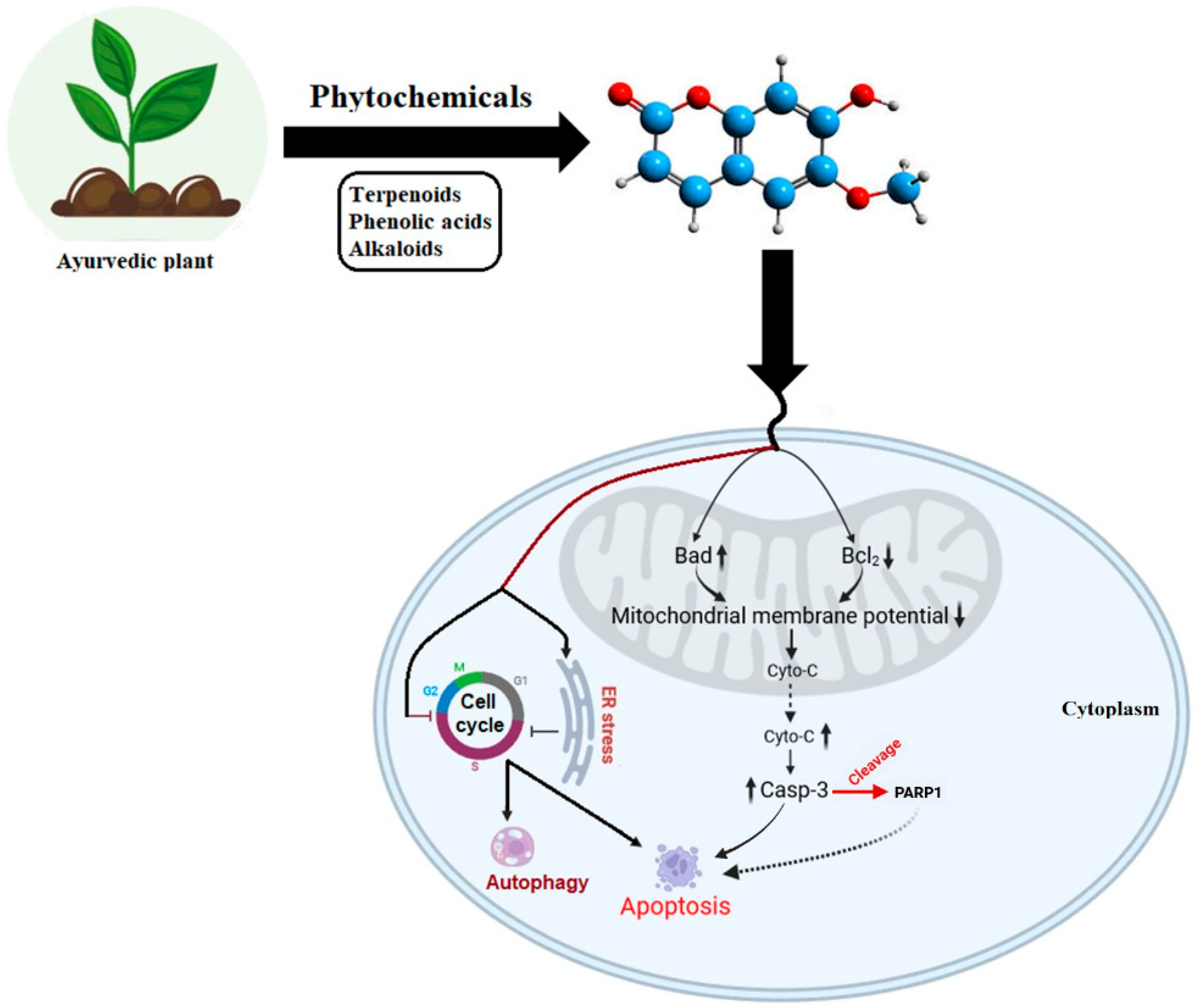
The Bcl-2 family of proteins regulates programmed cell death through the balance between pro-apoptotic members (Bax, BAD) and anti-apoptotic members (Bcl-2, Bcl-xl). Dysregulation of this balance disrupts mitochondrial membrane potential, leading to cytochrome C release and formation of the apoptosome complex with Apaf-1 and procaspase-9, thereby activating caspase-3 [103,104]. Ayurvedic plant-derived phytochemicals have been shown to modulate these apoptotic cascades by altering mitochondrial potential, increasing annexin-V/PI-positive apoptotic cells, and upregulating mRNA and protein expression of BAD, cytochrome C, and caspase-3, while downregulating Bcl-2 [79]. In lung cancer models, vasicine specifically enhances PARP activation, further amplifying apoptosis [79]. Additionally, key phytochemicals induce ER stress, trigger cell-cycle arrest, and activate autophagic signaling, promoting programmed cell death and inhibiting tumor progression [99].
Research demonstrated that Oridonin induces apoptosis in human carcinoma BEL-7402 cells via caspase-3 activation and downregulation of Bcl-2 expression, accompanied by Bax upregulation [105]. Rottlerin treatment in breast cancer stem cells (CSCs) inhibits Akt and mTOR phosphorylation while promoting AMPK phosphorylation, leading to apoptosis [106]. Glucosinolate-derived phenethyl isothiocyanates (PEITC) downregulate HER2, EGFR, and STAT3 expression and induce apoptosis in mice via caspase 3 and PARP cleavage. Additionally, NCTD demonstrates anticancer properties by inhibiting c-Met and mTOR [107,108]. Furthermore, PARP is known to play a crucial role in DNA damage and repair processes [109,110,111]. Overexpression of PARP can initiate apoptosis by promoting the release of mitochondrial apoptosis-inducing factors and inducing double-stranded DNA breaks. Studies on vasicine treatment in lung cancer cells have shown an increase in both mRNA and protein expression of PARP, along with DNA fragmentation [79]. This suggests that the PARP-mediated ADP ribosylation pathway could be an interesting area for future research, particularly in understanding cellular biomolecule modifications induced by Ayurvedic treatments (Figure 3).

6. ADP-Ribosylation and Inflammatory Pathways: Insights from Ayurvedic Medicine

ADPr, particularly PARylation, plays a critical role in modulating inflammation by influencing immune signalling pathways, cytokine production, and cell death processes. Among the PARP family, PARP1 is a central regulator of inflammatory responses. Its activation enhances transcription of pro-inflammatory genes through interactions with key transcription factors such as NF-κB and AP-1 [112]. However, excessive or prolonged PARP1 activation can lead to NAD+ and ATP depletion, resulting in cellular dysfunction and death-an underlying mechanism implicated in a variety of inflammatory diseases [113]. Notably, pharmacological inhibition of PARP has demonstrated anti-inflammatory efficacy in preclinical models of sepsis, arthritis, and colitis, supporting its potential as a therapeutic target in chronic inflammatory conditions [114,115].
Inflammation is increasingly recognized as a central link between metabolic disorders and cancer. Disruptions in immune regulation, redox balance, and cellular homeostasis not only promote inflammatory diseases but also create a tumour-supportive microenvironment [116]. Chronic inflammation, whether driven by genetic mutations (intrinsic pathway) or environmental stimuli (extrinsic pathway), contributes to the initiation and progression of malignancies. Key inflammatory mediators-including NF-κB, STAT3, and HIF-1α-activate transcription of pro-inflammatory genes such as cytokines, chemokines, COX-2, prostaglandins, iNOS, and nitric oxide. This cascade promotes the recruitment of tumour-associated macrophages (TAMs) and other immune cells into the tumor microenvironment, reinforcing inflammation and aiding tumour progression [117,118].
Mechanistically, reactive oxygen and nitrogen species (ROS/RNS) induce inflammatory responses by damaging DNA, lipids, and proteins, leading to gene mutations and the formation of advanced glycation end products (AGEs). These AGEs interact with their receptor (RAGE), activating NF-κB at sites of tissue injury and bypassing natural anti-inflammatory mechanisms, thereby perpetuating chronic inflammation [119,120]. Consequently, anti-inflammatory interventions-such as nonsteroidal anti-inflammatory drugs (NSAIDs) and selective COX-2 inhibitors-have shown efficacy in both suppressing tumour growth and reducing oxidative stress and angiogenesis [121]. In particular, blocking prostaglandin E2 (PGE2) production via inhibition of iNOS and COX-2 is emerging as a promising strategy for inflammation-associated cancer therapy.
Several Ayurvedic phytochemicals with established anti-inflammatory properties may intersect with ADPr pathways. For instance, Bhadradarvadi Kashayam modulates the NF-κB pathway, highlighting its potential as a natural anti-inflammatory agent [122,123,124]. Likewise, compounds such as curcumin (Curcuma longa), epigallocatechin gallate (EGCG) from green tea, resveratrol from grapes, and guggulsterone from Commiphora wightii exhibit potent anti-inflammatory and anticancer activities, partly through suppression of NF-κB-dependent transcription. Among these, guggulsterone stands out for its ability to induce apoptosis, inhibit tumor invasion, angiogenesis, and metastasis, and modulate STAT3 signaling [125]. These phytochemicals may influence inflammatory and ADPr-associated signaling, offering promising prospects for integrated cancer therapeutics. Future studies investigating their role in ADPr regulation could unveil novel anti-inflammatory mechanisms grounded in Ayurvedic medicine.

7. Integrative Cancer Therapies: ADP-Ribosylation in Ayurvedic and Allopathic Perspectives

Entire medical systems such as Ayurveda operate independently of biomedicine, drawing upon canonical texts and holistic, individualized therapeutic strategies [126,127]. While allopathic medicine effectively manages acute and life-threatening conditions, its symptom-centric approach often results in adverse side effects and limited long-term efficacy. In contrast, Ayurvedic formulations have been investigated for their potential molecular and therapeutic effects, particularly in chronic diseases such as hemiplegia, haemorrhoids, and arthritis, where conventional therapies remain inadequate [128,129]. Emerging studies suggest that these formulations exert beneficial effects through the modulation of inflammatory, oxidative stress, and metabolic signaling pathways, thereby promoting cellular homeostasis and tissue repair. Rooted in centuries of empirical wisdom, Ayurveda offers an integrative and mechanistically relevant approach to disease modulation beyond conventional pharmacotherapy [130,131].
An emerging approach in cancer management emphasizes holistic systems such as Ayurveda [132,133], which views cancer as a tridoshic imbalance involving vata, pitta, and kapha [131]. Ayurvedic care aims to restore systemic balance, enhance resilience, and mitigate the adverse effects of biomedical therapies. It recognizes genetic, dietary, and lifestyle factors as major contributors, paralleling modern perspectives [134,135,136]. The roles of kapha in tissue growth, pitta in malignant transformation, and vata in metastasis are central, with granthi and arbuda considered traditional analogues of cancer [131].
Recent advances in molecular biology have opened up new possibilities for decoding the therapeutic effects of Ayurvedic medicine by linking them to conserved cellular signalling pathways. One such promising link lies in ADPr, which plays crucial roles in DNA damage repair, transcriptional regulation, cell death, and importantly, immune modulation [112,113]. Inflammation-associated PARP1 activation is well-documented in both acute and chronic disease contexts, including cancer [112,113]. Sustained PARP1 activation leads to NAD+ depletion and mitochondrial dysfunction, contributing to inflammatory and tumour-promoting microenvironments [114]. Notably, emerging studies indicate that several phytochemicals used in Ayurvedic formulations-such as curcumin, resveratrol, and rosmarinic acid modulate ADPr by either inhibiting PARPs or affecting the expression of PAR-regulated genes, suggesting a plausible mechanistic bridge between Ayurvedic interventions and molecular oncology [137,138,139]. Despite the standardized, evidence-based framework of allopathic medicine, alternative medical systems continue to provide diverse therapeutic perspectives with varying degrees of validation [4,131]. A regional survey by Choi et al. involving nearly 900 participants from Southeast Asia revealed that more than half preferred traditional and complementary medicine (T&CM), particularly Ayurveda, for cancer care. The study emphasized integrating traditional and biomedical approaches to improve patient outcomes [140].
Allopathic cancer management employs surgery, radiotherapy, chemotherapy, immunotherapy, hormonal therapy, and targeted therapeutics, supported by recent advances in nanotechnology, RNA profiling, and CRISPR-mediated interventions [141,142]. While its strong empirical and regulatory foundation ensures credibility, alternative systems often lack similar standardization and reproducibility, which constrains their broader acceptance [131,143].
Ayurveda offers a distinct yet complementary paradigm, focusing on detoxification, immune modulation, and the restoration of systemic equilibrium through Dosh, Dhatu, Mal, Upadhatu, and Oaj regulation [4,131]. Mechanistically, emerging evidence suggests that Ayurvedic phytochemicals influence redox homeostasis, inflammatory signaling, and NAD+ metabolism—processes tightly linked to ADPr dynamics. Since ADPr governs DNA repair, metabolic adaptation, and cell death pathways, its modulation by plant-derived bioactives could underpin the anticancer efficacy of Ayurvedic formulations. Thus, Ayurveda’s holistic framework, when integrated with molecular oncology, may enable rationally designed combinatorial therapies targeting ADPr-mediated signaling networks in cancer.

8. Phytochemical–Nanoparticle Coupling and Targeted Delivery Strategies for Modulating ADP-Ribosylation Pathways in Cancer

In the context of Ayurvedic-inspired nanocarriers, it is noteworthy that classical formulations such as the bhasma preparations from the Rasashastra tradition reflect several fundamental principles of modern nanoscale delivery systems. Bhasmas are produced through repeated purification (śodhana) and incineration (māraṇa) processes, yielding metal or mineral particles in the nanometre range (typically 5–50 nm) that are highly absorbable and bio-assimilable [144,145]. These preparations exhibit physicochemical properties analogous to contemporary nanocarriers-such as high surface area, enhanced systemic distribution (“śighra vyāpī”), micro-dose efficacy (“alpamātra”), and targeted delivery behaviour (“yogavāhī”)—demonstrating their advanced pharmacological rationale [144,146]. By highlighting the nanoscale essence of bhasmas, this perspective emphasizes how Ayurvedic carrier-based concepts provide both a philosophical and mechanistic foundation for modern Phyto-nanomedicine design. Nanotechnology thus represents a natural extension of these ancient principles, offering new opportunities for diagnostic and therapeutic innovations. Bhasmas can be viewed as early prototypes of nanoparticle-based delivery, following a traditional “top-down” synthesis approach, and their future standardization should integrate modern nanotechnological characterization methods [144].
The integration of nanotechnology with Ayurvedic phytochemicals has opened a promising avenue for enhancing bioavailability, specificity, and efficacy of natural compounds in cancer therapy [147,148]. A growing body of research suggests that nano formulations of phytochemicals not only improve solubility and pharmacokinetics but also enable precision targeting of critical oncogenic pathways such as ADPr, particularly those mediated by PARPs. PARPs, especially PARP1 and PARP2, play a pivotal role in DNA repair, chromatin remodelling, and cellular stress responses [149]. Dysregulation of PARP activity is a hallmark of several cancers, and PARP inhibitors (PARPi) like Olaparib have already received FDA approval [150,151]. However, synthetic inhibitors often suffer from toxicity, resistance, and limited tumour selectivity. This has stimulated interest in plant-derived modulators as natural alternatives or adjuvants.
Phytochemicals such as curcumin, resveratrol, quercetin, and rosmarinic acid possess documented PARP-inhibitory or modulatory effects. Their nano-encapsulation, using liposomes, PLGA (poly-lactic-co-glycolic acid) nanoparticles, solid lipid nanoparticles (SLNs), or gold nanoparticles (AuNPs), has shown promise in targeting PARP-dependent signalling and DNA repair pathways in preclinical cancer models [55,152]. For instance, curcumin-loaded nanoparticles have demonstrated PARP1 suppression in models of breast and colorectal cancer, enhancing DNA damage and apoptotic cell death when combined with ionizing radiation or chemotherapeutics [152]. Similarly, resveratrol-loaded PLGA nanoparticles have improved cellular uptake and induced DNA damage through modulation of PARP cleavage in glioma and ovarian cancer models [153]. In another example, quercetin-conjugated gold nanoparticles were shown to inhibit PARP1 activity in prostate cancer cells, amplifying DNA damage and reducing cell viability more effectively than free quercetin [154]. Mechanistically, the crosstalk between phytochemical-induced oxidative stress and the PARP axis is central to these outcomes. Many polyphenols induce mitochondrial dysfunction and ER stress, leading to single strand breaks and consequent PARP activation. Overactivation of PARP can cause cellular energy crisis and necrosis, which, when precisely modulated by phytochemicals, may tilt the balance toward apoptotic cell death in cancer cells while sparing healthy tissues [155,156].
While Ayurvedic medicine-based nanotechnology in cancer therapy faces significant challenges, including issues of standardization, potential toxicity, and translational bottlenecks [157,158,159], these obstacles can be systematically addressed through the application of green nanofabrication techniques, rigorous analytical characterization, and robust preclinical validation. Additionally, the integration of multidisciplinary approaches, including the design of engineered nanoparticles for cancer vaccination and immunotherapy, further enhances the safety, specificity, and therapeutic potential of these nano formulations [160,161,162]. The convergence of traditional Ayurvedic principles with modern nanoscience thus holds the promise of establishing a rational, scientifically credible, and translationally viable foundation for next-generation anticancer therapeutics.
Recent efforts have increasingly focused on scaling these preclinical findings toward translational endpoints. Several nano-formulated phytochemicals are currently being evaluated in early-phase clinical trials or investigational studies. Table 1 [163,164,165,166,167,168,169,170,171,172,173,174,175,176,177,178,179] summarises representative examples of phytochemical–nanoparticle conjugates, their target cancer types, and associated PARP-driven mechanistic insights. For instance, nano-curcumin, which indirectly modulates PARP1-mediated responses through oxidative stress pathways, has already completed multiple Phase I/II trials in cancers such as pancreatic and colorectal carcinoma [180,181]. Another promising direction is the development of polyphenol-based nano-drug delivery systems designed to co-deliver phytochemicals alongside PARP inhibitors, thereby enhancing synergistic anti-tumour effects while minimizing toxicity. These combinatorial platforms mark a paradigm shift in dual-targeting strategies-integrating the wisdom of traditional Ayurveda with the precision of modern molecular oncology. Looking ahead, rigorous exploration of Ayurvedic phytochemical–nanoparticle formulations in the context of ADPr signalling remains essential. High-throughput screening of ADPr-related endpoints, imaging-guided biodistribution analyses, and computational modelling of phytochemical–PARP interactions could further accelerate progress. As clinical investigations mature, such natural compound-based nanomedicines hold the potential to emerge either as standalone therapeutic agents or as sensitizers augmenting existing DNA repair-targeted regimens [182,183].

9. Concluding Reflections

Ayurvedic phytochemicals mediate two distinct cellular pathways: one linked to organelle-induced stress responses involving mitochondria and the endoplasmic reticulum that culminate in apoptotic signalling (Figure 3), and another potential mechanism in which such stress may trigger DNA damage, particularly in cancer cells. This can activate DNA damage response pathways, including the spatiotemporally regulated activity of PARPs, influencing ADPr-driven survival or death outcomes (Figure 2). Although our proposed model (Figure 2) outlines these processes, detailed investigations are still required to understand how phytochemical-induced stress determines cell fate through ADPr. This review integrates traditional Ayurvedic principles with modern biomedical evidence to provide molecular insights into cancer progression and therapy. It highlights how phytochemicals influence signalling networks associated with apoptosis, stress responses, and genomic stability. The synthesis of literature reveals phytochemicals as promising agents that bridge ancient wisdom and molecular oncology.
A major highlight is the identification of key research gaps that limit translational progress. While single-compound studies offer mechanistic clarity, they often face limitations such as resistance and poor pharmacokinetic behaviour. In contrast, whole-plant extracts or multi-compound formulations central to Ayurvedic practice may offer synergistic efficacy, better bioavailability, and reduced toxicity [33]. Integrating Ayurvedic constitutional types (Prakriti: Vata, Pitta, Kapha) with genomic and molecular data also presents opportunities for personalized cancer management [4,184]. Such approaches reinforce Ayurveda’s relevance in precision medicine and therapeutic innovation.

10. Future Perspectives

Future studies should focus on decoding how phytochemicals influence genome stability, particularly through interactions with DNA repair pathways and redox regulation. Many natural compounds affect chromatin remodelling, repair protein recruitment, and oxidative signalling, offering therapeutic potential in cancers driven by genomic instability [80,185]. The psychosomatic component of cancer also warrants greater attention. Chronic stress and depression influence cancer initiation, progression, and prognosis through activation of the hypothalamic–pituitary–adrenal axis and sympathetic signalling [186,187,188]. Ayurvedic lifestyle interventions including diet, daily routines, yoga, breathing practices, and meditation can serve as non-pharmacological approaches to reduce stress-induced cancer risk and improve overall outcomes.
Advances in molecular biology such as CRISPR/Cas9, ZFNs, and TALENs have enhanced the precision of gene targeting [143]. Combining gene-editing with phytochemical modulation could generate innovative therapeutic strategies. Bioactive compounds including alkaloids, tannins, saponins, phenolics, and flavonoids continue to demonstrate antiproliferative, antioxidant, and pro-apoptotic activity [189]. For example, Vasicine from Adhatoda vasica shows selective cytotoxicity toward A549 lung cancer cells while sparing normal fibroblasts, underscoring its potential as an anticancer candidate [79,190]. Despite promising outcomes, clinical translation remains challenging due to issues of solubility, stability, and bioavailability. Nanocarrier-based delivery systems offer a way forward by improving stability, controlled release, and targeted delivery of phytocompounds [191]. Integrating Ayurvedic phytochemicals with nanotechnology and exploring their influence on ADPr and PARP pathways could open new avenues in precision oncology and immunomodulation.
Future research should prioritize the mechanistic validation of phytochemical–ADPr interactions, the establishment of standardized nano formulation protocols, comprehensive safety assessments, and well-structured clinical trials. Of particular interest is the exploration of how Ayurvedic phytochemicals influence p53 regulation, Sirtuin-mediated ADPr, and upstream–downstream signalling hierarchies that dictate cell fate decisions. Bridging traditional pharmacology with nanomedicine and molecular oncology will be pivotal for developing integrative, sustainable, and effective therapeutic approaches [4,33,131,184].
In summary, advancing the scientific foundation of Ayurveda through molecular-level inquiry and technological innovation will not only refine our understanding of cancer biology but also guide the evolution of inclusive and precision-based cancer care for the future.

Author Contributions

Conceptualization, K.S. and P.K.; writing—original draft preparation, K.S. and P.K.; writing—review and editing, G.S.V.S.R.R., S.K.N., P.C., K.S. and P.K.; project administration, K.S. and P.K.; funding acquisition, P.K. All authors have read and agreed to the published version of the manuscript.

Funding

This work was supported by SRM University-AP. Dr. Pulak Kar is the recipient of the SRM University-AP Seed Grant [Grant No. SRMAP/URG/SEED/2024-25/042].

Institutional Review Board Statement

Not applicable.

Informed Consent Statement

Not applicable.

Data Availability Statement

Not applicable.

Acknowledgments

Financial support from SRM University-AP (Grant No: SRMAP/URG/SEED/2024-25/042) for our research is gratefully acknowledged. G.S.R.R thanks SRM University-AP for providing PhD fellowships. We also extend our sincere thanks to Ivan Ahel and Osamu Suyari from the Dunn School of Pathology, Oxford University, for their help and support in our work.

Conflicts of Interest

The authors declare no conflicts of interest.

Abbreviations

The following abbreviations are used in this manuscript:
TMEtumor microenvironment
CAMcomplementary and alternative medicine
NSBnano swarna bhasma
ADPrADP-ribosylation
ARTADP-ribosyl transferases
NAD+β-nicotinamide adenine dinucleotide
PARPpoly (ADP-ribose) polymerase
3D QSAR three-dimensional quantitative structure–activity relationship
CDK1cyclin-dependent kinase 1
BaxBcl-2-associated X protein
BRCA1breast cancer gene 1
BRCA2breast cancer gene 2
SIRT6sirtuin 6
BAF170BRG1-Associated Factor 170
ILinterleukin
NF-κBNuclear factor kappa-light-chain-enhancer of activated B cells
RANKLreceptor activator of NF-kappa B ligand
MDRmultidrug resistance
MAPKmitogen-activated protein kinase
ERKextracellular signal-regulated kinase
PKAprotein kinase A
TNF-αtumor necrosis factor-alpha
RAD51radiation sensitive 51.
Aktprotein kinase B
Bcl-2B-cell lymphoma 2
COX-2cyclooxygenase-2
MMP-9matrix metalloproteinase-9
STAT3signal transducer and activator of transcription 3
ROSreactive oxygen species
EGCGepigallocatechin-3-gallate
WFAWithaferin A
BRAFv-Raf murine sarcoma viral oncogene homolog B1
Raf-1v-Raf-1 murine leukemia viral oncogene homolog 1
HSPheat shock protein
NHEJnon-homologous end joining
CBPCREB-binding protein
p300E1A-associated protein p300
HATshistone acetyltransferases
ATRataxia telangiectasia and Rad3-related
DR4death receptors 4
DR5death receptors 5
γH2AXphosphorylated histone H2A variant X
p65/RelAv-Rel avian reticuloendotheliosis viral oncogene homolog A
BEL-7402human hepatocellular carcinoma cell lines
LDHlactate dehydrogenase
BADBcl-2-associated death promoter
PELprimary effusion lymphoma
ERendoplasmic reticulum
JNKc-jun N-terminal kinase
PI3Kphosphoinositide 3-kinase
mTORmammalian target of rapamycin
MCF-7Michigan Cancer Foundation-7
Bcl-xLB-cell lymphoma-extra large
CSCscancer stem cells
PEITCphenethyl isothiocyanates
HER2human epidermal growth factor receptor 2
EGFRepidermal growth factor receptor
AP-1activator protein 1
HIF-1αhypoxia-inducible factor 1-alpha
iNOSinducible nitric oxide synthase
FDAfood and drug administration
TAMstumor-associated macrophages
RNSreactive nitrogen species
AGEsadvanced glycation end products
RAGEreceptor for advanced glycation end-products
NSAIDsnonsteroidal anti-inflammatory drugs
EGCGepigallocatechin gallate
DDRDNA damage response
CRISPRclustered regularly interspaced short palindromic repeats
Cas9CRISPR-associated protein 9
ZFNszinc finger nucleases
TALENstranscription activator-like effector nucleases
DSBsdouble-strand breaks

References

  1. Armitage, P.; Doll, R. The age distribution of cancer and a multi-stage theory of carcinogenesis. Br. J. Cancer 1954, 8, 1–12. [Google Scholar] [CrossRef]
  2. Sugimura, T. Multistep carcinogenesis: A 1992 perspective. Science 1992, 258, 603–607. [Google Scholar] [CrossRef] [PubMed]
  3. Gorga, F. The Molecular Basis of Cancer. Bridgewater Rev. 1998, 17, 3–6. Available online: https://vc.bridgew.edu/br_rev/vol17/iss2/5 (accessed on 30 October 2025).
  4. Arnold, J.T. Integrating ayurvedic medicine into cancer research programs part 1: Ayurveda background and applications. J. Ayurveda Integr. Med. 2023, 14, 100676. [Google Scholar] [CrossRef] [PubMed]
  5. Shi, R.; Tang, Y.Q.; Miao, H. Metabolism in tumor microenvironment: Implications for cancer immunotherapy. MedComm 2020, 1, 47–68. [Google Scholar] [CrossRef] [PubMed]
  6. Vohra, R.; Singh, R.; Shrivastava, R. A scoping review on ‘Maharishi Amrit Kalash’, an ayurveda formulation for cancer prevention and management. J. Ayurveda Integr. Med. 2024, 15, 100866. [Google Scholar] [CrossRef]
  7. Tralongo, P.; Pescarenico, M.G.; Surbone, A.; Bordonaro, S.; Berretta, M.; DI Mari, A. Physical needs of long-term cancer patients. Anticancer Res. 2017, 37, 4733–4746. [Google Scholar] [CrossRef]
  8. Given, B.A.; Given, C.W.; Vachon, E.; Hershey, D. Do we have a clue: The treatment burden for the patient with cancer? Cancer Nurs. 2016, 39, 423–424. [Google Scholar] [CrossRef]
  9. Valdivieso, M.; Kujawa, A.M.; Jones, T.; Baker, L.H. Cancer survivors in the United States: A review of the literature and a call to action. Int. J. Med. Sci. 2012, 9, 63–73. [Google Scholar] [CrossRef]
  10. Mateo, J.; Steuten, L.; Aftimos, P.; André, F.; Davies, M.; Garralda, E.; Geissler, J.; Husereau, D.; Martinez-Lopez, I.; Normanno, N.; et al. Delivering precision oncology to patients with cancer. Nat. Med. 2022, 28, 658–665. [Google Scholar] [CrossRef]
  11. Samanta, K.; Reddy, G.S.V.S.R.; Sharma, N.K.; Kar, P. Deciphering the Role of Functional Ion Channels in Cancer Stem Cells (CSCs) and Their Therapeutic Implications. Int. J. Mol. Sci. 2025, 26, 7595. [Google Scholar] [CrossRef] [PubMed]
  12. Kumar, S.; Dobos, G.J.; Rampp, T. The significance of Ayurvedic medicinal plants. J. Evid. Based Complement. Altern. Med. 2017, 22, 494–501. [Google Scholar] [CrossRef] [PubMed]
  13. Tandon, N.; Yadav, S.S. Contributions of Indian council of medical research (ICMR) in the area of medicinal plants/traditional medicine. J. Ethnopharmacol. 2017, 197, 39–45. [Google Scholar] [CrossRef]
  14. Pandey, S.; Walpole, C.; Shaw, P.N.; Cabot, P.J.; Hewavitharana, A.K.; Batra, J. Bio-guided fractionation of papaya leaf juice for delineating the components responsible for the selective anti-proliferative effects on prostate cancer cells. Front. Pharmacol. 2018, 9, 1319. [Google Scholar] [CrossRef]
  15. Khoobchandani, M.; Katti, K.K.; Karikachery, A.R.; Thipe, V.C.; Srisrimal, D.; Dhurvas Mohandoss, D.K.; Darshakumar, R.D.; Joshi, C.M.; Katti, K.V. New approaches in breast cancer therapy through green nanotechnology and nano-Ayurvedic medicine—Pre-clinical and pilot human clinical investigations. Int. J. Nanomed. 2020, 15, 181–197. [Google Scholar] [CrossRef]
  16. Huang, L.; Luo, S.; Tong, S.; Lv, Z.; Wu, J. The development of nanocarriers for natural products. Wiley Interdiscip. Rev. Nanomed. Nanobiotechnol. 2024, 16, e1967. [Google Scholar] [CrossRef]
  17. Gao, S.; Zhou, J.R.; Yokomizo, K.; Fang, J. Nano-drug delivery system of natural products for disease prevention and treatment. Expert Opin. Drug Deliv. 2025, 22, 1057–1067. [Google Scholar] [CrossRef] [PubMed]
  18. Katti, K.; Chanda, N.; Shukla, R.; Zambre, A.; Suibramanian, T.; Kulkarni, R.R.; Kannan, R.; Katti, K.V. Green nanotechnology from cumin phytochemicals: Generation of biocompatible gold nanoparticles. Int. J. Green Nanotechnol. Biomed. 2009, 1, B39–B52. [Google Scholar] [CrossRef]
  19. Yayintas, O.T.; Demir, N.; Canbolat, F.; Ayna, T.K.; Pehlivan, M. Characterization, biological activity, and anticancer effect of green-synthesized gold nanoparticles using Nasturtium officinale L. BMC Complement. Med. Ther. 2024, 24, 346. [Google Scholar] [CrossRef]
  20. Nune, S.K.; Chanda, N.; Shukla, R.; Katti, K.; Kulkarni, R.R.; Thilakavathi, S.; Mekapothula, S.; Kannan, R.; Katti, K.V. Green nanotechnology from tea: Phytochemicals in tea as building blocks for production of biocompatible gold nanoparticles. J. Mater. Chem. 2009, 19, 2912–2920. [Google Scholar] [CrossRef]
  21. Khoobchandani, M.; Katti, K.; Maxwell, A.; Fay, W.P.; Katti, K.V. Laminin receptor-avid nanotherapeutic EGCg-AuNPs as a potential alternative therapeutic approach to prevent restenosis. Int. J. Mol. Sci. 2016, 17, 316. [Google Scholar] [CrossRef]
  22. Thipe, V.C.; Keyster, M.; Katti, K.V. Sustainable Nanotechnology: Mycotoxin Detection and Protection. In Nanobiotechnology Applications in Plant Protection; Abd-Elsalam, K., Prasad, R., Eds.; Nanotechnology in the Life Sciences; Springer: Cham, Switzerland, 2018; pp. 323–349. [Google Scholar] [CrossRef]
  23. Yang, L.Y.; Lei, S.Z.; Xu, W.J.; Lai, Y.-X.; Zhang, Y.-Y.; Wang, Y.; Wang, Z.-L. Rising above: Exploring the therapeutic potential of natural product-based compounds in human cancer treatment. Tradit. Med. Res. 2025, 10, 18. [Google Scholar] [CrossRef]
  24. Menze, N.; Van der Watt, A.S.J.; Moxley, K.; Seedat, S. Profiles of traditional healers and their healing practices in the Eastern Cape province of South Africa. S. Afr. J. Psychiatry 2018, 24, a1305. [Google Scholar] [CrossRef]
  25. Yang, L.; Wang, Z. Natural Products, Alone or in Combination with FDA-Approved Drugs, to Treat COVID-19 and Lung Cancer. Biomedicines 2021, 9, 689. [Google Scholar] [CrossRef]
  26. Dutta, R.; Khalil, R.; Green, R.; Mohapatra, S.S.; Mohapatra, S. Withania somnifera (Ashwagandha) and Withaferin A: Potential in integrative oncology. Int. J. Mol. Sci. 2019, 20, 5310. [Google Scholar] [CrossRef] [PubMed]
  27. Ogiwara, H.; Ui, A.; Shiotani, B.; Zou, L.; Yasui, A.; Kohno, T. Curcumin suppresses multiple DNA damage response pathways and has potency as a sensitizer to PARP inhibitor. Carcinogenesis 2013, 34, 2486–2497. [Google Scholar] [CrossRef]
  28. Aggarwal, B.B.; Harikumar, K.B. Potential therapeutic effects of curcumin, the anti-inflammatory agent, against neurodegenerative, cardiovascular, pulmonary, metabolic, autoimmune and neoplastic diseases. Int. J. Biochem. Cell Biol. 2009, 41, 40–59. [Google Scholar] [CrossRef] [PubMed]
  29. Mishra, L.C.; Singh, B.B.; Dagenais, S. Scientific basis for the therapeutic use of Withania somnifera (Ashwagandha): A review. Altern. Med. Rev. 2000, 5, 334–346. [Google Scholar] [PubMed]
  30. Kunnumakkara, A.B.; Anand, P.; Aggarwal, B.B. Curcumin inhibits proliferation, invasion, angiogenesis and metastasis of different cancers through interaction with multiple cell signaling proteins. Cancer Lett. 2008, 269, 199–225. [Google Scholar] [CrossRef] [PubMed]
  31. Sehgal, N.; Gupta, A.; Valli, R.K.; Joshi, S.D.; Mills, J.T.; Hamel, E.; Khanna, P.; Jain, S.C.; Thakur, S.S.; Ravindranath, V. Withania somnifera reverses Alzheimer’s disease pathology by enhancing low-density lipoprotein receptor-related protein in liver. Proc. Natl. Acad. Sci. USA 2012, 109, 3510–3515. [Google Scholar] [CrossRef]
  32. Patwardhan, B.; Gautam, M. Botanical immunodrugs: Scope and opportunities. Drug Discov. Today. 2005, 10, 495–502. [Google Scholar] [CrossRef] [PubMed]
  33. Aggarwal, B.B.; Prasad, S.; Reuter, S.; Kannappan, R.; Yadev, V.R.; Park, B.; Kim, J.H.; Gupta, S.C.; Phromnoi, K.; Sundaram, C.; et al. Identification of novel anti-inflammatory agents from Ayurvedic medicine for prevention of chronic diseases: “reverse pharmacology” and “bedside to bench” approach. Curr. Drug Targets 2011, 12, 1595–1653. [Google Scholar] [CrossRef]
  34. Gupte, R.; Liu, Z.; Kraus, W.L. PARPs and ADP-ribosylation: Recent advances linking molecular functions to biological outcomes. Genes Dev. 2017, 31, 101–126. [Google Scholar] [CrossRef]
  35. Ray Chaudhuri, A.; Nussenzweig, A. The multifaceted roles of PARP1 in DNA repair and chromatin remodelling. Nat. Rev. Mol. Cell Biol. 2017, 18, 610–621. [Google Scholar] [CrossRef]
  36. Zrinej, J.; Elmchichi, L.; Alaqarbeh, M.; Tahar Lakhlifia, T.; Bouachrineac, M. Computational approach: 3D-QSAR, molecular docking, ADMET, molecular dynamics simulation investigations, and retrosynthesis of some curcumin analogues as PARP-1 inhibitors targeting colon cancer. New J. Chem. 2023, 47, 20987–21009. [Google Scholar] [CrossRef]
  37. El Hassab, M.A.; Eldehna, W.M.; Hassan, G.S.; Abou-Seri, S.M. Multi-stage structure-based virtual screening approach combining 3D pharmacophore, docking and molecular dynamic simulation towards the identification of potential selective PARP-1 inhibitors. BMC Chem. 2025, 19, 30. [Google Scholar] [CrossRef]
  38. Bono, A.; La Monica, G.; Alamia, F.; Mingoia, F.; Gentile, C.; Peri, D.; Lauria, A.; Martorana, A. In Silico Mixed Ligand/Structure-Based Design of New CDK-1/PARP-1 Dual Inhibitors as Anti-Breast Cancer Agents. Int. J. Mol. Sci. 2023, 24, 13769. [Google Scholar] [CrossRef]
  39. Zhu, Y.; Mehmoodc, A.; Li, D. Unraveling the inhibitory potential of Rosetta designed de novo cyclic peptides on PARP7 through molecular dynamics simulations. New J. Chem. 2024, 48, 7347–7355. [Google Scholar] [CrossRef]
  40. Wierbiłowicz, K.; Yang, C.S.; Almaghasilah, A.; Wesołowski, P.A.; Pracht, P.; Dworak, N.M.; Masur, J.; Wijngaarden, S.; Filippov, D.V.; Wales, D.J.; et al. Parp7 generates an ADP-ribosyl degron that controls negative feedback of androgen signaling. EMBO J. 2025, 44, 4720–4744. [Google Scholar] [CrossRef] [PubMed]
  41. Caba, K.; Tran-Nguyen, V.K.; Rahman, T.; Ballester, P.J. Comprehensive machine learning boosts structure-based virtual screening for PARP1 inhibitors. J. Cheminform. 2024, 16, 40. [Google Scholar] [CrossRef] [PubMed]
  42. Goel, A.; Kunnumakkara, A.B.; Aggarwal, B.B. Curcumin as “Curecumin”: From kitchen to clinic. Biochem. Pharmacol. 2008, 75, 787–809. [Google Scholar] [CrossRef]
  43. Sharma, R.A.; McLelland, H.R.; Hill, K.A.; Ireson, C.R.; Euden, S.A.; Manson, M.M.; Pirmohamed, M.; Marnett, L.J.; Gescher, A.J.; Steward, W.P. Pharmacodynamic and pharmacokinetic study of oral curcuma extract in patients with colorectal cancer. Clin. Cancer Res. 2001, 7, 1894–1900. [Google Scholar] [PubMed]
  44. Aghababaei, F.; Hadidi, M. Recent Advances in Potential Health Benefits of Quercetin. Pharmaceuticals 2023, 16, 1020. [Google Scholar] [CrossRef]
  45. Kesarwani, K.; Gupta, R. Bioavailability enhancers of herbal origin: An overview. Asian Pac. J. Trop. Biomed. 2013, 3, 253–266. [Google Scholar] [CrossRef] [PubMed]
  46. Maeda, J.; Roybal, E.J.; Brents, C.A.; Uesaka, M.; Aizawa, Y.; Kato, T.A. Natural and glucosyl flavonoids inhibit poly(ADP-ribose) polymerase activity and induce synthetic lethality in BRCA mutant cells. Oncol. Rep. 2014, 31, 551–556. [Google Scholar] [CrossRef] [PubMed]
  47. Asgharian, P.; Tazekand, A.P.; Hosseini, K.; Forouhandeh, H.; Ghasemnejad, T.; Ranjbar, M.; Hasan, M.; Kumar, M.; Beirami, S.M.; Tarhriz, V.; et al. Potential mechanisms of quercetin in cancer prevention: Focus on cellular and molecular targets. Cancer Cell Int. 2022, 22, 257. [Google Scholar] [CrossRef]
  48. Mao, Z.; Hine, C.; Tian, X.; Van Meter, M.; Au, M.; Vaidya, A.; Seluanov, A.; Gorbunova, V. SIRT6 promotes DNA repair under stress by activating PARP1. Science 2011, 332, 1443–1446. [Google Scholar] [CrossRef] [PubMed]
  49. Zhang, H.; Zhang, J.; Zhang, H.X. Effect of quercetin on the protein-substrate interactions in SIRT6: Insight from MD simulations. J. Mol. Graph. Model 2024, 130, 108778. [Google Scholar] [CrossRef]
  50. Wang, X.; Zhang, Y. Potential terahertz therapeutic strategy for the prevention or mitigation of Alzheimer’s disease pathology. Light Sci. Appl. 2023, 12, 254. [Google Scholar] [CrossRef]
  51. Xavier, C.P.; Pereira-Wilson, C. Medicinal plants of the genuses Salvia and Hypericum are sources of anticolon cancer compounds: Effects on PI3K/Akt and MAP kinases pathways. PharmaNutrition 2016, 4, 112–122. [Google Scholar] [CrossRef]
  52. Rocha, J.; Eduardo-Figueira, M.; Barateiro, A.; Fernandes, A.; Brites, D.; Bronze, R.; Duarte, C.M.; Serra, A.T.; Pinto, R.; Freitas, M.; et al. Anti-inflammatory effect of rosmarinic acid and an extract of Rosmarinus officinalis in rat models of local and systemic inflammation. Basic Clin. Pharmacol. Toxicol. 2015, 116, 398–413. [Google Scholar] [CrossRef]
  53. Liu, W.Y.; Wang, H.; Xu, X.; Wang, X.; Han, K.K.; You, W.D.; Yang, Y.; Zhang, T. Natural compound rosmarinic acid displays anti-tumor activity in colorectal cancer cells by suppressing nuclear factor-kappa B signaling. World J. Clin. Oncol. 2025, 16, 105341. [Google Scholar] [CrossRef]
  54. Su, C.; Gius, J.P.; Van Steenberg, J.; Haskins, A.H.; Heishima, K.; Omata, C.; Iwayama, M.; Murakami, M.; Mori, T.; Maruo, K.; et al. Hypersensitivity of BRCA2 deficient cells to rosemary extract explained by weak PARP inhibitory activity. Sci. Rep. 2017, 7, 16704. [Google Scholar] [CrossRef]
  55. Subongkot, T.; Ngawhirunpat, T.; Opanasopit, P. Development of ultra deformable liposomes with fatty acids for enhanced dermal rosmarinic acid delivery. Pharmaceutics 2021, 13, 404. [Google Scholar] [CrossRef]
  56. Aggarwal, S.; Ichikawa, H.; Takada, Y.; Sandur, S.K.; Shishodia, S.; Aggarwal, B.B. Curcumin (diferuloylmethane) down-regulates expression of cell proliferation and antiapoptotic and metastatic gene products through suppression of IkappaBalpha kinase and Akt activation. Mol. Pharmacol. 2006, 69, 195–206. [Google Scholar] [CrossRef]
  57. Singh, S.; Aggarwal, B.B. Activation of transcription factor NF-kappa B is suppressed by curcumin (diferuloylmethane). J. Biol. Chem. 1995, 270, 24995–25000. [Google Scholar] [CrossRef] [PubMed]
  58. Chao, W.W.; Kuo, Y.H.; Lin, B.F. Anti-inflammatory activity of new compounds from Andrographis paniculata by NF-kappaB transactivation inhibition. J. Agric. Food Chem. 2010, 58, 2505–2512. [Google Scholar] [CrossRef] [PubMed]
  59. Chao, W.W.; Lin, B.F. Isolation and identification of bioactive compounds in Andrographis paniculata (Chuanxinlian). Chin. Med. 2010, 5, 17. [Google Scholar] [CrossRef] [PubMed]
  60. Qu, J.; Liu, Q.; You, G.; Ye, L.; Jin, Y.; Kong, L.; Guo, W.; Xu, Q.; Sun, Y. Advances in ameliorating inflammatory diseases and cancers by andrographolide: Pharmacokinetics, pharmacodynamics, and perspective. Med. Res. Rev. 2022, 42, 1147–1178. [Google Scholar] [CrossRef]
  61. Ichikawa, H.; Takada, Y.; Shishodia, S.; Jayaprakasam, B.; Nair, M.G.; Aggarwal, B.B. Withanolides potentiate apoptosis, inhibit invasion, and abolish osteoclastogenesis through suppression of nuclear factor-kappaB (NF-kappaB) activation and NF-kappaB-regulated gene expression. Mol. Cancer Ther. 2006, 5, 1434–1445. [Google Scholar] [CrossRef]
  62. Mulabagal, V.; Subbaraju, G.V.; Rao, C.V.; Sivaramakrishna, C.; Dewitt, D.L.; Holmes, D.; Sung, B.; Aggarwal, B.B.; Tsay, H.S.; Nair, M.G. Withanolide sulfoxide from Aswagandha roots inhibits nuclear transcription factor-kappa-B, cyclooxygenase and tumor cell proliferation. Phytother. Res. 2009, 23, 987–992. [Google Scholar] [CrossRef]
  63. Widodo, N.; Kaur, K.; Shrestha, B.G.; Takagi, Y.; Ishii, T.; Wadhwa, R.; Kaul, S.C. Selective killing of cancer cells by leaf extract of Ashwagandha: Identification of a tumor-inhibitory factor and the first molecular insights to its effect. Clin. Cancer Res. 2007, 13, 2298–2306. [Google Scholar] [CrossRef]
  64. Grogan, P.T.; Sarkaria, J.N.; Timmermann, B.N.; Cohen, M.S. Oxidative cytotoxic agent withaferin A resensitizes temozolomide-resistant glioblastomas via MGMT depletion and induces apoptosis through Akt/mTOR pathway inhibitory modulation. Investig. New Drugs 2014, 32, 604–617. [Google Scholar] [CrossRef]
  65. Gupta, A.; Gupta, P.; Bajpai, G. Tinospora cordifolia (Giloy): An insight on the multifarious pharmacological paradigms of a most promising medicinal ayurvedic herb. Heliyon 2024, 10, e26125. [Google Scholar] [CrossRef]
  66. Patil, S.; Ashi, H.; Hosmani, J.; Almalki, A.Y.; Alhazmi, Y.A.; Mushtaq, S.; Parveen, S.; Baeshen, H.A.; Varadarajan, S.; Raj, A.T.; et al. Tinospora cordifolia (Thunb.) Miers (Giloy) inhibits oral cancer cells in a dose-dependent manner by inducing apoptosis and attenuating epithelial-mesenchymal transition. Saudi J. Biol. Sci. 2021, 28, 4553–4559. [Google Scholar] [CrossRef]
  67. Kumar, G.H.; Priyadarsini, R.V.; Vinothini, G.; Letchoumy, P.V.; Nagini, S. The neem limonoids azadirachtin and nimbolide inhibit cell proliferation and induce apoptosis in an animal model of oral oncogenesis. Investig. New Drugs. 2010, 28, 392–401. [Google Scholar] [CrossRef]
  68. Wu, Q.; Kohli, M.; Bergen, H.R., 3rd; Cheville, J.C.; Karnes, R.J.; Cao, H.; Young, C.Y.; Tindall, D.J.; McNiven, M.A.; Donkena, K.V. Preclinical evaluation of the supercritical extract of Azadirachta indica (neem) leaves in vitro and in vivo on inhibition of prostate cancer tumor growth. Mol. Cancer Ther. 2014, 13, 1067–1077. [Google Scholar] [CrossRef]
  69. Visavadiya, N.P.; Narasimhacharya, A.V. Asparagus root regulates cholesterol metabolism and improves antioxidant status in hypercholesteremic rats. Evid. Based Complement. Alternat. Med. 2009, 6, 219–226. [Google Scholar] [CrossRef] [PubMed]
  70. Pathak, A.K.; Bhutani, M.; Nair, A.S.; Ahn, K.S.; Chakraborty, A.; Kadara, H.; Guha, S.; Sethi, G.; Aggarwal, B.B. Ursolic acid inhibits STAT3 activation pathway leading to suppression of proliferation and chemosensitization of human multiple myeloma cells. Mol. Cancer Res. 2007, 5, 943–955. [Google Scholar] [CrossRef]
  71. Mohanta, Y.K.; Biswas, K.; Mishra, A.K.; Patra, B.; Mishra, B.; Panda, J.; Avula, S.K.; Varma, R.S.; Panda, B.P.; Nayak, D. Amelioration of gold nanoparticles mediated through Ocimum oil extracts induces reactive oxygen species and mitochondrial instability against MCF-7 breast carcinoma. RSC Adv. 2024, 14, 27816–27830. [Google Scholar] [CrossRef]
  72. Jadhav, P.; Shaikh, A.Q.; Bachhav, R.S.; Rawal, D.; Lohar, S. A Review: Study of Tulsi as Immune Booster. Int. J. Res. Appl. Sci. Eng. Technol. 2025, 13, 770–775. [Google Scholar] [CrossRef]
  73. Hasan, M.R.; Alotaibi, B.S.; Althafar, Z.M.; Mujamammi, A.H.; Jameela, J. An update on the therapeutic anticancer potential of Ocimum sanctum L.: “Elixir of Life”. Molecules 2023, 28, 1193. [Google Scholar] [CrossRef]
  74. Nayak, P.; Thirunavoukkarasu, M. A review of the plant Boerhaavia diffusa: Its chemistry, pharmacology and therapeutical potential. J. Phytopharm. 2016, 5, 83–92. [Google Scholar] [CrossRef]
  75. Leslie Taylor, N.D. Boerhaavia diffusa. In The Healing Power of Rainforest Herbs; Square one Publishers, Inc.: New York, NY, USA, 2005; pp. 1–535. Available online: https://www.rain-tree.com/book2.htm (accessed on 30 October 2025).
  76. Manu, K.A.; Kuttan, G. Effect of Punarnavine, an alkaloid from Boerhaavia diffusa, on cell-mediated immune responses and TIMP-1 in B16F-10 metastatic melanoma-bearing mice. Immunopharmacol. Immunotoxicol. 2007, 29, 569–586. [Google Scholar] [CrossRef]
  77. Patel, N.; Mishra, R.; Rajput, D.; Gupta, A. A comprehensive review of the phytochemistry, pharmacology, pharmacokinetics, and green nanotechnological significance of Boerhavia diffusa Linn. Fitoterapia 2025, 184, 106599. [Google Scholar] [CrossRef]
  78. Aher, V.; Chattopadhyay, P.; Goyary, D.; Veer, V. Evaluation of the genotoxic and antigenotoxic potential of the alkaloid punarnavine from Boerhaavia diffusa. Planta Med. 2013, 79, 939–945. [Google Scholar] [CrossRef]
  79. Dey, T.; Dutta, P.; Manna, P.; Kalita, J.; Boruah, H.P.D.; Buragohain, A.K.; Unni, B. Anti-proliferative activities of Vasicinone on lung carcinoma cells mediated via activation of both mitochondria-dependent and independent pathways. Biomol. Ther. 2018, 26, 409–416. [Google Scholar] [CrossRef]
  80. Masutani, M.; Gunji, A.; Tsutsumi, M.; Ogawa, K.; Kamada, N.; Shirai, T.; Jishage, K.-I.; Nakagama, H.; Sugimura, T. Role of Poly-ADP-Ribosylation in Cancer Development. In Madame Curie Bioscience Database; Landes Bioscience: Austin, TX, USA, 2013. Available online: https://www.ncbi.nlm.nih.gov/books/NBK6118/ (accessed on 30 October 2025).
  81. Bejan, D.S.; Sundalam, S.; Jin, H.; Morgan, R.K.; Kirby, I.T.; Siordia, I.R.; Tivon, B.; London, N.; Cohen, M.S. Structure-guided design and characterization of a clickable, covalent PARP16 inhibitor. Chem. Sci. 2022, 13, 13898–13906. [Google Scholar] [CrossRef]
  82. Masutani, M.; Nakagama, H.; Sugimura, T. Poly (ADP-ribose) and carcinogenesis. Genes Chromosomes Cancer 2003, 38, 339–348. [Google Scholar] [CrossRef]
  83. Prasad, S.C.; Thraves, P.J.; Bhatia, K.G.; Smulson, M.E.; Dritschilo, A. Enhanced poly (adenosine diphosphate ribose) polymerase activity and gene expression in Ewing’s sarcoma cells. Cancer Res. 1990, 50, 38–43. [Google Scholar] [PubMed]
  84. Bièche, I.; de Murcia, G.; Lidereau, R. Poly (ADP-ribose) polymerase gene expression status and genomic instability in human breast cancer. Clin. Cancer Res. 1996, 2, 1163–1167. [Google Scholar] [PubMed]
  85. Rajaee-Behbahani, N.; Schmezer, P.; Ramroth, H.; Bürkle, A.; Bartsch, H.; Dietz, A.; Becher, H. Reduced poly (ADP-ribosyl) ation in lymphocytes of laryngeal cancer patients: Results of a case-control study. Int. J. Cancer 2002, 98, 780–784. [Google Scholar] [CrossRef]
  86. Masutani, M.; Nozaki, T.; Sasaki, H.; Yamada, T.; Kohno, T.; Himizu, K.S.; Otoh, G.M.; Hiraishi, S.M.; Yokota, J.; Irohashi, H.S.; et al. Poly (ADP-ribose) polymerase-1 gene in human tumor cell lines: Its expression and structural alteration. Proc. Jpn. Acad. Ser. B 2004, 80, 114–118. [Google Scholar] [CrossRef]
  87. Augustin, A.; Spenlehauer, C.; Dumond, H.; Ménissier-De Murcia, J.; Piel, M.; Schmit, A.C.; Apiou, F.; Vonesch, J.L.; Kock, M.; Bornens, M.; et al. PARP-3 localizes preferentially to the daughter centriole and interferes with the G1/S cell cycle progression. J. Cell Sci. 2003, 116, 1551–1562. [Google Scholar] [CrossRef]
  88. Kunnumakkara, A.B.; Bordoloi, D.; Harsha, C.; Banik, K.; Gupta, S.C.; Aggarwal, B.B. Curcumin mediates anticancer effects by modulating multiple cell signaling pathways. Clin. Sci. 2017, 131, 1781–1799. [Google Scholar] [CrossRef]
  89. Cohen, S.M.; Mukerji, R.; Timmermann, B.N.; Samadi, A.K.; Cohen, M.S. A novel combination of withaferin A and sorafenib shows synergistic efficacy against both papillary and anaplastic thyroid cancers. Am. J. Surg. 2012, 204, 895–901. [Google Scholar] [CrossRef]
  90. Adhvaryu, M.R.; Reddy, N.; Parabia, M.H. Anti-tumor activity of four Ayurvedic herbs in Dalton lymphoma ascites bearing mice and their short-term in vitro cytotoxicity on DLA-cell-line. Afr. J. Tradit. Complement. Altern. Med. 2008, 5, 409–418. [Google Scholar] [CrossRef]
  91. Alvarez-Gonzalez, R.; Zentgraf, H.; Frey, M.; Mendoza-Alvarez, H. Functional Interactions of PARP-1 with p53. In Poly(ADP-Ribosyl)ation. Molecular Biology Intelligence Unit; Springer: Boston, MA, USA, 2006. [Google Scholar] [CrossRef]
  92. Choi, Y.E.; Park, E. Curcumin enhances poly(ADP-ribose) polymerase inhibitor sensitivity to chemotherapy in breast cancer cells. J. Nutr. Biochem. 2015, 26, 1442–1447. [Google Scholar] [CrossRef]
  93. Fischbach, A.; Krüger, A.; Hampp, S.; Assmann, G.; Rank, L.; Hufnagel, M.; Stöckl, M.T.; Fischer, J.M.F.; Veith, S.; Rossatti, P.; et al. The C-terminal domain of p53 orchestrates the interplay between non-covalent and covalent poly(ADP-ribosyl)ation of p53 by PARP1. Nucleic Acids Res. 2018, 46, 804–822. [Google Scholar] [CrossRef]
  94. Koshkina, D.O.; Maluchenko, N.V.; Korovina, A.N.; Lobanova, A.A.; Feofanov, A.V.; Studitsky, V.M. Resveratrol Inhibits Nucleosome Binding and Catalytic Activity of PARP1. Biomolecules 2024, 14, 1398. [Google Scholar] [CrossRef]
  95. Sari, A.N.; Bhargava, P.; Dhanjal, J.K.; Putri, J.F.; Radhakrishnan, N.; Shefrin, S.; Ishida, Y.; Terao, K.; Sundar, D.; Kaul, S.C.; et al. Combination of Withaferin-A and CAPE Provides Superior Anticancer Potency: Bioinformatics and Experimental Evidence to Their Molecular Targets and Mechanism of Action. Cancers 2020, 12, 1160. [Google Scholar] [CrossRef]
  96. Watson, J.L.; Hill, R.; Yaffe, P.B.; Greenshields, A.; Walsh, M.; Lee, P.W.; Giacomantonio, C.A.; Hoskin, D.W. Curcumin causes superoxide anion production and p53-independent apoptosis in human colon cancer cells. Cancer Lett. 2010, 297, 1–8. [Google Scholar] [CrossRef]
  97. Deng, Y.; Bi, R.; Guo, H.; Yang, J.; Du, Y.; Wang, C.; Wei, W. Andrographolide Enhances TRAIL-Induced Apoptosis via p53-Mediated Death Receptors Up-Regulation and Suppression of the NF-κB Pathway in Bladder Cancer Cells. Int. J. Biol. Sci. 2019, 15, 688–700. [Google Scholar] [CrossRef]
  98. Okuda, A.; Kurokawa, S.; Takehashi, M.; Maeda, A.; Fukuda, K.; Kubo, Y.; Nogusa, H.; Takatani-Nakase, T.; Okuda, S.; Ueda, K.; et al. Poly(ADP-ribose) polymerase inhibitors activate the p53 signaling pathway in neural stem/progenitor cells. BMC Neurosci. 2017, 18, 14. [Google Scholar] [CrossRef]
  99. Rahman, M.A.; Hannan, M.A.; Dash, R.; Rahman, M.H.; Islam, R.; Uddin, M.J.; Sohag, A.A.M.; Rahman, M.H.; Rhim, H. Phytochemicals as a Complement to Cancer Chemotherapy: Pharmacological Modulation of the Autophagy-Apoptosis Pathway. Front. Pharmacol. 2021, 12, 639628. [Google Scholar] [CrossRef]
  100. Masuelli, L.; Benvenuto, M.; Di Stefano, E.; Mattera, R.; Fantini, M.; De Feudis, G.; De Smaele, E.; Tresoldi, I.; Giganti, M.G.; Modesti, A.; et al. Curcumin blocks autophagy and activates apoptosis of malignant mesothelioma cell lines and increases the survival of mice intraperitoneally transplanted with a malignant mesothelioma cell line. Oncotarget 2017, 8, 34405. [Google Scholar] [CrossRef] [PubMed]
  101. Chen, J.C.; Chiu, M.H.; Nie, R.L.; Cordell, G.A.; Qiu, S.X. Cucurbitacins and cucurbitane glycosides: Structures and biological activities. Nat. Prod. Rep. 2005, 22, 386–399. [Google Scholar] [CrossRef] [PubMed]
  102. Ren, G.; Sha, T.; Guo, J.; Li, W.; Lu, J.; Chen, X. Cucurbitacin B induces DNA damage and autophagy mediated by reactive oxygen species (ROS) in MCF-7 breast cancer cells. J. Nat. Med. 2015, 69, 522–530. [Google Scholar] [CrossRef] [PubMed]
  103. Marsden, V.S.; Ekert, P.G.; Van Delft, M.; Vaux, D.L.; Adams, J.M.; Strasser, A. Bcl-2-regulated apoptosis and cytochrome c release can occur independently of both caspase-2 and caspase-9. J. Cell Biol. 2004, 165, 775–780. [Google Scholar] [CrossRef]
  104. Kar, P.; Samanta, K.; Shaikh, S.; Chowdhury, A.; Chakraborti, T.; Chakraborti, S. Mitochondrial calpain system: An overview. Arch. Biochem. Biophys. 2010, 495, 1–7. [Google Scholar] [CrossRef]
  105. Zhang, J.F.; Liu, J.J.; Liu, P.Q.; Lin, D.J.; Li, X.D.; Chen, G.H. Oridonin inhibits cell growth by induction of apoptosis on human hepatocelluar carcinoma BEL-7402 cells. Hepatol. Res. 2006, 35, 104–110. [Google Scholar] [CrossRef]
  106. Kumar, D.; Shankar, S.; Srivastava, R.K. Rottlerin-induced autophagy leads to the apoptosis in breast cancer stem cells: Molecular mechanisms. Mol. Cancer 2013, 12, 171. [Google Scholar] [CrossRef]
  107. Gupta, P.; Srivastava, S.K. Antitumor activity of phenethyl isothiocyanate in HER2-positive breast cancer models. BMC Med. 2012, 10, 80. [Google Scholar] [CrossRef]
  108. Sun, Z.L.; Dong, J.L.; Wu, J. Juglanin induces apoptosis and autophagy in human breast cancer progression via ROS/JNK promotion. Biomed. Pharmacother. 2017, 85, 303–312. [Google Scholar] [CrossRef]
  109. Suskiewicz, M.J.; Prokhorova, E.; Rack, J.G.M.; Ahel, I. ADP-ribosylation from molecular mechanisms to therapeutic implications. Cell 2023, 186, 4475–4495. [Google Scholar] [CrossRef] [PubMed]
  110. Pleschke, J.M.; Kleczkowska, H.E.; Strohm, M.; Althaus, F.R. Poly (ADP-ribose) binds to specific domains in DNA damage checkpoint proteins. J. Biol. Chem. 2000, 275, 40974–40980. [Google Scholar] [CrossRef]
  111. Virag, L.; Marmer, D.J.; Szabó, C. Crucial role of apopain in the peroxynitrite-induced apoptotic DNA fragmentation. Free Radic. Biol. Med. 1998, 25, 1075–1082. [Google Scholar] [CrossRef] [PubMed]
  112. Hassa, P.O.; Hottiger, M.O. The functional role of poly(ADP-ribose)polymerase 1 as novel coactivator of NF-kappaB in inflammatory disorders. Cell Mol. Life Sci. 2002, 59, 1534–1553. [Google Scholar] [CrossRef] [PubMed]
  113. Andrabi, S.A.; Kim, N.S.; Yu, S.W.; Wang, H.; Koh, D.W.; Sasaki, M.; Klaus, J.A.; Otsuka, T.; Zhang, Z.; Koehler, R.C.; et al. Poly(ADP-ribose) (PAR) polymer is a death signal. Proc. Natl. Acad. Sci. USA 2006, 103, 18308–18313. [Google Scholar] [CrossRef]
  114. Kraus, W.L. PARPs and ADP-Ribosylation: 50 Years… and counting. Mol. Cell. 2015, 58, 902–910. [Google Scholar] [CrossRef]
  115. Bai, P.; Csóka, B. New route for the activation of poly(ADP-ribose) polymerase-1: A passage that links poly(ADP-ribose) polymerase-1 to lipotoxicity? Biochem. J. 2015, 469, e9–e11. [Google Scholar] [CrossRef] [PubMed]
  116. Sumantran, V.N.; Tillu, G. Cancer, inflammation, and insights from ayurveda. Evid. Based Complement. Altern. Med. 2012, 2012, 306346. [Google Scholar] [CrossRef] [PubMed]
  117. Hanahan, D.; Weinberg, R.A. Hallmarks of cancer: The next generation. Cell 2011, 144, 646–674. [Google Scholar] [CrossRef]
  118. Karin, M. Nuclear factor-κB in cancer development and progression. Nature 2006, 441, 431–436. [Google Scholar] [CrossRef]
  119. Riehl, A.; Nemeth, J.; Angel, P.; Hess, J. The receptor RAGE: Bridging inflammation and cancer. Cell Commun. Signal. 2009, 7, 12–18. [Google Scholar] [CrossRef]
  120. Bierhaus, A.; Schiekofer, S.; Schwaninger, M.; Andrassy, M.; Humpert, P.M.; Chen, J.; Hong, M.; Luther, T.; Henle, T.; Klöting, I.; et al. Diabetes-associated sustained activation of the transcription factor nuclear factor-kappaB. Diabetes 2001, 50, 2792–2808. [Google Scholar] [CrossRef]
  121. Shacter, E.; Weitzman, S.A. Chronic inflammation and cancer. Oncology 2002, 16, 217–226. [Google Scholar]
  122. Ali, A.M.M.T.; Narayana, S.D.S.; Lulu, S.S.; Nag, S.; Sundararajan, V. Targeting NF-κB pathway for the anti-inflammatory potential of Bhadradarvadi kashayam on stimulated RAW 264.7 macrophages. Heliyon 2023, 19, e19270. [Google Scholar] [CrossRef]
  123. Sharma, A.; Arora, P. Anti-cancer activity of Cedrus deodara in 1,2-dimethly hydrazine (DMH) induced anti-cancer model in rats. Asian J. Pharm. Res. Dev. 2018, 6, 82–86. [Google Scholar] [CrossRef]
  124. Hwang, J.M.; Yu, J.Y.; Jang, Y.O.; Kim, B.T.; Hwang, K.J.; Jeon, Y.M.; Lee, J.C. A phenolic acid phenethyl urea compound inhibits lipopolysaccharide-induced production of nitric oxide and pro-inflammatory cytokines in cell culture. Int. Immunopharmacol. 2010, 10, 526–532. [Google Scholar] [CrossRef]
  125. Asif Amin, M.; Fox, D.A.; Ruth, J.H. Synovial cellular and molecular markers in rheumatoid arthritis. Semin. Immunopathol. 2017, 39, 385–393. [Google Scholar] [CrossRef]
  126. Ritenbaugh, C.; Verhoef, M.; Fleishman, S.; Boon, H.; Leis, A. Whole systems research: A discipline for studying complementary and alternative medicine. Altern. Ther. Health Med. 2003, 9, 32–36. [Google Scholar]
  127. Bodeker, G. Integrative oncology meets immunotherapy: New prospects for combination therapy grounded in Eastern medical knowledge. Chin. J. Integr. Med. 2012, 18, 652–662. [Google Scholar] [CrossRef]
  128. Chopra, A.; Saluja, M.; Tillu, G.; Venugopalan, A.; Sarmukaddam, S.; Raut, A.K.; Bichile, L.; Narsimulu, G.; Handa, R.; Patwardhan, B. A Randomized Controlled Exploratory Evaluation of Standardized Ayurvedic Formulations in Symptomatic Osteoarthritis Knees: A Government of India NMITLI Project. Evid. Based Complement. Alternat. Med. 2011, 2011, 724291. [Google Scholar] [CrossRef]
  129. Mehra, R.; Makhija, R.; Vyas, N. A clinical study on the role of Ksara vasti and Triphala guggulu in Raktarsha (Bleeding piles). AYU 2011, 32, 192–195. [Google Scholar] [CrossRef]
  130. Braakhuis, B.J.; Tabor, M.P.; Kummer, J.A.; Leemans, C.R.; Brakenhoff, R.H. A genetic explanation of Slaughter’s concept of field cancerization: Evidence and clinical implications. Cancer Res. 2003, 63, 1727–1730. [Google Scholar]
  131. Dhruva, A.; Hecht, F.M.; Miaskowski, C.; Kaptchuk, T.J.; Bodeker, G.; Abrams, D.; Lad, V.; Adler, S.R. Correlating traditional Ayurvedic and modern medical perspectives on cancer: Results of a qualitative study. J. Altern. Complement. Med. 2014, 20, 364–370. [Google Scholar] [CrossRef] [PubMed]
  132. Jabbari, P.; Yazdanpanah, O.; Benjamin, D.J.; Kalebasty, A.R. The Role of Ayurveda in Prostate Cancer Management. Integr. Cancer Ther. 2025, 24, 15347354251330906. [Google Scholar] [CrossRef] [PubMed]
  133. Bode, M. Revitalizing Ayurveda. Asian Med. 2025, 20, 1–31. [Google Scholar] [CrossRef]
  134. McTiernan, A.; Irwin, M.; Vongruenigen, V. Weight, physical activity, diet, and prognosis in breast and gynecologic cancers. J. Clin. Oncol. 2010, 28, 4074–4080. [Google Scholar] [CrossRef]
  135. Bhatti, P.; Cushing-Haugen, K.L.; Wicklund, K.G.; Doherty, J.A.; Rossing, M.A. Nightshift work and risk of ovarian cancer. Occup. Environ. Med. 2013, 70, 231–237. [Google Scholar] [CrossRef] [PubMed]
  136. Kamdar, B.B.; Tergas, A.I.; Mateen, F.J.; Bhayani, N.H.; Oh, J. Nightshift work and risk of breast cancer: A systematic review and meta-analysis. Breast Cancer Res. Treat. 2013, 138, 291–301. [Google Scholar] [CrossRef] [PubMed]
  137. El-Huneidi, W.; Anjum, S.; Mohammed, A.K.; Bin Eshaq, S.; Abdrabh, S.; Bustanji, Y.; Soares, N.C.; Semreen, M.H.; Alzoubi, K.H.; Abu-Gharbieh, E.; et al. Rosemarinic acid protects β-cell from STZ-induced cell damage via modulating NF-κβ pathway. Heliyon 2023, 9, e19234. [Google Scholar] [CrossRef] [PubMed]
  138. Gbr, A.A.; Abdel Baky, N.A.; Mohamed, E.A.; Zaky, H.S. Cardioprotective effect of pioglitazone and curcumin against diabetic cardiomyopathy in type 1 diabetes mellitus: Impact on CaMKII/NF-κB/TGF-β1 and PPAR-γ signaling pathway. Naunyn Schmiedeberg’s Arch. Pharmacol. 2021, 394, 349–360. [Google Scholar] [CrossRef] [PubMed]
  139. Özkaya, D.; Nazıroğlu, M. Curcumin diminishes cisplatin-induced apoptosis and mitochondrial oxidative stress through inhibition of TRPM2 channel signaling pathway in mouse optic nerve. J. Recept. Signal Transduct. Res. 2020, 40, 97–108. [Google Scholar] [CrossRef]
  140. Choi, S.J.; Kunwor, S.K.; Im, H.B.; Hwang, J.H.; Choi, D.; Han, D. Traditional and complementary medicine use among cancer patients in Nepal: A cross-sectional survey. BMC Complement. Med. Ther. 2022, 22, 70. [Google Scholar] [CrossRef]
  141. Sudhakar, A. History of cancer, ancient and modern treatment methods. J. Cancer Sci. Ther. 2009, 1, 1–4. [Google Scholar]
  142. Ottolino-Perry, K.; Diallo, J.S.; Lichty, B.D.; Bell, J.C.; McCart, J.A. Intelligent design: Combination therapy with oncolytic viruses. Mol. Ther. 2010, 18, 251–263. [Google Scholar] [CrossRef]
  143. Imran, A.; Qamar, H.Y.; Ali, Q.; Naeem, H.; Riaz, M.; Amin, S.; Kanwal, N.; Ali, F.; Sabar, M.F.; Nasir, I.A. Role of Molecular Biology in Cancer Treatment: A Review Article. Iran. J. Public Health 2017, 46, 1475–1485. [Google Scholar] [PubMed] [PubMed Central]
  144. Mahanta, P.; Nidagundi, P.S.; Sobagin, M.V. Concept & utility of Nanotechnology in the standardization of Rasadravya. J. Ayurveda Integr. Med. Sci. 2020, 4, 235–238. [Google Scholar] [CrossRef]
  145. Faraday, M. The Bakerian Lecture: Experimental Relations of Gold (and Other Metals) to Light. Philos. Trans. R. Soc. Lond. 1857, 147, 145–181. [Google Scholar] [CrossRef]
  146. Paul, S.; Chugh, A. Assessing the role of Ayurvedic ‘Bhasma’ as ethno-nanomedicine in the metal-based nanomedicine patent regime. J. Intellect. Prop. Rights 2011, 16, 509–515. [Google Scholar]
  147. Lv, Y.; Li, W.; Liao, W.; Jiang, H.; Liu, Y.; Cao, J.; Lu, W.; Feng, Y. Nano-Drug Delivery Systems Based on Natural Products. Int. J. Nanomed. 2024, 19, 541–569. [Google Scholar] [CrossRef]
  148. Aldea-Perona, A.M.; Beledo, J.F.; Frías Iniesta, J.; García, A.G.; Tamargo, J.; Zaragozá, F. Corrigendum to: An account on the history of pharmacology in Spain. Pharmacol. Res. 2024, 205, 107240. [Google Scholar] [CrossRef]
  149. Lüscher, B.; Ahel, I.; Altmeyer, M.; Ashworth, A.; Bai, P.; Chang, P.; Cohen, M.; Corda, D.; Dantzer, F.; Daugherty, M.D.; et al. ADP-ribosyltransferases, an update on function and nomenclature. FEBS J. 2022, 289, 7399–7410. [Google Scholar] [CrossRef]
  150. Zeng, Y.; Arisa, O.; Peer, C.J.; Fojo, A.; Figg, W.D. PARP inhibitors: A review of the pharmacology, pharmacokinetics, and pharmacogenetics. Semin. Oncol. 2024, 51, 19–24. [Google Scholar] [CrossRef]
  151. Rudolph, J.; Jung, K.; Luger, K. Inhibitors of PARP: Number crunching and structure gazing. Proc. Natl. Acad. Sci. USA 2022, 119, e2121979119. [Google Scholar] [CrossRef] [PubMed]
  152. Rai, M.; Ingle, A.P.; Pandit, R.; Paralikar, P.; Anasane, N.; Santos, C.A.D. Curcumin and curcumin-loaded nanoparticles: Antipathogenic and antiparasitic activities. Expert Rev. Anti Infect. Ther. 2020, 18, 367–379. [Google Scholar] [CrossRef]
  153. Pradhan, R.; Paul, S.; Das, B.; Sinha, S.; Dash, S.R.; Mandal, M.; Kundu, C.N. Resveratrol nanoparticle attenuates metastasis and angiogenesis by deregulating inflammatory cytokines through inhibition of CAFs in oral cancer by CXCL-12/IL-6-dependent pathway. J. Nutr. Biochem. 2023, 113, 109257. [Google Scholar] [CrossRef]
  154. Yilmaz, M.; Karanastasis, A.A.; Chatziathanasiadou, M.V.; Oguz, M.; Kougioumtzi, A.; Clemente, N.; Kellici, T.F.; Zafeiropoulos, N.E.; Avgeropoulos, A.; Mavromoustakos, T.; et al. Inclusion of Quercetin in Gold nanoparticles decorated with supramolecular hosts amplifies Its Tumor Targeting Properties. ACS Appl. Bio Mater. 2019, 2, 2715–2725. [Google Scholar] [CrossRef]
  155. Curtin, N.J.; Szabo, C. Poly(ADP-ribose) polymerase inhibition: Past, present and future. Nat. Rev. Drug Discov. 2020, 19, 711–736. [Google Scholar] [CrossRef] [PubMed]
  156. Virág, L.; Szabó, C. The therapeutic potential of poly (ADP-ribose) polymerase inhibitors. Pharmacol. Rev. 2002, 54, 375–429. [Google Scholar] [CrossRef]
  157. Wells, K.; Liu, T.; Zhu, L.; Yang, L. Immunomodulatory nanoparticles activate cytotoxic T cells for enhancement of the effect of cancer immunotherapy. Nanoscale 2024, 16, 17699–17722. [Google Scholar] [CrossRef]
  158. Qutub, M.; Hussain, U.M.; Tatode, A.; Premchandani, T.; Khan, R.; Umekar, M.; Taksande, J.; Singanwad, P. Nano-Engineered Epigallocatechin Gallate (EGCG) Delivery Systems: Overcoming Bioavailability Barriers to Unlock Clinical Potential in Cancer Therapy. AAPS PharmSciTech. 2025, 26, 137. [Google Scholar] [CrossRef] [PubMed]
  159. Ashique, S.; Afzal, O.; Yasmin, S.; Hussain, A.; Altamimi, M.A.; Webster, T.J.; Altamimi, A.S.A. Strategic nanocarriers to control neurodegenerative disorders: Concept, challenges, and future perspective. Int. J. Pharm. 2023, 633, 122614. [Google Scholar] [CrossRef] [PubMed]
  160. Latif, M.; Jiang, Y.; Kim, J. Additively manufactured flexible piezoelectric lead zirconate titanate-nanocellulose films with outstanding mechanical strength, dielectric and piezoelectric properties. Mater. Today Sci. 2024, 21, 100478. [Google Scholar] [CrossRef]
  161. Ayyadurai, P.; Ragavendran, C. Nano-bio-encapsulation of phyto-vaccines: A breakthrough in targeted cancer immunotherapy. Mol. Biol. Rep. 2024, 52, 58. [Google Scholar] [CrossRef]
  162. Aikins, M.E.; Xu, C.; Moon, J.J. Engineered Nanoparticles for Cancer Vaccination and Immunotherapy. Acc. Chem. Res. 2020, 53, 2094–2105. [Google Scholar] [CrossRef]
  163. Ranjbar, M.H.; Einafshar, E.; Javid, H.; Jafari, N.; Sajjadi, S.S.; Darban, R.A.; Hashemy, S.I. Enhancing the anticancer effects of rosmarinic acid in PC3 and LNCaP prostate cancer cells using titanium oxide and selenium-doped graphene oxide nanoparticles. Sci. Rep. 2025, 15, 11568. [Google Scholar] [CrossRef]
  164. Sinha, S.; Paul, S.; Acharya, S.S.; Das, C.; Dash, S.R.; Bhal, S.; Pradhan, R.; Das, B.; Kundu, C.N. Combination of Resveratrol and PARP inhibitor Olaparib efficiently deregulates homologous recombination repair pathway in breast cancer cells through inhibition of TIP60-mediated chromatin relaxation. Med. Oncol. 2024, 41, 49. [Google Scholar] [CrossRef]
  165. Li, H.; Qu, X.; Qian, W.; Song, Y.; Wang, C.; Liu, W. Andrographolide-loaded solid lipid nanoparticles enhance anti-cancer activity against head and neck cancer and precancerous cells. Oral Dis. 2022, 28, 142–149. [Google Scholar] [CrossRef]
  166. Yallapu, M.M.; Maher, D.M.; Sundram, V.; Bell, M.C.; Jaggi, M.; Chauhan, S.C. Curcumin induces chemo/radio-sensitization in ovarian cancer cells and curcumin nanoparticles inhibit ovarian cancer cell growth. J. Ovarian Res. 2010, 3, 11. [Google Scholar] [CrossRef] [PubMed]
  167. Hatami, M.; Kouchak, M.; Kheirollah, A.; Khorsandi, L.; Rashidi, M. Quercetin-loaded solid lipid nanoparticles exhibit antitumor activity and suppress the proliferation of triple-negative MDA-MB 231 breast cancer cells: Implications for invasive breast cancer treatment. Mol. Biol. Rep. 2023, 50, 9417–9430. [Google Scholar] [CrossRef]
  168. Andreeva, T.V.; Maluchenko, N.V.; Efremenko, A.V.; Lyubitelev, A.V.; Korovina, A.N.; Afonin, D.A.; Kirpichnikov, M.P.; Studitsky, V.M.; Feofanov, A.V. Epigallocatechin gallate affects the structure of chromatosomes, nucleosomes and their complexes with PARP1. Int. J. Mol. Sci. 2023, 24, 14187. [Google Scholar] [CrossRef]
  169. Vodnik, V.V.; Mojić, M.; Stamenović, U.; Otoničar, M.; Ajdžanović, V.; Maksimović-Ivanić, D.; Mijatović, S.; Marković, M.M.; Barudžija, T.; Filipović, B.; et al. Development of genistein-loaded gold nanoparticles and their antitumor potential against prostate cancer cell lines. Mater. Sci. Eng. C Mater. Biol. Appl. 2021, 124, 112078. [Google Scholar] [CrossRef]
  170. Verdura, S.; Cuyàs, E.; Ruiz-Torres, V.; Micol, V.; Joven, J.; Bosch-Barrera, J.; Menendez, J.A. Lung Cancer Management with Silibinin: A Historical and Translational Perspective. Pharmaceuticals 2021, 14, 559. [Google Scholar] [CrossRef] [PubMed]
  171. Agarwalla, P.; Mukherjee, S.; Sreedhar, B.; Banerjee, R. Glucocorticoid receptor-mediated delivery of nano gold-withaferin conjugates for reversal of epithelial-to-mesenchymal transition and tumor regression. Nanomedicine 2016, 11, 2529–2546. [Google Scholar] [CrossRef]
  172. Cristiano, M.C.; Froiio, F.; Spaccapelo, R.; Mancuso, A.; Nisticò, S.P.; Udongo, B.P.; Fresta, M.; Paolino, D. Sulforaphane-loaded ultradeformable vesicles as a potential natural nanomedicine for the treatment of skin cancer diseases. Pharmaceutics 2019, 12, 6. [Google Scholar] [CrossRef]
  173. Gupta, L.; Sharma, A.K.; Gothwal, A.; Khan, M.S.; Khinchi, M.P.; Qayum, A.; Singh, S.K.; Gupta, U. Dendrimer encapsulated and conjugated delivery of berberine: A novel approach mitigating toxicity and improving in vivo pharmacokinetics. Int. J. Pharm. 2017, 528, 88–99. [Google Scholar] [CrossRef]
  174. AbouAitah, K.; Stefanek, A.; Higazy, I.M.; Janczewska, M.; Swiderska-Sroda, A.; Chodara, A.; Wojnarowicz, J.; Szałaj, U.; Shahein, S.A.; Aboul-Enein, A.M.; et al. Effective Targeting of Colon Cancer Cells with Piperine Natural Anticancer Prodrug Using Functionalized Clusters of Hydroxyapatite Nanoparticles. Pharmaceutics 2020, 12, 70. [Google Scholar] [CrossRef] [PubMed]
  175. Ghasemi, M.; Nowroozzadeh, M.H.; Ghorat, F.; Iraji, A.; Hashempur, M.H. Piperine and its nanoformulations: A mechanistic review of their anti-cancer activities. Biomed. Pharmacother. 2025, 187, 118075. [Google Scholar] [CrossRef] [PubMed]
  176. Shi, H.; Wang, B.; Ma, H.; Li, Y.; Du, J.; Zhang, B.; Gao, Y.; Liu, Y.; Wu, C. Preparation of biomimetic selenium-baicalein nanoparticles and their targeted therapeutic application in nonsmall cell lung cancer. Mol. Pharm. 2024, 21, 4476–4489. [Google Scholar] [CrossRef]
  177. Kazmi, I.; Al-Abbasi, F.A.; Imam, S.S.; Afzal, M.; Nadeem, M.S.; Altayb, H.N.; Alshehri, S. Formulation and evaluation of Apigenin-loaded hybrid nanoparticles. Pharmaceutics 2022, 14, 783. [Google Scholar] [CrossRef]
  178. Wang, C.; Ren, X.; Han, Y.; Nan, D.; Zhang, Y.; Gao, Z. Development of a biomimetic nanoparticle platform for apigenin therapy in triple-negative breast cancer. Front. Oncol. 2025, 15, 1521529. [Google Scholar] [CrossRef]
  179. Adel, M.; Zahmatkeshan, M.; Akbarzadeh, A.; Rabiee, N.; Ahmadi, S.; Keyhanvar, P.; Rezayat, S.M.; Seifalian, A.M. Chemotherapeutic effects of Apigenin in breast cancer: Preclinical evidence and molecular mechanisms; enhanced bioavailability by nanoparticles. Biotechnol. Rep. 2022, 34, e00730. [Google Scholar] [CrossRef]
  180. Kanai, M.; Yoshimura, K.; Asada, M.; Imaizumi, A.; Suzuki, C.; Matsumoto, S.; Nishimura, T.; Mori, Y.; Masui, T.; Kawaguchi, Y.; et al. A phase I/II study of gemcitabine-based chemotherapy plus curcumin for patients with gemcitabine-resistant pancreatic cancer. Cancer Chemother. Pharmacol. 2011, 68, 157–164. [Google Scholar] [CrossRef]
  181. Gupta, S.C.; Patchva, S.; Aggarwal, B.B. Therapeutic roles of curcumin: Lessons learned from clinical trials. AAPS J. 2013, 15, 195–218. [Google Scholar] [CrossRef]
  182. Cetin, B.; Wabl, C.A.; Gumusay, O. The DNA damaging revolution. Crit. Rev. Oncol. Hematol. 2020, 156, 103117. [Google Scholar] [CrossRef]
  183. Phillipps, J.; Zhou, A.Y.; Butt, O.H.; Ansstas, G. PARP inhibition and immunotherapy: A promising duo in fighting cancer. Transl. Cancer Res. 2023, 12, 2433–2437. [Google Scholar] [CrossRef] [PubMed]
  184. Abbas, T.; Chaturvedi, G.; Prakrithi, P.; Pathak, A.K.; Kutum, R.; Dakle, P.; Narang, A.; Manchanda, V.; Patil, R.; Aggarwal, D.; et al. Whole exome sequencing in healthy individuals of extreme constitution types Reveals differential disease Risk: A novel approach towards predictive medicine. J. Pers. Med. 2022, 12, 489. [Google Scholar] [CrossRef] [PubMed]
  185. Hewlings, S.J.; Kalman, D.S. Curcumin: A Review of Its Effects on Human Health. Foods 2017, 6, 92. [Google Scholar] [CrossRef] [PubMed]
  186. Chida, Y.; Hamer, M.; Wardle, J.; Steptoe, A. Do stress-related psychosocial factors contribute to cancer incidence and survival? Nat. Clin. Pract. Oncol. 2008, 5, 466–475. [Google Scholar] [CrossRef] [PubMed]
  187. Minas, T.Z.; Kiely, M.; Ajao, A.; Ambs, S. An overview of cancer health disparities: New approaches and insights and why they matter. Carcinogenesis 2021, 42, 2–13. [Google Scholar] [CrossRef] [PubMed]
  188. Cole, S.W.; Nagaraja, A.S.; Lutgendorf, S.K.; Green, P.A.; Sood, A.K. Sympathetic nervous system regulation of the tumour microenvironment. Nat. Rev. Cancer 2015, 15, 563–572. [Google Scholar] [CrossRef]
  189. Gibbs, B.F. Differential modulation of IgE- dependent activation of human basophils by ambraxol and related secretolytic ana-logues. Int. J. Immunopathol. Pharmacol. 2009, 22, 919–927. [Google Scholar] [CrossRef]
  190. Shang, X.; Guo, X.; Li, B.; Pan, H.; Zhang, J.; Zhang, Y.; Miao, X. Microwave-assisted extraction of three bioactive alkaloids from Peganum harmala L. and their acaricidal activity against Psoroptes cuniculi in vitro. J. Ethnopharmacol. 2016, 192, 350–361. [Google Scholar] [CrossRef]
  191. Ahmad, R.; Srivastava, S.; Ghosh, S.; Khare, S.K. Phytochemical delivery through nanocarriers: A review. Colloids Surf. B Biointerfaces 2021, 197, 111389. [Google Scholar] [CrossRef]
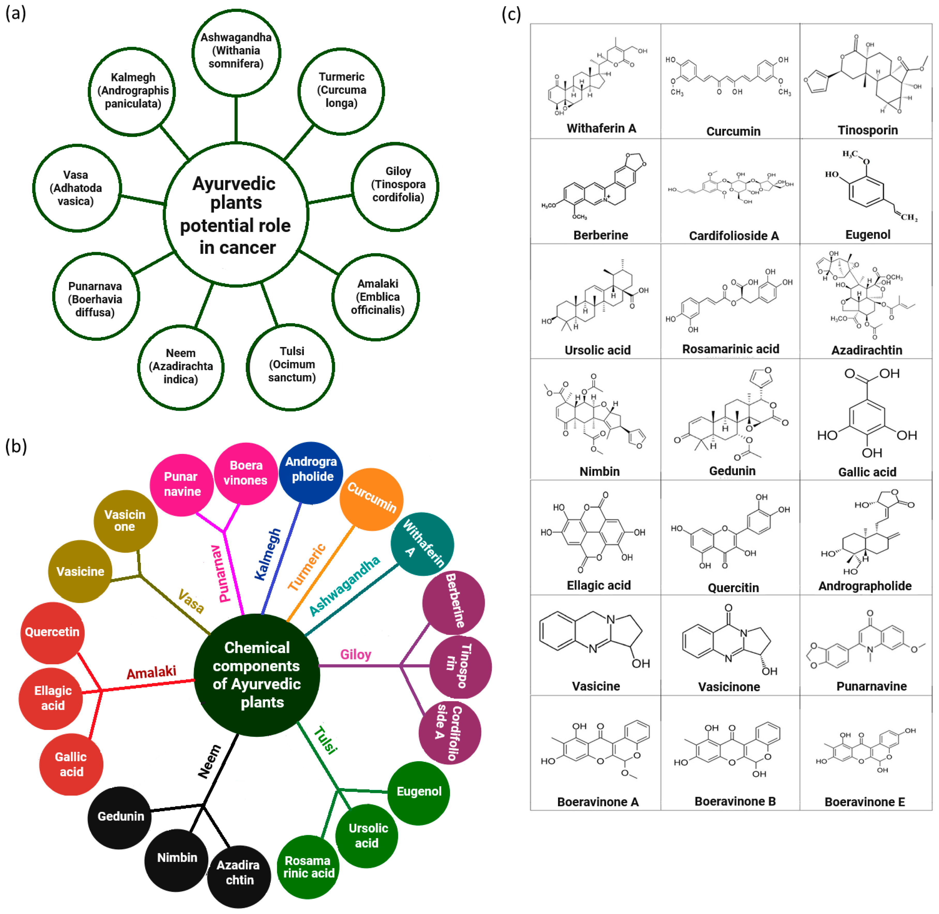
Figure 1. Ayurvedic phytochemicals exhibit promising potential in cancer therapy, with numerous compounds already recognized for their significant anticancer properties. (a) Highlights key Ayurvedic plants with potential roles in cancer management, while (b) depicts the major bioactive phytochemicals derived from these plants. (c) Presents the chemical structures of selected anticancer phytochemicals of Ayurvedic origin. For detailed insights into their molecular mechanisms, including involvement in ADPr and associated crosstalk pathways, please refer to the main text and corresponding bibliographic references.
Cells 14 01753 g001
Figure 2. Proposed model of phytochemical-mediated modulation of cellular stress and ADP-ribosylation signaling. Bioactive phytochemicals from medicinal plants may enter cells via passive diffusion, transporters, or receptor-mediated pathways, influencing endoplasmic reticulum (ER) and mitochondrial stress responses. These stresses activate the DNA damage response (DDR), particularly double-strand breaks (DSBs), leading to PARP1-driven ADPr (green arrow). Moderate activation promotes DNA repair and survival, whereas excessive activation depletes NAD+ and triggers cell death, apoptosis, or parthanatos (red arrow). The model proposes that phytochemicals fine-tune this balance, sensitizing cancer cells to apoptosis or enhancing repair, offering new directions for DDR-targeted therapies.
Cells 14 01753 g002
Figure 3. Molecular targets of Ayurvedic phytochemicals. The schematic illustrates the potential mechanisms by which Ayurvedic plant-derived phytochemicals activate apoptotic and autophagic pathways in human carcinoma cells. Treatment with Ayurvedic compounds upregulates pro-apoptotic markers such as BAD, cytochrome C, and caspase 3 (indicated by the upward arrow), while downregulating the anti-apoptotic protein Bcl-2 (indicated by the downward arrow). Activation of PARP further amplifies apoptotic signaling (indicated by the curved dotted arrow), as observed in lung cancer cells treated with vasicine [79]. These phytochemicals modulate signal transduction networks governing ER stress, cell-cycle arrest, and autophagy, promoting programmed cell death and inhibiting tumor growth [99].
Cells 14 01753 g003
Table 1. Potential Phytochemical–Nanoparticle Coupling Strategies Targeting ADP-ribosylation Pathways in Cancer.
Sl No.Name of the
Phytochemical
Deliverable Phytochemical–Nanoparticle ConjugateSize of the Formulation (nm)Zeta Potential Values (mV)Mechanistic Insights: ADPr/PARP PathwayCancer Type(s) StudiedClinical StatusReferences
1Rosamarinic acidRosmarinic acid titanium oxide and selenium-doped
graphene oxide nanoparticles (rosamarinic acid@Se-TiO2-GO
nanocomplex)
344.8 ± 43.2−33.1 ± 2.64Cleaved PARP-1, via p53 upregulation induced by HDAC2 downregulation, promoted apoptosis.Prostate cancerPreclinical
in vitro only
[163]
2ResveratrolLiposomal formulation--Synergizes with PARP inhibitors, modulates DNA repair pathways.Breast, prostatePreclinical[164]
3AndrographolideSolid Lipid Nanoparticles (ADG-SLNs)286.1 ± 8.03−20.5 ± 0.3ADG-SLNs enhance apoptosis in HN models; PARP/ADPr involvement (cleaved PARP-1, PARylation) remains to be tested.Head-and-neck cancer (HIOEC, Leuk-1, HN6, HN30 cells)Preclinical
in vitro only
[165]
4CurcuminPoly (lactic acid-co-glycolic acid) (PLGA) curcumin (Nano-CUR)70 ± 3.9-Enhances PARP inhibition, increases DNA damage, induces apoptosis.Breast, ovarianPreclinical[166]
5QuercetinSolid lipid nanoparticles154−27.7Inhibits PARP activity, enhances chemosensitivity.Lung, colon cancersPreclinical[167]
6Epigallocatechin gallate (EGCG)Chitosan nanoparticles--Downregulates PARP expression, increases ROS-mediated DNA damage.Prostate and breast cancersPreclinical[168]
7GenisteinGenistein–gold nanoparticles conjugates (Gen@AuNPs)65 ± 1.7−35 ± 2.5Enhanced antiproliferative effect; PARP/ADPr mechanisms (e.g., cleaved PARP-1 or PARP arbitrated ADPr) yet to be investigated. Prostate cancerPreclinical
(in vitro: PC3, DU145, and LNCaP cell lines)
[169]
8SilibininNo nano-conjugate reported --Prevents chemically induced lung tumors; overcomes drug resistance & metastatic traits; inhibits STAT3 in tumor and microenvironment-PARP/ADPr involvement needs further investigation.Lung cancersPreclinical[170]
9Withaferin A (WA)Gold nanoparticles (AuNP) conjugated with dexamethasone (GR ligand) and withaferin A (Au-Dex-WA nanoconjugate).--Glucocorticoid receptor (GR)-dependent cytotoxicity, epithelial–mesenchymal transition (EMT) reversal, ATP-binding cassette sub-family G member 2 (ABCG2) downregulation; potential PARP/ADPr involvement (needs further investigation).Mouse melanoma (EMT reversal, tumor regression); also studied in breast, lung (NSCLC), glioblastoma.Au-Dex-WA nanoconjugate remains preclinical; WA tested in early-phase clinical studies.[171]
10SulforaphaneUltra deformable vesicles (ethosomes®, transfersomes®)102 ± 6−21 ± 2ROS-mediated DNA damage and apoptosis via DR5, AP-1, MAPKs, mitochondrial dysfunction, and NF-κB inhibition; PARP/ADPr involvement yet to be explored.Skin cancer (melanoma, SK-MEL-28)Preclinical
in vitro (Melanoma cell lines) only
[172]
11BerberinePoly (amidoamine) (PAMAM) dendrimer encapsulated and conjugated formulation--Induced apoptosis via mitochondrial dysfunction, ROS generation, and modulation of Bcl-2 family proteins; possible PARP cleavage during apoptotic cascade (specific role of ADPr/PARP pathway not investigated—needs further exploration.Cervical cancerPreclinical
(in vitro—HeLa cells; in vivo—mouse xenograft model).
[173]
12PiperinePiperine-loaded hydroxyapatite, polymeric, and lipid nanoparticles; also, curcumin–piperine nanoparticle combinations.63.73 ± 1.07−20.46Piperine triggers DNA damage and caspase-dependent PARP-1 cleavage in apoptosis; nanoparticle delivery enhances bioavailability and sustained release, though ADPr signaling needs further investigation.Colon, prostate, and breast cancersPreclinical models[174,175]
13BaicaleinSelenium–Baicalein nanoparticles (ACM-SSe-BE), coated with A549 cell membrane for homologous targeting135.2 ± 2.52−32.23 ± 1.19Enhances ROS generation, promotes apoptosis and proliferation inhibition; possible ADPr/PARP involvement remains to be further investigatedA549 (non-small-cell lung cancer)Preclinical (in vitro and in vivo in animal models)[176]
14ApigeninPolymer–lipid hybrid nanoparticles (PLHNPs), macrophage-membrane-coated PEG micellar system (m@PEG-AGN), nanocrystals, micelles, liposomes, poly (lactic-co-glycolic acid) (PLGA)125.73 ± 5.57−26.71 ± 1.93Causes cell cycle arrest, ROS-induced DNA damage, apoptosis; suppresses metastasis (MMP/Akt); PARP signaling through ADPr in physiological/pathophysiological context needs further investigationBreast cancer (Triple-negative), colorectal carcinoma, and other cancer cell lines, often in vitro and/or in animal modelsApigenin remains at the preclinical stage. However, nanoformulations improve solubility, bioavailability & targeting, showing promise for clinical use[177,178,179]
Disclaimer/Publisher’s Note: The statements, opinions and data contained in all publications are solely those of the individual author(s) and contributor(s) and not of MDPI and/or the editor(s). MDPI and/or the editor(s) disclaim responsibility for any injury to people or property resulting from any ideas, methods, instructions or products referred to in the content.