Article

circSATB1 Modulates Cell Senescence in Age-Related Acute Myeloid Leukemia: A Mechanistic Proposal

1
Department of Clinical Medicine, School of Medical, Wuhan University of Science and Technology, Wuhan 430065, China
2
Department of Environmental Hygiene and Occupational Medicine, School of Public Health, Wuhan University of Science and Technology, Wuhan 430065, China
3
Hubei Province Key Laboratory of Occupational Hazard Identification and Control, Wuhan University of Science and Technology, Wuhan 430065, China
*
Author to whom correspondence should be addressed.
These authors contributed equally to this work.
Cells 2025, 14(15), 1181; https://doi.org/10.3390/cells14151181
Submission received: 27 June 2025 / Revised: 24 July 2025 / Accepted: 27 July 2025 / Published: 31 July 2025
(This article belongs to the Special Issue The Role of Cellular Senescence in Health, Disease, and Aging)

Abstract

Acute myeloid leukemia (AML) is a malignant hematological tumor with a high prevalence in elderly people, and circular RNA (circRNA) plays an important role in age-related diseases. Induction of cancer cell senescence is a highly promising therapeutic strategy; however, the presence of senescence-associated circRNAs in AML remains to be elucidated. Here, we show that the expression patterns of circRNAs differed between elderly AML patients and healthy volunteers. circSATB1 was significantly overexpressed in elderly patients and AML cells. Knockdown of circSATB1 resulted in the inhibition of proliferation and arrest of the cell cycle in the G0/G1 phase; no effect on apoptosis or DNA integrity was observed, and precocious cellular senescence was promoted, characterized by no change in telomere length. Database analysis revealed that there may be two miRNA and nine RNA-binding proteins (RBPs) involved in regulating the cellular functions of circSATB1. Our observations uncover circSATB1-orchestrated cell senescence in AML, which provides clues for finding more modest therapeutic targets for AML.

1. Introduction

AML, the most common type of acute leukemia in adults, is becoming increasingly prominent in terms of incidence and severity against the backdrop of global aging [1]. By 2050, the global population aged 65 and older is projected to increase from 761 million in 2021 to 1.6 billion, rising from 9% to 16% of the total population [2]. The growth rate of the population aged 80 and above is even faster, with an estimated 140 million in 2021 and 459 million (4.5% of the total population) by 2050 [3]. Most AML patients are aged 50 or older, with the highest incidence and mortality observed in those aged 70 and above [4]. From 2010 to 2017, the overall survival rate for AML patients over 70 was only 5% [5]. The incidence of AML exhibits a pronounced age-dependent pattern. According to the SEER (Surveillance, Epidemiology, and End Results) database, the median age at AML diagnosis is 67 years, with 50–60% of patients diagnosed at ≥65 years and over 30% at ≥75 years [6]. As global aging intensifies (the proportion of the population aged 60 and above is expected to reach 22% by 2050), the absolute number of elderly AML patients will continue to rise [7]. Older AML patients typically present with higher genetic risk (e.g., TP53 mutations and complex karyotype) and poorer performance status (PS ≥ 2 in over 40% of cases) [8], leading to lower treatment tolerance, reduced remission rates, and shorter survival. The complete remission (CR) rate for standard induction chemotherapy in patients ≥65 years is only 30–50%, with a 5-year overall survival (OS) rate of less than 10% [9], significantly lower than the 40–60% observed in younger patients.
CircRNA, a class of highly stable non-coding RNA with multidimensional regulatory functions, exhibits complex regulatory networks in the pathogenesis of age-related diseases and the aging process [10]. Following ischemic stroke, circSCMH1 enhances vascular endothelial cell repair and neurological recovery by modulating the ubiquitination of fat and obesity-associated (FTO) protein, reducing m6A modification of the lipid phosphatase phospholipid phosphatase 3 (Plpp3) and increasing its mRNA stability. In Alzheimer’s disease (AD) patients [10], circPSEN1 is significantly up-regulated, promoting amyloid-β (Aβ) production by inhibiting miR-137 expression and activating the nuclear factor of activated T cells 1 (NFATC1) and epidermal growth factor receptor (EGFR) pathways [11]. circSTX12 competitively binds to Casitas B-lineage lymphoma (CBL) protein, inhibiting the Hippo/YAP (Yes-associated protein) pathway, promoting adipogenic differentiation of bone marrow mesenchymal stem cells (BMSCs) and suppressing osteogenic differentiation, thereby contributing to bone loss in senile osteoporosis (OP). Antisense oligonucleotides (ASOs) targeting circSTX12 significantly improve bone microstructure in mouse models [12]. These studies highlight the central regulatory role of circRNA in age-related and aging-associated diseases, providing novel insights for targeted interventions.
Cellular senescence, first proposed by Hayflick in 1961, describes a state of irreversible growth arrest in actively proliferating cells [13]. Cellular senescence represents the third major tumor-suppressive mechanism following apoptosis and DNA damage repair. Senescent cells exhibit irreversible cell cycle arrest while retaining metabolic activity and secrete a variety of factors, including cytokines, chemokines, and extracellular matrix proteases, which collectively constitute the senescence-associated secretory phenotype (SASP). Therapy-induced senescence (TIS), characterized by irreversible cell cycle arrest and activation of immune clearance mechanisms, has emerged as a cutting-edge strategy in cancer treatment, particularly for elderly patients intolerant to chemotherapy or radiotherapy. Senescence-like phenotype mediated by IFI16 in glioblastoma (GBM) suppresses ferroptosis via Heme Oxygenase-1 (HMOX1) activation, conferring radiation resistance. The classic antidiabetic drug glyburide disrupts the binding of Interferon Gamma Inducible Protein 16 (IFI16) to transcription factors, restoring radio sensitivity [12]. Additionally, circRNA-induced tumor cell senescence is gaining attention. Inhibition of circPETH-147aa significantly increases Reactive Oxygen Species (ROS) accumulation, inducing DNA damage and cellular senescence [14]. ASOs targeting circITGB6 markedly suppress tumor progression in colorectal cancer liver metastasis models and induce a senescent phenotype [12]. However, research on circRNA-induced cellular senescence in AML remains limited.
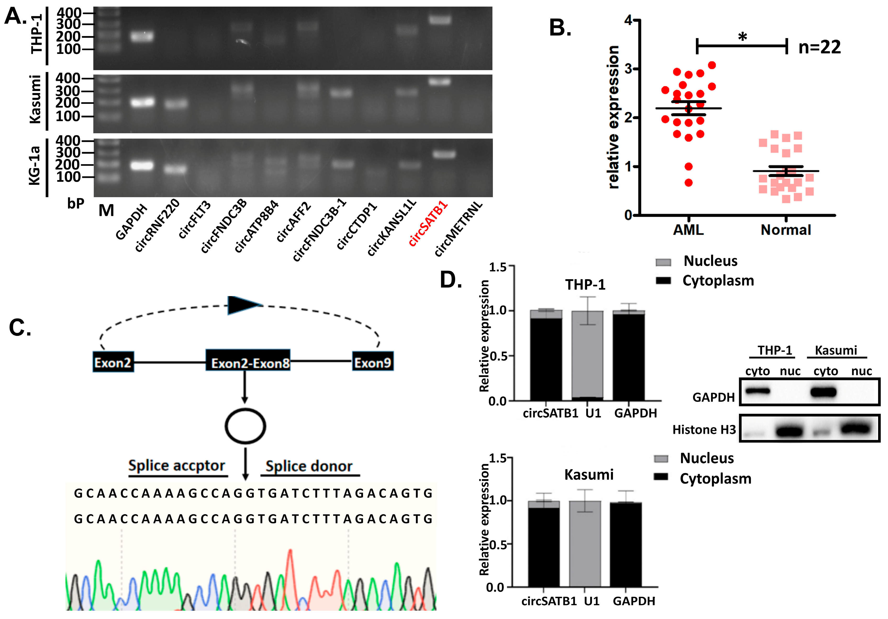
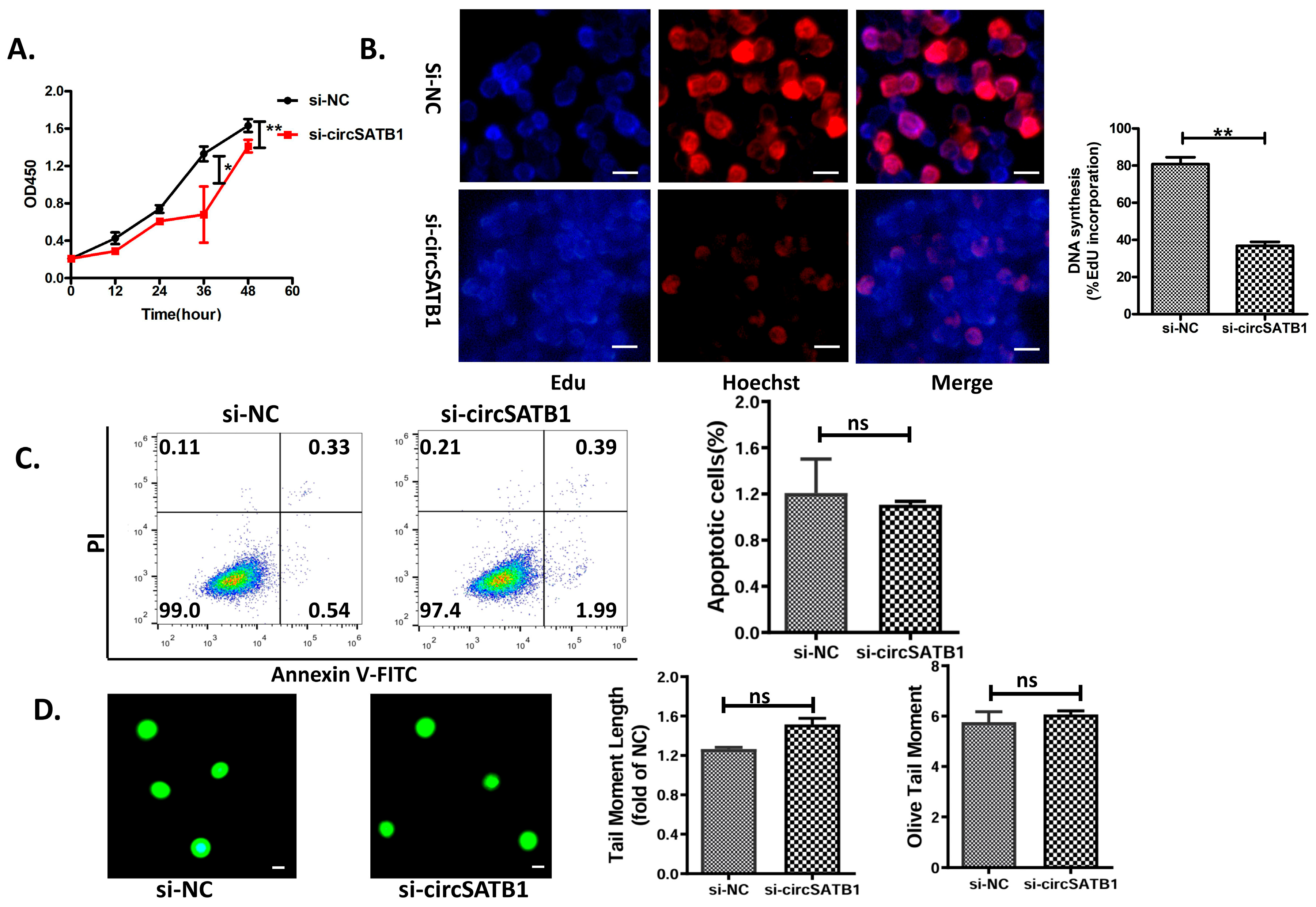
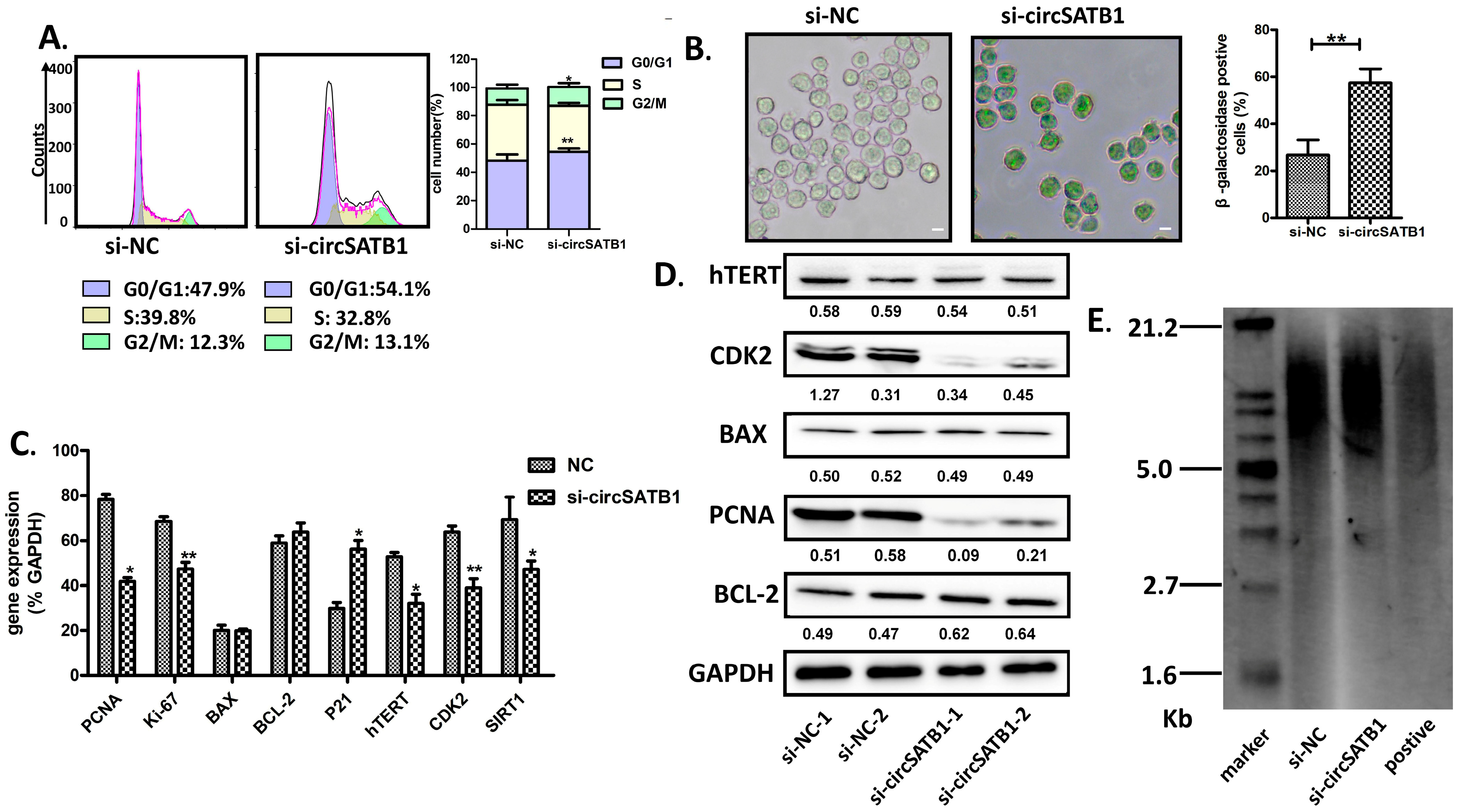
In this study, we identified circSATB1 as a significantly overexpressed circRNA in AML through whole-transcriptome sequencing and bioinformatics analysis. We validated its circular nature and confirmed its predominant cytoplasmic localization. Further siRNA knockdown experiments demonstrated that circSATB1 inhibition suppresses cell proliferation and promotes cellular senescence, with no significant effects on apoptosis or DNA damage. These findings suggest that circSATB1 holds considerable therapeutic potential in AML, possibly serving as a critical therapeutic target for inducing AML cell senescence.

2. Materials and Methods

2.1. Sample Collection and Transcriptome Sequencing

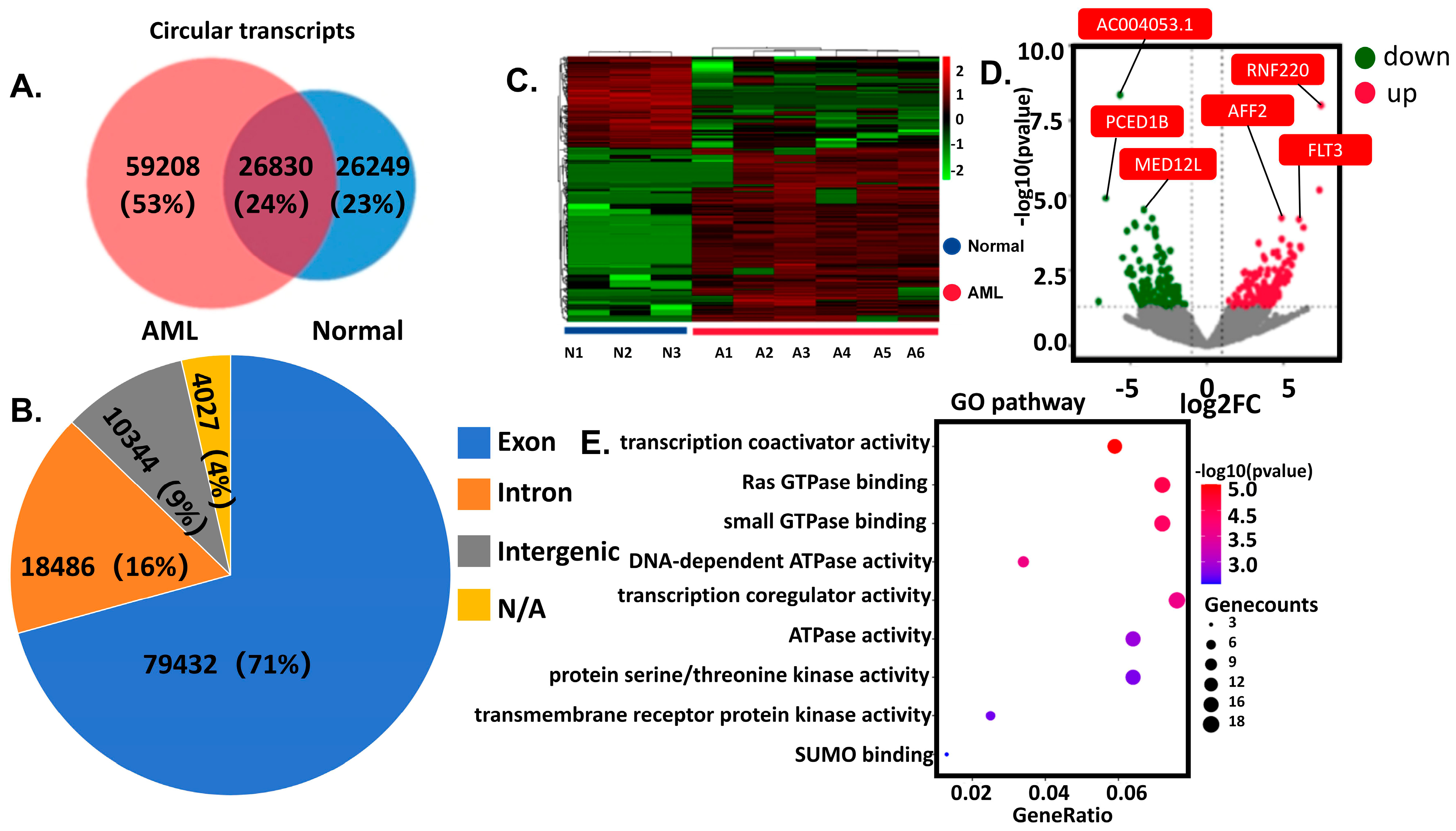
Discovery Cohort (RNA sequencing): Peripheral blood was collected from 6 AML patients and 3 healthy controls. Demographic and clinical characteristics have been comprehensively documented in Supplementary Material Table S1. Nuclear cells isolated via density gradient centrifugation underwent RNA sequencing (Illumina platform, Novogene, Sacramento, CA, USA). Differentially expressed circRNAs were identified through integrated computational analysis (CIRCexplorer2, CircFinder) with cross-database annotation. Differentially expressed genes (DEGs) were defined as |logFC| > 1 and p-value < 0.05. Validation Cohort (Clinical validation): circSATB1 expression was assessed in 44 independent anticoagulated blood samples (22 AML vs. 22 controls). All procedures complied with institutional ethics guidelines, with written informed consent obtained from participants.

2.2. Cell Culture and Treatment

KG-1a, THP-1, and Kasumi cells were purchased from the cell bank of Type Culture Collection of the Chinese Academy of Sciences (Shanghai, China). Cells were maintained in RPMI-1640 medium (HyClone, South Logan, UT, USA) supplemented with fetal bovine serum (FBS; Gibco, Carlabad, CA, USA, 10% for KG-1a and THP-1, 20% for Kasumi) and 1% penicillin/streptomycin (Gibco, Carlabad, CA, USA) under standard conditions (37 °C/5% CO2) with 2–3 times weekly passaging. siRNA transfection utilized Zlipo2000 (ZomanBio, Beijing, China) in 6-well plates (4–6 h incubation), followed by 24–72 h culture post-medium replacement. For siRNA transfection, Pepmute (ZomanBio, Beijing, China) complexes (40 pmol siRNA, RT incubation: 5 + 15 min) were applied to plates, with cells harvested after 24–72 h.
Nuclear-cytoplasmic fractionation was performed in THP-1 and Kasumi cells using pre-chilled Cell Fractionation Buffer (Thermo Fisher Scientific, Waltham, MA, USA) supplemented with PMSF (Beyotime, Shanghai, China, 1 mM final concentration). Sequential centrifugations (500× g, 4 °C, 5 min) isolated cellular components, followed by lysis in 2× Lysis Binding Solution (Thermo Fisher Scientific, Waltham, MA, USA; β-mercaptoethanol-supplemented to 1% (v/v)) and ethanol precipitation. RNA was purified via filter cartridge washes (Wash Solutions 1–3) and eluted in 95 °C Elution Buffer. Nuclear/cytoplasmic proteins were reserved for immunoblotting, while RNA underwent reverse transcription for qPCR analysis.

2.3. PCR and qPCR

Total RNA was reverse-transcribed using a Hifair® II 1st Strand cDNA Synthesis Kit (gDNA digester plus) (Yeasen, Shanghai, China) according to the manufacturer’s instructions. Briefly, 1 μg total RNA was mixed with gDNA digester and incubated at 42 °C for 30 min, and 85 °C for 5 min. For PCR, 2× HieffTM PCR Master Mix (With Dye) (Yeasen, Shanghai, China) was used. The cDNA and gDNA PCR products were evaluated using 2% agarose gel electrophoresis. The qPCR was conducted using 2× SYBR Green qPCR Master Mix (Bimake, Houston, TX, USA). GAPDH, β-actin, and U1 were used as controls. For quantification of gene expression, the 2−∆∆CT method was used, and the data were normalized to an endogenous control.

2.4. Western Blot Analysis

Cells were lysed in RIPA buffer (Beyotime, Shanghai, China), denatured in 5× loading buffer (100 °C, 10 min), and resolved via SDS-PAGE (10% or 15%). Proteins were transferred to PVDF membranes, blocked with 5% BSA (Biosharp, Hefei, China), and incubated overnight at 4 °C with primary antibodies: anti-GAPDH, Histone H3, CDK2, hTERT, BAX, PCNA and BCL-2 (Abclonal, Wuhan, China). Membranes were washed (TBST × 3), incubated with HRP-conjugated secondary antibodies (1:5000, 1 h, RT), and visualized using a ChemiDoc system (Bio-Rad, Hercules, CA, USA) with ECL (Beyotime Biotechnology, Shanghai, China). Band intensity was quantified via ImageJ (NIH, ImageJ 1.x).

2.5. Cell Proliferation

THP-1 cells were incubated with EdU (RiboBio, Guangzhou, China) in complete medium (1:1000 dilution) for 2 h post-transfection 48 h. Cells (1 × 10⁴/well, 96-well plate, n = 3) were fixed with 4% paraformaldehyde (Biosharp, Hefei, China), permeabilized (0.5% Triton X-100), and stained with Apollo reaction mix and Hoechst 33,342 (Beyotime, Shanghai, China; RT, dark, 30 min). Fluorescence imaging was performed using Optoelectronic Technology microscope (Mingmei, Guangzhou, China). Additionally, cell viability was assessed by a CCK-8 assay kit (Biosharp, Hefei, China) in Kasumi cells.

2.6. Cell Apoptosis

THP-1 cells were harvested by centrifugation (500× g, 5 min) post-transfection 48 h, washed twice with ice-cold PBS, and stained with Annexin V-FITC (Beyotime, Shanghai, China) in binding buffer (4 °C, dark, 15 min), followed by PI addition (4 °C, dark, 5 min). Unstained, FITC-only, and PI-only controls were included. Apoptotic subpopulations (Annexin V-FITC+/PI−: early apoptosis; Annexin V-FITC+/PI+: late apoptosis/necrosis) were quantified via flow cytometry (BD Biosciense, San Jose, CA, USA).

2.7. Cell Cycle

THP-1 cells were harvested by centrifugation (500× g, 5 min, RT), washed twice with ice-cold PBS, and fixed in 70% ethanol (−20 °C, ≥2 h). After rehydration, pellets were stained with PI (BD Biosciences, San Jose, CA, USA, 50 μg/mL), RNase A (Sigma-Aldrich, St. Louis, MO, USA, 100 μg/mL), and 0.1% Triton X-100 in PBS (Servicebio, Wuhan, China, 37 °C, 30 min, dark). Cell suspensions were filtered through 40 μm mesh and analyzed on a flow cytometer (488 nm laser; PI detection: bandpass filter centered at 585 nm with 40 nm bandwidth [585/40 nm]), with ≥10,000 events recorded per sample. Instrument calibration utilized unstained controls and fluorescent beads.

2.8. Comet Assay

The alkaline single-cell gel electrophoresis technique (comet assay) was used to measure the DNA damage in THP-1 cells using an OxiSelect Comet Assay Kit (Cell Biolabs, Chicago, IL, USA). Briefly, liquid agarose was pipetted onto a comet slide and chilled. Next, cell samples were combined with comet agarose, added on top of the base layer, lysed to form nucleoids containing supercoiled loops, and then immersed in alkaline solution. Then, the samples were electrophoresed for 30 min under voltage 25 V and current 300 mA to separate intact DNA from damaged fragments, stained with a diluent DNA dye (Cell Biolabs, Chicago, IL, USA), and visualized by epifluorescence microscopy using a FITC filter (MshOt, Guangzhou, China, 40×). Comet Assay Software Project (CaspLab-1.2.2) was used to quantify the tail parameter.

2.9. β-Galactosidase Staining

β-Galactosidase Staining was measured in THP-1 using the β-galactosidase staining kit (BeiboBio, Shanghai, China) according to the manufacturer’s protocol. Harvest cells by centrifugation at 300× g for 5 min at room temperature into a 1.5 mL centrifuge tube. Wash once with PBS, add 1 mL of β-galactosidase staining fixative, and fix at room temperature for 15 min. During fixation, slowly shake the tube on a shaker to avoid the cells from clumping together. After centrifugation (300× g, 5 min, RT), remove the cell fixative, wash the cells 3 times with PBS, each time for 3 min. Centrifuge (300× g, 5 min, RT), remove the PBS, and add 0.5–1 mL of staining working solution to each tube. Incubate at 37 °C overnight. Drop some of the stained cells onto a slide or into a 6-well plate, and observe under a common optical microscope.

2.10. Southern Blot

The measurements of terminal restriction fragment (TRF) length were applied using the TeloTAGGG telomere length assay kit (Roche, Basel, Switzerland). Briefly, DNA extraction from KG-1a cells was performed through sequential lysis, and nucleic acid purity was verified by NanoDrop 2000 spectrophotometry (Thermo Fisher, Waltham, MA, USA), confirming the A260/280 ratio (≥1.8). Equal volumes of HinfI and RsaI were combined for concurrent DNA digestion and positive control preparation, while the molecular marker was formulated by mixing 4 μL Bottle 6, 12 μL RNase-free water, and 4 μL Bottle 7. Electrophoresis was conducted in 0.8% agarose gel with 1× TAE running buffer (Servicebio, Wuhan, China) at 50 V for 5 h, followed by sequential post-electrophoretic processing: membrane transfer via capillary action (ambient temperature, overnight), UV cross-linking, prehybridization for 1 h and hybridization (42 °C, 3 h) in freshly prepared buffer, and chemiluminescent detection using Bottle 15 substrate.

2.11. Statistical Analysis

Statistical analyses were performed using GraphPad Prism software (version 7.0). Each experiment was independently repeated three times, and the representative data are shown. All the values are presented as mean ± SD of three biologically independent samples. Statistical analyses were performed using a two-way analysis of variance (ANOVA) when comparing at least three groups. The sample size is indicated in the corresponding figure legends. Statistical significance was defined as * p < 0.05, ** p < 0.01.

3. Results

Expression patterns of circRNAL in AML were mapped through RNA-seq of clinical samples and validated in cell lines; ultimately, circSATB1 was identified as the target for study. Subsequently, siRNA transfection was employed to investigate the cellular function, and its role in regulating premature cellular senescence in AML was corroborated. Finally, a prediction of the potential regulatory mechanism was made through bioinformatics analysis.

3.1. Identification of Differential circRNAs and Functional Enrichment

For the sequencing data, a total of 112,289 circRNAs were identified through bioinformatics tools such as Find-circ, CIRI, CircRNA finder, and Circexplorer. Among these, 59,208 (53%) were unique to AML patients, approximately twice the number found in healthy volunteers (Figure 1A). Source analysis of the identified circRNAs revealed that the majority were derived from exons (79,432, 71%), followed by introns (18,486, 16%), with a small proportion originating from intergenic regions or unannotated genomic loci (Figure 1B). Subsequent differential expression analysis identified 256 up-regulated and 135 down-regulated circRNAs (Figure 1C,D). Further functional enrichment analysis of the parental genes of these differentially expressed circRNAs demonstrated significant associations with pathways such as transcription coregulator activity, Ras-GTPase activity, and DNA-dependent ATPase activity (Figure 1E). The detailed enrichment data of the GO analysis results, including p-values and adjusted p-values (adj.p), have been comprehensively documented in Supplementary Material Table S2. These results indicate distinct circRNAs expression patterns between AML patients and healthy volunteers.

3.2. circSATB1 Highly Expressed in AML Clinical Samples and Cell Lines

Among the top ten differentially overexpressed circRNAs identified in AML (Table 1), we selected circSATB1 for further validation based on its consistent overexpression profile across three representative AML cell lines (THP-1, Kasumi-1, and KG-1a) as determined by PCR analysis (Figure 2A). Clinical validation in the Validation Cohort using qPCR (22 matched AML-control pairs) confirmed significant up-regulation of circSATB1 in AML specimens compared to normal controls (Figure 2B, p < 0.05), corroborating our initial sequencing findings and establishing circSATB1 as a promising candidate for functional characterization.
To unequivocally confirm the circular topology of circSATB1, we performed Sanger sequencing of PCR-amplified products from THP-1 cell cDNA. The obtained sequences showed perfect concordance (100% identity) with the predicted back-splice junction (chr3: 18419661|18462483) in UCSC genome annotations, verifying canonical head-to-tail splicing of exons 2–9 (Figure 2C). This molecular validation confirms circSATB1 as a bona fide circular RNA species. Subcellular fractionation experiments coupled with qPCR analysis revealed that circSATB1 exhibited predominant cytoplasmic localization (>85% of total cellular circSATB1) in THP-1 and Kasumi cells, and Western blot analysis of fractionated lysates (using U1 and GAPDH as nuclear/cytoplasmic markers, respectively) confirmed the purity of subcellular fractions (Figure 2D). This distribution pattern suggests circSATB1 may primarily function through cytoplasmic mechanisms, potentially involving post-transcriptional regulation or signaling pathway modulation.

3.3. circSATB1 Knockdown Diminished Cell Proliferation Without Apoptosis and DNA Damage

To investigate the functional role of circSATB1 in cellular proliferation, we designed a junction-specific siRNA targeting the back-splice junction of circSATB1, with scrambled siRNA (si-NC) serving as a negative control. Cellular viability was quantitatively evaluated using CCK-8 assays, and the results showed that circSATB1 knockdown significantly inhibited the proliferation of Kasumi cell lines (Figure 3A, p < 0.05). In addition, EdU incorporation assays were performed in KG-1a cells at 48 h post-transfection. Quantitative fluorescence imaging demonstrated a significant reduction in EdU-positive proliferating cells following circSATB1 knockdown compared to scramble controls (Figure 3B, 51.26% reduction in si-cicrSATB1 group, p < 0.01). To assess whether the observed growth inhibition was associated with apoptosis, Annexin V-FITC/PI dual-labeling flow cytometry was conducted in THP-1 cells. As shown in Figure 3C, no significant alterations in apoptotic rates were detected. The cumulative apoptotic incidence remained below 3%, with si-circSATB1 transfection yielding non-significant differences in early (1.99% vs. 0.54%) and late apoptotic populations (0.39% vs. 0.33%). Alkaline comet assay analysis in THP-1 cells demonstrated preserved genomic stability, with no significant differences observed in Olive Tail Moment and Tail DNA% between the circSATB1-knockdown and control groups (p > 0.05, Figure 3D). It is evident that circSATB1 modulates AML cells through a “non-aggressive” mechanism, characterized by cytostatic proliferation arrest without inducing apoptosis or compromising DNA integrity. This aligns with the hallmark features of cellular senescence, serving as a clear indicator of the aging state in these cells.

3.4. circSATB1 Knockdown-Attenuated Cell Senescence

To investigate the regulation of circSATB1 on the cell cycle, flow cytometric analysis in THP-1 cells was conducted, revealing that circSATB1 silencing induced significant cell cycle arrest. Specifically, circSATB1-depleted cells displayed pronounced G0/G1 phase accumulation (Figure 4A), indicative of proliferative cessation. Complementary senescence-associated β-galactosidase (SA-β-gal) staining assays confirmed the induction of cellular senescence in THP-1 cells, with a significant increase in characteristic blue staining compared to controls (Figure 4B), consistent with established senescence biomarkers. Subsequent investigation of SASP components revealed altered expression patterns of key regulatory molecules. Western blot quantification via ImageJ demonstrated significant down-regulation of proliferative markers PCNA (proliferating cell nuclear antigen), coupled with CDK2 (cyclin-dependent kinase 2) in circSATB1-deficient KG-1a cells (Figure 4C,D). Notably, hTERT (human telomerase reverse transcriptase) displayed discordant expression patterns across transcriptional and translational levels. While hTERT mRNA levels were significantly diminished in knockdown cells, protein expression remained statistically unaltered, suggesting potential post-transcriptional regulatory mechanisms, such as alternative splicing or translational compensation. To clarify the telomere maintenance dynamics, Southern blot analysis was employed. Quantitative assessment demonstrated comparable telomere restriction fragment lengths between control and circSATB1-knockdown groups in KG-1a cells (Figure 4E). These collective findings establish that circSATB1 depletion induces acute senescence in AML cells through telomere-independent pathways, distinct from classical replicative senescence mechanisms. This phenotype aligns with emerging paradigms of stress-induced premature senescence, potentially mediated through cell cycle checkpoint activation and SASP factor modulation.

3.5. circSATB1 Regulatory Network Prediction

To elucidate the potential mechanism by which circSATB1 regulates premature cellular senescence in AML cells, we performed predictive analyses for miRNAs that may interact with circSATB1 using the circBank and IDCSC databases. Two miRNAs, miR-450b-5p and miR-452-5p, were co-predicted by both databases (Figure 5A). Subsequently, the potential target genes of miR-450b-5p and miR-452-5p were predicted using three databases: TargetScan, miRTarBase, and miRDB, and the overlapping target genes from these databases were identified (Figure 5B,C). Based on the Score provided by miRDB, genes with a Score >90 were selected for further analysis. This yielded nine target genes for miR-450b-5p (CENPK, GABBR2, RBM47, ARID1A, UNC5D, ADAM19, LUM, SESN3, and KCNIP3) and two target genes for miR-452-5p (CASD1 and CDKN1B) (Figure 5D).
Additionally, RNA-binding proteins (RBPs) that may interact with circSATB1 were predicted using the catRAPID and RBPsuit databases. Eight RBPs were co-predicted: AGO1, AGO3, DGCR8, EIF4A3, FUS, FXR2, MOV10, and TNRC6. The protein–protein interaction (PPI) network of these RBPs was analyzed using the STRING database, and experimentally validated interactions (as documented in STRING) were visualized (Figure 5E).
These results demonstrate that circSATB1 is associated with cellular senescence, exhibits high expression in AML, and exerts an anti-senescence effect. Knockdown of circSATB1 promotes premature senescence and inhibits cell proliferation (Figure 6), suggesting its potential as a critical therapeutic target for inducing AML cell senescence.

4. Discussion

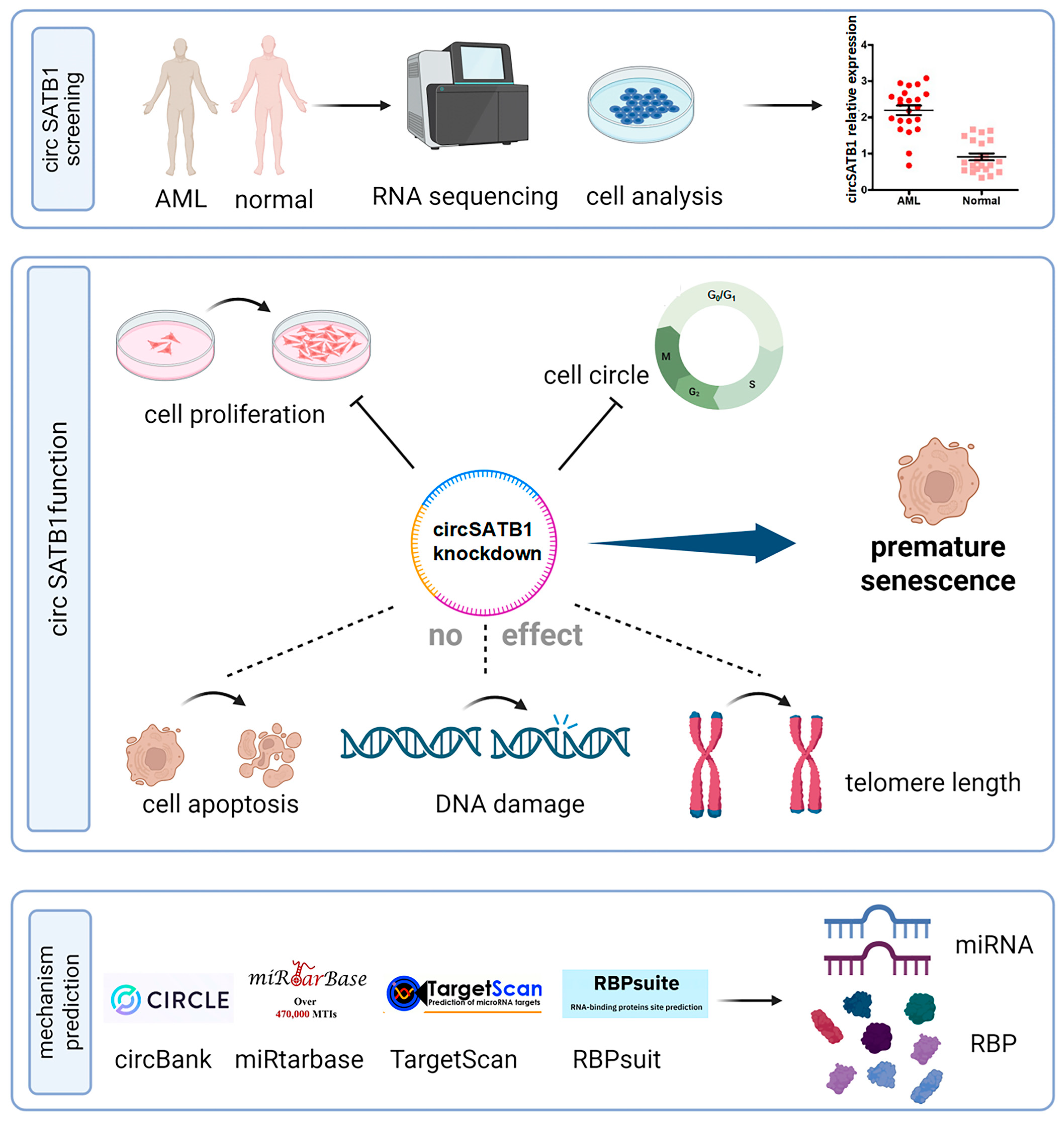
circSATB1, a previously uncharacterized circular RNA, was highly expressed in elderly AML patients and orchestrates a telomere-independent senescence program upon knockdown. Using multi-omics and functional validation, we demonstrate that (1) circSATB1 is significantly overexpressed in AML clinical samples and cell lines (Figure 2A,B); (2) its inhibition induces G0/G1 arrest and premature senescence without triggering apoptosis or DNA damage (Figure 3 and Figure 4); (3) this senescence phenotype occurs independently of telomere shortening (Figure 4E). These results establish circSATB1 as a novel regulator of cellular senescence in AML.
Population aging is a global public health challenge. Age-related diseases are becoming an increasingly serious public health concern [15]. It is both imperative and urgent to address these challenges through specific aging biomarkers. Also, their impact on age-related diseases is relatively unexplored, and their potential mechanisms to minimize or alleviate this rapid aging trend remain unclear. Recent research has proposed twelve hallmarks of aging, such as genomic instability, telomere attrition, mitochondrial dysfunction, cellular senescence, and chronic inflammation, which all contribute to understanding the progression of aging. Identifying biomarkers of aging and risks associated with age-related diseases is crucial for supporting public health initiatives for healthy aging, developing a priority control list, and enhancing regulation. Acute myeloid leukemia, the most common adult leukemia, predominantly affects individuals aged 60–70 years [16]. Elderly patients (≥65 years) exhibit significantly higher mortality rates, with a dismal 5-year relative survival rate of only 29.5% [17].
circRNAs play important roles in a variety of tumors. Signature circRNAs have been identified in Alzheimer’s disease [18], brain cell glioma [19], lung cancer [20], and other tumors in the elderly. In addition, tissue fluids and body fluids (saliva, blood, etc.) have been found to be rich in circRNAs [21], which are ideal biomarkers for liquid biopsy and may be a potential target for hematological tumor therapy, but it is not yet clear which circRNAs are involved in the development of AML. Certain circRNAs exhibit spatiotemporal expression patterns correlating with cell cycle phases, while others serve as molecular indicators of cellular senescence. As the third tumor barrier following apoptosis and DNA damage repair, senescence induces irreversible growth arrest while maintaining metabolic activity, thereby limiting cancer proliferation [22]. The Belgian group’s “one-two punch” strategy “CDC7 inhibitor-induced senescence” + “mTOR blocker-triggered apoptosis” effectively eliminated hepatocellular carcinoma cells with improved patient tolerance compared to conventional cytotoxic therapies [23]. Achieving cancer eradication while preserving quality of life remains a therapeutic dilemma, particularly for elderly AML patients. Therefore, exploring a similar therapeutic strategy to be adopted for AML is of significant interest. Identifying circRNAs that induce senescence in AML cells is a crucial step in this direction. Our study addresses this gap by identifying cirSATB1 as a senescence modulator in AML. While our data demonstrate that circSATB1 knockdown induces senescence (Figure 3 and Figure 4), the preliminary nature of the predicted regulatory networks (Figure 5) must be emphasized. Causal relationships between circSATB1 and senescence effectors remain to be established.
The ability of cancer cells to evade senescence is contingent on their sustained proliferative capacity. Cellular senescence can be classified into replicative senescence (telomere-dependent) and premature senescence (telomere-independent and stress-induced) [24,25,26,27,28]. In this study, we demonstrated that circSATB1 knockdown induced premature senescence in AML cells, characterized by maintenance of telomere length and obstruction of cell cycle progression. This suppression of uncontrolled AML cell proliferation offers a novel and mild therapeutic approach, providing a framework for identifying more tolerable AML treatment options. Such options include the induction of cellular senescence to halt disease progression, which holds significant prognostic value for elderly patients. Our findings align with emerging interest in senescence-inducing therapies [12,14,22,23], though the specific role of circRNAs in AML senescence was previously unexplored. The predicted interaction networks involving
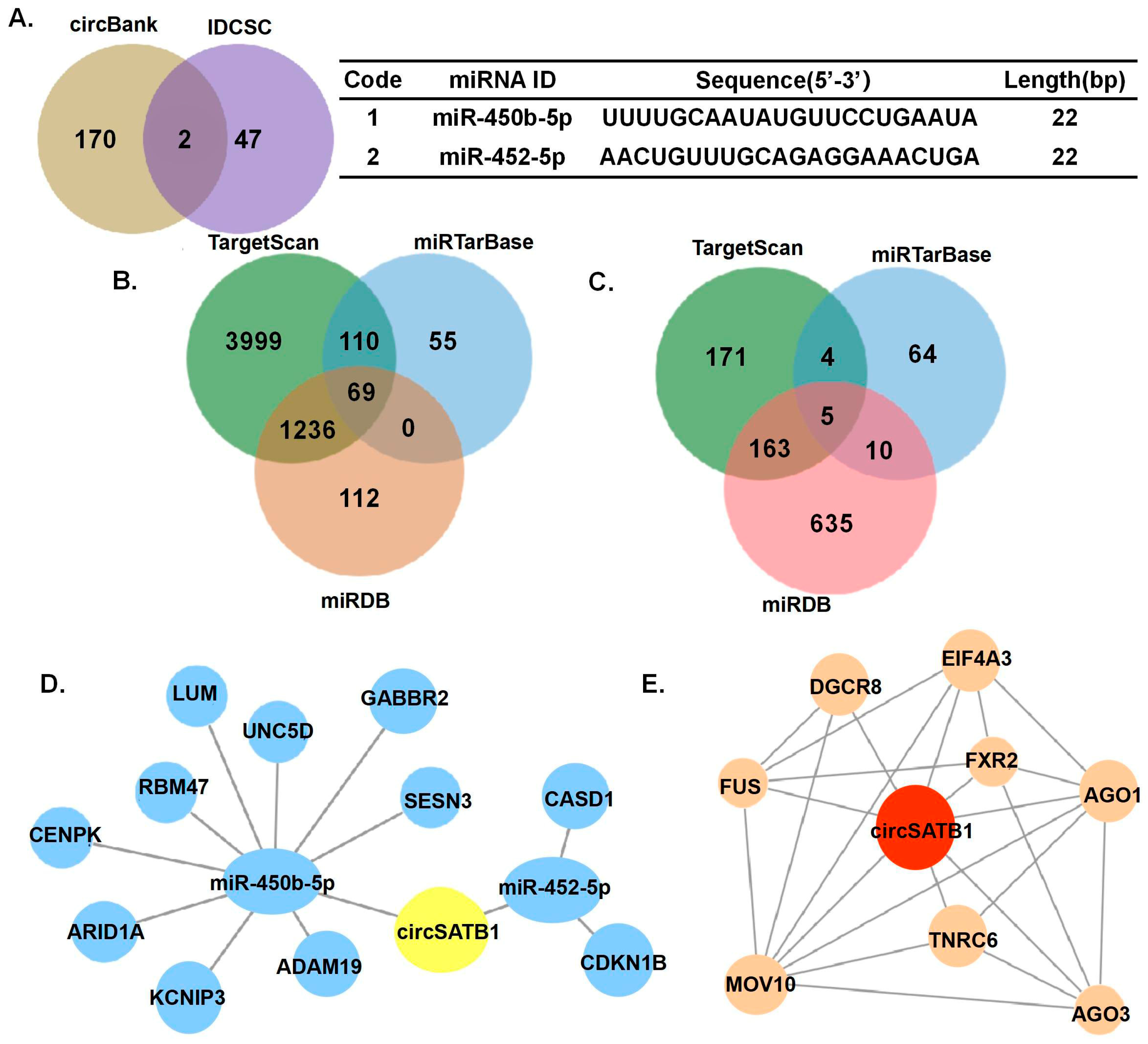
 miR-450b-5p, miR-452-5p, and specific RBPs (Figure 5) provide testable hypotheses for future mechanistic studies to understand how circSATB1 exerts its anti-senescence function. While our data provided evidence for identifying circSATB1 as a novel regulator of senescence in AML and suggest its potential as a therapeutic target, several limitations warrant acknowledgment: (1). Cohort size: Our conclusions are derived from in vitro analyses using AML cells from a limited cohort, which may not fully capture the heterogeneity of AML. Larger-scale validation in diverse populations is needed. (2). Model constraints: The exclusive use of in vitro models precludes assessment of systemic factors. Future work should integrate probing physiological relevance. (3). Mechanistic depth: Although we identified circSATB1 as a novel regulator of senescence in AML and suggest its potential as a therapeutic target, causal links require further deconvolution. Its therapeutic potential and mechanism of action remains untested.

5. Conclusions

In this study, we obtained 391 circRNAs significantly differentially expressed in AML by bioinformatics, which are potentially valuable in the study of AML. Experimental means were screened to obtain circSATB1, which is significantly highly expressed in AML and stably expressed in AML cells, and its circRNA properties were verified, and it was found to be mainly present in the cytoplasm and expressed in a variety of cancers. By knockdown of circSATB1 in AML cells, it was found that its knockdown could inhibit cell proliferation and promote premature cell senescence, while it had no significant effect on apoptosis and DNA damage. Preliminary prediction of the mechanism of action network of circSATB1 by bioinformatics means yielded two miRNAs and eight RBPs that might bind to circSATB1. These findings identify circSATB1 as a novel regulator of senescence in AML and suggest its potential as a therapeutic target. Future studies are warranted to validate the predicted regulatory networks and assess the therapeutic efficacy of targeting circSATB1 in vivo.

Supplementary Materials

The following supporting information can be downloaded at: https://www.mdpi.com/article/10.3390/cells14151181/s1, Table S1: Demographic and clinical characteristics of RNA sequencing donors; Table S2: Statistical results of GO enrichment analysis.

Author Contributions

Conceptualization, Y.F., L.H., X.W., L.Z., X.Y., Z.W., H.W. (Haodong Wu), Y.Z. and H.W. (Huiting Wang); methodology, Y.F., L.H., X.W., L.Z., X.Y., Z.W., H.W. (Haodong Wu) and Y.Z.; software, Y.F., L.H., X.W., L.Z., X.Y. and Z.W.; validation, Y.F., L.H., X.W. and L.Z.; formal analysis, Y.F., L.H., X.W., L.Z., X.Y., Z.W., H.W. (Haodong Wu), Y.Z. and H.W. (Huiting Wang); investigation, Y.F., L.H., X.W., X.Y., Z.W. and H.W. (Haodong Wu); resources, Y.F., L.H., X.W., L.Z. and X.Y.; data curation, Y.F., L.H., X.W. and L.Z.; writing—original draft preparation, Y.F., L.H., X.W., X.Y., Z.W., H.W. (Haodong Wu), Y.Z. and H.W. (Huiting Wang); writing—review and editing, Y.F., L.H., X.W., X.Y. and Z.W.; visualization, Y.F., L.H., X.W. and X.Y.; supervision, Y.F. and L.Z.; project administration, Y.F. and L.Z.; funding acquisition, Y.F. and L.Z. All authors have read and agreed to the published version of the manuscript.

Funding

This work was supported by the National Natural Science Foundation of China (No. 82200191) and Open Fund Project of Hubei Province Key Laboratory of Occupational Hazard Identification and Control (No. OHIC2023Y07).

Institutional Review Board Statement

The study was conducted in accordance with the Declaration of Helsinki, and informed consent was obtained according to protocols approved by the Ethics Committee of Wuhan University of Science and Technology (protocol code 2024153 and date of approval 19 June 2024).

Informed Consent Statement

Informed consent was obtained from all subjects involved in the study.

Data Availability Statement

The data presented in this study are available upon request from the corresponding author. The RNA sequencing datasets have been deposited to the NCBI’s Gene Expression Omnibus GEO database and are accessible through GEO Series accession numbers GSE229582 (https://www.ncbi.nlm.nih.gov/geo/query/acc.cgi?acc=GSE229582, accessed on 11 January 2025).

Acknowledgments

We acknowledge the valuable contribution of Renmin Hospital, Hubei University of Medicine for their excellent technical support and interpretation of data.

Conflicts of Interest

The authors declare no conflicts of interest.

Abbreviations

The following abbreviations are used in this manuscript:
AMLAcute myeloid leukemia
CRcomplete remission
OSoverall survival
ADAlzheimer’s disease
BMSCbone marrow mesenchymal stem cells
OPosteoporosis
ASOsAntisense oligonucleotides
TISTherapy-induced senescence
GBMglioblastoma
RBPRNA-binding proteins
qPCRQuantitative PCR (polymerase chain reaction)
PIPropidium Iodide
EdU5-Ethynyl-2′-deoxyuridine
SASPsenescence-associated secretory phenotype
TRFterminal restriction fragment

References

  1. Newell, L.F.; Cook, R.J. Advances in acute myeloid leukemia. BMJ Clin. Res. Ed. 2021, 375, n2026. [Google Scholar] [CrossRef] [PubMed]
  2. Li, K.; Guo, J.; Ming, Y.; Chen, S.; Zhang, T.; Ma, H.; Fu, X.; Wang, J.; Liu, W.; Peng, Y. A circular RNA activated by TGFβ promotes tumor metastasis through enhancing IGF2BP3-mediated PDPN mRNA stability. Nat. Commun. 2023, 14, 6876. [Google Scholar] [CrossRef] [PubMed]
  3. Carcelén-Fraile, M.d.C.; Déniz-Ramírez, N.d.P.; Sabina-Campos, J.; Aibar-Almazán, A.; Rivas-Campo, Y.; González-Martín, A.M.; Castellote-Caballero, Y. Exercise and Nutrition in the Mental Health of the Older Adult Population: A Randomized Controlled Clinical Trial. Nutrients 2024, 16, 1741. [Google Scholar] [CrossRef]
  4. Kayser, S.; Levis, M.J. The clinical impact of the molecular landscape of acute myeloid leukemia. Haematologica 2023, 108, 308–320. [Google Scholar] [CrossRef] [PubMed]
  5. Wysota, M.; Konopleva, M.; Mitchell, S. Novel Therapeutic Targets in Acute Myeloid Leukemia (AML). Curr. Oncol. Rep. 2024, 26, 409–420. [Google Scholar] [CrossRef] [PubMed]
  6. Mishra, S.K.; Millman, S.E.; Zhang, L. Metabolism in acute myeloid leukemia: Mechanistic insights and therapeutic targets. Blood 2023, 141, 1119–1135. [Google Scholar] [CrossRef]
  7. Wachter, F.; Pikman, Y. Pathophysiology of Acute Myeloid Leukemia. Acta Haematol. 2024, 147, 229–246. [Google Scholar] [CrossRef]
  8. Babakhanlou, R.; DiNardo, C.; Borthakur, G. IDH2 mutations in acute myeloid leukemia. Leuk. Lymphoma 2023, 64, 1733–1741. [Google Scholar] [CrossRef]
  9. Gómez-De León, A.; Demichelis-Gómez, R.; da Costa-Neto, A.; Gómez-Almaguer, D.; Rego, E.M. Acute myeloid leukemia: Challenges for diagnosis and treatment in Latin America. Hematology 2023, 28, 2158015. [Google Scholar] [CrossRef]
  10. Frost, B.; Dubnau, J. The Role of Retrotransposons and Endogenous Retroviruses in Age-Dependent Neurodegenerative Disorders. Annu. Rev. Neurosci. 2024, 47, 123–143. [Google Scholar] [CrossRef]
  11. Puri, S.; Hu, J.; Sun, Z.; Lin, M.; Stein, T.D.; Farrer, L.A.; Wolozin, B.; Zhang, X. Identification of circRNAs linked to Alzheimer’s disease and related dementias. Alzheimer’s Dement. 2023, 19, 3389–3405. [Google Scholar] [CrossRef]
  12. Gu, H.; Yu, W.; Feng, P.; Zeng, C.; Cao, Q.; Chen, F.; Wang, Z.; Shen, H.; Wu, Y.; Wang, S. Circular RNA circSTX12 regulates osteo-adipogenic balance and proliferation of BMSCs in senile osteoporosis. Cell. Mol. Life Sci. 2025, 82, 149. [Google Scholar] [CrossRef]
  13. Dong, Z.; Luo, Y.; Yuan, Z.; Tian, Y.; Jin, T.; Xu, F. Cellular senescence and SASP in tumor progression and therapeutic opportunities. Mol. Cancer 2024, 23, 181. [Google Scholar] [CrossRef]
  14. Lan, T.; Gao, F.; Cai, Y.; Lv, Y.; Zhu, J.; Liu, H.; Xie, S.; Wan, H.; He, H.; Xie, K.; et al. The protein circPETH-147aa regulates metabolic reprogramming in hepatocellular carcinoma cells to remodel immunosuppressive microenvironment. Nat. Commun. 2025, 16, 333. [Google Scholar] [CrossRef] [PubMed]
  15. Isola, J.V.V.; Hense, J.D.; Osório, C.A.P.; Biswas, S.; Alberola-Ila, J.; Ocañas, S.R.; Schneider, A.; Stout, M.B. Reproductive Ageing: Inflammation, immune cells, and cellular senescence in the aging ovary. Reproduction 2024, 168, e230499. [Google Scholar] [CrossRef] [PubMed]
  16. Alhajahjeh, A.; Bewersdorf, J.P.; Bystrom, R.P.; Zeidan, A.M.; Shimony, S.; Stahl, M. Acute myeloid leukemia (AML) with chromosome 3 inversion: Biology, management, and clinical outcome. Leuk. Lymphoma 2024, 65, 1541–1551. [Google Scholar] [CrossRef]
  17. Wienecke, C.P.; Heida, B.; Venturini, L.; Gabdoulline, R.; Krüger, K.; Teich, K.; Büttner, K.; Wichmann, M.; Puppe, W.; Neziri, B.; et al. Clonal relapse dynamics in acute myeloid leukemia following allogeneic hematopoietic cell transplantation. Blood 2024, 144, 296–307. [Google Scholar] [CrossRef]
  18. Su, L.; Zhang, Y.; Wang, Y.; Wei, H. Identification of a lncRNA/circRNA-miRNA-mRNA ceRNA Network in Alzheimer’s Disease. J. Integr. Neurosci. 2023, 22, 136. [Google Scholar] [CrossRef] [PubMed]
  19. Yan, Y.; Wang, H.; Hu, J.; Guo, T.; Dong, Q.; Yin, H.; Yuan, G.; Pan, Y. CircRNA-104718 promotes glioma malignancy through regulation of miR-218-5p/HMGB1 signalling pathway. Metab. Brain Dis. 2023, 38, 1531–1542. [Google Scholar] [CrossRef]
  20. Fan, Z.; Bai, Y.; Zhang, Q.; Qian, P. CircRNA circ_POLA2 promotes lung cancer cell stemness via regulating the miR-326/GNB1 axis. Environ. Toxicol. 2020, 35, 1146–1156. [Google Scholar] [CrossRef]
  21. Liu, Z.; Yang, J.; Wang, N.; Liu, J.; Geng, J.; Zhu, J.; Cong, B.; Sun, H.; Wu, R. Integrative lncRNA, circRNA, and mRNA analysis reveals expression profiles of six forensic body fluids/tissue. Int. J. Leg. Med. 2024, 138, 731–742. [Google Scholar] [CrossRef]
  22. Xu, Y.; Zhang, S.; Niu, H.; Ye, Y.; Hu, F.; Chen, S.; Li, X.; Luo, X.; Jiang, S.; Liu, Y.; et al. STIM1 accelerates cell senescence in a remodeled microenvironment but enhances the epithelial-to-mesenchymal transition in prostate cancer. Sci. Rep. 2015, 5, 11754. [Google Scholar] [CrossRef]
  23. Wang, C.; Vegna, S.; Jin, H.; Benedict, B.; Lieftink, C.; Ramirez, C.; de Oliveira, R.L.; Morris, B.; Gadiot, J.; Wang, W.; et al. Inducing and exploiting vulnerabilities for the treatment of liver cancer. Nature 2019, 574, 268–272. [Google Scholar] [CrossRef]
  24. Lin, J.; Epel, E. Stress and telomere shortening: Insights from cellular mechanisms. Ageing Res. Rev. 2022, 73, 101507. [Google Scholar] [CrossRef]
  25. Hertwig, F.; Peifer, M.; Fischer, M. Telomere maintenance is pivotal for high-risk neuroblastoma. Cell Cycle 2016, 15, 311–312. [Google Scholar] [CrossRef] [PubMed]
  26. Yalçin, Z.; Selenz, C.; Jacobs, J.J.L. Ubiquitination and SUMOylation in Telomere Maintenance and Dysfunction. Front. Genet. 2017, 8, 67. [Google Scholar] [CrossRef]
  27. Liu, J.; Wang, L.; Wang, Z.; Liu, J.P. Roles of Telomere Biology in Cell Senescence, Replicative and Chronological Ageing. Cells 2019, 8, 54. [Google Scholar] [CrossRef] [PubMed]
  28. Oshima, J.; Sidorova, J.M.; Monnat, R.J., Jr. Werner syndrome: Clinical features, pathogenesis and potential therapeutic interventions. Ageing Res. Rev. 2017, 33, 105–114. [Google Scholar] [CrossRef] [PubMed]
Figure 1. Analysis of sequencing results of circRNAs in AML. (A) Distribution of circRNAs in AML and healthy individuals. (B) Distribution of circRNAs by source. (C) Heat map showing differentially expressed circRNAs. (D) Volcano plot analysis. (E) Pathway enrichment analysis of differentially expressed circRNA transcripts of the parental gene. All pathways shown in the figure meet the statistical significance thresholds of p < 0.05, adj.p < 0.05, and q-value < 0.05.
Cells 14 01181 g001
Figure 2. Screening and identifying circSATB1. (A) PCR analysis (gel electrophoresis) of 10 circRNAs in AML cell lines (THP-1, Kasumi, KG-1a) with GAPDH as control. (B) qPCR analysis of circSATB1 expression in 22 clinical AML samples vs. matched normal controls, data presented as mean ± SD (* p < 0.05). (C) Sanger sequencing validation of circSATB1 junction site in PCR amplification products in THP-1. (D) Subcellular distribution of circSATB1 in THP-1 and Kasumi cells, identified by nuclear and cytoplasmic isolate assay. GAPDH as the cytoplasmic positive control, U1 and Histone H3 as the nuclear positive control.
Cells 14 01181 g002
Figure 3. Effects of circSATB1 knockdown on AML cell proliferation, apoptosis, and DNA integrity. (A) CCK-8 assays in Kasumi cells to detect cell viability in 84 h. (B) EdU assay in KG-1a to detect cell proliferation activity with quantitative results of DNA synthesis (right panel; p < 0.01 vs. si-NC). Left panel shows representative images (scale bar = 50 μm). (C) Flow cytometric analysis of apoptosis in THP-1 cells 48 h post-transfection. (D) Comet experiments in THP-1 cells, with voltage 25 V, current 300 mA, duration 30 min. n = 3 for each test. ns: no significance,* p < 0.05, ** p < 0.01 vs. si-NC.
Cells 14 01181 g003
Figure 4. Effects of circSATB1 knockdown on AML cell senescence. (A) Cell cycle analysis measured by flow cytometry in THP-1 cells. Representative histograms and quantified proportions of cells in each phase across replicates. (B) β-Galactosidase staining assay in THP-1. Representative images (scale bar = 50 μm) and quantification (p < 0.01 vs. si-NC). (C) qPCR and (D) Western blot assay to detect expression change in proliferation, apoptosis, and senescence after circSATB1 knockdown in KG-1a, with GAPDH as control. (E) Telomere length analysis in KG-1a cells detected by Southern blot. n = 3 for each test. All p-values were calculated by t-test. * p < 0.05, ** p < 0.01.
Cells 14 01181 g004
Figure 5. circSATB1-miRNA-mRNA and circSATB1-RBP regulatory network prediction. (A) CircBank and IDCSC database for circSATB1-bound miRNA prediction. (B,C) Venn plots show miRNA prediction for miR-450b-5p-bound and miR-452-5p-bound mRNA prediction. (D) circSATB1-miRNA-mRNA regulatory network prediction. (E) circSATB1-RBP interaction prediction.
Cells 14 01181 g005
Figure 6. Schematic characterization of the research workflow, functional characterization, and mechanistic prediction of circSATB1 in AML.
Cells 14 01181 g006
Table 1. Top 10 circRNAs highly expressed in AML.
Genomic CoordinatesMaternal GeneTypologyLengths (bp)
chr1:44877652|44878394RNF220exon742
chr13:28598997|28602425FLT3exon348
chr3:171969049|172028671FNDC3Bexon746
chr15:50330964|50366382ATP8B4exon334
chrX:147733519|147744289AFF2exon982
chr3:171969049|172025291FNDC3Bexon692
chr18:77455224|77464917CTDP1exon458
chr2:211018218|211019335KANSL1Lexon1117
chr3:18419661|18462483SATB1exon1599
chr17:81042813|81043199METRNLexon386
Disclaimer/Publisher’s Note: The statements, opinions and data contained in all publications are solely those of the individual author(s) and contributor(s) and not of MDPI and/or the editor(s). MDPI and/or the editor(s) disclaim responsibility for any injury to people or property resulting from any ideas, methods, instructions or products referred to in the content.

