Review

Fundus Autofluorescence in Inherited Retinal Disease: A Review

1
Department of Ophthalmology, Vagelos College of Physicians and Surgeons, Columbia University Irving Medical Center, New York, NY 10032, USA
2
Department of Ophthalmology, Bascom Palmer Eye Institute, School of Medicine, University of Miami Miller, Miami, FL 33136, USA
*
Author to whom correspondence should be addressed.
Cells 2025, 14(14), 1092; https://doi.org/10.3390/cells14141092
Submission received: 22 June 2025 / Revised: 14 July 2025 / Accepted: 15 July 2025 / Published: 16 July 2025
(This article belongs to the Special Issue Retinal Pigment Epithelium in Degenerative Retinal Diseases)

Abstract

Fundus autofluorescence (FAF) is a non-invasive retinal imaging technique that helps visualize naturally occurring fluorophores, such as lipofuscin, and provides valuable insight into retinal diseases—particularly inherited retinal diseases (IRDs). FAF is especially useful in detecting subclinical or early-stage IRDs and in monitoring disease progression over time. In Stargardt disease, areas of decreased autofluorescence correlate with disease progression and have been proposed as a biomarker for future clinical trials. FAF can also help differentiate Stargardt disease from other macular dystrophies. In retinitis pigmentosa, hyperautofluorescent rings are a common feature on FAF and serve as an important marker for disease monitoring, especially as changes align with those seen on other imaging modalities. FAF is valuable in tracking progression of choroideremia and may help identify disease carrier status. FAF has also improved the characterization of mitochondrial retinopathies such as maternally inherited diabetes and deafness. As a rapid and widely accessible imaging modality, FAF plays a critical role in both diagnosis and longitudinal care of patients with IRDs.

1. Introduction

Fundus autofluorescence (FAF) is a non-invasive imaging modality that allows for in vivo assessment of metabolic activity by detecting the distribution of lipofuscin, a fluorophore within the retinal pigment epithelium (RPE) [1,2,3,4,5]. Lipofuscin is produced as a byproduct of the visual cycle as photoreceptor outer segments are shed and subsequently phagocytosed by RPE cells [3,4,5]. Lipofuscin can then be excited using a scanning laser ophthalmoscope to capture an image of the fundus that demonstrates relative areas of lipofuscin density, serving as a reflection of metabolic activity. Given its wide availability and ease of acquisition, FAF serves as a practical and clinically relevant diagnostic tool.
FAF has demonstrated utility across a broad spectrum of retinal disorders, including age-related macular degeneration, central serous chorioretinopathy, chorioretinal inflammatory diseases, vitamin A deficiency, and various toxic or metabolic retinopathies [6,7,8,9,10,11,12]. One of its greatest applications lies in the diagnosis and follow-up of inherited retinal diseases (IRDs). IRDs represent a heterogeneous group of retinal disease that leads to progressive and irreversible degeneration of the photoreceptors and RPE. To date, nearly 300 different genetic etiologies have been identified, many of which have further varied phenotypic presentations [13,14]. FAF has proven effective in identifying and monitoring IRDs, especially when other clinical signs are subtle or absent. As such, there is growing interest in FAF as an outcome measure in IRD clinical trials.
In this review, we explore the clinical applications of FAF in diagnosing and evaluating IRDs, its emerging role in clinical trials, and future directions for its use.

2. Physiologic Basis and Clinical Relevance

Lipofuscin accumulates over time in the lysosomal storage bodies of RPE cells and serves as the primary fluorophore in FAF [4,5]. It is produced as part of the visual cycle in the outer segments of photoreceptors and is composed of a mixture of bisretinoids, including A2E, all-trans-retinal dimer, and related isomers [15,16]. The emission spectra of these constituents, which peak at roughly 600nm, form the primary basis for FAF [17,18]. Pathologic deterioration of the visual cycle in photoreceptors, and therefore diminished bisretinoid production or excess accumulation of bisretinoids in the RPE, leads to the ability to detect disease on FAF (Figure 1) [17,18]. In recent years, studies of patients with age-related macular degeneration who exhibit preserved FAF despite outer retinal atrophy have implicated additional fluorophores such as melanin, flavin proteins, collagen, and elastin as potential contributors to the FAF signal [19,20].
Modern FAF utilizes a confocal scanning ophthalmoscope, typically using an excitation wavelength of 488nm, to sweep across the retina [17,18]. The laser is projected through a pinhole and an emission filter with bandwidth of 500–700 nm is utilized to detect emission of fluorescence from excited fluorophores while a barrier filter with cut offs between 500 and 520 nm are used to block reflected light [2]. These images can be obtained within seconds and utilize incredibly low energies for excitation, thereby posing minimal harm. While traditionally obtained in a 30° × 30° or 55° × 55° field, contemporary wide-field scanning laser ophthalmoscopes can even image up to 200° in a single frame, with montaging capabilities extending the field to approximately 220° [Optos California, Optos, Dunfermline, UK) [21,22]. Image processing by the scanning laser ophthalmoscope reduces noise and enhances contrast in order to improve diagnostic utility [20].
Other advancements in autofluorescence imaging include the use of near-infrared autofluorescence (NIR-AF) and quantitative autofluorescence (qAF), both of which offer additional information outside of that provided by conventional FAF. NIR-AF operates on the same principles as FAF but uses an excitation wavelength of 795 nm to sweep the retina, which stimulates a different set of fluorophores, namely melanin [23,24]. Current evidence suggests that RPE melanin in addition to some choroidal fluorophores may be the primary source of NIR-AF singal [23,24]. Like FAF, NIR-AF has shown utility in the diagnosis and monitoring of retinal disease, including IRDs, although it is less widely used in clinical practice [25].
qAF is another application of FAF technology that allows for the measurement and quantification of autofluorescence intensity [26,27]. By incorporating an internal fluorescent reference, qAF corrects for variations in detector sensitivity and transmission and produces numerical values to quantify autofluorescence within a given image [26,27]. This modality allows for longitudinal comparison of images from individual patients, as well as comparison of qAF values between patients with a variety of disease pathology. Although not widely available, qAF has also demonstrated significant utility in the diagnosis and monitoring of IRDs [27].

3. Fundus Autofluorescence Features Across Inherited Retinal Disease

Stargardt Disease and Other Macular Dystrophies—Pattern Macular Dystrophy and Best Disease.
Stargardt disease is an autosomal recessive retinal dystrophy caused by biallelic variants in ABCA4, which encodes an ATP-binding cassette transporter that plays a crucial role in the visual cycle [28]. Dysfunction of the transporter leads to the accumulation of A2E in photoreceptor outer segments, which are then phagocytosed by RPE cells (Figure 1). Excessive accumulation of lipofuscin in the RPE then exerts toxic effects due to photooxidation and leads to RPE cell death [29]. Characterized by early onset and progressive central vision loss, Stargardt disease is one of the most common IRDs and one of the best studied in its relation to FAF.
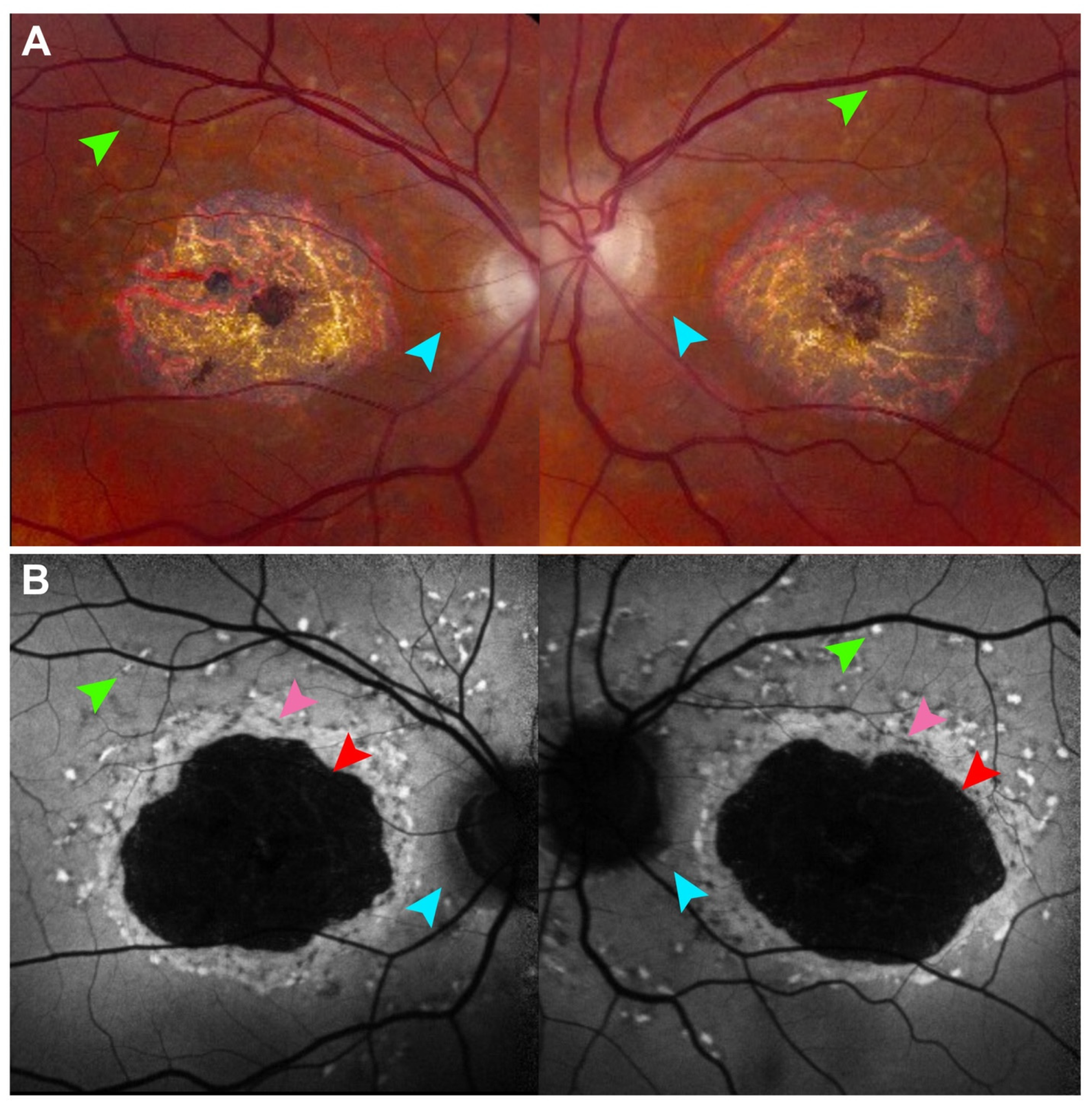
On FAF, Stargardt disease presents early with hyperautofluorescent deposits, frequently described as pisciform flecks, which correlate to areas of increased lipofuscin accumulation [30]. Key features of disease such as the presence of hyperautofluorescent flecks and peripapillary sparing are frequently better visualized on FAF and may predate the ability to detect disease features on fundus examination or on color fundus photography (Figure 2) [31]. As accumulation of toxic byproducts continue and RPE cells begin to die, these flecks then become atrophic and subsequently hypoautofluorescent. Due to the close relationship between disease pathophysiology and lipofuscin accumulation, FAF has received considerable attention as both a diagnostic tool and biomarker of disease progression.
The Progstar Study Group is a consortium dedicated to characterizing the natural history of Stargardt disease which has highlighted the utility of FAF as a tool to identify disease progression by measuring areas of hypoautofluorescence [32,33,34]. They found that areas of hypoautofluorescence grew, with rates of growth correlating with baseline lesion size, whereby larger lesions grew more rapidly [30,31,32]. These findings were corroborated by both prospective and retrospective studies and suggest that areas of atrophy may serve as both prognostic markers and outcome measurements in clinical trials [32,33,34].
A notable variant in Stargardt disease is the bulls eye maculopathy phenotype, associated with the hypomorphic c.5882G>A, p.(G1961E) allele [35]. This variation appears phenotypically similar to other macular and cone or cone–rod dystrophies. Disease is characterized by the appearance of a hyperautofluorescent bulls eye surrounding central macular atrophy and is known to progress relatively slowly compared to other forms of Stargardt disease. This phenotype presents a diagnostic challenge due to its numerous phenocopies such as RDS/PRPH2, CRX, or PROM1 associated macular dystrophy [36]. qAF has proven useful in distinguishing Stargardt disease from these masquerades, as qAF levels are significantly elevated in patients with Stargardt disease compared to both age-matched controls and individuals with other macular dystrophies [37].
RDS/PRPH2-associated macular dystrophy is another common IRD, accounting for approximately 5% of all IRDs. RDS/PRPH2 encodes a photoreceptor specific tetraspanin that plays an important role in outer segment function [38]. Although the precise mechanism remains unclear, these mutations also result in lipofuscin accumulation in the RPE. Variants in PRPH2 lead to phenotypically variable disease including pattern macular dystrophy, central areolar choroidal dystrophy, and adult vitelliform macular dystrophy [39]. FAF has been suggested to be the most valuable imaging modality in distinguishing the various phenotypic presentations of disease where other imaging modalities such as optical coherence tomography (OCT) may be less sensitive [39]. Notably, qAF levels in PRPH2-associated disease are moderately elevated compared to that of other macular dystrophies but remain below those observed in Stargardt disease [40]. The underlying mechanism of this intermediate accumulation is poorly understood [40].
Best Disease, or Best vitelliform macular dystrophy, is the most common autosomal dominant macular dystrophy and is characterized by mutations in BEST1, which encodes a calcium gated ion channel in the RPE [41]. Loss of BEST1 function has also been associated with accumulation of lipofuscin in the RPE, similar to Stargardt disease, although the mechanism remains poorly understood [41]. Best disease is characterized by the appearance of a vitelliform lesion in the macula that expands before involuting into an atrophic scar. Despite the central location of disease pathology, visual acuity tends to remain relatively well preserved until the later stages of the disease [42]. On FAF, this lesion initially appears hyperautofluorescent and becomes progressively hypoautofluorescent as the lesion becomes atrophic [43,44]. Areas of subretinal fluid may appear as a dependent hyperautofluorescent level within the vitelliform lesion [43,44]. Lesion area and diameter have been shown correlate with disease progression and may represent potential outcome measurements for future clinical trials [43,44,45].

4. Retinitis Pigmentosa and Other Rod–Cone Dystrophies

Retinitis pigmentosa (RP) encompasses a heterogeneous and diverse group of IRDs characterized by degeneration of rod followed by cone photoreceptors. Over 80 different genes have been implicated in disease pathogenesis, posing a significant challenge in diagnosis, monitoring of disease, and identification of biomarkers for clinical trial outcomes [46]. RP is typically characterized by a triad of bone spicule-like pigment migration, waxy pallor of the optic disk, and vascular attenuation on clinical examination. On FAF, RP typically presents with a central ellipsoid hyperautofluorescent ring, which has been shown to progressively shrink with disease progression, and diffuse hyperautofluorescence in the periphery (Figure 3). This ring, which is seen in over 50% of patients with RP and is undetectable on fundus examination, has been frequently studied as a disease marker for progression especially as it correlates well with other structural and functional modalities [47,48,49].
Changes in the hyperautofluorescent ring correlate strongly with retinal function, as the hyperautofluorescence demarcates the boundary between diseased and healthier photoreceptors [50,51,52]. As disease progresses, the ring begins to shrink and corresponds to evolving central vision loss [50,51,52]. Given the genotypic and phenotypic heterogeneity of disease, many efforts have been made to classify this change based on subgroupings such as inheritance pattern or by affected gene [51]. Prior studies have demonstrated that X-linked RP progresses faster than autosomal recessive RP, which in turn progresses faster than autosomal dominant RP, as evidenced by a more rapid decrease in ring size on FAF in X-linked RP [51].
Studies by individual gene are often limited to more common genetic etiologies of RP, including disease caused by RHO or USH2A, due to the limited number of affected patients. Mutations in RHO, the most common cause of autosomal dominant RP, lead to a slowly progressive disease, making RP due to mutations in RHO a target in gene therapy trials [53]. Prior studies have shown changes in hyperautofluorescent ring area as well as vertical and horizontal diameter to be relatively slow compared to other genetic etiologies of RP [53]. Similarly, mutations in USH2A, the most common cause of autosomal recessive RP, is also a focus of therapeutic interest [54]. The Foundation Fighting Blindness Consortium recently conducted a natural history study (RUSH2A) to identify clinically relevant endpoints for clinical trials [55]. Although FAF was not used, ellipsoid zone (EZ) length on OCT was used, which is highly correlated with ring diameter on FAF based on prior study with correlation coefficients upwards of 0.99 [54]. This suggests that although not used by this study, FAF changes likely remain a viable outcome measurement for clinical trials.
Despite the classic hyperautofluorescent ring appearance on FAF, other forms of ring morphology have also been described including double rings and irregular or asymmetric rings [56,57,58]. Double hyperautofluorescent rings have been described in disease associated with variants in RHO, RPGR, USH2A, as well as NR2E3, while irregular rings are more common with variants in RHO, RP1, and EYS [56,57,58]. These irregular rings may limit the applicability of hyperautofluorescent rings on FAF as potential outcome measures given their irregular morphology and difficulty characterizing progression; however, they may serve as diagnostic aids in narrowing genetic diagnoses.
The absence of autofluorescence on FAF can also be a critical diagnostic feature of RP caused by mutations in the visual cycle. The production of lipofuscin is dependent on the activity of the visual cycle within photoreceptors; therefore, in disease caused by variants in the visual cycle such as RPE65 or LRAT, severely diminished FAF has been described [59,60]. A recent study confirmed that mutations across all genes in the visual cycle may be associated with diminished FAF, which was corroborated by the use of qAF [59]. Notably, the advent of gene therapy for RPE65-mediated Lebers congenital amaurosis provided rare insight into the changes in FAF following treatment. A case report demonstrated restored qAF in a 9-year-old girl with biallelic mutations in RPE65 following treatment with voretigene neparvovec, supporting the role of FAF not only as a diagnostic aid but also as a potential biomarker of treatment [61]. More recently, longer-term follow-up studies have shown that nearly half of treated patients develop progressive chorioretinal atrophy, manifesting as circular areas of hypoautofluorescence that correspond to areas of subretinal bleb formation [62,63].
FAF in RP is particularly versatile when used alongside other structural and functional modalities. In addition to OCT, both OCT angiography and adaptive optics have provided valuable insights into disease pathogenesis [48,49]. The boundaries of hyperautofluorescent rings in RP have been shown to correlate with the blurring of cone photoreceptors on adaptive optics, reflecting a marked reduction in macular cone density [48]. Similarly, areas of preserved hyperautofluorescence are associated with intact retinal capillary density, whereas regions of decreased autofluorescence demonstrate reduced capillary density [49]. These findings underscore the value of FAF as a complementary tool given its strong correspondence with multiple ophthalmic imaging and diagnostic modalities.

5. Choroideremia

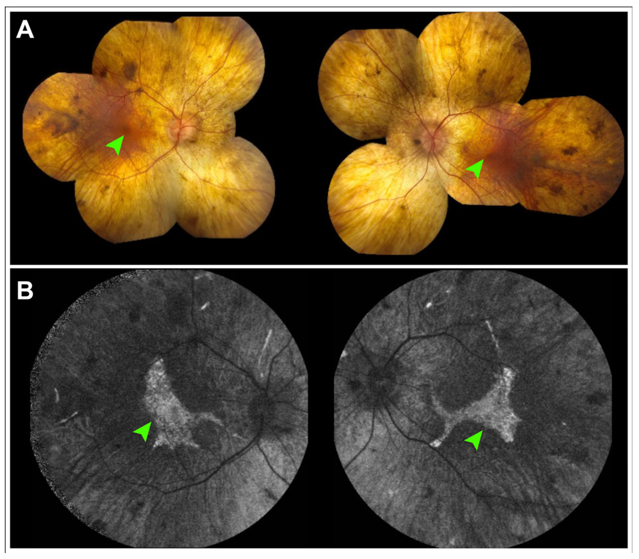
Choroideremia is an X-linked recessive IRD characterized by nyctalopia and progressive peripheral visual field loss [64]. Caused by mutations in CHM, which codes for REP-1, the disease leads to progressive degeneration of the RPE followed by secondary loss of the photoreceptors and choroid [64]. On examination, choroideremia presents as scalloped areas of chorioretinal atrophy across the posterior pole but often sparing the foveal center. FAF typically reveals areas of granular hyper- and hypoautofluorescence surrounded by broad patches of hypoautofluorescence corresponding to areas of atrophy (Figure 4).
FAF has been of particular interest in the evaluation of choroideremia, with several studies characterizing rate of autofluorescence loss and comparing it to other structural and functional outcome measurements such as outer nuclear layer thickness, EZ length, and sensitivity on microperimetry [65,66,67]. A recent natural history study by the NIGHT study group found that the area of involvement on FAF was a more sensitive measure of disease progression than best-corrected visual acuity, suggesting that disease area on FAF may be a viable outcome measurement for clinical trials [68]. Studies by the NIGHT group have also shown that disease progression in choroideremia may have directional preference, more prominently affecting the nasal subfields than others [65].
FAF also plays a role in the diagnosis and monitoring of heterozygous carriers of CHM. Although often asymptomatic, carriers may develop nyctalopia, and in severe cases, exhibit features and symptoms similar to actual disease [69,70]. Carriers with intermediate or severe phenotypes have disease that more closely resembles true choroideremia and FAF can be used to monitor disease progression. In carriers with milder phenotypes, patches of hyperautofluorescence alternating with hypoautofluorescence are seen on FAF, often described as a “mud splatter” pattern [69]. In this context, FAF can aid in diagnosing carrier status even in the absence of genetic testing and may inform family planning decisions.

6. Mitochondrial Disease

Mitochondrial disease has long been associated with phenotypically variable retinopathy. One of the most well-characterized mitochondrial retinopathies is maternally inherited diabetes and deafness (MIDD), which is frequently caused by a common point mutation in MT-TL1, m.3243A>G. MIDD typically presents as a pattern-like dystrophy with RPE and photoreceptor atrophy resembling the geographic atrophy seen in AMD [71]. On FAF, disease is characterized by hyperautofluorescent flecks, resembling those seen in Stargardt disease, radiating from central areas of hypoautofluorescence corresponding to chorioretinal atrophy. FAF in MIDD has proved valuable in both diagnosis and monitoring disease progression, especially when compared to examination or fundus photography alone [72]. One study by Ovens et al. demonstrated that areas of RPE atrophy can be readily tracked on FAF and may serve as relevant biomarkers in clinical trials [73]. Others have also demonstrated that patients with multifocal atrophy have more rapid disease progression, suggesting that baseline disease features may have prognostic value [74].
FAF is also valuable in the diagnosis and monitoring of other types of mitochondrial retinopathies. Birtel et al. proposed a classification system dividing mitochondrial retinopathies into three groups [75]. Type 1 is described as the small foci of hyper- and hypoautofluorescence on FAF that are frequently associated with large mitochondrial deletions, classically seen in Kearns Sayre and the chronic progressive external ophthalmoplegia spectrum of disease [75,76]. Type 2 is the phenotype most commonly associated with MIDD with hyperautofluorescent flecks and central areas of hypoautofluorescence [75]. Type 3 is associated with a diffuse granular hypoautofluorescence that extends outside of the arcades. This phenotype has been associated with variants in several genes, including MT-TL1, MT-RNR1, and MT-ATP6 [75]. In recent years, mutations in RNT4IP1, SSBP1, and WFS1—genes that play crucial roles in mitochondrial function—have been associated with both mitochondrial retinopathy and optic neuropathy [77,78,79,80].
Mitochondrial retinopathies are of particular interest due to the insight they may provide into more common ophthalmic conditions such as age-related macular degeneration [81,82]. Several studies have suggested that accumulation of mitochondrial damage in RPE cells is a key feature of disease pathogenesis [81,82,83]. Similarities in features on FAF have also been noted. Hyperautofluorescence at the borders of chorioretinal atrophy is seen in both AMD and mitochondrial retinopathies such as MIDD and heralds impending expansion of atrophy [73]. Consequently, keen interest has been paid to mitochondrial retinopathies to provide insights into improving our understanding of AMD.

7. Fundus Autofluorescence as a Quantitative Outcome Measure in Inherited Retinal Disease Natural History Studies and Clinical Trials

Natural history studies are critical precursors to therapeutic clinical trials for IRDs as they establish how disease progresses in the absence of treatment. Recently, such studies have become increasingly common, particularly for genetically defined forms of RP, with many incorporating FAF to monitor disease progression. In the past two years alone, multiple natural history studies for various forms of RP have been described [84,85,86,87].
FAF is an established primary outcome measure in clinical trials for IRDs, particularly for Stargardt disease. FAF enables objective quantitative assessment of retinal atrophy to evaluate whether treatment slows disease progression compared to untreated eyes.
Well-demarcated contiguous definitely decreased autofluorescence (DDAF) has been used in multiple clinical trials to assess treatment efficacy in Stargardt disease (Figure 2B). DDAF refers to areas of essentially complete FAF signal loss (≥90% darkness relative to the optic nerve head) and corresponds anatomically to geographic atrophy on OCT characterized by loss of RPE cells and overlying photoreceptors. In some Stargardt patients, the area of EZ loss exceeds the area of DDAF, suggesting that photoreceptor degeneration may precede retinal pigment epithelium atrophy [88].
A significant reduction in the progression of size of well-demarted DDAF with treatment is considered evidence of therapeutic benefit. However, contiguous well-demarcated DDAF is observed in only a subset of Stargardt patients and signifies a moderately advanced stage of disease.
Questionably decreased autofluorescence (QDAF) refers to areas with intermediate autofluorescence signal loss (50–90% darkness relative to the optic nerve head) and represents a transitional stage between healthy retina and advanced atrophy. QDAF regions are often poorly demarcated and progress to DDAF over time. Both QDAF area and the total autofluorescence area of DDAF and QDAF are being explored as potential outcome measures in clinical trials for Stargardt disease. However, the poorly defined borders of QDAF lesions pose challenges to obtaining reliable and reproducible measurements [89,90]. This becomes increasingly important as automated segmentation and artificial intelligence models are being proposed as adjuncts to human graders in clinical trials [89,90].

8. Conclusions

FAF is an indispensable tool in the diagnosis and monitoring of numerous retinal diseases. Across the spectrum of IRDs, including Stargardt disease and pattern dystrophies, RP, choroideremia, and mitochondrial retinopathies, FAF has demonstrated consistent utility in disease diagnosis, monitoring of progression, and guiding prognostication. Several biomarkers for potential clinical trials, such as changes in hyperautofluorescent ring size or area of hypoautofluorescent atrophy, are increasingly being considered as potential outcome measurements in clinical trials. As our understanding of IRDs deepens, FAF will likely remain a mainstay of both clinical care and research.

Author Contributions

Conceptualization, J.D.S. and J.K.O., Methodology, J.D.S. and J.K.O., Formal Analysis, J.D.S., J.K.O., O.M., Writing—Original Draft, J.K.O., O.M., Writing—Review and Editing, J.D.S., B.L.L., Supervision, J.D.S., B.L.L. All authors have read and agreed to the published version of the manuscript.

Funding

This research received no external funding.

Data Availability Statement

Not applicable.

Conflicts of Interest

The authors declare that there are no conflicts of interest.

References

  1. Delori, F.C.; Dorey, C.K.; Staurenghi, G.; Arend, O.; Goger, D.G.; Weiter, J.J. In vivo fluorescence of the ocular fundus exhibits retinal pigment epithelium lipofuscin characteristics. Investig. Ophthalmol. Vis. Sci. 1995, 36, 718–729. [Google Scholar]
  2. von Rückmann, A.; Fitzke, F.W.; Bird, A.C. Distribution of fundus autofluorescence with a scanning laser ophthalmoscope. Br. J. Ophthalmol. 1995, 79, 407–412. [Google Scholar] [CrossRef]
  3. Sparrow, J.R.; Gregory-Roberts, E.; Yamamoto, K.; Blonska, A.; Ghosh, S.K.; Ueda, K.; Zhou, J. The bisretinoids of retinal pigment epithelium. Prog. Retin. Eye Res. 2012, 31, 121–135. [Google Scholar] [CrossRef] [PubMed]
  4. Sparrow, J.R.; Boulton, M. RPE lipofuscin and its role in retinal pathobiology. Exp. Eye Res. 2005, 80, 595–606. [Google Scholar] [CrossRef] [PubMed]
  5. Yin, D. Biochemical basis of lipofuscin, ceroid, and age pigment-like fluorophores. Free Radic. Biol. Med. 1996, 21, 871–888. [Google Scholar] [CrossRef] [PubMed]
  6. Holz, F.G.; Bellmann, C.; Margaritidis, M.; Schütt, F.; Otto, T.P.; Völcker, H.E. Patterns of increased in vivo fundus autofluorescence in the junctional zone of geographic atrophy of the retinal pigment epithelium associated with age-related macular degeneration. Graefe’ s Arch. Clin. Exp. Ophthalmol. 1999, 237, 145–152. [Google Scholar] [CrossRef]
  7. Holz, F.G.; Bellman, C.; Staudt, S.; Schütt, F.; Völcker, H.E. Fundus autofluorescence and development of geographic atrophy in age-related macular degeneration. Investig. Ophthalmol. Vis. Sci. 2001, 42, 1051–1056. [Google Scholar] [CrossRef]
  8. Spaide, R.F. Autofluorescence imaging of acute posterior multifocal placoid pigment epitheliopathy. Retina 2006, 26, 479–482. [Google Scholar] [CrossRef]
  9. Haen, S.P.; Spaide, R.F. Fundus autofluorescence in multifocal choroiditis and panuveitis. Am. J. Ophthalmol. 2008, 145, 847–853. [Google Scholar] [CrossRef]
  10. Katz, M.L.; Eldred, G.E.; Robison, W.G., Jr. Lipofuscin autofluorescence: Evidence for vitamin A involvement in the retina. Mech. Ageing Dev. 1987, 39, 81–90. [Google Scholar] [CrossRef]
  11. Lenis, T.L.; Botsford, B.W.; Sarraf, D.; Papakostas, T.D. Didanosine-Associated Retinal Toxicity in a Patient with a Mutation in the CRB1 Gene. J. Vitreoretin. Dis. 2021, 6, 329–331. [Google Scholar] [CrossRef]
  12. Gelman, R.; Kiss, S.; Tsang, S.H. Multimodal imaging in a case of deferoxamine-induced maculopathy. Retin. Cases Brief Rep. 2014, 8, 306–309. [Google Scholar] [CrossRef]
  13. Schneider, N.; Sundaresan, Y.; Gopalakrishnan, P.; Beryozkin, A.; Hanany, M.; Levanon, E.Y.; Banin, E.; Ben-Aroya, S.; Sharon, D. Inherited retinal diseases: Linking genes, disease-causing variants, and relevant therapeutic modalities. Prog. Retin. Eye Res. 2022, 89, 101029. [Google Scholar] [CrossRef]
  14. Pichi, F.; Abboud, E.B.; Ghazi, N.G.; Khan, A.O. Fundus autofluorescence imaging in hereditary retinal diseases. Acta Ophthalmol. 2018, 96, e549–e561. [Google Scholar] [CrossRef]
  15. Eldred, G.E.; Lasky, M.R. Retinal age pigments generated by self-assembling lysosomotropic detergents. Nature 1993, 361, 724–726. [Google Scholar] [CrossRef]
  16. Li, J.; Yao, K.; Yu, X.; Dong, X.; Gan, L.; Luo, C.; Wu, Y. Identification of a novel lipofuscin pigment (iisoA2E) in retina and its effects in the retinal pigment epithelial cells. J. Biol. Chem. 2013, 288, 35671–35682. [Google Scholar] [CrossRef]
  17. Boulton, M.; Docchio, F.; Dayhaw-Barker, P.; Ramponi, R.; Cubeddu, R. Age-related changes in the morphology, absorption and fluorescence of melanosomes and lipofuscin granules of the retinal pigment epithelium. Vis. Res. 1990, 30, 1291–1303. [Google Scholar] [CrossRef]
  18. Sparrow, J.R.; Nakanishi, K.; Parish, C.A. The lipofuscin fluorophore A2E mediates blue light-induced damage to retinal pigmented epithelial cells. Investig. Ophthalmol. Vis. Sci. 2000, 41, 1981–1989. [Google Scholar]
  19. Schultz, R.; Hasan, S.; Curcio, C.A.; Smith, R.T.; Meller, D.; Hammer, M. Spectral and lifetime resolution of fundus autofluorescence in advanced age-related macular degeneration revealing different signal sources. Acta Ophthalmol. 2022, 100, e841–e846. [Google Scholar] [CrossRef] [PubMed]
  20. Hammer, M.; Sauer, L.; Klemm, M.; Peters, S.; Schultz, R.; Haueisen, J. Fundus autofluorescence beyond lipofuscin: Lesson learned from ex vivo fluorescence lifetime imaging in porcine eyes. Biomed. Opt. Express 2018, 9, 3078–3091. [Google Scholar] [CrossRef] [PubMed]
  21. Schmitz-Valckenberg, S.; Pfau, M.; Fleckenstein, M.; Staurenghi, G.; Sparrow, J.R.; Bindewald-Wittich, A.; Spaide, R.F.; Wolf, S.; Sadda, S.R.; Holz, F.G.; et al. Fundus autofluorescence imaging. Prog. Retin. Eye Res. 2021, 81, 100893. [Google Scholar] [CrossRef]
  22. Duisdieker, V.; Fleckenstein, M.; Zilkens, K.M.; Steinberg, J.S.; Holz, F.G.; Schmitz-Valckenberg, S. Long-Term Follow-Up of Fundus Autofluorescence Imaging Using Wide-Field Scanning Laser Ophthalmoscopy. Ophthalmologica 2015, 234, 218–226. [Google Scholar] [CrossRef] [PubMed]
  23. Keilhauer, C.N.; Delori, F.C. Near-infrared autofluorescence imaging of the fundus: Visualization of ocular melanin. Investig. Ophthalmol. Vis. Sci. 2006, 47, 3556–3564. [Google Scholar] [CrossRef] [PubMed]
  24. Weinberger, A.W.; Lappas, A.; Kirschkamp, T.; Mazinani, B.A.E.; Huth, J.K.; Mohammadi, B.; Walter, P. Fundus near infrared fluorescence correlates with fundus near infrared reflectance. Investig. Ophthalmol. Vis. Sci. 2006, 47, 3098–3108. [Google Scholar] [CrossRef]
  25. Kellner, S.; Weinitz, S.; Farmand, G.; Kellner, U. Near-Infrared Autofluorescence: Early Detection of Retinal Pigment Epithelial Alterations in Inherited Retinal Dystrophies. J. Clin. Med. 2024, 13, 6886. [Google Scholar] [CrossRef]
  26. Sparrow, J.R.; Duncker, T.; Schuerch, K.; Paavo, M.; de Carvalho, J.R.L., Jr. Lessons learned from quantitative fundus autofluorescence. Prog. Retin. Eye Res. 2020, 74, 100774. [Google Scholar] [CrossRef]
  27. Delori, F.; Greenberg, J.P.; Woods, R.L.; Fischer, J.; Duncker, T.; Sparrow, J.; Smith, R.T. Quantitative measurements of autofluorescence with the scanning laser ophthalmoscope. Investig. Ophthalmol. Vis. Sci. 2011, 52, 9379–9390. [Google Scholar] [CrossRef]
  28. Allikmets, R.; Singh, N.; Sun, H.; Shroyer, N.F.; Hutchinson, A.; Chidambaram, A.; Gerrard, B.; Baird, L.; Stauffer, D.; Peiffer, A.; et al. A photoreceptor cell-specific ATP-binding transporter gene (ABCR) is mutated in recessive Stargardt macular dystrophy. Nat. Genet. 1997, 15, 236–246, Erratum in Nat. Genet. 1997, 17, 122. [Google Scholar] [CrossRef]
  29. Radu, R.A.; Han, Y.; Bui, T.V.; Nusinowitz, S.; Bok, D.; Lichter, J.; Widder, K.; Travis, G.H.; Mata, N.L. Reductions in serum vitamin A arrest accumulation of toxic retinal fluorophores: A potential therapy for treatment of lipofuscin-based retinal diseases. Investig. Ophthalmol. Vis. Sci. 2005, 46, 4393–4401, Erratum in Investig. Ophthalmol. Vis. Sci. 2006, 47, 3735. [Google Scholar] [CrossRef]
  30. von Rückmann, A.; Fitzke, F.W.; Bird, A.C. In vivo fundus autofluorescence in macular dystrophies. Arch. Ophthalmol. 1997, 115, 609–615. [Google Scholar] [CrossRef]
  31. Burke, T.R.; Rhee, D.W.; Smith, R.T.; Tsang, S.H.; Allikmets, R.; Chang, S.; Lazow, M.A.; Hood, D.C.; Greenstein, V.C. Quantification of peripapillary sparing and macular involvement in Stargardt disease (STGD1). Investig. Ophthalmol. Vis. Sci. 2011, 52, 8006–8015. [Google Scholar] [CrossRef]
  32. Kong, X.; West, S.K.; Strauss, R.W.; Munoz, B.; Cideciyan, A.V.; Michaelides, M.; Ho, A.; Ahmed, M.; Schönbach, E.M.; Cheetham, J.K.; et al. Progression of Visual Acuity and Fundus Autofluorescence in Recent-Onset Stargardt Disease: ProgStar Study Report #4. Ophthalmol. Retin. 2017, 1, 514–523. [Google Scholar]
  33. Strauss, R.W.; Muñoz, B.; Ho, A.; Jha, A.; Michaelides, M.; Cideciyan, A.V.; Audo, I.; Birch, D.G.; Hariri, A.H.; Nittala, M.G.; et al. Progression of Stargardt Disease as Determined by Fundus Autofluorescence in the Retrospective Progression of Stargardt Disease Study (ProgStar Report No. 9). JAMA Ophthalmol. 2017, 135, 1232–1241. [Google Scholar] [CrossRef]
  34. Strauss, R.W.; Ho, A.; Jha, A.; Fujinami, K.; Michaelides, M.; Cideciyan, A.V.; Audo, I.; Birch, D.G.; Sadda, S.; Ip, M.; et al. Progression of Stargardt Disease as Determined by Fundus Autofluorescence over a 24-Month Period (ProgStar Report No. 17). Am. J. Ophthalmol. 2023, 250, 157–170. [Google Scholar] [CrossRef]
  35. Celia, W.; Greenstein, V.C.; Zernant-Rajang, J.; Smith, T.R.; Barile, G.; Allikmets, R.; Tsang, S.H. G1961E mutant allele in the Stargardt disease gene ABCA4 causes bull’s eye maculopathy. Exp. Eye Res. 2009, 89, 16–24. [Google Scholar] [CrossRef]
  36. Cremers, F.P.M.; Lee, W.; Collin, R.W.J.; Allikmets, R. Clinical spectrum, genetic complexity and therapeutic approaches for retinal disease caused by ABCA4 mutations. Prog. Retin. Eye Res. 2020, 79, 100861. [Google Scholar] [CrossRef]
  37. Duncker, T.; Tsang, S.H.; Lee, W.; Zernant, J.; Allikmets, R.; Delori, F.C.; Sparrow, J.R. Quantitative fundus autofluorescence distinguishes ABCA4-associated and non-ABCA4-associated bull’s-eye maculopathy. Ophthalmology 2015, 122, 345–355. [Google Scholar] [CrossRef] [PubMed]
  38. Ayyagari, R.; Borooah, S.; Durham, T.; Gelfman, C.; Bowman, A. Current and Future Directions in Developing Effective Treatments for PRPH2-Associated Retinal Diseases: A Workshop Report. Transl. Vis. Sci. Technol. 2024, 13, 16. [Google Scholar] [CrossRef] [PubMed]
  39. Antonelli, G.; Parravano, M.; Barbano, L.; Costanzo, E.; Bertelli, M.; Medori, M.C.; Parisi, V.; Ziccardi, L. Multimodal Study of PRPH2 Gene-Related Retinal Phenotypes. Diagnostics 2022, 12, 1851. [Google Scholar] [CrossRef] [PubMed]
  40. Duncker, T.; Tsang, S.H.; Woods, R.L.; Lee, W.; Zernant, J.; Allikmets, R.; Delori, F.C.; Sparrow, J.R. Quantitative Fundus Autofluorescence and Optical Coherence Tomography in PRPH2/RDS- and ABCA4-Associated Disease Exhibiting Phenotypic Overlap. Investig. Ophthalmol. Vis. Sci. 2015, 56, 3159–3170. [Google Scholar] [CrossRef]
  41. Duncker, T.; Greenberg, J.P.; Ramachandran, R.; Hood, D.C.; Smith, R.T.; Hirose, T.; Woods, R.L.; Tsang, S.H.; Delori, F.C.; Sparrow, J.R. Quantitative fundus autofluorescence and optical coherence tomography in best vitelliform macular dystrophy. Investig. Ophthalmol. Vis. Sci. 2014, 55, 1471–1482. [Google Scholar] [CrossRef]
  42. Mohler, C.W.; Fine, S.L. Long-term evaluation of patients with Best’ s vitelliform dystrophy. Ophthalmology 1981, 88, 688–692. [Google Scholar] [CrossRef] [PubMed]
  43. Jarc-Vidmar, M.; Kraut, A.; Hawlina, M. Fundus autofluorescence imaging in Best’ s vitelliform dystrophy. Klin. Monatsblätter Augenheilkd. 2003, 220, 861–867. [Google Scholar]
  44. Parodi, M.B.; Iacono, P.; Campa, C.; Del Turco, C.; Bandello, F. Fundus autofluorescence patterns in Best vitelliform macular dystrophy. Am. J. Ophthalmol. 2014, 158, 1086–1092. [Google Scholar] [CrossRef] [PubMed]
  45. Lima de Carvalho, J.R.; Oh, J.K.; Ragi, S.; Kim, J.; Manzi, R.S.; Sun, E.; Cabral, T.; Belfort, R.; Greenstein, V.C.; Sparrow, J.R.; et al. Multimodal Imaging and Dark-Adapted Chromatic Perimetry in BEST1 Vitelliform Macular Dystrophy: Identification of Outcome Measurements. Ophthalmol. Sci. 2025, 5, 100823. [Google Scholar] [CrossRef]
  46. Verbakel, S.K.; van Huet, R.A.C.; Boon, C.J.F.; den Hollander, A.I.; Collin, R.W.J.; Klaver, C.C.W.; Hoyng, C.B.; Roepman, R.; Klevering, B.J. Non-syndromic retinitis pigmentosa. Prog. Retin. Eye Res. 2018, 66, 157–186. [Google Scholar] [CrossRef]
  47. Lois, N.; Forrester, J.V. Fundus Autofluorescence; Lippincott Williams & Wilkins: Philadelphia, PA, USA, 2012. [Google Scholar]
  48. Tojo, N.; Nakamura, T.; Fuchizawa, C.; Oiwake, T.; Hayashi, A. Adaptive optics fundus images of cone photoreceptors in the macula of patients with retinitis pigmentosa. Clin. Ophthalmol. 2013, 7, 203–210. [Google Scholar] [CrossRef]
  49. Nassisi, M.; Lavia, C.; Mohand-Said, S.; Smirnov, V.; Antonio, A.; Condroyer, C.; Sancho, S.; Varin, J.; Gaudric, A.; Zeitz, C.; et al. Near-infrared fundus autofluorescence alterations correlate with swept-source optical coherence tomography angiography findings in patients with retinitis pigmentosa. Sci. Rep. 2021, 11, 3180. [Google Scholar] [CrossRef]
  50. Cabral, T.; Sengillo, J.D.; Duong, J.K.; Justus, S.; Boudreault, K.; Schuerch, K.; Belfort, R.; Mahajan, V.B.; Sparrow, J.R.; Tsang, S.H. Retrospective Analysis of Structural Disease Progression in Retinitis Pigmentosa Utilizing Multimodal Imaging. Sci. Rep. 2017, 7, 10347. [Google Scholar] [CrossRef]
  51. Jauregui, R.; Takahashi, V.K.L.; Park, K.S.; Cui, X.; Takiuti, J.T.; de Carvalho, J.R.L.; Tsang, S.H. Multimodal structural disease progression of retinitis pigmentosa according to mode of inheritance. Sci. Rep. 2019, 9, 10712. [Google Scholar] [CrossRef]
  52. Iga, Y.; Hasegawa, T.; Ikeda, H.O.; Hirota, Y.; Miyata, M.; Numa, S.; Otsuka, Y.; Tsujikawa, A. Progression of retinitis pigmentosa on static perimetry, optical coherence tomography, and fundus autofluorescence. Sci. Rep. 2023, 13, 22040. [Google Scholar] [CrossRef] [PubMed]
  53. Takahashi, V.K.L.; Takiuti, J.T.; Carvalho-Jr, J.R.L.; Xu, C.L.; Duong, J.K.; Mahajan, V.B.; Tsang, S.H. Fundus autofluorescence and ellipsoid zone (EZ) line width can be an outcome measurement in RHO-associated autosomal dominant retinitis pigmentosa. Graefe’ s Arch. Clin. Exp. Ophthalmol. 2019, 257, 725–731. [Google Scholar] [CrossRef] [PubMed]
  54. Gill, J.S.; Theofylaktopoulos, V.; Mitsios, A.; Houston, S.; Hagag, A.M.; Dubis, A.M.; Moosajee, M. Investigating Biomarkers for USH2A Retinopathy Using Multimodal Retinal Imaging. Int. J. Mol. Sci. 2022, 23, 4198. [Google Scholar] [CrossRef]
  55. Maguire, M.G.; Birch, D.G.; Duncan, J.L.; Ayala, A.R.; Ayton, L.N.; Cheetham, J.K.; Cheng, P.; Durham, T.A.; Ferris, F.L.; Hoyng, C.B.; et al. Endpoints and Design for Clinical Trials in USH2A-Related Retinal Degeneration: Results and Recommendations from the RUSH2A Natural History Study. Transl. Vis. Sci. Technol. 2024, 13, 15. [Google Scholar] [CrossRef]
  56. Marques, J.P.; Porto, F.B.O.; Carvalho, A.L.; Neves, E.; Chen, R.; Sampaio, S.A.M.; Murta, J.; Saraiva, J.; Silva, R. EYS-Associated Sector Retinitis Pigmentosa. Graefe’ s Arch. Clin. Exp. Ophthalmol. 2022, 260, 1405–1413. [Google Scholar] [CrossRef]
  57. Jauregui, R.; Chan, L.; Oh, J.K.; Cho, A.; Sparrow, J.R.; Tsang, S.H. Disease asymmetry and hyperautofluorescent ring shape in retinitis pigmentosa patients. Sci. Rep. 2020, 10, 3364. [Google Scholar] [CrossRef]
  58. Dowd-Schoeman, T.J.; Rosenbloom, J.; Ameri, H. Patterns of Autofluorescence in Common Genotypes of Retinitis Pigmentosa. Ophthalmic Surg. Lasers Imaging Retin. 2021, 52, 426–431. [Google Scholar] [CrossRef]
  59. Oh, J.K.; Lima de Carvalho, J.R., Jr.; Ryu, J.; Tsang, S.H.; Sparrow, J.R. Short-Wavelength and Near-Infrared Autofluorescence in Patients with Deficiencies of the Visual Cycle and Phototransduction. Sci. Rep. 2020, 10, 8998. [Google Scholar] [CrossRef]
  60. Lorenz, B.; Wabbels, B.; Wegscheider, E.; Hamel, C.P.; Drexler, W.; Preising, M.N. Lack of fundus autofluorescence to 488 nanometers from childhood on in patients with early-onset severe retinal dystrophy associated with mutations in RPE65. Ophthalmology 2004, 111, 1585–1594. [Google Scholar] [CrossRef]
  61. Levi, S.R.; Oh, J.K.; de Carvalho, J.R.L., Jr.; Mahajan, V.B.; Tsang, S.H.; Sparrow, J.R. Quantitative Autofluorescence Following Gene Therapy with Voretigene Neparvovec. JAMA Ophthalmol. 2020, 138, 919–921. [Google Scholar] [CrossRef]
  62. Kolesnikova, M.; Lima de Carvalho, J.R., Jr.; Parmann, R.; Kim, A.H.; Mahajan, V.B.; Tsang, S.H.; Sparrow, J.R. Chorioretinal atrophy following voretigene neparvovec despite the presence of fundus autofluorescence. Mol. Genet. Genom. Med. 2022, 10, e2038. [Google Scholar] [CrossRef]
  63. Lorenz, B.; Künzel, S.H.; Preising, M.N.; Scholz, J.P.; Chang, P.; Holz, F.G.; Herrmann, P. Single Center Experience with Voretigene Neparvovec Gene Augmentation Therapy in RPE65 Mutation-Associated Inherited Retinal Degeneration in a Clinical Setting. Ophthalmology 2024, 131, 161–178. [Google Scholar] [CrossRef]
  64. van den Hurk, J.A.; Schwartz, M.; van Bokhoven, H.; Van de Pol, T.J.R.; Bogerd, L.; Pinckers, A.J.L.G.; Bleeker-Wagemakers, E.M.; Pawlowitzki, I.H.; Rüther, K.; Ropers, H.-H.; et al. Molecular basis of choroideremia (CHM): Mutations involving the rab escort protein-1 (REP-1) gene. Hum. Mutat. 1997, 9, 110–117. [Google Scholar] [CrossRef]
  65. Poli, F.E.; MacLaren, R.E.; Cehajic-Kapetanovic, J. Retinal Patterns and the Role of Autofluorescence in Choroideremia. Genes 2024, 15, 1471. [Google Scholar] [CrossRef] [PubMed]
  66. Poli, F.E.; Yusuf, I.H.; Jolly, J.K.; Taylor, L.J.; Adeyoju, D.; Josan, A.S.; Birtel, J.; Issa, P.C.; Cehajic-Kapetanovic, J.; Da Cruz, L.; et al. Correlation Between Fundus Autofluorescence Pattern and Retinal Function on Microperimetry in Choroideremia. Transl. Vis. Sci. Technol. 2023, 12, 24. [Google Scholar] [CrossRef] [PubMed]
  67. Xue, K.; Oldani, M.; Jolly, J.K.; Edwards, T.; Groppe, M.; Downes, S.M.; MacLaren, R. Correlation of Optical Coherence Tomography and Autofluorescence in the Outer Retina and Choroid of Patients With Choroideremia. Investig. Ophthalmol. Vis. Sci. 2016, 57, 3674–3684. [Google Scholar] [CrossRef]
  68. Maclaren, R.E.; Lam, B.L.; Fischer, M.D.; Holz, F.G.; Pennesi, M.E.; Birch, D.G.; Sankila, E.-M.; Meunier, I.A.; Stepien, K.E.; Sallum, J.M.F.; et al. A Prospective, Observational, Non-interventional Clinical Study of Participants With Choroideremia: The NIGHT Study. Am. J. Ophthalmol. 2024, 263, 35–49. [Google Scholar] [CrossRef]
  69. Jauregui, R.; Park, K.S.; Tanaka, A.J.; Cho, A.; Paavo, M.; Zernant, J.; Francis, J.H.; Allikmets, R.; Sparrow, J.R.; Tsang, S.H. Spectrum of Disease Severity and Phenotype in Choroideremia Carriers. Am. J. Ophthalmol. 2019, 207, 77–86. [Google Scholar] [CrossRef]
  70. Paavo, M.; Carvalho, J.R.L., Jr.; Lee, W.; Sengillo, J.D.; Tsang, S.H.; Sparrow, J.R. Patterns and Intensities of Near-Infrared and Short-Wavelength Fundus Autofluorescence in Choroideremia Probands and Carriers. Investig. Ophthalmol. Vis. Sci. 2019, 60, 3752–3761. [Google Scholar] [CrossRef]
  71. Goto, Y.; Nonaka, I.; Horai, S. A mutation in the tRNALeu(UUR) gene associated with the MELAS subgroup of mitochondrial encephalomyopathies. Nature 1990, 348, 651–653. [Google Scholar] [CrossRef]
  72. Rath, P.P.; Jenkins, S.; Michaelides, M.; Smith, A.; Sweeney, M.G.; Davis, M.B.; Fitzke, F.W.; Bird, A.C. Characterisation of the macular dystrophy in patients with the A3243G mitochondrial DNA point mutation with fundus autofluorescence. Br. J. Ophthalmol. 2008, 92, 623–629. [Google Scholar] [CrossRef]
  73. Ovens, C.A.; Ahmad, K.; Fraser, C.L. Fundus Autofluorescence in Maternally Inherited Diabetes and Deafness: The Gold Standard for Monitoring Maculopathy? Neuroophthalmology 2019, 44, 168–173. [Google Scholar] [CrossRef]
  74. Müller, P.L.; Treis, T.; Pfau, M.; Degli Esposti, S.; Alsaedi, A.; Maloca, P.; Balaskas, K.; Webster, A.; Egan, C.; Tufail, A. Progression of Retinopathy Secondary to Maternally Inherited Diabetes and Deafness—Evaluation of Predicting Parameters. Am. J. Ophthalmol. 2020, 213, 134–144. [Google Scholar] [CrossRef] [PubMed]
  75. Birtel, J.; von Landenberg, C.; Gliem, M.; Gliem, C.; Reimann, J.; Kunz, W.S.; Herrmann, P.; Betz, C.; Caswell, R.; Nesbitt, V.; et al. Mitochondrial Retinopathy. Ophthalmol. Retin. 2022, 6, 65–79. [Google Scholar] [CrossRef] [PubMed]
  76. Oh, J.K.; Lima de Carvalho, J.R., Jr.; Nuzbrokh, Y.; Ryu, J.; Chemudupati, T.; Mahajan, V.B.; Sparrow, J.R.; Tsang, S.H. Retinal Manifestations of Mitochondrial Oxidative Phosphorylation Disorders. Investig. Ophthalmol. Vis. Sci. 2020, 61, 12. [Google Scholar] [CrossRef] [PubMed]
  77. Borrelli, E.; Bandello, F.; Boon, C.J.F.; Carelli, V.; Lenaers, G.; Reibaldi, M.; Sadda, S.R.; Sadun, A.A.; Sarraf, D.; Yu-Wai-Man, P.; et al. Mitochondrial retinopathies and optic neuropathies: The impact of retinal imaging on modern understanding of pathogenesis, diagnosis, and management. Prog. Retin. Eye Res. 2024, 101, 101264. [Google Scholar] [CrossRef]
  78. Meunier, I.; Bocquet, B.; Charif, M.; Dhaenens, C.-M.; Manes, G.; Amati-Bonneau, P.; Roubertie, A.; Zanlonghi, X.; Lenaers, G. A Rod-Cone Dystrophy Is Systematically Associated to the RTN4IP1 Recessive Optic Atrophy. Retina 2021, 41, 1771–1779. [Google Scholar] [CrossRef]
  79. Del Dotto, V.; Ullah, F.; Di Meo, I.; Magini, P.; Gusic, M.; Maresca, A.; Caporali, L.; Palombo, F.; Tagliavini, F.; Baugh, E.H.; et al. SSBP1 mutations cause mtDNA depletion underlying a complex optic atrophy disorder. J. Clin. Investig. 2020, 130, 108–125. [Google Scholar] [CrossRef]
  80. Tombolini, B.; Battista, M.; Borrelli, E.; Frontino, G.; Bandello, F.; Barboni, P.; Cascavilla, M.L. Wolfram Syndrome: Only a Neurodegenerative Disease or Also a Maculopathy? J. Neuroophthalmol. 2024, 44, e544–e546. [Google Scholar] [CrossRef]
  81. Fisher, C.R.; Ferrington, D.A. Perspective on AMD pathobiology: A bioenergetic crisis in the RPE. Investig. Ophthalmol. Vis. Sci. 2018, 59, AMD41–AMD47. [Google Scholar] [CrossRef]
  82. Terluk, M.R.; Kapphahn, R.J.; Soukup, L.M.; Gong, H.; Gallardo, C.; Montezuma, S.R.; Ferrington, D.A. Investigating mitochondria as a target for treating age-related macular degeneration. J. Neurosci. 2015, 35, 7304–7311. [Google Scholar] [CrossRef]
  83. Ferrington, D.A.; Fisher, C.R.; Kowluru, R.A. Mitochondrial defects drive degenerative retinal diseases. Trends Mol. Med. 2020, 26, 105–118. [Google Scholar] [CrossRef]
  84. MacLaren, R.E.; Duncan, J.L.; Fischer, M.D.; Lam, B.L.; Meunier, I.; Pennesi, M.E.; Sankila, E.-M.K.; Gow, J.A.; Li, J.; Tsang, S.-F.; et al. XOLARIS: A 24-Month, Prospective, Natural History Study of 201 Participants with Retinitis Pigmentosa GTPase Regulator-Associated X-Linked Retinitis Pigmentosa. Ophthalmol. Sci. 2024, 5, 100595. [Google Scholar] [CrossRef]
  85. Hashem, S.A.; Georgiou, M.; Wright, G.; Fujinami-Yokokawa, Y.; Laich, Y.; Varela, M.D.; de Guimaraes, T.A.; Mahroo, O.A.; Webster, A.R.; Fujinami, K.; et al. PDE6A-Associated Retinitis Pigmentosa, Clinical Characteristics, Genetics, and Natural History. Ophthalmol. Retin. 2025, 9, 278–287. [Google Scholar] [CrossRef] [PubMed]
  86. de Guimaraes, T.A.C.; Robson, A.G.; de Guimaraes, I.M.C.; Laich, Y.; Aychoua, N.; Wright, G.; Kalitzeos, A.; Mahroo, O.A.; Webster, A.R.; Michaelides, M. CDH23-Associated Usher Syndrome: Clinical Features, Retinal Imaging, and Natural History. Investig. Ophthalmol. Vis. Sci. 2024, 65, 27. [Google Scholar] [CrossRef] [PubMed]
  87. Kong, M.D.; Bailey, J.A.; Exinor, A.; Heaps, N.; Duong, J.; Hou, B.; Piamjitchol, C.; Demirkol, A.; Brodie, S.E.; Sparrow, J.R.; et al. Ellipsoid Zone Loss as an Outcome Measure in MAK-Associated Retinitis Pigmentosa. Am. J. Ophthalmol. 2025, in press. [Google Scholar] [CrossRef] [PubMed]
  88. Gomes, N.L.; Greenstein, V.C.; Carlson, J.N.; Tsang, S.H.; Smith, R.T.; Carr, R.E.; Hood, D.C.; Chang, S. A comparison of fundus autofluorescence and retinal structure in patients with Stargardt disease. Investig. Ophthalmol. Vis. Sci. 2009, 50, 3953–3959. [Google Scholar] [CrossRef]
  89. Kuehlewein, L.; Hariri, A.H.; Ho, A.; Dustin, L.; Wolfson, Y.; Strauss, R.W.; Scholl, H.P.N.; Sadda, S.R. Comparison of Manual and Semiautomated Fundus Autofluorescence Analysis of Macular Atrophy in Stargardt Disease Phenotype. Retina 2016, 36, 1216–1221. [Google Scholar] [CrossRef]
  90. Zhao, P.Y.; Branham, K.; Schlegel, D.; Fahim, A.T.; Jayasundera, K.T. Automated Segmentation of Autofluorescence Lesions in Stargardt Disease. Ophthalmol. Retin. 2022, 6, 1098–1104. [Google Scholar] [CrossRef]
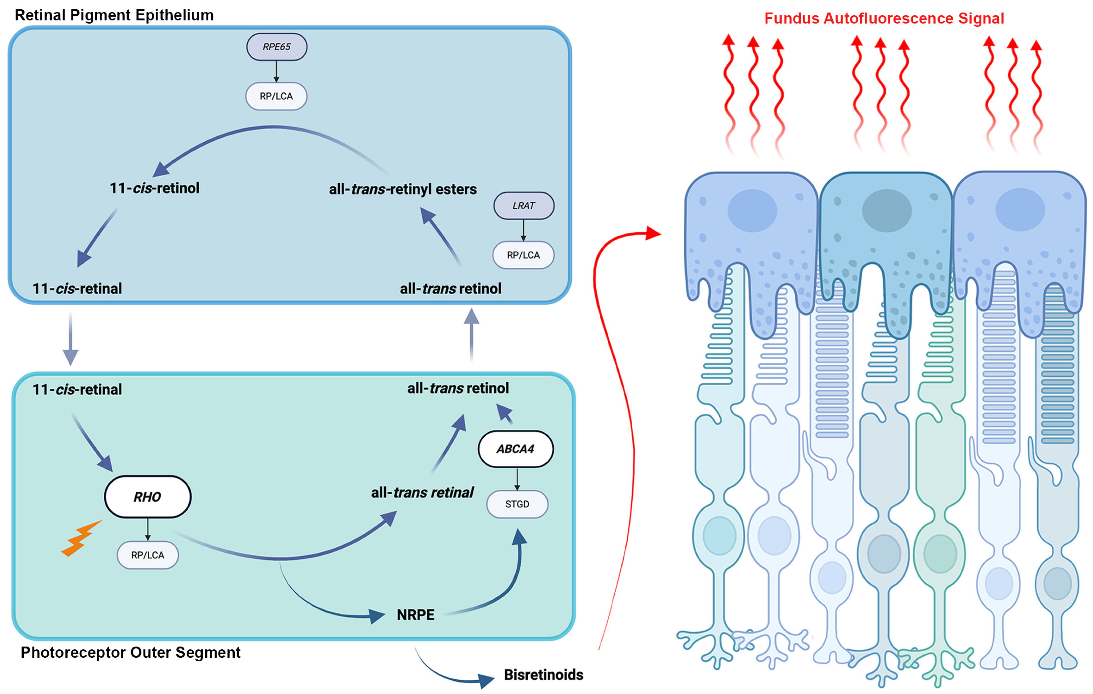
Figure 1. Retinoid visual cycle and the origin of fundus autofluorescence signal. An illustration of the retinoid visual cycle that takes place between the retinal pigment epithelium and the outer segments of photoreceptors is shown. Interruption of the visual cycle at any point can lead to the development of inherited retinal degenerations. Accumulation of bisretinoids in retinal pigment epithelium cells as a byproduct of the cycle is the source of fundus autofluorescence signal.
Cells 14 01092 g001
Figure 2. Fundus autofluorescence in Stargardt disease detects features hard to see on color fundus photography. (A) 30° field color fundus photography of a 77-year-old patient with Stargardt disease demonstrates central chorioretinal atrophy and faint gray flecks (green arrows) around central atrophy and along the superior arcades. (B) 30° field fundus autofluorescence better demonstrates hyperautofluorescent flecks and peripapillary sparing around the optic nerve which is nearly imperceptible on fundus photography (cyan arrows). The well-demarcated definitely decreased autofluorescence (DDAF) (red arrow) has been used in multiple clinical trials to assess treatment efficacy in Stargardt disease. The questionably decreased autofluorescence (QDAF) (pink arrow) refers to areas with intermediate autofluorescence signal loss and represents a transitional stage between healthy retina and advanced atrophy. QDAF regions are often poorly demarcated and progress to DDAF over time.
Cells 14 01092 g002
Figure 3. Hyperautofluorescent rings in retinitis pigmentosa seen on fundus autofluorescence compared to color fundus photography. (A) Montage color fundus photographs of a 36-year-old patient with autosomal dominant retinitis pigmentosa due to mutations in RHO demonstrate waxy pallor of the optic nerve, vascular attenuation and nasal and inferior bone spicules. (B) 55° field fundus autofluorescence demonstrates a central hyperautofluorescent ring, smaller in the right eye than the left, which is not seen on color fundus photographs (green arrows). Areas of both definitely decreased autofluorescence (red arrows) and questionably decreased autofluorescence can be seen (pink arrows).
Cells 14 01092 g003
Figure 4. Color fundus photographs and fundus autofluorescence in choroideremia. (A) Montage color fundus photographs of a 49-year-old patient with choroideremia demonstrates diffuse chorioretinal atrophy. (B) 55° field fundus autofluorescence demonstrates a small island of centrally preserved relative hyperautofluorescence that correlates to preserved photoreceptors (green arrows). The area of this central island has been measured as a biomarker of disease progression and is challenging to measure on fundus photography.
Cells 14 01092 g004
Disclaimer/Publisher’s Note: The statements, opinions and data contained in all publications are solely those of the individual author(s) and contributor(s) and not of MDPI and/or the editor(s). MDPI and/or the editor(s) disclaim responsibility for any injury to people or property resulting from any ideas, methods, instructions or products referred to in the content.