Next Article in Journal
Mesenchymal Stem Cell Secretome Attenuates PrP106-126-Induced Neurotoxicity by Suppressing Neuroinflammation and Apoptosis and Enhances Cell Migration
Previous Article in Journal
HSP110 Regulates the Assembly of the SWI/SNF Complex
Previous Article in Special Issue
How to Differentiate between Resistant and Susceptible Wheat Cultivars for Leaf Rust Fungi Using Antioxidant Enzymes and Histological and Molecular Studies?
 
 
Due to scheduled maintenance work on our database systems, there may be short service disruptions on this website between 10:00 and 11:00 CEST on June 14th.
Review

Rosmarinic Acid as Bioactive Compound: Molecular and Physiological Aspects of Biosynthesis with Future Perspectives

by
Dragana Jakovljević
1,
Marzena Warchoł
2 and
Edyta Skrzypek
2,*
1
Department of Biology and Ecology, Faculty of Science, University of Kragujevac, Radoja Domanovića 12, 34-000 Kragujevac, Serbia
2
The Franciszek Górski Institute of Plant Physiology, Polish Academy of Sciences, Niezapominajek 21, 30-239 Krakow, Poland
*
Author to whom correspondence should be addressed.
Cells 2025, 14(11), 850; https://doi.org/10.3390/cells14110850
Submission received: 11 April 2025 / Revised: 26 May 2025 / Accepted: 3 June 2025 / Published: 5 June 2025
(This article belongs to the Special Issue Antioxidants in Redox Homeostasis of Plant Development)

Abstract

:
The ester of caffeic acid with α-hydroxydihydrocaffeic acid, named rosmarinic acid (α-o-caffeoyl-3,4-dihydroxyphenyllactic acid; RA) can occur as oligomeric molecules, or in free, esterified, and glycosidic forms. Although it is commonly found among the members of the plants from the Lamiaceae (mints) and Boraginaceae (borages) families, only certain plant species produce a comparatively high concentration of RA. This valuable bioactive compound exhibits anti-cancer, anti-angiogenic, antioxidant, anti-inflammatory, antiviral, and antimicrobial properties, among others. As it is difficult to obtain high quantities of RA from natural sources, and since chemical manufacturing is costly and challenging, various biotechnological methods have recently been investigated to boost RA production. Plant cell tissue culture has been used to promote RA production in various plant species, particularly medicinal ones, with elicitation being the most commonly used technique. This review explores the main steps involved in RA biosynthesis in plants, including the molecular mechanisms and physiological alterations underlying its function, along with the primary mechanisms of RA accumulation in response to elicitation. Recent progress in synthetic biology-based RA synthesis, as well as metabolic engineering techniques to enhance the industrial production of this valuable bioactive constituent, are also discussed.

1. Introduction

Rosmarinic acid (α-o-caffeoyl-3,4-dihydroxyphenyllactic acid; RA) is an ester of caffeic acid with α-hydroxydihydrocaffeic acid. Its structure was reported in 1958 by Scarpati and Oriente [1], who were the first to isolate a previously undescribed main component of rosemary (Salvia rosmarinus Spenn.) leaves. Present-day studies have shown the occurrence of free RA in over a hundred plant species, ranging from 0.01 mg to 9.8 mg in 1 g of crude material, mainly in the Lamiaceae (mint) and Boraginaceae (borage) families, for which RA can be used as a chemotaxonomic determinant [2,3,4,5].
In the plant world, RA can occur in free, esterified, glycosidic forms, as well as in the form of oligomeric compounds. Unbound RA, its methyl ester, and melitric acids A and B have been isolated from the leaves of Melissa officinalis L. [6]. Melitric acids have a tridepside structure and are composed of three phenylpropanoic units. In relation to RA, they have an additional caffeic acid molecule attached by an ether bond. The further derivative of RA, sagerenic acid, present in Salvia officinalis L., is a dimer formed in the process of phytochemical cyclization [7]. Another compound, salvianolic acid K, was detected in the roots of Salvia deserta Schangin. and in Salvia officinalis L. [7]. The glycosidic form of RA is extremely rare; only a few compounds of this type have been described so far, mainly involving glucose [8]. These include 4′-O-β-d-glucopyranoside and 4,4′-O-di-β-d-glucopyranoside of RA, isolated from the fruits of the Indonesian medicinal plant Helicteres isora L. Both components show properties analogous to their aglycone, resulting from the presence of caffeoyl residues in the structure. Still, the main structural variation in RA occurs when two RA units combine to produce a compound, such as salvianolic acid B. Numerous related salvianolic acid derivatives and lithospermic acids seem to have their origins in this molecule as a precursor [9].
The free form of RA has been found in the following taxa: Lamiaceae (Agastache, Coleus, Dracocephalum, Esholtzia, Glechoma, Hyptis, Hyssopus, Lavandula, Lycopus, Majorana, Melissa, Mentha, Monarda, Nepeta, Ocimum, Origanum, Orthosiphon, Perilla, Prunella, Rabdosia, Rosmarinus, Salvia, Satureja, and Thymus); Apiaceae (Sanicula europaea L.); Boraginaceae (Anchusa, Lithospermum, and Symphytum); Plantaginaceae (Plantago lagopus L.); Sterculiaceae (Helicteres isora L.); and Sterculiaceae (Helicteres isora L.) [2]. Among the above-mentioned taxonomic groups, only a few plants have a relatively high concentration of RA. Due to the wide range of pharmacological properties they are commonly used in medicine to treat and prevent many diseases.
RA demonstrates anti-cancer, anti-angiogenic, antioxidant, anti-inflammatory, anti-viral, and anti-microbial properties by preventing cell damage caused by free radicals [4,5,10]. As a naturally occurring substance, RA has high therapeutic potential, as confirmed by both in vitro and in vivo studies. It stabilizes biological membranes, protects against the harmful effects of UV radiation and reactive oxygen species, and exhibits sedative effects on the central nervous system. It also has minor anti-allergic properties, and its ester derivatives possess choleretic properties. RA displays characteristics of a non-specific inhibitor of enzymes, reducing the activity of cyclooxygenase 1 and 2, lipoxygenase, hyaluronidase, b-hexosaminidase, ornithine decarboxylase, aldose reductase, α-amylase, carboxypeptidase A, adenylyl cyclase, and iodothyronine deiodinase to varying degrees. It stimulates the production of prostaglandin E2 and reduces the synthesis of leukotrienes B4 in leukocytes. It also has a protective effect on plasma lipoproteins, including the LDL fraction. Together with rutin, ursolic acid, or lycopene, it shows synergistic properties. RA reduces the permeability of capillary vessels, preventing the penetration of toxins and microorganisms. It also decreases the synthesis of inflammatory mediators (prostaglandins, prostacyclins, and thromboxane), thereby eliminating their effect on sensory endings and preventing platelet aggregation. RA also shows antimicrobial properties. Its virostatic activity against herpes simplexvirus type 1 (HSV-1) and human immunodeficiency virus (HIV-1) is due to its interaction with the viral and host protein structures, which delays the adsorption on the cell surface or impairs viral replication. As a nonspecific enzymatic inhibitor, RA reduces in vitro the activity of key HIV replication enzymes, reverse transcriptase, and integrase, thereby preventing the synthesis of a complementary viral DNA strand on the RNA matrix and its association with the host chromosome [5,10,11,12].
RA also plays an important role in the growth promotion and protection mechanisms against biotic and abiotic stresses in plants [13,14,15,16,17]. It shows strong antioxidant properties, which are essential for scavenging free radicals and defending cells from oxidative stress. The secretion of RA was observed in the roots of Ocimum basilicum L. when it was infected with the fungal pathogen Pythium ultimum, as well as during Fusarium attacks in barley [18,19]. RA can also be used for food preservation due to its ability to inhibit lipid peroxidation and the growth of bacteria, as well as an antibiotic substitute [20,21]. It is further applied in cosmetic preparations due to its antioxidant and anti-inflammatory properties [20].
In the natural environment, plants produce small amounts of RA, and its chemical synthesis is difficult and expensive. In order to increase production, various biotechnology approaches are being investigated. RA production using plant cell tissue culture has been carried out in numerous plant species, especially medicinal ones. The optimization of media and culture conditions, along with elicitation, are commonly applied methods to improve the production of secondary metabolites in cell cultures. Most plants do not produce secondary metabolites in satisfactory quantities, and their amounts are often inconsistent [20]. RA is accumulated very intensively in undifferentiated cell suspension, callus cultures, or shoot cultures—often in much higher quantities compared with whole plants [3,13]. Besides cell and callus culture, hairy roots culture appears to be a promising method for RA production. The success of RA production using in vitro systems has enabled the industrial-scale synthesis of RA in bioreactors [22]. RA production has also been achieved by expressing its biosynthesis enzymes from plants in yeast (Saccharomyces cerevisiae Meyen ex E.C. Hansen). This pilot study confirmed the opportunity to develop a fermentative procedure for RA production using Saccharomyces cerevisiae cells [23].
The interest in RA has continuously increased since it was first described. Recent research has explored not only the biosynthesis pathway and factors influencing the accumulation of RA in plant tissues but also the transcriptome level and genes encoding biosynthesis. The objective of this review is to present the molecular and physiological aspects of RA biosynthesis with future perspectives—especially the optimization of RA production methods for commercial use.

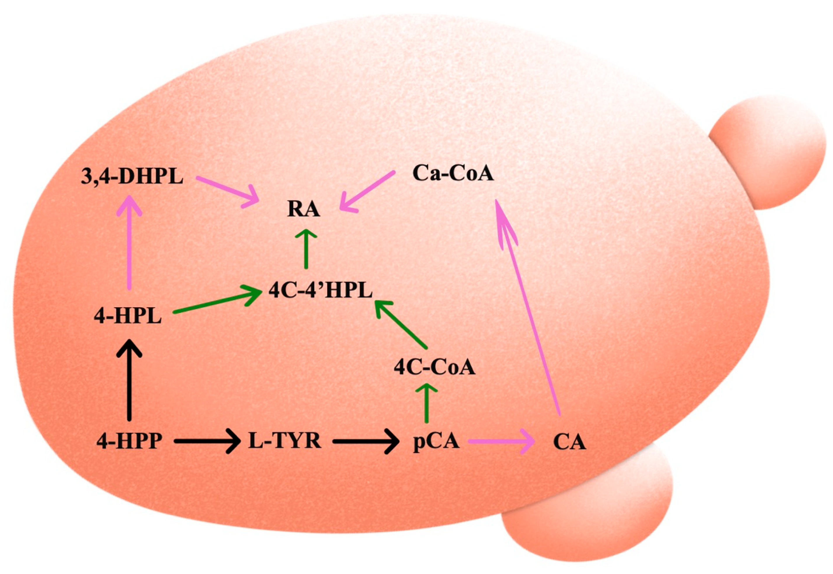
2. Rosmarinic Acid Biosynthetic Pathways in Plants

There are not many enzymes unique to the biosynthetic pathway required for RA synthesis. It is now believed that a limited number of enzymes needed to be “invented” specifically for the biosynthesis of RA, most likely based on genes involved in the production of caffeoylshikimic acid and chlorogenic acid. Additional biosynthetic steps may have been drawn from photorespiration, tocopherol/plastoquinone biosynthesis, and phenylpropanoid metabolism [24].
Beginning with the aromatic amino acids L-phenylalanine and L-tyrosine, the biosynthesis continues with their independent transformation [25]. The biosynthetic pathways starting with both amino acids, along with the enzymes involved in RA synthesis, are presented in Figure 1.

2.1. L-Phenylalanine

The enzyme phenylalanine ammonia-lyase (PAL; E.C. 4.3.1.5) catalyzes the initial step in the metabolism of phenylpropanoid compounds—a reaction involving the deamination of phenylalanine to cinnamic acid [26]. Through this common step, numerous aromatic metabolites are produced. They can be divided into classes (or subclasses) that include lignins, flavonoids, phenolic acids, coumarins, and stilbenes, among others. Variations in PAL enzyme activity can be attributed to abiotic and biotic environmental conditions [13]. In addition to the 4-methyldiene-imidazol-5-one (MIO) co-factor in the active site, this helix-containing protein has two structural segments—the N-terminal mobile extension and the shielding domain over the active center [27]. The expression of the PAL activity genes is spatially and temporally controlled. It is known that the synthesis of secondary metabolites cannot be regulated until adequate primary metabolite synthesis and photosynthetic activities have occurred. Although the PAL enzyme cannot be considered a component of the plant antioxidant system, the compounds it produces contribute significantly to the plant’s overall antioxidant capacity [28].
Following the deamination of L-phenylalanine to cinnamic acid, the cytochrome P450 monooxygenase cinnamate 4-hydroxylase (C4H; E.C. 1.14.13.11) then hydroxylates the fourth carbonic atom of the benzene ring in the cinnamic acid, forming 4-coumaric acid [22,29]. Hydroxycinnamic acids must be activated before proceeding to the next step. These acids come in a variety of activated forms. Although the coenzyme A thioester form is the most prevalent, hydroxycinnamic acid can also be activated via glucose esters or cinnamic acid esters (such as chlorogenic). In addition, 4-coumaroyl-coenzyme A ligase (4CL; E.C. 6.2.1.12) participates in the activation process [3], resulting in the formation of the intermediatory precursor 4-coumaroyl-CoA [22].

2.2. L-Tyrosine

Tyrosine is produced de novo in plants through the shikimate pathway, which also produces tryptophan and phenylalanine, the other two aromatic amino acids [30,31]. Tyrosine is a biosynthetic precursor to ubiquinone, plastoquinone, and tocopherols, all of which are necessary for plants to function. Additionally, RA and its derivatives in the Lamiaceae and Boraginaceae families are among the many specialized metabolites that are synthesized by several plant groups using tyrosine as a substrate [3]. The reversible transamination from tyrosine to 4-hydroxyphenylpyruvic acid (pHPP)—the first stage of the tyrosine conversion—is catalyzed by tyrosine aminotransferase (TAT; EC 2.6.1.5). Aromatic aminotransferases are typically located in the cytosol of plants. The importance of TAT as an entry-point enzyme in tyrosine-derived RA biosynthesis has been extensively studied [32,33,34,35,36], even though they are less significant in the biosynthesis of aromatic amino acids [37]. In order to function, aminotransferases need pyridoxal-5-phosphate. In Anchusa officinalis cell cultures, TAT demonstrated substrate selectivity to tyrosine, as well as to other amino moiety acceptors such as oxaloacetate and α-ketoglutarate [22].
In general, aminotransferases can be divided into four subgroups. According to Mehta et al. [38], aromatic aminotransferases and aminotransferases that transaminate aspartate or alanine are found in subgroup I. One large and one small domain are the main parts of the homodimeric aminotransferase’s two subunits. The contact between the subunit and the two domains forms the catalytic center. Pyridoxal-5′-phosphate (PLP) needs a conserved Lys-residue whose ɛ-amino group forms an internal aldimine with PLP in order to be covalently linked to the enzyme’s active site. The external aldimine is formed when the reacting amino acid displaces the existing group. After the α-proton is separated and hydrolyzed, the 2-oxoacid is eventually produced. The accepting 2-oxoacid—which is then transformed into the matching amino acid—regenerates the pyridoxamine-5′-phosphate, which is still attached to the enzyme, in the second reaction phase. Therefore, the entire process is categorized as a ping-pong mechanism from a mechanistic standpoint [37,39].
In a further step, the 4-hydroxyphenyllactic acid (pHPL) is produced via the reduction of pHPP by hydroxyphenylpyruvate reductase (HPPR, EC 1.1.1.237). In this reaction, both NADH and NADPH can serve as co-substrates. Although it has a modest affinity, HPPR also lowers the concentration of 3,4-dihydroxyphenylpyruvate [22,31,40].

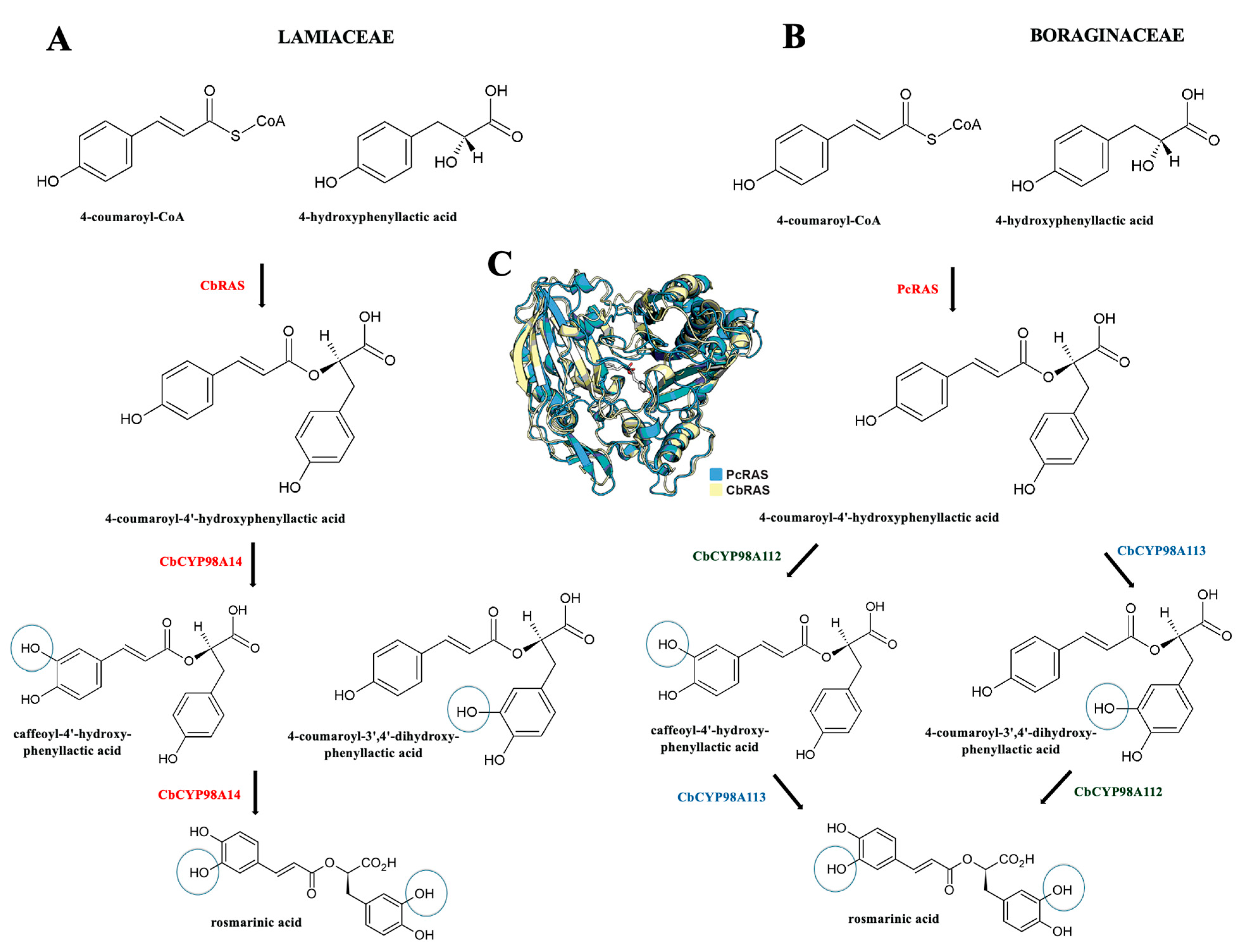
2.3. Rosmarinic Acid Synthase

4-hydroxyphenyllactate, derived from tyrosine, and 4-coumaric-CoA, derived from phenylalanine, are coupled by an ester bond in the subsequent biosynthesis phase. The enzyme 4-coumaroyl-CoA:4′-hydroxyphenyllactic acid 4-coumaroyltransferase, also known as rosmarinic acid synthase (RAS; E.C.2.3.1.140), catalyzes the reaction. Through the activity of RAS, 4-coumaroyl-4′-hydroxyphenyllactic acid (4C-pHPL) is created by the formation of an ester bond between two intermediate precursors, with the concurrent release of coenzyme A. In this step, the carboxyl group of 4-coumaric acid and the hydroxyl group of 4-hydroxyphenyllactate create an ester bond. This ester—known as 4-coumaroyl-4-hydroxyphenyllactate—is further hydroxylated on the 3 and 3′ locations of the aromatic ring, where cytochrome P450-dependent monooxygenase processes are primarily responsible for the introduction of the 3- and 3′-hydroxyl groups. [22,29]. According to Matsuno et al. [41], three cytochrome P450s are involved in RA biosynthesis, including C4H (identified as CYP73A3 and CYP73A30), and CYP98A6, which catalyzed the 3-hydroxylation of the hydroxycinnamoyl moiety of R-CHPL to form cafeoyl-4′-hydroxyphenyllactic acid, an immediate precursor of RA. Even though the precise biosynthetic mechanism of this conversion has not yet been fully characterized, RA may be further transformed into salvianolic acid B and lithospermic acid B [42,43].
Among the enzymes involved in RA biosynthesis, RAS can be regarded as the most specific [22]. The enzyme BAHD acetyltransferase has a molecular mass of 47,932 Da. The RAS amino acid sequence was elucidated from Coleus blumei [44], following full-length cDNA isolation (1290 bp long coding for 430 amino acids) and expression in E. coli. The RAS cDNA was also isolated from Melissa officinalis [45] and Lavandula angustifolia [46]. Recently, Levch et al. [47] performed a molecular phylogenic analysis involving a variety of Lamiaceae and Boraginaceae species, where a maximum likelihood phylogeny of BAHD acetyltransferase closely related to Phacelia campanularia RAS (PcRAS) revealed a Boraginaceae clade distinct from the Lamiaceae RAS clade. Figure 2 summarizes the independent biosynthesis of RA in both the Lamiaceae and Boraginaceae families based on the RAS assays.
As mentioned previously, the occurrence of RA is mostly associated with the Boraginaceae and Lamiaceae families. In this context, the cDNAs encoding RAS, along with a cytochrome P450 enzyme (CYP98A14) responsible for catalyzing the 3- and 3′-hydroxylation, have been cloned and heterologously expressed in the established RA biosynthetic pathway from Coleus blumei (Lamiaceae) [44,48]. More precisely, it has been discovered that CYP98A14 catalyzes a single meta-hydroxylation reaction on each of the aromatic rings of 4-coumaroyl-(R)-3-(4-hydroxyphenyl)lactate to produce RA, following RAS catalyzed-transfer of the 4-coumaroyl acyl group from 4-coumaroyl-CoA to the acyl acceptor molecule. Furthermore, the main differences among the above-mentioned plant families regarding RAS activity and RA biosynthesis are outlined in the study by Levsh et al. [47]. These authors combined a multi-omics approach with both the in vivo and in vitro functional characterization of candidate genes and discovered that two cytochrome P450 hydroxylases (PcCYP98A112 and PcCYP98A113) possessed specific activities during RA production in Phacelia campanularia (Boraginaceae). Namely, the findings indicates that, when it came to the P450 activities, the PcCYP98A112 catalyzed the 3-hydroxylation of the acyl–donor phenolic ring, while the acyl–acceptor ring was catalyzed by PcCYP98A113. It should be noticed that, although the precise catalytic order of the 4-coumaroyl-(R)3-(4-hydroxyphenyl)lactate remains unclear from the in vitro study, the in vivo results indicate that the in vivo RA production involves the combination of both P450 hydroxylases. Additionally, Zhou et al. [49] pointed to the distinct evolution of various RASs; RASs from the Lamiaceae family originate from early evolved shikimate/quinate hydroxycinnamoyl transferases (HCTs), while a spermidine hydroxycinnamoyl transferase (SHT) ancestor are related to the Boraginaceae RASs.

3. Molecular Mechanisms of Rosmarinic Acid Accumulation

Plant-produced chemicals can be separated into two categories—primary and secondary metabolites. Primary metabolites are essential for growth and development, while secondary metabolites provide a complex way for plants to adapt to unfavorable environmental conditions [50,51]. The chemical structure, position, type, quantity, and diversity of functional groups define their well-established antioxidant action [52]. PAL, as a key enzyme involved in phenylpropanoid metabolism, is sensitive to environmental stimuli. Consequently, plants may react to stress by changing the activity of PAL and accumulating phenylpropanoids. As a result, the ratio of various secondary metabolites varies greatly within and between taxa, as well as depending on the habitat, season, and plant material. Additionally, an increased amount of bioactive compounds may be induced in stressed plants compared to non-stressed plants. This can result in distinct plant scents, tastes, and quality, as well as altered yield potential [53,54]. For example, since the glycolysis process and amino acid metabolism (including amino acids from the alanine group) may be enhanced under the conditions of nutrient limitation, it is shown that PAL activity and other early stages of the phenylpropanoid pathway can be stimulated. This may lead to an increased synthesis of valuable secondary metabolites, including RA [13,55,56]. By studying the expression profiling of RA biosynthetic genes and some physiological responses from Mentha piperita L. under salinity and heat stress, Gholamnia et al. [57] demonstrated a significant decrease in RAS expression and a significant increase in C4H and HPPR expression in plants subjected to simultaneous stresses. In the same study, salinity at 25 °C raised the relative levels of proline and phenolic compounds, while after 72 h at a salinity of 120 mM at 35 °C, rosmarinic acid, soluble sugar, chlorophyll, and the K+/N+ ratio all declined by factors of 3.2, 1.8, 4.6, and 9, respectively.
The production of secondary metabolites can increase or decrease by up to 50% as a result of environmental influences [58]. Environmental effects on secondary metabolite biosynthesis have been explained by two proposed mechanisms—passive and active. The so-called “passive shift” involves using plant energy to produce active substances as defense compounds to protect plant survival, as seen in growth reduction [59], while the “active shift” involves upregulating the enzymes involved in secondary metabolism [60]. Plants use their receptors to recognize these harmful signals from the environment and initiate defense mechanisms, such as secondary metabolism, in response to stressful conditions [61]. For example, plants use receptors attached to the plasma membrane to identify pathogen-derived elicitors, which trigger the synthesis of low-molecular-weight chemicals (e.g., rosmarinic acid) as a defensive mechanism.
Elicitation is one of the best methods for improving the biotechnologically generated secondary metabolites. One of the primary techniques used to generate cell cultures with induced metabolite synthesis is the application of biotic or abiotic elicitors to the culture media [62]. From a biotechnological perspective, an elicitor can be regarded as an environmental element or a signal molecule that initiates a signal–transduction cascade that modulates the expression of genes linked to the production of secondary metabolites. An array of metabolic alterations is systemically started in plant cells to activate the plant’s innate immune system upon elicitation challenges [62,63]. Due to numerous interconnected reactions, the elicitation mechanism is highly complex. Furthermore, the elicitors’ origin, specificity, and concentration, as well as the plants’ development stage, the physiochemical environment of the contact, etc., all affect these occurrences [61]. Usually, plants’ response to elicitors starts in the cell’s plasma membrane. Although they may trigger the same signaling pathways, different membrane receptors perceive different elicitors [64]. Isolating elicitor signal molecules and identifying the associated receptors have been challenging, but it seems that different plant species may have common receptors for their recognition. For a variety of elicitors with different chemical structures, a number of elicitor-binding sites have been found in cell membranes. Although broad-spectrum elicitors can activate defenses in cultivars of several species, pathogen avirulence (Avr) genes and plant resistance (R) genes are essential in plant innate immunity, where specialized Avr gene products trigger defense responses only in cultivars with corresponding R genes [65]. The phytoalexinix activities of RA were demonstrated in the case of Ocimum basilicum roots. Namely, O. basilicum roots enhanced the RA production 2.67-fold upon elicitation with Pythium ultimum. Furthermore, RA showed antimicrobial activity against Pseudomonas aeruginosa and various soil-borne microorganisms. According to Bais et al. [18], RA is an essential antimicrobial component found in nature that, when challenged by microbes, may be released into the surrounding rhizosphere. Plant cells identify conserved microbe-associated molecular patterns (MAMPs)—a new term for universal and exogenous elicitors—to initiate defense responses, according to the novel explanation for plant innate immunity. On the other hand, a pathogen invasion may trigger the production of endogenous molecules or elicitors in plants, which are known as danger-associated molecular patterns. The identification of pathogen-secreted effectors, formerly referred to as particular elicitors, which are members of several groups such as proteins, glycans, and lipids, constitutes a second level of perception [62,66]. Today, it is well known that plant defense signaling molecules that mediate the plant defense response—such as methyl jasmonate (MeJA), salicylic acid (SA), jasmonic acid (JA), among others—can act as elicitors and cause the increased accumulation of RA. Table 1 summarizes the main elicitors commonly used to induce the accumulation of RA.

Effect of Elicitor on Rosmarinic Acid Accumulation

The effects of the foliar application of MeJA (50 μM) and AgNO3 (15 μM) on RA accumulation and the expression of PAL, TAT, HPPR, RAS, and CYP98A14 genes in Salvia officinalis and S. verticillata were assessed by Pesaraklu et al. [67]. The treatment of MeJA and Ag+ significantly increased the accumulation of RA in both of the tested species, according to the results. Furthermore, MeJA and Ag+ influenced the expression of the key genes PAL, TAT, HPPR, RAS, and CYP98A14 in both the phenylpropanoid and tyrosine pathways. Kianersi et al. [68] investigated the effects of MeJA on RA content and the alterations in the expression of key genes associated with biosynthesis (MoPAL, Mo4CL, and MoRAS) in Iranian lemon balm ecotypes (Melissa officinalis L.). According to the results, MeJA dosages considerably increased the RA content in both ecotypes when compared to the control samples. For both Iranian lemon balm ecotypes, RA accumulation was associated with increased expression levels of MoPAL, Mo4CL, and MoRAS in all treatments. In order to investigate whether MeJa is related to a signal transduction system in Prunella vulgaris hairy roots, Ru et al. [69] treated the hairy roots with MeJA and measured the accumulation of RA. The best induction efficiency—measured five days after application—was with 100 μM MeJA. Furthermore, a correlation study revealed that RA accumulation was substantially linked with PvPAL, PvHPPR, PvC4H, Pv4CL1, Pv4CL2, and PvCYP98A101 transcript expression, but not with PvRAS and PvTAT transcripts. Kianersi et al. [70] investigated the molecular mechanism of RA accumulation in two Salvia species (Salvia yangii and S. abrotanoides) upon MeJA elicitation. They found that, in comparison with untreated plants, 150 M MeJA increased RA content in S. yangii and S. abrotanoides by 1.66- and 1.54-fold, respectively. The effects of MeJA are most likely due to the activation of genes involved in the phenylpropanoid pathway, as evidenced by the increased numbers of transcripts for RAS, 4CL, and PAL. According to the study of Xiao et al. [86], the production of RA can be elevated by genetic manipulation, since the hairy root culture of Salvia miltiorrhiza produced several times higher concentration of RA due to the overexpression of a single gene (c4h, tat, and hppr), the overexpression of both tat and hppr, as well as the suppression of hppd. In the study by Khojasteh et al. [71], 100 μM MeJA was tested on cell suspension cultures of Satureja khuzistanica. Without influencing biomass production, MeJA more than tripled RA productivity, with the stimulated cultures reaching 3.9 g L−1. A maximum RA production of 3.1 g L−1 and biomass productivity of 18.7 g L−1 per day under MeJA elicitation were obtained when the cell culture was moved from a shake flask to a wave-mixed bioreactor. The production of RA was significantly enhanced by the elicitation period and concentration of MeJA in the hairy root culture of Lepechinia caulescens. When the treatments were elicited for 24 h, the highest accumulation occurred at a concentration of 300 µM of MeJA [72]. In the experiment with cell cultures of Agastache rugosa, the transcript levels of ArPAL, Ar4CL, and ArC4H increased 4.5-fold, 3.4-fold, and 3.5-fold, respectively, compared with the untreated controls, and the culture contained relatively high amounts of RA after exposure of cells to 50 µM MeJA [73]. The results also indicate that both RA and precursors—such as shikimate and aromatic amino acids—were induced as a response to MeJA treatment. In cell suspension cultures of Mentha × piperita, the effects of JA and MeJA on RA accumulation and cell development were also studied [74]. A total of 24 h after adding 100 μM MeJA, the maximum RA accumulation (117.95 mg g−1 DW) was recorded. Then, 48 h following the application of 200 μM JA, a comparable quantity (110.12 mg g−1 DW) was found. These values were over 1.5 times higher than those of the elicitation-free control sample. Elicitors had no noticeable impact on the secretion of RA into the culture media. RA concentrations outside of cells were comparable to those found in the control group. When compared to the control, the suspension cultures of M. piperita treated with elicitors were shown to have lower biomass accumulation. Zhou et al. [75] analyzed a transcriptome library of Salvia miltiorrhiza in response to MeJA. In addition to upregulating the expression of genes encoding important enzymes in the RA biosynthesis pathway, such as CYP98A14, overexpressing SmMYB1 markedly increased RA accumulation. SmMYB1 was found to activate the expression of CYP98A14 and genes encoding anthocyanin biosynthesis pathway enzymes, such as chalcone isomerase (CHI) and anthocyanidin synthase (ANS), according to dual-luciferase (dual-LUC) assays and/or electrophoretic mobility shift assays (EMSAs). Furthermore, it was demonstrated that, in contrast to SmMYB1 acting alone, the combination of SmMYB1 and SmMYC2 increased CYP98A14 expression. The level of gene expression for proteins associated with the RA biosynthesis pathway in Origanum vulgare in a consortium of three bacteria (Azotobacter chroococcum, Azospirillium brasilense, and Pseudomonas fluorescens) at two salinity levels and two elicitor levels (0.1 and 0.5 mM MeJA) were studied by Jafari et al. [76]. The findings demonstrated that, among all treatments, salt had a beneficial impact on DPPH radical scavenging capacity and a negative impact on RA induction and RAS gene expression. When 0.1 mM MeJA and bacterial consortia were applied together without salt, the expression of the RAS and C4H genes increased 3.37 and 6.6 times, respectively, in comparison to the control.
The results obtained by Gonçalves et al. [87] suggest that shoots of Thymus lotocephalus are a good source of antioxidant compounds, and that RA accumulation can be promoted through SA application and alterations in vitro culture conditions. In the callus culture of Salvia nemorosa, 0.5 μM SA increased RA content eight times compared to the control [77]. Li et al. [78] investigated the synthesis of RA under SA-induced pH changes in Salvia miltiorrhiza suspension cells, along with the PAL, TAT, and RAS gene expression. It was shown that SA decreased the cytosolic pH by inhibiting the activity of plasma membrane H+-ATPase. SA also upregulated the gene expression of TAT, PAL, and RAS, and as a result, enhanced the accumulation of those phenolic acids. Guo et al. [88] found that SA application led to the cytoplasmic acidification of Salvia miltiorrhiza cells and the alkalinization of the extracellular medium. Furthermore, Ca2+ protoplast mobilization was induced by SA, but the induction could be blocked by either organellar or plasma membranes, or a channel blocker. In the same study, both PAL activity and its gene expression showed the same patterns. On the other hand, a single application of salicylic acid (10 μM) on Thymus membranaceus culture media resulted in an increase in RA and phenolic levels, which in turn improved the extracts’ antioxidant properties [79]. The exogenous application of 250 and 500 μM SA led to the upregulation of PAL expression in Salvia officinalis (SoPAL) and S. virgata (SvPAL) [80]. Further analysis showed that, in S. virgata, at 500 μM SA, a higher accumulation of RA was achieved, while in Salvia officinalis, a higher RA accumulation was observed at 250 μM SA. It was determined that the RA accumulation in the tested species did not positively correlate with the level of PAL transcription. Thus, lower levels of RA were accumulated in the case of S. officinalis, even if the transcription rate of PAL increased at greater concentrations of SA. The synergistic effects of SA and H2O2 on RA production in Salvia miltiorrhiza cell cultures were investigated by Hao et al. [81]. The findings demonstrated that SA markedly increased RA accumulation, PAL activity, and H2O2 generation. Additionally, exogenous H2O2 increased RA synthesis and PAL activity. However, RA formation could be prevented if a quencher (DMTU) or an NADPH oxidase inhibitor (IMD) were introduced to suppress H2O2 generation. According to these findings, H2O2 is a secondary messenger for signal transduction, which SA can strongly activate, and which encourages the production of RA. The study of Khalil et al. [82] aimed to investigate the effect of applying various concentrations of SA on Thymus vulgaris L., while subjecting the plant to decreasing amounts of irrigation water. RA was detected as the major component with a higher percentage observed upon subjecting the plant to 3 mM SA. Stasińska-Jakubas et al. [83] foliar-applied three different elicitors (150 mg/L chitosan lactate (ChL), 10 mg L−1 selenite (Se), and 100 mg L−1 SA) to estimate the effect on the primary and secondary metabolism of Melissa officinalis. The applications of ChL and SA had the strongest elicitation impact, while the application of Se produced a slightly weaker effect. Furthermore, the highest concentration of RA was noted on the eighth day after elicitation, independent of the type of elicitor. Elicitation had the strongest effect on rosmarinic acid hexoside among the metabolites examined, with its level increasing several-fold compared to the control. Although some elicitor-induced alterations in parameters linked to photosynthesis and oxidative stress were observed, no apparent signs of chemical toxicity or impact on plant biomass were discovered.
In the study by Park et al. [84], the treatment of Agastache rugosa cell cultures with 500 mg L−1 YE and 30 mg L−1 silver nitrate increased the synthesis of RA and the expression of genes involved in the phenylpropanoid pathway. The quantity of YE and silver nitrate was correlated with the expression of RAS and HPPR. While PAL expression with silver nitrate treatment was 52.31 times higher than in the untreated controls after 24 h of elicitation, the transcript levels of HPPR under yeast extract treatment were 1.84-, 1.97-, and 2.86-fold higher than the control treatments after 3, 6, and 12 h, respectively. Additionally, YE supplementation recorded the highest quantity of RA at 4.98 mg g−1. The effects of YE on TAT gene expression and RA accumulation in Melissa officinalis seedlings were examined by Nasiri-Bezenjani et al. [85]. For the 17 h treatment, YE doses of 0.05%, 0.1%, and 0.2% markedly stimulated the RA biosynthesis. At 0.1% YE treatment, when RA biosynthesis significantly increased, the highest TAT gene expression was observed. The authors suggested that these findings could be linked to the oxidative stress that YE causes, as seen from the rising catalase and superoxide dismutase activity.
A considerable amount of research is focusing on the elucidation of RA biosynthesis and accumulation. Although the pathways of transduction and responsive genes have been clarified in some cases, it should be taken into account that different elicitors may be perceived by distinct membrane receptors, and that there is a broad variety of responses due to heterogeneity in the way of action. The general mechanism of action of elicitors was investigated by Ramirez-Estrada et al. [62]. In brief, second messengers are involved in the signal transduction pathways triggered by elicitors, which are first recognized by specific receptors. These messengers amplify the signal, initiating additional downstream reactions. The following is a summary of the sequential steps that occur in elicitor-induced defense reactions: perception of elicitor by receptor; reversible phosphorylation and dephosphorylation of proteins from plasma membrane and cytosol; cytosolic (Ca2+) enhancement; Cl and K+ efflux/H+ influx (cytoplasmic acidification and extracellular alkalinization); activation of MAPK; activation of NADPH oxidase and reactive oxygen and nitrogen species (ROS and RNS) production; expression of early defense genes; production of jasmonate; expression of the late defense response gene; and accumulation of secondary metabolites. According to Shakya et al. [61], ROS generation is an important aspect, since there is a linked role of G-proteins, ion channels, protein kinases, MAPKs, gene expression, and enzymatic reaction, which may reprogram the pathway of secondary metabolite production. All of the abovementioned should be taken into account when considering elicitor application for the induced synthesis of RA.

4. Synthetic Biology and Rosmarinic Acid Synthesis

The growing need for plant polyphenolic compounds cannot be met only by extracting phenolic compounds from natural plants, especially when it comes to RA, as previously discussed. As an alternate method for obtaining plant bioactive phenolic compounds, heterologous synthesis in plants has become popular in recent years. The benefit of this strategy is that since the phenylpropanoid pathway is already established, fewer genes need to be added. Plants, however, have competing pathways that have the potential to significantly disrupt the synthesis of polyphenols. Furthermore, the growth of these heterologous plants is influenced by the availability of fertile land and suitable weather conditions; this is a time-consuming process, as these plants are very challenging to genetically modify. Additionally, using plant cell cultures can have certain drawbacks, including heterogeneity, inconsistent yields, slow growth rates, unstable cultures, and vulnerability to aggregation and stress [89,90].
One of the most efficient ways to overcome this restriction and to generate large quantities of beneficial compounds, including RA, may be to create microbial cell factories that can produce polyphenols effectively. The development of synthetic biology techniques and the discovery of novel isoenzymes in plants or less complex organisms to construct heterologous pathways have made it easier to create effective microbial cell factories. Attempts have been made to enhance the host chassis in order to increase the process’ profitability [91]. These methods offer consistent quality, scalability, accessible extraction techniques, and the possibility of increased synthesis efficiency because they use renewable lignocellulosic biomass as a carbon source, which lessens the requirement for hazardous chemicals. Furthermore, the chemical variety of natural products, whose structural complexity might occasionally be difficult to obtain by multistep chemical synthesis, could be increased through biological synthesis [90].
Large-scale fermentations are made possible by the employment of microorganisms, and the lack of competing pathways improves future procedures [92]. The yeast Saccharomyces cerevisiae and the bacteria Escherichia coli are the most often utilized microbes for this purpose. In general, genetically modified microorganisms have several benefits, including the ability to grow in low-cost substrates, simplicity in manipulation, and quick production cycles that enable greater and faster output. Saccharomyces cerevisiae and Escherichia coli have excellent characterizations and are simple to manipulate, cultivate, and scale up. Additionally, Saccharomyces cerevisiae has internal compartments like plants and can undergo post-translational alterations like glycosylation. Furthermore, it is classified as food-grade, which permits its usage in pharmaceuticals and human nutrition [92,93]. A simplified route of RA synthesis using both plant and chimeric pathways in Saccharomyces cerevisiae is presented in Figure 3.
When it comes to RA synthesis, finding the best enzyme combination is the first step in achieving the highest conversion rate of intermediates to the desired final product [23]. Several candidates for the genes CYP98A14, RAS, and TAT with coding DNA sequences are available [33,48,94,95] as a result of studies conducted over the past ten years on the major enzymes involved in the production of RA across numerous plant species of the Lamiaceae family [23]. In this context, the BAHD family member—RA synthase LaAT1—from lavender (Lavandula angustifolia) was chosen for yeast expression and cloned separately on a vector containing the Arabidopsis gene At4CL5 (which encodes 4-coumarate/CoA ligase and is active on benzoates and hydroxycinnamates). When suitable combinations of donors and acceptor molecules were added, the diverse strains of Saccharomyces cerevisiae obtained for the co-expression of At4CL5 with the different BAHDs successfully produced a wide array of useful hydroxycinnamate and benzoate conjugates. Specifically, for the first time, the synthesis of glycerol hydroxycinnamate esters, quinate hydroxycinnamate esters like chlorogenic acid, as well as RA and its derivatives was demonstrated in yeast [90]. Furthermore, plasmids were used to express the key gene for the synthesis of the RA precursor in Saccharomyces cerevisiae to produce salvianolic acid B. RA was produced using a cell-free biosynthetic method in addition to de novo synthesis from basic carbon sources. An ATP and CoA double-regenerating system further raised the titer of RA to 320.04 mg L−1 by adding cofactors and substrates [96]. In the study of Babaei et al. [23], Saccharomyces cerevisiae was used for constructing a de novo biosynthetic pathway of RA utilizing glucose as a carbon source, and the ultimate titer of RA was 5.93 mg L−1. According to the authors, native cytochrome P450 reductase of Saccharomyces cerevisiae (NCP1 gene, accession code P16603) has 31% identity to the protein sequence of CPR from two RA producer cells, Salvia officinalis and Plectranthus scutellarioides, which makes Saccharomyces cerevisiae a potential host for RA production. On the other hand, an artificial L-tyrosine-containing RA biosynthetic pathway was created using 4-hydroxyphenylpyruvate as the only node in Escherichia coli in order to simplify the biosynthesis pathway [97]. An RA-producing strain of Saccharomyces cerevisiae was constructed in the study of Zhou et al. [98] by introducing OD-LDHY52A (optimized d-lactate dehydrogenase gene mutant), OMoRAS, and OPc4CL2 into a hyper-producer of caffeic acid. In brief, the final strain YRA113-15B showed a 63-fold improvement compared to the initial strain and produced 208 mg L−1 RA in a shake-flask culture.

5. Future Perspectives

The latest approach to the synthesis of RA involves utilizing biotechnological techniques, metabolic engineering, and elicitors. In our opinion, the efforts of scientists should be focused on the optimization of cell suspension cultures, which enable the repeatable and high production of RA under controlled conditions. Future research should result the development of detailed RA production protocols for various plant species, which will be available to anyone interested in this subject. Such availability will help extend research and applications in many fields, such as agriculture and medicine. By sharing these protocols, we can promote collaboration and innovation that will ultimately lead to the improved, environmentally friendly production of beneficial compounds like RA.
For RA production, it seems reasonable to use disposable single-use bioreactors, for example, in wave-mixed systems. These easy-to-use bioreactors have been successfully used for many species, and the extraction of secondary metabolites is cost-effective and environmentally friendly. Plant metabolic engineering offers a toolbox for overexpressing or silencing genes to control RA biosynthetic pathways in cells. Suppression of competing pathways, such as lignin production, increases the biosynthesis of phenolic precursors. In contrast, the overexpression of key genes in the RA biosynthetic pathway, such as c4h, tat, and hppr, leads to increased RA accumulation in transgenic lines.
By imitating environmental stress, elicitors activate the plant’s defense mechanisms and promote the synthesis of secondary metabolites such as RA, which are naturally produced as stress-protective substances. The use of elicitors such as MeJA, SA, and YE significantly increases the production of RA by activating its biosynthetic pathways. Elicitors also promote the expression of important genes and enzymes involved in RA production. In addition, elicitation is a simple and inexpensive method to increase RA production in controlled biotechnological systems, so elicitors can be used not only within in vitro tissue culture but also in soil and hydroponics. Combining elicitors with hydroponic cultures of medicinal plants is one of the new trends in organic farming, and the controlled conditions of this system allow for regular monitoring of the concentration of the obtained compounds. The cost-effectiveness of RA production using this method is also important, as it is crucial to meet the growing demand across various industrial sectors, including pharmaceuticals and nutraceuticals. Together, these methods constitute a bio-sustainable and innovative strategy for RA production, overcoming issues such as the overexploitation of natural plant resources and the effects of climate change.

6. Conclusions

In the plant world, the biosynthesis of RA begins with the aromatic amino acids L-phenylalanine and L-tyrosine, following their additional independent transformation. Among the enzymes involved in RA biosynthesis, RAS can be regarded as the most specific. Based on the BAHD acetyltransferase enzyme, some key differences can be observed among various members of the Lamiaceae and Boraginaceae families. Furthermore, recent progress has been made pointing to the distinct evolution of RASs in species from the abovementioned families. Both in vitro and in vivo studies have demonstrated that RA, a naturally occurring chemical compound, has great therapeutic potential. Additionally, RA is crucial for plant defense mechanisms against both biotic and abiotic stressors, as well as for promoting plant development. Although it is difficult to obtain high concentrations of RA from natural sources, with the application of the appropriate elicitor (MeJA, SA, or YE) on non-differentiated plant cell cultures in vitro, substantial amounts of RA can be obtained. Studies on this subject demonstrate that various elicitors may influence the expression of the key genes of the phenylpropanoid pathway, including PAL, TAT, HPPR, 4CL, C4H, RAS, and CYP98A14, leading to the correlated accumulation of RA. Still, it should be considered that a variety of responses can be obtained due to heterogeneity and differences in the elicitor’s way of action. Due to the recent progress in synthetic biology techniques, RA can also be effectively produced via microbial cell factories, with Saccharomyces cerevisiae and Escherichia coli as potential hosts to produce both RA and its derivates.
Research on RA needs to be extended to different crop species, considering developments in genomics, transcriptomics, and metabolomics technologies. Additionally, bioinformatical analysis must be combined. Furthermore, additional work is necessary to fully understand the receptors and signaling pathways involved. This will likely expand the range of processes and applications of rosmarinic acid synthesis. Taking all this into account, RA can be regarded as a promising subject for future investigation. Future work is expected to focus on expanding our understanding of the regulatory networks of rosmarinic acid from different plant taxa, broadening the application domains, and balancing the interactions between cell and tissue culture, the environment, and other organisms, building on the current foundations of rosmarinic acid research. The study and use of RA should be further encouraged to guarantee the stability and safety of products.

Author Contributions

Conceptualization, D.J.; writing—original draft preparation, D.J. and E.S.; writing—review and editing, D.J., M.W. and E.S. All authors have read and agreed to the published version of the manuscript.

Funding

This work was supported by the Ministry of Science, Technological Development and Innovation of the Republic of Serbia (Agreements No. 451-03-136/2025-03/200122).

Data Availability Statement

No new data were created or analyzed in this study.

Conflicts of Interest

The authors declare no conflicts of interest.

References

  1. Scarpati, M.L.; Oriente, G. Isolamento e costituzione dell’acido rosmarinico (dal Rosmarinus off.). Ric. Sci. 1958, 28, 2329–2333. [Google Scholar]
  2. Janicsák, G.; Máthé, I.; Miklóssy-Vári, V.; Blunden, G. Comparative studies of the rosmarinic and caffeic acid contents of Lamiaceae species. Biochem. Syst. Ecol. 1999, 27, 733–738. [Google Scholar] [CrossRef]
  3. Petersen, M.; Simmonds, M.S.J. Molecules of interest—Rosmarinic acid. Phytochemistry 2003, 62, 121–125. [Google Scholar] [CrossRef] [PubMed]
  4. Petersen, M. Rosmarinic acid: New aspects. Phytochem. Rev. 2013, 12, 207–227. [Google Scholar] [CrossRef]
  5. Kim, G.D.; Park, Y.S.; Jin, Y.H.; Park, C.S. Production and applications of rosmarinic acid and structurally related compounds. Appl. Microbiol. Biotechnol. 2015, 99, 2083–2092. [Google Scholar] [CrossRef]
  6. Agata, I.; Kusakabe, H.; Hatano, T.; Nishibe, S.; Okuda, T. Melitric acids A and B, new trimeric caffeic acid derivatives from Melissa officinalis. Chem. Pharm. Bull. 1993, 41, 1608–1611. [Google Scholar] [CrossRef]
  7. Lu, Y.R.; Foo, L.Y. Rosmarinic acid derivatives from Salvia officinalis. Phytochemistry 1999, 51, 91–94. [Google Scholar] [CrossRef]
  8. Satake, T.; Kamiya, K.; Saiki, Y.; Hama, T.; Fujimoto, U.; Kitanaka, S.; Kimura, Y.; Uzawa, J.; Endang, H.; Umar, M. Studies on the constituents of fruits of Helicteres isora L. Chem. Pharm. Bull. 1999, 47, 1444–1447. [Google Scholar] [CrossRef]
  9. Habtemariam, S. Molecular pharmacology of rosmarinic and salvianolic acids: Potential seeds for alzheimer’s and vascular dementia drugs. Int. J. Mol. Sci. 2018, 19, 458. [Google Scholar] [CrossRef]
  10. Bulgakov, V.P.; Inyushkina, Y.V.; Fedoreyev, S.A. Rosmarinic acid and its derivatives: Biotechnology and applications. Crit. Rev. Biotechnol. 2012, 32, 203–217. [Google Scholar] [CrossRef]
  11. Khojasteh, A.; Mirjalili, M.H.; Alcalde, M.A.; Cusido, R.M.; Eibl, R.; Palazon, J. Powerful plant antioxidants: A new biosustainable approach to the production of rosmarinic acid. Antioxidants 2020, 9, 1273. [Google Scholar] [CrossRef] [PubMed]
  12. Harindranath, H.; Susil, A.; Sekar, M.; Kumar, B.P. Unlocking the potential of rosmarinic acid: A review on extraction, isolation, quantification, pharmacokinetics and pharmacology. Phytomed. Plus 2024, 5, 100726. [Google Scholar] [CrossRef]
  13. Jakovljević, D.; Topuzović, M.; Stanković, M. Nutrient limitation as a tool for the induction of secondary metabolites with antioxidant activity in basil cultivars. Ind. Crops. Prod. 2019, 138, 111462. [Google Scholar] [CrossRef]
  14. Arikan, B.; Ozfidan-Konakci, C.; Alp, F.N.; Zengin, G.; Yildiztugay, E. Rosmarinic acid and hesperidin regulate gas exchange, chlorophyll fluorescence, antioxidant system and the fatty acid biosynthesis-related gene expression in Arabidopsis thaliana under heat stress. Phytochemistry 2022, 198, 113157. [Google Scholar] [CrossRef] [PubMed]
  15. Chowdhary, V.; Alooparampil, S.; Pandya, R.V.; Tank, J.G. Physiological function of phenolic compounds in plant defense. In Phenolic Compounds: Chemistry, Synthesis, Diversity, Non-Conventional Industrial, Pharmaceutical and Therapeutic Applications; Badria, F.A., Ed.; IntechOpen: London, UK, 2022; p. 185. [Google Scholar]
  16. Zhou, P.P.; Yue, C.L.; Zhang, Y.C.; Li, Y.; Da, X.Y.; Zhou, X.Q.; Ye, L.D. Alleviation of the byproducts formation enables highly efficient biosynthesis of rosmarinic acid in Saccharomyces cerevisiae. J. Agric. Food Chem. 2022, 70, 5077–5087. [Google Scholar] [CrossRef]
  17. Ku, Y.S.; Cheng, S.S.; Luk, C.Y.; Leung, H.S.; Chan, T.Y.; Lam, H.M. Deciphering metabolite signalling between plant roots and soil pathogens to design resistance. BMC Plant Biol. 2025, 25, 308. [Google Scholar] [CrossRef]
  18. Bais, H.P.; Walker, T.S.; Schweizer, H.P.; Vivanco, J.A. Root specific elicitation and antimicrobial activity of rosmarinic acid in hairy root cultures of Ocimum basilicum. Plant Physiol. Biochem. 2002, 40, 983–995. [Google Scholar] [CrossRef]
  19. Lanoue, A.; Burlat, V.; Henkes, G.J.; Koch, I.; Schurr, U.; Röse, U.S.R. De novo biosynthesis of defense root exudates in response to Fusarium attack in barley. New Phytol. 2010, 185, 577–588. [Google Scholar] [CrossRef]
  20. Park, S.U.; Uddin, M.R.; Xu, H.; Kim, Y.K.; Lee, S.Y. Biotechnological applications for rosmarinic acid production in plant. Afr. J. Biotechnol. 2008, 7, 4959–4965. [Google Scholar]
  21. Chen, Y.; Jiang, X.; Yang, Q.; Luo, S.; Xie, X.; Peng, C. Bioactivity of rosmarinic acid and its application in food industry: A review. Food Ferment. Ind. 2023, 49, 318–325. [Google Scholar]
  22. Trócsányi, E.; György, Z.; Zámboriné-Németh, V. New insights into rosmarinic acid biosynthesis based on molecular studies. Curr. Plant Biol. 2020, 23, 100162. [Google Scholar] [CrossRef]
  23. Babaei, M.; Zamfir, G.M.B.; Chen, X.; Christensen, H.B.; Kristensen, M.; Nielsen, J.; Borodina, I. Metabolic engineering of Saccharomyces cerevisiae for rosmarinic acid production. ACS Synth. Biol. 2020, 9, 1978–1988. [Google Scholar] [CrossRef]
  24. Petersen, M.; Abdullah, Y.; Benner, J.; Eberle, D.; Gehlen, K.; Hücherig, S.; Janiak, V.; Kim, K.H.; Sander, M.; Weitzel, C.; et al. Evolution of rosmarinic acid biosynthesis. Phytochemistry 2009, 70, 1663–1679. [Google Scholar] [CrossRef]
  25. Ellis, B.E.; Towers, G.H.N. Biogenesis of rosmarinic acid in Mentha. Biochem. J. 1970, 118, 291–297. [Google Scholar] [CrossRef]
  26. Tariq, H.; Asif, S.; Andleeb, A.; Hano, C.; Abbasi, B.H. Flavonoid production: Current trends in plant metabolic engineering and de novo microbial production. Metabolites 2023, 13, 124. [Google Scholar] [CrossRef]
  27. Zhang, X.B.; Liu, C.J. Multifaceted regulations of gateway enzyme phenylalanine ammonialyase in the biosynthesis of phenylpropanoids. Mol. Plant 2015, 8, 17–27. [Google Scholar] [CrossRef]
  28. Jakovljević, D. Exploring molecular diversity of plants for enhancement of natural products. In Biosynthesis of Natural Products in Plants: Bioengineering in Post-Genomics Era; Kumar, N., Ed.; Springer: Singapore, 2024; pp. 315–327. [Google Scholar]
  29. Petersen, M. Cytochrome P450-dependent hydroxylation in the biosynthesis of rosmarinic acid in Coleus. Phytochemistry 1997, 45, 1165–1172. [Google Scholar] [CrossRef]
  30. Maeda, H.; Dudareva, N. The shikimate pathway and aromatic amino acid biosynthesis in plants. Annu. Rev. Plant Biol. 2012, 63, 73–105. [Google Scholar] [CrossRef]
  31. Xu, J.J.; Fang, X.; Li, C.Y.; Yang, L.; Chen, X.Y. General and specialized tyrosine metabolism pathways in plants. Abiotech 2020, 1, 97–105. [Google Scholar] [CrossRef]
  32. Mizukami, H.; Ellis, B.E. Rosmarinic acid formation and differential expression of tyrosine aminotransferase isoforms in Anchusa officinalis cell suspension cultures. Plant Cell Rep. 1991, 10, 321–324. [Google Scholar] [CrossRef]
  33. Huang, B.B.; Yi, B.; Duan, Y.B.; Sun, L.N.; Yu, X.J.; Guo, J.; Chen, W.S. Characterization and expression profiling of tyrosine aminotransferase gene from Salvia miltiorrhiza (Dan-shen) in rosmarinic acid biosynthesis pathway. Mol. Biol. Rep. 2008, 35, 601–612. [Google Scholar] [CrossRef]
  34. Lu, X.L.; Hao, L.; Wang, F.; Huang, C.; Wu, S.W. Molecular cloning and overexpression of the tyrosine aminotransferase (TAT) gene leads to increased rosmarinic acid yield in Perilla frutescens. PCTOC 2013, 115, 69–83. [Google Scholar] [CrossRef]
  35. Kim, Y.B.; Uddin, M.R.; Kim, Y.; Park, C.G.; Park, S.U. Molecular cloning and characterization of tyrosine aminotransferase and hydroxyphenylpyruvate reductase, and rosmarinic acid accumulation in Scutellaria baicalensis. Nat. Prod. Commun. 2014, 9, 1311–1314. [Google Scholar] [CrossRef]
  36. Ru, M.; Wang, K.R.; Bai, Z.Q.; Peng, L.; He, S.X.; Wang, Y.; Liang, Z.S. A tyrosine aminotransferase involved in rosmarinic acid biosynthesis in Prunella vulgaris L. Sci. Rep. 2017, 7, 4892. [Google Scholar] [CrossRef]
  37. Busch, T.; Petersen, M. Identification and biochemical characterisation of tyrosine aminotransferase from Anthoceros agrestis unveils the conceivable entry point into rosmarinic acid biosynthesis in hornworts. Planta 2021, 253, 98. [Google Scholar] [CrossRef]
  38. Mehta, P.K.; Hale, T.I.; Christen, P. Aminotransferases: Demonstration of homology and division into evolutionary subgroups. Eur. J. Biochem. 1993, 214, 549–561. [Google Scholar] [CrossRef]
  39. Frey, P.A.; Hegeman, A.D. Enzymatic Reaction Mechanisms; Oxford University Press: Oxford, UK, 2007. [Google Scholar]
  40. Petersen, M.; Hausler, E.; Karwatzki, B.; Meinhard, J. Proposed biosynthetic pathway for rosmarinic acid in cell cultures of Coleus blumei Benth. Planta 1993, 189, 10–14. [Google Scholar] [CrossRef]
  41. Matsuno, M.; Nagatsu, A.; Ogihara, Y.; Ellis, B.E.; Mizukami, H. CYP98A6 from Lithospermum erythrorhizon encodes 4-coumaroyl-4′-hydroxyphenyllactic acid 3-hydroxylase involved in rosmarinic acid biosynthesis. FEBS Lett. 2002, 514, 219–224. [Google Scholar] [CrossRef]
  42. Xiao, Y.; Gao, S.H.; Di, P.; Chen, J.F.; Chen, W.S.; Zhang, L. Lithospermic acid B is more responsive to silver ions (Ag+) than rosmarinic acid in Salvia miltiorrhiza hairy root cultures. Biosci. Rep. 2010, 30, 33–40. [Google Scholar] [CrossRef]
  43. Xu, Y.P.; Geng, L.J.; Zhao, S.J. Biosynthesis of bioactive ingredients of Salvia miltiorrhiza and advanced biotechnologies for their production. Biotechnol. Biotechnol. Equip. 2018, 32, 1367–1377. [Google Scholar] [CrossRef]
  44. Berger, A.; Meinhard, J.; Petersen, M. Rosmarinic acid synthase is a new member of the superfamily of BAHD acyltransferases. Planta 2006, 224, 1503–1510. [Google Scholar] [CrossRef]
  45. Weitzel, C.; Petersen, M. Cloning and characterisation of rosmarinic acid synthase from Melissa officinalis L. Phytochemistry 2011, 72, 572–578. [Google Scholar] [CrossRef]
  46. Landmann, C.; Hücherig, S.; Fink, B.; Hoffmann, T.; Dittlein, D.; Coiner, H.A.; Schwab, W. Substrate promiscuity of a rosmarinic acid synthase from lavender (Lavandula angustifolia L.). Planta 2011, 234, 305–320. [Google Scholar] [CrossRef]
  47. Levsh, O.; Pluskar, T.; Carballo, V.; Mitchell, A.J.; Weng, J.K. Independent evolution of rosmarinic acid biosynthesis in two sister families under the Lamiids clade of flowering plants. J. Biol. Chem. 2019, 294, 15193–15205. [Google Scholar] [CrossRef]
  48. Eberle, D.; Ullmann, P.; Werck-Reichhart, D.; Petersen, M. cDNA cloning and functional characterisation of CYP98A14 and NADPH:cytochrome P450 reductase from Coleus blumei involved in rosmarinic acid biosynthesis. Plant Mol. Biol. 2009, 69, 239–253. [Google Scholar] [CrossRef]
  49. Zhou, J.L.; Zou, X.F.; Deng, Z.X.; Duan, L. Analysing a group of homologous BAHD enzymes provides insights into the evolutionary transition of rosmarinic acid synthases from hydroxycinnamoyl-CoA: Shikimate/quinate hydroxycinnamoyl transferases. Plants 2024, 13, 512. [Google Scholar] [CrossRef]
  50. Caretto, S.; Linsalata, V.; Colella, G.; Mita, G.; Lattanzio, V. Carbon fluxes between primary metabolism and phenolic pathway in plant tissues under stress. Int. J. Mol. Sci. 2015, 16, 26378–26394. [Google Scholar] [CrossRef]
  51. Yoshikawa, M.; Luo, W.F.; Tanaka, G.; Konishi, Y.; Matsuura, H.; Takahashi, K. Wounding stress induces phenylalanine ammonia lyases, leading to the accumulation of phenylpropanoids in the model liverwort Marchantia polymorpha. Phytochemistry 2018, 155, 30–36. [Google Scholar] [CrossRef]
  52. Moure, A.; Cruz, J.M.; Franco, D.; Domínguez, J.M.; Sineiro, J.; Domínguez, H.; Núñez, M.J.; Parajó, J.C. Natural antioxidants from residual sources. Food Chem. 2001, 72, 145–171. [Google Scholar] [CrossRef]
  53. Salami, M.; Rahimmalek, M.; Ehtemam, M.H.; Szumny, A.; Fabian, S.; Matkowski, A. Essential oil composition, antimicrobial activity and anatomical characteristics of Foeniculum vulgare Mill. fruits from dif-ferent regions of Iran. J. Essent. Oil-Bear. Plants 2016, 19, 1614–1626. [Google Scholar] [CrossRef]
  54. Zali, A.G.; Ehsanzadeh, P.; Szumny, A.; Matkowski, A. Genotype-specific response of Foeniculum vulgare grain yield and essential oil composition to proline treatment under different irrigation conditions. Ind. Crops. Prod. 2018, 124, 177–185. [Google Scholar] [CrossRef]
  55. Kiferle, C.; Maggini, R.; Pardossi, A. Influence of nitrogen nutrition on growth and accumulation of rosmarinic acid in sweet basil (Ocimum basilicum L.) grown in hydroponic culture. Aust. J. Crop Sci. 2013, 7, 321–327. [Google Scholar]
  56. Li, M.X.; Xu, J.S.; Wang, X.X.; Fu, H.; Zhao, M.L.; Wang, H.; Shi, L.X. Photosynthetic characteristics and metabolic analyses of two soybean genotypes revealed adaptive strategies to low-nitrogen stress. J. Plant Physiol. 2018, 229, 132–141. [Google Scholar] [CrossRef]
  57. Gholamnia, A.; Arani, A.M.; Sodaeizadeh, H.; Esfahani, S.T.; Ghasemi, S. Expression profiling of rosmarinic acid biosynthetic genes and some physiological responses from Mentha piperita L. under salinity and heat stress. Physiol. Mol. Biol. Plants 2022, 28, 545–557. [Google Scholar] [CrossRef]
  58. Farhadi, N.; Moghaddam, M. Application of recent advanced technologies for the improvement of medicinal and aromatic plants. In Biosynthesis of Bioactive Compounds in Medicinal and Aromatic Plants: Manipulation by Conventional and Biotechnological Approaches; Jafari, S.M., Ed.; Springer: Cham, Switzerland, 2023; pp. 235–255. [Google Scholar]
  59. Kleinwächter, M.; Selmar, D. New insights explain that drought stress enhances the quality of spice and medicinal plants: Potential applications. Agron. Sustain. Dev. 2015, 35, 121–131. [Google Scholar] [CrossRef]
  60. Yahyazadeh, M.; Meinen, R.; Hänsch, R.; Abouzeid, S.; Selmar, D. Impact of drought and salt stress on the biosynthesis of alkaloids in Chelidonium majus L. Phytochemistry 2018, 152, 204–212. [Google Scholar] [CrossRef]
  61. Shakya, P.; Marslin, G.; Siram, K.; Beerhues, L.; Franklin, G. Elicitation as a tool to improve the profiles of high-value secondary metabolites and pharmacological properties of Hypericum perforatum. J. Pharm. Pharmacol. 2019, 71, 70–82. [Google Scholar] [CrossRef]
  62. Ramirez-Estrada, K.; Vidal-Limon, H.; Hidalgo, D.; Moyano, E.; Golenioswki, M.; Cusidó, R.M.; Palazon, J. Elicitation, an effective strategy for the biotechnological production of bioactive high-added value compounds in plant cell factories. Molecules 2016, 21, 182. [Google Scholar] [CrossRef]
  63. Farmer, E.E.; Alméras, E.; Krishnamurthy, V. Jasmonates and related oxylipins in plant responses to pathogenesis and herbivory. Curr. Opin. Plant Biol. 2003, 6, 372–378. [Google Scholar] [CrossRef]
  64. Baraik, B.; Kumari, T.; Lal, S. Role of induced mutation and stresses in the production of bioactive compounds in plants. In Biosynthesis of Bioactive Compounds in Medicinal and Aromatic Plants: Manipulation by Conventional and Biotechnological Approaches; Jafari, S.M., Ed.; Springer: Cham, Switzerland, 2023; pp. 151–179. [Google Scholar]
  65. Garcia-Brugger, A.; Lamotte, O.; Vandelle, E.; Bourque, S.; Lecourieux, D.; Poinssot, B.; Wendehenne, D.; Pugin, A. Early signaling events induced by elicitors of plant defenses. Mol. Plant-Microbe Interact. 2006, 19, 711–724. [Google Scholar] [CrossRef]
  66. Delaunois, B.; Farace, G.; Jeandet, P.; Clément, C.; Baillieul, F.; Dorey, S.; Cordelier, S. Elicitors as alternative strategy to pesticides in grapevine? Current knowledge on their mode of action from controlled conditions to vineyard. Environ. Sci. Pollut. Res. Int. 2014, 21, 4837–4846. [Google Scholar] [CrossRef]
  67. Pesaraklu, A.; Radjabian, T.; Salami, S.A. Methyl jasmonate and Ag+ as effective elicitors for enhancement of phenolic acids contents in Salvia officinalis and Salvia verticillata, as two traditional medicinal plants. S. Afr. J. Bot. 2021, 141, 105–115. [Google Scholar] [CrossRef]
  68. Kianersi, F.; Azarm, D.A.; Pour-Aboughadareh, A.; Poczai, P. Change in secondary metabolites and expression pattern of key rosmarinic acid related genes in iranian lemon balm (Melissa officinalis L.) ecotypes using methyl jasmonate treatments. Molecules 2022, 27, 1715. [Google Scholar] [CrossRef]
  69. Ru, M.; Li, Y.H.; Guo, M.; Chen, L.Y.; Tan, Y.; Peng, L.; Liang, Z.S. Increase in rosmarinic acid accumulation and transcriptional responses of synthetic genes in hairy root cultures of Prunella vulgaris induced by methyl jasmonate. PCTOC 2022, 149, 371–379. [Google Scholar] [CrossRef]
  70. Kianersi, F.; Azarm, D.A.; Fatemi, F.; Jamshidi, B.; Pour-Aboughadareh, A.; Janda, T. The influence of methyl jasmonate on expression patterns of rosmarinic acid biosynthesis genes, and phenolic compounds in different species of Salvia subg. Perovskia Kar L. Genes 2023, 14, 871. [Google Scholar] [CrossRef]
  71. Khojasteh, A.; Mirjalili, M.H.; Palazon, J.; Eibl, R.; Cusido, R.M. Methyl jasmonate enhanced production of rosmarinic acid in cell cultures of Satureja khuzistanica in a bioreactor. Eng. Life Sci. 2016, 16, 740–749. [Google Scholar] [CrossRef]
  72. Vergara-Martínez, V.M.; Estrada-Soto, S.E.; Valencia-Díaz, S.; Garcia-Sosa, K.; Peña-Rodríguez, L.M.; Arellano-García, J.D.; Perea-Arango, I. Methyl jasmonate enhances ursolic, oleanolic and rosmarinic acid production and sucrose induced biomass accumulation, in hairy roots of Lepechinia caulescens. PeerJ 2021, 9, e11279. [Google Scholar] [CrossRef]
  73. Kim, Y.B.; Kim, J.K.; Uddin, M.R.; Xu, H.; Park, W.T.; Tuan, P.A.; Li, X.; Chung, E.; Lee, J.H.; Park, S.U. Metabolomics analysis and biosynthesis of rosmarinic acid in Agastache rugosa Kuntze treated with methyl jasmonate. PLoS ONE 2013, 8, e64199. [Google Scholar] [CrossRef]
  74. Krzyzanowska, J.; Czubacka, A.; Pecio, L.; Przybys, M.; Doroszewska, T.; Stochmal, A.; Oleszek, W. The effects of jasmonic acid and methyl jasmonate on rosmarinic acid production in Mentha x piperita cell suspension cultures. PCTOC 2012, 108, 73–81. [Google Scholar] [CrossRef]
  75. Zhou, W.; Shi, M.; Deng, C.P.; Lu, S.J.; Huang, F.F.; Wang, Y.; Kai, G.Y. The methyl jasmonate-responsive transcription factor SmMYB1 promotes phenolic acid biosynthesis in Salvia miltiorrhiza. Hortic. Res. 2021, 8, 10. [Google Scholar] [CrossRef]
  76. Jafari, S.H.; Arani, A.M.; Esfahani, S.T. The combined effects of Rhizobacteria and methyl jasmonate on rosmarinic acid production and gene expression profile in Origanum vulgare L. under salinity conditions. J. Plant Growth Regul. 2023, 42, 1472–1487. [Google Scholar] [CrossRef]
  77. Khoshsokhan, F.; Babalar, M.; Salami, S.A.; Sheikhakbari-Mehr, R.; Mirjalili, M.H. An efficient protocol for production of rosmarinic acid in Salvia nemorosa L. In Vitro Cell. Dev. Biol. Plan 2023, 59, 298–314. [Google Scholar] [CrossRef]
  78. Li, X.H.; Guo, H.B.; Qi, Y.X.; Liu, H.L.; Zhang, X.R.; Ma, P.D.; Liang, Z.S.; Dong, J.N. Salicylic acid-induced cytosolic acidification increases the accumulation of phenolic acids in Salvia miltiorrhiza cells. PCTOC 2016, 126, 333–341. [Google Scholar] [CrossRef]
  79. Pérez-Tortosa, V.; López-Orenes, A.; Martínez-Pérez, A.; Ferrer, M.A.; Calderón, A.A. Antioxidant activity and rosmarinic acid changes in salicylic acid-treated Thymus membranaceus shoots. Food Chem. 2012, 130, 362–369. [Google Scholar] [CrossRef]
  80. Ejtahed, R.S.; Radjabian, T.; Tafreshi, S.A.H. Expression analysis of phenylalanine ammonia lyase gene and rosmarinic acid production in Salvia officinalis and Salvia virgata shoots under salicylic acid elicitation. Appl. Biochem. Biotechnol. 2015, 176, 1846–1858. [Google Scholar] [CrossRef]
  81. Hao, W.F.; Guo, H.B.; Zhang, J.Y.; Hu, G.G.; Yao, Y.Q.; Dong, J.E. Hydrogen peroxide is involved in salicylic acid-elicited rosmarinic acid production in Salvia miltiorrhiza cell cultures. Sci. World J. 2014, 1, 843764. [Google Scholar]
  82. Khalil, N.; Fekry, M.; Bishr, M.; El-Zalabani, S.; Salama, O. Foliar spraying of salicylic acid induced accumulation of phenolics, increased radical scavenging activity and modified the composition of the essential oil of water stressed Thymus vulgaris L. Plant Physiol. Biochem. 2018, 123, 65–74. [Google Scholar] [CrossRef]
  83. Stasinska-Jakubas, M.; Hawrylak-Nowak, B.; Dresler, S.; Wójciak, M.; Rubinowska, K. Application of chitosan lactate, selenite, and salicylic acid as an approach to induce biological responses and enhance secondary metabolism in Melissa officinalis L. Ind. Crops. Prod. 2023, 205, 117571. [Google Scholar] [CrossRef]
  84. Park, W.T.; Arasu, M.V.; Al-Dhabi, N.A.; Yeo, S.K.; Jeon, J.; Park, J.S.; Lee, S.Y.; Park, S.U. Yeast extract and silver nitrate induce the expression of phenylpropanoid biosynthetic genes and induce the accumulation of rosmarinic acid in Agastache rugosa cell culture. Molecules 2016, 21, 426. [Google Scholar] [CrossRef]
  85. Nasiri-Bezenjani, M.A.; Riahi-Madvar, A.; Baghizadeh, A.; Ahmadi, A.R. Rosmarinic acid production and expression of tyrosine aminotransferase gene in Melissa officinalis seedlings in response to yeast extract. J. Agric. Sci. Technol. 2014, 16, 921–930. [Google Scholar]
  86. Xiao, Y.; Zhang, L.; Gao, S.H.; Saechao, S.K.; Di, P.; Chen, J.F.; Chen, W.S. The c4h, tat, hppr and hppd genes prompted engineering of rosmarinic acid biosynthetic pathway in Salvia miltiorrhiza hairy root cultures. PLoS ONE 2011, 6, e29713. [Google Scholar] [CrossRef]
  87. Gonçalves, S.; Mansinhos, I.; Rodríguez-Solana, R.; Pérez-Santín, E.; Coelho, N.; Romano, A. Elicitation improves rosmarinic acid content and antioxidant activity in Thymus lotocephalus shoot cultures. Ind. Crops. Prod. 2019, 137, 214–220. [Google Scholar] [CrossRef]
  88. Guo, H.B.; Zhu, N.; Deyholos, M.K.; Liu, J.; Zhang, X.R.; Dong, J.E. Calcium mobilization in salicylic acid-induced Salvia miltiorrhiza cell cultures and its effect on the accumulation of rosmarinic acid. Appl. Biochem. Biotechnol. 2015, 175, 2689–2702. [Google Scholar] [CrossRef]
  89. Ho, C.C.; Kumaran, A.; Hwang, L.S. Bio-assay guided isolation and identification of anti-Alzheimer active compounds from the root of Angelica sinensis. Food Chem. 2009, 114, 246–252. [Google Scholar] [CrossRef]
  90. Eudes, A.; Mouille, M.; Robinson, D.S.; Benites, V.T.; Wang, G.; Roux, L.; Tsai, Y.L.; Baidoo, E.E.K.; Chiu, T.Y.; Heazlewood, J.L.; et al. Exploiting members of the BAHD acyltransferase family to synthesize multiple hydroxycinnamate and benzoate conjugates in yeast. Microb. Cell Fact. 2016, 15, 198. [Google Scholar] [CrossRef]
  91. Rainha, J.; Gomes, D.; Rodrigues, L.R.; Rodrigues, J.L. Synthetic biology approaches to engineer Saccharomyces cerevisiae towards the industrial production of valuable poly-phenolic compounds. Life 2020, 10, 56. [Google Scholar] [CrossRef]
  92. Braga, A.; Rocha, I.; Faria, N. Microbial hosts as a promising platform for polyphenol production. In Natural Bio-Active Compounds: Volume 1: Production and Applications; Akhtar, M.S., Swamy, M.K., Sinniah, U.R., Eds.; Springer: Singapore, 2019; pp. 71–103. [Google Scholar]
  93. Hanson, P.K. Saccharomyces cerevisiae: A unicellular model genetic organism of enduring importance. Curr. Protoc. Essent. Lab. Tech. 2018, 16, e21. [Google Scholar] [CrossRef]
  94. Weitzel, C.; Petersen, M. Enzymes of phenylpropanoid metabolism in the important medicinal plant Melissa officinalis L. Planta 2010, 232, 731–742. [Google Scholar] [CrossRef]
  95. Prabhu, P.R.; Hudson, A.O. Identification and partial characterization of an L-tyrosine aminotransferase (TAT) from Arabidopsis thaliana. Biochem. Res. Int. 2010, 2010, 549572. [Google Scholar] [CrossRef]
  96. Yan, Y.; Jia, P.; Bai, Y.J.; Fan, T.P.; Zheng, X.H.; Cai, Y.J. Production of rosmarinic acid with ATP and CoA double regenerating system. Enzyme. Microb. Technol. 2019, 131, 109392. [Google Scholar] [CrossRef]
  97. Bloch, S.E.; Schmidt-Dannert, C. Construction of a chimeric biosynthetic pathway for the de novo biosynthesis of rosmarinic acid in Escherichia coli. Chembiochem 2014, 15, 2393–2401. [Google Scholar] [CrossRef] [PubMed]
  98. Zhou, Z.W.; Li, J.J.; Zhu, C.A.; Jing, B.Y.; Shi, K.; Yu, J.Q.; Hu, Z.J. Exogenous rosmarinic acid application enhances thermotolerance in tomatoes. Plants 2022, 11, 1172. [Google Scholar] [CrossRef] [PubMed]
Figure 1. Commonly proposed biosynthetic pathway of rosmarinic acid (redrawn from Petersen et al., 2009 [24]) with structure of enzymes involved. PAL—phenylalanine ammonia lyase; C4H—cinnamic acid 4-hydroxylase; 4CL—4-coumaric acid CoA-ligase; TAT—tyrosine aminotransferase; HPPR—hydroxyphenylpyruvate reductase; RAS—hydroxycinnamoyl-CoA:hydroxyphenyllactate hydroxycinnamoyltransferase (rosmarinic acid synthase); 4C-pHPL 3H, 4C-pHPL 3′H—4-coumaroyl-4′-hydroxyphenyllactate 3/3′-hydroxylases; Caf-pHPL 3′H—caffeoyl-4′-hydroxyphenyllactate 3′-hydroxylase; 4C-DHPL 3H—4-coumaroyl-3′,4′-dihydroxyphenyllactate 3-hydroxylase. The abbreviations of the enzyme names in the rosmarinic acid biosynthetic pathway are marked in red, and those of the structural formulas are marked in green. The structures of the presented enzymes are downloaded from www.brenda-enzymes.org (accessed on 12 March 2025).
Cells 14 00850 g001
Figure 2. Comparison of (A) Lamiaceae and (B) Boraginaceae biosynthetic pathways to rosmarinic acid starting from 4-coumaroyl-CoA and 4-hydroxyphenyllactic acid and an order of meta-hydroxylation of 4-coumaroyl-4′-hydroxyphenyllactic acid based on different activities of P450 hydroxylases from CYP98A family; (C) the structure of Coleus blumei RAS (CbRAS) and a structural model of Phacelia campanularia RAS (PcRAS) models (proposed by Levsh et al. [47]).
Cells 14 00850 g002
Figure 3. Simplified route of rosmarinic acid (RA) synthesis in Saccharomices cerevisiae. 4-HPP, 4-hydroxyphenylpyruvate; 4-HPL, 4-hydroxyphenyllactate; 3,4-DHPL, 3,4-dihydroxyphenyllactate; L-TYR, L-tyrosine; pCA, p-coumaric acid; CA, caffeic acid; 4C-CoA, 4-coumaroyl-coA; 4C-4′HPL, 4-coumaroyl-4′-hydroxyphenyllactic acid; Ca-CoA, caffeoyl-coA; RA, rosmarinic acid. The green arrows show the chimeric pathway, while the purple arrows show the plant pathway.
Cells 14 00850 g003
Table 1. Brief description of rosmarinic acid accumulation upon elicitation and process that elicitors influence in different plant species.
Plant SpeciesElicitor UsedCulture ConditionsExperimental OutcomesReferences
Salvia officinalis
Salvia verticillata
50 μM MeJA and
15 μM AgNO3
Foliar application of elicitorsMeJA and Ag+ influenced the expression of the key genes PAL, TAT, HPPR, RAS, and CYP98A14 in both phenylpropanoid and tyrosine pathways[67]
Melissa officinalis150 μM MeJAAerial plant parts at vegetative development stage were sprayedRA accumulation was associated with the transcript level of MoPAL, Mo4CL, and MoRAS[68]
Prunella vulgaris100 μM MeJAHairy root cultureRA accumulation linked with transcript expression of PvPAL, PvHPPR, PVC4H, PvCL1, PvCL2, and PvCYP98A101[69]
Salvia yangii
Salvia abrotanoides
150 μM MeJAAerial plant parts at vegetative development stage were sprayed1.66- and 1.54-fold increase in RA content due to the increased number of RAS, 4CL, and PAL transcripts[70]
Satureja khuzistanica100 μM MeJACell suspension cultureElicitor tripled RA production (3.9 g L−1)[71]
Lepechinia caulescens300 μM MeJAHairy root cultureThe highest concentration of RA was 24 h after elicitor treatment[72]
Agastache rugosa50 μM MeJACell culturesIncreased transcript levels of ArPAL, Ar4CL, and ArC4H[73]
Mentha × piperita100 μM MeJA and
200 μM jasmonic acid
Cell suspension culture1.5 times higher RA concentration (117.95 mg g−1 DW and 110.12 mg g−1 DW) than in elicitor-free culture[74]
Salvia miltiorrhiza50 μM MeJAtransgenic lines with hairy rootsSmMYB1 overexpression increased RA accumulation[75]
Origanum vulgare0.1 mM MeJA, and Azotobacter chroococcum, Azospirillium brasilense, Pseudomonas fluorescens consortiumMeJA solutions were sprayed on aerial parts of the plantsThe expression of RAS and C4H genes increased 3.37 and 6.6 times, respectively[76]
Salvia nemorosa0.5 μM SACallus culture8-fold increase in RA content compared to the control[77]
Salvia miltiorrhiza0.16 mM SACell culturesSA up-regulated the expression of TAT, PAL, and RAS and enhanced the RA accumulation[78]
Thymus membranaceus10 μM SAIn vitro shoot cultureIncreased RA and phenolic levels[79]
Salvia officinalis
Salvia virgata
250 and 500 μM SAIn vitro shoot cultureUp-regulation of SoPAL and SvPAL caused by elicitor treatments[80]
Salvia miltiorrhizaSA and H2O2Cell culturesSynergistic effects of applied elicitors positively influenced RA accumulation and PAL activity[81]
Thymus vulgaris3 mM SADrought-induced stressIncreased RA accumulation[82]
Melissa officinalis100 mg L−1 SAFoliar application of elicitorThe application of SA in combination with chitosan lactate had the strongest impact on RA accumulation[83]
Agastache rugosa500 mg L−1 YE and
30 mg L−1 silver nitrate
Cell suspension culture4.98 mg L−1 of RA after YE elicitation with several times higher transcript levels of HPPR[84]
Melisa officinalis0.1% YEElicitor applied on 30-days old seedlingsThe highest TAT gene expression in relation to RA accumulation[85]
RA—rosmarinic acid; MeJA—methyl jasmonate; SA—salicylic acid; YE—yeast extract.
Disclaimer/Publisher’s Note: The statements, opinions and data contained in all publications are solely those of the individual author(s) and contributor(s) and not of MDPI and/or the editor(s). MDPI and/or the editor(s) disclaim responsibility for any injury to people or property resulting from any ideas, methods, instructions or products referred to in the content.

