Article

Absence of Microglial Activation and Maintained Hippocampal Neurogenesis in a Transgenic Mouse Model of Crohn’s Disease

1
Department of Molecular Neurology, Universitätsklinikum Erlangen, Friedrich-Alexander-Universität Erlangen-Nürnberg, 91054 Erlangen, Germany
2
Department of Medicine 1, Universitätsklinikum Erlangen, Friedrich-Alexander-Universität Erlangen-Nürnberg, 91054 Erlangen, Germany
3
Deutsches Zentrum Immuntherapie, Universitätsklinikum Erlangen, 91054 Erlangen, Germany
4
Profile Center Immunomedicine, Friedrich-Alexander-Universität Erlangen-Nürnberg, 91054 Erlangen, Germany
*
Author to whom correspondence should be addressed.
Cells 2025, 14(11), 841; https://doi.org/10.3390/cells14110841
Submission received: 28 March 2025 / Revised: 1 June 2025 / Accepted: 2 June 2025 / Published: 4 June 2025
(This article belongs to the Special Issue Advances in Neurogenesis: 3rd Edition)

Abstract

:
Adult neurogenesis in the hippocampal dentate gyrus (DG) is not only essential for learning and pattern separation, but it is also involved in emotional regulation. This process is vulnerable to local and peripheral inflammation, which is partly mediated by microglia in the DG. As Crohn’s disease (CD) is associated with neuropsychiatric comorbidity, including depression and cognitive impairment, a reduction in adult hippocampal neurogenesis by chronic gut-derived inflammation has been hypothesized. Here, we present the first study that examined the influence of chronic ileocolitis on microglia in the DG and on adult hippocampal neurogenesis in a transgenic mouse model of CD, which is generated by a constitutive knockout of caspase 8 in intestinal epithelial cells (IECs, Casp8ΔIEC mice). Structural and transcriptional analyses revealed that microglial cell proliferation and density in the DG as well as the expression of genes associated with their homeostasis and activation in the forebrain were maintained in 14- and 24-week-old Casp8ΔIEC mice compared to Casp8fl controls. Furthermore, different stages of adult hippocampal neurogenesis, including progenitor cell proliferation, maturation, and apoptosis of newly generated cells, were predominantly unaffected by chronic ileocolitis, except a potential minor phenotypic shift in maturating cells in 24-week-old mice. Together, we demonstrate largely preserved adult hippocampal neurogenesis, lacking signs of local inflammatory microglial activation despite chronic inflammation of the gut.

Graphical Abstract

1. Introduction

Besides gastrointestinal symptoms, a high disease burden in inflammatory bowel diseases (IBDs), which comprise ulcerative colitis (UC) and Crohn’s Disease (CD), is conveyed by comorbid neuropsychiatric symptoms. Meta-analyses have discovered an increased prevalence of anxiety and depression, as well as cognitive impairment, in IBD patients [1,2,3]. Furthermore, anxiety and depression symptoms were even higher in patients with CD compared to UC patients [1]. The mechanisms underlying neuropsychiatric comorbidities are not fully understood, but increasing evidence suggests a bidirectional communication between the gut and the central nervous system (CNS) via the immune system, which may lead to the propagation of gut-derived inflammation into the brain (see for review [4]).
As a potential downstream effect, impaired adult hippocampal neurogenesis has been proposed to contribute to neuropsychiatric comorbidity in IBD. Adult hippocampal neurogenesis is defined as the generation of new neurons in the hippocampal dentate gyrus (DG) during adulthood, which is not only essential for learning, memory, and pattern separation but also significantly involved in emotional regulation [5]. Several trophic factors govern adult neurogenesis, including the brain-derived neurotrophic factor (BDNF), glial cell line-derived neurotrophic factor (GDNF), and insulin-like growth factor 1 (IGF1) [6,7,8]. Impairment of this highly vulnerable and plastic process is linked to cognitive impairment or affective disorders like anxiety and depression. In addition, a drug-mediated increase in adult hippocampal neurogenesis is able to reduce anxiety- and depression-related behaviors in mice [5,9,10]. Furthermore, it is well established that adult hippocampal neurogenesis is impaired by local neuroinflammation as well as peripheral inflammation, such as chronic complete Freund’s adjuvant-induced inflammatory arthritis and peripheral lipopolysaccharide-induced inflammation [11,12,13,14]. This detrimental effect of inflammation on adult hippocampal neurogenesis is substantially mediated by microglia, the resident macrophages of the brain [15,16]. Microglia are critical to maintain homeostasis in the adult hippocampal neurogenic niche through phagocytosis and chemokine release in healthy, aged, and diseased brains [12,16,17,18]. In the context of gut-derived peripheral inflammation, previous studies focused on the dextran sodium sulfate (DSS) chemically induced colitis mouse model, which led to conflicting results regarding effects on adult hippocampal neurogenesis (see [4] for review). So far, data on the effects of chronic ileocolitis on adult hippocampal neurogenesis in an animal model more specifically reflecting features of CD are missing.
In this study, we employed a genetically modified mouse model that spontaneously develops ileocolitis and symptoms analogous to those observed in CD, including a cohort of male and female adolescent (14-week-old) and adult (24-week-old) mice. The primary hypothesis was that chronic genetically induced colitis results in microglial activation in the DG associated with an impaired adult hippocampal neurogenesis. To the best of our knowledge, this is the first longitudinal study to investigate the impact of chronic colitis on adult hippocampal neurogenesis using a genetically modified mouse model that spontaneously develops ileocolitis in adolescent and adult mice.

2. Materials and Methods

2.1. Experimental Animals

All animal experiments were approved by the local government commission of animal health and were performed according to the EU Directive 2010/63 for the protection of animals used for scientific purposes. The animals were housed under specific-pathogen-free conditions in a 12 h light/dark cycle and provided with drinking water and standard laboratory chow ad libitum. Male and female experimental mice with an intestinal epithelial cell (IEC)-specific deletion of caspase 8 (Casp8ΔIEC) and Casp8fl control mice were used in this study [19]. Casp8ΔIEC mice spontaneously develop intestinal inflammation typically around 12 weeks of age, influenced by the composition of the gut microbiota and environmental factors [20,21]. To investigate how different durations of ileocolitis affect the DG, we selected two time points: 14 weeks, corresponding to early inflammation, and 24 weeks, representing a more advanced and sustained inflammatory state. All experimental groups contained mixed genders.

2.2. Tissue Sample Preparation

Mice were deeply anesthetized using isofluorane and transcardially perfused with ice-cold phosphate-buffered saline (PBS). The brain was exposed, and one hemisphere was snap-frozen and stored at −80 °C until RNA isolation, while the contralateral hemisphere was fixed in 4% paraformaldehyde for 4–8 h and rehydrated in 30% sucrose solution before sagittal slicing into 20 µm sections with a sliding microtome (Leica SM2010 R, Leica, Nussloch, Germany). The colon and intestine were dissected and rinsed with PBS. Tissue sections of the distal colon, ileum, and liver were snap-frozen and stored at −80 °C until RNA isolation. Additionally, tissue sections of the distal colon, ileum, and liver were fixed in 4.5% formaldehyde, dehydrated, and embedded in paraffin before cutting into 3 µm slices.

2.3. Histological Analysis

Histopathological analysis of colitis and ileitis was performed on formalin-fixed paraffin-embedded tissue after hematoxylin and eosin (H&E) staining.
The staining of the brain tissue was performed in every 12th sagittal brain section of each animal. Nissl staining of sagittal brain slices was performed by hydrating the samples in ethanol at decreasing concentrations (100%, 95%, 70%, 50%), rinsing in distilled water, staining in thionin staining solution (0.25% thionin, 36 mM NaOH, 200 mM acetic acid in H2O), rinsing in water, rehydrating in ethanol at increasing concentrations (50%, 70%, 95%, 100%), and clearing in Neo-Clear® (Merck, Darmstadt, Germany) before covering with Neo-Mount® (Merck, Darmstadt, Germany).
Immunofluorescence staining of free-floating sagittal brain sections was performed as previously described [22]. The sections were washed three times in a permeabilization buffer (0.05% Triton™ X-100 in Tris-buffered saline (TBS)), incubated in a citrate buffer (0.1 M citric acid, 0.1 M Tris-sodium citrate in H2O) for 30 min at 80 °C, and washed three times in the permeabilization buffer prior to being transferred into a blocking solution (3% donkey serum, 0.3% Triton™ X-100 in TBS) for 2 h at RT. Following incubation with primary antibodies overnight at 4 °C, the sections were washed three times in the permeabilization buffer and incubated with secondary antibodies for 2 h at RT. The primary antibodies used were a rat monoclonal antibody against a cluster of differentiation of 68 (CD68; 1:300, Bio-Rad, Feldkirchen, Germany), a goat polyclonal antibody against ionized calcium-binding adapter molecule 1 (Iba1; 1:500, Abcam), a rabbit polyclonal antibody against minichromosome maintenance complex component 2 (Mcm2; 1:300, Cell Signaling Technology, Danvers, MA, USA), and a goat polyclonal antibody against doublecortin (DCX; 1:250, Santa Cruz Biotechnology, Dallas, TX, USA). Secondary antibodies used were fluorophore-conjugated (Alexa Fluor 488, Alexa Fluor 568, Alexa Fluor 647) donkey antibodies (1:500; Invitrogen, Waltham, MA, USA). The sections were washed in TBS before and after staining the cell nuclei with 4′,6-Diamidino-2-phenylindole dihydrochloride (DAPI, Sigma-Aldrich, Taufkirchen, Germany; 1:10,000 in TBS) and mounted with ProLong™ Gold Antifade Mountant (Invitrogen, Waltham, MA, USA). Staining of apoptotic cells was performed with a terminal deoxynucleotide transferase-mediated dUTP-X nick end labeling (TUNEL) method using the In Situ Cell Death Detection Kit (Roche, Penzberg, Germany) according to the manufacturers protocol.

2.4. Scoring of Colitis Severity

Scoring of colitis severity was performed on H&E-stained colon and terminal ileum tissue sections as previously described [23,24]. Briefly, the sum of the individual scores ((a) intestinal epithelial integrity (0–3), (b) extent of mucosal inflammation (0–3), and (c) submucosal pathological changes (0–3)) was calculated for each animal. The score for the colon was added to the score of the corresponding terminal ileum (=total score) and used as an indicator of disease severity.

2.5. Histological Quantification

Images of Nissl-stained sections were acquired at an Hamamatsu Nanozoomer S60 (Hamamatsu, Hamamatsu City, Japan), and further analysis was performed using the software NDP.view2 version 2.9.29 (Hamamatsu, Hamamatsu City, Japan). The thickness of the DG granule cell layer (GCL) was calculated from the mean thickness of the upper and lower GCL of the dorsal DG measured in 3–4 Nissl-stained slices per mouse.
Iba1+ myeloid cells and Iba1+Mcm2+-proliferating myeloid cells in the DG were quantified in at least three representative z stacks with 1 µm intervals and a total thickness of 20 µm per section and in all brain sections containing the DG of each mouse. The numbers of Iba1+ cells were normalized to the analyzed volume, resulting in a density of Iba1+ cells per mm3. The phagocytic state of ramified Iba1+ myeloid cells in the DG was quantified by scoring the cell morphology based on cellular Iba1 expression and the expression of the lysosomal protein CD68 according to Wilton et al. [25]. Overall, 50-207 Iba1+ cells were analyzed per mouse in at least three representative z stacks per section and in all brain sections containing the DG of each mouse. A total number of Iba1+ cells (Casp8fl/Casp8ΔIEC) in the DG of 14-week-old mice (740/717) and 24-week-old mice (1105/1018) was scored. Z stacks with 2 µm interval and a total thickness of 20 µm of sections co-stained for Iba1, Mcm2, and CD68 were acquired with an Axio Observer Z1 inverted fluorescence microscope with an ApoTome (Zeiss, Oberkochen, Germany) under a 40× objective.
To determine the total numbers of Mcm2+-proliferating cells in the subgranular zone of the DG and the numbers of DCX+ immature neurons, tile scans of confocal z stacks with 1 µm intervals and a total thickness of 20 µm were acquired at an LSM780 confocal laser scanning microscope with an inverted Axio Observer (Zeiss, Oberkochen, Germany) under a 20× objective, covering the whole DG on every brain section. All Mcm2+ and DCX+ cells were quantified, and obtained numbers were multiplied by 12. Moreover, all Mcm2+DCX+, Mcm2+NeuN+, and DCX+NeuN+ cells were counted and their percentages normalized to either all Mcm2+ or all DCX+ cells were calculated.
TUNEL+ apoptotic cells were quantified in 3–4 brain sections per animal and in 3 z stacks with 1 µm intervals recorded per section under a 20× objective using a LSM780 confocal laser scanning microscope with an inverted Axio Observer (Zeiss, Oberkochen, Germany). TUNEL+ cell density normalized to analyzed DG volumes was determined.
Histological quantifications were performed using the Zeiss Zen 2012 software (black edition, version 8.1.0.484; blue edition, version 1.1.2.0).

2.6. RNA Isolation and qPCR

RNA was isolated from snap-frozen forebrain, distal colon, ileum, and liver. RNA from colon, ileum, and liver was extracted using the NucleoSpin RNA Mini kit for RNA purification (Macherey-Nagel, Dueren, Germany). RNA from forebrain tissue was isolated by mechanically homogenizing the tissue in QIAzol reagent (Qiagen, Hilden, Germany) and using the RNeasy Mini Kit (Qiagen, Hilden, Germany). Reverse transcription was performed using the GoScript™ Reverse Transcription System (Promega, Walldorf, Germany) according to the manufacturers protocol. Quantitative real-time PCR was performed using the SsoFast™ EvaGreen® Supermix (Bio-Rad, Feldkirchen, Germany) and the primers listed in Table 1. Samples were measured at a LightCycler®96 System with the LightCycler® software (version 1.5.1; Roche, Penzberg, Germany). Quantification of gene expression was performed relative to the mean mRNA expression of the housekeeping genes Hprt and Pgk1 using the ΔΔCt method.

2.7. Statistical Analysis

Statistical analyses were performed using GraphPad Prism 10 (Dotmatics, Boston, MA, USA) and R (version 4.4.3), RStudio (version 2024.12.1). Data were tested for normal distribution using a Shapiro–Wilk test. Colitis scores and histological data were further tested for statistically significant differences between genotypes or ages using two-way analysis of variance (ANOVA) followed by a Tukey’s multiple comparisons test. In TUNEL+ cell density data, the groups of 24-week-old Casp8fl and Casp8ΔIEC mice each contained one outlier exceeding all other values by more than 3-fold. The outliers were confirmed using the ROUT method [26] in GraphPad Prism and the Gubbs test in R and were excluded from further analyses. Microglial activation scores based on morphology and CD68 expression were analyzed via multiple paired t-tests as previously described [25]. Gene expression values derived from qPCR experiments were compared separately for 14-week-old and 24-week old cohorts between Casp8fl and Casp8ΔIEC mice via unpaired t-tests. The association between microglial parameters and “Colitis+Ileitis Score” was analyzed using a Pearson’s coefficient of correlation analysis using R. Results with a probability level of p < 0.05 were considered statistically significant. All data are presented as mean ± standard deviation; ns p > 0.05, * p ≤ 0.05, ** p ≤ 0.01, *** p ≤ 0.001.
The graphical abstract was created in BioRender. Masanetz, R. (2025) https://BioRender.com/si6b2qp.

3. Results

3.1. Casp8ΔIEC Mice Develop Ileocolitis and Extraintestinal Inflammation

In this study, Casp8ΔIEC mice were used to determine the effects of gut inflammation on the microglial compartment and adult neurogenesis in the hippocampal DG. The genetically modified Casp8ΔIEC mouse model of IBD spontaneously develops inflammation in the gastrointestinal tract, specifically in the terminal ileum and colon [19,20] depending on the microbiome, and displays a mild inflammatory phenotype in the liver [21,27]. Mice with ileocolitis (Casp8ΔIEC) were compared to control mice (Casp8fl) at the age of 14 and 24 weeks, respectively (Figure 1A). Histologic scoring of H&E-stained colon and terminal ileum tissue sections revealed moderate colitis and ileitis in 14- and 24-week-old Casp8ΔIEC mice compared to Casp8fl mice (Casp8fl vs. Casp8ΔIEC Colon + Ileum 14 weeks: 0.8 ± 0.3 vs. 7.8 ± 1.6, p < 0.001; 24 weeks: 1.1 ± 0.5 vs. 11.1 ± 2.9, p < 0.001; Colon 14 weeks: 0.6 ± 0.2 vs. 3.1 ± 1.6, p = 0.024; 24 weeks: 0.6 ± 0.5 vs. 5.6 ± 2.8, p < 0.001; Ileum 14 weeks: 0.2 ± 0.3 vs. 4.8 ± 1.1, p < 0.001; 24 weeks: 0.5 ± 0.4 vs. 5.4 ± 2.9, p < 0.001; Figure 1C). Besides colitis and ileitis, 14- and 24-week-old Casp8ΔIEC mice displayed mild liver inflammation, as indicated by increased mRNA levels of the inflammatory genes tumor necrosis factor (Tnf; Casp8fl vs. Casp8ΔIEC 14 weeks: 1.0 ± 0.6 vs. 3.8 ± 3.1, p = 0.061; 24 weeks: 1.0 ± 0.6 vs. 2.3 ± 0.3, p = 0.002; Figure 1D) and lipocalin 2 (Lcn2; Casp8fl vs. Casp8ΔIEC 14 weeks: 1.0 ± 0.6 vs. 131.8 ± 77.3, p = 0.002; 24 weeks: 1.0 ± 0.4 vs. 405.2 ± 327.1, p = 0.014; Figure 1D) but not interleukin 1 beta (Il1b; Casp8fl vs. Casp8ΔIEC 14 weeks: 1.0 ± 0.9 vs. 1.7 ± 0.8, p=0.198; 24 weeks: 1.0 ± 1.4 vs. 0.7 ± 0.3, p = 0.681; Figure 1D), pointing towards systemic inflammation.

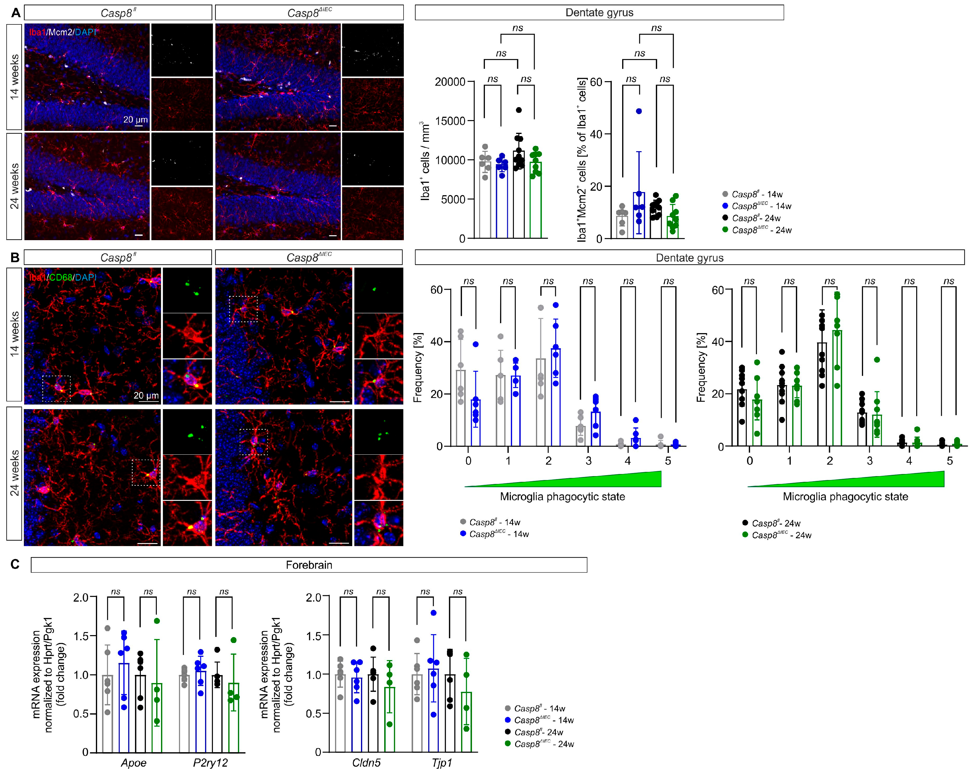
3.2. Microglial State in the Dentate Gyrus Is Maintained in Casp8ΔIEC Mice

Microglia are highly plastic brain-resident macrophages responsive to chronic peripheral inflammation [28,29] and involved in the regulation of adult hippocampal neurogenesis [12,16]. We therefore hypothesized that chronic ileocolitis in the Casp8ΔIEC mouse model of CD results in microglia activation and impairment of adult neurogenesis in the hippocampal DG. In the DG of Casp8ΔIEC mice, the cell density of ramified Iba1+ myeloid cells (Casp8fl vs. Casp8ΔIEC 14 weeks: 9738 ± 1341 mm3 vs. 9372 ± 886 mm3, p = 0.974; 24 weeks: 11,104 ± 2270 mm3 vs. 9679 ± 1282 mm3, p = 0.147; Figure 2A) and the percentage of proliferating Iba1+Mcm2+ double positive cells among all Iba1+ cells (Casp8fl vs. Casp8ΔIEC 14 weeks: 8.5 ± 3.6% vs. 17.6 ± 15.7%, p = 0.230; 24 weeks: 11.8 ± 2.9% vs. 8.4 ± 4.6%, p = 0.839; Figure 2A) were comparable to Casp8fl mice in both age groups. The activation of microglia was scored as previously described [25] based on the morphology and expression of the lysosomal protein CD68, reflecting the level of microglial phagocytic activity. In 14- and 24-week-old Casp8ΔIEC mice, the phagocytic phenotype of DG microglia was comparable to Casp8fl control mice (Casp8fl vs. Casp8ΔIEC 14 weeks, stage 0: 29.3 ± 11.6% vs. 18.0 ± 10.7%, p = 0.286; stage 1: 27.3 ± 9.4% vs. 27.2 ± 4.7%, p > 0.999; stage 2: 33.7 ± 15.2% vs. 37.5 ± 11.2%, p = 0.954; stage 3: 7.9 ± 3.7% vs. 13.3 ± 6.0%, p = 0.286; stage 4: 0.8 ± 0.8% vs. 3.3 ± 3.8%, p = 0.286; stage 5: 0.7 ± 1.5% vs. 0.7 ± 0.7%, p > 0.999; 24 weeks, stage 0: 21.8 ± 6.6% vs. 17.9 ± 7.9%, p = 0.958; stage 1: 23.4 ± 7.2% vs. 23.1 ± 4.6%, p = 0.995; stage 2: 39.8 ± 12.3% vs. 44.4 ± 14.4%, p = 0.958; stage 3: 12.9 ± 4.1% vs. 12.1 ± 8.7%, p = 0.995; stage 4: 1.4 ± 1.0% vs. 1.4 ± 2.1%, p = 0.995; stage 5: 0.6 ± 0.9% vs. 0.9 ± 1.0%, p = 0.958; Figure 2B), indicating that there is no overt inflammatory activation of microglia. An absent microglial response to gastrointestinal inflammatory cues in Casp8ΔIEC mice was supported by a lacking correlation with histological scores of colitis and ileitis within individual animals (Figure S1). This is corroborated by the mRNA expression analysis of genes associated with microglia homeostasis (purinergic receptor P2Y12, P2ry12; Casp8fl vs. Casp8ΔIEC 14 weeks: 1.0 ± 0.1 vs. 1.1 ± 0.2, p = 0.646; 24 weeks: Casp8fl: 1.0 ± 0.2 vs. 0.9 ± 0.4, p = 0.569; Figure 2C) and activation (apolipoprotein E, Apoe; Casp8fl vs. Casp8ΔIEC 14 weeks: 1.0 ± 0.4 vs. 1.2 ± 0.4, p = 0.611; 24 weeks: 1.0 ± 0.3 vs. 0.9 ± 0.6, p = 0.707; Figure 2C) in the forebrain, which revealed comparable expression among groups. Moreover, the gene expression of the tight junction proteins claudin 5 (Cldn5; Casp8fl vs. Casp8ΔIEC 14 weeks: 1.0 ± 0.2 vs. 1.0 ± 0.2, p = 0.705; 24 weeks: 1.0 ± 0.2 vs. 0.8 ± 0.3, p = 0.379; Figure 2C) and zona occludens 1 (ZO-1; tight junction protein 1, Tjp1; Casp8fl vs. Casp8ΔIEC 14 weeks: 1.0 ± 0.3 vs. 1.1 ± 0.4, p = 0.728; 24 weeks: 1.0 ± 0.3 vs. 0.8 ± 0.4, p = 0.389; Figure 2C), which are critical for blood–brain barrier integrity, were similar between groups, indicating no major blood–brain barrier disruption. These findings indicate that there is no major inflammatory microglial response or blood–brain barrier leakage in the DG of Casp8ΔIEC mice.

3.3. Adult Neurogenesis in the Dentate Gyrus Is Preserved Despite Ileocolitis

During adulthood, new neurons are born and mature in the subgranular zone (neurogenic niche) of the DG before integrating into the GCL. This process is susceptible to regulation by inflammatory factors derived from microglia [30]. In the Casp8ΔIEC mouse model, the thickness of the DG GCL was comparable between groups (Casp8fl vs. Casp8ΔIEC 14 weeks: 65.6 ± 5.3 µm vs. 64.2 ± 4.6 µm, p = 0.970; 24 weeks: 65.4 ± 6.2 µm vs. 67.4 ± 7.2 µm, p = 0.999; Figure 3A), indicating no major loss of GCL neurons or degeneration of the DG. Moreover, the absolute numbers of proliferating Mcm2+ cells in the subgranular zone of the DG did not significantly differ between groups (Casp8fl vs. Casp8ΔIEC 14 weeks: 4328 ± 1911 vs. 3934 ± 1013, p = 0.960; 24 weeks: 3626 ± 1146 vs. 3748 ± 3427, p > 0.999; Figure 3C). Besides absolute numbers, beginning neuronal differentiation of proliferating Mcm2+ progenitor cells indicated by the percentage of expression of the immature neuroblast marker DCX was not altered by ileocolitis (Casp8fl vs. Casp8ΔIEC 14 weeks: 70.9 ± 2.6% vs. 68.4 ± 5.6%, p = 0.923; 24 weeks: 59.6 ± 8.5% vs. 61.0 ± 6.9%, p > 0.999; Figure 3C). To address the further maturation of neural progenitor cells during experimental ileocolitis, we characterized DCX+ neuroblasts in the DG and found no significant changes in absolute numbers of DCX+ cells between Casp8ΔIEC mice and Casp8fl controls (Casp8fl vs. Casp8ΔIEC 14 weeks: 10,660 ± 4516 vs. 10,538 ± 3112, p > 0.999; 24 weeks: 8328 ± 4380 vs. 8013 ± 7236, p = 0.957; Figure 3D). A distinct co-expression analysis to compare the ratios of rather early DCX+Mcm2+ neuroblasts revealed no changes between groups (Casp8fl vs. Casp8ΔIEC 14 weeks: 28.9 ± 2.4% vs. 25.7 ± 1.8%, p = 0.695; 24 weeks: 28.2 ± 6.9% vs. 28.0 ± 4.5%, p = 0.989; Figure 3D), while the percentages of more mature DCX+NeuN+ neuroblasts showed a minor, but significant, increase in 24-week-old Casp8fl control mice compared to younger Casp8fl mice, and age-matched Casp8ΔIEC mice with ileocolitis (Casp8fl vs. Casp8ΔIEC 14 weeks: 18.1 ± 3.2% vs. 17.9 ± 2.1%, p = 0.996; 24 weeks: 22.3 ± 3.7% vs. 19.5 ± 3.7%, p = 0.037; Figure 3D) revealed no major shift in maturation between all four experimental groups. Furthermore, the density of TUNEL+ apoptotic cells were comparable between diseased Casp8ΔIEC and healthy Casp8fl mice at both ages (Casp8fl vs. Casp8ΔIEC 14 weeks: 1489 ± 1020 mm3 vs. 1090 ± 521 mm3, p > 0.999; 24 weeks: 1867 ± 1643 mm3 vs. 994 ± 583 mm3, p = 0.907; Figure 3E), indicating no changes in cell death during the neurogenic process between the groups. Finally, we assessed gene expression of neurotrophic factors supporting adult hippocampal neurogenesis. In line with the maintained stages of neurogenesis detected by histology, we did not observe alterations in the expression of Bdnf (Casp8fl vs. Casp8ΔIEC 14 weeks: 1.0 ± 0.6 vs. 0.9 ± 0.3, p = 0.673; 24 weeks: 1.0 ± 0.3 vs. 0.8 ± 0.4, p = 0.472; Figure 3F), Gdnf (Casp8fl vs. Casp8ΔIEC 14 weeks: 1.0 ± 1.0 vs. 0.8 ± 0.5, p = 0.665; 24 weeks: 1.0 ± 0.7 vs. 1.1 ± 1.2, p = 0.920; Figure 3F), and Igf1 (Casp8fl vs. Casp8ΔIEC 14 weeks: 1.0 ± 1.0 vs. 1.0 ± 0.6, p = 0.966; 24 weeks: 1.0 ± 0.4 vs. 0.9 ± 0.5, p = 0.686; Figure 3F) between mice with ileocolitis and controls in both age cohorts. In conclusion, adult hippocampal neurogenesis, including proliferation, maturation, apoptotic cell death, and expression of neurotrophic factors, is not altered in the presence of ileocolitis in Casp8ΔIEC mice.

4. Discussion

IBD poses a rising global health care burden [31,32,33,34]. Besides chronic intestinal inflammation, extraintestinal manifestations have been observed in nearly half of all IBD patients [35] and are more prevalent among CD patients [35,36]. Extraintestinal manifestations in patients diagnosed with IBD encompass the nervous system [37,38]. Specifically, IBD patients frequently exhibit neuropsychiatric symptoms, including depression and anxiety, which often correlate with inflammatory disease activity [1]. Thus, neuropsychiatric symptoms in IBD may be mediated by the propagation of gut-derived inflammation to the brain.
The Casp8ΔIEC mice utilized in this study exhibited spontaneous development of moderate ileocolitis, with some variation in the disease severity based on the histologic scoring. In addition to colitis and ileitis, Casp8ΔIEC mice demonstrated an increased expression of the inflammatory genes Tnf and Lcn2 in the liver, suggesting mild hepatic inflammation as previously reported [21,27]. In recent years, the interaction between the gut, the liver, and the brain—the gut–liver–brain axis—has gained tremendous research attention [39].
In light of these findings and in alignment with previous studies indicating compromised adult hippocampal neurogenesis in the context of experimental colitis [28,40,41], we hypothesized that the peripheral inflammation in the IEC-specific Casp8ΔIEC mouse model leads to microglia activation and subsequently to an impaired adult hippocampal neurogenesis. The process of adult hippocampal neurogenesis in the DG is influenced by various immune components, including microglia cells, which regulate cell proliferation, differentiation, and survival of newborn neurons and can act proneurogenically, depending on the inflammatory microenvironment [15,16,42]. In the brain, microglia account for the largest immune cell population. These parenchymal macrophages rapidly respond to changes in the tissue environment, as induced by external stimuli, such as inflammation [43,44]. The impact of inflammation on the highly plastic process of hippocampal neurogenesis is evident, as pharmacologically blocking peripherally induced brain inflammation by treatment with the nonsteroidal anti-inflammatory drug (NSAID) indomethacin restored hippocampal neurogenesis in mice [14].
We thus investigated the impact of chronic ileocolitis in the Casp8ΔIEC mouse model on the myeloid cell compartment and adult neurogenic niche of the hippocampal DG. Our results demonstrate that, despite peripheral inflammation, in both the adolescent (14-week-old) and adult (24-week-old) Casp8ΔIEC mice, signs of inflammatory microglial activation and blood–brain barrier leakage in the DG were not detected. Furthermore, adult hippocampal neurogenesis is maintained despite chronic gut-derived peripheral inflammation in Casp8ΔIEC mice. The detected minimal decrease in NeuN expression in DCX+ neuroblasts in 24-week-old Casp8ΔIEC mice compared to Casp8fl controls can mainly be attributed to an unexpected slight increase in proportional NeuN-positivity in Casp8fl mice between 14 and 24 weeks of age but is very unlikely to reflect a biologically relevant impairment in neuroblast maturation due to ileocolitis, especially as all other neurogenesis-related parameters are preserved. This indicates that not only microglia but also other cellular and molecular drivers of neuroinflammation, as well as microbial factors previously described in IBD [4,45], are unable to trigger an overt microglial response and substantially impair adult neurogenesis in the DG of Casp8ΔIEC mice. Previous studies employing the DSS-induced colitis model have reported conflicting results regarding microglial density and activation state in the hippocampus [28,40,41,46]. While an increased density of microglia and microglial activation were only observed in the hippocampus of acute DSS-induced colitis mice [28,40,46], elevated inflammatory cytokine expression was detected in the hippocampus of both acute and chronic DSS-induced colitis mice [28,47], whereas the density of microglia was not altered in the hippocampus of chronic DSS-induced colitis compared to control mice [41].
Moreover, conflicting results have been reported regarding the effect of DSS-induced colitis on the generation and survival of new neurons in the hippocampal DG [28,40,41]. In acute DSS-induced colitis mice, Vitali et al. [41] observed reduced densities of glial fibrillary acidic protein (Gfap)-expressing radial glia-like stem cells and SRY-box transcription factor 2 (Sox2)-expressing intermediate progenitor cells with no changes in the densities of Ki67+-proliferating cells and DCX+ immature neurons in the hippocampal DG of 8-week-old male C57BL/6 mice fed with 0.7% DSS for 7 days plus 7 days recovery, whereas Takahashi et al. [40] reported significantly reduced numbers of 5-Bromo-2′-Desoxyuridine (BrdU)-positive proliferating cells and DCX+ immature neurons in the DG in 6-7-week-old male ddY mice fed with 1.5% DSS for 7 days compared to control mice. In contrast, Gampierakis et al. [28] found significantly increased densities of Ki67+-proliferating cells, Sox2+ intermediate progenitor cells, and DCX+ immature neurons in the DG of 2.5-month-old male C57BL/6 mice fed with 2.5% DSS for 7 days compared to control mice. In chronic DSS-induced colitis, Zonis et al. [48] observed reduced numbers of Sox2+ and DCX+ cells in the DG subgranular zone, which is in line with the reduced hippocampal gene and protein expression of Sox2, DCX, and DCX reported by Barnes et al. [47]. In contrast, Vitali et al. [41] observed no difference in the densities of Gfap+ radial glia-like stem cells, Sox2+, and DCX+ cells but a reduced density of Ki67+-proliferating cells in the hippocampus of chronic DSS-induced colitis mice. The reported findings do not imply a dose-dependent effect, and the heterogeneous results may be attributed to the differences in the experimental design paradigm, age, and genetic background of animals, disease severity, and the resulting systemic inflammatory response.
Thus, we employed the IEC-specific Casp8ΔIEC transgenic mouse model to overcome the limitation of the highly variable and less specific colitis induction applied in previous studies employing the DSS-induced colitis model. While numbers of proliferating progenitor cells and maturating neuroblasts as well as apoptosis in the DG of Casp8ΔIEC mice were comparable to those in the DG of Casp8fl mice, minimal changes in the expression of NeuN within maturating DCX+ neuroblasts are possible, as mice age and ileocolitis progresses. In the light of the high vulnerability of adult hippocampal neurogenesis and previous findings in DSS-treated rodents, our finding of largely maintained adult hippocampal neurogenesis may appear surprising, but preserved adult hippocampal neurogenesis has previously been described in other models of even more severe chronic peripheral inflammation [22,49]. As peripheral inflammation was shown to affect the brain in a regionally different manner [29], the present findings do not allow the conclusion of the overall cerebral resilience to ileocolitis. Future studies therefore must compare regional immune responses in the brain of the Casp8ΔIEC model. This may include further brain regions which might be involved in neuropsychiatric comorbidity of IBD independent of the hippocampus. Moreover, an impairment of adult hippocampal neurogenesis may be restricted to certain subtypes of systemic inflammation and may be predicted by more thorough immunophenotyping of ileocolitis and systemic inflammation in IBD patients in the future.

5. Conclusions

In summary, hippocampal neurogenesis and microglial response in the DG are largely unaffected by ileocolitis in the Casp8ΔIEC transgenic mouse model of CD.

Supplementary Materials

The following supporting information can be downloaded at: https://www.mdpi.com/article/10.3390/cells14110841/s1, Figure S1: Correlation of microglial parameters with ileocolitis severity. Pearson correlation analysis revealed no correlation between Colitis+Ileitis score and (A) Iba1+ cell density in the DG (R2 = −0.2200), (B) percentage of Iba1+Mcm2+ cells of all Iba1+ cells in the DG (R2 = 0.0156), and (C–H) microglial activation scores 0–5 (R2 = −0.2419, R2 = −0.0013, R2 = 0.0845, R2 = 0.1517, R2 = 0.1230, R2 = −0.0218, respectively). Regression line equations and R2 values are indicated in the respective plot.

Author Contributions

Conceptualization, C.G. and P.S.; methodology, C.G., I.S. and P.S.; formal analysis, H.M., R.K.M. and P.S.; investigation, H.M., R.K.M. and P.S.; resources, C.G., I.S., J.W. and P.S.; writing—original draft preparation, R.K.M.; writing—review and editing, C.G., I.S., J.W. and P.S.; visualization, R.K.M.; supervision, R.K.M., J.W., C.G. and P.S.; project administration, C.G., J.W. and P.S.; funding acquisition, C.G., J.W. and P.S. All authors have read and agreed to the published version of the manuscript.

Funding

This research was funded by the Deutsche Forschungsgemeinschaft (KFO5024-505539112 (A04, B01, Z01), C.G., J.W. and P.S.; TRR241–375876048 (A02), FOR2886–405969122 (A02), TRR305–429280966 (B08), TRR369–501752319 (A02), DFG GU 1431/13-1, C.G.), the Interdisciplinary Center for Clinical Research of the Friedrich-Alexander-Universität Erlangen-Nürnberg (Junior Project J94, P.S. and R.K.M.; MD thesis scholarship, H.M.; Jochen-Kalden funding program N5, C.G.) and the Else Kröner-Fresenius-Stiftung (Else Kröner Memorial Fellowship, P.S.).

Institutional Review Board Statement

The animal study was performed in accordance with the EU Directive 2010/63 for the protection of animals used for scientific purposes and was approved by the local government commission of animal health (approval date: 5 April 2024).

Informed Consent Statement

Not applicable.

Data Availability Statement

The original contributions presented in this study are included in the article. Further inquiries can be directed to the corresponding author.

Acknowledgments

The present study was conducted in accordance with the academic requirements of the Friedrich-Alexander-Universität Erlangen-Nürnberg for obtaining the degree ‘Dr. med.’ of Hanna Mundlos. We kindly thank Lucas Hoffmann from the Department of Neuropathology, Universitätsklinikum Erlangen, for the use of the Hamamatsu Nanozoomer S60 and extend our gratitude to Carina Gauer from Erlangen for her intellectual contributions to this study.

Conflicts of Interest

The authors declare no conflicts of interest.

Abbreviations

The following abbreviations are used in this manuscript:
ANOVAAnalysis of variance
APOEApolipoprotein E
BDNFBrain-derived neurotrophic factor
BRDU5-Bromo-2′-Desoxyuridine
CASP8Caspase 8
CDCrohn’s disease
CLDN5Claudin 5
CNSCentral nervous system
DAPI4′,6-Diamidino-2-phenylindole dihydrochloride
DCXDoublecortin
DGDentate gyrus
DSSDextran sodium sulfate
GCLGranule cell layer
GDNFGlial cell line-derived neurotrophic factor
GFAPGlial fibrillary acidic protein
H&EHematoxylin and eosin
IBA1Ionized calcium-binding adapter molecule 1
IBDInflammatory bowel disease
IECIntestinal epithelial cell
IGF1Insulin-like growth factor 1
IL1BInterleukin 1 beta
LCN2Lipocalin 2
MCM2Minichromosome maintenance complex component 2
P2RY12Purinergic receptor P2Y12
PBSPhosphate-buffered saline
QPCRReal-time quantitative polymerase chain reaction
RTRoom temperature
SOX2SRY-box transcription factor 2
TBSTris-buffered saline
TJP1Tight junction protein 1
TNFTumor necrosis factor
TUNELTerminal deoxynucleotide transferase-mediated dUTP-X nick end labeling
UCUlcerative colitis
ZO-1Zona occludens 1

References

  1. Barberio, B.; Zamani, M.; Black, C.J.; Savarino, E.V.; Ford, A.C. Prevalence of symptoms of anxiety and depression in patients with inflammatory bowel disease: A systematic review and meta-analysis. Lancet Gastroenterol. Hepatol. 2021, 6, 359–370. [Google Scholar] [CrossRef] [PubMed]
  2. Fairbrass, K.M.; Lovatt, J.; Barberio, B.; Yuan, Y.; Gracie, D.J.; Ford, A.C. Bidirectional brain–gut axis effects influence mood and prognosis in IBD: A systematic review and meta-analysis. Gut 2022, 71, 1773. [Google Scholar] [CrossRef]
  3. Hopkins, C.W.P.; Powell, N.; Norton, C.; Dumbrill, J.L.; Hayee, B.H.; Moulton, C.D. Cognitive Impairment in Adult Inflammatory Bowel Disease: A Systematic Review and Meta-Analysis. J. Acad. Consult.-Liaison Psychiatry 2021, 62, 387–403. [Google Scholar] [CrossRef] [PubMed]
  4. Masanetz, R.K.; Winkler, J.; Winner, B.; Günther, C.; Süß, P. The Gut–Immune–Brain Axis: An Important Route for Neuropsychiatric Morbidity in Inflammatory Bowel Disease. Int. J. Mol. Sci. 2022, 23, 11111. [Google Scholar] [CrossRef]
  5. Toda, T.; Parylak, S.L.; Linker, S.B.; Gage, F.H. The role of adult hippocampal neurogenesis in brain health and disease. Mol. Psychiatry 2019, 24, 67–87. [Google Scholar] [CrossRef]
  6. Bonafina, A.; Trinchero, M.F.; Ríos, A.S.; Bekinschtein, P.; Schinder, A.F.; Paratcha, G.; Ledda, F. GDNF and GFRα1 Are Required for Proper Integration of Adult-Born Hippocampal Neurons. Cell Rep. 2019, 29, 4308–4319.e4. [Google Scholar] [CrossRef]
  7. Ribeiro, F.F.; Xapelli, S. Intervention of Brain-Derived Neurotrophic Factor and Other Neurotrophins in Adult Neurogenesis. In Recent Advances in NGF and Related Molecules: The Continuum of the NGF “Saga”; Calzà, L., Aloe, L., Giardino, L., Eds.; Springer International Publishing: Cham, Switzerland, 2021; pp. 95–115. [Google Scholar]
  8. Yuan, H.; Chen, R.; Wu, L.; Chen, Q.; Hu, A.; Zhang, T.; Wang, Z.; Zhu, X. The Regulatory Mechanism of Neurogenesis by IGF-1 in Adult Mice. Mol. Neurobiol. 2015, 51, 512–522. [Google Scholar] [CrossRef] [PubMed]
  9. Hill, A.S.; Sahay, A.; Hen, R. Increasing Adult Hippocampal Neurogenesis is Sufficient to Reduce Anxiety and Depression-Like Behaviors. Neuropsychopharmacology 2015, 40, 2368–2378. [Google Scholar] [CrossRef]
  10. Jones, K.L.; Zhou, M.; Jhaveri, D.J. Dissecting the role of adult hippocampal neurogenesis towards resilience versus susceptibility to stress-related mood disorders. NPJ Sci. Learn. 2022, 7, 16. [Google Scholar] [CrossRef]
  11. Ayyubova, G.; Kodali, M.; Upadhya, R.; Madhu, L.N.; Attaluri, S.; Somayaji, Y.; Shuai, B.; Rao, S.; Shankar, G.; Shetty, A.K. Extracellular vesicles from hiPSC-NSCs can prevent peripheral inflammation-induced cognitive dysfunction with inflammasome inhibition and improved neurogenesis in the hippocampus. J. Neuroinflamm. 2023, 20, 297. [Google Scholar] [CrossRef]
  12. Biscaro, B.; Lindvall, O.; Tesco, G.; Ekdahl, C.T.; Nitsch, R.M. Inhibition of Microglial Activation Protects Hippocampal Neurogenesis and Improves Cognitive Deficits in a Transgenic Mouse Model for Alzheimer’s Disease. Neurodegener. Dis. 2012, 9, 187–198. [Google Scholar] [CrossRef]
  13. Lindvall, O.; Kokaia, Z. Neurogenesis following Stroke Affecting the Adult Brain. Cold Spring Harb. Perspect. Biol. 2015, 7, a019034. [Google Scholar] [CrossRef] [PubMed]
  14. Monje, M.L.; Toda, H.; Palmer, T.D. Inflammatory blockade restores adult hippocampal neurogenesis. Science 2003, 302, 1760–1765. [Google Scholar] [CrossRef] [PubMed]
  15. Ekdahl, C.T.; Claasen, J.-H.; Bonde, S.; Kokaia, Z.; Lindvall, O. Inflammation is detrimental for neurogenesis in adult brain. Proc. Natl. Acad. Sci. USA 2003, 100, 13632–13637. [Google Scholar] [CrossRef]
  16. Ekdahl, C.T.; Kokaia, Z.; Lindvall, O. Brain inflammation and adult neurogenesis: The dual role of microglia. Neuroscience 2009, 158, 1021–1029. [Google Scholar] [CrossRef]
  17. Sierra, A.; Encinas, J.M.; Deudero, J.J.; Chancey, J.H.; Enikolopov, G.; Overstreet-Wadiche, L.S.; Tsirka, S.E.; Maletic-Savatic, M. Microglia shape adult hippocampal neurogenesis through apoptosis-coupled phagocytosis. Cell Stem Cell 2010, 7, 483–495. [Google Scholar] [CrossRef]
  18. Vukovic, J.; Colditz, M.J.; Blackmore, D.G.; Ruitenberg, M.J.; Bartlett, P.F. Microglia Modulate Hippocampal Neural Precursor Activity in Response to Exercise and Aging. J. Neurosci. 2012, 32, 6435. [Google Scholar] [CrossRef] [PubMed]
  19. Günther, C.; Martini, E.; Wittkopf, N.; Amann, K.; Weigmann, B.; Neumann, H.; Waldner, M.J.; Hedrick, S.M.; Tenzer, S.; Neurath, M.F.; et al. Caspase-8 regulates TNF-α-induced epithelial necroptosis and terminal ileitis. Nature 2011, 477, 335–339. [Google Scholar] [CrossRef]
  20. Stolzer, I.; Kaden-Volynets, V.; Ruder, B.; Letizia, M.; Bittel, M.; Rausch, P.; Basic, M.; Bleich, A.; Baines, J.F.; Neurath, M.F.; et al. Environmental Microbial Factors Determine the Pattern of Inflammatory Lesions in a Murine Model of Crohn’s Disease–Like Inflammation. Inflamm. Bowel Dis. 2019, 26, 66–79. [Google Scholar] [CrossRef]
  21. Kaden-Volynets, V.; Günther, C.; Zimmermann, J.; Beisner, J.; Becker, C.; Bischoff, S.C. Deletion of the Casp8 gene in mice results in ileocolitis, gut barrier dysfunction, and malassimilation, which can be partially attenuated by inulin or sodium butyrate. Am. J. Physiol.-Gastrointest. Liver Physiol. 2019, 317, G493–G507. [Google Scholar] [CrossRef]
  22. Masanetz, R.K.; Baum, W.; Schett, G.; Winkler, J.; Süß, P. Cellular plasticity and myeloid inflammation in the adult brain are independent of the transcriptional modulator DREAM. Neurosci. Lett. 2023, 796, 137061. [Google Scholar] [CrossRef] [PubMed]
  23. Hefele, M.; Stolzer, I.; Ruder, B.; He, G.-W.; Mahapatro, M.; Wirtz, S.; Neurath, M.F.; Günther, C. Intestinal epithelial Caspase-8 signaling is essential to prevent necroptosis during Salmonella Typhimurium induced enteritis. Mucosal Immunol. 2018, 11, 1191–1202. [Google Scholar] [CrossRef] [PubMed]
  24. Stolzer, I.; Schickedanz, L.; Chiriac, M.T.; López-Posadas, R.; Grassl, G.A.; Mattner, J.; Wirtz, S.; Winner, B.; Neurath, M.F.; Günther, C. STAT1 coordinates intestinal epithelial cell death during gastrointestinal infection upstream of Caspase-8. Mucosal Immunol. 2022, 15, 130–142. [Google Scholar] [CrossRef]
  25. Wilton, D.K.; Mastro, K.; Heller, M.D.; Gergits, F.W.; Willing, C.R.; Fahey, J.B.; Frouin, A.; Daggett, A.; Gu, X.; Kim, Y.A.; et al. Microglia and complement mediate early corticostriatal synapse loss and cognitive dysfunction in Huntington’s disease. Nat. Med. 2023, 29, 2866–2884. [Google Scholar] [CrossRef] [PubMed]
  26. Motulsky, H.J.; Brown, R.E. Detecting outliers when fitting data with nonlinear regression—A new method based on robust nonlinear regression and the false discovery rate. BMC Bioinform. 2006, 7, 123. [Google Scholar] [CrossRef]
  27. Dorner, H.; Stolzer, I.; Mattner, J.; Kaminski, S.; Leistl, S.; Edrich, L.-M.; Schwendner, R.; Hobauer, J.; Sebald, A.; Leikam, S.; et al. Gut Pathobiont–Derived Outer Membrane Vesicles Drive Liver Inflammation and Fibrosis in Primary Sclerosing Cholangitis–Associated Inflammatory Bowel Disease. Gastroenterology 2024, 167, 1183–1197.e1116. [Google Scholar] [CrossRef]
  28. Gampierakis, I.-A.; Koutmani, Y.; Semitekolou, M.; Morianos, I.; Polissidis, A.; Katsouda, A.; Charalampopoulos, I.; Xanthou, G.; Gravanis, A.; Karalis, K.P. Hippocampal neural stem cells and microglia response to experimental inflammatory bowel disease (IBD). Mol. Psychiatry 2021, 26, 1248–1263. [Google Scholar] [CrossRef]
  29. Süß, P.; Hoffmann, A.; Rothe, T.; Ouyang, Z.; Baum, W.; Staszewski, O.; Schett, G.; Prinz, M.; Krönke, G.; Glass, C.K.; et al. Chronic Peripheral Inflammation Causes a Region-Specific Myeloid Response in the Central Nervous System. Cell Rep. 2020, 30, 4082–4095.e4086. [Google Scholar] [CrossRef]
  30. Araki, T.; Ikegaya, Y.; Koyama, R. The effects of microglia- and astrocyte-derived factors on neurogenesis in health and disease. Eur. J. Neurosci. 2021, 54, 5880–5901. [Google Scholar] [CrossRef]
  31. Kaplan, G.G.; Windsor, J.W. The four epidemiological stages in the global evolution of inflammatory bowel disease. Nat. Rev. Gastroenterol. Hepatol. 2021, 18, 56–66. [Google Scholar] [CrossRef]
  32. El Hadad, J.; Schreiner, P.; Vavricka, S.R.; Greuter, T. The Genetics of Inflammatory Bowel Disease. Mol. Diagn. Ther. 2024, 28, 27–35. [Google Scholar] [CrossRef] [PubMed]
  33. Nambu, R.; Warner, N.; Mulder, D.J.; Kotlarz, D.; McGovern, D.P.B.; Cho, J.; Klein, C.; Snapper, S.B.; Griffiths, A.M.; Iwama, I.; et al. A Systematic Review of Monogenic Inflammatory Bowel Disease. Clin. Gastroenterol. Hepatol. 2022, 20, e653–e663. [Google Scholar] [CrossRef] [PubMed]
  34. Piovani, D.; Danese, S.; Peyrin-Biroulet, L.; Nikolopoulos, G.K.; Lytras, T.; Bonovas, S. Environmental Risk Factors for Inflammatory Bowel Diseases: An Umbrella Review of Meta-analyses. Gastroenterology 2019, 157, 647–659.e644. [Google Scholar] [CrossRef]
  35. Khrom, M.; Long, M.; Dube, S.; Robbins, L.; Botwin, G.J.; Yang, S.; Mengesha, E.; Li, D.; Naito, T.; Bonthala, N.N.; et al. Comprehensive Association Analyses of Extraintestinal Manifestations in Inflammatory Bowel Disease. Gastroenterology 2024, 167, 315–332. [Google Scholar] [CrossRef]
  36. Kilic, Y.; Kamal, S.; Jaffar, F.; Sriranganathan, D.; Quraishi, M.N.; Segal, J.P. Prevalence of Extraintestinal Manifestations in Inflammatory Bowel Disease: A Systematic Review and Meta-analysis. Inflamm. Bowel Dis. 2023, 30, 230–239. [Google Scholar] [CrossRef]
  37. Ferro, J.M.; Oliveira, S.N.; Correia, L. Neurologic manifestations of inflammatory bowel diseases. In Handbook of Clinical Neurology; Biller, J., Ferro, J.M., Eds.; Elsevier: Amsterdam, The Netherlands, 2014; Volume 120, Chapter 40; pp. 595–605. [Google Scholar]
  38. Hall, C.V.; Radford-Smith, G.; Savage, E.; Robinson, C.; Cocchi, L.; Moran, R.J. Brain signatures of chronic gut inflammation. Front. Psychiatry 2023, 14, 1250268. [Google Scholar] [CrossRef] [PubMed]
  39. Yan, M.; Man, S.; Sun, B.; Ma, L.; Guo, L.; Huang, L.; Gao, W. Gut liver brain axis in diseases: The implications for therapeutic interventions. Signal Transduct. Target. Ther. 2023, 8, 443. [Google Scholar] [CrossRef]
  40. Takahashi, K.; Kurokawa, K.; Hong, L.; Miyagawa, K.; Mochida-Saito, A.; Takeda, H.; Tsuji, M. Hippocampal and gut AMPK activation attenuates enterocolitis-like symptoms and co-occurring depressive-like behavior in ulcerative colitis model mice: Involvement of brain-gut autophagy. Exp. Neurol. 2024, 373, 114671. [Google Scholar] [CrossRef]
  41. Vitali, R.; Prioreschi, C.; Lorenzo Rebenaque, L.; Colantoni, E.; Giovannini, D.; Frusciante, S.; Diretto, G.; Marco-Jiménez, F.; Mancuso, M.; Casciati, A.; et al. Gut–Brain Axis: Insights from Hippocampal Neurogenesis and Brain Tumor Development in a Mouse Model of Experimental Colitis Induced by Dextran Sodium Sulfate. Int. J. Mol. Sci. 2022, 23, 11495. [Google Scholar] [CrossRef]
  42. Amanollahi, M.; Jameie, M.; Heidari, A.; Rezaei, N. The Dialogue Between Neuroinflammation and Adult Neurogenesis: Mechanisms Involved and Alterations in Neurological Diseases. Mol. Neurobiol. 2023, 60, 923–959. [Google Scholar] [CrossRef]
  43. Chausse, B.; Malorny, N.; Lewen, A.; Poschet, G.; Berndt, N.; Kann, O. Metabolic flexibility ensures proper neuronal network function in moderate neuroinflammation. Sci. Rep. 2024, 14, 14405. [Google Scholar] [CrossRef] [PubMed]
  44. Sabogal-Guáqueta, A.M.; Marmolejo-Garza, A.; Trombetta-Lima, M.; Oun, A.; Hunneman, J.; Chen, T.; Koistinaho, J.; Lehtonen, S.; Kortholt, A.; Wolters, J.C.; et al. Species-specific metabolic reprogramming in human and mouse microglia during inflammatory pathway induction. Nat. Commun. 2023, 14, 6454. [Google Scholar] [CrossRef] [PubMed]
  45. Chang, J.T. Pathophysiology of Inflammatory Bowel Diseases. N. Engl. J. Med. 2020, 383, 2652–2664. [Google Scholar] [CrossRef] [PubMed]
  46. Nakagawasai, O.; Yamada, K.; Takahashi, K.; Odaira, T.; Sakuma, W.; Ishizawa, D.; Takahashi, N.; Onuma, K.; Hozumi, C.; Nemoto, W.; et al. Liver hydrolysate prevents depressive-like behavior in an animal model of colitis: Involvement of hippocampal neurogenesis via the AMPK/BDNF pathway. Behav. Brain Res. 2020, 390, 112640. [Google Scholar] [CrossRef]
  47. Barnes, S.E.; Zera, K.A.; Ivison, G.T.; Buckwalter, M.S.; Engleman, E.G. Brain profiling in murine colitis and human epilepsy reveals neutrophils and TNFα as mediators of neuronal hyperexcitability. J. Neuroinflamm. 2021, 18, 199. [Google Scholar] [CrossRef]
  48. Zonis, S.; Pechnick, R.N.; Ljubimov, V.A.; Mahgerefteh, M.; Wawrowsky, K.; Michelsen, K.S.; Chesnokova, V. Chronic intestinal inflammation alters hippocampal neurogenesis. J. Neuroinflamm. 2015, 12, 65. [Google Scholar] [CrossRef]
  49. Süß, P.; Kalinichenko, L.; Baum, W.; Reichel, M.; Kornhuber, J.; Loskarn, S.; Ettle, B.; Distler, J.H.W.; Schett, G.; Winkler, J.; et al. Hippocampal structure and function are maintained despite severe innate peripheral inflammation. Brain Behav. Immun. 2015, 49, 156–170. [Google Scholar] [CrossRef]
Figure 1. Ileocolitis in the Casp8ΔIEC mouse model of inflammatory bowel disease. (A) Casp8ΔIEC mice and Casp8fl control mice aged 14 and 24 weeks were used. (B) Representative H&E images of colon and terminal ileum tissue sections of 24-week-old Casp8ΔIEC and Casp8fl mice. Scale bar: 100 µm. (C) The combined and the individual histologic scores of the colon and ileum were significantly higher in 14- and 24-week-old Casp8ΔIEC mice (14 weeks: n = 6, 3 female/3 male; 24: weeks: n = 9, 6 female/3 male) compared to age-matched Casp8fl mice (14 weeks: n = 6, 3 female/3 male; 24: weeks: n = 9, 6 female/3 male). (D) Gene expression analysis revealed increased mRNA levels of Tnf and Lcn2 but not Il1b in the liver of 14- and 24-week-old Casp8ΔIEC mice (14 weeks: n = 6, 3 female/3 male; 24: weeks: n = 4, 4 female/0 male) compared to age-matched Casp8fl mice (14 weeks: n = 6, 3 female/3 male; 24: weeks: n = 6, 6 female/0 male). Significance in (C) was tested by two-way ANOVA followed by Tukey’s post hoc test. Significance in (D) was tested by unpaired t-tests. Numbers above brackets indicate p-values for pairwise comparisons. All data are shown as mean ± standard deviation; ns p > 0.05, * p ≤ 0.05, ** p ≤ 0.01, *** p ≤ 0.001.
Cells 14 00841 g001
Figure 2. Microglia proliferation and activation in the DG. (A) The densities of ramified Iba1+ myeloid cells (microglia) and Iba1+Mcm2+-proliferating microglia were similar between groups. (B) Similar number of phagocytic microglia scored by cell morphology and CD68 expression were observed in Casp8ΔIEC and Casp8fl mice in both age groups. (C) In the forebrain, expression of genes associated with microglia activation (Apoe) and homeostasis (P2ry12) and of genes encoding tight junction proteins (Cldn5, Tjp1) was similar in all groups. Data in (A,B) are representative of n = 6 (3 female/3 male) 14-week-old and n = 9 (6 female/3 male) 24-week-old Casp8ΔIEC mice as well as n = 6 (3 female/3 male) 14-week-old and n = 10 (7 female/3 male) 24-week-old Casp8fl controls and were tested for significance using two-way ANOVA followed by Tukey’s post hoc test ((A), Iba1+ cells), two-way ANOVA followed by Tukey’s post hoc test ((A), Iba1+Mcm2+ cells), or multiple unpaired t-tests (B), respectively. Data in (C) are representative of n = 6 (3 female/ 3 male) 14-week-old and n = 4 (4 female/0 male) 24-week-old Casp8ΔIEC mice as well as n = 6 (3 female/3 male) 14-week-old and n = 6 (6 female/0 male) 24-week-old Casp8fl controls and were tested for significance by unpaired t-tests. All data are shown as mean ± standard deviation; ns p > 0.05.
Cells 14 00841 g002
Figure 3. Adult hippocampal neurogenesis. (A) The thickness of the DG GCL was similar in all groups. Scale bar: 200 µm. (B) Representative images showing immunofluorescent co-staining of the DG for Mcm2 (white), DCX (green), NeuN (red), and DAPI (blue) in 14- and 24-week-old Casp8fl and Casp8ΔIEC mice. Scale bar: 100 µm. (C) Quantification of total proliferating Mcm2+ cells and the percentage of Mcm2+ cells co-expressing DCX showing no difference between mice with ileocolitis and controls. (D) Quantification of total maturating DCX+ neuroblasts and the percentages of DCX+ cells co-expressing Mcm2 or NeuN, respectively, showing a slight increase in DCX+NeuN+ cells in 24-week-old Casp8fl mice compared to 14-week-old Casp8fl and 24-week-old Casp8ΔIEC mice. (E) Densities of TUNEL+ apoptotic cells per mm3 were comparable between Casp8ΔIEC and Casp8fl mice in both age groups. (F) Expression of genes encoding the trophic factors Bdnf, Gdnf, and Igf1 in the forebrain was similar in 14- and 24-week-old Casp8ΔIEC mice compared to age-matched Casp8fl mice. Data in (A,F) represent n = 6 (3 female/3 male) 14-week-old and n = 4 (4 female/0 male) 24-week-old Casp8ΔIEC mice as well as n = 6 (3 female/3 male) 14-week-old and n = 6 (6 female/0 male) 24-week-old Casp8fl controls. Data in (C,D) represent n = 5 (3 female/2 male) 14-week-old and n = 9 (6 female/3 male) 24-week-old Casp8ΔIEC mice as well as n = 6 (3 female/3 male) 14-week-old and n = 10 (7 female/3 male) 24-week-old Casp8fl controls. Data in (E) represent n = 6 (3 female/3 male) 14-week-old and n = 7 (5 female/2 male) 24-week-old Casp8ΔIEC mice as well as n = 6 (3 female/3 male) 14-week-old and n = 8 (5 female/3 male) 24-week-old Casp8fl controls. Data in (A,CE) were tested for significant differences between genotypes and ages by 2way ANOVA followed by Tukey’s post hoc test. Data in (F) were tested for significant differences between genotypes via unpaired t-tests. All data are shown as means ± standard deviation; ns p > 0.05, * p ≤ 0.05.
Cells 14 00841 g003
Table 1. Sequences of primers used for qPCR.
GeneSequence Forward Primer (5′-3′)Sequence Reverse Primer (5′-3′)
ApoeGAACAACCCGCCTCGTGAAGCTCCTTCCGAAACAAGTCC
BdnfGCTCATCTTTGCCAGAGCCCAGCAGCTTTCTCAACGCCT
Cldn5GTTAAGGCACGGGTAGCACTTACTTCTGTGACACCGGCAC
GdnfACCCTGCTAGAAAACGCGAGACGGAGATCCGGGCAAAAG
HprtGTCATGTCGACCCTCAGTCCGCAAGTCTTTCAGTCCTGTCC
Igf1ATACAGCCAACGGGAAACAGCAACAAAGCTGGATGCCTGTC
Il1bTGACAGTGATGAGAATGACCTGCCACGGGAAAGACACAGGTA
Lcn2ACGGACTACAACCAGTTCGCATGCATTGGTCGGTGGGG
P2ry12AGTGCAAGAACACTCAAGGCGACGGTGTACAGCAATGGGA
Pgk1GTCGTGATGAGGGTGGACTTAACGGACTTGGCTCCATTGT
TnfTAGCCCACGTCGTAGCAAACGCAGCCTTGTCCCTTGAAGA
Tjp1GGAGATGTTTATGCGGACGGCCATTGCTGTGCTCTTAGCG
Disclaimer/Publisher’s Note: The statements, opinions and data contained in all publications are solely those of the individual author(s) and contributor(s) and not of MDPI and/or the editor(s). MDPI and/or the editor(s) disclaim responsibility for any injury to people or property resulting from any ideas, methods, instructions or products referred to in the content.

