Abstract
The cell membrane is frequently subjected to damage, either through physical or chemical means. The swift restoration of the cell membrane’s integrity is crucial to prevent the leakage of intracellular materials and the uncontrolled influx of extracellular ions. Consequently, wound repair plays a vital role in cell survival, akin to the importance of DNA repair. The mechanisms involved in wound repair encompass a series of events, including ion influx, membrane patch formation, endocytosis, exocytosis, recruitment of the actin cytoskeleton, and the elimination of damaged membrane sections. Despite the absence of a universally accepted general model, diverse molecular models have been proposed for wound repair in different organisms. Traditional wound methods not only damage the cell membrane but also impact intracellular structures, including the underlying cortical actin networks, microtubules, and organelles. In contrast, the more recent improved laserporation selectively targets the cell membrane. Studies on Dictyostelium cells utilizing this method have introduced a novel perspective on the wound repair mechanism. This review commences by detailing methods for inducing wounds and subsequently reviews recent developments in the field.
1. Introduction
The cell membrane serves as a crucial barrier between the extracellular and intracellular spaces, yet it is consistently vulnerable to physical or chemical damage. Such injuries compromise the membrane’s integrity, leading to an influx of undesirable substances into the cell and cytoplasmic loss. Local wounds on the cell membrane also impact cell polarity during migration [1] and influence the division axis and symmetrical division in cell division [2]. Mechanically active tissues, like mammalian skeletal and cardiac muscles, frequently experience membrane wounds due to repeated contractions [3,4,5]. Ischemia-reperfusion injury followed by heart attack and stroke also damages the cell membrane [6]. Infection by pathogenic funguses, bacteria, and viruses and their pore-forming toxins can also result in membrane wounds at the cell membrane [7,8,9,10]. Loss of the wound repair function is observed in various diseases, including diabetes [11], muscular dystrophies [12,13], acute kidney injury [14], and vitamin deficiencies [15]. Recent studies have identified defects in wound repair as common in Parkinson’s and Alzheimer’s diseases [16,17,18]. Plant cells, affected by freezing damage in cold seasons, also possess the capability to repair damaged membranes [19,20,21]. Similar to DNA repair, wound repair is a physiologically vital phenomenon for living cells. Moreover, many methods for introducing extracellular substances into cells, such as microinjection and electroporation, rely on cellular wound repair. See also recent good reviews [6,22,23,24,25,26].
The mechanisms of wound repair have been extensively studied across various model organisms, including mammalian cells [27,28], amphibian oocytes [29,30,31,32], echinoderm oocytes [33,34,35], fruit flies [36,37,38], nematodes [12,39,40], amoebae [41,42], yeast [43,44], ciliate [45], plant cells [19,46,47], and Dictyostelium cells [48]. A common feature among these mechanisms is the essential role of Ca2+ influx from an external medium in the wound repair process. While the “membrane patch hypothesis” suggests that cytosolic membrane vesicles accumulate at the wound site to form an impermanent “patch” for emergency wound pore plugging [34,49,50,51], alternative hypotheses that do not involve patching have also been proposed [25,27,52]. However, there is no universally accepted general model for the mechanisms driving the repair process.
In larger cells like Xenopus oocytes and Drosophila embryos, an actomyosin ring surrounds the wound site, similar to the contractile ring in dividing cells, facilitating wound pore closure [36,53,54,55]. Conversely, in smaller cells like yeast, animal culture cells, and Dictyostelium cells, actin transiently accumulates at the wound site [43,56,57,58]. The absence of actin polymerization hinders wound pore closure in, mammalian culture cells, muscle cells, and Dictyostelium cells [51,59,60]. Notably, myosin II’s accumulation at the wound site and its contribution to wound repair in small cells remain controversial [43,57,58,61].
This article reviews recent advancements in cellular wound repair, with a specific focus on the wound response and repair process in Dictyostelium cells. The discussion encompasses techniques for studying the wound repair mechanism, an overview of previous information on wound repair, and future perspectives in the field.
2. The Cell Can Repair a Wounded Cell Membrane
The presence of a cell membrane wound repair mechanism must be noticed during the initial phases of single-cell microsurgery and microinjection experiments [30,31,62,63,64,65]. For example, when a Dictyostelium cell is divided into two fragments using a microneedle, the nucleate fragment exhibits normal migration, whereas the anucleate fragment is incapable of doing so [66]. This experiment underscores the nucleus’s indispensable role in cell migration and simultaneously emphasizes the prompt repair of the cell membrane, which was wounded during microsurgery—a pioneering observation in Dictyostelium cells.
The majority of wound experiments have focused on a limited life stage of cells. The life cycle of Dictyostelium discoideum is broadly categorized into four stages: vegetative, aggregation, multicellular, and culmination. After the starvation of vegetative cells, individual cells aggregate to form streams towards the aggregation center. Aggregation is mediated by the chemotaxis of cells toward cAMP excreted from the aggregation centers. This process results in the formation of a multicellular organism and eventually leads to the development of fruiting bodies consisting of spores and stalks. Wound repair is observed at all stages in Dictyostelium cells (Figure 1A), including spore cells, which are dormant cells with a rigid cell wall. Furthermore, wound repair is noted at different stages of the cell cycle, such as interphase and the mitotic stage, in Dictyostelium cells [2].
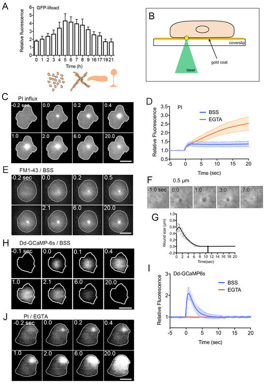
Figure 1.
Wound repair of the cell membrane. (A) Relative amplitudes of actin accumulation at the wound site over time following starvation (0 h). As illustrated in the lower drawings, upon initiation of vegetative cell starvation, individual cells aggregate, forming streams direct toward the aggregation center. This process leads to the creation of a multicellular structure, culminating in the development of a fruiting body. Importantly, wound repair is observed at every stage of the lifecycle in Dictyostelium discoideum. (B) Schematic representation of the enhanced laserporation with gold coating. The wound diameter is usually set at 0.5 μm for Dictyostelium cells. (C) A representative sequence of fluorescence images capturing PI influx after laserporation. (D) Temporal profiles of PI influx in the presence (BSS, control) and absence (EGTA) of external Ca2+. The wound laser beam was applied at 0 sec. (E) A typical sequence of fluorescence images illustrating FM dye influx after laserporation. (F) Laserporation of a cell expressing GFP-cAR1 resulted in the appearance of a black spot on the cell membrane. The black spot transiently expanded, then contracted, and finally closed. (G) The time course of the black spot diameter. (H) A sequence of fluorescence images featuring a cell expressing GCAMP6s after laserporation. (I) Temporal profiles of GCAMP6s fluorescence intensities in the presence (BSS) and absence (EGTA) of external Ca2+. (J) A typical sequence of fluorescence images illustrating PI influx after laserporation in the absence (EGTA) of external Ca2+. Scale bars, 10 µm. Figures are posted from [48,51,67,68] with proper permission.
3. Monitoring of Wound Repair
Various methods have been employed to investigate the wound repair mechanism. In early experiments, the cell membrane was wounded mainly by microneedle poking in large cells such as protozoan amebae [41,65,69], amphibian eggs [30,62,64], and echinoderm eggs [50]. For small cells like yeast, animal cultured cells, and Dictyostelium cells, laser ablation has been predominantly used due to the technical challenges and time constraints associated with microneedle poking in such small cells. Recent research also utilizes laser ablation for large cells, offering precise-sized wounds and accurate timing. However, both laser ablation and previous methods not only damage the cell membrane but also impact intracellular structures, including cortical actin networks, microtubules, and organelles.
To address this, we have developed an improved laser ablation method that selectively injures only the cell membrane. As depicted in Figure 1B, after placing cells on a carbon or gold-coated coverslip, a laser beam is focused on the coat underneath the cells. The laser energy absorbed by the coat generates heat and/or plasmon [48,70], selectively injuring the cell membrane attached to the coat [48]. This method has been originally invented for the introduction of extracellular substances into cells [71]. Instead of wounding individual cells, for biochemical analysis, a large number of cells can be wounded by treating with pore-forming agents or detergents [72,73,74].
For monitoring the wounding process, propidium iodide (PI) or FM1-43 has been widely used. PI, a cell-impermeant dye emitting fluorescence upon binding to RNA or DNA, and FM1-43, a cell-impermeable fluorescent lipid analog emitting fluorescence upon insertion into the membrane, are placed in the external medium. Their entry into the cytosol is monitored by the increase in fluorescence upon wounding. As shown in Figure 1C, PI fluorescence begins to increase at the wound site upon injury, spreading over the cytosol, suggesting PI entry through the wound pores. Figure 1D (BSS) illustrates the time course of PI fluorescence intensity in the cytosol of wounded cells, indicating that PI influx ceases within 2–3 s after injury, terminating urgent wound repair within this timeframe. Figure 1E demonstrates the influx of FM1-43 dye upon wounding, also showing that the dye enters from the wound pore and spreads across the cytoplasm.
To visualize the wound pore in Dictyostelium cells, cells expressing GFP-cAR1 (cAMP receptor) as a membrane protein marker are wounded. Immediately after wounding, a black spot appears at the laser application site (Figure 1F). This black spot is not generated by photobleaching, as it transiently expands slightly, then shrinks, and eventually closes (Figure 1G). This closure is not uniform but occurs from the wound edge to the center.
4. Ca2+ Influx as the First Signal
The initial signal common to all examined cells across various species is the influx of Ca2+ from the wound pore [30,32,41,50,75]. Monitoring this influx is feasible using a Ca2+ indicating fluorescent dye or a GFP-based Ca2+ indicator. Figure 1H presents a time series of fluorescence images of Dictyostelium cells expressing GCAMP6s, a GFP-based fluorescent Ca2+ indicator. In Figure 1I (BSS), the time course of fluorescence intensities in the cytosol is depicted. Intracellular Ca2+ concentration (Cai2+) promptly rises upon wounding, returning to resting levels within approximately 7 s. In the absence of external Ca2+, Cai2+ remains unchanged upon wounding (Figure 1I, EGTA), indicating that the influx of Ca2+ triggers the increase in Cai2+. Without external Ca2+, PI influx persists, leading to the eventual death of wounded cells (Figure 1D, EGTA, and Figure 1J). Additionally, the black spot observed in experiments using GFP-cAR1 does not close without Ca2+ influx. A concentration higher than 0.1 mM of Ca2+ in the external medium is necessary for wound repair in Dictyostelium cells [51].
Ca2+ influx induces the release of Ca2+ from intracellular stores through the calcium-induced calcium release (CICR) mechanism [76,77]. Deleting CICR reduces the amplitude of Cai2+ but does not impact wound repair, indicating that a local increase in Cai2+ is crucial, not a global one [67]. On the other hand, MCOLN1, an endosomal and lysosomal Ca2+-channel, is crucial for cell membrane repair in muscle cells, emphasizing the significance of Ca2+ release from intracellular stores in wound repair [78].
Various intracellular targets of Ca2+ for wound repair include dysfelin, mitsugmin 53 (MG53), neuroblast differentiation-associated protein (AHNAK), calpain, calmodulin, annexins, the endosomal sorting complex required for transport (ESCRT), protein kinase C, and actin-related proteins. These will be discussed in detail later.
5. Closing Wound Pores
5.1. Spontaneous Self-Sealing
Ruptured artificial lipid bilayer membranes exhibit spontaneous resealing [79]. Similarly, extremely small wound pores, such as those generated by electroporation in live cells at the nanometer scale, are thought to undergo spontaneous resealing due to thermodynamically unfavorable lipid disorder (Figure 2A). However, even with such small pores, there may be a necessity for an active wound repair mechanism [80]. Additionally, membrane pores created by pore-forming toxins, despite being nanometer-sized, require an active wound repair mechanism [7,23,81].
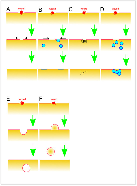
Figure 2.
Various models for wound repair mechanisms. (A) Spontaneous self-sealing. (B) Self-sealing by regulation of surface tension. Black arrows indicate the direction of the membrane flow. (C) Sealing by protein aggregation. (D) Sealing by membrane patch. (E) Endocytosis of damaged membrane. (F) Vesicle budding and shedding to the outside. These illustrations are simplified for a better understanding of basic concepts.
5.2. Self-Sealing by Regulation of Surface Tension
Given the tension on the cell surface, larger pores cannot spontaneously reseal against the cell surface tension. The wound-induced influx of Ca2+ triggers the fusion of exocytic vesicles with the cell membrane, extending beyond the wound site and enlarging the plasma membrane (Figure 2B). This process results in a reduction of cell surface tension, facilitating spontaneous resealing and closure of the wounded pore [82]. In large cells, although the actomyosin ring exerts force to close the wound pore against the opening force of the cell surface tension, self-sealing alone is insufficient, and a membrane patch is also required for sealing, as described later.
5.3. Sealing by Protein Aggregation
The wounded pores have been suggested to be clogged by the aggregation of proteins, including annexins and actin (Figure 2C). Annexin A5 self-assembles into two-dimensional arrays on the membrane upon Ca2+ activation, a crucial aspect of its role in plasma membrane repair in mammalian cells [83]. Similar clogging phenomena have been reported for other annexins, which collaborate with wound repair-related proteins such as actin, dysferin, and MG53 [84,85,86,87]. We will delve into these proteins in more detail later. Actin accumulates at the wound site, potentially serving a clogging function through actin gelation. However, this accumulation does not happen immediately upon wounding but occurs after the cessation of PI influx.
5.4. Sealing by Membrane Patch
Mammalian red blood cells, lacking endomembranes, including nuclei, take a longer time to reseal wound membranes or fail to repair in physiological conditions, suggesting that endomembranes are necessary for wound repair [88]. The membrane patch hypothesis was initially proposed in large cells like echinoderm and frog oocytes (Figure 2D). A local increase in Ca2+ induces the fusion of small cytoplasmic vesicles with each other, creating a continuous membrane plug at the wound site along with the plasma membrane [34,35]. More recently, cortical granules in Xenopus oocytes have been identified as such intracellular compartments, and their fusion was visualized in live cells [89,90]. This wound repair process is succeeded by the constriction of an actomyosin ring, akin to the contractile ring involved in cytokinesis.
Various sources for the membrane patch, including lysosomes [91,92], endosomes [93,94], MG53-rich vesicles [75], dysferlin-containing vesicles [95,96], or AHNAK-positive “enlargeosomes” [97,98], have been proposed. However, these vesicles and organelles might not meet the spatiotemporal requirements for rapid and efficient wound repair, considering the possibility of multiple wounds with very short intervals [67].
Recently, we proposed that the vesicles for the membrane plug are newly generated at the wound site in Dictyostelium cells [51]. In influx experiments using FM dye, most of the FM fluorescence diffuses in the cytosol, but a portion of FM dye remains at the wounded site and increases in size (Figure 1E), indicating membrane accumulation at the wound site. In the PI influx experiment (Figure 1C), a portion of PI fluorescence also persists at the wound site, suggesting that cytoplasm, including PI dye, is entrapped in the newly enclosed vesicles. It is improbable that the membrane plug originates from the broken cell membrane due to the limited amount of the broken cell membrane. Additionally, vesicles are unlikely to be transported from other locations since pharmacological disruption of microtubules and actin did not impede membrane accumulation. Therefore, we propose that the vesicles for the membrane plug are generated de novo at the wound site, although the mechanism for this generation remains unclear.
As described earlier, experiments using GFP-cAR1 show that the wound pore is not repaired from the wound edge to the center. Therefore, the wound edge grows toward the center through vesicles repeatedly fusing with the edge of the cell membrane, rather than forming a fused large patch to plug the wound pore.
5.5. Endocytosis of Damaged Membrane
Ca2+-triggered endocytosis is suggested to eliminate damaged membrane (Figure 2E). The membrane, including the damaged portion, invaginates inward, and the resulting bud is removed by releasing it into the cell, dependent on Ca2+ [74,99]. Upon wounding, acid sphingomyelinase is secreted into the extracellular space through Ca2+-dependent lysosomal exocytosis. This enzyme hydrolyzes sphingomyelin in the cell membrane into ceramide, facilitating membrane invagination and vesiculation [100]. Ceramide formation by sphingomyelinase also induces caveolae-mediated endocytosis, internalizing the wounded membrane [99,101,102,103,104,105]. It has been also reported that clathrin- and dynamin-mediated endocytosis facilitates removing the wounds by pore-forming proteins or toxins [106,107].
In Dictyostelium cells, neither endocytosis nor exocytosis appears to contribute to membrane accumulation for wound repair [51], despite the rapid turnover of the cell membrane through endocytosis–exocytosis coupling [108,109,110,111]. Notably, caveolin proteins are not present in Dictyostelium [112] and inhibitors of sphingomyelinase do not affect the wound repair in Dictyostelium cells (our preliminary observations). Additionally, clathrin- and dynamin-mediated endocytosis does not contribute to wound repair in Dictyostelium cells [48].
5.6. Vesicle Budding and Shedding to the Outside
Rather than endocytosis involving the inward budding and scission of the damaged membrane, vesicle budding or blebbing toward the outside of the cell, followed by scission, facilitates the removal and shedding of damaged membrane or pore-forming reagents (Figure 2F). Membrane-binding proteins for wound repair, such as the endosomal sorting complex required for transport (ESCRT) and annexins, facilitate this type of shedding in a Ca2+-dependent manner in mammal cells [10,44,84,99,113,114].
In Dictyostelium cells, FM dye that accumulates at the wound site remains there for the duration of our observation. The wounded sites do not move relative to the substrate and eventually shed onto the substrate as cells migrate [1].
6. Membrane-Binding Proteins in Wound Repair
For wound repair, various membrane-binding proteins, including annexins, the ESCRT complex, synaptotagmin, and dysferin, have been proposed. These proteins also act as sensors, detecting damage to the cell membrane due to their calcium-dependency [115].
6.1. Annexins
Annexins, a highly conserved and ubiquitous family of Ca2+- and phospholipid-binding proteins, play a crucial role in wound repair [116,117,118,119,120,121]. In vertebrates, 12 annexin subfamilies (A1–A11 and A13) have been identified. Annexins such as A1, A2, A5, and A6 accumulate at the wound site by binding to the inner cell membrane, particularly acidic phospholipids like phosphatidylserine, in response to a Ca2+ influx. Through membrane binding and interactions with other proteins, such as S100 family proteins, annexin prevents further expansion of the wound pore, reduces membrane tension, and prepares the membrane for resealing [59,122,123].
Annexins induce curvature in the free-edge membranes and generate constriction force to close the wound pore through annexin crosslinking [124,125,126]. Annexins can also be cross-linked by transglutamilases in a Ca2+-dependent manner [127], which have also been implicated in plasma membrane repair [128].
Moreover, annexins have been proposed to assemble into multimeric lattice structures, recruiting M53-laden vesicles and mini-dysferlin72, effectively clogging the wound pore [13,83,120,129]. Some annexins, such as annexin 1 and 2, can bind to actin and stabilize actin filaments at the wound site [85,119,130,131,132].
Dictyostelium possesses two annexin genes, annexin C1 (annexin VII or synexin) and annexin C2 (annexin I) [133]. Both can bind phosphatidylserine in a Ca2+-dependent manner [134,135]. Only annexin C1 accumulates at the wound site immediately after wounding. Wounded annexin C1-null cells exhibit irregular curves with multiple peaks in PI influx, Ca2+ influx, and actin dynamics. Additionally, annexin C1-null cells have a significantly reduced survival rate following injury, suggesting that annexin C1 partially contributes to wound repair in Dictyostelium cells [48,67].
6.2. ESCRT Complexes
The endosomal sorting complex required for transport (ESCRT) is categorized into five protein complexes (ESCRT-0, ESCRT-I, ESCRT-II, ESCRT-III, and Vps4). These complexes play integral roles in various cellular processes, including endosomal budding transport, virus budding, and cytokinesis. The ESCRT complex constricts and severs narrow necks during membrane budding processes [44,136,137,138,139,140,141,142,143,144]. Additionally, ESCRT complexes have been implicated in shedding wounded membranes as extracellular vesicles [44,114,144]. Upon injury, ESCRT complexes promptly accumulate at the wound site, protrude the wounded membrane as a bud or bleb, and subsequently cut it off to release extracellular vesicles. This ESCRT-mediated abscission of the wounded membrane appears to limit smaller-sized wounds (<100 nm in diameter) [145,146].
Interestingly, ESCRT complexes also participate in repairing damaged membranes of intracellular organelles, such as the nuclear envelope and lysosomes [147,148,149]. Furthermore, ESCRT complexes mediate the sealing of holes in the nascent nuclear envelope and nascent autophagosome [150].
While ESCRT complexes themselves are not sensitive to Ca2+, Ca2+-sensitive proteins like ALG-2 and calmodulin confer Ca2+ sensitivity on ESCRT complexes [10,144,151]. Recently, it has been reported that annexin A6 also plays a similar role in the secretion of exosomes [152].
In Dictyostelium cells, components of the ESCRT complexes accumulate at the wound site immediately upon injury, depending on the influx of Ca2+ [51]. However, in ESCRT null cells, PI influx ceases normally, and actin dynamics are observed, similar to wild-type cells. This suggests that ESCRT complexes are not essential for wound repair in Dictyostelium cells [48].
During cytokinesis in animal cells, ESCRT complexes and annexins accumulate at the cleavage furrow and/or midbody and are considered to play a role in cytokinesis [153,154,155,156,157,158]. Dictyostelium cells lack a midbody and undergo division through physical cutting via the constriction of the contractile ring and the traction force of the two daughter fragments migrating in opposite directions [159,160]. This suggests that this abscission might be a form of ‘physiological wound’. However, our preliminary observations indicate that neither ESCRT components nor annexins accumulate at the torn edges, suggesting the existence of a novel mechanism for cytokinetic abscission in Dictyostelium cells.
6.3. Synaptotagmin
Synaptotagmin comprises a family of Ca2+-binding and membrane-trafficking proteins, with particular emphasis on its well-characterized role in the release of synaptic vesicles in neurons, where it regulates Ca2+-dependent exocytosis [161]. Synaptotagmin 7, specifically, participates in the repair of wounded membranes through Ca2+-dependent lysosome exocytosis [92,162]. Knockdown mice lacking synaptotagmin 7 exhibit defects in wound repair [163]. While Dictyostelium possesses a synaptotagmin-like protein, there is currently no available information regarding its role in wound repair.
6.4. Dysferlin
Dysferlin, a membrane protein within the Ferlin family involved in vesicle fusion, is notably abundant in skeletal and cardiac muscle [164]. Dysferlin binds to vesicles containing acid phospholipids, such as phosphatidylserine, through the dysferlin C2 domain, relying on Ca2+ [165]. Mice deficient in dysferlin display defects in wound repair in muscle cells and develop muscular dystrophy [12]. Dysferlin is proposed to generate a membrane patch by recruiting and fusing vesicles in wounded skeletal and cardiac muscle cells [96,166]. Dysferlin organizes vesicle fusion with the assistance of binding partners like S100A10, annexin A2, AHNAK, caveolin-3, and TRIM72 (MG53) [13,75,102,115,167]. TRIM72, also known as MG53, which is highly expressed in muscle cells, assembles into a higher-ordered structure on the phosphatidylserine-enriched membranes. This assembly and association with the membrane depend on its oligomeric assembly and ubiquitination activity, facilitating vesicle transport to the wound site [168]. Notably, Dictyostelium cells lack a dysferlin homolog.
7. Cytoskeletons
The most comprehensively understood molecular mechanism for wound repair is derived from research in Xenopus oocytes. The entry of Ca2+ activates the small G-protein Rho, leading to the accumulation of myosin II [53,55]. Myosin II is a type II myosin that can assemble into bipolar filaments, generating a contractile force by interacting with actin filaments. Ca2+ influx also activates cdc42, another small G-protein, leading to actin assembly [90,169,170,171,172]. An actomyosin ring structure, akin to the contractile ring during cytokinesis, forms around the wound pore, facilitating wound closure. However, the appearance of the actomyosin ring is limited to large cells. In this section, we will explore the contributions of actin, myosin, actin-binding proteins, and microtubules.
7.1. Actin
Actin transiently accumulates at the wound site upon injury in various organisms [173]. Previous studies on cultured mammalian cells have indicated that, before wound-induced actin accumulation, pre-existing cortical actin networks at and around wound pores are largely removed [122,174,175]. This removal is believed to be necessary for the access of exocytic vesicles to the cell membrane and subsequent actin accumulation at the wound site [91,176]. Although the molecular mechanism of actin removal is not fully understood, Ca2+-dependent proteases such as calpain [177,178,179,180] or Ca2+-dependent actin-depolymerizing factors, such as actin-related proteins severing or depolymerizing actin filaments, might remove the actin cortex.
Conversely, in Dictyostelium cells, the removal of cortical actin networks is substantially undetectable upon wounding [48]. This discrepancy may be caused by differences in wounding methods. Conventional wounding methods disrupt not only the cell membrane but also the actin cortex, microtubules, and organelles, whereas the method used for Dictyostelium cells only disrupts the cell membrane, not the inner structures. Disruption of Ca2+-storing organelles such as the endoplasmic reticulum might result in the disassembly of actin filaments by activating Ca2+-dependent proteases or Ca2+-dependent actin-depolymerizing factors. Alternatively, although the dynamic instability of actin filaments is shown only in vitro, in contrast to that of microtubules [181,182], disassembly of actin filaments might expand to a much larger area due to catastrophic disassembly when a part of the meshwork is disrupted by the conventional wounding method.
Figure 3A–C show typical time courses of fluorescence images and fluorescence intensities of GFP-lifeact, a marker of actin filaments expressed in Dictyostelium cells upon wounding. Actin transiently accumulates at the wound site. In the presence of latrunculin A, a depolymerizer of actin filaments, actin does not accumulate at the wound site, and PI influx does not stop (Figure 3D, LatA), resulting in the failure of wound repair [51]. Therefore, actin accumulation is essential for wound repair.
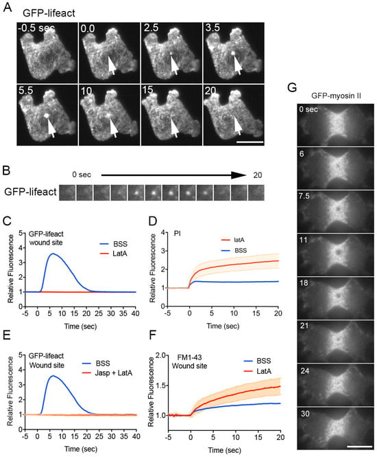
Figure 3.
Role of actin in wound repair. (A) Representative sequence of fluorescence images featuring a cell expressing GFP-lifeact upon wounding. Arrows indicate the wound site. (B) Sequence of fluorescence images at the wound sites of cells expressing GFP-lifeact. (C) Temporal profiles of relative fluorescence intensity of GFP-lifeact at the wound site in the presence (LatA) and absence (BSS) of latrunculin A. (D) Temporal profiles of PI influx in the cytosol in the presence (LatA) and absence (BSS) of latrunculin A. (E) Temporal profiles of GFP-lifeact fluorescence intensities at the wound site in the presence and absence of jasplakinolide and latrunculin A (Jasp + LatA). (F) Temporal profiles of FM fluorescence intensities at the wound site in the presence (LatA) and absence (BSS) of latrunculin A. (G) Representative sequence of fluorescence images of a dividing cell expressing GFP-myosin II upon wounding. Scale bars, 10 µm. Figures are posted from [48,51,67,68] with proper permission.
There are two possible mechanisms for actin accumulation at the wound site: (1) preexisting cortical actin filaments flow (moving along the cell membrane) toward the wound site (flow model), and (2) monomeric actin polymerizes at the wound site (de novo synthesis model). In Xenopus oocytes, the actomyosin ring is generated by the flow of cortical actin filaments toward the wound site, accompanied by dynamic actin polymerization [183]. However, such a flow is not observed around the wound site in Dictyostelium cells with curable wounds [48].
When cells are incubated with jasplakinolide, a stabilizer for actin filaments, for 30 min and then with latrunculin A, the number of polymerizable actin monomers should be significantly reduced, although the pre-built actin structures are stabilized by jasplakinolide. Upon wounding, actin does not accumulate at the wound site (Figure 3E, Jasp + LatA), suggesting that the de novo synthesis model is preferable because actin cannot newly polymerize without polymerizable actin monomers in the presence of both inhibitors. Therefore, it is plausible that actin polymerizes de novo at the wound site.
Although the aggregation of actin filaments itself may directly plug the wound pore, this is not the case in Dictyostelium cells because the wound pore substantially closes about 2 s after wounding, as observed from the influx of PI. On the other hand, actin begins to accumulate about 2.5 s after wounding. As shown in Figure 3D, PI influx in the presence of latrunculin A shows two phases: an initial rapid influx phase and a subsequent slow influx phase. PI influx in the control substantially stops after the initial phase. Therefore, the role of actin is not to directly constrict or plug the pore but rather to maintain pore closure after the emergent closing by the membrane plug.
Figure 3F shows the time course of the fluorescence intensity of FM1-43 at the wound site in the presence and absence of latrunculin A. The membrane accumulates at the wound site by two steps in the control (BSS): the initial rapid accumulation upon wounding (urgent membrane plug) and thereafter gradually increasing accumulation at a very low level. In the presence of latrunculin A, the size of the membrane pore gradually increases, and an increasing amount of membrane accumulates, suggesting an additional role for actin accumulation to prevent further membrane accumulation, although the detailed mechanism is elusive. Since PI influx does not cease in the presence of latrunculin A, the urgent membrane plug in the first step is incomplete, and the actin accumulation plays a role in the completion of the plug.
7.2. Myosin II
To explain the actin ring constriction mechanism independent of myosin II, it is proposed that actin continuously assembles on the inside of the actin ring and disassembles at the outer edge (actin treadmilling mechanism) in Xenopus oocytes [52].
Myosin II does not accumulate at the wound site in small cells such as yeast, Dictyostelium, and cultured animal cells. Moreover, pharmacological inhibition has demonstrated the dispensability of myosin II in C. elegans hypodermal cells [184] and sea urchin coelomocytes [185]. Conversely, in mammalian cultured cells, myosin IIA interacts with MG53 to regulate vesicle trafficking to the wound site [186], while myosin IIB facilitates wound-induced exocytosis and the membrane resealing process [57].
Dictyostelium cells possess a single copy of the myosin II (heavy chain) gene. Interestingly, myosin II disappears from a much larger area than the wound size (Figure 3G). Furthermore, myosin II null Dictyostelium cells can repair wounds comparably to wild-type cells [68]. Consequently, myosin II is not essential for wound repair in Dictyostelium cells. Notably, the disappearance of myosin II filaments is independent of both its phosphorylation and motor activities, suggesting that it occurs without accompanying the disassembly of myosin II filaments [68,187,188]. A local decrease in tension in the cortical actin network at the wound region may release myosin II filaments from the cortex, as myosin II filaments preferentially bind to stretched actin filaments [189].
One plausible reason why small cells do not rely on myosin II is that the power of myosin II may be necessary to prevent the wound pore from opening in large animal cells due to the significantly higher surface tension in large cells compared to smaller cells. Another reason may involve a different type of myosin replacing myosin II to contribute to the closing constriction power of the wound pore. Groups of type I myosins, MyoB and MyoC, transiently accumulate at the wound site in Dictyostelium cells [48]. A third reason is the notable difference in wound sizes used for the experiments between large and small cells: about 100 µm in diameter for the former and around 1 µm.
When Dictyostelium cells are wounded with different sizes, wounds larger than 1.5 µm in diameter result in cell lysis with a 50% frequency. Most cells subjected to 2 µm wounds cannot survive [67]. However, in such significantly wounded cells, we observed a ring composed not only of actin but also myosin II filaments (unpublished data). If the wound size is 1.5 µm in diameter, the wound area is estimated to occupy 1.2% of the whole cell surface area for Dictyostelium cells. On the other hand, for Xenopus oocytes, if the wound size is 100 µm in diameter, it is estimated to be 8%, suggesting that Xenopus oocytes can tolerate much larger wounds than Dictyostelium cells. Interestingly, actin and myosin II flow toward the wound center in such significantly wounded Dictyostelium cells, resembling the flows observed in Xenopus oocytes (unpublished data). The participation of myosin II may depend on the wound size and the cell surface tension.
7.3. Actin-Related Proteins (ARPs)
The transient accumulation of actin at the wound site is orchestrated by the assembly and subsequent disassembly of actin filaments, a process regulated by actin-related proteins (ARPs). Various direct modulators of actin polymerization, such as formins, Arp2/3 complex, SCAR/WAVE (suppressor of cAR/WASP family verprolin homologous protein), and WASP (Wiskott-Aldrich Syndrome protein), have been identified as participants in actin assembly at the wound site across multiple organisms [175,185,190,191,192]. Formins and Arp2/3 complex direct elongation of unbranched and branched actin filaments, respectively. Arp2/3 complex is activated by WASP family proteins such as WASP, SCAR/WAVE, whereas formins are individually active.
In Dictyostelium cells, 13 out of 27 examined ARPs accumulate at the wound site. Severin (an F-actin-severing protein akin to gelsolin) rapidly accumulates at the wound site, followed by WASP, Arp3 (a component of the Arp2/3 complex activated by WASP), ABP34 (an actin-bundling protein), and MyoB (a type I myosin), which accumulate significantly earlier than actin. Alpha-actinin (an actin-crosslinking protein), filamin (another actin-crosslinking protein), and CAP32 (an actin capping protein) begin to accumulate simultaneously with actin. MyoC (a type I myosin), CARMIL (a multidomain scaffold protein), and fimbrin (an actin-bundling protein) start accumulating after actin. Coronin and cofilin accumulate during the actin disassembly stage, consistent with the consensus that coronin inactivates the Arp2/3 complex [193], and ADF/cofilin promotes actin disassembly [194].
Similar to myosin II, EfaA1 (elongation factor 1 alpha 1, an actin-bundling protein), DlpA (dynamin-like protein A), MHCKC (myosin heavy chain kinase C), and cortexillins A and B (actin-binding proteins) transiently disappear from the wound site [68]. Intriguingly, with the exception of EfaA1, most of these proteins localize at the cleavage furrow during cytokinesis and at the rear regions during cell migration [195,196,197,198,199,200]. Incidentally, signaling proteins localizing at the cleavage furrow, such as PakA (p21-activated protein kinase A) [201], PTEN (phosphatase and tensin homolog deleted on chromosome 10) [202,203], and GAPA (IQGAP-related protein) [204], also transiently disappear at the wound site. Figure 4 provides a summary of the durations of their appearance and disappearance.
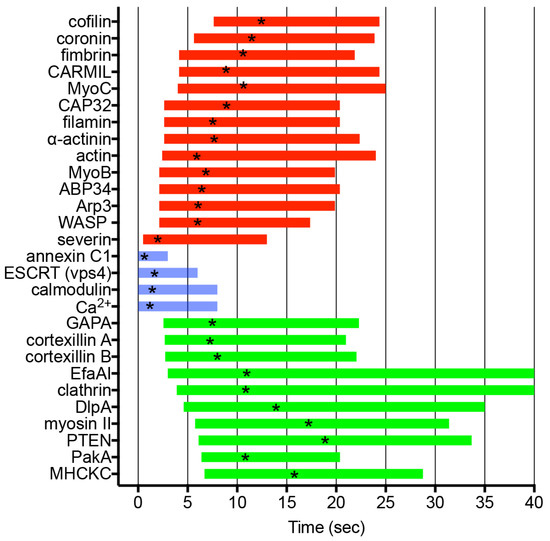
Figure 4.
Dynamics of ARPs and signal-related proteins. The graph illustrates the duration of appearance (red) and disappearance (green) of individual ARPs and signal-related proteins, including actin at wound sites. Additionally, the durations of Ca2+ influx, calmodulin dynamics, ESCRT component vps4 dynamics, and annexin C1 dynamics are depicted (blue). Asterisks in the duration bars indicate peak times.
The necessity of these ARPs for wound-induced actin dynamics has been explored through inhibition experiments using knockout mutants or pharmacological inhibition in Dictyostelium cells. Inhibition of WASP, Arp2/3, formin, and profilin significantly reduces actin accumulation at the wound site [48]. Although most ARPs accumulating at the wound site are found to be dispensable, they likely possess redundant roles in the regulation of the actin cytoskeleton, a hypothesis that warrants further investigation using double or triple knockout mutants [205].
7.4. Microtubules
Microtubules exhibit distinct behaviors in wound repair across different organisms. In Xenopus oocytes, microtubules accumulate and form a radial array around the wound site [191]. In epithelial kidney cells, microtubules locally disassemble upon wounding and elongate toward the wound site [206]. However, in Drosophila embryos, microtubules do not undergo significant changes upon wounding, yet pharmacological disruption of microtubules reduces wound repair [36]. Microtubules have been reported to transport small vesicles, such as lysosomes and vesicles derived from the Golgi apparatus, to the wound site, relying on motor proteins like kinesins and myosins [96,207,208]. In addition, it has been reported that microtubules regulate the actomyosin recruitment to the wound site in Xenopus oocytes [191].
In contrast, microtubules neither accumulate nor elongate toward the wound site upon wounding in Dictyostelium cells. Wound repair defects are not detected in the presence of a microtubule depolymerizer [51]. Therefore, vesicles are not transported along microtubules, and microtubules are not essential for wound repair in Dictyostelium cells. In these cells, the membrane at the wound site is not transported from other locations; instead, vesicles are generated de novo at the wound site, as mentioned earlier.
8. Signals for the Wound Repair
The downstream signals following Ca2+ influx upon wounding have been investigated. The Ca2+-sensitive membrane-binding proteins as described above (Section 6) can directly play a role in wound repair upon influx of Ca2+. We will see other signal pathways.
8.1. Protein Kinase C
Protein kinase C (PKC), activated by signals such as increases in Cai2+, is implicated in actin assembly in Xenopus oocytes and yeast during wound repair [43,209]. Elevating Cai2+ levels through treatment with a Ca ionophore results in the transient translocation of actin to the cell cortex [210,211,212]. PKC has been proposed to regulate small G proteins, which in turn regulates actin-related proteins for dynamics of actin at the wound site [170,213]. However, PKC does not seem to contribute to the wound repair in Dictyostelium cells [48].
8.2. Small G Proteins
The Rho family of small G proteins (Rho, Rac, Cdc42) serves as an upstream signal for regulating actin dynamics at the wound sites in Xenopus and Drosophila oocytes. These GTPases, including Rho, Rac, and Cdc42, are recruited to wounds in distinct spatiotemporal patterns [37,171,214]. Through their downstream effectors such as WASP, Scar/WAVE, and WASH, they recruit actin to the wound site, forming an actomyosin ring along the wound edge [172,190]. The Rab family of small G proteins regulate vesicle transport, including vesicle attachment to motor proteins and their tethering to target membranes [215]. The Rab3a, one of the Rab family, binds to lysosomes with myosin II heavy chain A, which is required for the lysosome exocytosis for wound repair in HeLa cells and melanocytes [216,217].
Dictyostelium cells possess 20 Rac proteins but lack Rho and Cdc42 [218]. A Rac inhibitor significantly diminishes actin accumulation amplitude and delays both the initiation and termination times, indicating that Rac regulates wound-induced actin dynamics. The specific type of Rac contributing to the actin accumulation requires further clarification. The Ras family also regulates actin polymerization, the deletion of both RasG and RasC does not affect wound-induced actin accumulation [48].
8.3. Reactive Oxygen Species (ROS)
The wound-induced influx of Ca2+ causes mitochondria to associate with the wounded membrane [219] and activates the uptake of Ca2+ into mitochondria in skeletal muscle cells, which generates reactive oxygen species (ROS). ROS locally activate RhoA, triggering actin accumulation at the wound site [40,220,221]. It is also reported that oxidation mediates assembly of MG53 for wound repair [75].
8.4. Calmodulin
Calmodulin, a multifunctional Ca2+-binding messenger protein, is reported to contribute to cellular wound repair in neurons, green algae, and Dictyostelium cells [51,222,223]. Calmodulin activates calpain, a Ca2+-dependent protease, to degrade fodrin, an actin-related protein, and thereby disassembles the actin cortex, which facilitates the resealing of wounded membrane in neurons and axons [224].
In Dictyostelium cells, calmodulin transiently accumulates at the wound site immediately after injury, depending on Ca2+ influx. In the presence of W7, a calmodulin inhibitor, calmodulin fails to accumulate at the wound site, and wound-induced PI and FM1-43 influx persists, contrary to the control, indicating the essential role of calmodulin in wound repair. W7 inhibits annexin accumulation but does not affect ESCRT complexes accumulation. Notably, calmodulin may regulate ESCRT complexes for exosomal biogenesis in human cultured cells [225]. W7 inhibits actin accumulation, while latrunculin A does not inhibit calmodulin accumulation in Dictyostelium cells, suggesting that calmodulin acts upstream of annexin and actin.
Chemotaxis signaling pathways, including the regulation of dynamics in the membrane and cytoskeleton, have been extensively investigated in Dictyostelium cells as well as neutrophils [226,227]. We explored the wound repair signaling pathways by referencing the chemotaxis signaling pathways. Figure 5 provides an overview of the signaling pathways involved in wound repair in Dictyostelium cells. After the influx of Ca2+ upon wounding, calmodulin and annexin C1 accumulate immediately at the wound site, which triggers the de novo generation of vesicles and mutual fusion of vesicle–vesicle and vesicle–cell membrane to make an urgent membrane plug. The TORC2, Dock/Elmo, PIP2-derived product, and PLA2 pathways are activated, which is common in the chemotaxis signaling pathway. Racs, WASPs, and then formins and Arp2/3 are involved in these pathways, and further downstream, many ARPs regulate the actin dynamics at the wound site.
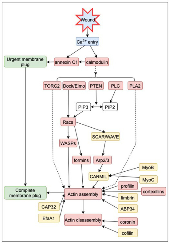
Figure 5.
Signaling pathways for wound repair in Dictyostelium. ARPs and signal-related proteins contributing to wound repair, identified through null mutants or pharmacological inhibitors, are highlighted in red. Certain proteins (yellow) exhibit accumulation or disappearance at the wound site, though their specific contributions are yet to be fully elucidated. Data are based on [48,51,67,68].
9. Wound Repair Model for Dictyostelium Cells
Figure 6 provides a comprehensive overview of wound repair in Dictyostelium cells. Upon wounding, the influx of Ca2+ through the wound pore serves as a signal for the accumulation of calmodulin and annexin C1 at the wound site, triggering the formation of an urgent membrane plug. This process involves the de novo generation of membrane vesicles, followed by their mutual fusion with both other vesicles and the cell membrane.
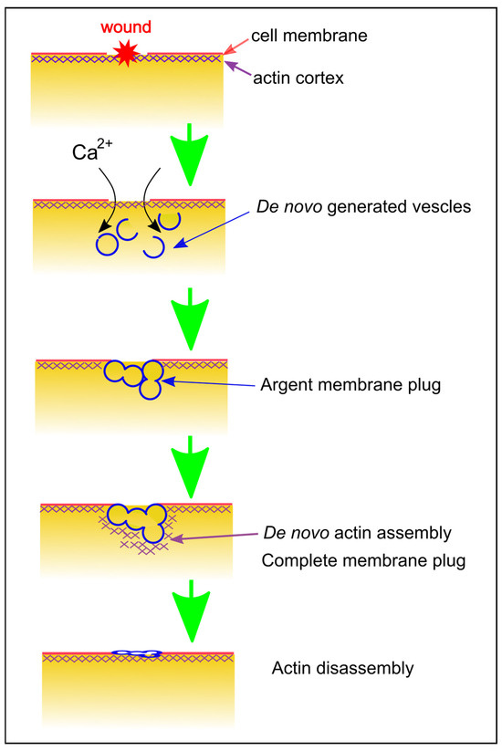
Figure 6.
Summary of Wound Repair in Dictyostelium Cells. This schematic diagram illustrates the wound repair mechanism in Dictyostelium cells. Upon wounding, Ca2+ enters through the wound pore, initiating the de novo generation of vesicles and the mutual fusion of vesicle–vesicle and vesicle–cell membrane, forming an immediate membrane plug. Actin accumulates to finalize the plug, relying on Ca2+ and calmodulin. Following the disassembly of the actin structure, the remaining damaged membrane is shed as the cell undergoes migration.
In the second step, actin accumulates through de novo polymerization to complete the membrane plug. The signals for actin accumulation are transmitted via multiple signaling pathways. Eventually, the actin accumulation completes the membrane plug. The dynamics of actin assembly and subsequent disassembly are finely regulated by numerous ARPs, each recruited at specific times. In the final step, the repaired membranes may not precisely match the original unwounded membrane. Ultimately, these membranes are shed as cells migrate away, completing the wound repair process.
10. Conclusions and Perspective
The molecular mechanism underlying wound repair is intricate, and presently, no universal mechanism exists across different organisms. It is plausible that diverse mechanisms have evolved to ensure the survival of cells. Moving forward, it is crucial to approach research from the following perspectives: (1) Investigation of the membrane source for the membrane plug: Exploration into the origin of the membrane forming the membrane plug is warranted. If the membrane is generated de novo, examination of the mechanisms involved is necessary. (2) Exploration of force requirements for wound closure: Identification of the forces involved in wound closure and determination of the types of forces contributing to the process are necessary. Insights into these dynamics can be gained through direct measurements of force and tension around the wound pore. (3) Conducting ultrastructural observations: Essential observations of the wound pore closing process should be carried out using electron microscopy. Given the high repair speed, rapid freezing preparation is essential for accurate analysis. (4) Comprehensive identification of players: Utilization of genetic tools to comprehensively identify all factors related to wound repair is essential. Clarification of their spatiotemporal distribution is crucial for gaining a holistic understanding. (5) Conducting reconstitution experiments: Wound repair experiments using artificial membranes, including essential components, should be performed in vitro. (6) Conducting research on cell membrane damage in a wider variety of organisms. This will enable us to understand how organisms have acquired mechanisms for cell membrane damage. (7) Assessment of contributions to applied fields: Evaluation of the potential applications of research in this field, particularly in generating therapeutic medicine for the rapid recovery of diseases characterized by impaired wound repair, is necessary. Exogenous delivery of recombinant repair proteins such as MG53, annexins, or synthetic molecules has been shown to significantly enhance membrane repair in vivo and has proven effective for the treatment of muscular and neuronal damage [17,228,229,230,231]. Additionally, the exploration of implications for developing agricultural fungicides is warranted.
Funding
This work was partially supported by JSPS KAKENHI Grant Number 20K06642 and Yamaguchi University Fund.
Institutional Review Board Statement
Not applicable.
Informed Consent Statement
Not applicable.
Data Availability Statement
All relevant data are available from the authors on reasonable request.
Acknowledgments
I would like to thank Md. Shahabe Uddin Talukder, Mst. Shaela Pervin, Md. Istiaq Obaidi Tanvir, Takashi Matsumura, Koushiro Fujimoto, Masahito Tanaka and Go Itoh for their helps.
Conflicts of Interest
The author declares no conflicts of interest.
Abbreviation
| ABP | actin binding proteins |
| ADF | actin depolymerizing factor |
| AHNAK | neuroblast differentiation-associated protein |
| ALG-2 | alpha-1,3/1,6-mannosyltransferase |
| Arp2/3 | actin related protein 2/3 |
| ARPs | actin related protens |
| CAP32 | F-actin capping protein 32 |
| CARMIL | capping protein, Arp2/3, myosin I linker |
| DlpA | dynamin-like protein A |
| Dock/Elmo | dedicator of cytokinesis/engulfment and cell motility |
| EfaA1 | elongation factor alpha 1 |
| ESCRT | endosomal sorting complex required for transport |
| GAPA | IQGAP-related protein |
| MG53 | Mitsugmin 53, a TRIM (tripartite motif) family protein |
| MHCKC | myosin heavy chain kinase C |
| MyoB | myosin IB |
| PakA | p21-activated protein kinase A |
| PCK | protein kinase C |
| PIP2 | Phosphatidylinositol 4,5-bisphosphate |
| PLA2 | phospholipase A2 |
| PLC | phospholipase C |
| PTEN | phosphatase and tensin homolog deleted on chromosome 10 |
| ROS | reactive oxygen species |
| SCAR | suppressor of cAR |
| TORC2 | target of rapamycin C2 |
| WASH | Wiskott–Aldrich syndrome protein and SCAR Homolog |
| WASP | Wiskott-Aldrich Syndrome protein |
| WAVE | WASP family verprolin homologous protein |
References
- Pervin, M.S.; Yumura, S. Manipulation of cell migration by laserporation-induced local wounding. Sci. Rep. 2019, 9, 4291. [Google Scholar] [CrossRef]
- Tanvir, M.I.O.; Yumura, S. Effects of wounds in the cell membrane on cell division. Sci. Rep. 2023, 13, 1941. [Google Scholar] [CrossRef]
- McNeil, P.L.; Ito, S. Gastrointestinal cell plasma membrane wounding and resealing in vivo. Gastroenterology 1989, 96, 1238–1248. [Google Scholar] [CrossRef] [PubMed]
- McNeil, P.L.; Khakee, R. Disruptions of muscle fiber plasma membranes. Role in exercise-induced damage. Am. J. Pathol. 1992, 140, 1097–1109. [Google Scholar]
- McNeil, P.L. Cellular and molecular adaptations to injurious mechanical stress. Trends Cell Biol. 1993, 3, 302–307. [Google Scholar] [CrossRef] [PubMed]
- Dias, C.; Nylandsted, J. Plasma membrane integrity in health and disease: Significance and therapeutic potential. Cell Discov. 2021, 7, 4. [Google Scholar] [CrossRef]
- Etxaniz, A.; Gonzalez-Bullon, D.; Martin, C.; Ostolaza, H. Membrane Repair Mechanisms against Permeabilization by Pore-Forming Toxins. Toxins 2018, 10, 234. [Google Scholar] [CrossRef]
- Luisoni, S.; Suomalainen, M.; Boucke, K.; Tanner, L.B.; Wenk, M.R.; Guan, X.L.; Grzybek, M.; Coskun, U.; Greber, U.F. Co-option of Membrane Wounding Enables Virus Penetration into Cells. Cell Host Microbe 2015, 18, 75–85. [Google Scholar] [CrossRef] [PubMed]
- Stow, J.L.; Condon, N.D. The cell surface environment for pathogen recognition and entry. Clin. Trans. Immunol. 2016, 5, e71. [Google Scholar] [CrossRef]
- Westman, J.; Plumb, J.; Licht, A.; Yang, M.; Allert, S.; Naglik, J.R.; Hube, B.; Grinstein, S.; Maxson, M.E. Calcium-dependent ESCRT recruitment and lysosome exocytosis maintain epithelial integrity during Candida albicans invasion. Cell Rep. 2022, 38, 110187. [Google Scholar] [CrossRef]
- Howard, A.C.; McNeil, A.K.; Xiong, F.; Xiong, W.C.; McNeil, P.L. A novel cellular defect in diabetes: Membrane repair failure. Diabetes 2011, 60, 3034–3043. [Google Scholar] [CrossRef] [PubMed]
- Bansal, D.; Miyake, K.; Vogel, S.S.; Groh, S.; Chen, C.C.; Williamson, R.; McNeil, P.L.; Campbell, K.P. Defective membrane repair in dysferlin-deficient muscular dystrophy. Nature 2003, 423, 168–172. [Google Scholar] [CrossRef] [PubMed]
- Waddell, L.B.; Lemckert, F.A.; Zheng, X.F.; Tran, J.; Evesson, F.J.; Hawkes, J.M.; Lek, A.; Street, N.E.; Lin, P.; Clarke, N.F.; et al. Dysferlin, annexin A1, and mitsugumin 53 are upregulated in muscular dystrophy and localize to longitudinal tubules of the T-system with stretch. J. Neuropathol. Exp. Neurol. 2011, 70, 302–313. [Google Scholar] [CrossRef] [PubMed]
- Duann, P.; Li, H.; Lin, P.; Tan, T.; Wang, Z.; Chen, K.; Zhou, X.; Gumpper, K.; Zhu, H.; Ludwig, T.; et al. MG53-mediated cell membrane repair protects against acute kidney injury. Sci. Transl. Med. 2015, 7, 279ra36. [Google Scholar] [CrossRef] [PubMed]
- Labazi, M.; McNeil, A.K.; Kurtz, T.; Lee, T.C.; Pegg, R.B.; Angeli, J.P.F.; Conrad, M.; McNeil, P.L. The antioxidant requirement for plasma membrane repair in skeletal muscle. Free. Radic. Biol. Med. 2015, 84, 246–253. [Google Scholar] [CrossRef] [PubMed]
- Agnihotri, A.; Aruoma, O.I. Alzheimer’s Disease and Parkinson’s Disease: A Nutritional Toxicology Perspective of the Impact of Oxidative Stress, Mitochondrial Dysfunction, Nutrigenomics and Environmental Chemicals. J. Am. Coll. Nutr. 2020, 39, 16–27. [Google Scholar] [CrossRef] [PubMed]
- Bulgart, H.R.; Goncalves, I.; Weisleder, N. Leveraging Plasma Membrane Repair Therapeutics for Treating Neurodegenerative Diseases. Cells 2023, 12, 1660. [Google Scholar] [CrossRef]
- Maiti, P.; Manna, J.; Dunbar, G.L. Current understanding of the molecular mechanisms in Parkinson’s disease: Targets for potential treatments. Transl. Neurodegener. 2017, 6, 28. [Google Scholar] [CrossRef]
- Schapire, A.L.; Valpuesta, V.; Botella, M.A. Plasma membrane repair in plants. Trends Plant Sci. 2009, 14, 645–652. [Google Scholar] [CrossRef]
- Yamazaki, T.; Takata, N.; Uemura, M.; Kawamura, Y. Arabidopsis synaptotagmin SYT1, a type I signal-anchor protein, requires tandem C2 domains for delivery to the plasma membrane. J. Biol. Chem. 2010, 285, 23165–23176. [Google Scholar] [CrossRef]
- Vyse, K.; Penzlin, J.; Sergeant, K.; Hincha, D.K.; Arora, R.; Zuther, E. Repair of sub-lethal freezing damage in leaves of Arabidopsis thaliana. BMC Plant Biol. 2020, 20, 35. [Google Scholar] [CrossRef]
- Ammendolia, D.A.; Bement, W.M.; Brumell, J.H. Plasma membrane integrity: Implications for health and disease. BMC Biol. 2021, 19, 71. [Google Scholar] [CrossRef] [PubMed]
- Barisch, C.; Holthuis, J.C.M.; Cosentino, K. Membrane damage and repair: A thin line between life and death. Biol. Chem. 2023, 404, 467–490. [Google Scholar] [CrossRef] [PubMed]
- Horn, A.; Jaiswal, J.K. Structural and signaling role of lipids in plasma membrane repair. Curr. Top. Membr. 2019, 84, 67–98. [Google Scholar] [PubMed]
- Nakamura, M.; Dominguez, A.N.M.; Decker, J.R.; Hull, A.J.; Verboon, J.M.; Parkhurst, S.M. Into the breach: How cells cope with wounds. Open Biol. 2018, 8, 180135. [Google Scholar] [CrossRef]
- Xu, S.; Yang, T.J.; Xu, S.; Gong, Y.N. Plasma membrane repair empowers the necrotic survivors as innate immune modulators. Semin. Cell Dev. Biol. 2023, 156, 93–106. [Google Scholar] [CrossRef] [PubMed]
- Andrews, N.W.; Corrotte, M. Plasma membrane repair. Curr. Biol. 2018, 28, R392–R397. [Google Scholar] [CrossRef] [PubMed]
- Togo, T.; Alderton, J.M.; Bi, G.Q.; Steinhardt, R.A. The mechanism of facilitated cell membrane resealing. J. Cell Sci. 1999, 112, 719–731. [Google Scholar] [CrossRef] [PubMed]
- Bement, W.M.; Capco, D.G. Analysis of inducible contractile rings suggests a role for protein kinase C in embryonic cytokinesis and wound healing. Cell Motil. Cytoskelet. 1991, 20, 145–157. [Google Scholar] [CrossRef]
- Gingell, D. Contractile responses at the surface of an amphibian egg. J. Embryol. Exp. Morphol. 1970, 23, 583–609. [Google Scholar] [CrossRef]
- Holtfreter, J. Properties and functions of the surface coat in amphibian embryos. J. Exp. Zool. 1943, 93, 251–323. [Google Scholar] [CrossRef]
- Stanisstreet, M. Calcium and wound healing in Xenopus early embryos. J. Embryol. Exp. Morphol. 1982, 67, 195–205. [Google Scholar] [CrossRef] [PubMed]
- Bi, G.Q.; Alderton, J.M.; Steinhardt, R.A. Calcium-regulated exocytosis is required for cell membrane resealing. J. Cell Biol. 1995, 131, 1747–1758. [Google Scholar] [CrossRef] [PubMed]
- McNeil, P.L.; Vogel, S.S.; Miyake, K.; Terasaki, M. Patching plasma membrane disruptions with cytoplasmic membrane. J. Cell Sci. 2000, 113, 1891–1902. [Google Scholar] [CrossRef] [PubMed]
- Terasaki, M.; Miyake, K.; McNeil, P.L. Large plasma membrane disruptions are rapidly resealed by Ca2+-dependent vesicle-vesicle fusion events. J. Cell Biol. 1997, 139, 63–74. [Google Scholar] [CrossRef] [PubMed]
- Abreu-Blanco, M.T.; Verboon, J.M.; Parkhurst, S.M. Cell wound repair in Drosophila occurs through three distinct phases of membrane and cytoskeletal remodeling. J. Cell. Biol. 2011, 193, 455–464. [Google Scholar] [CrossRef]
- Abreu-Blanco, M.T.; Verboon, J.M.; Parkhurst, S.M. Coordination of Rho family GTPase activities to orchestrate cytoskeleton responses during cell wound repair. Curr. Biol. 2014, 24, 144–155. [Google Scholar] [CrossRef]
- Razzell, W.; Wood, W.; Martin, P. Swatting flies: Modelling wound healing and inflammation in Drosophila. Dis. Model. Mech. 2011, 4, 569–574. [Google Scholar] [CrossRef]
- Ma, Y.; Xie, J.; Wijaya, C.S.; Xu, S. From wound response to repair—Lessons from C. elegans. Cell Regen. 2021, 10, 5. [Google Scholar] [CrossRef]
- Xu, S.; Chisholm, A.D. C. elegans epidermal wounding induces a mitochondrial ROS burst that promotes wound repair. Dev. Cell 2014, 31, 48–60. [Google Scholar] [CrossRef] [PubMed]
- Jeon, K.W.; Jeon, M.S. Cytoplasmic filaments and cellular wound healing in Amoeba proteus. J. Cell Biol. 1975, 67, 243–249. [Google Scholar] [CrossRef]
- Szubinska, B. Closure of the plasma membrane around microneedle in Amoeba proteus. An ultrastructural study. Exp. Cell Res. 1978, 111, 105–115. [Google Scholar] [CrossRef]
- Kono, K.; Saeki, Y.; Yoshida, S.; Tanaka, K.; Pellman, D. Proteasomal degradation resolves competition between cell polarization and cellular wound healing. Cell 2012, 150, 151–164. [Google Scholar] [CrossRef] [PubMed]
- Radulovic, M.; Stenmark, H. ESCRTs in membrane sealing. Biochem. Soc. Trans. 2018, 46, 773–778. [Google Scholar] [CrossRef] [PubMed]
- Zhang, K.S.; Blauch, L.R.; Huang, W.; Marshall, W.F.; Tang, S.K.Y. Microfluidic guillotine reveals multiple timescales and mechanical modes of wound response in Stentor coeruleus. BMC Biol. 2021, 19, 63. [Google Scholar] [CrossRef] [PubMed]
- Foissner, I.; Wasteneys, G.O. The characean internodal cell as a model system for studying wound healing. J. Microsc. 2012, 247, 10–22. [Google Scholar] [CrossRef] [PubMed]
- Schapire, A.L.; Voigt, B.; Jasik, J.; Rosado, A.; Lopez-Cobollo, R.; Menzel, D.; Salinas, J.; Mancuso, S.; Valpuesta, V.; Baluska, F.; et al. Arabidopsis synaptotagmin 1 is required for the maintenance of plasma membrane integrity and cell viability. Plant Cell 2008, 20, 3374–3388. [Google Scholar] [CrossRef] [PubMed]
- Yumura, S.; Talukder, M.S.U.; Pervin, M.S.; Tanvir, M.I.O.; Matsumura, T.; Fujimoto, K.; Tanaka, M.; Itoh, G. Dynamics of Actin Cytoskeleton and Their Signaling Pathways during Cellular Wound Repair. Cells 2022, 11, 3166. [Google Scholar] [CrossRef] [PubMed]
- McNeil, P.L.; Terasaki, M. Coping with the inevitable: How cells repair a torn surface membrane. Nat. Cell Biol. 2001, 3, E124–E129. [Google Scholar] [CrossRef]
- Steinhardt, R.A.; Bi, G.; Alderton, J.M. Cell membrane resealing by a vesicular mechanism similar to neurotransmitter release. Science 1994, 263, 390–393. [Google Scholar] [CrossRef]
- Talukder, M.S.U.; Pervin, M.S.; Tanvir, M.I.O.; Fujimoto, K.; Tanaka, M.; Itoh, G.; Yumura, S. Ca2+-calmodulin dependent wound repair in Dictyostelium cell membrane. Cells 2020, 9, 1058. [Google Scholar] [CrossRef]
- Moe, A.M.; Golding, A.E.; Bement, W.M. Cell healing: Calcium, repair and regeneration. Semin. Cell Dev. Biol. 2015, 45, 18–23. [Google Scholar] [CrossRef] [PubMed]
- Bement, W.M.; Mandato, C.A.; Kirsch, M.N. Wound-induced assembly and closure of an actomyosin purse string in Xenopus oocytes. Curr. Biol. 1999, 9, 579–587. [Google Scholar] [CrossRef] [PubMed]
- Hui, J.; Stjepic, V.; Nakamura, M.; Parkhurst, S.M. Wrangling Actin Assemblies: Actin Ring Dynamics during Cell Wound Repair. Cells 2022, 11, 2777. [Google Scholar] [CrossRef] [PubMed]
- Kiehart, D.P. Wound healing: The power of the purse string. Curr. Biol. 1999, 9, R602–R605. [Google Scholar] [CrossRef] [PubMed]
- DeKraker, C.; Goldin-Blais, L.; Boucher, E.; Mandato, C.A. Dynamics of actin polymerisation during the mammalian single-cell wound healing response. BMC Res. Notes 2019, 12, 420. [Google Scholar] [CrossRef] [PubMed]
- Togo, T.; Steinhardt, R.A. Nonmuscle myosin IIA and IIB have distinct functions in the exocytosis-dependent process of cell membrane repair. Mol. Biol. Cell 2004, 15, 688–695. [Google Scholar] [CrossRef] [PubMed]
- Yumura, S.; Hashima, S.; Muranaka, S. Myosin II does not contribute to wound repair in Dictyostelium cells. Biol. Open 2014, 3, 966–973. [Google Scholar] [CrossRef]
- Jaiswal, J.K.; Lauritzen, S.P.; Scheffer, L.; Sakaguchi, M.; Bunkenborg, J.; Simon, S.M.; Kallunki, T.; Jaattela, M.; Nylandsted, J. S100A11 is required for efficient plasma membrane repair and survival of invasive cancer cells. Nat. Commun. 2014, 5, 3795. [Google Scholar] [CrossRef]
- McDade, J.R.; Archambeau, A.; Michele, D.E. Rapid actin-cytoskeleton-dependent recruitment of plasma membrane-derived dysferlin at wounds is critical for muscle membrane repair. FASEB J. 2014, 28, 3660–3670. [Google Scholar] [CrossRef]
- Shen, S.S.; Steinhardt, R.A. The mechanisms of cell membrane resealing in rabbit corneal epithelial cells. Curr. Eye Res. 2005, 30, 543–554. [Google Scholar] [CrossRef]
- Bluemink, J.G. Cortical wound healing in the amphibian egg: An electron microscopical study. J. Ultrastruct. Res. 1972, 41, 95–114. [Google Scholar] [CrossRef][Green Version]
- Jeon, K.W.; Danielli, J.F. Micrurgical studies with large free-living amebas. Int. Rev. Cytol. 1971, 30, 49–89. [Google Scholar] [PubMed]
- Luckenbill, L.M. Dense material associated with wound closure in the axolotl egg (A. mexicanum). Exp. Cell Res. 1971, 66, 263–267. [Google Scholar] [CrossRef]
- Szubinska, B. “New membrane” formation in Amoeba proteus upon injury of individual cells. Electron microscope observations. J. Cell Biol. 1971, 49, 747–772. [Google Scholar] [CrossRef] [PubMed]
- Swanson, J.A.; Taylor, D.L. Local and spatially coordinated movements in Dictyostelium discoideum amoebae during chemotaxis. Cell 1982, 28, 225–232. [Google Scholar] [CrossRef]
- Pervin, M.S.; Itoh, G.; Talukder, M.S.U.; Fujimoto, K.; Morimoto, Y.V.; Tanaka, M.; Ueda, M.; Yumura, S. A study of wound repair in Dictyostelium cells by using novel laserporation. Sci. Rep. 2018, 8, 7969. [Google Scholar] [CrossRef] [PubMed]
- Tanvir, M.I.O.; Itoh, G.; Adachi, H.; Yumura, S. Dynamics of Myosin II Filaments during Wound Repair in Dividing Cells. Cells 2021, 10, 1229. [Google Scholar] [CrossRef]
- Taylor, D.L.; Wang, Y.L.; Heiple, J.M. Contractile basis of ameboid movement. VII. The distribution of fluorescently labeled actin in living amebas. J. Cell Biol. 1980, 86, 590–598. [Google Scholar] [CrossRef]
- Nakatoh, T.; Osaki, T.; Tanimoto, S.; Jahan, M.G.S.; Kawakami, T.; Chihara, K.; Sakai, N.; Yumura, S. Cell behaviors within a confined adhesive area fabricated using novel micropatterning methods. PLoS ONE 2022, 17, e0262632. [Google Scholar] [CrossRef]
- Yumura, S. A novel low-power laser-mediated transfer of foreign molecules into cells. Sci. Rep. 2016, 6, 22055. [Google Scholar] [CrossRef]
- Bischofberger, M.; Gonzalez, M.R.; van der Goot, F.G. Membrane injury by pore-forming proteins. Curr. Opin. Cell Biol. 2009, 21, 589–595. [Google Scholar] [CrossRef]
- Calvello, R.; Mitolo, V.; Acquafredda, A.; Cianciulli, A.; Panaro, M.A. Plasma membrane damage sensing and repairing. Role of heterotrimeric G-proteins and the cytoskeleton. Toxicol. Vitr. 2011, 25, 1067–1074. [Google Scholar] [CrossRef]
- Idone, V.; Tam, C.; Goss, J.W.; Toomre, D.; Pypaert, M.; Andrews, N.W. Repair of injured plasma membrane by rapid Ca2+-dependent endocytosis. J. Cell Biol. 2008, 180, 905–914. [Google Scholar] [CrossRef] [PubMed]
- Cai, C.; Masumiya, H.; Weisleder, N.; Matsuda, N.; Nishi, M.; Hwang, M.; Ko, J.K.; Lin, P.; Thornton, A.; Zhao, X.; et al. MG53 nucleates assembly of cell membrane repair machinery. Nat. Cell Biol. 2009, 11, 56–64. [Google Scholar] [CrossRef] [PubMed]
- Fisher, P.R.; Wilczynska, Z. Contribution of endoplasmic reticulum to Ca(2+) signals in Dictyostelium depends on extracellular Ca(2+). FEMS Microbiol. Lett. 2006, 257, 268–277. [Google Scholar] [CrossRef] [PubMed][Green Version]
- Malchow, D.; Lusche, D.F.; De Lozanne, A.; Schlatterer, C. A fast Ca2+-induced Ca2+-release mechanism in Dictyostelium discoideum. Cell Calcium 2008, 43, 521–530. [Google Scholar] [CrossRef] [PubMed]
- Cheng, X.; Zhang, X.; Gao, Q.; Ali Samie, M.; Azar, M.; Tsang, W.L.; Dong, L.; Sahoo, N.; Li, X.; Zhuo, Y.; et al. The intracellular Ca(2)(+) channel MCOLN1 is required for sarcolemma repair to prevent muscular dystrophy. Nat. Med. 2014, 20, 1187–1192. [Google Scholar] [CrossRef] [PubMed]
- Gozen, I.; Dommersnes, P. Pore dynamics in lipid membranes. Eur. Phys. J. Spec. Top. 2014, 223, 1813–1829. [Google Scholar] [CrossRef]
- Hai, A.; Spira, M.E. On-chip electroporation, membrane repair dynamics and transient in-cell recordings by arrays of gold mushroom-shaped microelectrodes. Lab. Chip 2012, 12, 2865–2873. [Google Scholar] [CrossRef] [PubMed]
- Lata, K.; Singh, M.; Chatterjee, S.; Chattopadhyay, K. Membrane Dynamics and Remodelling in Response to the Action of the Membrane-Damaging Pore-Forming Toxins. J. Membr. Biol. 2022, 255, 161–173. [Google Scholar] [CrossRef] [PubMed]
- Togo, T.; Krasieva, T.B.; Steinhardt, R.A. A decrease in membrane tension precedes successful cell-membrane repair. Mol. Biol. Cell 2000, 11, 4339–4346. [Google Scholar] [CrossRef] [PubMed]
- Bouter, A.; Gounou, C.; Berat, R.; Tan, S.; Gallois, B.; Granier, T.; d’Estaintot, B.L.; Poschl, E.; Brachvogel, B.; Brisson, A.R. Annexin-A5 assembled into two-dimensional arrays promotes cell membrane repair. Nat. Commun. 2011, 2, 270. [Google Scholar] [CrossRef] [PubMed]
- Babiychuk, E.B.; Monastyrskaya, K.; Potez, S.; Draeger, A. Blebbing confers resistance against cell lysis. Cell Death Differ. 2011, 18, 80–89. [Google Scholar] [CrossRef] [PubMed]
- Demonbreun, A.R.; Quattrocelli, M.; Barefield, D.Y.; Allen, M.V.; Swanson, K.E.; McNally, E.M. An actin-dependent annexin complex mediates plasma membrane repair in muscle. J. Cell Biol. 2016, 213, 705–718. [Google Scholar] [CrossRef] [PubMed]
- Roostalu, U.; Strahle, U. In vivo imaging of molecular interactions at damaged sarcolemma. Dev. Cell 2012, 22, 515–529. [Google Scholar] [CrossRef]
- Swaggart, K.A.; Demonbreun, A.R.; Vo, A.H.; Swanson, K.E.; Kim, E.Y.; Fahrenbach, J.P.; Holley-Cuthrell, J.; Eskin, A.; Chen, Z.; Squire, K.; et al. Annexin A6 modifies muscular dystrophy by mediating sarcolemmal repair. Proc. Natl. Acad. Sci. USA 2014, 111, 6004–6009. [Google Scholar] [CrossRef]
- McNeil, P.L.; Miyake, K.; Vogel, S.S. The endomembrane requirement for cell surface repair. Proc. Natl. Acad. Sci. USA 2003, 100, 4592–4597. [Google Scholar] [CrossRef]
- Davenport, N.R.; Bement, W.M. Cell repair: Revisiting the patch hypothesis. Commun. Integr. Biol. 2016, 9, e1253643. [Google Scholar] [CrossRef]
- Davenport, N.R.; Sonnemann, K.J.; Eliceiri, K.W.; Bement, W.M. Membrane dynamics during cellular wound repair. Mol. Biol. Cell 2016, 27, 2272–2285. [Google Scholar] [CrossRef]
- McNeil, P.L. Repairing a torn cell surface: Make way, lysosomes to the rescue. J. Cell Sci. 2002, 115, 873–879. [Google Scholar] [CrossRef]
- Reddy, A.; Caler, E.V.; Andrews, N.W. Plasma membrane repair is mediated by Ca2+-regulated exocytosis of lysosomes. Cell 2001, 106, 157–169. [Google Scholar] [CrossRef] [PubMed]
- Eddleman, C.S.; Ballinger, M.L.; Smyers, M.E.; Fishman, H.M.; Bittner, G.D. Endocytotic formation of vesicles and other membranous structures induced by Ca2+ and axolemmal injury. J. Neurosci. 1998, 18, 4029–4041. [Google Scholar] [CrossRef] [PubMed][Green Version]
- Raj, N.; Greune, L.; Kahms, M.; Mildner, K.; Franzkoch, R.; Psathaki, O.E.; Zobel, T.; Zeuschner, D.; Klingauf, J.; Gerke, V. Early Endosomes Act as Local Exocytosis Hubs to Repair Endothelial Membrane Damage. Adv. Sci. 2023, 10, e2300244. [Google Scholar] [CrossRef] [PubMed]
- Lek, A.; Evesson, F.J.; Lemckert, F.A.; Redpath, G.M.; Lueders, A.K.; Turnbull, L.; Whitchurch, C.B.; North, K.N.; Cooper, S.T. Calpains, cleaved mini-dysferlinC72, and L-type channels underpin calcium-dependent muscle membrane repair. J. Neurosci. 2013, 33, 5085–5094. [Google Scholar] [CrossRef] [PubMed]
- McDade, J.R.; Michele, D.E. Membrane damage-induced vesicle-vesicle fusion of dysferlin-containing vesicles in muscle cells requires microtubules and kinesin. Hum. Mol. Genet. 2014, 23, 1677–1686. [Google Scholar] [CrossRef]
- Cocucci, E.; Racchetti, G.; Podini, P.; Rupnik, M.; Meldolesi, J. Enlargeosome, an exocytic vesicle resistant to nonionic detergents, undergoes endocytosis via a nonacidic route. Mol. Biol. Cell 2004, 15, 5356–5368. [Google Scholar] [CrossRef] [PubMed][Green Version]
- Meldolesi, J. Surface wound healing: A new, general function of eukaryotic cells. J. Cell Mol. Med. 2003, 7, 197–203. [Google Scholar] [CrossRef] [PubMed]
- Andrews, N.W.; Almeida, P.E.; Corrotte, M. Damage control: Cellular mechanisms of plasma membrane repair. Trends Cell Biol. 2014, 24, 734–742. [Google Scholar] [CrossRef]
- Holopainen, J.M.; Angelova, M.I.; Kinnunen, P.K. Vectorial budding of vesicles by asymmetrical enzymatic formation of ceramide in giant liposomes. Biophys. J. 2000, 78, 830–838. [Google Scholar] [CrossRef]
- Castro-Gomes, T.; Corrotte, M.; Tam, C.; Andrews, N.W. Plasma Membrane Repair Is Regulated Extracellularly by Proteases Released from Lysosomes. PLoS ONE 2016, 11, e0152583. [Google Scholar] [CrossRef]
- Corrotte, M.; Almeida, P.E.; Tam, C.; Castro-Gomes, T.; Fernandes, M.C.; Millis, B.A.; Cortez, M.; Miller, H.; Song, W.; Maugel, T.K.; et al. Caveolae internalization repairs wounded cells and muscle fibers. eLife 2013, 2, e00926. [Google Scholar] [CrossRef] [PubMed]
- Draeger, A.; Babiychuk, E.B. Ceramide in plasma membrane repair. Handb. Exp. Pharmacol. 2013, 341–353. [Google Scholar]
- Idone, V.; Tam, C.; Andrews, N.W. Two-way traffic on the road to plasma membrane repair. Trends Cell Biol. 2008, 18, 552–559. [Google Scholar] [CrossRef] [PubMed]
- Tam, C.; Idone, V.; Devlin, C.; Fernandes, M.C.; Flannery, A.; He, X.; Schuchman, E.; Tabas, I.; Andrews, N.W. Exocytosis of acid sphingomyelinase by wounded cells promotes endocytosis and plasma membrane repair. J. Cell Biol. 2010, 189, 1027–1038. [Google Scholar] [CrossRef]
- Corrotte, M.; Fernandes, M.C.; Tam, C.; Andrews, N.W. Toxin pores endocytosed during plasma membrane repair traffic into the lumen of MVBs for degradation. Traffic 2012, 13, 483–494. [Google Scholar] [CrossRef] [PubMed]
- Thiery, J.; Keefe, D.; Saffarian, S.; Martinvalet, D.; Walch, M.; Boucrot, E.; Kirchhausen, T.; Lieberman, J. Perforin activates clathrin- and dynamin-dependent endocytosis, which is required for plasma membrane repair and delivery of granzyme B for granzyme-mediated apoptosis. Blood 2010, 115, 1582–1593. [Google Scholar] [CrossRef] [PubMed]
- Aguado-Velasco, C.; Bretscher, M.S. Circulation of the plasma membrane in Dictyostelium. Mol. Biol. Cell 1999, 10, 4419–4427. [Google Scholar] [CrossRef] [PubMed]
- Tanaka, M.; Kikuchi, T.; Uno, H.; Okita, K.; Kitanishi-Yumura, T.; Yumura, S. Turnover and flow of the cell membrane for cell migration. Sci. Rep. 2017, 7, 12970. [Google Scholar] [CrossRef]
- Tanaka, M.; Fujimoto, K.; Yumura, S. Regulation of the Total Cell Surface Area in Dividing Dictyostelium Cells. Front. Cell Dev. Biol. 2020, 8, 238. [Google Scholar] [CrossRef]
- Wu, L.G.; Hamid, E.; Shin, W.; Chiang, H.C. Exocytosis and endocytosis: Modes, functions, and coupling mechanisms. Annu. Rev. Physiol. 2014, 76, 301–331. [Google Scholar] [CrossRef]
- Kirkham, M.; Nixon, S.J.; Howes, M.T.; Abi-Rached, L.; Wakeham, D.E.; Hanzal-Bayer, M.; Ferguson, C.; Hill, M.M.; Fernandez-Rojo, M.; Brown, D.A.; et al. Evolutionary analysis and molecular dissection of caveola biogenesis. J. Cell Sci. 2008, 121, 2075–2086. [Google Scholar] [CrossRef] [PubMed]
- Babiychuk, E.B.; Monastyrskaya, K.; Potez, S.; Draeger, A. Intracellular Ca2+ operates a switch between repair and lysis of streptolysin O-perforated cells. Cell Death Differ. 2009, 16, 1126–1134. [Google Scholar] [CrossRef] [PubMed]
- Jimenez, A.J.; Maiuri, P.; Lafaurie-Janvore, J.; Divoux, S.; Piel, M.; Perez, F. ESCRT machinery is required for plasma membrane repair. Science 2014, 343, 1247136. [Google Scholar] [CrossRef] [PubMed]
- Li, Z.; Shaw, G.S. Role of calcium-sensor proteins in cell membrane repair. Biosci. Rep. 2023, 43, BSR20220765. [Google Scholar] [CrossRef] [PubMed]
- Blackwood, R.A.; Ernst, J.D. Characterization of Ca2(+)-dependent phospholipid binding, vesicle aggregation and membrane fusion by annexins. Biochem. J. 1990, 266, 195–200. [Google Scholar] [CrossRef] [PubMed]
- Boye, T.L.; Nylandsted, J. Annexins in plasma membrane repair. Biol. Chem. 2016, 397, 961–969. [Google Scholar] [CrossRef] [PubMed]
- Koerdt, S.N.; Ashraf, A.P.K.; Gerke, V. Annexins and plasma membrane repair. Curr. Top. Membr. 2019, 84, 43–65. [Google Scholar]
- Lauritzen, S.P.; Boye, T.L.; Nylandsted, J. Annexins are instrumental for efficient plasma membrane repair in cancer cells. Semin. Cell Dev. Biol. 2015, 45, 32–38. [Google Scholar] [CrossRef]
- Lennon, N.J.; Kho, A.; Bacskai, B.J.; Perlmutter, S.L.; Hyman, B.T.; Brown, R.H.J. Dysferlin interacts with annexins A1 and A2 and mediates sarcolemmal wound-healing. J. Biol. Chem. 2003, 278, 50466–50473. [Google Scholar] [CrossRef]
- McNeil, A.K.; Rescher, U.; Gerke, V.; McNeil, P.L. Requirement for annexin A1 in plasma membrane repair. J. Biol. Chem. 2006, 281, 35202–35207. [Google Scholar] [CrossRef]
- Ashraf, A.P.K.; Gerke, V. Plasma membrane wound repair is characterized by extensive membrane lipid and protein rearrangements in vascular endothelial cells. Biochim. Biophys. Acta Mol. Cell Res. 2021, 1868, 118991. [Google Scholar] [CrossRef]
- Ashraf, A.P.K.; Gerke, V. The resealing factor S100A11 interacts with annexins and extended synaptotagmin-1 in the course of plasma membrane wound repair. Front. Cell Dev. Biol. 2022, 10, 968164. [Google Scholar] [CrossRef]
- Boye, T.L.; Maeda, K.; Pezeshkian, W.; Sonder, S.L.; Haeger, S.C.; Gerke, V.; Simonsen, A.C.; Nylandsted, J. Annexin A4 and A6 induce membrane curvature and constriction during cell membrane repair. Nat. Commun. 2017, 8, 1623. [Google Scholar] [CrossRef]
- Boye, T.L.; Jeppesen, J.C.; Maeda, K.; Pezeshkian, W.; Solovyeva, V.; Nylandsted, J.; Simonsen, A.C. Annexins induce curvature on free-edge membranes displaying distinct morphologies. Sci. Rep. 2018, 8, 10309. [Google Scholar] [CrossRef] [PubMed]
- Mularski, A.; S√∏nder, S.L.; Heitmann, A.S.B.; Pandey, M.P.; Khandelia, H.; Nylandsted, J.; Simonsen, A.C. Interplay of membrane crosslinking and curvature induction by annexins. Sci. Rep. 2022, 12, 22568. [Google Scholar] [CrossRef] [PubMed]
- Ando, Y.; Imamura, S.; Owada, M.K.; Kannagi, R. Calcium-induced intracellular cross-linking of lipocortin I by tissue transglutaminase in A431 cells. Augmentation by membrane phospholipids. J. Biol. Chem. 1991, 266, 1101–1108. [Google Scholar] [CrossRef] [PubMed]
- Kawai, Y.; Wada, F.; Sugimura, Y.; Maki, M.; Hitomi, K. Transglutaminase 2 activity promotes membrane resealing after mechanical damage in the lung cancer cell line A549. Cell Biol. Int. 2008, 32, 928–934. [Google Scholar] [CrossRef] [PubMed]
- Matsuda, C.; Miyake, K.; Kameyama, K.; Keduka, E.; Takeshima, H.; Imamura, T.; Araki, N.; Nishino, I.; Hayashi, Y. The C2A domain in dysferlin is important for association with MG53 (TRIM72). PLoS Curr. 2012, 4, e5035add8caff4. [Google Scholar] [CrossRef] [PubMed]
- Hayes, M.J.; Rescher, U.; Gerke, V.; Moss, S.E. Annexin-actin interactions. Traffic 2004, 5, 571–576. [Google Scholar] [CrossRef] [PubMed]
- Nakamura, M.; Verboon, J.M.; Parkhurst, S.M. Prepatterning by RhoGEFs governs Rho GTPase spatiotemporal dynamics during wound repair. J. Cell Biol. 2017, 216, 3959–3969. [Google Scholar] [CrossRef]
- Monastyrskaya, K.; Babiychuk, E.B.; Hostettler, A.; Wood, P.; Grewal, T.; Draeger, A. Plasma membrane-associated annexin A6 reduces Ca2+ entry by stabilizing the cortical actin cytoskeleton. J. Biol. Chem. 2009, 284, 17227–17242. [Google Scholar] [CrossRef] [PubMed]
- Marko, M.; Prabhu, Y.; Muller, R.; Blau-Wasser, R.; Schleicher, M.; Noegel, A.A. The annexins of Dictyostelium. Eur. J. Cell Biol. 2006, 85, 1011–1022. [Google Scholar] [CrossRef] [PubMed]
- Bonfils, C.; Greenwood, M.; Tsang, A. Expression and characterization of a Dictyostelium discoideum annexin. Mol. Cell Biochem. 1994, 139, 159–166. [Google Scholar] [CrossRef] [PubMed]
- Doring, V.; Veretout, F.; Albrecht, R.; Muhlbauer, B.; Schlatterer, C.; Schleicher, M.; Noegel, A.A. The in vivo role of annexin VII (synexin): Characterization of an annexin VII-deficient Dictyostelium mutant indicates an involvement in Ca(2+)-regulated processes. J. Cell Sci. 1995, 108, 2065–2076. [Google Scholar] [CrossRef] [PubMed]
- Babst, M. A protein’s final ESCRT. Traffic 2005, 6, 2–9. [Google Scholar] [CrossRef] [PubMed]
- Carlton, J. The ESCRT machinery: A cellular apparatus for sorting and scission. Biochem. Soc. Trans. 2010, 38, 1397–1412. [Google Scholar] [CrossRef]
- Christ, L.; Raiborg, C.; Wenzel, E.M.; Campsteijn, C.; Stenmark, H. Cellular Functions and Molecular Mechanisms of the ESCRT Membrane-Scission Machinery. Trends Biochem. Sci. 2017, 42, 42–56. [Google Scholar] [CrossRef]
- Franquelim, H.G.; Schwille, P. Revolving around constriction by ESCRT-III. Nat. Cell Biol. 2017, 19, 754–756. [Google Scholar] [CrossRef]
- Henne, W.M.; Stenmark, H.; Emr, S.D. Molecular mechanisms of the membrane sculpting ESCRT pathway. Cold Spring Harb. Perspect. Biol. 2013, 5, a016766. [Google Scholar] [CrossRef]
- Hurley, J.H. ESCRTs are everywhere. EMBO J. 2015, 34, 2398–2407. [Google Scholar] [CrossRef] [PubMed]
- McCullough, J.; Clippinger, A.K.; Talledge, N.; Skowyra, M.L.; Saunders, M.G.; Naismith, T.V.; Colf, L.A.; Afonine, P.; Arthur, C.; Sundquist, W.I.; et al. Structure and membrane remodeling activity of ESCRT-III helical polymers. Science 2015, 350, 1548–1551. [Google Scholar] [CrossRef] [PubMed]
- Peel, S.; Macheboeuf, P.; Martinelli, N.; Weissenhorn, W. Divergent pathways lead to ESCRT-III-catalyzed membrane fission. Trends Biochem. Sci. 2011, 36, 199–210. [Google Scholar] [CrossRef]
- Scheffer, L.L.; Sreetama, S.C.; Sharma, N.; Medikayala, S.; Brown, K.J.; Defour, A.; Jaiswal, J.K. Mechanism of Ca2+-triggered ESCRT assembly and regulation of cell membrane repair. Nat. Commun. 2014, 5, 5646. [Google Scholar] [CrossRef] [PubMed]
- Jimenez, A.J.; Maiuri, P.; Lafaurie-Janvore, J.; Perez, F.; Piel, M. Laser induced wounding of the plasma membrane and methods to study the repair process. Methods Cell Biol. 2015, 125, 391–408. [Google Scholar]
- Jimenez, A.J.; Perez, F. Plasma membrane repair: The adaptable cell life-insurance. Curr. Opin. Cell Biol. 2017, 47, 99–107. [Google Scholar] [CrossRef]
- Bohannon, K.P.; Hanson, P.I. ESCRT puts its thumb on the nanoscale: Fixing tiny holes in endolysosomes. Curr. Opin. Cell Biol. 2020, 65, 122–130. [Google Scholar] [CrossRef]
- Denais, C.M.; Gilbert, R.M.; Isermann, P.; McGregor, A.L.; te Lindert, M.; Weigelin, B.; Davidson, P.M.; Friedl, P.; Wolf, K.; Lammerding, J. Nuclear envelope rupture and repair during cancer cell migration. Science 2016, 352, 353–358. [Google Scholar] [CrossRef]
- Radulovic, M.; Schink, K.O.; Wenzel, E.M.; Nahse, V.; Bongiovanni, A.; Lafont, F.; Stenmark, H. ESCRT-mediated lysosome repair precedes lysophagy and promotes cell survival. EMBO J. 2018, 37, e99753. [Google Scholar] [CrossRef]
- Zhen, Y.; Radulovic, M.; Vietri, M.; Stenmark, H. Sealing holes in cellular membranes. EMBO J. 2021, 40, e106922. [Google Scholar] [CrossRef]
- Maki, M.; Suzuki, H.; Shibata, H. Structure and function of ALG-2, a penta-EF-hand calcium-dependent adaptor protein. Sci. China Life Sci. 2011, 54, 770–779. [Google Scholar] [CrossRef]
- Williams, J.K.; Ngo, J.M.; Lehman, I.M.; Schekman, R. Annexin A6 mediates calcium-dependent exosome secretion during plasma membrane repair. Elife 2023, 12, e86556. [Google Scholar] [CrossRef] [PubMed]
- Benaud, C.; Le Dez, G.; Mironov, S.; Galli, F.; Reboutier, D.; Prigent, C. Annexin A2 is required for the early steps of cytokinesis. EMBO Rep. 2015, 16, 481–489. [Google Scholar] [CrossRef] [PubMed]
- Elia, N.; Sougrat, R.; Spurlin, T.A.; Hurley, J.H.; Lippincott-Schwartz, J. Dynamics of endosomal sorting complex required for transport (ESCRT) machinery during cytokinesis and its role in abscission. Proc. Natl. Acad. Sci. USA 2011, 108, 4846–4851. [Google Scholar] [CrossRef] [PubMed]
- Gulluni, F.; Martini, M.; Hirsch, E. Cytokinetic Abscission: Phosphoinositides and ESCRTs Direct the Final Cut. J. Cell Biochem. 2017, 118, 3561–3568. [Google Scholar] [CrossRef] [PubMed]
- Merigliano, C.; Burla, R.; La Torre, M.; Del Giudice, S.; Teo, H.; Liew, C.W.; Chojnowski, A.; Goh, W.I.; Olmos, Y.; Maccaroni, K.; et al. AKTIP interacts with ESCRT I and is needed for the recruitment of ESCRT III subunits to the midbody. PLoS Genet. 2021, 17, e1009757. [Google Scholar] [CrossRef] [PubMed]
- Morita, E.; Sandrin, V.; Chung, H.Y.; Morham, S.G.; Gygi, S.P.; Rodesch, C.K.; Sundquist, W.I. Human ESCRT and ALIX proteins interact with proteins of the midbody and function in cytokinesis. EMBO J. 2007, 26, 4215–4227. [Google Scholar] [CrossRef] [PubMed]
- Tomas, A.; Futter, C.; Moss, S.E. Annexin 11 is required for midbody formation and completion of the terminal phase of cytokinesis. J. Cell Biol. 2004, 165, 813–822. [Google Scholar] [CrossRef]
- Jahan, M.G.S.; Yumura, S. Traction force and its regulation during cytokinesis in Dictyostelium cells. Eur. J. Cell Biol. 2017, 96, 515–528. [Google Scholar] [CrossRef]
- Taira, R.; Yumura, S. A novel mode of cytokinesis without cell-substratum adhesion. Sci. Rep. 2017, 7, 17694. [Google Scholar] [CrossRef]
- DeBello, W.M.; Betz, H.; Augustine, G.J. Synaptotagmin and neurotransmitter release. Cell 1993, 74, 947–950. [Google Scholar] [CrossRef]
- Martinez, I.; Chakrabarti, S.; Hellevik, T.; Morehead, J.; Fowler, K.; Andrews, N.W. Synaptotagmin VII Regulates Ca2+-Dependent Exocytosis of Lysosomes in Fibroblasts. J. Cell Biol. 2000, 148, 1141–1150. [Google Scholar] [CrossRef]
- Chakrabarti, S.; Kobayashi, K.S.; Flavell, R.A.; Marks, C.B.; Miyake, K.; Liston, D.R.; Fowler, K.T.; Gorelick, F.S.; Andrews, N.W. Impaired membrane resealing and autoimmune myositis in synaptotagmin VII-deficient mice. J. Cell Biol. 2003, 162, 543–549. [Google Scholar] [CrossRef]
- Liu, J.; Aoki, M.; Illa, I.; Wu, C.; Fardeau, M.; Angelini, C.; Serrano, C.; Urtizberea, J.A.; Hentati, F.; Hamida, M.B.; et al. Dysferlin, a novel skeletal muscle gene, is mutated in Miyoshi myopathy and limb girdle muscular dystrophy. Nat. Genet. 1998, 20, 31–36. [Google Scholar] [CrossRef] [PubMed]
- Peulen, O.; Rademaker, G.; Anania, S.; Turtoi, A.; Bellahcene, A.; Castronovo, V. Ferlin Overview: From Membrane to Cancer Biology. Cells 2019, 8, 954. [Google Scholar] [CrossRef] [PubMed]
- Han, R.; Bansal, D.; Miyake, K.; Muniz, V.P.; Weiss, R.M.; McNeil, P.L.; Campbell, K.P. Dysferlin-mediated membrane repair protects the heart from stress-induced left ventricular injury. J. Clin. Investig. 2007, 117, 1805–1813. [Google Scholar] [CrossRef] [PubMed]
- Huang, Y.; Laval, S.H.; van Remoortere, A.; Baudier, J.; Benaud, C.; Anderson, L.V.; Straub, V.; Deelder, A.; Frants, R.R.; den Dunnen, J.T.; et al. AHNAK, a novel component of the dysferlin protein complex, redistributes to the cytoplasm with dysferlin during skeletal muscle regeneration. FASEB J. 2007, 21, 732–742. [Google Scholar] [CrossRef] [PubMed]
- Park, S.H.; Han, J.; Jeong, B.-C.; Song, J.H.; Jang, S.H.; Jeong, H.; Kim, B.H.; Ko, Y.-G.; Park, Z.-Y.; Lee, K.E.; et al. Structure and activation of the RING E3 ubiquitin ligase TRIM72 on the membrane. Nat. Struct. Mol. Biol. 2023, 30, 1695–1706. [Google Scholar] [CrossRef]
- Benink, H.A.; Bement, W.M. Concentric zones of active RhoA and Cdc42 around single cell wounds. J. Cell Biol. 2005, 168, 429–439. [Google Scholar] [CrossRef]
- Holmes, W.R.; Golding, A.E.; Bement, W.M.; Edelstein-Keshet, L. A mathematical model of GTPase pattern formation during single-cell wound repair. Interface Focus 2016, 6, 20160032. [Google Scholar] [CrossRef] [PubMed]
- Simon, C.M.; Vaughan, E.M.; Bement, W.M.; Edelstein-Keshet, L. Pattern formation of Rho GTPases in single cell wound healing. Mol. Biol. Cell 2013, 24, 421–432. [Google Scholar] [CrossRef]
- Sokac, A.M.; Co, C.; Taunton, J.; Bement, W. Cdc42-dependent actin polymerization during compensatory endocytosis in Xenopus eggs. Nat. Cell Biol. 2003, 5, 727–732. [Google Scholar] [CrossRef] [PubMed]
- Ebstrup, M.L.; Dias, C.; Heitmann, A.S.B.; Sonder, S.L.; Nylandsted, J. Actin Cytoskeletal Dynamics in Single-Cell Wound Repair. Int. J. Mol. Sci. 2021, 22, 10886. [Google Scholar] [CrossRef] [PubMed]
- Godin, L.M.; Vergen, J.; Prakash, Y.S.; Pagano, R.E.; Hubmayr, R.D. Spatiotemporal dynamics of actin remodeling and endomembrane trafficking in alveolar epithelial type I cell wound healing. Am. J. Physiol. Lung Cell Mol. Physiol. 2011, 300, L615–L623. [Google Scholar] [CrossRef] [PubMed]
- Wales, P.; Schuberth, C.E.; Aufschnaiter, R.; Fels, J.; Garcia-Aguilar, I.; Janning, A.; Dlugos, C.P.; Schafer-Herte, M.; Klingner, C.; Walte, M.; et al. Calcium-mediated actin reset (CaAR) mediates acute cell adaptations. eLife 2016, 5, e19850. [Google Scholar] [CrossRef] [PubMed]
- Miyake, K.; McNeil, P.L.; Suzuki, K.; Tsunoda, R.; Sugai, N. An actin barrier to resealing. J. Cell Sci. 2001, 114, 3487–3494. [Google Scholar] [CrossRef] [PubMed]
- Bittner, G.D.; Spaeth, C.S.; Poon, A.D.; Burgess, Z.S.; McGill, C.H. Repair of traumatic plasmalemmal damage to neurons and other eukaryotic cells. Neural Regen. Res. 2016, 11, 1033–1042. [Google Scholar] [CrossRef] [PubMed]
- Mellgren, R.L.; Zhang, W.; Miyake, K.; McNeil, P.L. Calpain is required for the rapid, calcium-dependent repair of wounded plasma membrane. J. Biol. Chem. 2007, 282, 2567–2575. [Google Scholar] [CrossRef]
- Redpath, G.M.; Woolger, N.; Piper, A.K.; Lemckert, F.A.; Lek, A.; Greer, P.A.; North, K.N.; Cooper, S.T. Calpain cleavage within dysferlin exon 40a releases a synaptotagmin-like module for membrane repair. Mol. Biol. Cell 2014, 25, 3037–3048. [Google Scholar] [CrossRef]
- Spaeth, C.S.; Boydston, E.A.; Figard, L.R.; Zuzek, A.; Bittner, G.D. A model for sealing plasmalemmal damage in neurons and other eukaryotic cells. J. Neurosci. 2010, 30, 15790–15800. [Google Scholar] [CrossRef]
- Romet-Lemonne, G.; Jégou, A. The dynamic instability of actin filament barbed ends. J. Cell Biol. 2021, 220, e202102020. [Google Scholar] [CrossRef]
- Tang, V.W.; Nadkarni, A.V.; Brieher, W.M. Catastrophic actin filament bursting by cofilin, Aip1, and coronin. J. Biol. Chem. 2020, 295, 13299–13313. [Google Scholar] [CrossRef]
- Bement, W.M.; Yu, H.Y.; Burkel, B.M.; Vaughan, E.M.; Clark, A.G. Rehabilitation and the single cell. Curr. Opin. Cell Biol. 2007, 19, 95–100. [Google Scholar] [CrossRef]
- Xu, S.; Chisholm, A.D. A Galphaq-Ca(2)(+) signaling pathway promotes actin-mediated epidermal wound closure in C. elegans. Curr. Biol. 2011, 21, 1960–1967. [Google Scholar] [CrossRef] [PubMed]
- Henson, J.H.; Nazarian, R.; Schulberg, K.L.; Trabosh, V.A.; Kolnik, S.E.; Burns, A.R.; McPartland, K.J. Wound Closure in the Lamellipodia of Single Cells: Mediation by Actin Polymerization in the Absence of an Actomyosin Purse String. Mol. Biol. Cell 2002, 13, 1001–1014. [Google Scholar] [CrossRef] [PubMed]
- Lin, P.; Zhu, H.; Cai, C.; Wang, X.; Cao, C.; Xiao, R.; Pan, Z.; Weisleder, N.; Takeshima, H.; Ma, J. Nonmuscle myosin IIA facilitates vesicle trafficking for MG53-mediated cell membrane repair. FASEB J. 2012, 26, 1875–1883. [Google Scholar] [CrossRef] [PubMed]
- Yumura, S. Myosin II dynamics and cortical flow during contractile ring formation in Dictyostelium cells. J. Cell Biol. 2001, 154, 137–146. [Google Scholar] [CrossRef] [PubMed]
- Yumura, S.; Kitanishi-Yumura, T. Release of myosin II from the membrane-cytoskeleton of Dictyostelium discoideum mediated by heavy-chain phosphorylation at the foci within the cortical actin network. J. Cell Biol. 1992, 117, 1231–1239. [Google Scholar] [CrossRef] [PubMed]
- Uyeda, T.Q.; Iwadate, Y.; Umeki, N.; Nagasaki, A.; Yumura, S. Stretching actin filaments within cells enhances their affinity for the myosin II motor domain. PLoS ONE 2011, 6, e26200. [Google Scholar] [CrossRef] [PubMed]
- Hui, J.; Nakamura, M.; Dubrulle, J.; Parkhurst, S.M. Coordinated efforts of different actin filament populations are needed for optimal cell wound repair. Mol. Biol. Cell 2023, 34, ar15. [Google Scholar] [CrossRef]
- Mandato, C.A.; Bement, W.M. Actomyosin transports microtubules and microtubules control actomyosin recruitment during Xenopus oocyte wound healing. Curr. Biol. 2003, 13, 1096–1105. [Google Scholar] [CrossRef]
- Matsubayashi, Y.; Coulson-Gilmer, C.; Millard, T.H. Endocytosis-dependent coordination of multiple actin regulators is required for wound healing. J. Cell Biol. 2015, 210, 419–433. [Google Scholar] [CrossRef] [PubMed]
- Cai, L.; Marshall, T.W.; Uetrecht, A.C.; Schafer, D.A.; Bear, J.E. Coronin 1B coordinates Arp2/3 complex and cofilin activities at the leading edge. Cell 2007, 128, 915–929. [Google Scholar] [CrossRef]
- Pollard, T.D.; Borisy, G.G. Cellular motility driven by assembly and disassembly of actin filaments. Cell 2003, 112, 453–465. [Google Scholar] [CrossRef] [PubMed]
- Fujimoto, K.; Tanaka, M.; Rana, A.Y.K.M.M.; Jahan, M.G.S.; Itoh, G.; Tsujioka, M.; Uyeda, T.Q.P.; Miyagishima, S.Y.; Yumura, S. Dynamin-like protein B of Dictyostelium contributes to cytokinesis cooperatively with other dynamins. Cells 2019, 8, 781. [Google Scholar] [CrossRef]
- Liang, W.; Licate, L.; Warrick, H.; Spudich, J.; Egelhoff, T. Differential localization in cells of myosin II heavy chain kinases during cytokinesis and polarized migration. BMC Cell Biol. 2002, 3, 19. [Google Scholar] [CrossRef]
- Masud Rana, A.Y.; Tsujioka, M.; Miyagishima, S.; Ueda, M.; Yumura, S. Dynamin contributes to cytokinesis by stabilizing actin filaments in the contractile ring. Genes. Cells 2013, 18, 621–635. [Google Scholar] [CrossRef] [PubMed]
- Nagasaki, A.; Itoh, G.; Yumura, S.; Uyeda, T.Q. Novel myosin heavy chain kinase involved in disassembly of myosin II filaments and efficient cleavage in mitotic Dictyostelium cells. Mol. Biol. Cell 2002, 13, 4333–4342. [Google Scholar] [CrossRef][Green Version]
- Weber, I.; Gerisch, G.; Heizer, C.; Murphy, J.; Badelt, K.; Stock, A.; Schwartz, J.M.; Faix, J. Cytokinesis mediated through the recruitment of cortexillins into the cleavage furrow. EMBO J. 1999, 18, 586–594. [Google Scholar] [CrossRef]
- Yumura, S.; Mori, H.; Fukui, Y. Localization of actin and myosin for the study of ameboid movement in Dictyostelium using improved immunofluorescence. J. Cell Biol. 1984, 99, 894–899. [Google Scholar] [CrossRef]
- Chung, C.Y.; Firtel, R.A. PAKa, a putative PAK family member, is required for cytokinesis and the regulation of the cytoskeleton in Dictyostelium discoideum cells during chemotaxis. J. Cell Biol. 1999, 147, 559–576. [Google Scholar] [CrossRef]
- Janetopoulos, C.; Borleis, J.; Vazquez, F.; Iijima, M.; Devreotes, P. Temporal and spatial regulation of phosphoinositide signaling mediates cytokinesis. Dev. Cell 2005, 8, 467–477. [Google Scholar] [CrossRef]
- Pramanik, M.K.; Iijima, M.; Iwadate, Y.; Yumura, S. PTEN is a mechanosensing signal transducer for myosin II localization in Dictyostelium cells. Genes. Cells 2009, 14, 821–834. [Google Scholar] [CrossRef]
- Adachi, H.; Takahashi, Y.; Hasebe, T.; Shirouzu, M.; Yokoyama, S.; Sutoh, K. Dictyostelium IQGAP-related protein specifically involved in the completion of cytokinesis. J. Cell Biol. 1997, 137, 891–898. [Google Scholar] [CrossRef]
- Rivero, F.; Furukawa, R.; Fechheimer, M.; Noegel, A.A. Three actin cross-linking proteins, the 34 kDa actin-bundling protein, alpha-actinin and gelation factor (ABP-120), have both unique and redundant roles in the growth and development of Dictyostelium. J. Cell Sci. 1999, 112, 2737–2751. [Google Scholar] [CrossRef]
- Togo, T. Disruption of the plasma membrane stimulates rearrangement of microtubules and lipid traffic toward the wound site. J. Cell Sci. 2006, 119, 2780–2786. [Google Scholar] [CrossRef]
- Bi, G.Q.; Morris, R.L.; Liao, G.; Alderton, J.M.; Scholey, J.M.; Steinhardt, R.A. Kinesin- and myosin-driven steps of vesicle recruitment for Ca2+-regulated exocytosis. J. Cell Biol. 1997, 138, 999–1008. [Google Scholar] [CrossRef] [PubMed]
- Tuck, E.; Cavalli, V. Roles of membrane trafficking in nerve repair and regeneration. Commun. Integr. Biol. 2010, 3, 209–214. [Google Scholar] [CrossRef] [PubMed]
- Vaughan, E.M.; You, J.S.; Elsie Yu, H.Y.; Lasek, A.; Vitale, N.; Hornberger, T.A.; Bement, W.M. Lipid domain-dependent regulation of single-cell wound repair. Mol. Biol. Cell 2014, 25, 1867–1876. [Google Scholar] [CrossRef] [PubMed]
- Battaglia, D.E.; Gaddum-Rosse, P. Influence of the calcium ionophore A23187 on rat egg behavior and cortical F-actin. Gamete Res. 1987, 18, 141–152. [Google Scholar] [CrossRef] [PubMed]
- Yumura, S. Reorganization of actin and myosin II in Dictyostelium amoeba during stimulation by cAMP. Cell Struct. Funct. 1993, 18, 379–388. [Google Scholar] [CrossRef] [PubMed]
- Yumura, S. Rapid translocation of myosin II in vegetative Dictyostelium amoebae during chemotactic stimulation by folic acid. Cell Struct. Funct. 1994, 19, 143–151. [Google Scholar] [CrossRef] [PubMed][Green Version]
- Holmes, W.R.; Liao, L.; Bement, W.; Edelstein-Keshet, L. Modeling the roles of protein kinase Cbeta and eta in single-cell wound repair. Mol. Biol. Cell 2015, 26, 4100–4108. [Google Scholar] [CrossRef] [PubMed]
- Verboon, J.M.; Parkhurst, S.M. Rho family GTPases bring a familiar ring to cell wound repair. Small GTPases 2015, 6, 1–7. [Google Scholar] [CrossRef] [PubMed]
- Zhen, Y.; Stenmark, H. Cellular functions of Rab GTPases at a glance. J. Cell Sci. 2015, 128, 3171–3176. [Google Scholar] [CrossRef] [PubMed]
- Encarnação, M.; Espada, L.; Escrevente, C.; Mateus, D.; Ramalho, J.; Michelet, X.; Santarino, I.; Hsu, V.W.; Brenner, M.B.; Barral, D.C.; et al. A Rab3a-dependent complex essential for lysosome positioning and plasma membrane repair. J. Cell Biol. 2016, 213, 631–640. [Google Scholar] [CrossRef] [PubMed]
- Vieira, O.V. Rab3a and Rab10 are regulators of lysosome exocytosis and plasma membrane repair. Small GTPases 2016, 9, 349–351. [Google Scholar] [CrossRef]
- Rivero, F.; Xiong, H. Rho Signaling in Dictyostelium discoideum. Int. Rev. Cell Mol. Biol. 2016, 322, 61–181. [Google Scholar]
- Sharma, N.; Medikayala, S.; Defour, A.; Rayavarapu, S.; Brown, K.J.; Hathout, Y.; Jaiswal, J.K. Use of quantitative membrane proteomics identifies a novel role of mitochondria in healing injured muscles. J. Biol. Chem. 2012, 287, 30455–30467. [Google Scholar] [CrossRef]
- Horn, A.; Van der Meulen, J.H.; Defour, A.; Hogarth, M.; Sreetama, S.C.; Reed, A.; Scheffer, L.; Chandel, N.S.; Jaiswal, J.K. Mitochondrial redox signaling enables repair of injured skeletal muscle cells. Sci. Signal 2017, 10, eaaj1978. [Google Scholar] [CrossRef]
- Horn, A.; Jaiswal, J.K. Splitting up to heal: Mitochondrial shape regulates signaling for focal membrane repair. Biochem. Soc. Trans. 2020, 48, 1995–2002. [Google Scholar] [CrossRef]
- Goddard, R.H.; La, C.J.W. Calmodulin and wound healing in the coenocytic green alga Ernodesmis verticillata (Kutzing) Borgesen: Immunofluorescence and effects of antagonists. Planta 1991, 183, 281–293. [Google Scholar] [CrossRef] [PubMed]
- Poon, A.D.; McGill, S.H.; Bhupanapadu Sunkesula, S.R.; Burgess, Z.S.; Dunne, P.J.; Kang, E.E.; Bittner, G.D. Ca2+/calmodulin-dependent protein kinase II and Dimethyl Sulfoxide affect the sealing frequencies of transected hippocampal neurons. J. Neurosci. Res. 2018, 96, 1208–1222. [Google Scholar] [CrossRef] [PubMed]
- Xie, X.Y.; Barrett, J.N. Membrane resealing in cultured rat septal neurons after neurite transection: Evidence for enhancement by Ca(2+)-triggered protease activity and cytoskeletal disassembly. J. Neurosci. 1991, 11, 3257–3267. [Google Scholar] [CrossRef] [PubMed]
- Ono, K.; Niwa, M.; Suzuki, H.; Kobayashi, N.B.; Yoshida, T.; Sawada, M. Calmodulin as a Key Regulator of Exosomal Signal Peptides. Cells 2023, 12, 158. [Google Scholar] [CrossRef] [PubMed]
- Devreotes, P.; Horwitz, A.R. Signaling networks that regulate cell migration. Cold Spring Harb. Perspect. Biol. 2015, 7, a005959. [Google Scholar] [CrossRef]
- Veltman, D.M.; Keizer-Gunnik, I.; Van Haastert, P.J. Four key signaling pathways mediating chemotaxis in Dictyostelium discoideum. J. Cell Biol. 2008, 180, 747–753. [Google Scholar] [CrossRef] [PubMed]
- Carmeille, R.; Bouvet, F.; Tan, S.; Croissant, C.; Gounou, C.; Mamchaoui, K.; Mouly, V.; Brisson, A.R.; Bouter, A. Membrane repair of human skeletal muscle cells requires Annexin-A5. Biochim. Biophys. Acta 2016, 1863, 2267–2279. [Google Scholar] [CrossRef] [PubMed]
- Demonbreun, A.R.; Fallon, K.S.; Oosterbaan, C.C.; Bogdanovic, E.; Warner, J.L.; Sell, J.J.; Page, P.G.; Quattrocelli, M.; Barefield, D.Y.; McNally, E.M. Recombinant annexin A6 promotes membrane repair and protects against muscle injury. J. Clin. Investig. 2019, 129, 4657–4670. [Google Scholar] [CrossRef]
- Paleo, B.J.; Madalena, K.M.; Mital, R.; McElhanon, K.E.; Kwiatkowski, T.A.; Rose, A.L.; Lerch, J.K.; Weisleder, N. Enhancing membrane repair increases regeneration in a sciatic injury model. PLoS ONE 2020, 15, e0231194. [Google Scholar] [CrossRef]
- Weisleder, N.; Takizawa, N.; Lin, P.; Wang, X.; Cao, C.; Zhang, Y.; Tan, T.; Ferrante, C.; Zhu, H.; Chen, P.J.; et al. Recombinant MG53 protein modulates therapeutic cell membrane repair in treatment of muscular dystrophy. Sci. Transl. Med. 2012, 4, 139ra85. [Google Scholar] [CrossRef] [PubMed]
Disclaimer/Publisher’s Note: The statements, opinions and data contained in all publications are solely those of the individual author(s) and contributor(s) and not of MDPI and/or the editor(s). MDPI and/or the editor(s) disclaim responsibility for any injury to people or property resulting from any ideas, methods, instructions or products referred to in the content. |
© 2024 by the author. Licensee MDPI, Basel, Switzerland. This article is an open access article distributed under the terms and conditions of the Creative Commons Attribution (CC BY) license (https://creativecommons.org/licenses/by/4.0/).