Article

Predictive Value of Flow Cytometry Quantification of BAL Lymphocytes and Neutrophils in ILD

by
Erika M. Novoa-Bolivar
1,
José A. Ros
2,
Sonia Pérez-Fernández
3,
José A. Campillo
1,
Ruth López-Hernández
1,
Rosana González-López
1,
Almudena Otálora-Alcaraz
1,
Cristina Ortuño-Hernández
1,
Lourdes Gimeno
1,
Inmaculada Ruiz-Lorente
1,
Diana Ceballos-Francisco
1,
Manuel Muro
1,
Elena Solana
2,
Pablo Martinez-Camblor
4 and
Alfredo Minguela
1,*
1
Immunology Service, Clinical University Hospital Virgen de la Arrixaca (HCUVA), Biomedical Research Institute of Murcia Pascual Parrilla (IMIB), 30120 Murcia, Spain
2
Pneumology Service, Clinical University Hospital Virgen de la Arrixaca (HCUVA), Biomedical Research Institute of Murcia Pascual Parrilla (IMIB), 30120 Murcia, Spain
3
Department of Statistics and Operations Research and Mathematics Didactics, University of Oviedo, 33007 Asturias, Spain
4
Department of Biomedical Data Science, Geisel School of Medicine at Dartmouth, 7 Lebanon Street, Suite 309, Hinman Box 7261, Hanover, NH 03755, USA
*
Author to whom correspondence should be addressed.
Cells 2024, 13(24), 2066; https://doi.org/10.3390/cells13242066
Submission received: 14 November 2024 / Revised: 6 December 2024 / Accepted: 11 December 2024 / Published: 13 December 2024
(This article belongs to the Special Issue Flow Cytometry in Immunology Research)

Abstract

:
Interstitial lung diseases (ILDs) are pathologies affecting the pulmonary interstitium and, less frequently, the alveolar and vascular epithelia. Bronchoalveolar lavage (BAL) is commonly used in ILD evaluation since it allows the sampling of the lower respiratory tract. The prognostic value of BAL cell counts in ILD is unknown. Flow cytometry quantification of lymphocytes and neutrophils in BAL of 1074 real-life consecutive patients were retrospectively correlated with clinical, radiological, anatomopathological, functional/spirometry, and evolutionary data. Cut-offs with predictive value were established at 7% and 5% for lymphocytes and neutrophils, respectively. Three risk stratification groups (Risk-LN) were established: FAVORABLE (lymphocytes > 7% and neutrophils < 5%), INTERMEDIATE (rest of patients), and UNFAVORABLE (lymphocytes < 7% and neutrophils > 5%), showing 75th percentile overall survival (OS) of 10.0 ± 1.4, 5.8 ± 0.6, and 3.0 ± 0.3 years (p < 0.001), respectively. A scoring model combining Risk-LN and the age of the patients with great predictive capacity for OS on fibrotic and non-fibrotic ILDs is proposed. This score is an independent predictive factor (HR = 1.859, p = 0.002) complementary to the fibrosis status (HR = 2.081, p < 0.001) and the type of treatment. Flow cytometry of BAL provides rapid and accurate quantification of lymphocytes and neutrophils, allowing the establishment of a risk score model that is useful in the clinical management of fibrotic and non-fibrotic ILDs from the time of diagnosis.

1. Introduction

Interstitial lung disease (ILD) is a group of heterogeneous pathologies with different etiologies and comorbidities that commonly involve the pulmonary interstitium and, less frequently, also the alveolar and vascular epithelium. Repair processes are involved in the disease, with varying degrees of inflammation and fibrosis [1,2]. Idiopathic pulmonary fibrosis (IPF) is the most lethal ILD. Clinical guidelines have defined several factors associated with worse prognosis in IPF, such as age, presence of symptoms, impaired lung function, greater severity of pulmonary fibrosis, or the presence of usual interstitial pneumonia (UIP). Scores have been established to stratify risk mortality in IPF; the most widely used is the GAP [3], which includes gender (G), age (A), and physiology (P) functional tests such as diffusing capacity of the lungs for carbon monoxide (DLCO) and forced vital capacity (FVC). The GAP score does not predict a drop in pulmonary function [4], nor does it take into account the presence of comorbidities, which has led to introducing other variables to adapt the score, such as the Charlson comorbidity index [5], changes in body mass index [6], the drop in oxygen saturation in the 6 min walk test [7,8,9], hospital admissions [5], or echocardiography data [10]. The GAP score has been adapted for the rest of the ILDs, such as the ILD-GAP [11], which has proven to be useful in different ILDs [12,13]. In progressive pulmonary fibrosis (PPF), age is a factor in poorer pulmonary function and mortality; furthermore, the degree of fibrosis is related to worse prognosis [14]. In patients with systemic sclerosis-associated interstitial lung disease (SSc-ILD), high-resolution computed tomography (HRTC) [15] and ultrasound [16] of the lung can help in the risk stratification of disease severity.
Some studies on ILD have evaluated the possible prognostic value of leukocyte quantification in bronchoalveolar lavage (BAL) [17,18], especially during acute processes [19,20]. Results have shown that higher percentages of neutrophils are associated with an increased risk of exacerbations [13], and of mortality during exacerbations [19,20], because neutrophils might be related to more alveolar damage and fibrosis. In contrast, higher percentages of lymphocytes are associated with better prognoses [19,20]. However, these results have not been translated into prognostic scores; therefore, the usefulness of BAL cytology remains limited to the diagnostic algorithm of some ILDs [21].
This retrospective study evaluates the prognostic value of BAL leukocyte subset quantification at diagnosis in a large series of real-life ILDs. The results show that neutrophil and lymphocyte counts have opposite prognostic values and can be used for risk stratification. The main novelty of our work is that leukocyte quantification is performed by flow cytometry, providing faster and more accurate results than conventional cytology, and it can be used, together with age and radiological patterns, to estimate a new, highly predictive survival score.

2. Materials and Methods

2.1. Patients and Samples

This retrospective and observational study from real-life medicine included clinical, radiological, and anatomopathological data from patients referred from public hospitals of Murcia Region, Spain. A total of 1483 consecutive BAL samples were analyzed by flow cytometry between 2000 and 2018. Exclusion criteria were lung or other cancers (n = 211), asthma (n = 86), chronic obstructive pulmonary disease (n = 82), or tuberculosis (n = 30). Finally, the study included 731 BAL samples from patients with ILD and 231 from patients with pulmonary infections at the moment of BAL. As control-BAL group, 112 BAL samples were included from patients with suspected lung disease (mainly due to the presence of micronodules on radiological images) that after years of follow-up did not show clinicopathological evidence of lung disease in the electronic medical record. Furthermore, to compare the outcomes of ILD patients, 243 patients with monoclonal gammopathy of undetermined significance (MGUS), without pulmonary disease and free of adverse prognostic factors [22], were included as a survival control of the general population.
The BAL sampling procedure was performed following the official American Thoracic Society clinical practice guidelines [21].
The end of evolutionary data collection was on 1 April 2023. Anamnesis, clinical examination, radiology (radiography and HRTC), BAL cytomorphology and microbiology, and anatomopathological and functional pulmonary studies were performed according to clinical practice in each hospital. Functional and spirometry data were available only in 182 out of 731 ILDs (24.89%). Pulmonary fibrosis was computed with the presence of reticular changes, traction bronchiectasis, and honeycombing in the radiological study. Diagnostic criteria of ILD subtype were based on the ATS/ERS classification [23]. Immunomodulatory treatment was administered according to standard practice [24]. Antifibrotics were available from 2014 onward, with limited access according to local restrictions. Based on information from the electronic medical record, patients were grouped into 3 treatment groups: (1) patients who did not require systemic immunomodulatory treatment; (2) patients who received only corticosteroids; and (3) patients who required other immunomodulatory treatments (rituximab, azathioprine, mycophenolate mofetil, cyclophosphamide, or tacrolimus) after corticosteroids, as described previously [25].
The institutional review board (IRB-00005712) approved the study. Written informed consent was obtained from all patients in accordance with the Declaration of Helsinki.

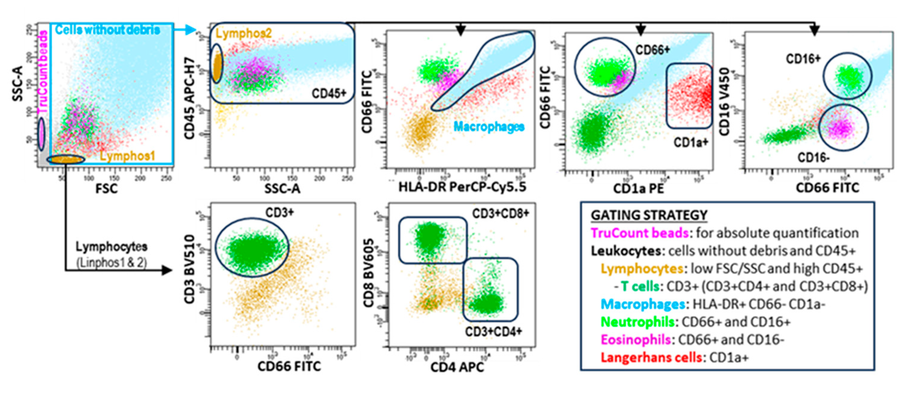
2.2. Immunophenotype Studies

BAL samples from different hospitals in Murcia Region were transported refrigerated at 4–8 °C in isothermal containers and processed immediately in our laboratory upon receipt. Briefly, the volume and appearance of samples were recorded. All the volume was immediately centrifuged at 1800 rpm, the supernatant was aspirated, and the pellet was washed once with 15 mL of FACSFlow (Becton Dickinson; BD; San Jose, CA, USA). Finally, samples were resuspended in 0.5 mL of FACSFlow (BD) and 50 µL stained immediately in TruCount tubes (BD) with the following antibody mix: CD1a PE (HI149), CD3 BV510 (SK7), CD4 APC (SK3), CD8 PE-Cy7 (SK1), CD16 V450 (3G8), CD19 APC (SJ25C1), CD20 FITC (L27), CD45 APC-H7 (2D1), and HLA-DR PerCp (L243) from BD and CD66 FITC (Kat4c) from Dako (Santa Clara, CA, USA). Samples were vortexed and incubated for 10 min at room temperature in the dark, then lysed with ammonium chloride solution (BD) for 7 min, and a minimum of 0.5 million cells were acquired immediately in 8-color FACSCanto-II flow cytometer (BD). Photomultiplier (PMT) voltages were adjusted daily using CS&T beads (BD). Fluorescence compensations were adjusted using FC beads (BD) every two months and finely adjusted on a daily basis using negative events as reference for each fluorochrome [25,26].
DiVATM Software 9.0 (BD, San Jose, CA, USA) was used for analysis following the gating strategy described in Figure 1. No dead cell exclusion dyes were used; therefore, both alive and dead cells (identified by the loss in FSC and SSC) were included, as long as they maintained the expression of leukocyte markers, especially CD45 [25].

2.3. Statistical Analysis

As described previously [25], data were collected in Excel (Microsoft Corporation, Redmond, WA, USA) and analyzed in SPSS 21.0 (Armonk, NY, USA). Kaplan–Meier and Log-Rank tests were used for survival estimation. Overall survival (OS) was defined as the time from first BAL analysis to death, with living patients censused on the date of last follow-up. Outcome of patient groups expressed in years was estimated as the 75th-percentile-OS (75p-OS). Comparisons between qualitative variables were performed by the two-tailed Fisher’s exact test. Quantitative variables were compared using ANOVA and DMS post hoc tests. Receiver operating characteristic (ROC) analysis was used to explore patient OS and to determine the optimal cut-off for quantitative variables. Multivariate analysis of predictive factors for OS was performed using the Cox proportional hazards model (stepwise regression). Hazard ratio (HR) and 95% confidence interval were estimated. p < 0.05 was considered statistically significant.

3. Results

3.1. Clinical, Biological, and Therapeutic Characteristics of Study Groups

Table 1 shows the biological and clinical characteristics of the patient and control groups as well as the main treatments for each pulmonary pathology.
Age and sex showed comparable values among the study groups (controls, ILDs, and infectious pulmonary diseases). Some ILD pathologies were predominantly observed among men, such as pneumoconiosis (96.3%), Eosinophilic-ILD (80.0%), IPF (71.7%), LIP (69.2%), and PLCH (66.7%). Comparable age was observed among the groups, although patients with PLCH were younger (36.7 years) compared with the age of the rest of the ILD patients (58.7 years). Connective tissue disease ILDs were not included as a group, since very few of these patients had a BAL study. It should also be noted that one-third of the patients did not require systemic immunosuppressive treatment.

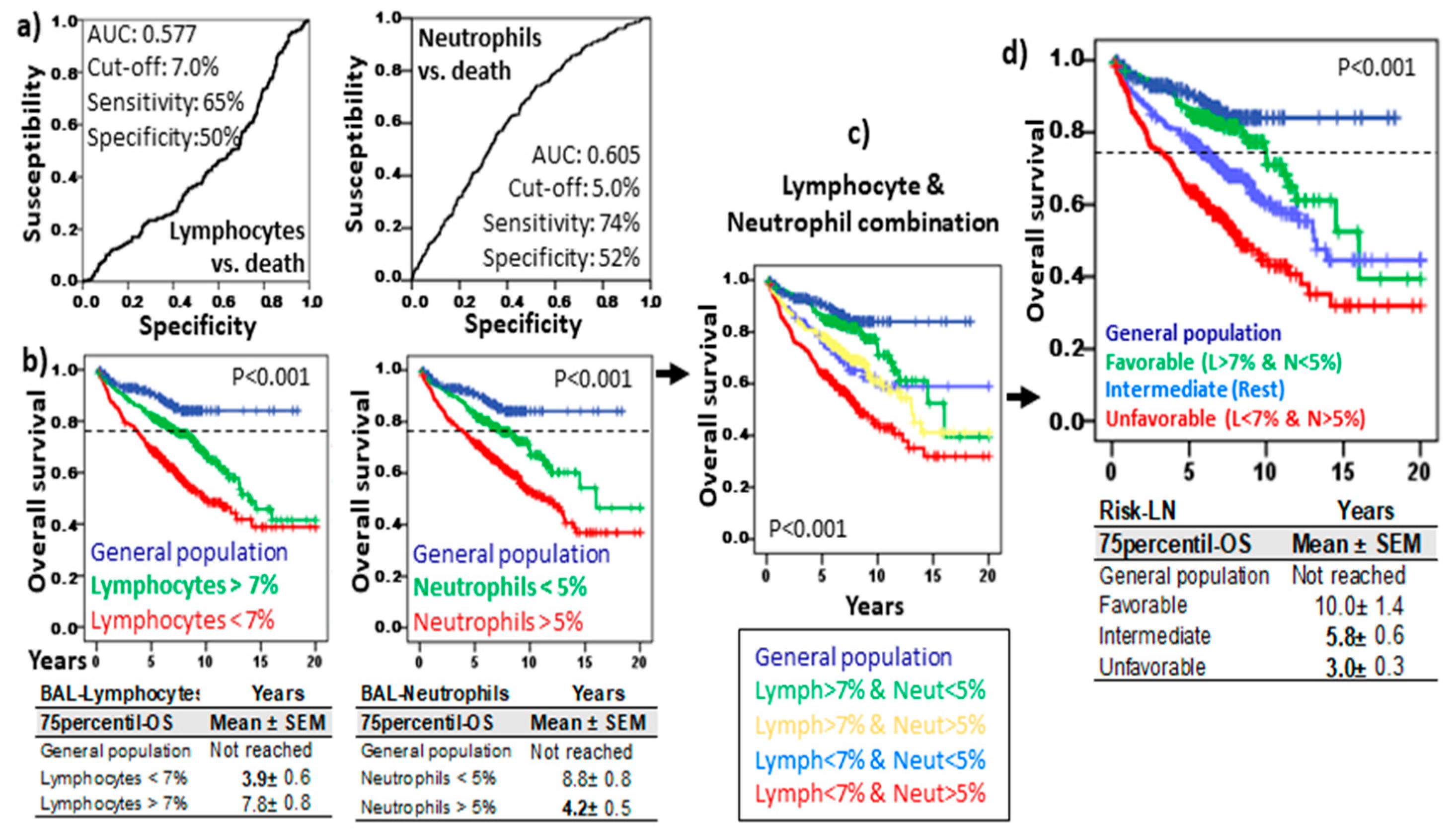
3.2. Risk Stratification of ILDs Based on Lymphocyte and Neutrophil Counts in BAL (Risk-LN)

Table 2 shows the percentage and absolute counts of the main leukocyte subsets in the bronchoalveolar lavage of the main disease groups. In the BAL control group, macrophages represented 89.8% of the cellularity with a mean of 98.2 ± 4.6 macrophages/µL. ILDs with or without fibrosis showed higher mean values of lymphocytes (22.4 ± 1% and 20.8 ± 1.6%, respectively) and neutrophils (7.7 ± 0.5 and 8.5 ± 0.8%, respectively) than BAL controls. IPF and infectious diseases had higher values of neutrophils (16.9 ± 1.5% and 46.0 ± 1.8%, respectively, p < 0.001) than BAL controls and other ILDs with or without fibrosis.
ROC analysis revealed cut-offs of 7.0% and 5.0% for lymphocytes and neutrophils, respectively, with predictive value on the OS (see Figure 2a for details). Longer 75p-OS was observed in patients with lymphocytes > 7.0% (7.8 ± 0.8 vs. 3.9 ± 0.6 years, p < 0.001) and neutrophils < 5.0% (8.8 ± 0.8 vs. 4.2 ± 0.5 years, p < 0.001) (Figure 2b). The combination of lymphocyte and neutrophil cut-offs showed four groups with different survival rates (Figure 2c,d): patients with lymphocytes > 7% and neutrophils < 5% showed the longest 75p-OS (10.0 ± 1.4 years, p < 0.001), with values comparable to those of the general population (75p-OS not reached); patients with lymphocytes < 7% and neutrophils > 5% showed the shortest 75p-OS (3.0 ± 0.3 years); the rest of the patients showed intermediate values for 75p-OS (5.8 ± 0.6 years). Therefore, an accurate counting of lymphocytes and neutrophils by flow cytometry in the BAL of patients with ILD and infectious diseases can provide a survival risk stratification.
Risk-LN predictive capacity was maintained in both ILD (10.0 ± 1.4, 5.7 ± 0.6, and 2.2 ± 0.4 years, p < 0.001) and infectious pulmonary diseases (not reached, 6.1 ± 0.6, and 4.0 ± 0.4 years, p < 0.01) with comparable 75p-OS values for favorable, intermediate, and unfavorable risk groups, respectively (Figure 3).
Because the analyzed samples came not only from our hospital but also from other hospitals, we tested whether the Risk-LN stratification could be influenced by transport conditions or processing time. Results showed very similar 75p-OS for local samples, which were processed in less than 30 min, and samples from other hospitals, which were processed in 60 to 90 min, from patients with favorable (12.0 ± 1.93 vs. 9.1 ± 1.69 years), intermediate (5.9 ± 1.43 vs. 5.2 ± 1.03 years), or unfavorable (3.3 ± 0.86 vs. 2.5 ± 0.88 years) Risk-LN stratifications (Supplementary Figure S1).

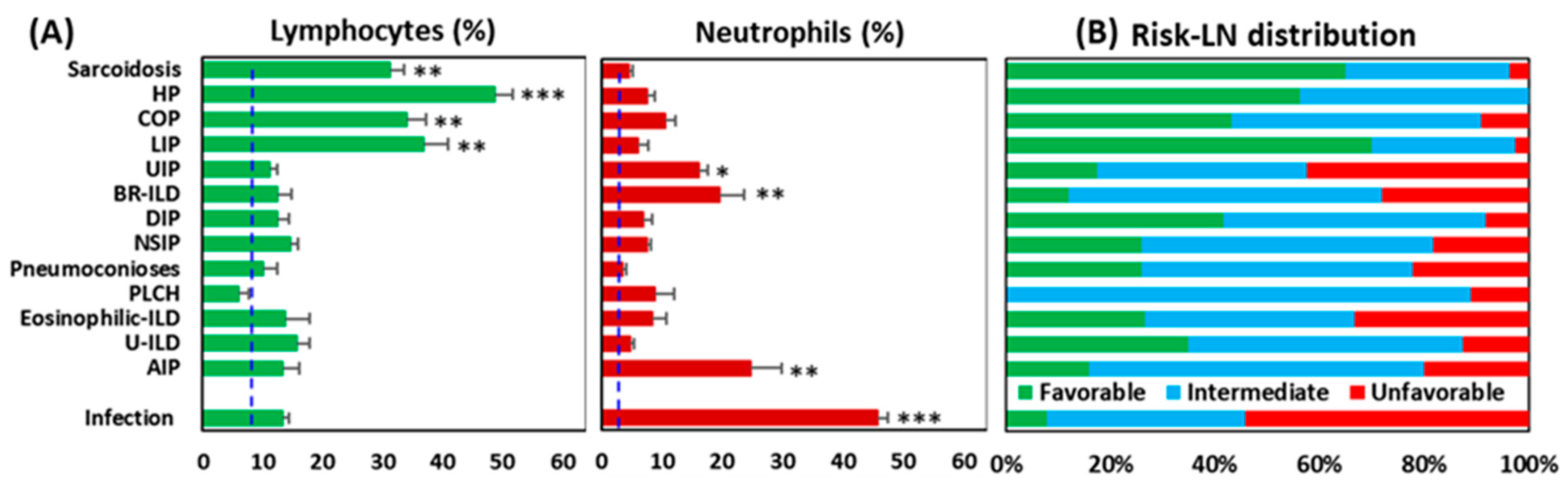
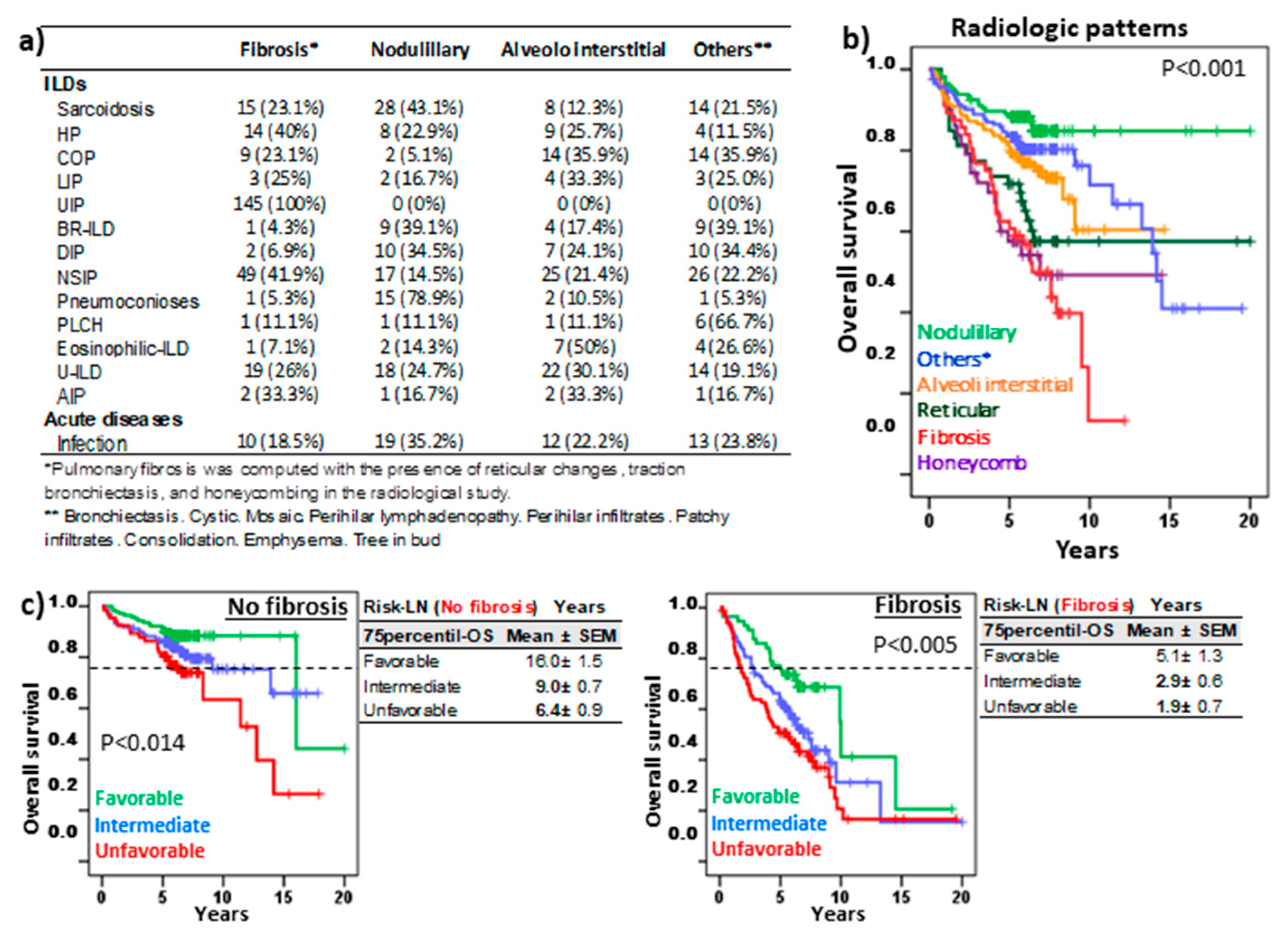
3.3. Uneven Distribution of Risk-LN Groups Among Pulmonary Pathologies

The Risk-LN groups had an uneven distribution among the pulmonary pathologies (Figure 4). Favorable and intermediate groups were mostly represented in ILDs with a predominance of lymphocytes (sarcoidosis, HP, COP, and LIP), whereas the unfavorable group was mostly represented in pathologies with a predominance of neutrophils (infectious pulmonary disease, AIP, BR-ILD, and UIP). In pathologies with a predominance of macrophages, eosinophils, or Langerhans cells (DIP, NSIP, pneumoconiosis, PLCH, Eosinophilic-ILD, and U-ILD), the distribution of the Risk-LN groups was more even.

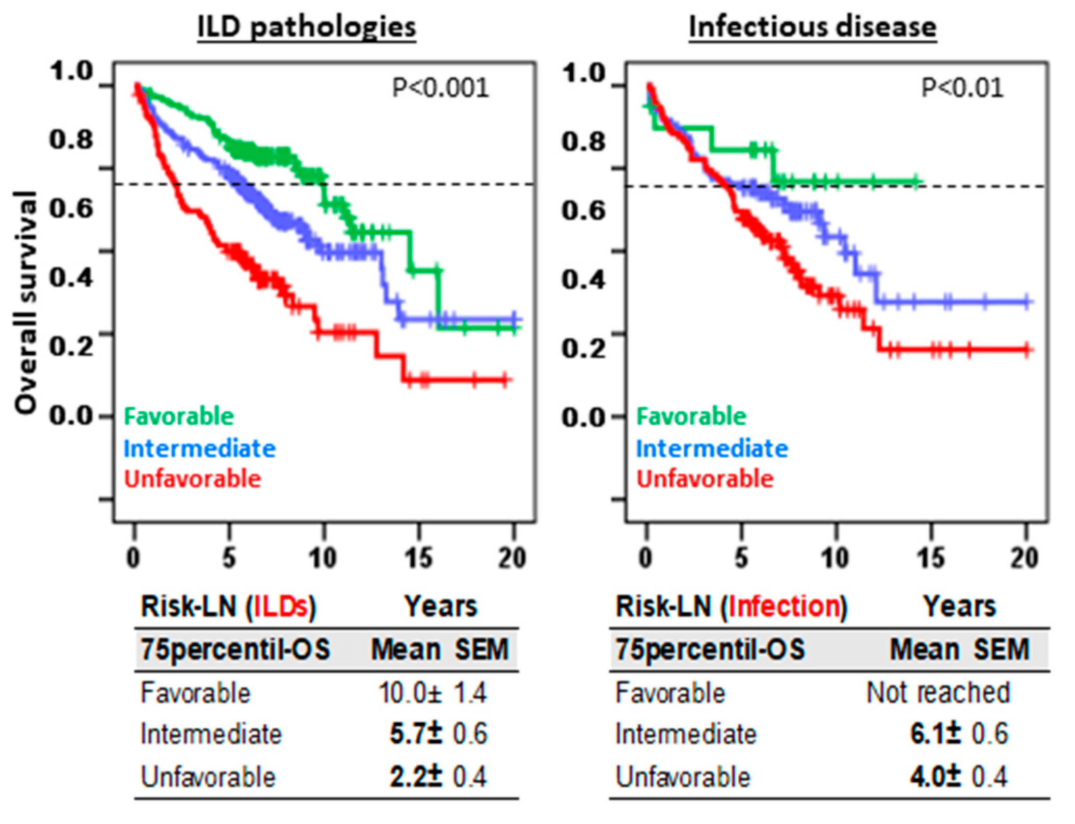
3.4. Risk-LN Stratification Preserves Its Predictive Value in Pathologies With or Without Pulmonary Fibrosis

Figure 5a shows the distribution of the most frequent radiological patterns in the different lung pathologies. As expected, the radiological patterns associated with pulmonary fibrosis (presence of reticular changes, traction bronchiectasis, and honeycombing) presented lower 75p-OS (2.9 ± 1.3 years) than nodular (not reached), alveoli interstitial (8.3 ± 0.6 years), or the rest of the patterns (11.4 ± 1.1 years) (Figure 5b). However, Risk-LN stratification maintained its prognostic value in patients without fibrosis (16.0 ± 1.5, 9.0 ± 0.7, and 6.4 ± 0.9 years, p = 0.014) or with fibrosis (5.1 ± 1.3 vs. 2.9 ± 0.6, and 1.9 ± 1.3 years, p < 0.005), respectively, for the favorable, intermediate, and unfavorable risk groups.

3.5. Risk-LN Predictive Capacity Was Independent of the Type of Treatment

Figure 6a shows patient outcomes according to the type of systemic therapy. Patients that did not receive systemic therapy showed longer 75p-OS (10.1 ± 1.4, p < 0.001) than those treated with other immunosuppressants (6.3 ± 1.3 years) or corticosteroids (4.3 ± 0.86 years).
Nonetheless, Risk-LN stratification maintained its predictive value in patients without systemic treatment (11.5 ± 0.98, 7.7 ± 0.76, and 5.8 ± 1.3 years, p < 0.05) or those treated with systemic corticosteroids (10.0 ± 2.5, 4.4 ± 2.1, and 1.7 ± 0.4 years, p < 0.001) or other immunosuppressants (9.9 ± 2.5, 6.4 ± 1.3, and 2.5 ± 0.7 years, p < 0.03) for the favorable, intermediate, and unfavorable risk groups.

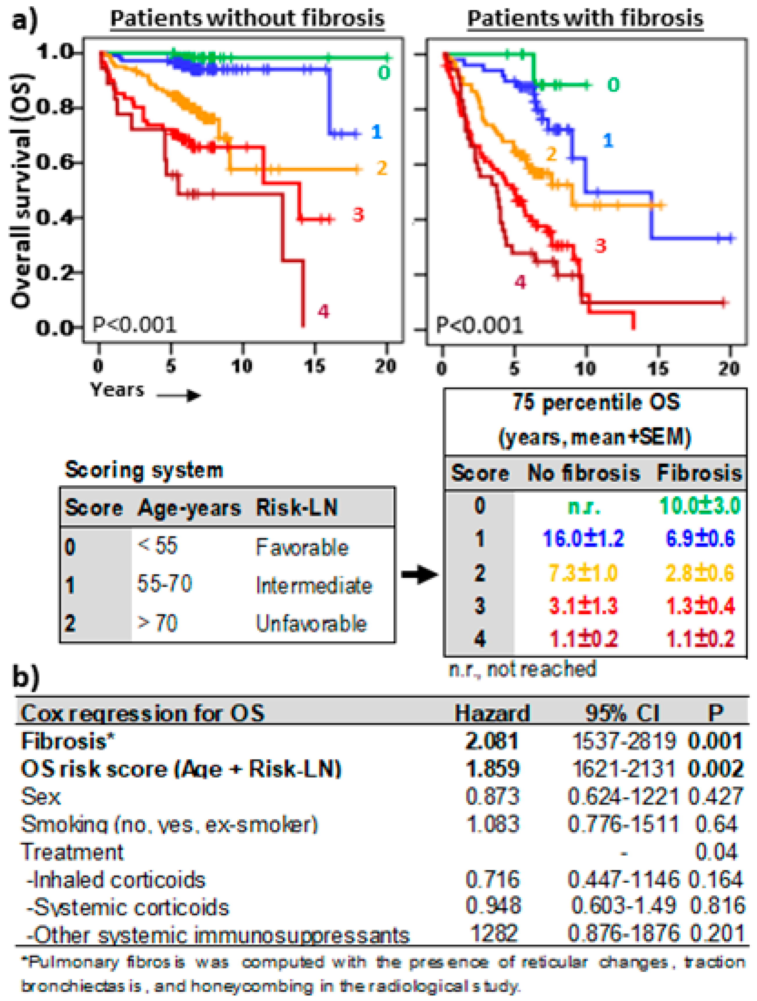
3.6. Risk-LN, DLCO, and Fibrosis Are Independent and Complementary Predictive Factors in ILD

Supplementary Figure S2a shows the functional and spirometry parameters in the different ILDs. Although DLCO data were available in only 25.1% of patients, the ROC analysis revealed a cut-off of 55.5% for DLCO with predictive value on the OS. Patients with DLCO < 55.5% showed shorter 75p-OS (3.0 ± 0.6 vs. 9.1 ± 1.2 years, p < 0.001) than patients with DLCO > 55.5%. Nonetheless, Risk-LN could still identify patients with unfavorable risk among patients with DLCO > 55.5%, who showed shorter 75p-OS (4.1 ± 0.9 years vs. not reached, p < 0.001) compared with patients with favorable or intermediate risk. In patients with DLCO < 55.5%, unfavorable Risk-LN was associated with shorter 75p-OS (2.0 ± 0.9 vs. 6.3 ± 0.7 and 3.9 ± 0.8 years, p < 0.05) compared with patients with favorable or intermediate risk, respectively (Supplementary Figure S2c).
Risk-LN stratification was an independent predictive factor for OS (HR = 1.619, p = 0.008) that complemented the other independent predictive factors in ILDs, such as age (HR = 1.046, p = 0.001), fibrosis (HR = 2.048, p = 0.015), and DLCO (HR = 0.979, p = 0.001) (Supplementary Figure S2d).

3.7. Risk-Scoring Model for Overall Survival Useful in Fibrotic and Non-Fibrotic ILDs

Finally, a risk score model (Figure 7a) with predictive capacity for OS in fibrotic and non-fibrotic ILDs was proposed combining the Risk-LN (0, favorable; 1, intermediate; and 2, unfavorable) and the age range of patients (0, <50 years; 1, 50–70 years; and 2, >70 years). Decreasing 75p-OSs were observed in patients with 0, 1, 2, 3, and 4 scores, in both non-fibrotic (not reached, 16.0 ± 1.2, 7.3 ± 1.0, 3.1 ± 1.3, and 1.1 ± 0.2 years, p < 0.001) and fibrotic (10.0 ± 3.0, 6.9 ± 0.6, 2.8 ± 0.6, 1.3 ± 0.4, and 1.1 ± 0.2 years, p < 0.001) pulmonary pathologies. This OS risk score was an independent predictive factor (HR = 1.859, p = 0.002) complementary to the fibrosis status (HR = 2.081, p < 0.001) and the type of treatment (Figure 7b).
A quick and automated estimate of the risk score can be obtained at “https://bal-ildcalculator.imib.es/”. In addition, a diagnostic approach to the most probable pathology associated with that cellular pattern will be offered by applying logistic regression models to BAL leukocyte subsets of the patient. Examples of real cases are shown in Supplementary Figure S3.

4. Discussion

Several models estimating the mortality risk in IPF [3] or general ILD [27] have been proposed to guide the clinical management of these patients. Models have taken into consideration clinical symptoms, pulmonary function, the extent of UIP, and even the Charlson comorbidity index score (CCIS) (ILD-GAPC) [27]. However, the immune cells infiltrating pulmonary tissue that are involved in lung deterioration and/or the fibrotic process have not been tested in these models. Our results show that lymphocytes and neutrophils, the inflammatory leukocytes that most frequently infiltrate the lung in ILD, play opposing roles in the clinical evolution of patients. While the former seem to protect, neutrophils are associated with a more accelerated progression of the disease and shorter patient survival. Flow cytometry of BAL can offer a quick and accurate quantification of these leukocyte subsets, revealing cut-offs with predictive capacity in ILD and thus allowing the establishment of a risk stratification based on the number of lymphocytes and neutrophils (Risk-LN). Risk-LN stratification can help to identify patients with a very unfavorable risk from the time of diagnosis as well as patients who have favorable risk and survival rates similar to the general population (without lung pathology), and therefore, who would not need close surveillance or therapies in the short term.
Furthermore, the Risk-LN stratification combined with the age range of patients allowed for the development of a “scoring” model that is able to predict the survival of patients with great precision in both non-fibrotic and fibrotic ILDs. With this predictive model, patients with very favorable long-term survival estimates—longer than 16 and 10 years in patients without and with pulmonary fibrosis, respectively—can be identified from diagnosis with the first BAL sample. In contrast, patients with very poor long-term overall survival estimates—shorter than 1.1 years—were also identified in both non-fibrotic and fibrotic ILDs. Unfortunately, the unavailability of functional and spirometry tests in some hospitals in our region between 2000 and 2015 has prevented the inclusion of the independent prognostic factor DLCO in the estimation of the risk score. Nonetheless, the score system described in this manuscript is simple and could provide a useful tool for guiding treatment decisions in the most common lung pathologies (quick score estimation is accessible to pulmonologists worldwide at https://bal-ildcalculator.imib.es/. Even so, independent studies in more recent patients will be necessary to determine the contribution of DLCO to the score model proposed in this study.
Although the protective effect of lymphocytes [20] and the harmful effect of neutrophils infiltrating the lung [17,18,20,28,29,30,31], or even in the peripheral blood [32,33,34], has been well stablished, the inclusion of lymphocyte and neutrophil quantification in clinical practice is still far from happening. Different reasons may be delaying the inclusion of these parameters in the management of lung pathology: first, the use of prognostic markers outside FPI is still unusual among pulmonologists; second, in real-life medicine, BAL cytology reports do not always include the number of leukocyte subsets, and if they do, the results may be delayed for several weeks. In this sense, flow cytometry can offer accurate results in less than an hour. However, flow cytometry continues to be a methodology with low penetration in pulmonology, although it has become an essential tool in many other clinical disciplines [35]. Technology improvement has facilitated the use of antibody panels large enough to correctly discriminate the different leukocyte subpopulations, thus clearly differentiating neutrophils from eosinophils, which can be difficult with panels with fewer antibodies. Nowadays, accessibility to this type of analysis has become widespread in most large hospitals, with professionals perfectly qualified to study BAL. Furthermore, the analysis strategy shown in this study, including the dead cells, can facilitate the accessibility of this technology in smaller hospitals that do not have onsite flow cytometry, since the Risk-LN predictive value was maintained even in samples that needed to be transported and were processed later than 60 to 90 min after extraction. Furthermore, our results support recent findings indicating that flow cytometry may be a good alternative to cytopathological analysis, as it provides comparable data when analyzing major leukocyte subsets, including lymphocytes and neutrophils [36].
The harmful effect shown by neutrophils in our series and in others [37], in the absence of the protective effect of lymphocytes, could serve as a guide for clinicians to establish early preventive therapies to mitigate pulmonary deterioration, particularly in patients with unfavorable Risk-LNs (predominance of neutrophils over lymphocytes) and short survival estimates (scores higher than 2 in non-fibrotic patients and scores higher than 1 in fibrotic patients). Although Risk-LN stratification preserved its predictive value in patients without treatment and in patients with different types of immunosuppressive therapies (corticosteroids or other immunosuppressants), it would have been interesting to evaluate its predictive capacity in patients with the new antifibrotic treatments [38,39,40]. However, just a minority of patients from our series received this therapy; therefore, this should be addressed in future studies.
In conclusion, although the predictive capacity of the Risk-LN score should be confirmed in larger prospective studies to evaluate its prognostic value, and it should incorporate other variables with prognostic value such as DLCO, our results clearly show that flow cytometry of BAL can offer predictive information independent of DLCO and the fibrotic status with potential utility for treatment personalization in ILD from the time of diagnosis.

Supplementary Materials

The following supporting information can be downloaded at: https://www.mdpi.com/article/10.3390/cells13242066/s1, Supplementary Figure S1: Impact of processing time on Risk-LN stratification. Kaplan–Meier and Log-Rank test for overall survival (OS) according to Risk-LN groups for patients from our hospital, whose samples were processed in less than 30 min, and of samples from other hospitals, processed in 60 to 90 min. The 75th-percentile-OS is indicated in the figure for each risk group. Supplementary Figure S2: Risk-LN stratification complements the predictive value of DLCO in ILD patients. (a) Functional and spirometry parameters in ILD patients: Diffusing capacity of the lungs for carbon monoxide (DLCO), forced expiratory volume in the first second (FEV1), and forced vital capacity (FVC). (b) Receiver operating characteristic curve (ROC) of DLCO related to overall survival (OS). Area under the curve (AUC) and cut-offs with the highest sensitivity and specificity is shown, and Kaplan–Meier and Log-Rank tests for OS of patients according to DLCO above or below 55.5%. (c) Kaplan–Meier and Log-Rank tests for OS of ILD patients according to DLCO above or below 55.5% and Risk-LN. The 75th-percentile-OS is shown in each section. (d) Cox regression analysis for OS in ILD patients for sex, age, fibrosis, DLCO (%), and Risk-LN. Supplementary Figure S3: BAL ILD calculator.

Author Contributions

Conceptualization, A.M. and J.A.R.; methodology, A.O.-A., C.O.-H., R.G.-L., I.R.-L., L.G., D.C.-F. and E.M.N.-B.; software, A.M., L.G., R.G.-L., R.L.-H., I.R.-L., S.P.-F., P.M.-C., E.M.N.-B. and A.M.; validation, E.M.N.-B., R.G.-L., A.O.-A., C.O.-H., I.R.-L., L.G., D.C.-F. and R.L.-H.; formal analysis, A.M., J.A.R., J.A.C., M.M., S.P.-F., P.M.-C. and E.M.N.-B.; investigation, I.R.-L., L.G., R.L.-H., A.O.-A. and A.M.; resources, E.M.N.-B., R.G.-L., J.A.R., M.M., P.M.-C., I.R.-L. and A.M.; data curation, E.M.N.-B., R.G.-L., C.O.-H., J.A.R., R.L.-H. and A.M.; writing—original draft preparation, E.M.N.-B., I.R.-L., A.M., J.A.R., D.C.-F., L.G., M.M., S.P.-F., P.M.-C. and J.A.C.; writing—review and editing, J.A.R., J.A.C., R.L.-H., M.M., S.P.-F. and A.M.; visualization, A.M., E.M.N.-B., R.G.-L., I.R.-L., L.G., A.O.-A. and M.M.; supervision, S.P.-F., M.M., E.S., P.M.-C. and A.M.; project administration, J.A.R., J.A.C., M.M., E.S., P.M.-C. and A.M.; funding acquisition, J.A.R., A.M. and S.P.-F. All authors have read and agreed to the published version of the manuscript.

Funding

The Biomedical Informatics and Bioinformatics department at IMIB was funded by Consejería de Salud de la Comunidad Autónoma de la Región de Murcia.

Institutional Review Board Statement

This study was approved on 8 May 2024 by the ethics committee of Clinic University Hospital Virgen de la Arrixaca (IRB-00005712).

Informed Consent Statement

Written informed consent was obtained from all patients in accordance with the Declaration of Helsinki.

Data Availability Statement

The data that support the findings of this study are available from the corresponding author upon reasonable request.

Acknowledgments

We would like to thank clinicians attending lung pathology from all public hospitals in Murcia Region for sending samples to our flow cytometry unit; to all laboratory technicians, particularly Carmen Martínez Solano, Alba Fernández Torres, and José Moya Martínez; and to the Biomedical Informatics and Bioinformatics department at the Biomedical Research Institute of Murcia Pascual Parrilla (IMIB).

Conflicts of Interest

The authors declare no conflicts of interest.

References

  1. Bagnato, G.; Harari, S. Cellular interactions in the pathogenesis of interstitial lung diseases. Eur. Respir. Rev. 2015, 24, 102–114. [Google Scholar] [CrossRef]
  2. Moss, B.J.; Ryter, S.W.; Rosas, I.O. Pathogenic Mechanisms Underlying Idiopathic Pulmonary Fibrosis. Annu. Rev. Pathol. Mech. Dis. 2022, 17, 515–546. [Google Scholar] [CrossRef] [PubMed]
  3. Ley, B.; Ryerson, C.J.; Vittinghoff, E.; Ryu, J.; Tomassetti, S.; Lee, J.S.; Poletti, V.; Buccioli, M.; Elicker, B.M.; Jones, K.D.; et al. A multidimensional index and staging system for idiopathic pulmonary fibrosis. Ann. Intern. Med. 2012, 156, 684–691. [Google Scholar] [CrossRef] [PubMed]
  4. Salisbury, M.L.; Xia, M.; Zhou, Y.; Zhou, Y.; Murray, S.; Tayob, N.; Brown, K.K.; Wells, A.U.; Schmidt, S.L.; Martinez, F.J.; et al. Idiopathic Pulmonary Fibrosis: Gender-Age-Physiology Index Stage for Predicting Future Lung Function Decline. Chest 2016, 149, 491–498. [Google Scholar] [CrossRef] [PubMed]
  5. Ley, B.; Bradford, W.Z.; Weycker, D.; Vittinghoff, E.; du Bois, R.M.; Collard, H.R. Unified baseline and longitudinal mortality prediction in idiopathic pulmonary fibrosis. Eur. Respir. J. 2015, 45, 1374–1381. [Google Scholar] [CrossRef]
  6. Suzuki, Y.; Mori, K.; Aono, Y.; Kono, M.; Hasegawa, H.; Yokomura, K.; Naoi, H.; Hozumi, H.; Karayama, M.; Furuhashi, K.; et al. Combined assessment of the GAP index and body mass index at antifibrotic therapy initiation for prognosis of idiopathic pulmonary fibrosis. Sci. Rep. 2021, 11, 18579. [Google Scholar] [CrossRef] [PubMed]
  7. Chandel, A.; Pastre, J.; Valery, S.; King, C.S.; Nathan, S.D. Derivation and validation of a simple multidimensional index incorporating exercise capacity parameters for survival prediction in idiopathic pulmonary fibrosis. Thorax 2023, 78, 368–375. [Google Scholar] [CrossRef] [PubMed]
  8. Chandel, A.; King, C.S.; Ignacio, R.V.; Pastre, J.; Shlobin, O.A.; Khangoora, V.; Aryal, S.; Nyquist, A.; Singhal, A.; Flaherty, K.R.; et al. External validation and longitudinal application of the DO-GAP index to individualise survival prediction in idiopathic pulmonary fibrosis. ERJ Open Res. 2023, 9, 00124-2023. [Google Scholar] [CrossRef]
  9. Lee, J.H.; Jang, J.H.; Jang, H.-J.; Kim, S.Y.; Chung, M.P.; Yoo, H.; Jeong, S.H.; Song, J.W.; Lee, H.L.; Choi, S.M.; et al. New prognostic scoring system for mortality in idiopathic pulmonary fibrosis by modifying the gender, age, and physiology model with desaturation during the six-minute walk test. Front. Med. 2023, 10, 1052129. [Google Scholar] [CrossRef]
  10. Zhu, W.-W.; Li, H.; Li, Y.-D.; Sun, L.; Kong, L.; Ye, X.; Cai, Q.; Lv, X.-Z. Risk assessment in interstitial lung disease: The incremental prognostic value of cardiopulmonary ultrasound. BMC Pulm. Med. 2021, 21, 237. [Google Scholar] [CrossRef]
  11. Ryerson, C.J.; Vittinghoff, E.; Ley, B.; Lee, J.S.; Mooney, J.J.; Jones, K.D.; Elicker, B.M.; Wolters, P.J.; Koth, L.L.; King, T.E.; et al. Predicting survival across chronic interstitial lung disease: The ILD-GAP model. Chest 2014, 145, 723–728. [Google Scholar] [CrossRef] [PubMed]
  12. Almeida, L.M.; Fernandes, A.L.; Cardoso, C.G.; Lima, B.; Neves, I.; Novais-Bastos, H.; Mota, P.C.; Melo, N.; Moura, C.S.; Guimarães, S.; et al. Mortality risk prediction with ILD-GAP index in a fibrotic hypersensitivity pneumonitis cohort. Ther. Adv. Respir. Dis. 2022, 16, 17534666221135316. [Google Scholar] [CrossRef] [PubMed]
  13. Shi, S.; Chen, L.; Liu, X.; Yu, M.; Wu, C.; Xiao, Y. Development of a scoring system with multidimensional markers for fibrosing interstitial lung disease. Sci. Rep. 2022, 12, 14217. [Google Scholar] [CrossRef] [PubMed]
  14. Chen, X.; Guo, J.; Yu, D.; Jie, B.; Zhou, Y. Predictors of Mortality in Progressive Fibrosing Interstitial Lung Diseases. Front. Pharmacol. 2021, 12, 754851. [Google Scholar] [CrossRef] [PubMed]
  15. Ruaro, B.; Baratella, E.; Confalonieri, P.; Wade, B.; Marrocchio, C.; Geri, P.; Busca, A.; Pozzan, R.; Andrisano, A.G.; Cova, M.A.; et al. High-Resolution Computed Tomography: Lights and Shadows in Improving Care for SSc-ILD Patients. Diagnostics 2021, 11, 1960. [Google Scholar] [CrossRef] [PubMed]
  16. Hughes, M.; Bruni, C.; Cuomo, G.; Sedie, A.D.; Gargani, L.; Gutierrez, M.; Lepri, G.; Ruaro, B.; Santiago, T.; Suliman, Y.; et al. The role of ultrasound in systemic sclerosis: On the cutting edge to foster clinical and research advancement. J. Scleroderma Relat. Disord. 2021, 6, 123–132. [Google Scholar] [CrossRef] [PubMed]
  17. Salonen, J.; Lampela, H.; Keskitalo, E.; Vähänikkilä, H.; Purokivi, M.; Kaarteenaho, R. Bronchoalveolar lavage differential cell count on prognostic assessment of patients with stable or acute interstitial lung disease: A retrospective real-life study. Clin. Immunol. 2020, 220, 108594. [Google Scholar] [CrossRef]
  18. Kinder, B.W.; Brown, K.K.; Schwarz, M.I.; Ix, J.H.; Kervitsky, A.; King, T.E. Baseline BAL neutrophilia predicts early mortality in idiopathic pulmonary fibrosis. Chest 2008, 133, 226–232. [Google Scholar] [CrossRef] [PubMed]
  19. Kono, M.; Miyashita, K.; Hirama, R.; Oshima, Y.; Takeda, K.; Mochizuka, Y.; Tsutsumi, A.; Miwa, H.; Miki, Y.; Hashimoto, D.; et al. Prognostic significance of bronchoalveolar lavage cellular analysis in patients with acute exacerbation of interstitial lung disease. Respir. Med. 2021, 186, 106534. [Google Scholar] [CrossRef] [PubMed]
  20. Hirasawa, Y.; Nakada, T.-A.; Shimazui, T.; Abe, M.; Isaka, Y.; Sakayori, M.; Suzuki, K.; Yoshioka, K.; Kawasaki, T.; Terada, J.; et al. Prognostic value of lymphocyte counts in bronchoalveolar lavage fluid in patients with acute respiratory failure: A retrospective cohort study. J. Intensiv. Care 2021, 9, 21. [Google Scholar] [CrossRef] [PubMed]
  21. Meyer, K.C.; Raghu, G.; Baughman, R.P.; Brown, K.K.; Costabel, U.; du Bois, R.M.; Drent, M.; Haslam, P.L.; Kim, D.S.; Nagai, S.; et al. An official American Thoracic Society clinical practice guideline: The clinical utility of bronchoalveolar lavage cellular analysis in interstitial lung disease. Am. J. Respir. Crit. Care Med. 2012, 185, 1004–1014. [Google Scholar] [CrossRef] [PubMed]
  22. Vasco-Mogorrón, M.A.; Campillo, J.A.; Periago, A.; Cabañas, V.; Berenguer, M.; García-Garay, M.C.; Gimeno, L.; Soto-Ramírez, M.F.; Martínez-Hernández, M.D.; Muro, M.; et al. Blood-based risk stratification for pre-malignant and symptomatic plasma cell neoplasms to improve patient management. Am. J. Cancer Res. 2021, 11, 2736–2753. [Google Scholar] [PubMed]
  23. Travis, W.D.; Costabel, U.; Hansell, D.M.; King, T.E., Jr.; Lynch, D.A.; Nicholson, A.G.; Ryerson, C.J.; Ryu, J.H.; Selman, M.; Wells, A.U.; et al. An official American Thoracic Society/European Respiratory Society statement: Update of the international multidisciplinary classification of the idiopathic interstitial pneumonias. Am. J. Respir. Crit. Care Med. 2013, 188, 733–748. [Google Scholar] [CrossRef] [PubMed]
  24. Bosch, L.v.D.; Luppi, F.; Ferrara, G.; Mura, M. Immunomodulatory treatment of interstitial lung disease. Ther. Adv. Respir. Dis. 2022, 16, 17534666221117002. [Google Scholar] [CrossRef]
  25. Novoa-Bolivar, E.M.; Ros, J.A.; Pérez-Fernández, S.; Campillo, J.A.; López-Hernández, R.; González-López, R.; Otalora-Alcaraz, A.; Ortuño-Hernández, C.; Gimeno, L.; Ruiz-Lorente, I.; et al. Neutrophils and Lymphocytes: Yin and Yang of Lung Fibrosis and Patient Outcome in Diffuse Interstitial Lung Diseases. Biomedicines 2024, 12, 2439. [Google Scholar] [CrossRef] [PubMed]
  26. Martínez-Sánchez, M.V.; Fuster, J.L.; Campillo, J.A.; Galera, A.M.; Bermúdez-Cortés, M.; Llinares, M.E.; Ramos-Elbal, E.; Pascual-Gázquez, J.F.; Fita, A.M.; Martínez-Banaclocha, H.; et al. Expression of NK Cell Receptor Ligands on Leukemic Cells Is Associated with the Outcome of Childhood Acute Leukemia. Cancers 2021, 13, 2294. [Google Scholar] [CrossRef]
  27. Fujii, H.; Hara, Y.; Saigusa, Y.; Tagami, Y.; Murohashi, K.; Nagasawa, R.; Aoki, A.; Izawa, A.; Seki, K.; Watanabe, K.; et al. ILD-GAP Combined with the Charlson Comorbidity Index Score (ILD-GAPC) as a Prognostic Prediction Model in Patients with Interstitial Lung Disease. Can. Respir. J. 2023, 2023, 5088207. [Google Scholar] [CrossRef]
  28. Kanaoka, K.; Minami, S.; Ihara, S.; Komuta, K. High neutrophils and low lymphocytes percentages in bronchoalveolar lavage fluid are prognostic factors of higher in-hospital mortality in diffuse alveolar hemorrhage. BMC Pulm. Med. 2021, 21, 288. [Google Scholar] [CrossRef] [PubMed]
  29. D’Alessandro, M.; Bergantini, L.; Carleo, A.; Cameli, P.; Perrone, A.; Fossi, A.; Sestini, P.; Bargagli, E. Neutrophil-to-lymphocyte ratio in bronchoalveolar lavage from IPF patients: A novel prognostic biomarker? Minerva Med. 2022, 113, 526–531. [Google Scholar] [CrossRef]
  30. Ryu, W.K.; Moon, Y.; Park, M.H.; Lim, J.H.; Kim, Y.S.; Lee, K.-H.; Kwak, S.M.; Kim, C.; Nam, H.-S. A Preliminary Study on the Prognostic Impact of Neutrophil to Lymphocyte Ratio of the Bronchoalveolar Lavage Fluid in Patients with Lung Cancer. Diagnostics 2021, 11, 2201. [Google Scholar] [CrossRef]
  31. Crowley, L.E.; Stockley, R.A.; Thickett, D.R.; Dosanjh, D.; Scott, A.; Parekh, D. Neutrophil dynamics in pulmonary fibrosis: Pathophysiological and therapeutic perspectives. Eur. Respir. Rev. 2024, 33, 240139. [Google Scholar] [CrossRef] [PubMed]
  32. Achaiah, A.; Rathnapala, A.; Pereira, A.; Bothwell, H.; Dwivedi, K.; Barker, R.; Iotchkova, V.; Benamore, R.; Hoyles, R.K.; Ho, L.-P. Neutrophil lymphocyte ratio as an indicator for disease progression in Idiopathic Pulmonary Fibrosis. BMJ Open Respir. Res. 2022, 9, e001202. [Google Scholar] [CrossRef] [PubMed]
  33. Song, M.; Graubard, B.I.; Rabkin, C.S.; Engels, E.A. Neutrophil-to-lymphocyte ratio and mortality in the United States general population. Sci. Rep. 2021, 11, 464. [Google Scholar] [CrossRef]
  34. Lee, H.; Um, S.-J.; Kim, Y.S.; Kim, D.K.; Jang, A.S.; Choi, H.S.; Kim, Y.H.; Kim, T.E.; Yoo, K.H.; Jung, K.-S. Association of the Neutrophil-to-Lymphocyte Ratio with Lung Function and Exacerbations in Patients with Chronic Obstructive Pulmonary Disease. PLoS ONE 2016, 11, e0156511. [Google Scholar] [CrossRef]
  35. Pang, K.; Dong, S.; Zhu, Y.; Zhu, X.; Zhou, Q.; Gu, B.; Jin, W.; Zhang, R.; Fu, Y.; Yu, B.; et al. Advanced flow cytometry for biomedical applications. J. Biophotonics 2023, 16, e202300135. [Google Scholar] [CrossRef] [PubMed]
  36. Bratke, K.; Weise, M.; Stoll, P.; Virchow, J.C.; Lommatzsch, M. Flow Cytometry as an Alternative to Microscopy for the Differentiation of BAL Fluid Leukocytes. Chest 2024, 166, 793–801. [Google Scholar] [CrossRef]
  37. Inomata, T.; Konno, S.; Nagai, K.; Suzuki, M.; Nishimura, M. Neutrophil predominance in bronchoalveolar lavage fluid is associated with disease severity and progression of HRCT findings in pulmonary Mycobacterium avium infection. PLoS ONE 2018, 13, e0190189. [Google Scholar] [CrossRef]
  38. Collins, B.F.; Raghu, G. Antifibrotic therapy for fibrotic lung disease beyond idiopathic pulmonary fibrosis. Eur. Respir. Rev. 2019, 28, 190022. [Google Scholar] [CrossRef] [PubMed]
  39. Maher, T.M.; Strek, M.E. Antifibrotic therapy for idiopathic pulmonary fibrosis: Time to treat. Respir. Res. 2019, 20, 205. [Google Scholar] [CrossRef] [PubMed]
  40. Finnerty, J.P.; Ponnuswamy, A.; Dutta, P.; Abdelaziz, A.; Kamil, H. Efficacy of antifibrotic drugs, nintedanib and pirfenidone, in treatment of progressive pulmonary fibrosis in both idiopathic pulmonary fibrosis (IPF) and non-IPF: A systematic review and meta-analysis. BMC Pulm. Med. 2021, 21, 411. [Google Scholar] [CrossRef] [PubMed]
Figure 1. Flow cytometry analysis of the leukocyte subsets contained in BAL samples. Leukocyte subsets were identified following the hierarchical and logical gating strategy shown in the figure. TruCount beads were used to calculate the absolute cell counts per microliter following the manufacturer’s instructions.
Cells 13 02066 g001
Figure 2. Risk stratification of ILD and infectious disease based on lymphocyte and neutrophil counts of BAL. (a) Receiver operating characteristic curve (ROC) of lymphocytes and neutrophils related to overall survival (OS). Area under the curve (AUC), cut-off, and the highest sensitivity and specificity are shown. (b) Kaplan–Meier and Log-Rank tests for OS of ILD and infectious disease patients with lymphocytes and neutrophils below and over their respective cut-offs and OS of general population (without lung diseases). (c) Kaplan–Meier and Log-Rank tests for OS of ILD patients according to the combinations of lymphocytes (above or below 7%) and neutrophils (above or below 5%). (d) Kaplan–Meier and Log-Rank tests for OS of ILD patients according to the Risk-Lymphocyte/Neutrophil stratification (Risk-LN): favorable (lymphocyte > 7% and neutrophil < 5%), unfavorable (lymphocyte < 7% and neutrophil > 5%), and intermediate (rest). The 75th percentile OS (Mean ± SD) are shown for the patients and general population in each section.
Cells 13 02066 g002
Figure 3. Risk-Lymphocyte/Neutrophil (Risk-LN) stratification offers predictive information in both ILDs and infectious diseases. Kaplan–Meier and Log-Rank tests for overall survival (OS) according to Risk-LN in patients with ILD pathologies and in patients with infectious disease. The 75th-percentile-OS is shown.
Cells 13 02066 g003
Figure 4. Distribution of Risk-Lymphocyte/Neutrophil (Risk-LN) stratification groups among pulmonary pathologies. (A) Percentage of lymphocytes and neutrophils in the flow cytometry analysis of BAL samples from patients with different pulmonary pathologies. * p < 0.05, ** p < 0.01, *** p < 0.001 in the ANOVA and DMS post hoc tests. The dashed lines indicate the mean value of lymphocytes (6.5%) and neutrophils (2.12%) in the control BAL group. (B) Distribution of Risk-LN groups among pulmonary pathologies: favorable (lymphocyte > 7% and neutrophil < 5%), unfavorable (lymphocyte < 7% and neutrophil > 5%), and intermediate (rest).
Cells 13 02066 g004
Figure 5. Risk-Lymphocyte/Neutrophil stratification (Risk-LN) in ILD patients with pulmonary fibrosis. (a) Frequency of the most common lung imaging patterns among ILD subtypes. (b) Kaplan–Meier and Log-Rank tests for overall survival (OS) of ILD patients according to their predominant lung pattern. * other radiological patterns. (c) Kaplan–Meier and Log-Rank tests for OS of ILD patients with or without fibrosis according to Risk-LN stratification. The 75th-percentile-OS is shown in each case.
Cells 13 02066 g005
Figure 6. Risk-Lymphocyte/Neutrophil (Risk-LN) stratification in ILD patients with different systemic immunosuppressive treatments. (a) Kaplan–Meier and Log-Rank tests for overall survival (OS) of ILD patients according to the type of treatment: no systemic treatment, corticosteroids, or other immunosuppressants (rituximab, azathioprine, mycophenolate mofetil, cyclophosphamide, tacrolimus). (b) Kaplan–Meier and Log-Rank tests for OS of ILD patients according to the Risk-Lymphocyte/Neutrophil stratification (Risk-LN) and the type of treatment. The 75th-percentile-OS is shown in each case.
Cells 13 02066 g006
Figure 7. Overall survival risk scoring systems in ILDs. (a) Kaplan–Meier and Log-Rank tests for overall survival (OS) of ILD patients according to a scoring model including age range (<55 years = 0, 55–70 years = 1, >55 years = 2) and Risk-Lymphocyte/Neutrophil stratification (Risk-LN) (favorable = 0, intermediate = 1, unfavorable = 2). The 75th-percentile-OS was estimated separately for patients with or without pulmonary fibrosis. (b) Cox regression analysis for OS in ILD patients for sex, type of ILD, smoking status, type of treatment, fibrosis status, and the OS risk score.
Cells 13 02066 g007
Table 1. Biological, clinical, and therapeutic characteristics of patient and control groups.
PatientsSex Age Fibrosis 1Treatments (%) 2
(N)(% man)(Mean ± SD)(%)NTSCOther
Control groups35553.2%61.2 ± 13.5-
General population 324353.1%67.9 ± 12.3-
BAL control 411253.3%53.5 ± 16.60%
Interstitial lung diseases (ILD)73158.1%58.7 ± 16.438.2%323218
Sarcoidosis8248.20%55.6 ± 14.819.5%343815
Hypersensitivity pneumonitis (HP)4849.0%50.4 ± 17.629.1%134613
Organized cryptogenic pneumonia (COP)4446.50%62.3 ± 17.418.1%185221
Lymphocytic interstitial pneumonia (LIP)3769.20%53.2 ± 15.98.1%492416
Usual interstitial pneumonia (UIP) 514571.70%65.8 ± 12.3100%253519
Respiratory bronchiolitis ILD (RB-ILD)2540.0%53.3 ± 22.88.0%36368
Desquamative interstitial pneumonitis (DIP)3552.8%54.4 ± 20.85.7%462317
Nonspecific interstitial pneumonia (NSIP)15648.1%59.3 ± 15.533.9%312830
Pneumoconiosis2796.3%56.5 ± 16.018.5%42234
Pulmonary Langerhans cell histiocytosis (PLCH)966.7%36.7 ± 15.811.1%671111
Eosinophilic-ILD1780.0%48.4 ± 22.75.9%0766
Unclassifiable-ILD (U-ILD)8060.0%61.3 ± 12.425.0%491510
Acute interstitial pneumonia (AIP)2656.0%57.5 ± 21.915.3%292412
Pulmonary infectious diseases23159.0%62.1 ± 15.615.1%272218
1 Pulmonary fibrosis was computed with the presence of reticular changes, traction bronchiectasis, and honeycombing in the radiological study. 2 Immunomodulatory treatment was administered according to standard practice [24]: NT: no treatment; SC: systemic corticosteroid; Other: rituximab, azathioprine, mycophenolate mofetil, cyclophosphamide, or tacrolimus. 3 Patients with monoclonal gammopathy of undetermined significance (MGUS), without pulmonary disease and free of adverse prognostic factors [22]. 4 BAL performed for etiological affiliation, but during the follow-up, no ILD pathology was evident. 5 Following ATS/ERS diagnostic criteria, idiopathic pulmonary fibrosis was included in the UIP group.
Table 2. Percentage and absolute counts of the main leukocyte subsets in the bronchoalveolar lavage of the main disease groups.
BAL ControlILD Without FibrosisILD with FibrosisIPFInfectious Diseases
(n = 112)(n = 452)(n = 279)(n = 145)(n = 231)
%Cels/µL%Cels/µL%Cels/µL%Cels/µL%Cels/µL
Lymphocytes6.3 ± 0.66.9 ± 0.722.4 ± 1.0 *24 ± 1.2 *20.8 ± 1.6 *22.7 ± 2.0 *10.5 ± 1.2 *11.1 ± 1.4 *13 ± 1.1 *14.6 ± 1.3 *
Macrophages89.8 ± 0.798.2 ± 4.665.9 ± 1.1 *70.1 ± 1.5 *67.1 ± 1.8 *72.9 ± 3.2 *69.1 ± 1.8 *71.8 ± 2.8 *37.9 ± 1.5 *44.5 ± 2.6 *
Neutrophils2.1 ± 0.22.5 ± 0.47.7 ± 0.5 *8.8 ± 1.0 *8.5 ± 0.8 *11.1 ± 2.4 *16.9 ± 1.5 *18.2 ± 1.8 *46.0 ± 1.8 *62.7 ± 7.1 *
Eosinophils0.4 ± 0.10.5 ± 0.21.6 ± 0.31.6 ± 0.31.3 ± 0.21.6 ± 0.41.1 ± 0.21.1 ± 0.20.9 ± 0.11.1 ± 0.2
CD1a + D.C.0.5 ± 0.10.6 ± 0.10.7 ± 0.10.7 ± 0.10.7 ± 0.10.8 ± 0.10.9 ± 0.11.1 ± 0.10.5 ± 0.10.7 ± 0.1
CD4 + Lymphs2.8 ± 0.33.0 ± 0.410.8 ± 0.611.5 ± 0.710.4 ± 1.011.0 ± 1.15.6 ± 0.85.9 ± 0.96.3 ± 0.77.1 ± 0.8
CD8 + Lymphs2.5 ± 0.32.7 ± 0.38.8 ± 0.59.5 ± 0.77.7 ± 0.98.6 ± 1.13.5 ± 0.43.8 ± 0.64.7 ± 0.55.3 ± 0.6
D.C., Langerhans dendritic cells; ILD, interstitial lung disease; IPF, idiopathic pulmonary fibrosis. * p < 0.001 in the ANOVA and DMS post-hoc tests compared to BAL-control groups.
Disclaimer/Publisher’s Note: The statements, opinions and data contained in all publications are solely those of the individual author(s) and contributor(s) and not of MDPI and/or the editor(s). MDPI and/or the editor(s) disclaim responsibility for any injury to people or property resulting from any ideas, methods, instructions or products referred to in the content.

