Review

The Role of Hedgehog Signaling Pathway in Head and Neck Squamous Cell Carcinoma

1
Department of Maxillofacial Surgery, The Ludwik Rydygier Specialist Hospital, Osiedle Zlotej Jesieni 1, 31-826 Krakow, Poland
2
Dental Surgery Outpatient Clinic, 4th Military Clinical Hospital, Weigla 5, 53-114 Wroclaw, Poland
3
Department of Immunopathology and Molecular Biology, Wroclaw Medical University, Bujwida 44, 50-345 Wroclaw, Poland
*
Authors to whom correspondence should be addressed.
Cells 2023, 12(16), 2083; https://doi.org/10.3390/cells12162083
Submission received: 17 July 2023 / Revised: 12 August 2023 / Accepted: 15 August 2023 / Published: 17 August 2023

Abstract

:
Head and neck squamous cell carcinoma (HNSCC) is the sixth leading malignancy worldwide, with a poor prognosis and limited treatment options. Molecularly targeted therapies for HNSCC are still lacking. However, recent reports provide novel insights about many molecular alterations in HNSCC that may be useful in future therapies. Therefore, it is necessary to identify new biomarkers that may provide a better prediction of the disease and promising targets for personalized therapy. The poor response of HNSCC to therapy is attributed to a small population of tumor cells called cancer stem cells (CSCs). Growing evidence indicates that the Hedgehog (HH) signaling pathway plays a crucial role in the development and maintenance of head and neck tissues. The HH pathway is normally involved in embryogenesis, stem cell renewal, and tissue regeneration. However, abnormal activation of the HH pathway is also associated with carcinogenesis and CSC regulation. Overactivation of the HH pathway was observed in several tumors, including basal cell carcinoma, that are successfully treated with HH inhibitors. However, clinical studies about HH pathways in HNSCC are still rare. In this review, we summarize the current knowledge and recent advances regarding the HH pathway in HNSCC and discuss its possible implications for prognosis and future therapy.

1. Introduction

Head and neck squamous cell carcinoma (HNSCC) is the most common head and neck malignancy, with a global incidence of about 890,000 cases annually [1]. HNSCC arises from the upper aerodigestive tract mucosa and is located in sites like the lip, oral cavity, nasopharynx, pharynx, and larynx [1]. The etiology of HNSCC is multifactorial, including tobacco smoking, alcohol abuse, betel chewing, and human papillomavirus (HPV) or Epstein–Barr virus (EBV) infections [2]. Currently, radical surgical resection of tumors in combination with radio/chemotherapy is the standard treatment option that provides limited efficacy in advanced cases. Despite intensive development in oncology in recent years, HNSCC is characterized by a poor prognosis and remains among the most lethal cancers worldwide [1,2]. During the last few decades, the 5-year survival rate of patients with HNSCC has not improved significantly and is estimated at about 60% [3]. In addition, the prevalence of HNSCC is increasing worldwide [1,2]. Progress in treatment is low, and novel targeted therapies for HNSCC are marginal [4]. Up to date, of a wide range of molecular antibodies, only cetuximab, nivolumab, and pembrolizumab are available for the treatment of recurrent or metastatic HNSCC [2,4]. Thus, it is very important to develop novel therapeutic options based on molecular alterations detected in HNSCC. Molecularly targeted therapies are used clinically with success in other tumors like melanoma, lung cancer, and breast cancer [5]. Nonetheless, molecular-targeted therapies are still limited treatment options in HNSCC [6].
Among the many molecular alterations occurring in HNSCC, researchers have recently focused on signaling pathways that normally regulate several processes related to cell proliferation, differentiation, and stemness, while their aberrant activation has been detected in various cancers [6,7]. An especially promising target for novel therapies is the Hedgehog (HH) signaling pathway, which was first detected in 1980 by Christiane Nűsslein-Volhard and Eric Wieschaus during mutation screening in Drosophila melanogaster [8]. Further studies showed that HH signaling plays a crucial role not only in embryogenesis but also in many human disorders, including cancer [9]. Based on these findings, inhibition of the HH pathway was introduced in selected carcinomas, where it is currently used with clinical success [10]. However, reports studying the role of HH signaling in HNSCC are limited, and its clinical significance is still not determined.
In this paper, we aim to summarize the current knowledge and recent advances regarding the HH signaling pathway in HNSCC and discuss its possible relevance in tumorigenesis, progression, and future therapy, indicating new directions for further research.

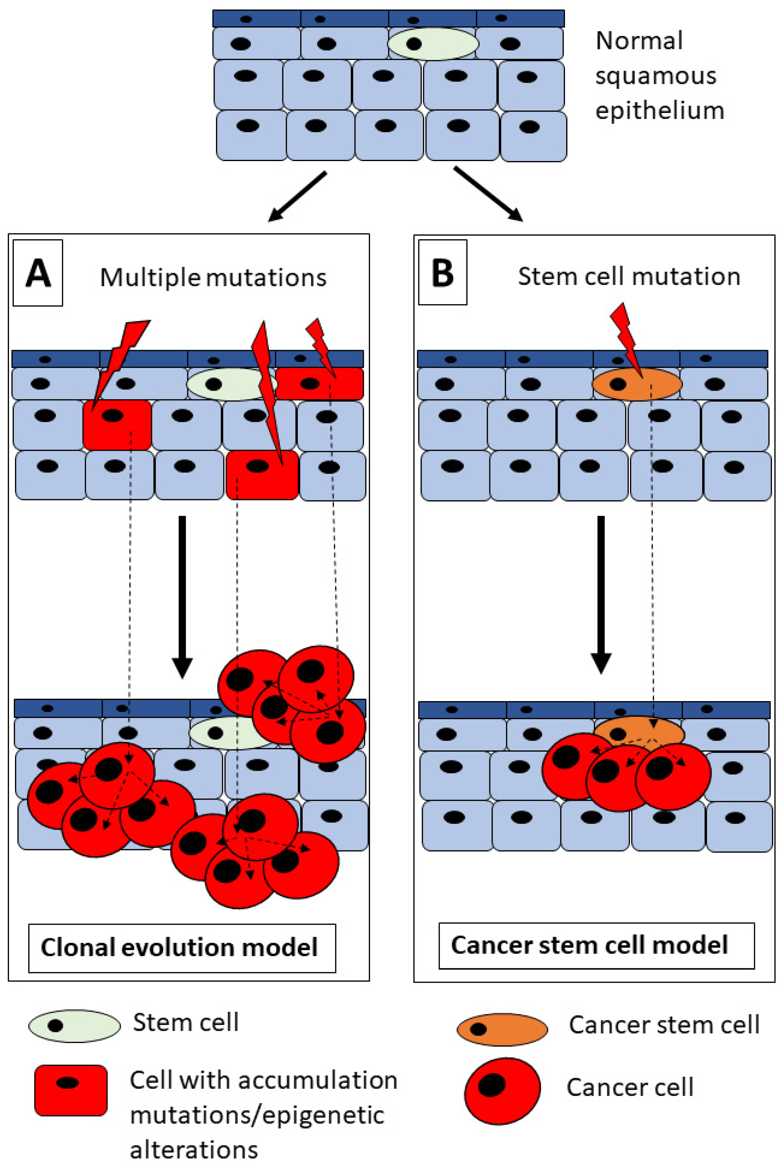
2. HNSCC and Tumorigenesis Model

The poor prognosis of HNSCC is caused by the rapid dissemination of tumor cells to cervical lymph nodes, resistance to chemo/radiotherapy, and a high risk of local tumor recurrence [11]. Generally, initial tumors may be cured by a combination of surgery and radio/chemotherapy, but successful therapy is limited proportionally to the clinical stage of the tumor [6]. However, even if the above therapies are combined, a clinical response is observed in nearly 100% of the early tumors but only in one-third of the advanced tumors [6]. The low response of HNSCC to current treatment modalities is associated with their intratumoral heterogeneity, which reflects differences in cell subpopulations existing in the tumor tissue and explains heterogeneous sensitivity to anticancer therapy [11,12]. This heterogeneity of HNSCC seems to be a result of several independent factors like genetic mutations, environmental impact, or intracellular modifications [13]. As shown in Figure 1, two models of HNSCC tumorigenesis have been proposed that may explain the cellular heterogeneity of HNSCC: the clonal evolution model and the cancer stem cell (CSC) model of tumorigenesis [7,13,14].
The clonal evolution model of tumor development demonstrated by Peter Nowell suggests that cancer is an evolutionary process induced by the growth and expansion of subclones that carry selectively advantageous genetic alterations [15]. The high genetic instability observed in subclones of cancer cells reflects the accumulation of gene mutations [15]. The clonal evolution model of tumor growth suggests that all cells existing in the tumor have the same potential to form tumors, what may explain the progressive growth and resistance to radio/chemotherapy in selected tumors [15,16]. Cell clonal analyses showed that HNSCC progression might be associated with the clonal evolution model of tumor development [16]. Moreover, a genetic sequence comparison of HNSCC tissues from the primary tumor and metastatic focus showed that there are two different patterns of clonal dynamics [13,17]. Nonetheless, published evidence supports the hierarchical model of solid tumor growth [14]. The proposed CSC model suggests that CSCs are the highest in this hierarchy, symmetrically dividing to complete the CSC population, whereas asymmetric division results in daughter cells with a low potential for tumorigenesis [10,13,16]. CSCs present the same properties as normal tissue stem cells, like self-renewal ability, uncontrolled proliferation, and differentiation [6,13]. A hierarchical organization of tumors is the primary difference between the CSC model and the clonal evolution model observed in many solid tumors, including HNSCC [14,16,18]. Although these models are still under study, it was shown in Figure 1 that these two models are considered for HNSCC development because CSCs might undergo clonal evolution during tumor growth under the influence of external factors like hypoxia, nutrient deficiency, or anticancer therapy [14]. In light of this fact, various CSC populations may exist in a single tumor, and their cellular composition has an impact on cancer progression and resistance to chemo/radiotherapy [10,14,19]. Interestingly, Kreso and Dick revealed that differentiated cancer cells forming a tumor (non-CSCs) might acquire stem cell features and thereby transform into “induced” CSCs [19].
It was hypothesized that some types of cancer cells with unique features have been described as CSCs, which become highly malignant, leading to cancer recurrence, metastasis, and resistance to radio/chemotherapy [6,7,12,20]. It was shown that CSCs are a small subpopulation of cancer cells that share common molecular features and express similar biomarkers to embryonic and normal adult stem cells [6,13]. It was postulated that CSCs within the bulk tumor are resistant to conventional anticancer therapies, leading to tumor recurrence [13]. Interestingly, it was noted that CSCs are not an individual cell population but consist of multiple heterogeneous subclones. It is difficult to define which one contributes to metastasis, tumorigenicity, and resistance to chemo/radiotherapy [6,7,12]. These properties were related to the activation of signaling pathways, such as HH, WNT, NOTCH, PI3K, Hippo, or nuclear factor-kappa B (NF-κB), that control detoxification, activate resistance to apoptosis and autophagy, increase the presence of drug transporter proteins, and regulate stem cell transcription factors [10,18]. Many studies imply that it is essential to expand our knowledge about the signaling pathways related to head and neck CSCs in order to develop novel targeted therapies for HNSCC [6,7,10,13,14,18].

3. The HH Signaling Pathway

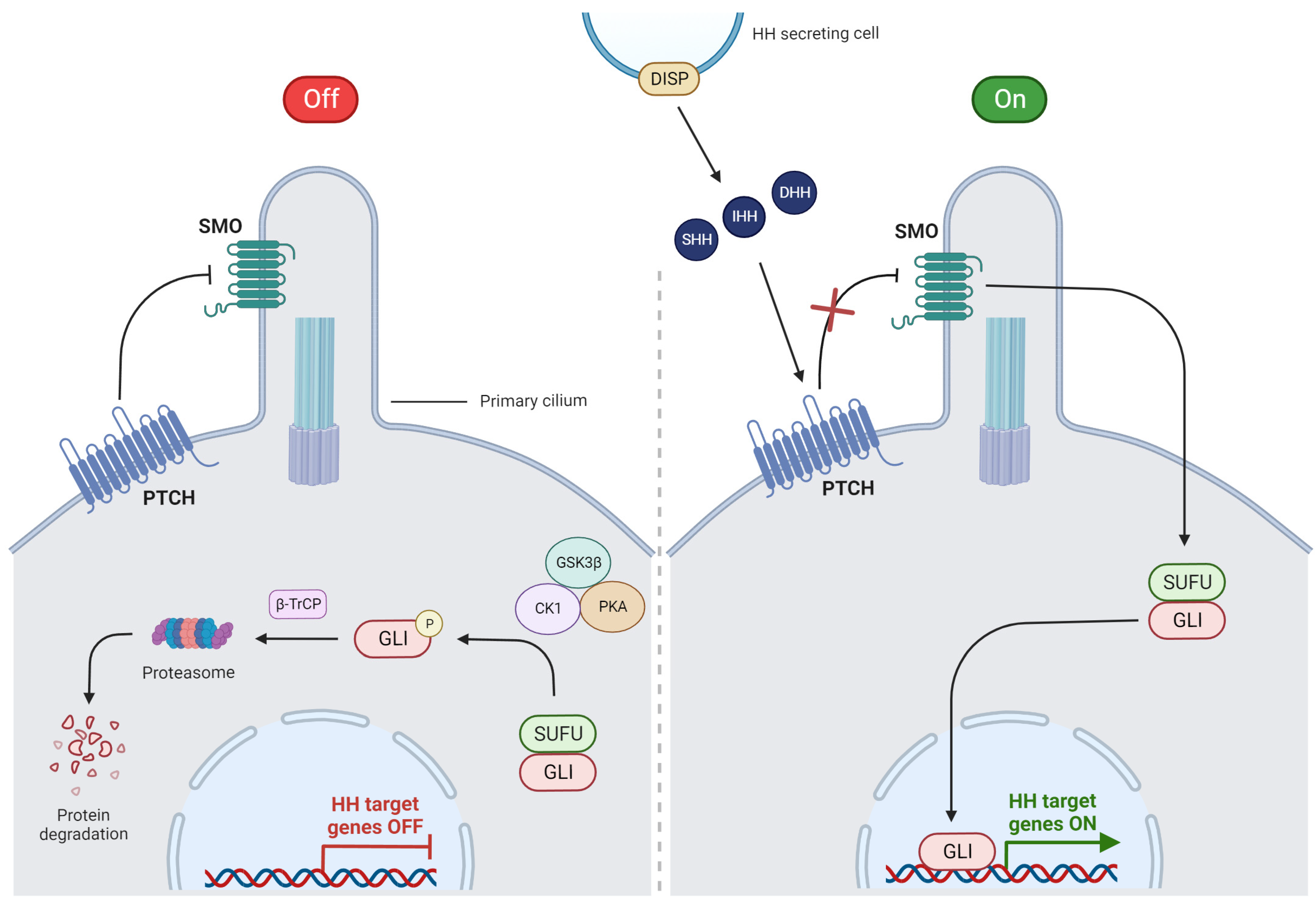
3.1. Overview of HH Signaling

The HH pathway is a strictly conserved evolutionary signaling cascade that controls embryonic development and regulates normal cell growth and differentiation under physiological conditions [21,22]. This pathway also plays a key role in the maintenance of adult stem cells [23]. The HH pathway consists of receptors and ligands that cooperate under this signaling pathway activation [24]. HH is a 45 kDa protein that is the result of the autoproteolytic cleavage of 20 kDa HH ligand precursor proteins. This leads to the production of an N-terminal protein, followed by dual lipid modifications covalently bound to palmitic acid and cholesterol [25]. HH has three gene homologs: Sonic HH (SHH), Desert HH (DHH), and Indian HH (IHH), which express the following proteins: SHH, DHH, and IHH [21,25]. HH proteins are modified post-translationally and released by the secreting cell as HH ligands with the participation of Dispatched (DISP), a transmembrane transporter protein [21,23]. These ligands play distinct roles during embryonic development. SHH is the most widely present HH ligand in vertebrates, and its paracrine effects on adjacent cells are the most common feature of transduction signals. However, HH may also have an autocrine effect [23,24]. SHH is important for neuronal development, whereas IHH is relevant in skeletal development, especially in bone formation. Additionally, DHH is crucial to the gonads’ development [25]. The HH signaling pathway is complex and consists of HH ligands, the 12-transmembrane receptor Patched (PTCH) with two large extracellular loops crucial for HH ligand binding, the 7-transmembrane protein Smoothened (SMO), and the glioma-associated oncogene (GLI) transcriptional factor that is crucial for the production of HH target genes [24,25]. In general, HH signaling might be activated by two different mechanisms: canonical or non-canonical pathways [18,26].

3.2. Canonical Pathway

Activation of HH signaling by the canonical mechanism requires the involvement of the primary cilium with its specialized transmembrane receptors [10]. One of these receptors is PTCH1, which is placed near the primary cilium, specific structure that is exposed on the cell membrane and binds with HH ligands such as SHH, DHH, and IHH [10,27]. When one of these ligands binds to PTCH1, the receptor is internalized, causing accumulation of SMO protein in the primary cilium, which in turn signals the suppressor of fused (SUFU) to release GLI proteins. This permits GLI to translocate into the nucleus in order to start the transcription of HH target genes [10,21,28]. GLI1 is responsible for the genes associated with cell proliferation (cyclins D1, D2, and N-myc) and survival (BCL-2), while GLI2 and GLI3 have repressive functions [10,21,24]. In the absence of HH ligands, the PTCH receptor inhibits SMO protein, and the HH pathway is blocked [10]. SMO is not able to activate GLI, which remains connected with SUFU in the cytoplasm [10]. Accumulating GLI is phosphorylated by a complex of glycogen synthase kinase 3β (GSK3β), casein kinase 1 (CK1), and protein kinase A (PKA) [28]. Then, phosphorylated GLI is detected by β-transducin repeat-containing protein (β-TrCP) and directed to degradation in the proteasome. All processes of the canonical HH pathway are illustrated in Figure 2.

3.3. Non-Canonical Pathway

In the non-canonical mechanism of HH signaling, activation of GLI proteins can be performed without the participation of SMO, but this way is not yet fully understood [10]. Up to now, there are three known types of non-canonical HH pathways [21]. In contrast to the canonical pathway, the non-canonical HH pathway is activated without HH ligands but with the inhibition of PTCH1 (type I) and/or SMO (type II) or through the immediate activation of GLI proteins by other signaling pathways, like a “bypass”, independently from the signaling through the PTCH1/SMO complex (type III) [26]. In the non-canonical HH pathway, GLI1 may be activated by various signaling pathways, and this crosstalk between different signaling pathways seems to be responsible for anticancer treatment failure [27,28]. It was observed that breast CSCs with increased HH signaling gain adaptation to hypoxic conditions and promote breast tumor metastasis [27]. Additionally, the HH pathway crosstalks with several signaling pathways like mTOR, MAPK, and AKT, which activate genes involved in cell survival (BCL-2), and this mechanism was described as promoting HNSCC progression [28]. Other target genes involved in the oncogenic role of the HH pathway are genes controlling cell proliferation (cyclins D1, E1, and N-myc), promoting angiogenesis (components of platelet-derived growth factor (PDGF) and vascular epithelial growth factor (VEGF), and cell migration (components of epithelial-mesenchymal transition (EMT)) [6]. The role of HH signaling in tumor invasion and migration has been confirmed in several experimental studies [12,29]. Dysfunction of many signaling pathways, including HH, WNT, NOTCH, JAK/STAT, PI3K/PTEN, and NF-kB, was found in CSCs [26]. Recent reports suggest that dysfunction of various signaling pathways may provide properties typical for CSCs, like self-renewal and differentiation [16,17,21,25]. The precise mechanisms underlying the function of signaling pathways and their relationships in normal stem cells and CSCs have not yet been fully elucidated [30].

4. HH Pathway in Head and Neck Tissue Formation

HH signaling is originally involved in the proper development of several organs, including head and neck tissues like teeth, lips, palate, and salivary glands [31]. Therefore, dysfunction of the HH pathway during embryogenesis results in a variety of congenital craniofacial deformities, such as holoprosencephaly, cleft lip and palate, and tooth dysplasia [32]. Of the three HH ligands, the most expressed in humans is SHH, which is pivotal in head and neck tissue formation [32].
Orofacial cleft (OFC) is the most common birth defect, with incidence in about 1/500–700 living newborns [33]. Up until now, genetic studies have revealed 24 mutations in PTCH1 in patients with OFC that lead to the loss of PTCH1 functions [33]. Dysregulation of the HH pathway results in incomplete fusion of embryonic facial processes and the creation of cleft lips and palates [34]. An experimental study conducted by Heyne et al. showed that inhibition of HH signaling with vismodegib caused OFC in mice [35].
In teeth, HH signaling plays a key role in physiological morphogenesis by controlling crown and root formation at multiple stages of odontogenesis [36]. It was reported that the inactivation of HH signaling is correlated with tooth malformations. The HH pathway is also considered a regulator of dental stem cells, which may lead to providing new dental therapies based on regenerative medicine [31]. Furthermore, dysregulation of HH signaling is related to the pathogenesis of odontogenic tumors like odontogenic keratocyst (OKC) and ameloblastoma (AB) [37]. Recent reports revealed overexpression of HH pathway components in a high percentage of OKCs and ABs, what indicates the role of HH signaling in the formation of these tumors [38,39]. Based on in vitro studies, these findings suggest the intracystic use of HH inhibitors as an alternative option in the treatment of these tumors [40].

5. Aberration of HH Signaling Pathway in HNSCC

The importance of HH signaling in oncogenesis was first described in 1996 in Gorlin–Goltz syndrome, a rare autosomal dominant disease that is related to a high predisposition to OKC, basal cell carcinoma (BCC), medulloblastoma, and rhabdomyosarcoma [41]. A mutation of the PTCH1 gene was identified by two independent research groups in patients suffering from this syndrome [42,43]. Inhibition of the HH pathway in patients with unresectable BCC was introduced to clinical practice in 2012 with good efficacy [44]. Based on these optimistic findings, aberrant activation of components belonging to the HH pathway was observed in the next few years in other tumors like gliomas, breast, lung, gastric, and prostate cancer [45,46,47,48,49]. However, studies about HH signaling in HNSCC are still rare, especially clinical ones.
The first analysis of the HH pathway in HNSCC was conducted by Schneider et al. They compared the expression of HH proteins in skin squamous cell carcinoma (SSCC) and HNSCC [50]. Schneider observed an association between increased expression of SHH and worse overall survival (OS) in patients with HNSCC [50]. Currently, there are several reports regarding the expression of the HH pathway in HNSCC, mostly using the immunohistochemical method. Their findings and clinical correlations with clinicopathological parameters are collected in Table 1. It is known that HH signaling molecules are significantly overexpressed in HNSCC tissue, contrary to normal mucosa [51,52]. Moreover, Gonzalez et al. identified a significant increase in HH protein expression from normal mucosa, through dysplasia, to HNSCC tissue, what indicates that HH signaling is involved in tumor initiation [53]. Several studies have demonstrated that the HH pathway plays a crucial role in tumor growth and invasion in HNSCC. Therefore, increased expression of SHH, PTCH1, SMO, GLI1, GLI2, and GLI3 was detected significantly more frequently in advanced HNSCC tumors [54,55,56,57,58,59,60,61]. In most cases, progressing HNSCC invades the mandible or maxilla, what leads to bone destruction. It was identified that HH signaling may enhance the development of HNSCC by modulating osteoclast differentiation and osteolysis [62,63]. An experimental study performed by Qiao et al. showed that inhibition of the HH pathway by GDC-0449 (an SMO inhibitor) decreased osteolytic activity induced by HNSCC [63]. Likewise, other authors observed an association between HH expression and lymph node invasion, what may confirm the involvement of HH signaling in HNSCC progression and dissemination [54,55,59,60,64]. Interestingly, the methylation of ZIC4, an important regulator of HH signaling, was correlated with lymph node metastasis in OSCC [65]. The significance of the HH pathway in the promotion of metastasis in HNSCC was supported by Fan et al., who observed a correlation between HH pathway components and EMT [60]. Furthermore, overexpression of GLI1, GLI2, PTCH1, and SMO was correlated with a higher histological grade of the tumor, what may suggest that HH signaling plays a key role in HNSCC malignancy [55,58,66].
The correlation between the expression of HH proteins and prognosis in patients with HNSCC is still not well established. Most studies revealed that increased SHH, PTCH1, SMO, and GLI1 expression are significantly correlated with worse OS [50,57,59,60,67,68]. Contrary to these results, Enzenhofer et al. observed better OS in HPV-negative HNSCC patients with GLI1 and GLI2 overexpression [69]. Additionally, SHH, GLI1, and SMO overexpression were significantly correlated with tumor recurrence and shorter disease-free survival (DFS) [54,67,68]. However, only two of all the studies were confirmed by multivariate COX regression analysis, indicating that the overexpression of GLI1 and SHH may serve as independent poor prognostic biomarkers in HNSCC [57]. Further studies with larger cohorts are required to define the prognostic significance of the HH pathway components in patients with HNSCC.
Table 1. The summary of experimental studies evaluating the clinical significance of Hedgehog pathway components in head and neck squamous cell carcinoma.
HH Protein OverexpressionClinical FindingsAuthor, Year, Reference
SHHWorse OSSchneider et al., 2011 [50]
Lymph node metastasisFan et al., 2014 [60]
Advanced clinical stage, lymph node metastasis, tumor recurrenceHuaitong et al., 2017 [54]
Worse OS, shorter DFSNoman et al., 2020 [67]
Lower pT stageLu et al., 2020 [58]
Poor prognosis, worse OS, advanced pT stageCierpikowski et al., 2021 [57]
PTCH1Lymph node metastasis, worse OSWang et al., 2012 [59]
Higher tumor gradeLeovic et al., 2012 [66]
Advanced pT stage, tumor gradeLu et al., 2020 [58]
SMOLower clinical stageCavicchioli Buim et al., 2011 [70]
Worse OS, shorter DFSRichtig et al., 2019 [68]
Advanced pT stage, higher tumor gradeLu et al., 2020 [58]
Lymph node metastasisSchlaepfer Sales et al., 2021 [64]
GLI1Poor prognosis, regional–distant metastasisChung et al., 2011 [71]
Larger tumor size, tumor recurrence, lymph node metastasis, worse OSWang et al., 2012 [59]
Lower tumor gradeLeovic et al., 2012 [66]
Advanced clinical stage, lymph node metastasis, worse OSFan et al., 2014 [60]
Better OSEnzenhofer et al., 2016 [69]
Advanced clinical stage, lymph node metastasis, tumor recurrenceHuaitong et al., 2017 [54]
Lymph node metastasis, higher tumor gradeChen et al., 2018 [55]
Advanced clinical stageDantas et al., 2021 [61]
GLI2Better OS, longer DFSEnzenhofer et al., 2016 [69]
Advanced clinical stage, higher tumor gradeChen et al., 2018 [55]
GLI3Advanced pT stageRodrigues et al., 2018 [56]
Abbreviations: OS—overall survival, DFS—disease-free survival.

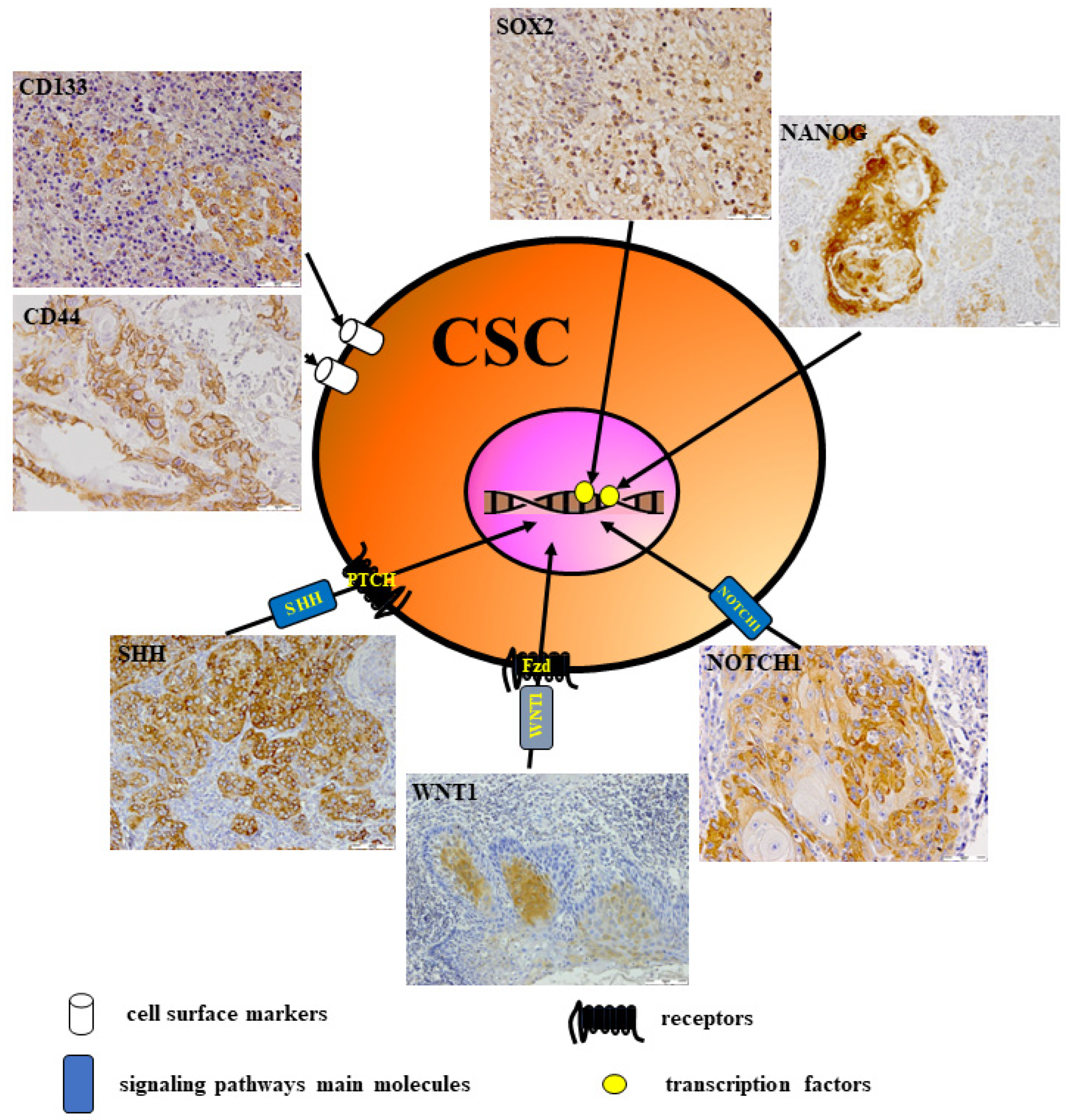
6. CSCs in Head and Neck Cancer

The key reason for the aggressive clinical course of head and neck cancer (HNC) is tumor heterogeneity, which reflects differences within cell subpopulations existing in the tumor tissue [6,7]. It was hypothesized that in HNC, there is a specific type of cancer cells with unique features, defined as CSCs. CSCs are proposed as a promising target for HNC therapy. They may also be a valuable biomarker to detect HNC patients with resistance to therapy or a high risk of tumor recurrence [7,72]. CSCs have been identified as a small population of cancer cells in tumor tissue in terms of 0.01–2% of total tumor mass that have similar features to embryonic and normal stem cells [6,73]. CSCs have asymmetrical cell division and self-renewal capability, which induce a heterogeneous subpopulation of cancer cells involved in angiogenesis and immortality [6,7,73]. It is well known that CSCs are more malignant than other subsets of cancer cells in tumor masses. This heightened malignancy contributes to cancer recurrence, metastases, resistance to chemo/radiotherapy, and quick adaptation to variations in the tumor microenvironment, like nutrient deficiency, hypoxia, or increased DNA repair capacity, thereby avoiding cell apoptosis [6,7,74]. It was found that oncogenic mutations accumulated in somatic stem cells induce the transformation of normal stem cells into CSCs [6,73]. The CSC population has been identified in acute myeloid leukemia (AML) as cells showing CD34 and CD38 expression or co-expression of both markers, CD34/CD38. These cells are able to develop AML when inoculated into immunocompromised mice [75]. A few years later, CSC populations were also found in solid tumors [76,77]. Afterward, CSCs have been widely investigated and identified in various types of solid tumors, including HNC, but their role has not been fully described [6,7,76,78]. Several investigators showed that putative CSCs could be isolated from HNSCC tissue by their unique surface biomarker pattern expression [7,73,76,79]. There are several available techniques for isolating CSCs from tumor tissue [7,72,73,76,80]. Generally, CSCs are separated by the detection of combined specific biomarkers that are located on the normal or CSC surface [6,7,24,81,82,83]. Fluorescence-activated cell sorting (FACS) and magnetic-activated cell sorting (MACS) were the first techniques used for CSC separation, which allow multi-biomarker sorting of stem cells at the same time with high purity and strong specificity [80,84]. Other methods used for CSC separation from tumors are based on cell culture with Hoechst 33342 or the colony-forming ability of CSCs [7,24,78]. So far, multiple markers, independently or in combination, have been documented by many authors as putative CSC markers in various tumors, including HNC [6,7,72,73,76,79]. These lists of putative CSC markers include specific biomolecules such as surface markers, drug transporters, enzymes, and components of several signaling pathways [7,72,73,76,78,81]. The majority of these biomarkers are cell surface and cytoplasmic proteins, sometimes located in nuclei. Therefore, they can be detected by antibodies using flow cytometry or immunohistochemistry [78,81,85]. In many solid tumors, including HNSCC, CSCs have been isolated and identified by the detection of surface proteins such as CD44, CD133, CD90, and CD117 [7,73,76,78,79,81,82,83]. It was noted that expression of stem cell surface biomarkers, e.g., CD133, CD44, CD24, and CD117, is associated with metastasis to lymph nodes, poor prognosis, and resistance to chemo/radiotherapy in patients with HNSCC [76,79,83,86]. Recently, Yu and Cirillo summarized the major CSC markers in HNC. They described evidence for several well-accepted markers of stemness, including CD44, aldehyde dehydrogenase 1 (ALDH1), SOX2, OCT3/4, and NANOG, as reliable means of identifying and isolating CSCs [87]. However, up to now, no specific biomarker of CSC has allowed for the isolation of a pure CSC population from HNSCC, what confirms the fact that the population of CSCs is very heterogeneous [7,73,78,79]. As presented in Figure 3, HNSCC tissue shows expression of different markers that are described as biomarkers expressed by stem cells, as confirmed in our previous studies [88,89].
CD44 is an adhesive, transmembrane glycoprotein located on the surface of normal and HNSCC cells as a receptor that is involved in extracellular matrix (ECM) communication [6,7,78]. In cancer, CD44 participates in cell migration, motility, survival regulation, and stemness maintenance [7]. The presence of CD44 has also been observed in normal epithelial tissue. However, overexpression of CD44 in HNSCC tissue was associated with a high rate of CSC proliferation, self-renewal, and metastasis [90,91]. Recent data showed that CD44 might exist in different variants, playing a distinct biological role in cells, but only its variant, CD44v3, is overexpressed in CSCs and is related to HNSCC progression [86,91]. Similarly, CD44v6 expression was correlated with tumor grade, lymph node invasion, and a worse clinical outcome in HNSCC patients [84]. Therefore, CD44 expression has been considered by some authors as a possible prognostic biomarker in patients with HNSCC [91,92,93]. Another cell surface marker expressed on CSCs in HNSCC is the glycoprotein CD24, which decreases cell adhesion and increases carcinoma cell metastasis [94]. It was noted that CD24+ cells promoted angiogenesis and showed high resistance to chemotherapy in HNSCC [7,78,95,96].
The ALDH proteins are cytosolic enzymes that participate in numerous physiological and pathological processes in embryonic and adult tissue, including tumorigenesis [7,97]. The presence of ALDH1 in CSCs was observed in co-expression with other related markers, such as MMP-9 and SNAIL, what confirms the relationship between ALDH1 and EMT [97,98]. ALDH1+ cells found in HNSCC tissue defined a subset of tumors showing a more tumorigenic phenotype with higher resistance to radio/chemotherapy [97]. Increased expression of ALDH1 in HNSCC tissue samples is correlated with poor prognosis in patients [6,7,78]. The activity of the ALDH1 isoenzymes was markedly upregulated in head and neck CSCs, and ALDH1 was considered a specific CSC biomarker, mainly when co-expressed with CD44 [78,93]. It was observed that ALDH+ cells influence the behavior of oral CSCs and promote a high capability of tumor formation, increasing invasion and chemo/radiotherapy resistance [72].
CD133 expression was detected in normal stem cells and CSCs [6,7,78]. The presence of CD133 was correlated with the high tumorigenicity and invasiveness of HNSCC [6,7,98]. It was found that CD133 overexpression in HNSCC is correlated with tumor grade, lymph node metastasis, and a worse clinical outcome in patients [99,100]. The presence of CD133 correlated with increased proliferation of cells, an EMT phenotype, CSC-sphere formation, and increased in vivo tumorigenicity [74]. CD133+ cell populations showed an increased ability to migrate, invade, form colonies, and resist therapy when compared with CD133-negative populations [98]. It is postulated that CD133 expression might be used as a valuable prognostic biomarker for survival in HNSCC patients [97,98]. Interestingly, it was reported that tumors positive for both CD133 and CD44 showed a more aggressive phenotype when compared to tumors positive for only CD133 or CD44 expression [78].
Head and neck CSCs express the same profile of proteins as those that regulate embryonic stem cells (ESCs), in particular OCT4, SOX2, and NANOG, which were also reported to be expressed in various cancer tissues and were correlated with poor prognosis [7,72,73,76]. These proteins are considered to be specific regulators that determine the stemness properties of CSCs and have an impact on cell behavior, such as self-renewal ability, inhibition of apoptosis, and cell motility [97].
OCT4 (octamer-binding transcription factor 4) has been involved in tumor development, invasion, and metastasis of many tumors, including OSCC [7,73,78]. OCT4 has been overexpressed in various tumors, such as HNSCC, lung, breast, liver, and ovarian cancer [7,73]. High OCT4 expression is associated with a poor survival rate of OSCC and enhanced OSCC progression [6,7,78]. Overexpression of OCT4 was found more frequently in recurrent or metastatic OSCC, what suggests that OCT4 may be a potential marker of aggressive OSCC [95,97]. Growing evidence indicates that OCT4 may be an independent prognostic marker for HNSCC based on its correlation with disease progression [7,78,95,98].
NANOG (Nanog Homeobox) has the ability to inhibit differentiation by maintaining ESC pluripotency, but its overexpression was also observed in HNSCC [73,76,79]. The higher expression of NANOG was reported in precancer and cancer tissue, contrary to the normal mucosa, what suggests its role in HNSCC tumorigenesis [7,73]. Recent data showed that NANOG could not only promote tumor initiation but also increase the formation of CSC colonies in HNSCC [79]. Furthermore, other reports suggested that overexpression of NANOG enhanced EMT and increased resistance to chemo/radiotherapy in HNSCC [96,98]. NANOG was also noted as a potential prognostic factor for OS in HNSCC [98]. Additionally, the increased expression of NANOG is associated with higher tumor grades in different cancers, including HNSCC [7,73]. There are several reports suggesting that NANOG may serve as a regulating factor in the preservation of CSC stemness [7,78].
SOX2 (Sex determining region Y-box 2) is a nuclear transcription factor that plays a crucial role in the development of pluripotent cells [100]. SOX2 induces various signal transduction pathways like tumorigenesis, inhibition of apoptosis, migration, invasion, and resistance to radio/chemotherapy [6,7,88]. Increased transcriptional activity of SOX2 seems to be critical for HNSCC growth and development [78,98]. Furthermore, the silence of SOX2 in head and neck CSCs inhibits their ability to uncontrolled self-renew and tumorigenesis [7]. Chung et al. found that high SOX2 mRNA gene expression in HNSCC indicates that SOX2 might be a prognostic factor for HNSCC patients that can predict the response to radiotherapy [101]. Interesting data were presented by the authors, who showed that co-expression of OCT4 and SOX2 is associated with the initial tumor stage and a longer DFS. It suggests the possibility of a novel targeted therapy option against SOX2 and OCT4 [98,102]. As reported by Fu et al., the downregulation of SOX2 and OCT4 expression in advanced HNSCC indicates that SOX2 and OCT4 overexpression noted in initial tumors might decrease gradually during HNSCC progression [102]. A meta-analysis performed by Fan et al. revealed that the expression of CD133, NANOG, and OCT4 showed prognostic significance in patients with HNSCC [103].
CSCs also possess certain remarkable features that allow them to define their metastatic capabilities, drug resistance, invasiveness, and hierarchical differentiation [7,79]. Several reports noted that adenosine triphosphate (ATP)-binding cassette (ABC) transporters are the main factors in the resistance of CSCs to radio/chemotherapy [6,74,78]. ABC transporters are present in normal cells, but their overexpression in cancer cells increases the chemo/radioresistance of tumors [104]. CSCs efficiently express ABC transporter proteins, such as MDR1/ABCB1, MRP1/ABCC1, and ABCG2, that belong to multidrug resistance proteins and ensure cancer cells’ resistance to anticancer therapy [73,105]. Moreover, it was reported that CSC populations have efflux ability due to the high activity of ABC transporters and ALDH, which decreases oxidative stress and increases resistance to cytotoxic agents [6,24]. Agents inducing DNA damage and apoptosis through chemotherapy or radiotherapy could be inhibited because CSCs protect tumor cells from apoptosis by activating DNA repair [73].
Several signaling pathways in normal stem cells contribute to their proliferation, differentiation, self-renewal, and survival. However, they are abnormally activated or repressed during tumorigenesis and facilitate CSC growth and self-renewal [18,24,90]. These signaling pathways may also induce up- or downstream expression of several genes, including growth factors and apoptotic and anti-apoptotic proteins, in CSCs [24,90]. These signaling pathways are not regulated by a single regulator but by an interwoven network of signaling mediators to regulate CSC growth [18]. The acquisition of CSC features was linked to the activation of various signaling pathways. These pathways include WNT, NOTCH, HH, PI3K, JAK/STAT, Hippo, and NF-κB, which induce resistance to apoptosis, promote the overexpression of drug transporter proteins, and enhance the activation of specific stem cell transcription factors [18]. High activation of several signaling pathways, including WNT, NOTCH, and HH, was observed in our and other studies, what might suggest that CSCs are responsible for resistance to standard therapies in patients with HNSCC [18,88,89,101].

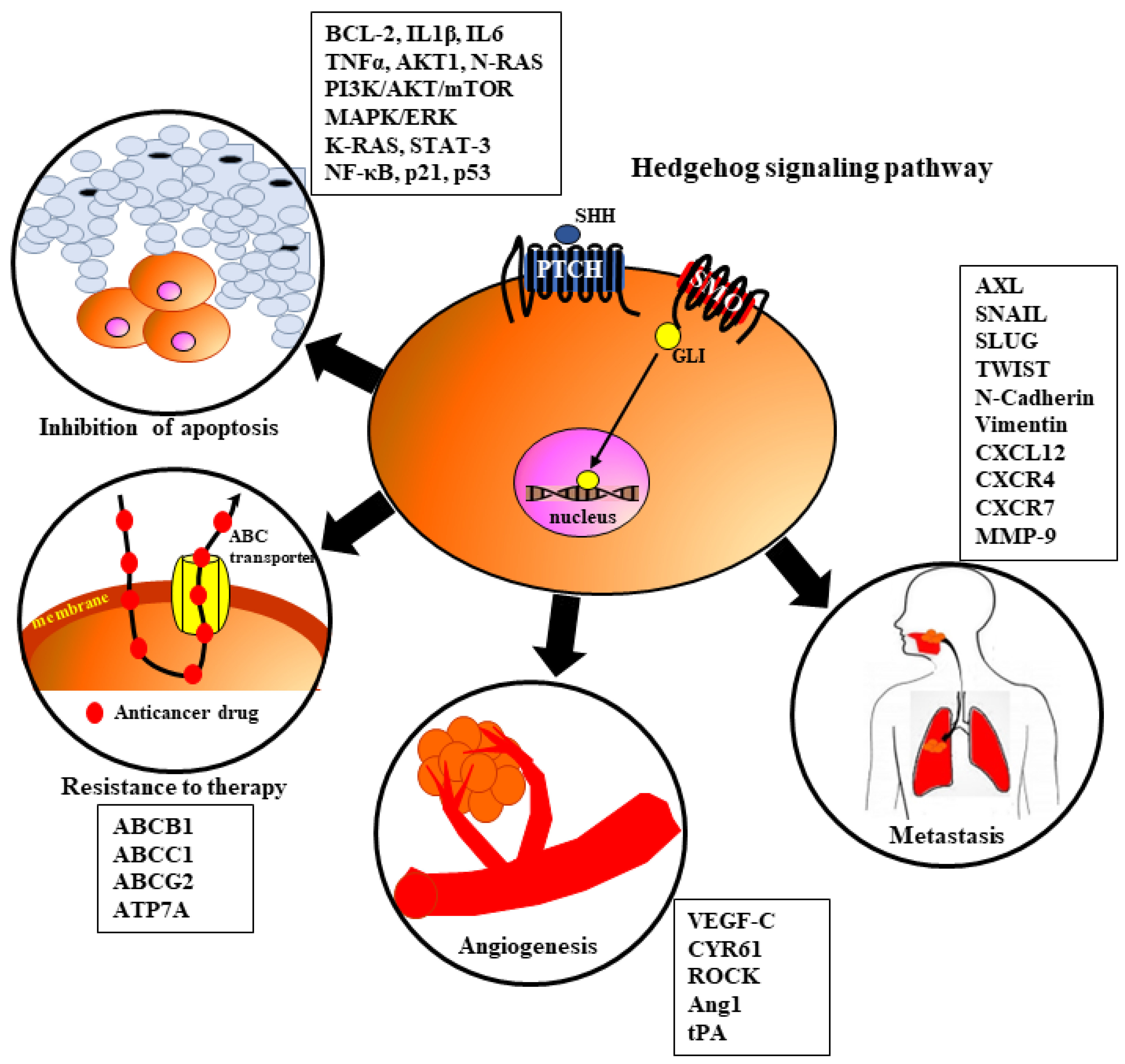
7. The Role of HH Signaling in HNSCC Tumorigenesis

Growing evidence revealed that dysregulation of HH signaling is frequently observed in CSCs [7,9,10,13,18]. Several authors postulated that HH, directly and indirectly, has an impact on the biological behavior of many cancers, including HNSCC, by influencing various processes related to tumorigeneses like angiogenesis, metastasis, resistance to therapy, and inhibition of apoptosis (Figure 4) [21,22,69,106,107,108,109,110,111]. Overactivation of HH signaling was documented in various solid tumors. According to the published report, dysregulation of the HH pathway is associated with the development of one-third of all cancers [22,112]. Upregulation of any component belonging to the HH pathway induces its overactivation and thereby leads to carcinogenesis [21,22,29,112]. As described earlier, there are proposed three mechanisms of aberrant HH signaling activation that could be considered in HNSCC development: type I—linked with ligand-independent mechanisms of HH signaling; type II—connected with ligand-dependent oncogenic HH signaling in autocrine/juxtracrine mechanisms; and type III—associated with ligand-dependent HH signaling in paracrine/reverse mechanisms [21,22]. During HNSCC development, HH signaling might be involved in tumor progression, promote tumor growth, and protect tumor cells against treatment modalities [21]. Genetic mutations of components belonging to HH signaling are commonly observed in HNSCC [21].

7.1. Angiogenesis

Angiogenesis is a key process determining tumor growth and invasion that is balanced by numerous pro-angiogenic and anti-angiogenic factors [113]. Newly formed blood vessels within the tumor support its rapid progression with required nutrients, facilitate the dissemination of cancer cells from the primary site, and reduce the concentration of cytotoxic agents in tumor tissue [113,114]. Endothelial cells (ECs) express the VEGF receptor (VEGFR), which is bound by VEGF-A, leading to the activation of many signaling cascades that play an important role in cell migration and ECM remodeling [115,116]. It is well known that HH, NOTCH, PI3K/AKT, and MAPK/ERK pathways are involved in the activation of ECs to promote angiogenesis in HNSCC [115,116,117]. As presented in Figure 4, HH signaling induces ECs and surrounding tissue to release many proangiogenic proteins (Ang1, Ang2, and VEGF) [116,118]. In 1998, Pepicelli et al. [119] described for the first time the involvement of the HH pathway in embryonic vascular formation. Similarly, Geng et al. [120] found that HH signaling affects vasculogenesis and angiogenesis, especially during embryogenesis, but its role in tumor angiogenesis is still undefined. In addition, HH ligands are involved in blood vessel maturation, integrity, and arterial differentiation [121]. Some authors pointed out that molecular and cellular mechanisms responsible for HH activation in angiogenesis are not well understood, and published results are conflicting [118,121]. There are data showing that hyperactivation of the HH signaling pathway promotes tumor angiogenesis. A potential association between activated HH signaling and high mRNA levels of VEGF-C was found in many cancers [118,121,122]. It was reported that HH signaling influences angiogenesis induced by ECs through the Rho/RhoA and Rho kinase (ROCK) pathway [123]. This observation was confirmed by experimental data, which revealed that inhibition of the HH signaling pathway by GDC-0449 and cyclopamine decreases the vascular density of colon cancer and OSCC xenografts in animal models [106,124]. While it was reported that SHH expression in low-HH-DLD-1 xenografts promotes neoangiogenesis by inducing tumor vascularity, tumors implanted in Hhip+ mice present higher angiogenesis [106,118,125]. There are reports showing that EMT and CSCs share the same molecular cascades that are crucial for tumor progression through the migration of tumor cells and the formation of metastases [113]. Additionally, analysis of genes related to EMT and CSCs revealed similarities in gene expression, including the HH pathway [113].
Upactivation of the HH signaling pathway in cancer cells induces GLI1 and SHH overexpression, which increases the expression of proangiogenic factors, including VEGF-A, MMP-2, MMP-9, heparanase, and cysteine-rich angiogenic inducer 61 (Cyr61), in different tumor cells [118,124,126]. It was suggested that the function of ECs might be regulated directly by HH ligands secreted by tumor cells [124,127]. SHH overexpression was observed in OSCC, whereas expression of PTCH1 and GLI1/2 was noted in the vascular cells in the front of the tumor [124,127]. SHH may induce neoangiogenesis by increasing the expression of proangiogenic proteins in tumors (e.g., VEGF-A) or by promoting EC proliferation by GLI1 overexpression [118]. HH ligands are expressed not only by tumor cells but also by ECs, which are present mostly in OSCCs and gliomas [118,124]. Blockade of endothelial Scube2 and SHH suppresses angiogenesis in OSCC [54,128]. Most data concludes in reporting that hyperactivation of signaling pathways in tumors might enhance angiogenesis. However, there are opposite results that found that inhibition of SMO using IPI-926 (saridegib) increased vessel density in pancreatic cancer [107]. Also, another SMO inhibitor, NVP-LDE225 (erismodegib), restored vessel density in pancreatic ductal adenocarcinoma, decreased pericyte coverage, enhanced vessel permeability, and increased the proportion of immature microvessels [129]. Interesting data were demonstrated by Takabatake et al. [117], who showed the new significance of HH signaling in the development of OSCC tumor vasculature. The authors suggested that HH signaling facilitates tumor invasion not only in an autocrine manner when stimulating the proliferation of tumor cells but also in a paracrine manner when inducing neoangiogenesis [117]. They also indicated that PTCH expression was not present in blood vessels within OSCC [117]. Nonetheless, OSCCs with overexpression of SHH presented PTCH-positive tumor blood vessels, which confirms that SHH signaling has autocrine and paracrine effects on neoangiogenesis [117]. Similar mechanisms were identified in pancreatic cancer by other authors. They showed that HH signaling promotes neoangiogenesis indirectly by increasing PTCH1 and GLI1 expression and inhibiting antiangiogenic molecules such as THBS2 and TIMP2. They also found that HH signaling directly promotes neoangiogenesis by inducing GTPases belonging to the RHO family in the expression of VEGF [110].

7.2. Metastasis

The mechanisms of metastasis induced by HH signaling are understood. However, there are several reports documenting that the HH pathway induces invasion and metastasis through the activation and interaction between different signaling pathways, including HH [117,118,123]. HH promotes metastasis in various solid tumors. Moreover, high activity of the HH, PI3K/AKT/mTOR, and ERK pathways was reported in advanced HNSCC [117,130]. It was found to be a positive association between GLI1 and MMP-9 expression and an inverse correlation between the expression of SHH/GLI1 and E-cadherin in OSCC, which indicates that SHH/GLI-1 is involved in the progression of OSCC and the formation of metastasis [60]. Other studies revealed the role of GLI3 in OSCC metastasis through increased stemness and cell proliferation [56]. The presence of PTCH1, GLI1, and GLI2 protein expression in the invasive front of OSCC might allow cell migration and facilitate metastasis to distant organs [56]. Moreover, PTCH1 expression was frequently observed in advanced stages of HNSCC and was significantly associated with recurrence in patients with OSCC [59].
There are data showing that high upregulation of the HH pathway mediates the function of GLI1, facilitates the CXCL12-induced migration of cancer cells, and increases the metastatic potential of tumor cells [123]. The crucial role of GLI in EMT activation is related to inducing SNAIL expression, what leads to decreased adhesion between cells and influences migration effectiveness [123]. Analysis of the OSCC microenvironment indicated that SHH has an autocrine effect, triggering OSCC invasion. SHH might modulate parenchyma–stromal OSCC by paracrine effect, and both functions of SHH promote metastasis [117].
Downregulation of the GLI family mediates the activation of EMT, leading to the acquisition of a mesenchymal phenotype by epithelial cells [122,123,131]. The loss of cell-cell adhesion and polarity and the gaining of invasive and migratory properties contribute to the aggressive behavior of cancer cells [122,123,131]. HH signaling promotes EMT by decreasing the expression of E-cadherin and increasing the expression of β-catenin and vimentin, thereby enhancing the migration and metastasis of carcinoma cells, which are regulated by factors like SNAIL, SLUG, and TWIST via GLI [131]. There was a positive correlation between HH signaling and SNAIL and MMP-9 expression, as well as a negative correlation with E-cadherin. This suggests that the HH pathway may play a significant role in the progression of HNSCC by regulating EMT [98]. Such a mechanism has been reported in animal models induced by xenografts of gastric adenocarcinoma. In this context, metastatic cells with high expression of GLI2 undergo rapid proliferation through basement tissues, colonize gastric mucosa, and form tumor islands [98,131].

7.3. Resistance to Therapy

The current treatment of patients with HNSCC includes a combination of surgery with radio/chemotherapy, which depends on the tumor location and its clinical stage [83]. About one-third of stage I/II HNSCC patients present good survival rates after surgical resection and chemo or radiotherapy alone [83]. Nonetheless, about two-thirds of HNSCC patients in stages III/IV require a more precise therapeutic strategy [83]. First-line standard chemotherapy contains cisplatin or carboplatin plus 5-fluorouracil (5-FU) and cetuximab, whereas a combined therapy strategy is based on 5-FU, docetaxel, and cisplatin [132,133]. However, the acquisition of chemotherapy resistance is frequently observed in HNSCC patients and leads to tumor recurrence and metastasis [132]. It was reported that there are several mechanisms that allow HNSCC cells to avoid cell death/apoptosis after chemo/radiotherapy. These include DNA repair, drug efflux, apoptosis inhibition, activation of the epidermal growth factor receptor (EGFR), NF-κB, and several signaling pathways [109,111,133]. There are studies indicating that the activity of the HH signaling pathway causes chemo/radioresistance in many tumors, including HNSCC [109,122,134]. Moreover, HH participates in several mechanisms like increased DNA damage repair, EMT, drug efflux, alteration or structural modification of drug target proteins, hypoxia, and reduced apoptosis that induce resistance to therapy [109,135,136]. Several reports found that the upstream and downstream expression of HH components, e.g., GLIs, SHH/IHH, SMO, PTCH1, and SUFU, are involved in the chemo/radioresistance of cancer cells [109,135]. Some evidence implicates that HH is a member of the regulatory mechanism that mediates not only oncogenesis but is also responsible for cancer stem cell resistance to therapy in HNSCC [109,133,135]. Moreover, the association between HH signaling and stemness-related genes, e.g., OCT4 and SOX2, suggests that HH might regulate CSCs’ behavior [137]. This hypothesis was confirmed by other authors, who found a correlation between SHH and SOX2 in OSCC. The subset of tumors with co-expression of both biomarkers showed resistance to standard therapy and a shorter survival time [88].
It is well known that HH signaling controls complex mechanisms of DNA repair machinery in normal and tumor cells [109]. This function of the HH pathway is very important when cisplatin is used in HNSCC patients’ therapy because cisplatin induces DNA damage [109,133,135]. Increased expression of EERC1, a protein associated with nucleotide excision repair (NER), was significantly correlated with resistance to cisplatin in HNSCC patients [138]. Additionally, a new association between HH signaling and DNA repair mechanisms with the participation of O-6-methylguanine-DNA methyltransferase (MGMT) was identified in gliomas [109,139]. Cisplatin therapy may be modulated by epigenetic alterations like hypermethylation of different gene promoters [132]. A similar mechanism of DNA methylation was caused by HH activation [109]. Recent data showed that the promoter CpG islands of the neurofilament light polypeptide (NEFL) gene are involved in resistance to cisplatin therapy in the HNSCC cell line [132]. Initial data presented by Wang et al. [140] showed that HH components like SHH and PTCH1 are expressed in the esophageal cancer tissue of patients with micrometastases after receiving chemoradiotherapy. Based on this observation, the researchers postulated that the HH pathway activation mediates chemo/radiotherapy resistance [140]. Interestingly, expression of SHH, PTCH1, and SMO was significantly increased in HNSCCs that were cured with chemotherapy, in contrast to chemotherapy-naive HNSCCs [58]. Other group results showed that high expression of PTCH1, GLI1, and GLI2 activity was connected with more invasive ability, EMT activity, and increased expression of ABC transporters, what leads to enhanced chemoresistance in many solid tumors [141]. Keysar et al. showed that HNSCC cells that acquired resistance to EGFR inhibition revealed upregulation of GLI1 [142]. On the other hand, authors indicated that HNSCC resistance to cisplatin and cetuximab treatment might result from upregulation of the mesenchymal markers (ZEB2, TWIST1, and SNAIL) by transcription of target genes of the HH pathway, which facilitate acquired resistance [142]. The clear correlation between GLI and ABC transporter proteins such as ABCA2, ABCB1, ABCB4, ABCB7, ABCC2, and ABCG1 was revealed by the use of GLI1 inhibitors, which led to reduced expression of ABC transporters in colorectal cancer cells [143]. The role of HH in the resistance of HNSCC to radiotherapy was confirmed by GLI1 upregulation after irradiation [144]. The association between HH signaling, hypoxia-inducible factor 1-alpha (HIF-1α), and temozolomide (TMZ) treatment was reported in an experimental study [109]. It was documented that hypoxia contributes to resistance to TMZ therapy through HIF-1α/SHH/GLI1-dependent alteration of MGMT [139]. The authors found that GLI mediates the expression of MGMT, which promotes resistance to TMZ [139]. There are results presenting that the HIF-1α/SHH/GLI1 axis is involved in the resistance of HNSCC to therapy and that HIF-1α reduces the response of HNSCC to radiotherapy [78,111,122,145]. There are several pieces of evidence documenting the association between CSCs and their signaling pathways and resistance to chemotherapy [109,113,145]. Head and neck CSCs are able to avoid response to therapy due to their overexpression of ABC drug transporters and activation of several pathways [146]. The NOTCH, HH, WNT/β-catenin, PI3K/AKT/mTOR, TGF-β, and STAT pathways are activated and participate in the drug resistance specific for CSCs [147,148,149]. The CSCs with dysregulated HH signaling induced resistance to chemotherapy when platinum, 5-FU, paclitaxel, and doxorubicin were used [150,151]. Song et al. found that SMO and GLI1 expression in the cisplatin (DDP)-resistant A2780 cells (A2780/DDP) was higher than that in native A2780 cells [134]. It was also reported that HH signaling controls esophageal CSC traits, which may increase the resistance of ESCC to chemotherapy [109]. Some authors found an interaction between the GLI1 transcription factor and the promoter of ABCG2 in gastric CSCs [151]. Similar results were noted by other authors, who found that expression of SHH, GLI2, and ABCB1 was increased in the cisplatin-resistant OC SK-OV-3 cell line. Additionally, decreased ABCB1 expression was noted when GLI2 was inhibited with GANT-61 [152]. Interestingly, Cui et al. revealed that the sensitivity of ESCC to cisplatin was dependent on HH activity, what confirms that HH signaling is a key factor in resistance to anticancer therapy [153]. The authors also found that cisplatin therapy enhanced SHH signaling activation by increasing GLI1 and PTCH1 expression, which normally induces upregulation of ABCB1 to reduce cisplatin enrichment in the ALDH-positive ESCC [153]. The subpopulation of CSCs characterized by high activity of ALDH1 was attributed to the radio- and chemoresistance of CSCs in HNSCC [132].

7.4. Inhibition of Apoptosis

Apoptosis is one of the main human defense mechanisms by which tumor cells are eliminated during anticancer treatment [154]. However, inhibition of apoptosis is one of the specific properties used by HNSCC to resist cisplatin [133]. Inhibitor of apoptosis protein (IAP) is able to block apoptosis by inhibiting caspases, thereby decreasing cleaved caspase proteins, what explains the resistance of oral CSCs to apoptosis after irradiation [99,155]. It was observed that resistance mechanisms, including activation of anti-apoptotic proteins like BCL-2 family members, are frequently observed in HNSCC cells, especially in CSCs [155]. Inhibitors of BCL-2 combined with cetuximab and radiotherapy revealed good results in the elimination of CSCs in HNSCC cell lines [155]. The apoptosis resistance was induced by the reduced cleaved caspase protein of oral CSCs after irradiation [155]. In laryngeal carcinoma, co-expression of BMI1 with CD133 showed promotion of proliferation and anti-apoptotic effects [99,156].

8. The HH Signaling and Viral Infections

There is rising evidence that various viruses target HH signaling to facilitate its transcription, leading to effective and uncontrolled viral replication [117]. The activation of the HH pathway might be destroyed by different viral infections such as HPV, EBV, influenza A (IAV), hepatitis B virus (HBV), hepatitis C virus (HCV), human immunodeficiency virus (HIV), or Kaposi’s sarcoma herpesvirus (KSHV) [123]. It has been found that new vessels are induced by the PDGF-SHH axis. However, this mechanism might be disturbed by KSHV infection, which utilizes the host PDGF receptor (PDGFR) and generates tumorigenesis [157]. Moreover, it is well known that HBV and HCV are involved in the activation of HH signaling in hepatocellular carcinoma (HCC), what contributes to several processes related strictly to HCC tumorigenesis like EMT, maintenance of CSCs, metastasis, and resistance to therapy [158]. Some authors revealed the aberration of HH signaling by HCV in liver fibrosis and HCC but also proposed the possibility of HH inhibitors as a potential target in future therapy [159,160]. Researchers are still investigating the relationship between HCV and HH signaling components to establish the role of both factors in the pathogenesis of HCC [158]. The association between the HH signaling pathway and HPV/E6 protein was confirmed in the murine model of cervical cancer [161]. It has also been found that HPV oncogenes (E6/E7) might increase GLI overexpression in cervical cancer [161].
It is well documented that some of these viruses have oncogenic potential. They play a crucial role in the etiology of HNSCC. Infection with HPV is increasingly common in oropharyngeal squamous cell carcinoma (OPSCC), especially among young non-smokers from developed countries [2,162]. Likewise, EBV infection is associated with nasopharyngeal carcinoma [1]. However, little is known about the associations between HH signaling and HPV and EBV infections in HNSCC. It was postulated that HPV infection might promote carcinogenesis by modifying the stem cell phenotype and transforming oral epithelial stem cells into oral CSCs [73]. GLI1 was proposed as a potential driver in HPV-negative HNSCC [163]. Overexpression of GLI1 and GLI2 has been considered a prognostic parameter for DFS and OS in HPV-negative HNSCC patients undergoing surgical resection and postoperative radiotherapy [69]. Our previous study revealed that some sets of OSCC with SHH expression showed HPV infection, but the relationship has not been statistically significant [57]. Likewise, Richtig et al. did not reveal any correlations between GLI1, SMO, and p16 expression in the HNSCC cohort [68]. Interestingly, such a correlation between p16 and PTCH1 expression was identified by Leovic et al. in OPSCC [66]. Aberrant activation of HH signaling seems to play a significant role in EBV-induced nasopharyngeal carcinoma [158]. It was reported that HH signaling in epithelial cells is activated by EBV, which results in the overexpression of stemness genes [158].

9. Crosstalk between HH Signaling and Other Pathways

Nowadays, it is not surprising that cell signaling pathways strictly influence themselves through activation or suppression [164]. The crosslink between HH and other stem cell pathways like WNT and NOTCH physiologically plays a significant role in embryogenesis and tissue homeostasis. However, when dysregulated, it may lead to tumorigenesis [165]. The development of HNSCC is a multifactorial and multistep process that is related to the aberration of not only HH signaling but also several other pathways [25,166,167]. In this context, inhibition of only one selected signaling pathway in anticancer treatment may be insufficient, what explains rapid tumor recurrence after an initial good clinical response to the therapy of HNSCC [164].
Despite the fact that the crosstalk between HH and other pathways is known, it is still not fully elucidated. HH signaling is closely related to the WNT pathway by producing WNT ligands like WNT2b, WNT4, and WNT7a, which are required for activation of the WNT pathway [164]. It was noted that GLI1-3 proteins and SUFU may regulate WNT signaling by influencing the accumulation of β-catenin [168,169,170]. Recently, GSK-3β was shown to be important for both HH and WNT signaling [171]. In turn, the WNT pathway produces GLI, which is an essential protein for HH signaling [164]. β-catenin was found to influence GLI stability, thereby enhancing or suppressing the HH pathway [172,173].
The previous genetic analysis identified that NOTCH signaling modulates HH pathway activity by inducing key proteins belonging to the HH pathway, like GLI2, GLI3, and SMO [174]. Several studies reported that molecules of NOTCH signaling, such as Jag1, NICD, Maml1, RBPjK, and NOTCH targets like Hes1, may regulate the HH pathway by interaction with the GLI protein [164,175,176,177]. In addition, NOTCH signaling was identified as a key modulator of the HH pathway by modulation of intracellular transport in the primary cilium, which is a crucial structure for the HH cascade [178]. In turn, the HH pathway controls NOTCH signaling through the expression of its ligands [164]. The close relationship between HH and NOTCH pathways may confirm the fact that HH mutations (mostly PTCH1) were identified recently in T-cell lymphoblastic leukemia, which is associated with NOTCH alteration [179].
A better understanding of this complex crosstalk between signaling pathways involved in tumorigenesis is crucial for developing novel targeted therapies for HNSCC. Simultaneous targeting of several signaling pathways may provide more effective treatment for patients with HNSCC, but additional studies are necessary to confirm this hypothesis.

10. Targeting the HH Pathway in HNSCC

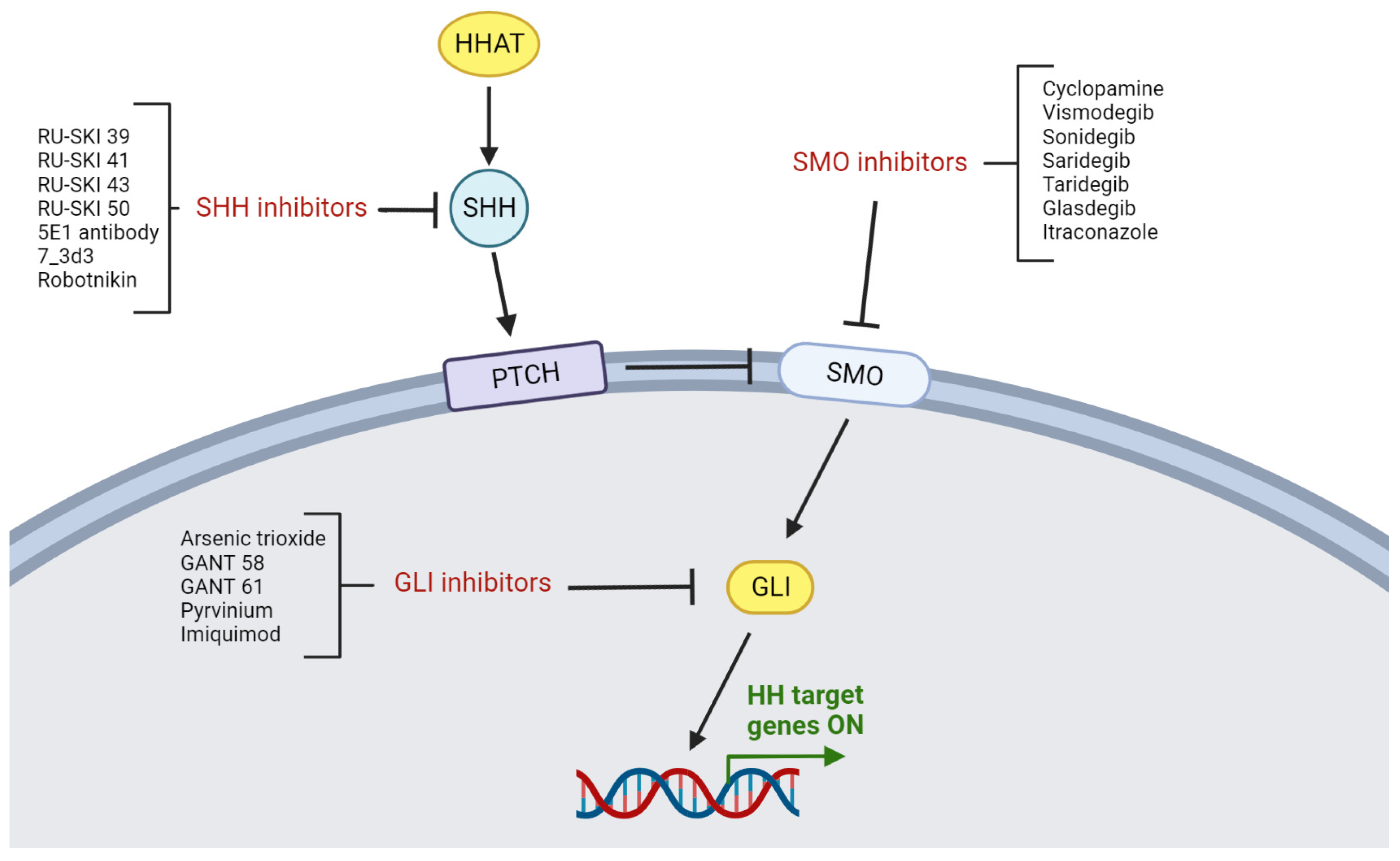
10.1. Characterization of HH Inhibition

The proven significance of HH signaling in a variety of human tumors has led researchers to target this pathway in anticancer therapy. The first discovered HH inhibitor was cyclopamine, which is a steroidal alkaloid that naturally originated from the corn lily [10]. The possible association between corn lily and blockade of HH signaling was observed in sheep that gave birth to lambs with cyclopia [28]. The discovery of cyclopamine contributed to further studies regarding HH signaling. Nonetheless, cyclopamine is not currently used in treatment due to its low solubility and high toxicity [10]. During the last two decades, over 50 HH inhibitors that block signaling pathways at different steps have been introduced into preclinical or clinical studies [9]. Some of them are currently used in the treatment of various tumors. Interestingly, several natural compounds like curcumin and resveratrol inhibit HH signaling [180,181].
As described in Figure 5, the blockade of HH signaling may be performed with the use of inhibitors against specific proteins at each stage of the HH cascade. Based on the above, inhibitors of the HH pathway may be classified as SHH, SMO, and GLI inhibitors.

10.2. SHH Inhibitors

HH ligand inhibitors block the binding of HH ligands to the PTCH receptor, thereby inhibiting HH signaling at the first stage of this complex cascade. It is well-known from various tumors that SHH overexpression is associated with increased HH signaling [27]. Therefore, inhibition of HH ligands may be an attractive target that is currently evaluated in preclinical studies in other tumors [44]. However, there have been no published reports yet on the use of SHH inhibitors in HNSCC.

10.3. SMO Inhibitors

The most common target for HH inhibitors is the SMO protein. The breakthrough moment in studies regarding HH signaling was the acceptance of vismodegib in 2012 by the Food and Drug Administration (FDA) for treating unresectable BCC [44]. In the next few years, the FDA also approved the use of sonidegib in patients with BCC and glasdegib in patients with AML [10]. Based on this success, many SMO inhibitors were developed and studied in various tumors, including HNSCC. Nonetheless, one of the crucial difficulties in anticancer therapy with SMO inhibitors is the development of drug resistance, which limits their long-term efficacy [9,10].
The positive effect of HH inhibition in HNSCC was first observed by Mozet et al., who studied the influence of cyclopamine, cisplatin, and docetaxel on HNSCC cell colony formation [182]. It was reported that HH inhibition by cyclopamine suppresses colony formation in HNSCC. Furthermore, cyclopamine may increase the chemosensitivity of HNSCC cells when combined in therapy with cisplatin or docetaxel [182]. Similar observations were noted by Gan et al., who revealed that inhibition of HH signaling with cyclopamine enhances HNSCC sensitivity to radiotherapy [183]. The influence of HH inhibition on radiotherapy sensitization in HNSCC was confirmed by Hehlgans et al. [184]. Vismodegib downregulated the HH pathway and sensitized HNSCC cells to radiotherapy [184]. Additionally, it was found that vismodegib decreases the expression of GLI1 in HNSCC cell lines [184]. Recently, the use of vismodegib and itraconazole was also studied in OSCC cell lines, which induced apoptosis and altered the morphology of tumor cells [185]. Based on the encouraging results of the in vitro study, Freitas et al. suggested that SMO inhibitors, such as vismodegib and itraconazole, may be considered for potential future OSCC therapy. Therefore, further clinical trials of HH inhibitors in OSCC patients are required [185].
Downregulation of the HH pathway in HNSCCs after the use of SMO inhibitors was represented by decreased expression of HH components in several preclinical studies [183,184,185]. Considering these promising results, more in vivo studies are necessary in order to verify its efficacy in clinical practice. It is well known from other solid tumors that the initial successful effect of SMO inhibitors is often reduced over time by drug resistance development [10]. Therefore, it is currently postulated to simultaneously inhibit different signaling pathways in order to overcome therapeutic resistance in tumors [186]. Based on the above, the concept of dual targeting of the HH and EGFR pathways in HNSCC has been proposed by several researchers. They reported favorable effects of such combined therapy with the use of HH and EGFR inhibitors in experimental studies [142,187,188]. The dual inhibition of EGFR and HH pathways by cetuximab and vismodegib in combination with cisplatin and docetaxel decreased the cell proliferation and colony-forming ability of HNSCC cells [187]. Likewise, there are studies reporting that blockade of the mTOR pathway increased the effect of HH inhibition in esophageal cancer and HNSCC [183,189].
Recently, interesting results were reported by Kleszcz et al., who presented a combined therapy with inhibitors of several signaling pathways in HNSCC [190]. They analyzed the therapeutic effects caused by selected inhibitors of the following signaling pathways, such as HH (vismodegib), WNT/β-catenin (PRI-724), PI3K (HS-173), and EGFR (erlotinib), separately and in combination on the tongue (CAL 27) and hypopharynx (FaDu) cancer cell lines [190]. The study showed a better response of the hypopharynx than tongue cell lines to the dual inhibition of WNT and HH signaling [190]. A similar effect was observed when simultaneous inhibition with PRI-724 and erlotinib or PRI-724 and the PI3K inhibitor (HS-173) was used [190]. Moreover, the authors postulated that simultaneous inhibition of the WNT and HH pathways decreased CAL 27 and FaDu cell migration and had better anti-proliferative than pro-apoptotic effects. In contrast, PRI-724 combined with erlotinib or HS-173 significantly increased the apoptosis of cell populations, but its individual usage showed lower apoptotic efficacy [190]. Currently, it is widely postulated that a multitherapeutic approach with several anticancer drugs may provide a better response than single therapy, even when molecularly targeted [191]. Therefore, numerous clinical studies in other tumors have been performed that study the efficacy of treatment with HH inhibitors in combination with standard chemotherapy or other targeted therapies [191].
Up to now, only two clinical trials have evaluated SMO inhibitors in HNSCC. The first survey performed by Bowles et al. (ID NCT01255800) studied the combination of IPI-926 (saridegib) with cetuximab in recurrent or metastatic HNSCC patients [192]. This clinical study demonstrated that IPI-926 may be safely combined with cetuximab in HNSCC patients [192]. The dual blockade of EGFR and HH signaling showed tolerable toxicity [192]. Moreover, tumor biopsies during the study revealed that clinical response to therapy was associated with the suppression of EGFR and HH genes [192]. Nonetheless, partial clinical response to treatment was seen only in 12.5% of patients with HNSCC [192]. The authors suggested that inhibition of the HH pathway may increase EGFR signaling, what may indicate that the dosage of IPI-926 enhances response to cetuximab in HNSCC [192]. This observation is in agreement with previous studies showing that HH signaling is a negative regulator of the EGFR signaling pathway and that inhibition of the HH pathway sensitizes tumor cells to EGFR inhibition [142]. On the other hand, in the author’s opinion, the lack of responses in cetuximab-pre-treated patients indicated that HH inhibition may not be enough to convert EGFR-resistant tumors back into EGFR-sensitive tumors. Therefore, HH inhibitors should be used prior to cetuximab in order to increase EGFR-dependent cetuximab sensitivity [192]. However, the experimental data showed that this effect is different in HPV-positive and HPV-negative HNSCCs, and the combination of EGFR and HH inhibition was more effective in HPV-negative tumors [142]. However, the clinical pilot study performed by Bowles et al. had several limitations. The studied group of patients was very limited (n = 9) and heterogeneous (different HPV status, cetuximab-naïve, and cetuximab-pre-treated patients). Therefore, the authors concluded that further exploration of this combined therapy is recommended in a more numerous and homogenous cohort [192].
The revealed discrepancy between in vitro and in vivo studies might be a result of both genetic and epigenetic alterations leading to persistent GLI activation that allows tumors to evade inhibition of SMO. This contributes to the development of resistance to therapy and rapid tumor recurrence [191]. A recent report by Yao et al. showed that both JNK/AP-1 and TGFβ/Smad3 pathways simultaneously activate the nuclear myocardin-related transcription factor (nMRTF) that connects with SRF and serves as a coactivator for GLI1 in resistant BCC [193]. It is currently well known that various signaling pathways like WNT, MAPK, NF-κB, PI3K, and TGFβ may activate GLI, what causes that the inhibition of the HH pathway with SMO inhibitors not to be fully effective [191].
The second clinical trial conducted by Mayo Clinic (ID NCT04007744) is still recruiting and has not been completed. This phase I clinical trial evaluates the combined use of sonidegib and pembrolizumab in various tumors, including metastatic HNSCC. However, the results are not yet available.

10.4. GLI Inhibitors

The GLI transcription factor is a final protein of the HH pathway, which implies that it is directly involved in HH target gene expression. Therefore, inhibition of GLI seems to be a promising and more effective target for anticancer drugs, contrary to other HH proteins. To date, several GLI inhibitors have been developed, but only a few of them have been tested in vitro in HNSCCs. The most studied GLI antagonist is arsenic trioxide (ATO), which was approved in 2000 by the FDA for the treatment of acute promyelocytic leukemia [194]. However, the anticancer effectiveness of ATO was also tested in solid tumors, including HNSCC [28,195]. The latest in vitro study on OSCC cell lines conducted by Nogueira et al. noted that ATO is a promising cytotoxic agent in OSCC [196]. Treatment with ATO on OSCC cells resulted in reduced GLI1 expression, alterations in the cells’ morphology, nuclear fragmentation, and increased apoptosis [196]. Nonetheless, the main problem is the fact that high doses of ATO are required to effectively inhibit in vivo solid tumors, which in turn causes strong side effects [194]. Based on these findings, current studies are focused on combined therapies with low doses of ATO and other cytotoxic drugs. It was reported in several in vitro studies that ATO may enhance sensitivity to cisplatin and increase their synergistic activity [194,197,198]. Furthermore, other studies suggest that ATO may increase the radiosensitivity of HNSCC [199].
Similar observations were reported in studies evaluating the influence of other GLI inhibitors—GANT61—on OSCC cell lines [200,201]. In vitro experiments showed that GANT61 decreases GLI expression and promotes apoptosis in OSCC cell lines [201]. Likewise, Zubčić et al. observed inhibited proliferation and growth of HNSCC cell lines in response to treatment with GANT61 and lithium chloride, a GSK3β inhibitor [202]. However, none of the GLI inhibitors have been evaluated in clinical studies with the participation of patients suffering from HNSCC.

11. Future Perspectives and Conclusions

To our knowledge, this is the first comprehensive review regarding the HH pathway in HNSCC. We made an effort to collect and summarize recent findings about HH signaling in HNSCC and pointed out the role of this pathway in the biology of HNSCC and its clinical outcome. Based on the latest studies, we may confirm that the HH pathway is aberrantly activated in advanced HNSCC; however, the clinical studies are very limited. As we summarized in this paper, HH signaling is involved in several crucial events during HNSCC oncogenesis, like tumor initiation, neoangiogenesis, cancer invasion, resistance to chemo/radiotherapy, recurrence, and metastasis. Furthermore, increased expression of proteins belonging to the HH pathway is correlated with worse survival and a poor prognosis in HNSCC patients, what may suggest the prognostic significance of HH signaling in HNSCC. Given the present lack of molecularly targeted therapies for patients with HNSCC and low survival rates, inhibition of HH signaling might be a promising intervention in future HNSCC therapies. Therefore, further clinical studies evaluating the efficacy and safety of HH inhibitors in HNSCC treatment are urgently needed. However, due to the strict and complex crosstalk between HH and other signaling pathways, it seems that combined therapy with simultaneous inhibition of several tumorigenic pathways may be required to provide breakthroughs in HNSCC treatment.

Author Contributions

Conceptualization, P.C.; writing—original draft preparation, P.C., A.L. and J.B.; writing—review and editing, P.C. and J.B.; visualization, P.C.; supervision, J.B. All authors have read and agreed to the published version of the manuscript.

Funding

This research received no external funding.

Institutional Review Board Statement

Not applicable.

Informed Consent Statement

Not applicable.

Data Availability Statement

Not applicable.

Acknowledgments

The authors would like to thank Piotr Grelewski for his help in preparing the figures.

Conflicts of Interest

The authors declare no conflict of interest.

References

  1. Johnson, D.E.; Burtness, B.; Leemans, C.R.; Lui, V.W.Y.; Bauman, J.E.; Grandis, J.R. Head and Neck Squamous Cell Carcinoma. Nat. Rev. Dis. Primers 2020, 6, 92. [Google Scholar] [CrossRef]
  2. Amaral, M.N.; Faísca, P.; Ferreira, H.A.; Gaspar, M.M.; Reis, C.P. Current Insights and Progress in the Clinical Management of Head and Neck Cancer. Cancers 2022, 14, 6079. [Google Scholar] [CrossRef]
  3. Du, E.; Mazul, A.L.; Farquhar, D.; Brennan, P.; Anantharaman, D.; Abedi-Ardekani, B.; Weissler, M.C.; Hayes, D.N.; Olshan, A.F.; Zevallos, J.P. Long-Term Survival in Head and Neck Cancer: Impact of Site, Stage, Smoking, and Human Papillomavirus Status. Laryngoscope 2019, 129, 2506–2513. [Google Scholar] [CrossRef] [PubMed]
  4. Goel, B.; Tiwari, A.K.; Pandey, R.K.; Singh, A.P.; Kumar, S.; Sinha, A.; Jain, S.K.; Khattri, A. Therapeutic Approaches for the Treatment of Head and Neck Squamous Cell Carcinoma–An Update on Clinical Trials. Transl. Oncol. 2022, 21, 101426. [Google Scholar] [CrossRef] [PubMed]
  5. Min, H.-Y.; Lee, H.-Y. Molecular Targeted Therapy for Anticancer Treatment. Exp. Mol. Med. 2022, 54, 1670–1694. [Google Scholar] [CrossRef] [PubMed]
  6. Baniebrahimi, G.; Mir, F.; Khanmohammadi, R. Cancer Stem Cells and Oral Cancer: Insights into Molecular Mechanisms and Therapeutic Approaches. Cancer Cell Int. 2020, 20, 113. [Google Scholar] [CrossRef] [PubMed]
  7. Tahmasebi, E.; Alikhani, M.; Yazdanian, A.; Yazdanian, M.; Tebyanian, H.; Seifalian, A. The Current Markers of Cancer Stem Cell in Oral Cancers. Life Sci. 2020, 249, 117483. [Google Scholar] [CrossRef] [PubMed]
  8. Nüsslein-Volhard, C.; Wieschaus, E. Mutations Affecting Segment Number and Polarity in Drosophila. Nature 1980, 287, 795–801. [Google Scholar] [CrossRef]
  9. Jiang, J. Hedgehog Signaling Mechanism and Role in Cancer. Semin. Cancer Biol. 2022, 85, 107–122. [Google Scholar] [CrossRef]
  10. Nguyen, N.M.; Cho, J. Hedgehog Pathway Inhibitors as Targeted Cancer Therapy and Strategies to Overcome Drug Resistance. Int. J. Mol. Sci. 2022, 23, 1733. [Google Scholar] [CrossRef]
  11. Ouban, A. SALL4 Stemness Agent Expression in Oral Squamous Cell Cancer and Its Clinical Significance. Biotechnol. Biotechnol. Equip. 2021, 35, 665–673. [Google Scholar] [CrossRef]
  12. Batlle, E.; Clevers, H. Cancer Stem Cells Revisited. Nat. Med. 2017, 23, 1124–1134. [Google Scholar] [CrossRef]
  13. Chen, D.; Wang, C.-Y. Targeting Cancer Stem Cells in Squamous Cell Carcinoma. Precis. Clin. Med. 2019, 2, 152–165. [Google Scholar] [CrossRef]
  14. Song, S.Y.; Seo, D. Cancer Stem Cells: Biological Features and Targeted Therapeutics. Hanyang Med. Rev. 2015, 35, 250–257. [Google Scholar] [CrossRef]
  15. Nowell, P.C. The Clonal Evolution of Tumor Cell Populations. Science 1976, 194, 23–28. [Google Scholar] [CrossRef] [PubMed]
  16. Cameron, S.R.; Dahler, A.L.; Endo-Munoz, L.B.; Jabbar, I.; Thomas, G.P.; Leo, P.J.; Poth, K.; Rickwood, D.; Guminski, A.; Saunders, N.A. Tumor-Initiating Activity and Tumor Morphology of HNSCC Is Modulated by Interactions between Clonal Variants within the Tumor. Lab. Investig. 2010, 90, 1594–1603. [Google Scholar] [CrossRef] [PubMed]
  17. Melchardt, T.; Magnes, T.; Hufnagl, C.; Thorner, A.R.; Ducar, M.; Neureiter, D.; Tränkenschuh, W.; Klieser, E.; Gaggl, A.; Rösch, S.; et al. Clonal Evolution and Heterogeneity in Metastatic Head and Neck Cancer-An Analysis of the Austrian Study Group of Medical Tumour Therapy Study Group. Eur. J. Cancer 2018, 93, 69–78. [Google Scholar] [CrossRef]
  18. Espinosa-Sánchez, A.; Suárez-Martínez, E.; Sánchez-Díaz, L.; Carnero, A. Therapeutic Targeting of Signaling Pathways Related to Cancer Stemness. Front. Oncol. 2020, 10, 1533. [Google Scholar] [CrossRef] [PubMed]
  19. Kreso, A.; Dick, J.E. Evolution of the Cancer Stem Cell Model. Cell Stem Cell 2014, 14, 275–291. [Google Scholar] [CrossRef]
  20. Notarstefano, V.; Belloni, A.; Sabbatini, S.; Pro, C.; Orilisi, G.; Monterubbianesi, R.; Tosco, V.; Byrne, H.J.; Vaccari, L.; Giorgini, E. Cytotoxic Effects of 5-Azacytidine on Primary Tumour Cells and Cancer Stem Cells from Oral Squamous Cell Carcinoma: An In Vitro FTIRM Analysis. Cells 2021, 10, 2127. [Google Scholar] [CrossRef]
  21. Doheny, D.; Manore, S.G.; Wong, G.L.; Lo, H.-W. Hedgehog Signaling and Truncated GLI1 in Cancer. Cells 2020, 9, 2114. [Google Scholar] [CrossRef] [PubMed]
  22. Skoda, A.M.; Simovic, D.; Karin, V.; Kardum, V.; Vranic, S.; Serman, L. The Role of the Hedgehog Signaling Pathway in Cancer: A Comprehensive Review. Bosn. J. Basic Med. Sci. 2018, 18, 8–20. [Google Scholar] [CrossRef] [PubMed]
  23. Petrova, R.; Joyner, A.L. Roles for Hedgehog Signaling in Adult Organ Homeostasis and Repair. Development 2014, 141, 3445–3457. [Google Scholar] [CrossRef] [PubMed]
  24. Yang, L.; Shi, P.; Zhao, G.; Xu, J.; Peng, W.; Zhang, J.; Zhang, G.; Wang, X.; Dong, Z.; Chen, F.; et al. Targeting Cancer Stem Cell Pathways for Cancer Therapy. Signal Transduct. Target. Ther. 2020, 5, 8. [Google Scholar] [CrossRef] [PubMed]
  25. Patni, A.P.; Harishankar, M.K.; Joseph, J.P.; Sreeshma, B.; Jayaraj, R.; Devi, A. Comprehending the Crosstalk between Notch, Wnt and Hedgehog Signaling Pathways in Oral Squamous Cell Carcinoma—Clinical Implications. Cell. Oncol. 2021, 44, 473–494. [Google Scholar] [CrossRef] [PubMed]
  26. Po, A.; Silvano, M.; Miele, E.; Capalbo, C.; Eramo, A.; Salvati, V.; Todaro, M.; Besharat, Z.M.; Catanzaro, G.; Cucchi, D.; et al. Noncanonical GLI1 Signaling Promotes Stemness Features and in Vivo Growth in Lung Adenocarcinoma. Oncogene 2017, 36, 4641–4652. [Google Scholar] [CrossRef] [PubMed]
  27. Niyaz, M.; Khan, M.S.; Mudassar, S. Hedgehog Signaling: An Achilles’ Heel in Cancer. Transl. Oncol. 2019, 12, 1334–1344. [Google Scholar] [CrossRef]
  28. Girardi, D.; Barrichello, A.; Fernandes, G.; Pereira, A. Targeting the Hedgehog Pathway in Cancer: Current Evidence and Future Perspectives. Cells 2019, 8, 153. [Google Scholar] [CrossRef]
  29. Riquelme, I.; Roa, J.C. Activated Status of Hedgehog Pathway in Oral Squamous Cell Carcinoma (OSCC): The Door Is Still Open. Transl. Cancer Res. 2016, 5, 14–16. [Google Scholar] [CrossRef]
  30. Matsui, W.H. Cancer Stem Cell Signaling Pathways. Medicine 2016, 95, S8. [Google Scholar] [CrossRef]
  31. Hosoya, A.; Shalehin, N.; Takebe, H.; Shimo, T.; Irie, K. Sonic Hedgehog Signaling and Tooth Development. Int. J. Mol. Sci. 2020, 21, 1587. [Google Scholar] [CrossRef]
  32. Abramyan, J. Hedgehog Signaling and Embryonic Craniofacial Disorders. J. Dev. Biol. 2019, 7, 9. [Google Scholar] [CrossRef] [PubMed]
  33. He, Q.; Hao, X.; Bao, S.; Wu, X.; Xu, L.; Hou, Y.; Huang, Y.; Peng, L.; Huang, H.; Ding, Y.; et al. A392V and R945X Mutations Cause Orofacial Clefts via Impairing PTCH1 Function. Genomics 2022, 114, 110507. [Google Scholar] [CrossRef] [PubMed]
  34. Kurosaka, H. The Roles of Hedgehog Signaling in Upper Lip Formation. BioMed Res. Int. 2015, 2015, 901041. [Google Scholar] [CrossRef] [PubMed]
  35. Heyne, G.W.; Melberg, C.G.; Doroodchi, P.; Parins, K.F.; Kietzman, H.W.; Everson, J.L.; Ansen-Wilson, L.J.; Lipinski, R.J. Definition of Critical Periods for Hedgehog Pathway Antagonist-Induced Holoprosencephaly, Cleft Lip, and Cleft Palate. PLoS ONE 2015, 10, e0120517. [Google Scholar] [CrossRef] [PubMed]
  36. Seppala, M.; Fraser, G.J.; Birjandi, A.A.; Xavier, G.M.; Cobourne, M.T. Sonic Hedgehog Signaling and Development of the Dentition. J. Dev. Biol. 2017, 5, 6. [Google Scholar] [CrossRef]
  37. Mishra, P.; Panda, A.; Bandyopadhyay, A.; Kumar, H.; Mohiddin, G. Sonic Hedgehog Signalling Pathway and Ameloblastoma—A Review. J. Clin. Diagn. Res. 2015, 9, ZE10–ZE13. [Google Scholar] [CrossRef]
  38. Gurgel, C.A.S.; Buim, M.E.C.; Carvalho, K.C.; Sales, C.B.S.; Reis, M.G.; de Souza, R.O.; de Faro Valverde, L.; de Azevedo, R.A.; Dos Santos, J.N.; Soares, F.A.; et al. Transcriptional Profiles of SHH Pathway Genes in Keratocystic Odontogenic Tumor and Ameloblastoma. J. Oral Pathol. Med. 2014, 43, 619–626. [Google Scholar] [CrossRef]
  39. Rodrigues, K.S.; Santos, H.B.d.P.; de Morais, E.F.; Freitas, R.d.A. Immunohistochemical Analysis of SHH, SMO and GLI-1 Proteins in Epithelial Odontogenic Lesions. Braz. Dent. J. 2022, 33, 91–99. [Google Scholar] [CrossRef]
  40. Zhai, J.; Zhang, H.; Zhang, J.; Zhang, R.; Hong, Y.; Qu, J.; Chen, F.; Li, T. Effect of the Sonic Hedgehog Inhibitor GDC-0449 on an in Vitro Isogenic Cellular Model Simulating Odontogenic Keratocysts. Int. J. Oral Sci. 2019, 11, 4. [Google Scholar] [CrossRef]
  41. Okamoto, N.; Naruto, T.; Kohmoto, T.; Komori, T.; Imoto, I. A Novel PTCH1 Mutation in a Patient with Gorlin Syndrome. Hum. Genome Var. 2014, 1, 14022. [Google Scholar] [CrossRef]
  42. Hahn, H.; Wicking, C.; Zaphiropoulous, P.G.; Gailani, M.R.; Shanley, S.; Chidambaram, A.; Vorechovsky, I.; Holmberg, E.; Unden, A.B.; Gillies, S.; et al. Mutations of the Human Homolog of Drosophila Patched in the Nevoid Basal Cell Carcinoma Syndrome. Cell 1996, 85, 841–851. [Google Scholar] [CrossRef]
  43. Johnson, R.L.; Rothman, A.L.; Xie, J.; Goodrich, L.V.; Bare, J.W.; Bonifas, J.M.; Quinn, A.G.; Myers, R.M.; Cox, D.R.; Epstein, E.H.; et al. Human Homolog of Patched, a Candidate Gene for the Basal Cell Nevus Syndrome. Science 1996, 272, 1668–1671. [Google Scholar] [CrossRef] [PubMed]
  44. Gampala, S.; Yang, J.-Y. Hedgehog Pathway Inhibitors against Tumor Microenvironment. Cells 2021, 10, 3135. [Google Scholar] [CrossRef] [PubMed]
  45. Zhao, H.; Tang, H.; Xiao, Q.; He, M.; Zhao, L.; Fu, Y.; Wu, H.; Yu, Z.; Jiang, Q.; Yan, Y.; et al. The Hedgehog Signaling Pathway Is Associated with Poor Prognosis in Breast Cancer Patients with the CD44+/CD24− Phenotype. Mol. Med. Rep. 2016, 14, 5261–5270. [Google Scholar] [CrossRef] [PubMed]
  46. Lim, S.; Lim, S.M.; Kim, M.-J.; Park, S.Y.; Kim, J.-H. Sonic Hedgehog Pathway as the Prognostic Marker in Patients with Extensive Stage Small Cell Lung Cancer. Yonsei Med. J. 2019, 60, 898. [Google Scholar] [CrossRef] [PubMed]
  47. Jian-Hui, C.; Er-Tao, Z.; Si-Le, C.; Hui, W.; Kai-Ming, W.; Xin-Hua, Z.; Chuang-Qi, C.; Shi-Rong, C.; Yu-Long, H. CD44, Sonic Hedgehog, and Gli1 Expression Are Prognostic Biomarkers in Gastric Cancer Patients after Radical Resection. Gastroenterol. Res. Pract. 2016, 2016, 1013045. [Google Scholar] [CrossRef]
  48. Lv, L.; Yang, Z.; Ma, T.; Xuan, Y. Gli1, a Potential Cancer Stem Cell Marker, Is Strongly Associated with Prognosis in Prostate Cancer. Int. J. Clin. Exp. Pathol. 2018, 11, 4957–4966. [Google Scholar]
  49. Tu, Y.; Niu, M.; Xie, P.; Yue, C.; Liu, N.; Qi, Z.; Gao, S.; Liu, H.; Shi, Q.; Yu, R.; et al. Smoothened Is a Poor Prognosis Factor and a Potential Therapeutic Target in Glioma. Sci. Rep. 2017, 7, 42630. [Google Scholar] [CrossRef]
  50. Schneider, S.; Thurnher, D.; Kloimstein, P.; Leitner, V.; Petzelbauer, P.; Pammer, J.; Brunner, M.; Erovic, B.M. Expression of the Sonic Hedgehog Pathway in Squamous Cell Carcinoma of the Skin and the Mucosa of the Head and Neck. Head Neck 2011, 33, 244–250. [Google Scholar] [CrossRef]
  51. Srinath, S.; Iyengar, A.R.; Mysorekar, V. Sonic Hedgehog in Oral Squamous Cell Carcinoma: An Immunohistochemical Study. J. Oral Maxillofac. Pathol. 2016, 20, 377–383. [Google Scholar] [CrossRef]
  52. Dimitrova, K.; Stoehr, M.; Dehghani, F.; Dietz, A.; Wichmann, G.; Bertolini, J.; Mozet, C. Overexpression of the Hedgehog Signalling Pathway in Head and Neck Squamous Cell Carcinoma. Onkologie 2013, 36, 279–286. [Google Scholar] [CrossRef] [PubMed]
  53. Gonzalez, A.C.; Ferreira, M.; Ariel, T.; Reis, S.R.; Andrade, Z.; Peixoto Medrado, A. Immunohistochemical Evaluation of Hedgehog Signalling in Epithelial/Mesenchymal Interactions in Squamous Cell Carcinoma Transformation: A Pilot Study. J. Oral Pathol. Med. 2016, 45, 173–179. [Google Scholar] [CrossRef] [PubMed]
  54. Huaitong, X.; Yuanyong, F.; Yueqin, T.; Peng, Z.; Wei, S.; Kai, S. Microvesicles Releasing by Oral Cancer Cells Enhance Endothelial Cell Angiogenesis via Shh/RhoA Signaling Pathway. Cancer Biol. Ther. 2017, 18, 783–791. [Google Scholar] [CrossRef]
  55. Chen, G.; Yan, M.; Li, R.R.; Chen, W.T. Sonic Hedgehog Signalling Activation Contributes to ALCAM Over-Expression and Poor Clinical Outcome in Patients with Oral Squamous Cell Carcinoma. Chin. J. Dent. Res. 2018, 21, 31–40. [Google Scholar] [CrossRef]
  56. Rodrigues, M.F.S.D.; Miguita, L.; De Andrade, N.P.; Heguedusch, D.; Rodini, C.O.; Moyses, R.A.; Toporcov, T.N.; Gama, R.R.; Tajara, E.E.; Nunes, F.D. GLI3 Knockdown Decreases Stemness, Cell Proliferation and Invasion in Oral Squamous Cell Carcinoma. Int. J. Oncol. 2018, 53, 2458–2472. [Google Scholar] [CrossRef] [PubMed]
  57. Cierpikowski, P.; Lis-Nawara, A.; Bar, J. Sonic Hedgehog Is a Novel Prognostic Biomarker in Patients with Oral Squamous Cell Carcinoma. Neoplasma 2021, 68, 867–874. [Google Scholar] [CrossRef] [PubMed]
  58. Lu, X.; Wang, Z.; Huang, H.; Wang, H. Hedgehog Signaling Promotes Multidrug Resistance by Regulation of ABC Transporters in Oral Squamous Cell Carcinoma. J. Oral Pathol. Med. 2020, 49, 897–906. [Google Scholar] [CrossRef] [PubMed]
  59. Wang, Y.-F.; Chang, C.-J.; Lin, C.-P.; Chang, S.-Y.; Chu, P.-Y.; Tai, S.-K.; Li, W.-Y.; Chao, K.S.C.; Chen, Y.-J. Expression of Hedgehog Signaling Molecules as a Prognostic Indicator of Oral Squamous Cell Carcinoma. Head Neck 2012, 34, 1556–1561. [Google Scholar] [CrossRef]
  60. Fan, H.-X.; Wang, S.; Zhao, H.; Liu, N.; Chen, D.; Sun, M.; Zheng, J.-H. Sonic Hedgehog Signaling May Promote Invasion and Metastasis of Oral Squamous Cell Carcinoma by Activating MMP-9 and E-Cadherin Expression. Med. Oncol. 2014, 31, 41. [Google Scholar] [CrossRef]
  61. Dantas, R.C.M.; Guimarães, V.S.N.; de Souza, R.O.; Valverde, L.F.; Vidal, M.T.A.; Nogueira, R.L.R.; da Rocha, L.O.S.; Araújo, G.T.; Dos Santos, J.N.; Rocha, C.A.G. Immunodetection of Epithelial-Mesenchymal Transition and Tumor Proliferation Markers in GLi-1-Positive Oral Squamous Cell Carcinoma. Appl. Immunohistochem. Mol. Morphol. 2021, 29, 335–344. [Google Scholar] [CrossRef] [PubMed]
  62. Honami, T.; Shimo, T.; Okui, T.; Kurio, N.; Hassan, N.M.M.; Iwamoto, M.; Sasaki, A. Sonic Hedgehog Signaling Promotes Growth of Oral Squamous Cell Carcinoma Cells Associated with Bone Destruction. Oral Oncol. 2012, 48, 49–55. [Google Scholar] [CrossRef] [PubMed]
  63. Qiao, Q.; Xu, L.; Li, Q.; Wang, Y.; Lu, H.; Zhao, N.; Pu, Y.; Wang, L.; Guo, Y.; Guo, C. Bone Morphogenetic Protein Receptor 1α Promotes Osteolytic Lesion of Oral Squamous Cell Carcinoma by SHH-Dependent Osteoclastogenesis. Cancer Sci. 2022, 113, 1639–1651. [Google Scholar] [CrossRef]
  64. Sales, C.B.S.; Guimarães, V.S.; Valverde, L.F.; Dias, R.B.; Freitas, R.D.; Rocha, L.d.O.S.d.; de Miranda, M.C.; Bôas, D.S.V.; Agra, I.M.G.; dos Santos, J.N.; et al. Glypican-1, -3, -5 (GPC1, GPC3, GPC5) and Hedgehog Pathway Expression in Oral Squamous Cell Carcinoma. Appl. Immunohistochem. Mol. Morphol. AIMM 2021, 29, 345–351. [Google Scholar] [CrossRef]
  65. Paluszczak, J.; Wiśniewska, D.; Kostrzewska-Poczekaj, M.; Kiwerska, K.; Grénman, R.; Mielcarek-Kuchta, D.; Jarmuż-Szymczak, M. Prognostic Significance of the Methylation of Wnt Pathway Antagonists-CXXC4, DACT2, and the Inhibitors of Sonic Hedgehog Signaling-ZIC1, ZIC4, and HHIP in Head and Neck Squamous Cell Carcinomas. Clin. Oral Investig. 2017, 21, 1777–1788. [Google Scholar] [CrossRef] [PubMed]
  66. Leovic, D.; Sabol, M.; Ozretic, P.; Musani, V.; Car, D.; Marjanovic, K.; Zubcic, V.; Sabol, I.; Sikora, M.; Grce, M.; et al. Hh-Gli Signaling Pathway Activity in Oral and Oropharyngeal Squamous Cell Carcinoma. Head Neck 2012, 34, 104–112. [Google Scholar] [CrossRef]
  67. Noman, A.S.M.; Parag, R.R.; Rashid, M.I.; Rahman, M.Z.; Chowdhury, A.A.; Sultana, A.; Jerin, C.; Siddiqua, A.; Rahman, L.; Shirin, A.; et al. Widespread Expression of Sonic Hedgehog (Shh) and Nrf2 in Patients Treated with Cisplatin Predicts Outcome in Resected Tumors and Are Potential Therapeutic Targets for HPV-Negative Head and Neck Cancer. Ther. Adv. Med. Oncol. 2020, 12, 1758835920911229. [Google Scholar] [CrossRef]
  68. Richtig, G.; Aigelsreiter, A.M.; Asslaber, M.; Weiland, T.; Pichler, M.; Eberhard, K.; Sygulla, S.; Schauer, S.; Hoefler, G.; Aigelsreiter, A. Hedgehog Pathway Proteins SMO and GLI Expression as Prognostic Markers in Head and Neck Squamous Cell Carcinoma. Histopathology 2019, 75, 118–127. [Google Scholar] [CrossRef]
  69. Enzenhofer, E.; Parzefall, T.; Haymerle, G.; Schneider, S.; Kadletz, L.; Heiduschka, G.; Pammer, J.; Oberndorfer, F.; Wrba, F.; Loader, B.; et al. Impact of Sonic Hedgehog Pathway Expression on Outcome in HPV Negative Head and Neck Carcinoma Patients after Surgery and Adjuvant Radiotherapy. PLoS ONE 2016, 11, e0167665. [Google Scholar] [CrossRef]
  70. Cavicchioli Buim, M.E.; Gurgel, C.A.S.; Gonçalves Ramos, E.A.; Lourenço, S.V.; Soares, F.A. Activation of Sonic Hedgehog Signaling in Oral Squamous Cell Carcinomas: A Preliminary Study. Hum. Pathol. 2011, 42, 1484–1490. [Google Scholar] [CrossRef]
  71. Chung, C.H.; Dignam, J.J.; Hammond, M.E.; Klimowicz, A.C.; Petrillo, S.K.; Magliocco, A.; Jordan, R.; Trotti, A.; Spencer, S.; Cooper, J.S.; et al. Glioma-Associated Oncogene Family Zinc Finger 1 Expression and Metastasis in Patients with Head and Neck Squamous Cell Carcinoma Treated with Radiation Therapy (RTOG 9003). J. Clin. Oncol. 2011, 29, 1326–1334. [Google Scholar] [CrossRef] [PubMed]
  72. Peitzsch, C.; Nathansen, J.; Schniewind, S.I.; Schwarz, F.; Dubrovska, A. Cancer Stem Cells in Head and Neck Squamous Cell Carcinoma: Identification, Characterization and Clinical Implications. Cancers 2019, 11, 616. [Google Scholar] [CrossRef] [PubMed]
  73. Fukumoto, C.; Uchida, D.; Kawamata, H. Diversity of the Origin of Cancer Stem Cells in Oral Squamous Cell Carcinoma and Its Clinical Implications. Cancers 2022, 14, 3588. [Google Scholar] [CrossRef] [PubMed]
  74. Peitzsch, C.; Kurth, I.; Ebert, N.; Dubrovska, A.; Baumann, M. Cancer Stem Cells in Radiation Response: Current Views and Future Perspectives in Radiation Oncology. Int. J. Radiat. Biol. 2019, 95, 900–911. [Google Scholar] [CrossRef]
  75. Bonnet, D.; Dick, J.E. Human Acute Myeloid Leukemia Is Organized as a Hierarchy That Originates from a Primitive Hematopoietic Cell. Nat. Med. 1997, 3, 730–737. [Google Scholar] [CrossRef] [PubMed]
  76. Prince, M.E.; Sivanandan, R.; Kaczorowski, A.; Wolf, G.T.; Kaplan, M.J.; Dalerba, P.; Weissman, I.L.; Clarke, M.F.; Ailles, L.E. Identification of a Subpopulation of Cells with Cancer Stem Cell Properties in Head and Neck Squamous Cell Carcinoma. Proc. Natl. Acad. Sci. USA 2007, 104, 973–978. [Google Scholar] [CrossRef]
  77. Visvader, J.E.; Lindeman, G.J. Cancer Stem Cells: Current Status and Evolving Complexities. Cell Stem Cell 2012, 10, 717–728. [Google Scholar] [CrossRef] [PubMed]
  78. Zisis, V.; Venou, M.; Poulopoulos, A.; Andreadis, D. Cancer Stem Cells in Head and Neck Squamous Cell Carcinoma: Treatment Modalities. Balk. J. Dent. Med. 2021, 25, 73–79. [Google Scholar] [CrossRef]
  79. Heft Neal, M.E.; Brenner, J.C.; Prince, M.E.P.; Chinn, S.B. Advancement in Cancer Stem Cell Biology and Precision Medicine-Review Article Head and Neck Cancer Stem Cell Plasticity and the Tumor Microenvironment. Front. Cell Dev. Biol. 2021, 9, 660210. [Google Scholar] [CrossRef]
  80. Miltenyi, S.; Müller, W.; Weichel, W.; Radbruch, A. High Gradient Magnetic Cell Separation with MACS. Cytometry 1990, 11, 231–238. [Google Scholar] [CrossRef]
  81. Kaseb, H.O.; Fohrer-Ting, H.; Lewis, D.W.; Lagasse, E.; Gollin, S.M. Identification, Expansion and Characterization of Cancer Cells with Stem Cell Properties from Head and Neck Squamous Cell Carcinomas. Exp. Cell Res. 2016, 348, 75–86. [Google Scholar] [CrossRef] [PubMed]
  82. Ma, Z.; Zhang, C.; Liu, X.; Fang, F.; Liu, S.; Liao, X.; Tao, S.; Mai, H. Characterisation of a Subpopulation of CD133 + Cancer Stem Cells from Chinese Patients with Oral Squamous Cell Carcinoma. Sci. Rep. 2020, 10, 8875. [Google Scholar] [CrossRef] [PubMed]
  83. Picon, H.; Guddati, A.K. Cancer Stem Cells in Head and Neck Cancer. Am. J. Stem Cells 2021, 10, 28–35. [Google Scholar] [PubMed]
  84. de Wynter, E.A.; Coutinho, L.H.; Pei, X.; Marsh, J.C.; Hows, J.; Luft, T.; Testa, N.G. Comparison of Purity and Enrichment of CD34+ Cells from Bone Marrow, Umbilical Cord and Peripheral Blood (Primed for Apheresis) Using Five Separation Systems. Stem Cells 1995, 13, 524–532. [Google Scholar] [CrossRef] [PubMed]
  85. Desai, A.; Yan, Y.; Gerson, S.L. Concise Reviews: Cancer Stem Cell Targeted Therapies: Toward Clinical Success. Stem Cells Transl. Med. 2019, 8, 75–81. [Google Scholar] [CrossRef] [PubMed]
  86. Bourguignon, L.Y.W.; Earle, C.; Shiina, M. Activation of Matrix Hyaluronan-Mediated CD44 Signaling, Epigenetic Regulation and Chemoresistance in Head and Neck Cancer Stem Cells. Int. J. Mol. Sci. 2017, 18, 1849. [Google Scholar] [CrossRef] [PubMed]
  87. Yu, S.S.; Cirillo, N. The Molecular Markers of Cancer Stem Cells in Head and Neck Tumors. J. Cell. Physiol. 2020, 235, 65–73. [Google Scholar] [CrossRef]
  88. Cierpikowski, P.; Lis-Nawara, A.; Bar, J. SHH Expression Is Significantly Associated With Cancer Stem Cell Markers in Oral Squamous Cell Carcinoma. Anticancer Res. 2021, 41, 5405–5413. [Google Scholar] [CrossRef]
  89. Cierpikowski, P.; Lis-Nawara, A.; Bar, J. Prognostic Value of WNT1, NOTCH1, PDGFRβ, and CXCR4 in Oral Squamous Cell Carcinoma. Anticancer Res. 2023, 43, 591–602. [Google Scholar] [CrossRef]
  90. Walcher, L.; Kistenmacher, A.-K.; Suo, H.; Kitte, R.; Dluczek, S.; Strauß, A.; Blaudszun, A.-R.; Yevsa, T.; Fricke, S.; Kossatz-Boehlert, U. Cancer Stem Cells-Origins and Biomarkers: Perspectives for Targeted Personalized Therapies. Front. Immunol. 2020, 11, 1280. [Google Scholar] [CrossRef]
  91. Boxberg, M.; Götz, C.; Haidari, S.; Dorfner, C.; Jesinghaus, M.; Drecoll, E.; Boskov, M.; Wolff, K.D.; Weichert, W.; Haller, B.; et al. Immunohistochemical Expression of CD44 in Oral Squamous Cell Carcinoma in Relation to Histomorphological Parameters and Clinicopathological Factors. Histopathology 2018, 73, 559–572. [Google Scholar] [CrossRef] [PubMed]
  92. Dubey, P.; Gupta, R.; Mishra, A.; Kumar, V.; Bhadauria, S.; Bhatt, M.L.B. Evaluation of Correlation between CD44, Radiotherapy Response, and Survival Rate in Patients with Advanced Stage of Head and Neck Squamous Cell Carcinoma (HNSCC). Cancer Med. 2022, 11, 1937–1947. [Google Scholar] [CrossRef] [PubMed]
  93. Jakob, M.; Sharaf, K.; Schirmer, M.; Leu, M.; Küffer, S.; Bertlich, M.; Ihler, F.; Haubner, F.; Canis, M.; Kitz, J. Role of Cancer Stem Cell Markers ALDH1, BCL11B, BMI-1, and CD44 in the Prognosis of Advanced HNSCC. Strahlenther. Onkol. 2021, 197, 231–245. [Google Scholar] [CrossRef] [PubMed]
  94. Zimmerer, R.M.; Ludwig, N.; Kampmann, A.; Bittermann, G.; Spalthoff, S.; Jungheim, M.; Gellrich, N.-C.; Tavassol, F. CD24+ Tumor-Initiating Cells from Oral Squamous Cell Carcinoma Induce Initial Angiogenesis in Vivo. Microvasc. Res. 2017, 112, 101–108. [Google Scholar] [CrossRef] [PubMed]
  95. Tsai, L.-L.; Hu, F.-W.; Lee, S.-S.; Yu, C.-H.; Yu, C.-C.; Chang, Y.-C. Oct4 Mediates Tumor Initiating Properties in Oral Squamous Cell Carcinomas through the Regulation of Epithelial-Mesenchymal Transition. PLoS ONE 2014, 9, e87207. [Google Scholar] [CrossRef] [PubMed]
  96. Modur, V.; Joshi, P.; Nie, D.; Robbins, K.T.; Khan, A.U.; Rao, K. CD24 Expression May Play a Role as a Predictive Indicator and a Modulator of Cisplatin Treatment Response in Head and Neck Squamous Cellular Carcinoma. PLoS ONE 2016, 11, e0156651. [Google Scholar] [CrossRef] [PubMed]
  97. Wang, J.; Wu, Y.; Gao, W.; Li, F.; Bo, Y.; Zhu, M.; Fu, R.; Liu, Q.; Wen, S.; Wang, B. Identification and Characterization of CD133+CD44+ Cancer Stem Cells from Human Laryngeal Squamous Cell Carcinoma Cell Lines. J. Cancer 2017, 8, 497–506. [Google Scholar] [CrossRef]
  98. Shahoumi, L.A. Oral Cancer Stem Cells: Therapeutic Implications and Challenges. Front. Oral Health 2021, 2, 685236. [Google Scholar] [CrossRef]
  99. Shibata, M.; Hoque, M.O. Targeting Cancer Stem Cells: A Strategy for Effective Eradication of Cancer. Cancers 2019, 11, 732. [Google Scholar] [CrossRef]
  100. Boiani, M.; Schöler, H.R. Regulatory Networks in Embryo-Derived Pluripotent Stem Cells. Nat. Rev. Mol. Cell Biol. 2005, 6, 872–884. [Google Scholar] [CrossRef]
  101. Chung, J.H.; Jung, H.R.; Jung, A.R.; Lee, Y.C.; Kong, M.; Lee, J.-S.; Eun, Y.-G. SOX2 Activation Predicts Prognosis in Patients with Head and Neck Squamous Cell Carcinoma. Sci. Rep. 2018, 8, 1677. [Google Scholar] [CrossRef]
  102. Fu, T.-Y.; Hsieh, I.-C.; Cheng, J.-T.; Tsai, M.-H.; Hou, Y.-Y.; Lee, J.-H.; Liou, H.-H.; Huang, S.-F.; Chen, H.-C.; Yen, L.-M.; et al. Association of OCT4, SOX2, and NANOG Expression with Oral Squamous Cell Carcinoma Progression. J. Oral Pathol. Med. 2016, 45, 89–95. [Google Scholar] [CrossRef]
  103. Fan, Z.; Li, M.; Chen, X.; Wang, J.; Liang, X.; Wang, H.; Wang, Z.; Cheng, B.; Xia, J. Prognostic Value of Cancer Stem Cell Markers in Head and Neck Squamous Cell Carcinoma: A Meta-Analysis. Sci. Rep. 2017, 7, 43008. [Google Scholar] [CrossRef] [PubMed]
  104. Shin, K.-H.; Kim, R.H. An Updated Review of Oral Cancer Stem Cells and Their Stemness Regulation. Crit. Rev. Oncog. 2018, 23, 189–200. [Google Scholar] [CrossRef]
  105. Ajmeera, D.; Ajumeera, R. Drug Repurposing: A Novel Strategy to Target Cancer Stem Cells and Therapeutic Resistance. Genes Dis. 2023, 11, 148–175. [Google Scholar] [CrossRef]
  106. Chen, W.; Tang, T.; Eastham-Anderson, J.; Dunlap, D.; Alicke, B.; Nannini, M.; Gould, S.; Yauch, R.; Modrusan, Z.; DuPree, K.J.; et al. Canonical Hedgehog Signaling Augments Tumor Angiogenesis by Induction of VEGF-A in Stromal Perivascular Cells. Proc. Natl. Acad. Sci. USA 2011, 108, 9589–9594. [Google Scholar] [CrossRef] [PubMed]
  107. Olive, K.P.; Jacobetz, M.A.; Davidson, C.J.; Gopinathan, A.; McIntyre, D.; Honess, D.; Madhu, B.; Goldgraben, M.A.; Caldwell, M.E.; Allard, D.; et al. Inhibition of Hedgehog Signaling Enhances Delivery of Chemotherapy in a Mouse Model of Pancreatic Cancer. Science 2009, 324, 1457–1461. [Google Scholar] [CrossRef] [PubMed]
  108. Yuan, S.; Tao, F.; Zhang, X.; Zhang, Y.; Sun, X.; Wu, D. Role of Wnt/β-Catenin Signaling in the Chemoresistance Modulation of Colorectal Cancer. BioMed Res. Int. 2020, 2020, 9390878. [Google Scholar] [CrossRef]
  109. Chai, J.Y.; Sugumar, V.; Alshanon, A.F.; Wong, W.F.; Fung, S.Y.; Looi, C.Y. Defining the Role of GLI/Hedgehog Signaling in Chemoresistance: Implications in Therapeutic Approaches. Cancers 2021, 13, 4746. [Google Scholar] [CrossRef]
  110. Bausch, D.; Fritz, S.; Bolm, L.; Wellner, U.F.; Fernandez-Del-Castillo, C.; Warshaw, A.L.; Thayer, S.P.; Liss, A.S. Hedgehog Signaling Promotes Angiogenesis Directly and Indirectly in Pancreatic Cancer. Angiogenesis 2020, 23, 479–492. [Google Scholar] [CrossRef]
  111. Makena, M.R.; Ranjan, A.; Thirumala, V.; Reddy, A.P. Cancer Stem Cells: Road to Therapeutic Resistance and Strategies to Overcome Resistance. Biochim. Biophys. Acta Mol. Basis Dis. 2020, 1866, 165339. [Google Scholar] [CrossRef] [PubMed]
  112. Berman, D.M.; Karhadkar, S.S.; Maitra, A.; Montes De Oca, R.; Gerstenblith, M.R.; Briggs, K.; Parker, A.R.; Shimada, Y.; Eshleman, J.R.; Watkins, D.N.; et al. Widespread Requirement for Hedgehog Ligand Stimulation in Growth of Digestive Tract Tumours. Nature 2003, 425, 846–851. [Google Scholar] [CrossRef]
  113. Dzobo, K.; Senthebane, D.A.; Ganz, C.; Thomford, N.E.; Wonkam, A.; Dandara, C. Advances in Therapeutic Targeting of Cancer Stem Cells within the Tumor Microenvironment: An Updated Review. Cells 2020, 9, 1896. [Google Scholar] [CrossRef] [PubMed]
  114. Lugano, R.; Ramachandran, M.; Dimberg, A. Tumor Angiogenesis: Causes, Consequences, Challenges and Opportunities. Cell. Mol. Life Sci. 2020, 77, 1745–1770. [Google Scholar] [CrossRef] [PubMed]
  115. Ricciuti, B.; Foglietta, J.; Bianconi, V.; Sahebkar, A.; Pirro, M. Enzymes Involved in Tumor-Driven Angiogenesis: A Valuable Target for Anticancer Therapy. Semin. Cancer Biol. 2019, 56, 87–99. [Google Scholar] [CrossRef] [PubMed]
  116. Jeng, K.-S.; Chang, C.-F.; Lin, S.-S. Sonic Hedgehog Signaling in Organogenesis, Tumors, and Tumor Microenvironments. Int. J. Mol. Sci. 2020, 21, 758. [Google Scholar] [CrossRef] [PubMed]
  117. Takabatake, K.; Shimo, T.; Murakami, J.; Anqi, C.; Kawai, H.; Yoshida, S.; Wathone Oo, M.; Haruka, O.; Sukegawa, S.; Tsujigiwa, H.; et al. The Role of Sonic Hedgehog Signaling in the Tumor Microenvironment of Oral Squamous Cell Carcinoma. Int. J. Mol. Sci. 2019, 20, 5779. [Google Scholar] [CrossRef]
  118. Chapouly, C.; Guimbal, S.; Hollier, P.-L.; Renault, M.-A. Role of Hedgehog Signaling in Vasculature Development, Differentiation, and Maintenance. Int. J. Mol. Sci. 2019, 20, 3076. [Google Scholar] [CrossRef]
  119. Pepicelli, C.V.; Lewis, P.M.; McMahon, A.P. Sonic Hedgehog Regulates Branching Morphogenesis in the Mammalian Lung. Curr. Biol. 1998, 8, 1083–1086. [Google Scholar] [CrossRef]
  120. Geng, L.; Cuneo, K.C.; Cooper, M.K.; Wang, H.; Sekhar, K.; Fu, A.; Hallahan, D.E. Hedgehog Signaling in the Murine Melanoma Microenvironment. Angiogenesis 2007, 10, 259–267. [Google Scholar] [CrossRef]
  121. Yao, Q.; Renault, M.-A.; Chapouly, C.; Vandierdonck, S.; Belloc, I.; Jaspard-Vinassa, B.; Daniel-Lamazière, J.-M.; Laffargue, M.; Merched, A.; Desgranges, C.; et al. Sonic Hedgehog Mediates a Novel Pathway of PDGF-BB-Dependent Vessel Maturation. Blood 2014, 123, 2429–2437. [Google Scholar] [CrossRef] [PubMed]
  122. Xia, R.; Xu, M.; Yang, J.; Ma, X. The Role of Hedgehog and Notch Signaling Pathway in Cancer. Mol. Biomed. 2022, 3, 44. [Google Scholar] [CrossRef] [PubMed]
  123. Iriana, S.; Asha, K.; Repak, M.; Sharma-Walia, N. Hedgehog Signaling: Implications in Cancers and Viral Infections. Int. J. Mol. Sci. 2021, 22, 1042. [Google Scholar] [CrossRef] [PubMed]
  124. Kuroda, H.; Kurio, N.; Shimo, T.; Matsumoto, K.; Masui, M.; Takabatake, K.; Okui, T.; Ibaragi, S.; Kunisada, Y.; Obata, K.; et al. Oral Squamous Cell Carcinoma-Derived Sonic Hedgehog Promotes Angiogenesis. Anticancer Res. 2017, 37, 6731–6737. [Google Scholar] [CrossRef] [PubMed]
  125. Agrawal, V.; Kim, D.Y.; Kwon, Y.-G. Hhip Regulates Tumor-Stroma-Mediated Upregulation of Tumor Angiogenesis. Exp. Mol. Med. 2017, 49, e289. [Google Scholar] [CrossRef]
  126. Zhu, H.; Carpenter, R.L.; Han, W.; Lo, H.-W. The GLI1 Splice Variant TGLI1 Promotes Glioblastoma Angiogenesis and Growth. Cancer Lett. 2014, 343, 51–61. [Google Scholar] [CrossRef] [PubMed]
  127. Valverde, L.d.F.; Pereira, T.d.A.; Dias, R.B.; Guimarães, V.S.N.; Ramos, E.A.G.; Santos, J.N.; Gurgel Rocha, C.A. Macrophages and Endothelial Cells Orchestrate Tumor-Associated Angiogenesis in Oral Cancer via Hedgehog Pathway Activation. Tumour Biol. 2016, 37, 9233–9241. [Google Scholar] [CrossRef]
  128. Lin, Y.-C.; Liu, C.-Y.; Kannagi, R.; Yang, R.-B. Inhibition of Endothelial SCUBE2 (Signal Peptide-CUB-EGF Domain-Containing Protein 2), a Novel VEGFR2 (Vascular Endothelial Growth Factor Receptor 2) Coreceptor, Suppresses Tumor Angiogenesis. Arterioscler. Thromb. Vasc. Biol. 2018, 38, 1202–1215. [Google Scholar] [CrossRef]
  129. Roy Chaudhuri, T.; Straubinger, N.L.; Pitoniak, R.F.; Hylander, B.L.; Repasky, E.A.; Ma, W.W.; Straubinger, R.M. Tumor-Priming Smoothened Inhibitor Enhances Deposition and Efficacy of Cytotoxic Nanoparticles in a Pancreatic Cancer Model. Mol. Cancer Ther. 2016, 15, 84–93. [Google Scholar] [CrossRef]
  130. Liu, C.; Qi, M.; Li, L.; Yuan, Y.; Wu, X.; Fu, J. Natural Cordycepin Induces Apoptosis and Suppresses Metastasis in Breast Cancer Cells by Inhibiting the Hedgehog Pathway. Food Funct. 2020, 11, 2107–2116. [Google Scholar] [CrossRef]
  131. Sun, M.; Zhang, N.; Wang, X.; Li, Y.; Qi, W.; Zhang, H.; Li, Z.; Yang, Q. Hedgehog Pathway Is Involved in Nitidine Chloride Induced Inhibition of Epithelial-Mesenchymal Transition and Cancer Stem Cells-like Properties in Breast Cancer Cells. Cell Biosci. 2016, 6, 44. [Google Scholar] [CrossRef] [PubMed]
  132. Ortiz-Cuaran, S.; Bouaoud, J.; Karabajakian, A.; Fayette, J.; Saintigny, P. Precision Medicine Approaches to Overcome Resistance to Therapy in Head and Neck Cancers. Front. Oncol. 2021, 11, 614332. [Google Scholar] [CrossRef] [PubMed]
  133. Kanno, Y.; Chen, C.-Y.; Lee, H.-L.; Chiou, J.-F.; Chen, Y.-J. Molecular Mechanisms of Chemotherapy Resistance in Head and Neck Cancers. Front. Oncol. 2021, 11, 640392. [Google Scholar] [CrossRef] [PubMed]
  134. Song, X.; Yan, L.; Lu, C.; Zhang, C.; Zhu, F.; Yang, J.; Jing, H.; Zhang, Y.; Qiao, J.; Guo, H. Activation of Hedgehog Signaling and Its Association with Cisplatin Resistance in Ovarian Epithelial Tumors. Oncol. Lett. 2018, 15, 5569–5576. [Google Scholar] [CrossRef] [PubMed]
  135. Sari, I.N.; Phi, L.T.H.; Jun, N.; Wijaya, Y.T.; Lee, S.; Kwon, H.Y. Hedgehog Signaling in Cancer: A Prospective Therapeutic Target for Eradicating Cancer Stem Cells. Cells 2018, 7, 208. [Google Scholar] [CrossRef] [PubMed]
  136. Meng, E.; Hanna, A.; Samant, R.S.; Shevde, L.A. The Impact of Hedgehog Signaling Pathway on DNA Repair Mechanisms in Human Cancer. Cancers 2015, 7, 1333–1348. [Google Scholar] [CrossRef] [PubMed]
  137. Aramini, B.; Masciale, V.; Grisendi, G.; Bertolini, F.; Maur, M.; Guaitoli, G.; Chrystel, I.; Morandi, U.; Stella, F.; Dominici, M.; et al. Dissecting Tumor Growth: The Role of Cancer Stem Cells in Drug Resistance and Recurrence. Cancers 2022, 14, 976. [Google Scholar] [CrossRef]
  138. Xuelei, M.; Jingwen, H.; Wei, D.; Hongyu, Z.; Jing, Z.; Changle, S.; Lei, L. ERCC1 Plays an Important Role in Predicting Survival Outcomes and Treatment Response for Patients with HNSCC: A Meta-Analysis. Oral Oncol. 2015, 51, 483–492. [Google Scholar] [CrossRef]
  139. Wei, M.; Ma, R.; Huang, S.; Liao, Y.; Ding, Y.; Li, Z.; Guo, Q.; Tan, R.; Zhang, L.; Zhao, L. Oroxylin A Increases the Sensitivity of Temozolomide on Glioma Cells by Hypoxia-Inducible Factor 1α/Hedgehog Pathway under Hypoxia. J. Cell. Physiol. 2019, 234, 17392–17404. [Google Scholar] [CrossRef]
  140. Wang, D.; Nagle, P.W.; Wang, H.H.; Smit, J.K.; Faber, H.; Baanstra, M.; Karrenbeld, A.; Chiu, R.K.; Plukker, J.T.M.; Coppes, R.P. Hedgehog Pathway as a Potential Intervention Target in Esophageal Cancer. Cancers 2019, 11, 821. [Google Scholar] [CrossRef]
  141. Zhou, X.-T.; Ding, J.; Li, H.-Y.; Zuo, J.-L.; Ge, S.-Y.; Jia, H.-L.; Wu, J. Hedgehog Signalling Mediates Drug Resistance through Targeting TAP1 in Hepatocellular Carcinoma. J. Cell. Mol. Med. 2020, 24, 4298–4311. [Google Scholar] [CrossRef] [PubMed]
  142. Keysar, S.B.; Le, P.N.; Anderson, R.T.; Morton, J.J.; Bowles, D.W.; Paylor, J.J.; Vogler, B.W.; Thorburn, J.; Fernandez, P.; Glogowska, M.J.; et al. Hedgehog Signaling Alters Reliance on EGF Receptor Signaling and Mediates Anti-EGFR Therapeutic Resistance in Head and Neck Cancer. Cancer Res. 2013, 73, 3381–3392. [Google Scholar] [CrossRef] [PubMed]
  143. Po, A.; Citarella, A.; Catanzaro, G.; Besharat, Z.M.; Trocchianesi, S.; Gianno, F.; Sabato, C.; Moretti, M.; De Smaele, E.; Vacca, A.; et al. Hedgehog-GLI Signalling Promotes Chemoresistance through the Regulation of ABC Transporters in Colorectal Cancer Cells. Sci. Rep. 2020, 10, 13988. [Google Scholar] [CrossRef] [PubMed]
  144. Hyuga, T.; Alcantara, M.; Kajioka, D.; Haraguchi, R.; Suzuki, K.; Miyagawa, S.; Kojima, Y.; Hayashi, Y.; Yamada, G. Hedgehog Signaling for Urogenital Organogenesis and Prostate Cancer: An Implication for the Epithelial-Mesenchyme Interaction (EMI). Int. J. Mol. Sci. 2019, 21, 58. [Google Scholar] [CrossRef] [PubMed]
  145. Siqueira, J.M.; Heguedusch, D.; Rodini, C.O.; Nunes, F.D.; Rodrigues, M.F.S.D. Mechanisms Involved in Cancer Stem Cell Resistance in Head and Neck Squamous Cell Carcinoma. Cancer Drug Resist. 2023, 6, 116–137. [Google Scholar] [CrossRef] [PubMed]
  146. Steinbichler, T.B.; Dudás, J.; Skvortsov, S.; Ganswindt, U.; Riechelmann, H.; Skvortsova, I.-I. Therapy Resistance Mediated by Cancer Stem Cells. Semin. Cancer Biol. 2018, 53, 156–167. [Google Scholar] [CrossRef] [PubMed]
  147. Fang, P.; Zhou, L.; Lim, L.Y.; Fu, H.; Yuan, Z.-X.; Lin, J. Targeting Strategies for Renal Cancer Stem Cell Therapy. Curr. Pharm. Des. 2020, 26, 1964–1978. [Google Scholar] [CrossRef]
  148. Luo, M.; Wicha, M.S. Targeting Cancer Stem Cell Redox Metabolism to Enhance Therapy Responses. Semin. Radiat. Oncol. 2019, 29, 42–54. [Google Scholar] [CrossRef]
  149. Safa, A.R. Drug and Apoptosis Resistance in Cancer Stem Cells: A Puzzle with Many Pieces. Cancer Drug Resist. 2022, 5, 850–872. [Google Scholar] [CrossRef]
  150. Pádua, D.; Barros, R.; Amaral, A.L.; Mesquita, P.; Freire, A.F.; Sousa, M.; Maia, A.F.; Caiado, I.; Fernandes, H.; Pombinho, A.; et al. A SOX2 Reporter System Identifies Gastric Cancer Stem-Like Cells Sensitive to Monensin. Cancers 2020, 12, 495. [Google Scholar] [CrossRef]
  151. Yu, B.; Gu, D.; Zhang, X.; Li, J.; Liu, B.; Xie, J. GLI1-Mediated Regulation of Side Population Is Responsible for Drug Resistance in Gastric Cancer. Oncotarget 2017, 8, 27412–27427. [Google Scholar] [CrossRef] [PubMed]
  152. Zhang, H.; Hu, L.; Cheng, M.; Wang, Q.; Hu, X.; Chen, Q. The Hedgehog Signaling Pathway Promotes Chemotherapy Resistance via Multidrug Resistance Protein 1 in Ovarian Cancer. Oncol. Rep. 2020, 44, 2610–2620. [Google Scholar] [CrossRef] [PubMed]
  153. Cui, W.; Fang, T.; Duan, Z.; Xiang, D.; Wang, Y.; Zhang, M.; Zhai, F.; Cui, X.; Yang, L. Dihydroartemisinin Sensitizes Esophageal Squamous Cell Carcinoma to Cisplatin by Inhibiting Sonic Hedgehog Signaling. Front. Cell Dev. Biol. 2020, 8, 596788. [Google Scholar] [CrossRef] [PubMed]
  154. Um, H.-D. Bcl-2 Family Proteins as Regulators of Cancer Cell Invasion and Metastasis: A Review Focusing on Mitochondrial Respiration and Reactive Oxygen Species. Oncotarget 2016, 7, 5193–5203. [Google Scholar] [CrossRef] [PubMed]
  155. Guy, J.-B.; Espenel, S.; Louati, S.; Gauthier, A.; Garcia, M.-A.; Vial, N.; Malésys, C.; Ardail, D.; Alphonse, G.; Wozny, A.-S.; et al. Combining Radiation to EGFR and Bcl-2 Blockade: A New Approach to Target Cancer Stem Cells in Head and Neck Squamous Cell Carcinoma. J. Cancer Res. Clin. Oncol. 2021, 147, 1905–1916. [Google Scholar] [CrossRef] [PubMed]
  156. Chen, H.; Zhou, L.; Dou, T.; Wan, G.; Tang, H.; Tian, J. BMI1’S Maintenance of the Proliferative Capacity of Laryngeal Cancer Stem Cells. Head Neck 2011, 33, 1115–1125. [Google Scholar] [CrossRef]
  157. Cavallin, L.E.; Ma, Q.; Naipauer, J.; Gupta, S.; Kurian, M.; Locatelli, P.; Romanelli, P.; Nadji, M.; Goldschmidt-Clermont, P.J.; Mesri, E.A. KSHV-Induced Ligand Mediated Activation of PDGF Receptor-Alpha Drives Kaposi’s Sarcomagenesis. PLoS Pathog. 2018, 14, e1007175. [Google Scholar] [CrossRef]
  158. Zhou, Y.; Huang, J.; Jin, B.; He, S.; Dang, Y.; Zhao, T.; Jin, Z. The Emerging Role of Hedgehog Signaling in Viral Infections. Front. Microbiol. 2022, 13, 870316. [Google Scholar] [CrossRef]
  159. Pereira, T.d.A.; Witek, R.P.; Syn, W.-K.; Choi, S.S.; Bradrick, S.; Karaca, G.F.; Agboola, K.M.; Jung, Y.; Omenetti, A.; Moylan, C.A.; et al. Viral Factors Induce Hedgehog Pathway Activation in Humans with Viral Hepatitis, Cirrhosis, and Hepatocellular Carcinoma. Lab. Investig. 2010, 90, 1690–1703. [Google Scholar] [CrossRef]
  160. Granato, M.; Zompetta, C.; Vescarelli, E.; Rizzello, C.; Cardi, A.; Valia, S.; Antonelli, G.; Marchese, C.; Torrisi, M.R.; Faggioni, A.; et al. HCV Derived from Sera of HCV-Infected Patients Induces pro-Fibrotic Effects in Human Primary Fibroblasts by Activating GLI2. Sci. Rep. 2016, 6, 30649. [Google Scholar] [CrossRef]
  161. Rojo-León, V.; García, C.; Valencia, C.; Méndez, M.-A.; Wood, C.; Covarrubias, L. The E6/E7 Oncogenes of Human Papilloma Virus and Estradiol Regulate Hedgehog Signaling Activity in a Murine Model of Cervical Cancer. Exp. Cell Res. 2019, 381, 311–322. [Google Scholar] [CrossRef] [PubMed]
  162. Pinkiewicz, M.; Dorobisz, K.; Zatoński, T. Human Papillomavirus-Associated Head and Neck Cancers. Where Are We Now? A Systematic Review. Cancer Manag. Res. 2022, 14, 3313–3324. [Google Scholar] [CrossRef] [PubMed]
  163. Fertig, E.J.; Markovic, A.; Danilova, L.V.; Gaykalova, D.A.; Cope, L.; Chung, C.H.; Ochs, M.F.; Califano, J.A. Preferential Activation of the Hedgehog Pathway by Epigenetic Modulations in HPV Negative HNSCC Identified with Meta-Pathway Analysis. PLoS ONE 2013, 8, e78127. [Google Scholar] [CrossRef] [PubMed]
  164. Kumar, V.; Vashishta, M.; Kong, L.; Wu, X.; Lu, J.J.; Guha, C.; Dwarakanath, B.S. The Role of Notch, Hedgehog, and Wnt Signaling Pathways in the Resistance of Tumors to Anticancer Therapies. Front. Cell Dev. Biol. 2021, 9, 650772. [Google Scholar] [CrossRef] [PubMed]
  165. Carballo, G.B.; Honorato, J.R.; de Lopes, G.P.F.; Spohr, T.C.L. de S.E. A Highlight on Sonic Hedgehog Pathway. Cell Commun. Signal. 2018, 16, 11. [Google Scholar] [CrossRef] [PubMed]
  166. Xie, J.; Huang, L.; Lu, Y.-G.; Zheng, D.-L. Roles of the Wnt Signaling Pathway in Head and Neck Squamous Cell Carcinoma. Front. Mol. Biosci. 2021, 7, 590912. [Google Scholar] [CrossRef] [PubMed]
  167. Porcheri, C.; Meisel, C.T.; Mitsiadis, T. Multifactorial Contribution of Notch Signaling in Head and Neck Squamous Cell Carcinoma. Int. J. Mol. Sci. 2019, 20, 1520. [Google Scholar] [CrossRef] [PubMed]
  168. He, J.; Sheng, T.; Stelter, A.A.; Li, C.; Zhang, X.; Sinha, M.; Luxon, B.A.; Xie, J. Suppressing Wnt Signaling by the Hedgehog Pathway through SFRP-1*. J. Biol. Chem. 2006, 281, 35598–35602. [Google Scholar] [CrossRef]
  169. Ulloa, F.; Itasaki, N.; Briscoe, J. Inhibitory Gli3 Activity Negatively Regulates Wnt/β-Catenin Signaling. Curr. Biol. 2007, 17, 545–550. [Google Scholar] [CrossRef]
  170. Taylor, M.D.; Zhang, X.; Liu, L.; Hui, C.-C.; Mainprize, T.G.; Scherer, S.W.; Wainwright, B.; Hogg, D.; Rutka, J.T. Failure of a Medulloblastoma-Derived Mutant of SUFU to Suppress WNT Signaling. Oncogene 2004, 23, 4577–4583. [Google Scholar] [CrossRef]
  171. Yang, P.; Li, C.; Zhou, Q.; Zhang, X.; Kou, Y.; Feng, Q.; Wang, H.; Su, R.; Hasegawa, T.; Liu, H.; et al. Notum Leads to Potential Pro-Survival of OSCC through Crosstalk between Shh and Wnt/β-Catenin Signaling via p-GSK3β. Int. J. Biochem. Cell Biol. 2022, 153, 106316. [Google Scholar] [CrossRef] [PubMed]
  172. Noubissi, F.K.; Goswami, S.; Sanek, N.A.; Kawakami, K.; Minamoto, T.; Moser, A.; Grinblat, Y.; Spiegelman, V.S. Wnt Signaling Stimulates Transcriptional Outcome of the Hedgehog Pathway by Stabilizing GLI1 MRNA. Cancer Res. 2009, 69, 8572–8578. [Google Scholar] [CrossRef] [PubMed]
  173. Zinke, J.; Schneider, F.T.; Harter, P.N.; Thom, S.; Ziegler, N.; Toftgård, R.; Plate, K.H.; Liebner, S. β-Catenin-Gli1 Interaction Regulates Proliferation and Tumor Growth in Medulloblastoma. Mol. Cancer 2015, 14, 17. [Google Scholar] [CrossRef] [PubMed]
  174. Li, Y.; Hibbs, M.A.; Gard, A.L.; Shylo, N.A.; Yun, K. Genome-Wide Analysis of N1ICD/RBPJ Targets In Vivo Reveals Direct Transcriptional Regulation of Wnt, SHH, and Hippo Pathway Effectors by Notch1. Stem Cells 2012, 30, 741–752. [Google Scholar] [CrossRef]
  175. Schreck, K.C.; Taylor, P.; Marchionni, L.; Gopalakrishnan, V.; Bar, E.E.; Gaiano, N.; Eberhart, C.G. The Notch Target Hes1 Directly Modulates Gli1 Expression and Hedgehog Signaling: A Potential Mechanism of Therapeutic Resistance. Clin. Cancer Res. 2010, 16, 6060–6070. [Google Scholar] [CrossRef] [PubMed]
  176. Steg, A.D.; Katre, A.A.; Goodman, B.; Han, H.-D.; Nick, A.M.; Stone, R.L.; Coleman, R.L.; Alvarez, R.D.; Lopez-Berestein, G.; Sood, A.K.; et al. Targeting the Notch Ligand Jagged1 in Both Tumor Cells and Stroma in Ovarian Cancer. Clin. Cancer Res. 2011, 17, 5674–5685. [Google Scholar] [CrossRef]
  177. Quaranta, R.; Pelullo, M.; Zema, S.; Nardozza, F.; Checquolo, S.; Lauer, D.M.; Bufalieri, F.; Palermo, R.; Felli, M.P.; Vacca, A.; et al. Maml1 Acts Cooperatively with Gli Proteins to Regulate Sonic Hedgehog Signaling Pathway. Cell Death Dis. 2017, 8. [Google Scholar] [CrossRef]
  178. Stasiulewicz, M.; Gray, S.D.; Mastromina, I.; Silva, J.C.; Björklund, M.; Seymour, P.A.; Booth, D.; Thompson, C.; Green, R.J.; Hall, E.A.; et al. A Conserved Role for Notch Signaling in Priming the Cellular Response to Shh through Ciliary Localisation of the Key Shh Transducer Smo. Development 2015, 142, 2291–2303. [Google Scholar] [CrossRef]
  179. Burns, M.A.; Liao, Z.W.; Yamagata, N.; Pouliot, G.P.; Stevenson, K.E.; Neuberg, D.S.; Thorner, A.R.; Ducar, M.; Silverman, E.A.; Hunger, S.P.; et al. Hedgehog Pathway Mutations Drive Oncogenic Transformation in High-Risk T-Cell Acute Lymphoblastic Leukemia. Leukemia 2018, 32, 2126–2137. [Google Scholar] [CrossRef]
  180. Patel, H.; Joshi, J.; Raval, A.; Shah, F. Identification of Natural Compounds to Inhibit Sonic Hedgehog Pathway in Oral Cancer. Anticancer Agents Med. Chem. 2022, 22, 905–913. [Google Scholar] [CrossRef]
  181. Jiang, J.; Liu, Z.; Zhou, X.; Peng, F.; Wang, Z.; Li, F.; Li, M. Resveratrol Induces Apoptosis, Suppresses Migration, and Invasion of Cervical Cancer Cells by Inhibiting the Hedgehog Signaling Pathway. BioMed Res. Int. 2022, 2022, 8453011. [Google Scholar] [CrossRef] [PubMed]
  182. Mozet, C.; Stoehr, M.; Dimitrova, K.; Dietz, A.; Wichmann, G. Hedgehog Targeting by Cyclopamine Suppresses Head and Neck Squamous Cell Carcinoma and Enhances Chemotherapeutic Effects. Anticancer Res. 2013, 33, 2415–2424. [Google Scholar] [PubMed]
  183. Gan, G.N.; Eagles, J.; Keysar, S.B.; Wang, G.; Glogowska, M.J.; Altunbas, C.; Anderson, R.T.; Le, P.N.; Morton, J.J.; Frederick, B.; et al. Hedgehog Signaling Drives Radioresistance and Stroma-Driven Tumor Repopulation in Head and Neck Squamous Cancers. Cancer Res. 2014, 74, 7024–7036. [Google Scholar] [CrossRef]
  184. Hehlgans, S.; Booms, P.; Güllülü, Ö.; Sader, R.; Rödel, C.; Balermpas, P.; Rödel, F.; Ghanaati, S. Radiation Sensitization of Basal Cell and Head and Neck Squamous Cell Carcinoma by the Hedgehog Pathway Inhibitor Vismodegib. Int. J. Mol. Sci. 2018, 19, 2485. [Google Scholar] [CrossRef] [PubMed]
  185. Freitas, R.D.; Dias, R.B.; Vidal, M.T.A.; Valverde, L.d.F.; Costa, R.G.A.; Damasceno, A.K.A.; Sales, C.B.S.; Rocha, L.d.O.S.d.; dos Reis, M.G.; Soares, M.B.P.; et al. Inhibition of CAL27 Oral Squamous Carcinoma Cell by Targeting Hedgehog Pathway With Vismodegib or Itraconazole. Front. Oncol. 2020, 10, 563838. [Google Scholar] [CrossRef] [PubMed]
  186. Xie, H.; Paradise, B.D.; Ma, W.W.; Fernandez-Zapico, M.E. Recent Advances in the Clinical Targeting of Hedgehog/GLI Signaling in Cancer. Cells 2019, 8, 394. [Google Scholar] [CrossRef] [PubMed]
  187. Liebig, H.; Günther, G.; Kolb, M.; Mozet, C.; Boehm, A.; Dietz, A.; Wichmann, G. Reduced Proliferation and Colony Formation of Head and Neck Squamous Cell Carcinoma (HNSCC) after Dual Targeting of EGFR and Hedgehog Pathways. Cancer Chemother. Pharmacol. 2017, 79, 411–420. [Google Scholar] [CrossRef] [PubMed]
  188. Liu, J.; Xie, Y.; Wu, S.; Lv, D.; Wei, X.; Chen, F.; Wang, Z. Combined Effects of EGFR and Hedgehog Signaling Blockade on Inhibition of Head and Neck Squamous Cell Carcinoma. Int. J. Clin. Exp. Pathol. 2017, 10, 9816–9828. [Google Scholar]
  189. Wang, Y.; Ding, Q.; Yen, C.-J.; Xia, W.; Izzo, J.G.; Lang, J.-Y.; Li, C.-W.; Hsu, J.L.; Miller, S.A.; Wang, X.; et al. The Crosstalk of MTOR/S6K1 and Hedgehog Pathways. Cancer Cell 2012, 21, 374–387. [Google Scholar] [CrossRef]
  190. Kleszcz, R.; Frąckowiak, M.; Dorna, D.; Paluszczak, J. Combinations of PRI-724 Wnt/β-Catenin Pathway Inhibitor with Vismodegib, Erlotinib, or HS-173 Synergistically Inhibit Head and Neck Squamous Cancer Cells. Int. J. Mol. Sci. 2023, 24, 10448. [Google Scholar] [CrossRef]
  191. Booker, B.E.; Steg, A.D.; Kovac, S.; Landen, C.N.; Amm, H.M. The Use of Hedgehog Antagonists in Cancer Therapy: A Comparison of Clinical Outcomes and Gene Expression Analyses. Cancer Biol. Ther. 2020, 21, 873–883. [Google Scholar] [CrossRef]
  192. Bowles, D.W.; Keysar, S.B.; Eagles, J.R.; Wang, G.; Glogowska, M.J.; McDermott, J.D.; Le, P.N.; Gao, D.; Ray, C.E.; Rochon, P.J.; et al. A Pilot Study of Cetuximab and the Hedgehog Inhibitor IPI-926 in Recurrent/Metastatic Head and Neck Squamous Cell Carcinoma. Oral Oncol. 2016, 53, 74–79. [Google Scholar] [CrossRef] [PubMed]
  193. Yao, C.D.; Haensel, D.; Gaddam, S.; Patel, T.; Atwood, S.X.; Sarin, K.Y.; Whitson, R.J.; McKellar, S.; Shankar, G.; Aasi, S.; et al. AP-1 and TGFß Cooperativity Drives Non-Canonical Hedgehog Signaling in Resistant Basal Cell Carcinoma. Nat. Commun. 2020, 11, 5079. [Google Scholar] [CrossRef] [PubMed]
  194. Hu, W.-C.; Teo, W.-H.; Huang, T.-F.; Lee, T.-C.; Lo, J.-F. Combinatorial Low Dose Arsenic Trioxide and Cisplatin Exacerbates Autophagy via AMPK/STAT3 Signaling on Targeting Head and Neck Cancer Initiating Cells. Front. Oncol. 2020, 10, 463. [Google Scholar] [CrossRef]
  195. Boyko-Fabian, M.; Niehr, F.; Distel, L.; Budach, V.; Tinhofer, I. Increased Growth-Inhibitory and Cytotoxic Activity of Arsenic Trioxide in Head and Neck Carcinoma Cells with Functional P53 Deficiency and Resistance to EGFR Blockade. PLoS ONE 2014, 9, e98867. [Google Scholar] [CrossRef] [PubMed]
  196. Nogueira, R.L.R.; de Araújo, T.B.S.; Valverde, L.F.; Silva, V.A.O.; Cavalcante, B.R.R.; Rossi, E.A.; Allahdadi, K.J.; dos Reis, M.G.; Pereira, T.A.; Coletta, R.D.; et al. Arsenic Trioxide Triggers Apoptosis of Metastatic Oral Squamous Cells Carcinoma with Concomitant Downregulation of GLI1 in Hedgehog Signaling. Biomedicines 2022, 10, 3293. [Google Scholar] [CrossRef]
  197. Nakaoka, T.; Ota, A.; Ono, T.; Karnan, S.; Konishi, H.; Furuhashi, A.; Ohmura, Y.; Yamada, Y.; Hosokawa, Y.; Kazaoka, Y. Combined Arsenic Trioxide-Cisplatin Treatment Enhances Apoptosis in Oral Squamous Cell Carcinoma Cells. Cell. Oncol. 2014, 37, 119–129. [Google Scholar] [CrossRef]
  198. KOTOWSKI, U.; HEIDUSCHKA, G.; BRUNNER, M.; EROVIC, B.M.; MARTINEK, H.; THURNHER, D. Arsenic Trioxide Enhances the Cytotoxic Effect of Cisplatin in Head and Neck Squamous Cell Carcinoma Cell Lines. Oncol. Lett. 2012, 3, 1326–1330. [Google Scholar] [CrossRef] [PubMed]
  199. Kumar, P.; Gao, Q.; Ning, Y.; Wang, Z.; Krebsbach, P.H.; Polverini, P.J. Arsenic Trioxide Enhances the Therapeutic Efficacy of Radiation Treatment of Oral Squamous Carcinoma While Protecting Bone. Mol. Cancer Ther. 2008, 7, 2060–2069. [Google Scholar] [CrossRef]
  200. Yan, M.; Wang, L.; Zuo, H.; Zhang, Z.; Chen, W.; Mao, L.; Zhang, P. HH/GLI Signalling as a New Therapeutic Target for Patients with Oral Squamous Cell Carcinoma. Oral Oncol. 2011, 47, 504–509. [Google Scholar] [CrossRef]
  201. de Araújo, T.B.S.; Rocha, L.d.O.S.d.; Vidal, M.T.A.; Coelho, P.L.C.; dos Reis, M.G.; Souza, B.S.d.F.; Soares, M.B.P.; Pereira, T.A.; Della Coletta, R.; Bezerra, D.P.; et al. GANT61 Reduces Hedgehog Molecule (GLI1) Expression and Promotes Apoptosis in Metastatic Oral Squamous Cell Carcinoma Cells. Int. J. Mol. Sci. 2020, 21, 6076. [Google Scholar] [CrossRef]
  202. Zubčić, V.; Rinčić, N.; Kurtović, M.; Trnski, D.; Musani, V.; Ozretić, P.; Levanat, S.; Leović, D.; Sabol, M. GANT61 and Lithium Chloride Inhibit the Growth of Head and Neck Cancer Cell Lines Through the Regulation of GLI3 Processing by GSK3β. Int. J. Mol. Sci. 2020, 21, 6410. [Google Scholar] [CrossRef]
Figure 1. Two models of carcinogenesis for head and neck squamous cell carcinoma: (A) In the stochastic model, any tumor cell has equal ability to initiate tumors. (B) In the hierarchical or cancer stem cell (CSC) model, only CSCs are able to originate new tumors.
Cells 12 02083 g001
Figure 2. The schematic model of the canonical HH signaling pathway. On the left side, when there is a lack of HH ligand, the PTCH receptor inhibits SMO, which turns off HH signaling, and GLI is degraded in the proteasome. On the right side, when the HH ligand is present, the PTCH receptor stops inhibition of SMO, which allows GLI to translocate into the nucleus and turn on HH signaling.
Cells 12 02083 g002
Figure 3. The schematic view of cancer stem cells (CSCs) in head and neck squamous cell carcinoma tissue with expression of main cell surface markers (CD44 and CD133), transcription factors (SOX2 and NANOG), and selected molecules belonging to crucial signaling pathways (SHH, WNT1, and NOTCH1). Immunohistochemical images were adapted from our previous studies [88,89].
Cells 12 02083 g003
Figure 4. The schematic presentation shows the involvement of Hedgehog signaling in the regulation of crucial processes related to HNSCC tumorigenesis: inhibition of apoptosis, resistance to therapy, angiogenesis, and metastasis by upregulation of different proteins.
Cells 12 02083 g004
Figure 5. The potential therapeutic approaches for inhibition of the Hedgehog signaling pathway.
Cells 12 02083 g005
Disclaimer/Publisher’s Note: The statements, opinions and data contained in all publications are solely those of the individual author(s) and contributor(s) and not of MDPI and/or the editor(s). MDPI and/or the editor(s) disclaim responsibility for any injury to people or property resulting from any ideas, methods, instructions or products referred to in the content.

