Article

Assessment of BPV-1 Mediated Matrix Metalloproteinase Genes Deregulation in the In Vivo and In Vitro Models Designed to Explore Molecular Nature of Equine Sarcoids

by
Przemysław Podstawski
1,2,*,
Katarzyna Ropka-Molik
1,*,
Ewelina Semik-Gurgul
1,
Marcin Samiec
3,
Maria Skrzyszowska
3,
Zenon Podstawski
2,
Tomasz Szmatoła
1,4,
Maciej Witkowski
5,6 and
Klaudia Pawlina-Tyszko
1
1
Department of Animal Molecular Biology, National Research Institute of Animal Production, Krakowska 1 Street, 32-083 Balice, Poland
2
Department of Animal Reproduction, Anatomy and Genomics, University of Agriculture in Kraków, Mickiewicza 24/28, 30-059 Krakow, Poland
3
Department of Reproductive Biotechnology and Cryoconservation, National Research Institute of Animal Production, Krakowska 1 Street, 32-083 Balice, Poland
4
Center for Experimental and Innovative Medicine, University of Agriculture in Krakow, Rędzina 1c Street, 30-248 Krakow, Poland
5
Institute of Veterinary Medicine, University Centre of Veterinary Medicine JU-AU, Mickiewicza 24/28, 30-248 Krakow, Poland
6
Szpital Koni Służewiec, Puławska 266, 02-684 Warszawa, Poland
*
Authors to whom correspondence should be addressed.
Cells 2022, 11(8), 1268; https://doi.org/10.3390/cells11081268
Submission received: 5 March 2022 / Revised: 2 April 2022 / Accepted: 6 April 2022 / Published: 8 April 2022
(This article belongs to the Special Issue Cellular and Molecular Mechanisms of Cancer Metastasis)

Abstract

:
Matrix metalloproteinases (MMPs) represent a family of enzymes capable of biocatalytically breaking down the structural and functional proteins responsible for extracellular matrix (ECM) integrity. This capability is widely used in physiological processes; however, imbalanced MMP activity can trigger the onset and progression of various pathological changes, including the neoplasmic transformation of different cell types. We sought to uncover molecular mechanisms underlying alterations in transcriptional profiles of genes coding for MMPs, which were comprehensively identified in equine adult dermal tissue bioptates, sarcoid-derived explants, and ex vivo expanded adult cutaneous fibroblast cell (ACFC) lines subjected to inducible oncogenic transformation into sarcoid-like cells. The results strongly support the hypothesis that the transcriptional activity of MMP genes correlates with molecular modifications arising in equine dermal cells during their conversion into sarcoid cells. The alterations in MMP transcription signatures occurs in both sarcoid tissues and experimentally transformed equine ACFC lines expressing BPV1-E4^E1 transgene, which were characterized by gene up- and down-regulation patterns.

1. Introduction

Matrix metalloproteinases (MMPs) are proteins responsible for the disassembly of the extracellular matrix (ECM), which allows for cell migration and the release of various signaling factors. MMPs can regulate growth factors, whose activity is required to mediate different processes at a given tissue development stage [1,2]. Depending on their specific substrate, these proteins can be divided and assigned into six classes: collagenases, gelatinases, stromelysins, matrilisins, membrane-type MMPs, and other MMPs [1]. A wide variety of MMPs are involved in many physiological processes that are indispensable for remodeling of the tissue environment, e.g., wound healing. Importantly, the destabilization of their expression pattern in the tissue can lead to the promotion of many pathological changes such as intestinal inflammation in humans or recurrent airway obstruction and laminitis in horses, and various tumors (i.e., lung cancer, liver metastases, and gastric carcinomas) [3]. Moreover, MMPs are believed to incur the initiation and progression of neoplastic transformation processes by breaking down the ECM proteins. Related to the degradation function of ECM is the ability of cancer cells to metastasize. The breakdown of ECM and the various growth factors, cytokines, and chemokines released as a result of this process make metalloproteinases involved in every step of metastasis, starting with the induction of epithelial to mesenchymal transition (EMT), resistance to anoikis, supporting the angiogenesis process, and activating cancer cells invasion and colonization of new tissues. Therefore, metalloproteinases are considered to be one of the major supportive factors for cancer. The large influence of metalloproteinases on metastasis, a process that is the most common cause of death from cancer, makes them an important aspect of research in the context of cancer development [4,5]. However, molecular pathways regulating the transcriptional activity of particular genes in this family have not been precisely recognized so far. Some MMPs have been found to be over-expressed in neoplastic tissues, and others have been shown to be over-expressed in healthy tissues as a result of a natural response to inflammatory processes [3].
The most common skin tumor in the Equidae family is the sarcoid (Figure 1). It manifests itself in many forms, which vary according to the extent of severity, and it can decrease the quality of animal life. It has been established that sarcoids do not metastasize [6]. There is general agreement that bovine delta-papillomaviruses induce sarcoids, i.e., bovine papillomaviruses types 1 and 2, and possibly 13 (BPV1, BPV2, and BPV13) [7,8], but the molecular nature of the BPV oncoprotein-associated tumor formation process is not entirely understood.
Therefore, research targeted to comprehensively unravel the role of MMP expression and deregulation in the oncogenic transformation of equine dermal cells into sarcoids and their spread is imperative. Consequently, the objective of the current study was to explore the transcriptional profile of a panel of MMP genes in healthy equine skin and sarcoid tumors as well as in expanded adult cutaneous fibroblast cells (ACFCs) transfected with BPV1-E4^E1 fusion gene, which can be considered as an in vitro model for one of the development steps of sarcoid disease.

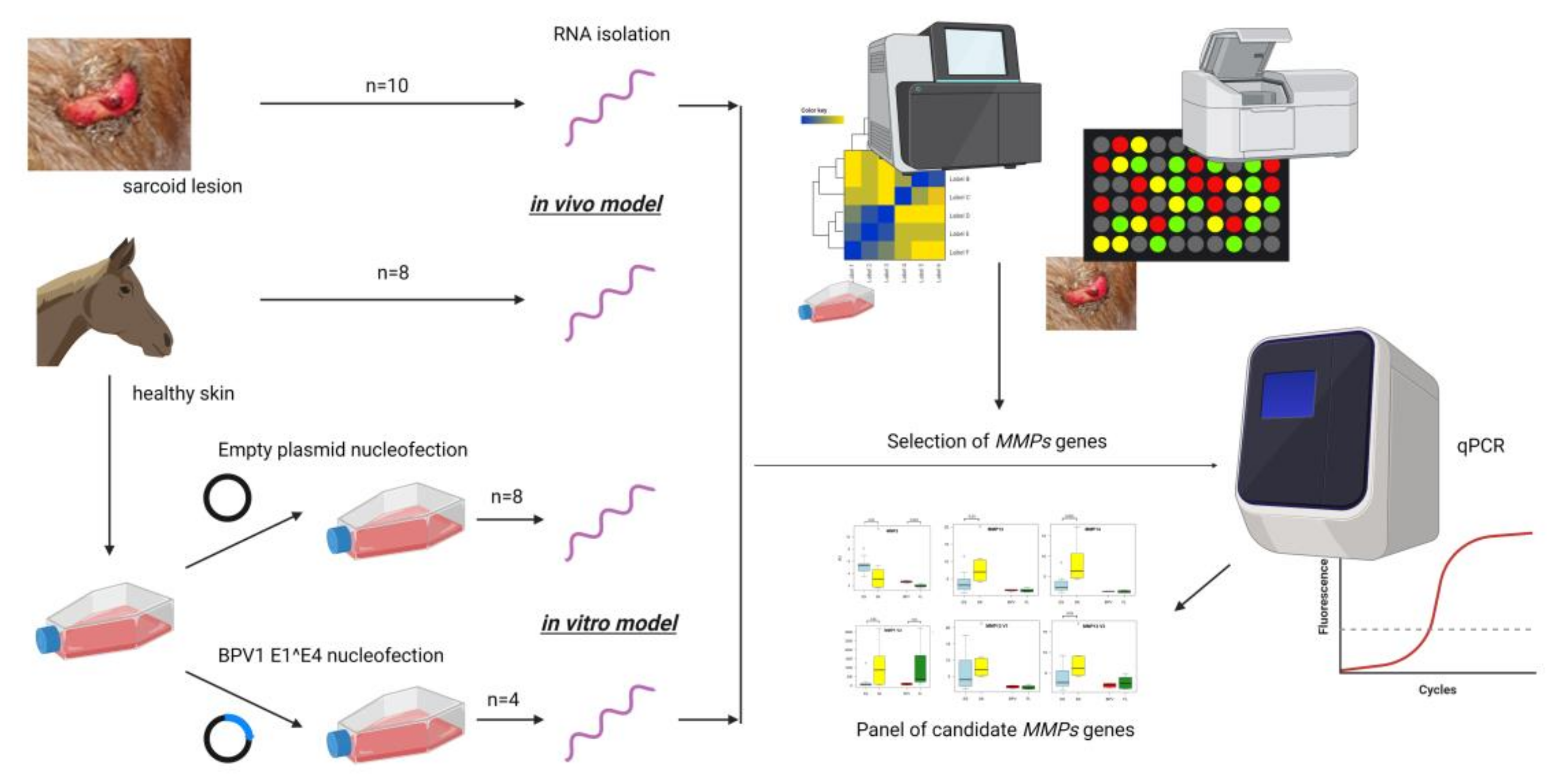
2. Materials and Methods

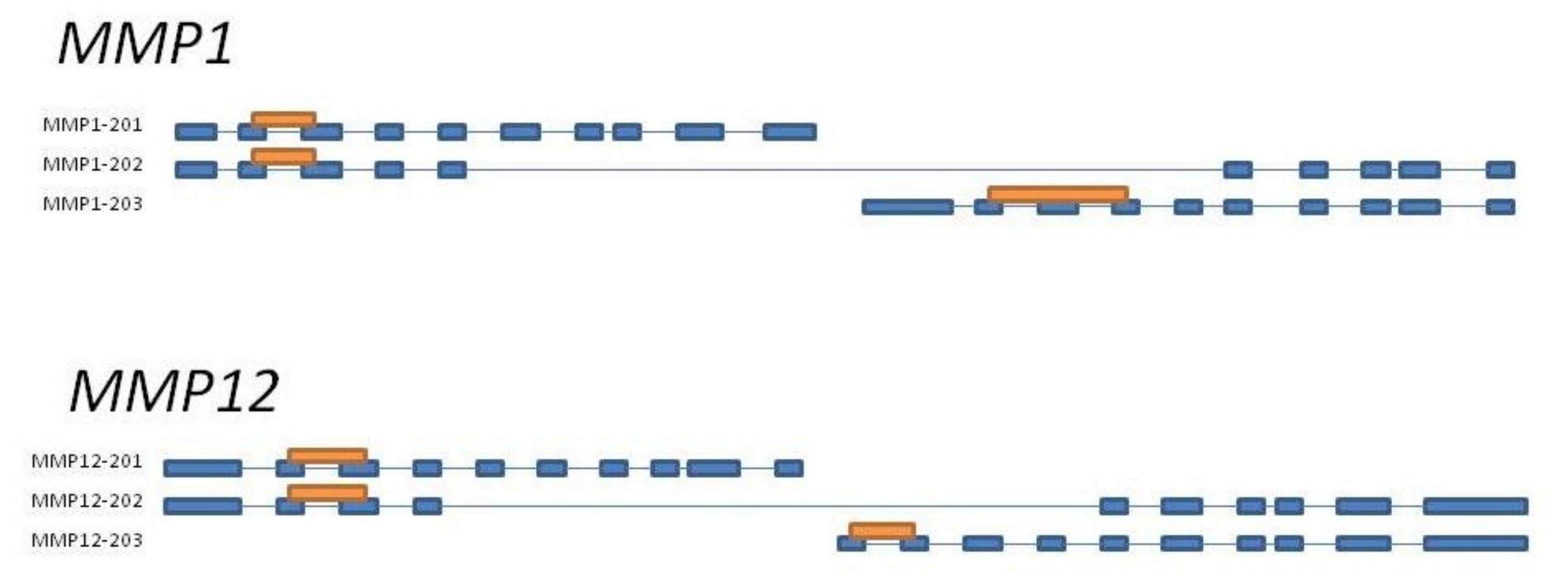
2.1. MMP Genes Selection

The experimental workflow is presented in Figure 2. The high-throughput analysis of transcriptomic signatures [9] was used to identify the MMP genes for further analyses based on applying cDNA microarrays to compare the gene expression patterns in healthy skin and sarcoid tissue samples. Additionally, using the RNA-seq technique, a panel of studies was accomplished to estimate the MMP gene expression profiles in both BPV-E4^E1 transfected ACFC lines and their counterparts transfected with empty vectors as a negative control group [10]. Considering both data sets generated (designated as GSE83430 and GSE193906), genes that belonged to the MMP family and their corresponding fold change values were achieved. The data obtained from microarrays were reanalyzed in a way similar to previous work [9], however without final filtering of genes by fold-change (p-value < 0.05). The RNA-seq data were obtained via sequencing of cDNA libraries on NextSeq 500 Illumina platform (Illumina) using NextSeq 500/550 High Output KIT v 2.5 (75 cycles) and according to protocol. The libraries were prepared using TruSeq RNA Kit v2 kit (Illumina) and evaluated using Qubit fluorimeter (Invitrogen, Thermo Fisher, Santa Clara, CA, USA). The obtained reads were mapped to EquCab3 reference genome (STAR software v 2.7.8). Next, the mapped reads were annotated and counted to specific gene thresholds using Ensembl gtf file version 100 (via htseq-count software v 0.13.5). Differential expression analysis was performed with the use of Deseq2 software (significance showed as p-value < 0.05 after multiple testing corrections).

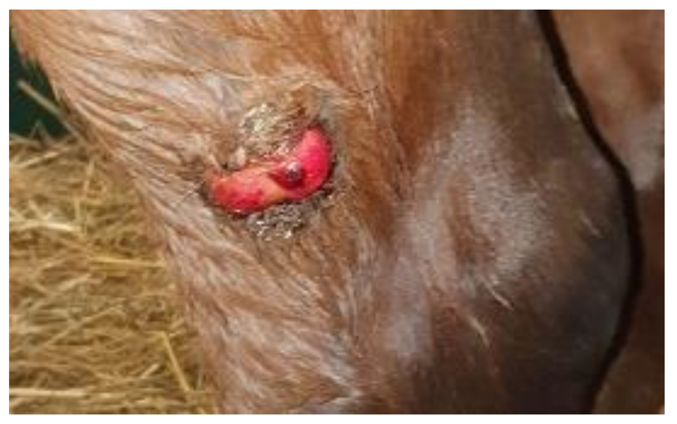
2.2. Sample Preparation

The biological samples comprised (i) equine sarcoid explants (n = 10) collected during standard veterinarian tumor removal, (ii) healthy skin bioptates (n = 8) collected at a local slaughterhouse, and (iii) ex vivo expanded ACFC lines. For all samples, the presence of BPV1 and 2 DNA was validated by PCR (AmpliTaq Gold™ 360 Master Mix; Thermo Fisher; according to the protocol) using specific primers as previously reported by Teifke et al. [12]. Skin bioptates scoring positive for BPV1 and 2 DNA were excluded from further experiments. The established ACFC lines were allotted to two groups: control cell lines transfected with an empty vector (pcDNA4/TO/myc-his/B; n = 8) and cell lines transfected with BPV1-E4^E1 gene constructs (pcDNA4/E4^E1/TO/myc-his/B; n = 4). The detailed procedures applied to establish ACFC lines and transfectant cells described by Podstawski et al. [10]. In short, fibroblast lines derived postmortem from equine skin were nucleofected with two plasmids included in the T-REx kit (Invitrogen, Waltham, MA, USA, Thermo Scientific): pcDNATM 6/TR and pcDNA™ 4/TO/myc-His/B (with or without (negative control) ligated BPV1-E1^E6 gene construct). Nucleofection procedure was performed with AmaxaTM Normal Human Dermal Fibroblast– Adult (NHDF-Adult) NucleofectorTM Kit (Lonza, CELLLAB, Warsaw, Poland). Positively transfected cell lines were obtained during antibiotic selection with 200 µg/mL zeocin (Invitrogen) and 6 µg/mL blasticidin S (Thermo Scientific, Waltham, MA, USA), which was held for one week.

2.3. Quantitative PCR Analysis

The conditions and parameters of qPCR method were thoroughly specified in our previous report [13]. Briefly, the RNA was isolated with PureLink RNA Mini Kit (Invitrogen, Thermo Fisher, according to the protocol). The quality and quantity of obtained RNA were checked using TapeStaion 2200 (Agilent Technologies, Santa Clara, CA, USA) and RNA Screen Tape (Agilent Technologies, Santa Clara, CA, USA). cDNA strand synthesis was performed with High-Capacity cDNA reverse transcription kit (Applied Biosystems, Thermo Scientific, according to the protocol). The qPCR was carried out with AmpliQ 5× HOT EvaGreen qPCR Mix Plus (ROX) (BIOTUM, Novazym, Poznań, Poland, according to the protocol) and specific primers (Table 1, Figure 3). Each sample was performed in three replications. Obtained results were calculated with the ΔΔCT method [14] based on two endogenous controls: β-actin (ACTB) and ubiquitin B (UBB) [15].

2.4. Statistical Analysis

The obtained RQ values were firstly tested for distribution purposes with a Shapiro–Francia normality test followed by Mann–Whitney U-test to assess the inter-group variability. Both tests were carried out using R software v 4.1 [16].

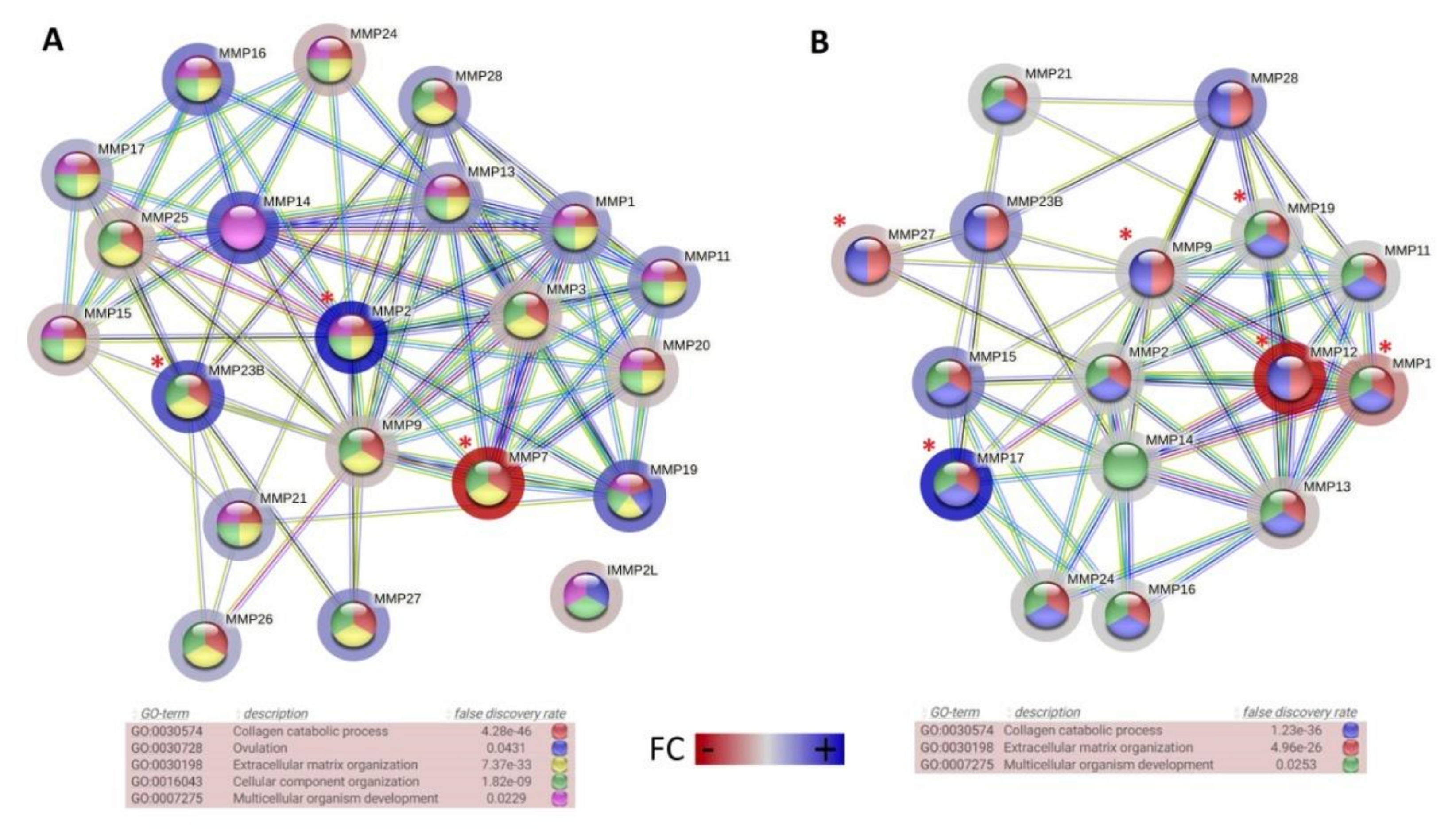
3. Results and Discussion

The present study was designed to determine changes in the MMP gene expression profiles that can be positively correlated with the development of equine sarcoids. For this purpose, to comprehensively evaluate the molecular patterns of sarcoid-dependent neoplasmic transformation, the in vivo and in vitro models have been applied. To comparatively examine the transcriptomic signatures of MMP genes, the expression profile in equine sarcoid tissue samples and BPV1-E4^E1 nucleofected ACFC-derived tumor cells were compared to healthy dermal tissues and ACFC lines not subjected to oncogenic transformation. First, the high throughput data was used to detect the MMP genes expressed in the investigated groups of cells and tissues and to demonstrate significant differences between them. The comparative analysis of MMP genes between sarcoid and healthy skin tissue samples allowed us to detect a total of 21 genes belonging to the MMP family, out of which three representatives (MMP2, MMP7, and MMP23B) underwent significantly differential expression patterns (Figure 4A). In turn, the whole transcriptome sequencing-mediated analysis of BPV1-E4^E1 transgenic equine ACFC-derived neoplastic cell lines and their counterparts nucleofected with empty vectors revealed the expression of 16 MMP family genes, out of which six members (MMP1, –9, –12, –17, –19, and –27) showed significantly differential expression (Figure 4B).
Next, the expression profiling using qPCR confirmed modifying the transcriptional activities for MMP genes representing both in vitro and in vivo models of sarcoid-dependent oncogenic transformation. Additionally, for two genes (MMP1 and MMP12), the splice variant analysis was also performed to prove the possible involvement of transcripts encoding different MMP isoforms in the processes of either transgenically induced neoplastic transformation in BPV-E4^E1 nucleofected ACFCs into sarcoid-like cells or pathophysiological conversion of dermal tissues into sarcoids.
Expression of several MMP genes was already investigated in equine sarcoids in terms of ECM remodeling [18]. It was hypothesized that ECM remodeling is a pivotal step of etiopathogenesis of sarcoid-dependent neoplasia of equine dermal tissues that provides clear evidence for the occurrence of aberrant expression of MMP genes. Martano et al. have also proven that the altered transcriptional activities of MMP and TIMP genes can lead to imbalance between collagen synthesis and degradation processes and, consequently, can result in skin neoplasia progression [18]. In turn, the study results by Yuan et al. [16] have confirmed that MMP1, –2, and –9 genes were up-regulated in sarcoid tumors and their over-expression was highly associated with the invasion fibroblast-derived sarcoid cells. Compared to the published data, we provide strong evidence of significant differences in the expression levels of MMP2 and MMP9 genes, which turned out to be up-regulated in both the ex vivo and in vitro model of sarcoid-specific neoplasmic transformation (Figure 5). Furthermore, an augmented transcriptional activity of MMP17 gene was also observed in dermal sarcoid samples and BPV1-E4^E1 nucleofected ACFC derivatives. MMP17 belongs to genes, whose enhanced expression has been found to be related to onset and progression of inflammatory processes. Therefore, it’s up-regulation may be a biomarker of inflammation evoked in sarcoid tissues and oncogenically transformed cells [19].
The whole transcriptome analysis on the in vitro model reflecting BPV1 infection in equine dermal fibroblast cells confirms the remarkable deregulation of MMP genes in BPV1-E4 and BPV1-E4^E1 transgenic ACFC neoplastic cell lines [10]. In 2008, Yuan et al. [20] evaluated the impact of BPV1 on the transcriptional profile of equine fibroblast cell lines. They pinpointed that the MMP1 gene was one of the most deregulated gene associated with the virus infection. Taking into account different splice isoforms of the MMP1 gene, the present research has identified a significant down-regulation of this gene in sarcoid tissue samples and nucleofected ACFC lines. Such modification was the most evident for V3 variant designated as ENSECAT00000025715.2 (fold changes equal to −5.90 and −12.26; p-values < 0.05 and < 0.01, respectively) (Figure 5). Moreover, as depicted in Figure 5, a similar expression profile was detected for all analyzed MMP12 isoforms, which were characterized by significantly and insignificantly diminished levels of V3 variant termed as ENSECAT00000020742.2 in sarcoid tissue explants (fold change equal to −1.95; p-value < 0.05) and BPV1-E4^E1 nucleofected ACFC lines (FC 1.31; p-value ≥ 0.05), respectively. This can indicate that BPV1 infection contributes to diminished MMP1 and MMP12 expression levels. Moreover, different involvement of individual splice variants may suggest the importance of their individual roles in the neoplastic process.
Interestingly, transcription of three other genes—MMP13 (p-value < 0.05), MMP14 (p-value < 0.001), and MMP27 (p-value ≥ 0.05)—was down-regulated in sarcoid tissue samples as compared to healthy skin bioptates. Observed MMP27 down-regulation contrasts with the results of other studies performed on tumors, where MMP27 mRNA was equally expressed in normal breast and breast cancer tissues [21]. These findings can support the hypothesis that MMP27 protein can play an important role in oncogenic transformation in a variety of tumor types. In turn, down-regulation of MMP13 and MMP14 genes can be one of the most important reasons for sarcoids’ very or negligibly low capability to metastasize, since MMP14 gene silencing brings about the negative control of cell migration [22]. In contrast, the MMP13 gene is considered to be a poor prognostic biomarker related to cell migration in several cancer types (i.e., colorectal cancer and breast cancer) [23,24].

4. Conclusions

To conclude, the present report strongly supports the hypothesis that the transcriptional activity of MMP genes is correlated with molecular modifications arising in equine dermal cells during their molecular conversion into sarcoid cells. The alterations in the MMP transcriptomic signatures that were shown to occur in sarcoid tissues and transformed equine ACFC lines expressing BPV1-E4^E1 transgene were characterized by both MMPs up- and down-regulation patterns. Moreover, for MMP1 and MMP12 genes, significant involvement of selected splice variants may suggest that only selected isoforms are crucial for the growth and development of sarcoids.

Author Contributions

Conceptualization, P.P. and K.R.-M.; Analysis of data and their interpretation, K.R.-M., P.P., M.S. (Marcin Samiec), T.S. and E.S.-G.; Performance of experiments and preparation of results, P.P., M.S. (Marcin Samiec), M.S. (Maria Skrzyszowska), T.S., K.R.-M., K.P.-T.; M.W. and Z.P. Writing the article—original draft, P.P., K.R.-M. and M.S. (Marcin Samiec); Writing the article—review and editing, T.S., E.S.-G., K.P.-T. and M.S. (Maria Skrzyszowska); Supervision and funding acquisition, K.R.-M., P.P. and M.S. (Marcin Samiec); Graphic and photographic documentation, P.P. and T.S.; Language correction of article, T.S. and M.S. (Marcin Samiec). All authors have read and agreed to the published version of the manuscript.

Funding

The present study was financially supported by the DI2016 012746 “Diamond Grant” from the Ministry of Science and Higher Education, Republic of Poland to P.P. and K.R.-M. Moreover, a panel of the studies focused on transgenic research (nucleofection-mediated transgenization of equine ACFCs) and cell culture engineering was partially supported by the statutory grant No. 04-19-11-21 from the National Research Institute of Animal Production in Balice near Kraków to M.S. (Marcin Samiec).

Institutional Review Board Statement

The Polish Act on the Protection of Animals Used for Scientific or Educational Purposes of 15 January 2015 (which implements Directive 2010/63/EU of the European Parliament on the protection of animals used for scientific purposes) states that ethic approval by the Animal Ethics Committee is not mandatory for research conducted on biological material collected postmortem during slaughter procedures. Transfection methods were performed based on procedure registration in the Ministry of Environment (number: 01.2-112/2017).

Informed Consent Statement

Not applicable.

Data Availability Statement

Not applicable.

Acknowledgments

The authors would like to thank Wojciech Witarski for significant role in constructs creation and support in laboratory analyses and valuable advice that made this research more remarkable.

Conflicts of Interest

The authors declare no conflict of interest.

References

  1. Cui, N.; Hu, M.; Khalil, R.A. Biochemical and Biological Attributes of Matrix Metalloproteinases. Prog. Mol. Biol. Transl. Sci. 2017, 147, 1–73. [Google Scholar] [CrossRef] [PubMed] [Green Version]
  2. Alaseem, A.; Alhazzani, K.; Dondapati, P.; Alobid, S.; Bishayee, A.; Rathinavelu, A. Matrix Metalloproteinases: A Challenging Paradigm of Cancer Management. Semin. Cancer Biol. 2019, 56, 100–115. [Google Scholar] [CrossRef] [PubMed]
  3. Clutterbuck, A.L.; Harris, P.; Allaway, D.; Mobasheri, A. Matrix Metalloproteinases in Inflammatory Pathologies of the Horse. Vet. J. 2010, 183, 27–38. [Google Scholar] [CrossRef] [PubMed]
  4. Gonzalez-Avila, G.; Sommer, B.; Mendoza-Posada, D.A.; Ramos, C.; Garcia-Hernandez, A.A.; Falfan-Valencia, R. Matrix Metalloproteinases Participation in the Metastatic Process and Their Diagnostic and Therapeutic Applications in Cancer. Crit. Rev. Oncol. Hematol. 2019, 137, 57–83. [Google Scholar] [CrossRef]
  5. Deryugina, E.I.; Quigley, J.P. Matrix Metalloproteinases and Tumor Metastasis. Cancer Metastasis Rev. 2006, 25, 9–34. [Google Scholar] [CrossRef]
  6. Knottenbelt, D.C. A Suggested Clinical Classification for the Equine Sarcoid. Clin. Tech. Equine Pract. 2005, 4, 278–295. [Google Scholar] [CrossRef]
  7. Nasir, L.; Reid, S.W.J. Bovine Papillomaviral Gene Expression in Equine Sarcoid Tumours. Virus Res. 1999, 61, 171–175. [Google Scholar] [CrossRef]
  8. Lunardi, M.; de Alcântara, B.K.; Otonel, R.A.A.; Rodrigues, W.B.; Alfieri, A.F.; Alfieri, A.A. Bovine Papillomavirus Type 13 DNA in Equine Sarcoids. J. Clin. Microbiol. 2013, 51, 2167–2171. [Google Scholar] [CrossRef] [Green Version]
  9. Semik, E.; Gurgul, A.; Ząbek, T.; Ropka-Molik, K.; Koch, C.; Mählmann, K.; Bugno-Poniewierska, M. Transcriptome Analysis of Equine Sarcoids. Vet. Comp. Oncol. 2017, 15, 1370–1381. [Google Scholar] [CrossRef]
  10. Podstawski, P.; Samiec, M.; Skrzyszowska, M.; Szmatoła, T.; Semik-Gurgul, E.; Ropka-Molik, K. The Induced Expression of BPV E4 Gene in Equine Adult Dermal Fibroblast Cells as a Potential Model of Skin Sarcoid-like Neoplasia. Int. J. Mol. Sci. 2022, 23, 1970. [Google Scholar] [CrossRef]
  11. BioRender. Available online: https://biorender.com/ (accessed on 31 March 2022).
  12. Teifke, J.P.; Kidney, B.A.; Löhr, C.V.; Yager, J.A. Detection of Papillomavirus-DNA in Mesenchymal Tumour Cells and Not in the Hyperplastic Epithelium of Feline Sarcoids. Vet. Dermatol. 2003, 14, 47–56. [Google Scholar] [CrossRef] [PubMed]
  13. Podstawski, P.; Witarski, W.; Szmatoła, T.; Bugno-Poniewierska, M.; Ropka-Molik, K. Mobility and Invasion Related Gene Expression Patterns in Equine Sarcoid. Animals 2020, 10, 880. [Google Scholar] [CrossRef] [PubMed]
  14. Pfaffl, M.W.; Tichopad, A.; Prgomet, C.; Neuvians, T.P. Determination of Stable Housekeeping Genes, Differentially Regulated Target Genes and Sample Integrity: BestKeeper—Excel-Based Tool Using Pair-Wise Correlations. Biotechnol. Lett. 2004, 26, 509–515. [Google Scholar] [CrossRef] [PubMed]
  15. Bogaert, L.; Van Poucke, M.; De Baere, C.; Peelman, L.; Gasthuys, F.; Martens, A. Selection of a Set of Reliable Reference Genes for Quantitative Real-Time PCR in Normal Equine Skin and in Equine Sarcoids. BMC Biotechnol. 2006, 6, 24. [Google Scholar] [CrossRef] [PubMed] [Green Version]
  16. R: The R Project for Statistical Computing. Available online: https://www.r-project.org/ (accessed on 21 January 2022).
  17. String: Functional Protein Association Networks. Available online: https://string-db.org/ (accessed on 21 January 2022).
  18. Martano, M.; Corteggio, A.; Restucci, B.; De Biase, M.E.; Borzacchiello, G.; Maiolino, P. Extracellular Matrix Remodeling in Equine Sarcoid: An Immunohistochemical and Molecular Study. BMC Vet. Res. 2016, 12, 24. [Google Scholar] [CrossRef] [PubMed] [Green Version]
  19. English, W.R.; Puente, X.S.; Freije, J.M.P.; Knäuper, V.; Amour, A.; Merryweather, A.; López-Otín, C.; Murphy, G. Membrane Type 4 Matrix Metalloproteinase (MMP17) Has Tumor Necrosis Factor-α Convertase Activity but Does Not Activate Pro-MMP2. J. Biol. Chem. 2000, 275, 14046–14055. [Google Scholar] [CrossRef] [PubMed] [Green Version]
  20. Yuan, Z.Q.; Gault, E.A.; Gobeil, P.; Nixon, C.; Campo, M.S.; Nasir, L. Establishment and Characterization of Equine Fibroblast Cell Lines Transformed in Vivo and in Vitro by BPV-1: Model Systems for Equine Sarcoids. Virology 2008, 373, 352–361. [Google Scholar] [CrossRef] [Green Version]
  21. Köhrmann, A.; Kammerer, U.; Kapp, M.; Dietl, J.; Anacker, J. Expression of Matrix Metalloproteinases (MMPs) in Primary Human Breast Cancer and Breast Cancer Cell Lines: New Findings and Review of the Literature. BMC Cancer 2009, 9, 188. [Google Scholar] [CrossRef] [Green Version]
  22. Zarrabi, K.; Dufour, A.; Li, J.; Kuscu, C.; Pulkoski-Gross, A.; Zhi, J.; Hu, Y.; Sampson, N.S.; Zucker, S.; Cao, J. Inhibition of Matrix Metalloproteinase 14 (MMP-14)-Mediated Cancer Cell Migration. J. Biol. Chem. 2011, 286, 33167–33177. [Google Scholar] [CrossRef] [Green Version]
  23. Yamada, T.; Oshima, T.; Yoshihara, K.; Tamura, S.; Kanazawa, A.; Inagaki, D.; Yamamoto, N.; Sato, T.; Fujii, S.; Numata, K.; et al. Overexpression of MMP-13 Gene in Colorectal Cancer with Liver Metastasis. Anticancer Res. 2010, 30, 2693–2699. [Google Scholar]
  24. Ameli, F.; Ghafourina Nassab, F.; Masir, N.; Kahtib, F. Tumor-Derived Matrix Metalloproteinase-13 (MMP-13) Expression in Benign and Malignant Breast Lesions. Asian Pac. J. Cancer Prev. 2021, 22, 2603–2609. [Google Scholar] [CrossRef] [PubMed]
Figure 1. Example of sarcoid lesion—sarcoid surgically removed from a two-year-old horse (noble half-blood horse); sarcoid was localized on gaskin (by Witkowski M).
Cells 11 01268 g001
Figure 2. The experimental schedule. Created with BioRender.com (accessed on 31 March 2022) [11].
Cells 11 01268 g002
Figure 3. Schematic presentation of primers designed for MMP1 and MMP12 genes to amplify all splicing variants. MMP–201–203 are splicing variants according to Ensembl accession number (MMP1-ENSECAG00000023733; MMP12-ENSECAG00000019445); primer amplification region is presented as an orange rectangle, and blue boxes represent exons.
Cells 11 01268 g003
Figure 4. The Gene ontology terms and interaction between sets of MMP genes identified using cDNA microarray in sarcoid and healthy skin tissues (A) and using RNA-seq approach in control ACFC lines nucleofected with empty vector or with BPV1-E4^E1 transgene (B); significant differentially expressed genes were marked with asterisks (p-value < 0.05); the fold change (FC) directions were marked with colors (String software [17] Equus Caballus reference).
Cells 11 01268 g004
Figure 5. The differences in MMP gene expression levels between analyzed groups of equine sarcoids (ES), skin (SK) samples, control ACFC lines nucleofected with empty vector (FL), and ACFC lines nucleofected with BPV-E4^E1 transgene (BPV) (R software v 4.1) [16].
Cells 11 01268 g005
Table 1. Specific primer sets used for amplification of MMPs cDNA. We investigated specific splice isoforms and exons spans.
GeneAccesion NumberPrimersProduct Length [bp]LocalisationSplice VariantsSplice Variants Accesion Number
MMP1 V1V2ENSECAG00000023733F: GCTGAAAGTGACTGGGAAGC
R: GGTCCACATCTGCTCTTGGT
179Exon 2 and 32/3ENSECAT00000022856.2
ENSECAT00000067257.1
MMP1 V3ENSECAG00000023733F: CCCAAGTGGGAACGAAATAA
R: ATTCCATTGGGTCCATCAAA
224Exon 12 and 141/3ENSECAT00000025715.2
MMP2ENSECAG00000000953F: TCCCTTTCCTCTTCAACGGC
R: CCGTATTTGCCGTCCTTGTC
112Exon 42/2ENSECAT00000044147.1
ENSECAT00000004278.3
MMP9ENSECAG00000013081F: CGTGTTTCCCTTCACCTTCG
R: GGTCGTAGTTGGCGGTAGT
141Exon 63/3ENSECAT00000056131.1
ENSECAT00000014089.3
ENSECAT00000068636.1
MMP12 V1V2ENSECAG00000019445F: TGGACATGATGCACAGACCT
R: CAGCCTCGCCTGTGTTAATC
222Exon 2 and 32/3ENSECAT00000002326.3
ENSECAT00000077256.1
MMP12 V3ENSECAG00000019445F:GATCTGCAAGGGACGAGGAT
R:CACCTCCACAGTGTCAGAGT
194Exon 11 and 121/3ENSECAT00000020742.2
MMP13ENSECAG00000005506F:GCTCCGAGAAATGCAGTCTT
R: GCCATTTGAGAGTTCGAGGG
140Exon 21/1ENSECAT00000007039.2
MMP14ENSECAG00000008351F: CATGATCTTCTTCGCTGAGGG
R: GGTGTCGCCTCCAATGTTG
109Exon 41/1ENSECAT00000008899.3
MMP17ENSECAG00000013201F: CACTACGCCCTCAAAGTCTG
R: AGGGATAGCGGTCATTGTGG
118Exon 42/2ENSECAT00000013924.2
ENSECAT00000013881.2
MMP27ENSECAG00000019404F: CGGTGACTGGAAAACTGGAT
R: CCCTTGGAAATCTTGGTGAA
238Exon 2 and 31/1ENSECAT00000020635.3
Publisher’s Note: MDPI stays neutral with regard to jurisdictional claims in published maps and institutional affiliations.

