Article

Iron Regulatory Protein 1 Inhibits Ferritin Translation Responding to OsHV-1 Infection in Ark Clams, Scapharca Broughtonii

1
Qingdao Key Laboratory of Mariculture Epidemiology and Biosecurity, Key Laboratory of Maricultural Organism Disease Control, Ministry of Agriculture, Yellow Sea Fisheries Research Institute, Chinese Academy of Fishery Sciences, Qingdao 266071, China
2
Function Laboratory for Marine Fisheries Science and Food Production Processes, Qingdao National Laboratory for Marine Science and Technology, Qingdao 266071, China
3
Guangxi Key Laboratory for Agricultural Resources Chemistry and Biotechnology, College of Biology and Pharmacy, Yulin Normal University, Yulin 537000, China
*
Author to whom correspondence should be addressed.
Cells 2022, 11(6), 982; https://doi.org/10.3390/cells11060982
Submission received: 16 February 2022 / Revised: 11 March 2022 / Accepted: 11 March 2022 / Published: 13 March 2022

Abstract

:
Elemental iron is an indispensable prosthetic group of DNA replication relative enzymes. The upregulation of ferritin translation by iron regulatory proteins (IRP1) in host cells is a nutritional immune strategy to sequester available iron to pathogens. The efficient replication of Ostreid herpesvirus 1 (OsHV-1), a lethal dsDNA virus among bivalves, depends on available iron. OsHV-1 infection was found to trigger iron limitation in ark clams; however, it is still an enigma how OsHV-1 successfully conducted rapid replication, escaping host iron limitations. In this study, we identified the IRP1 protein (designated as SbIRP-1) in the ark clam (Scapharca broughtonii) and found it could bind to the iron-responsive element (IRE) of ferritin (SbFn) mRNA based on electrophoretic mobility shift assay (EMSA). Knockdown of SbIRP-1 expression (0.24 ± 1.82-fold of that in NC group, p < 0.01) by RNA interference resulted in the accumulation of SbFn in hemocytes (1.79 ± 0.01-fold, p < 0.01) post-24 h of enhanced RNA interference injection. During OsHV-1 infection, SbFn mRNA was significantly upregulated in hemocytes from 24 h to 60 h, while its protein level was significantly reduced from 24 h to 48 h, with the lowest value at 36 h post-infection (0.11 ± 0.01-fold, p < 0.01). Further analysis by RNA immunoprecipitation assays showed that OsHV-1 could enhance the binding of SbIRP-1 with the SbFn IRE, which was significantly increased (2.17 ± 0.25-fold, p < 0.01) at 36 h post-infection. Consistently, SbIRP-1 protein expression was significantly increased in hemocytes from 12 h to 48 h post OsHV-1 infection (p < 0.01). In conclusion, the results suggest that OsHV-1 infection could suppress post-transcriptional translation of SbFn through the regulation of SbIRP-1, which likely contributes to OsHV-1 evasion of SbFn-mediating host iron limitation.

1. Introduction

Iron performs a critical role in cellular metabolism as a component of prosthetic groups in electron transport proteins and various enzymes involved in DNA synthesis, ATP production, etc. [1,2]. Virus replication is highly dependent on the host cell, and its efficient replication requires an available iron-rich microenvironment [2,3]. A virus with a life cycle of DNA stages requires dNTPs provided by ribonucleotide reductase (RR), which contains an iron tyrosyl radical center essential for activity [4,5]. In a previous study, the membrane permeable elater (2,2’-bipyridine) was found to inhibit the activation of vaccinia virus RR and strongly inhibited viral replication [6]. To ensure that infected cells are enriched with iron and favor viral rapid replication, some viruses selectively infect iron-rich cells by targeting transferrin receptor 1 (TfR1) [7] or disrupt host iron homeostasis by interfering with the regulation of host iron-metabolism-related proteins, such as hepcidin, MHC class I-like protein HFE (Homeostatic Iron Regulator), TfR1, and ferritin [8,9,10,11,12]. Similarly, pathogens such as bacteria and fungi have also parallelly evolved various strategies against iron limitation in hosts, such as secreting siderophores to compete for available iron in the host, or directly acquiring host iron via heme/hemoprotein receptors [2,13].
Ferritin complex has a hollow nanocage capable of storing up to 4500 iron atoms, is involved in iron metabolism, and widely exists in organisms [14,15]. The majority of cellular iron is sequestered and stored in ferritins in a non-toxic but bioavailable form [16]. Cellular ferritin levels are mainly regulated post-transcriptionally by iron regulatory proteins (IRP1 and IRP2), which could anchor to the iron response element (IRE) in the 5’ untranslated region (UTR) of the ferritin mRNA [17]. IRP1 is a bifunctional protein which predominantly responds to intracellular iron levels by assembling/disassembling the [4Fe-4S] cluster to switch between aconitase (closed conformation) and trans-regulated forms (open conformation) [18]. IRP2 is homologous to IRP1, but functions without the [4Fe-4S] cluster. The content of IRP2 is regulated at the protein level and is rapidly degraded by the proteasome in iron-replete cells [19]. Low levels of cellular iron promote the binding of IRPs to the IRE in the 5’ UTR of ferritin mRNA, leading to the downregulation of ferritin and the accumulation of free iron. When cellular iron is excessive, it will promote the assembly of IRP1 with the [4Fe-4S] cluster, switching from the IRE-binding form to the aconitase, and trigger proteasomal degradation of IRP2 to facilitate the translation of ferritin [19]. Additionally, the expression of ferritin is also regulated at the transcriptional level through the activation of transcription factors including nuclear-factor-E2related factor 2 and nuclear factor kappa B [20]. Both oxidative stress and inflammation were reported to promote the transcription of ferritin genes [21].
The IRE is conservatively found in the 5’UTR of the ferritin mRNA in invertebrates [22]. IRP1 homologues with IRE-binding activity have been identified in various invertebrates such as Drosophila melanogaster [23], Aedes aegypti [24], crayfish Pacifastacus leniusculus [25], and the earthworms Eisenia andrei [26]. Recombinant Manduca sexta IRP1 was found to inhibit the in vitro translation of ferritin mRNA through interactions with the IRE [27]. Meanwhile, iron-dependent translational control through IRE/IRP interactions is verified in M. sexta. Low concentrations of iron administration reduced the binding activity of M. sexta fat body IRP1 with the IRE and resulted in increased levels of ferritin [28]. In addition to the direct response to cellular iron concentrations, the IRE/IRP interaction in invertebrates was also affected by immune challenge. Mosquito cells exhibited an increase in IRP1/IRE binding activity when they were exposed to lipopolysaccharide [24].
OsHV-1, as a linear double-stranded DNA virus, belongs to the Malacoherpesviridae family and becomes a main epidemic virus pathogen among bivalves [29,30]. The transcriptome of OsHV-1 revealed high levels of DNA polymerase and RR transcripts consistent with high viral loads in the host [31]. High expression of these DNA replication-related enzymes is likely to be critical for the rapid progression of infection and it is notable that all these enzymes require iron to function [32]. Our previous studies demonstrated that in vivo, the iron microenvironment was disturbed in OsHV-1-infected ark clams, and the expression pattern of SbFn was dramatically affected post-virus infection [33]. To further elucidate how OsHV-1 influences host cellular iron homeostasis, the potential regulatory role of IRPs on cellular iron homeostasis was investigated in OsHV-1-infected ark clams. Firstly, an IRP1 member (SbIRP-1) was identified from ark clams (S.broughtonii) whose mRNA expression pattern was detected post-OsHV-1. Meanwhile, the regulatory function of SbIRP-1 was further surveyed. The results indicated the regulatory role of the SbIRP-1-SbFn pathway on iron homeostasis.

2. Materials and Methods

2.1. Animals and Challenge Experiment

The Healthy adult ark clams (two years old) were collected from a local farm in Rizhao (Shandong Province, China). Before processing, the ark clams were acclimatized for one week in natural seawater supplied with oxygen at 18 °C (20 ark clams in each 40 L tank). For experimental infections, OsHV-1 suspension was firstly prepared as previously described, with modifications [34]. Briefly, mantle tissues of OsHV-1-infected ark clams were collected and homogenized. After short centrifugation at 1000 rpm, 4 °C, the supernatant was filtered sequentially using filters with pore sizes of 5 μm, 2 μm, 0.45 μm, and 0.22 μm and stored at 4 °C before use. One hundred ark clams were randomly divided into five groups. Four groups of ark clams were injected with 100 μL OsHV-1 suspension (~106 copies of viral DNA/μL), while the remaining group was injected with the same volume of negative tissue homogenate as the control. For RNA extraction, triple tissue samples of hemocytes were collected respectively from three ark clams at 0 h, 12 h, 24 h, 36 h, 48 h and 60 h post injection. Different tissues, including mantle, gill, adductor muscle, foot, hemocyte, and hepatopancreas were obtained from untreated ark clams by using sterilized scissors and tweezers. All samples were added with 1 mL TRIzol reagent (Invitrogen, Waltham, MA, USA) and stored at −80 °C before RNA extraction.

2.2. Total RNA Extraction and cDNA Synthesis

Total RNA was extracted from 50 mg of each tissue sample using TRIzol reagent (Invitrogen) according to the manufacturer’s protocol. The first strand of cDNA was synthesized using the template of DNase I-treated total RNA according to the manufacturer’s protocol (TaKaRa, Shiga, Japan). The synthesis reaction was processed at 37 °C for 20 min and stopped by heating at 85 °C for 5 s. The cDNA mixture was diluted to 1:40 and stored at −80 °C.

2.3. Clone and Bioinformatic Analyses of SbIRP-1

The specific primers SbIRP-F and SbIRP-R (Table 1) were designed by Primer Premier 5 software to clone the full sequence of SbIRP-1 based on the sequence information of ark clam genomic data [35]. Then, the above PCR product was purified and cloned into the pMD 19-T simple vector (TaKaRa) and sequenced.
For characterization of the SbIRP-1 sequence, the amino acid sequences of SbIRP-1 were deduced by the ExPASy Protein Analysis System v3.0. http://www.expasy.org/ (accessed on 15 December 2021) and comparatively analyzed by the BLAST algorithm http://www.ncbi.nlm.nih.gov/blast (accessed on 16 December 2021). The protein domains were revealed by SMART v9.0. http://smart.embl-heidelberg.de/ (accessed on 16 December 2021). Multiple sequence alignment of SbIRP-1 with IRPs of other species was performed by the ClustalX multiple alignment program (Conway Institute UCD Dublin, Dublin, Ireland) and the result was generated in ESPript v3.0. https://espript.ibcp.fr/ESPript/cgi-bin/ESPript.cgi (accessed on 20 December 2021). A phylogenetic tree of IRPs was constructed by the neighbor-joining algorithm using the Mega program v6.0. (Tokyo Metropolitan University, Tokyo, Japan), and 1000 bootstrap replicates were performed. The three-dimensional structures of IRPs were constructed using SWISS-MODEL https://swissmodel.expasy.org (accessed on 21 December 2021) and showed by PyMol program v2.3.0. (Schrödinger, New York, NY, USA).

2.4. RT-qPCR Analysis of SbIRP-1 and SbFn mRNA

The expression levels of SbIRP-1 and SbFn were measured by SYBR RT-qPCR reagent (TOYOBO, Osaka, Japan) based on Bio-Rad CFX Connect real-time PCR system. Specific primers, qSbIRP-F, and qSbIRP-R (Table 1) were used for amplifying a 172 bp fragment of SbIRP-1, qSbFn-F, and qSbFn-R (Table 1) for a 153 bp fragment of SbFn. 60S ribosomal protein subunit fragment, amplified with primers qSbRL15-F and qSbRL15-R (Table 1), was chosen as an internal reference. The relative expression level of mRNA was analyzed by the 2ΔΔCt method [36].

2.5. SbIRP-1 Recombinant Protein Expression and Polyclonal Antibody Preparation

The vector construction and expression of recombinant SbIRP-1 were performed as previously described [37]. The coding region of SbIRP-1 was amplified with the primers SbIRP-Nco I and SbIRP-Xho I (Table 1). The amplified fragments were purified and digested with Nco I and Xho I before being inserted into the same double restriction enzyme linearized expression vector pET-28a. The constructed vector pET-28a-SbIRP-1 was transferred into BL21 (DE3) Chemically Competent Cell (TransGen, Beijing, China). Transformed cells were induced with 0.2 mM IPTG and cultured overnight at 18 °C. Histidine-labeled rSbIRP-1 was purified using ProteinPure Ni-NTA resin (TransGen) and concentrated by ultrafiltration after dialysis. For polyclonal antibody preparation, 2 mg of rSbIRP-1 protein was used as an antigen mixed thoroughly with an equal volume of Freund’s complete adjuvant and injected into New Zealand white rabbit at the first immunization, which was followed by a second and third immunization with 2 mg of rSbIRP-1 protein and the same volume of Freund’s incomplete adjuvant at 10-day intervals. The serum was harvested and stored at −80 °C before used. The specificity of the SbIRP-1 polyclonal antibody was tested by western blot of the rSbIRP-1 and hemocyte lysate.

2.6. The Knockdown of SbIRP-1

Small interfering RNA (siSbIRP) specific to SbIRP-1 and negative control (siNC) were designed and synthesized by Sangon Biotech (Shanghai, China) (Table 1). For RNA interference, siSbIRP (100 μL, 0.1 μg per μL) and siNC (100 μL, 0.1 μg per μL) were injected into the ark clams of experimental and control groups, respectively. To enhance the effect of RNA interference, a second injection was performed 12 h after the first injection using the same dosage. Hemocytes from the experimental and control groups were sampled 24 h after the second injection for further analysis of RNA and protein levels of SbIRP-1.

2.7. Electrophoretic Mobility Shift Assay

To test the binding activity of SbIRP-1 to SbFn-IRE, EMSA was performed using a chemiluminescent EMSA kit (Beyotime, Shanghai, China) according to the manufacturer’s instructions. IREs were predicted using SIREs web server v2.0. http://ccbg.imppc.org/sires/ (accessed on 10 October 2021) and RNAfold Web Server http://www.unafold.org/ (accessed on 24 November 2021). Biotin-labeled SbFn-IRE probes and mutation SbFn-IRE probes, and unlabeled SbFn-IRE probes were synthesized by Sangon Biotech (Shanghai, China). The sequences of the EMSA probes were listed in Table 1. For EMSA, the probes were heated at 95 °C for 3 min, then slowly cooled to room temperature, annealed to form a stem-loop structure, and then incubated with 1 μg of rSbIRP-1 for 30 min at 25 °C. Cold competitor probes (unlabeled SbFn-IRE probes) at 20×, 10× and 5× were used to confirm the binding specificity. Samples were separated using 6% native polyacrylamide gel, then transferred to nylon membrane and crosslinking by UV. Transferred probes were detected with HRP-conjugated streptavidin.

2.8. RNA Immunoprecipitation

RNAChIP was performed using the RNA ChIP-IT kit (Active Motif, La Hulpe, Belgium) according to the manufacturer’s instructions. Briefly, approximately 1 × 10⁷ hemocytes were cross-linked with 1% glutaraldehyde for 10 min at room temperature and terminated with 125 mM glycine. Cells were lysed on ice for 30 min using ice-cold NP40 lysis buffer containing protease inhibitor cocktail (Roche, Switzerland) and RNase Inhibitors (TaKaRa). Cell lysates were digested using DNase I before the immunoprecipitation reaction; a portion of treated cell lysates was saved as input RNA template at −80 °C before use. Immunoprecipitation was performed using SbIRP-1 polyclonal antibody or normal rabbit IgG (ABclonal, Wuhan, China) as control with equal amounts of DNase I-treated cell lysates overnight at 4 °C, respectively. RNA binding with SbIRP-1 was eluted from the magnetic beads, reversely cross-linked, and purified using TRIzol reagent (Invitrogen) according to the manufacturer’s protocol. The first-strand cDNA of purified RNA was synthesized using PrimeScript™ RT reagent Kit with gDNA Eraser (TaKaRa) and quantified by qPCR. Data were normalized to the corresponding RNA input. The sequence information of the primers was listed in Table 1.

2.9. Western Blot

Total protein of the hemocytes was extracted using the cell lysis buffer (Beyotime) containing a protease inhibitor cocktail (Roche). The protein concentration was quantified using a BCA protein assay kit (Transgen). The extracted protein samples were separated using 12% SDS-PAGE and transferred onto nitrocellulose (NC) membranes. After blocking with 5% skimmed milk powder, membranes were incubated overnight at 4 °C with SbIRP-1 polyclonal antibody (1:500), SbFn (1:500) polyclonal antibody [33] or SbTubulin polyclonal antibody (1:1000). Membranes were then incubated with HRP-conjugated goat anti-mouse Ig and goat anti-rabbit Ig secondary antibodies (1:3000, Abclonal) for 3 h at room temperature with shaking. Between each step, the NC membranes were washed thrice for 5 min with TBST. Protein bands were visualized in an automated chemiluminescent gel imaging system using Western lighting ECL substrate (Thermo Fisher Scientific, Waltham, MA, USA).

2.10. Statistical Analysis

All experiments were repeated thrice independently, and the data are presented as mean ± SD. Statistical analysis of the data was carried out using the Statistical Package for Social Sciences (SPSS) 21.0. Differences between groups were assessed using one-way ANOVA. Differences between means are considered significant at p < 0.05 and extremely significant at p < 0.01.

3. Results

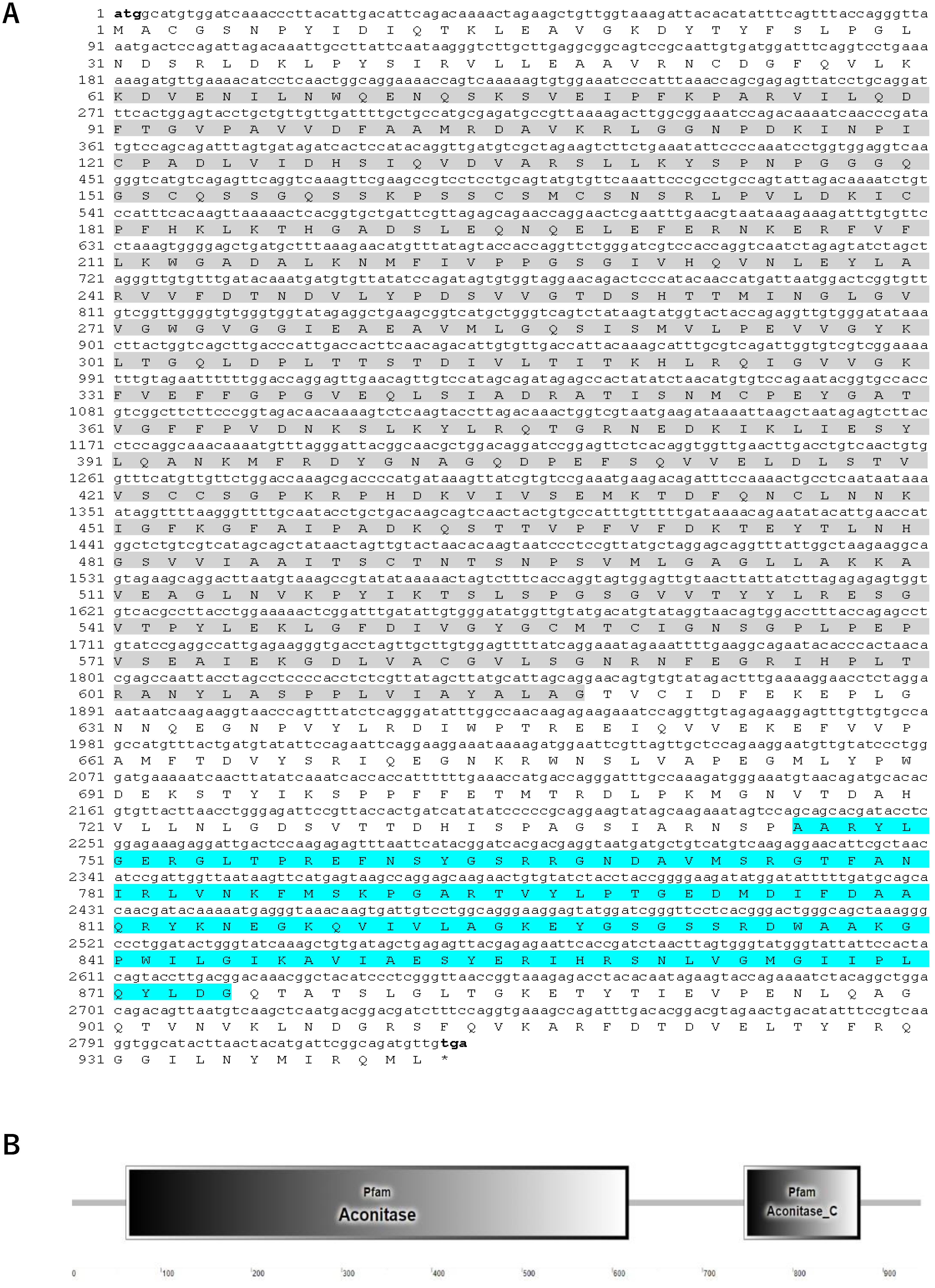
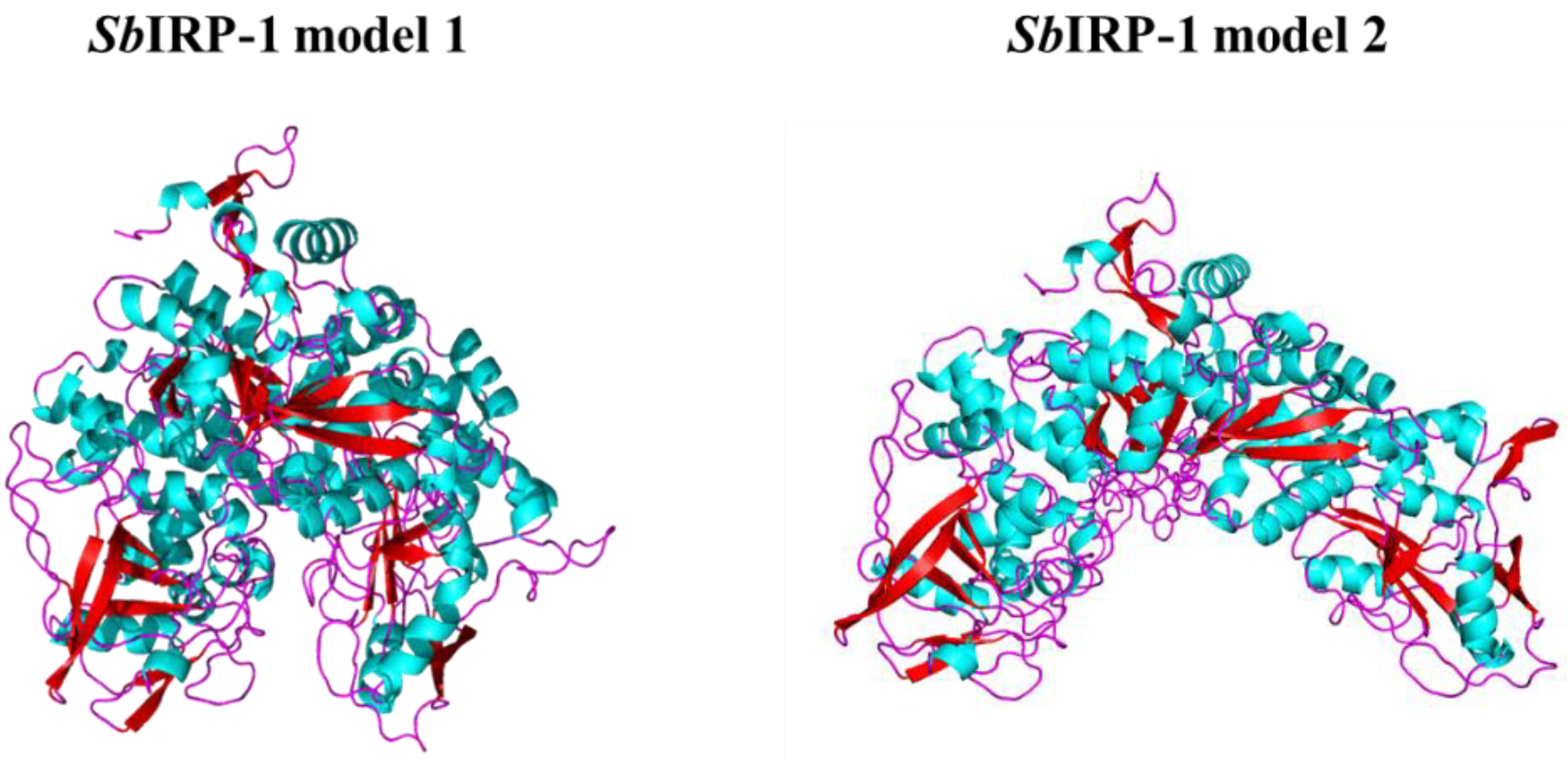
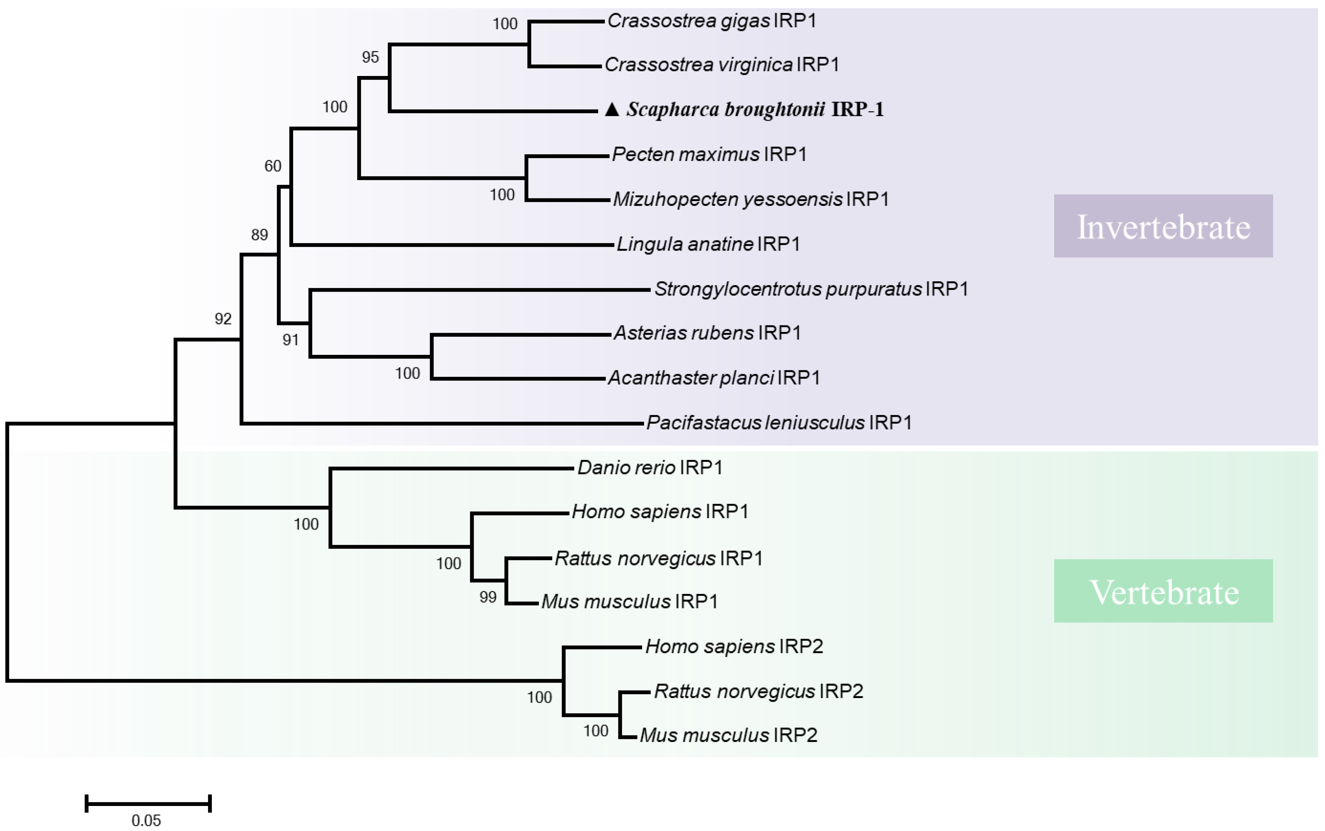
3.1. Molecular Characterization of SbIRP-1 Statistical Analysis

The full-length cDNA of SbIRP-1 with an ORF of 2829 bp encodes a putative protein of 942 amino acids (with a predicted molecular mass of 103.3 kDa and a theoretical pI of 6.16) (Figure 1A). SMART prediction domain analysis showed that SbIRP-1 contained conservative aconitase (Lys61-Gly618) and aconitase C-terminal (Ala746-Gly875) domains (Figure 1B). Multiple sequence comparisons showed that SbIRP-1 possessed 83% similarity to IRP1 form Crassostrea virginica (XP_022311890.1) and 69% similarity to IRP1 form Homo sapiens (NP_001265281.1), but the aconitase domain (Ser138-Gly189) of SbIRP-1 exhibited low homology with those of other molluscan IRP1s and was absent in vertebrate IRP1s (Figure 2). Conserved active sites were found in SbIRP-1, including three [4Fe-4S] cluster-ligating cysteine residues (Cys491, Cys557, and Cys560), which performed essential roles in iron regulation of RNA binding and aconitase activity, and four arginine residues (Arg590, Arg595, Arg753, and Arg834) in the aconitase active-site cleft were essential for RNA recognition. Additionally, three putative RNA binding regions (124DLVIDHSIQV133) [38], (200EFERNKERFVFL211) and (378GRNEDKIKLIE388) [39] were also conserved in the SbIRP-1.
The three-dimensional structure of SbIRP-1 was calculated using the SWISS-MODEL program. The results revealed two alternative conformations of SbIRP-1; model-template alignments showed that SbIRP-1 (amino acids 5–941) shared 72.57% sequence identity with the human cytosolic aconitase (IRP1) model 1 (PDB ID: 2b3y.1.A) assembling with the [4Fe-4S] cluster, and 73.25% sequence identity with the rabbit cytosolic aconitase (IRP1) model 2 (PDB ID: 3sn2.1.A) disassembling with the [4Fe-4S] cluster. The QMEAN Z-scores for the structures of SbIRP-1 obtained based on the homology modeling of the above two models were −1.22 and −1.90, respectively, indicating that the proposed homology model is reliable and acceptable [40] (Figure 3).
The phylogenetic tree of the IRPs was shown in Figure 4. IRP1 proteins from vertebrates (including fish and mammals) and invertebrates are clustered together forming an independent branch. The other branch consisted of three IRP2 proteins from vertebrates. Thereinto, SbIRP-1 is more closely clustered with the other invertebrate IRP1 proteins.

3.2. Tissue Distribution and Expression Profile Post OsHV-1 Infection of SbIRP-1

The spatial expression of the SbIRP-1 gene in the mantle, gills, adductor muscle, foot, hemocytes, and hepatopancreas were detected by qPCR, which revealed significant differences in the mRNA expression levels of the SbIRP-1 gene among various tissues. The mRNA expression level of the SbIRP-1 gene was the highest in hemocytes (14.43 ± 1.82-fold, p < 0.01) among all detected tissues, followed by the hepatopancreas (3.16 ± 1.01-fold, p < 0.05) and the adductor muscle (3.01 ± 0.18-fold, p < 0.01) (Figure 5A).
SbIRP-1 mRNA expression level was strongly affected by OsHV-1 infection. The mRNA expression level of SbIRP-1 genes in hemocytes significantly increased (1.58 ± 0.12-fold, p < 0.01) at 48 h post-OsHV-1 infection (Figure 5B).

3.3. Recombinant Expression, Purification and Antiserum Preparation of SbIRP-1

After IPTG induction, one major protein with an apparent molecular weight of around 100 kDa was detected (Figure 6A, Lane 1, 2), which was consistent with the predicted molecular mass of SbIRP-1. After purification, a single band of about 110 kDa representing rSbIRP-1 was observed in SDS-PAGE (Figure 6A, Lane 5). Western blotting assay of the hemocyte sample with SbIRP-1 polyclonal antibodies revealed that there was a distinct band of about 110 kDa (Figure 6A, Lane 6), which corresponded to the predicted molecular weight of SbIRP-1.

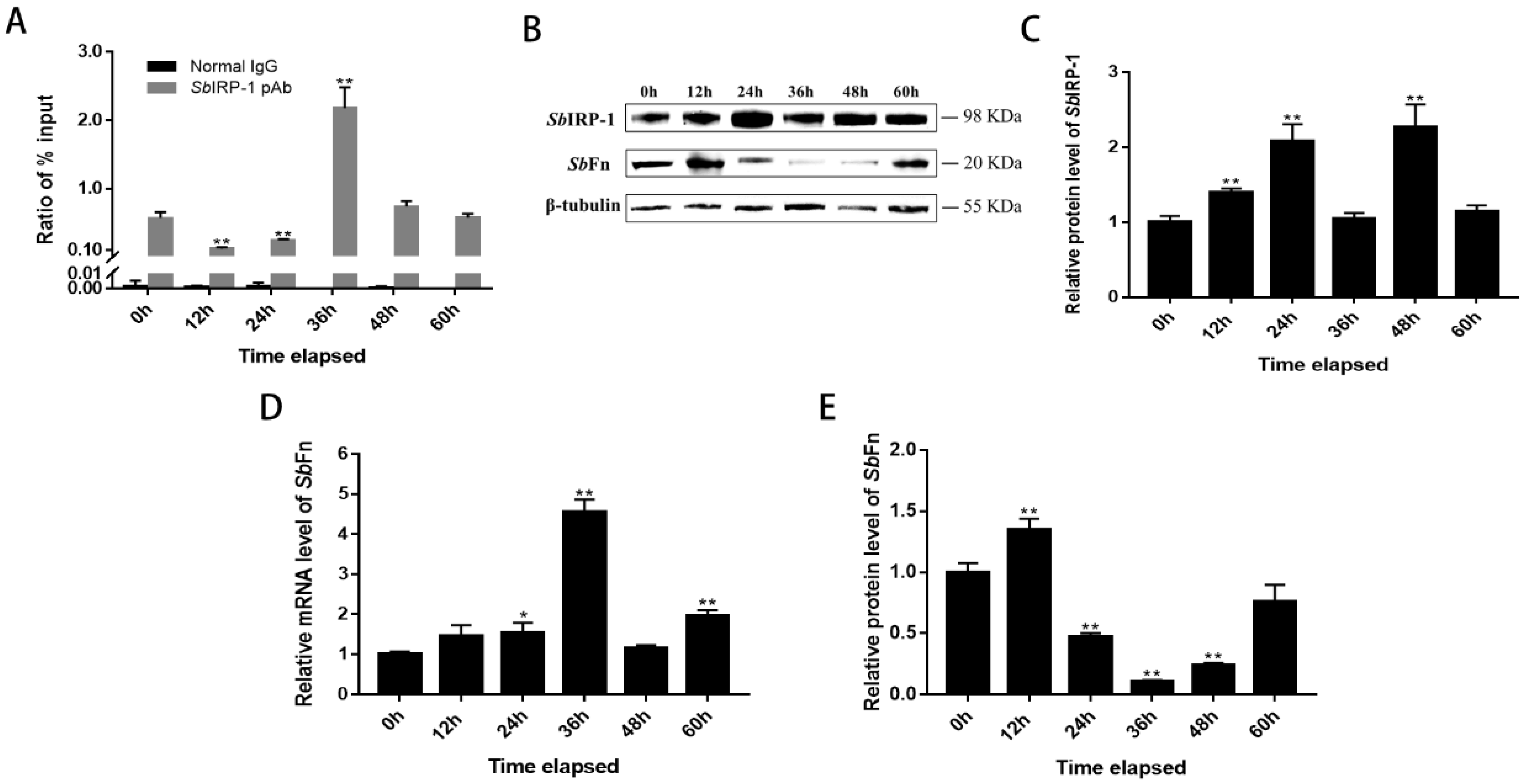
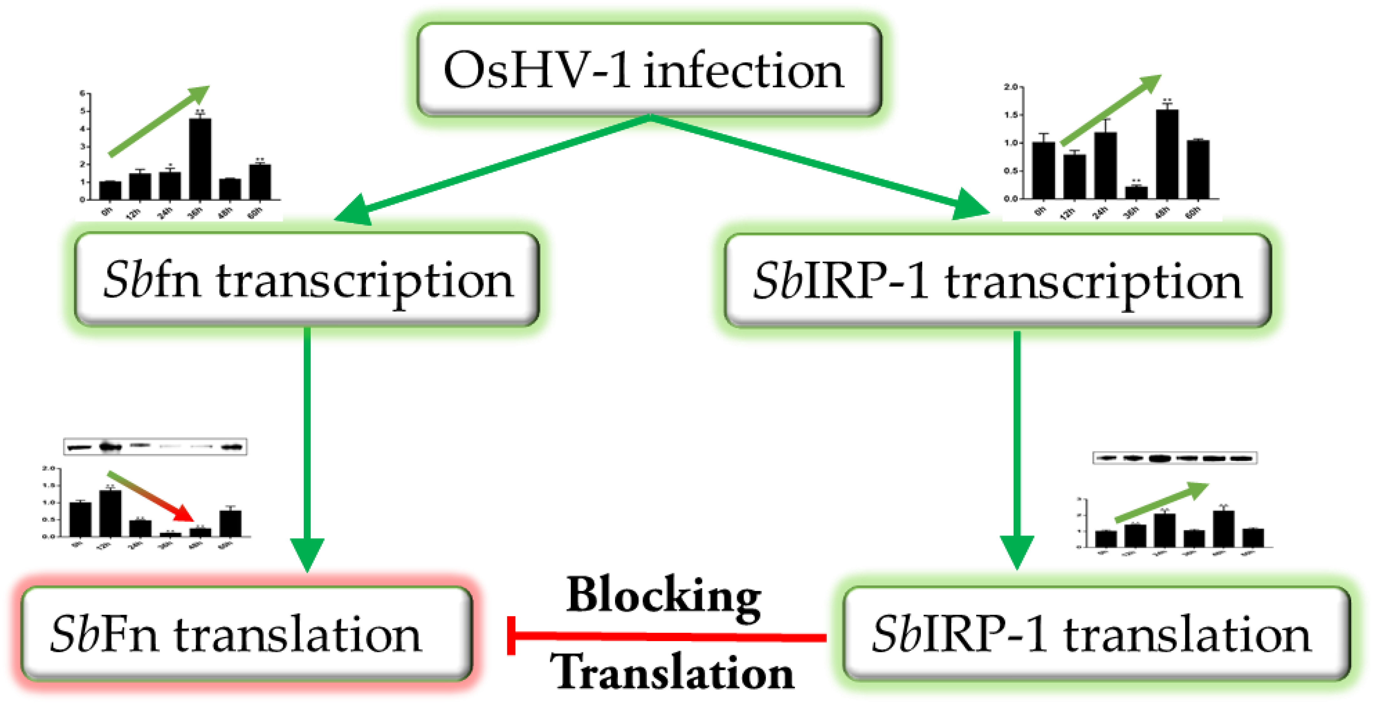
3.4. The Role of SbIRP-1 on the Post-Transcriptional Regulation of SbFn

IRPs mediate cellular post-transcriptional iron regulation by interacting with IREs located in the 5′-UTR mRNA of target genes. The 5′-UTR of SbFn mRNA was found equipped with a stem-loop structure resembling the IRE of the human-ferritin-heavy chain mRNA, which was located 95 bp downstream of the transcription start site. The IRE of SbFn mRNA showed a conserved six-membered loop sequence 5′ CAGUGN 3′ and a bulging C between the upper and lower stems (Figure 6B). To elucidate whether SbIRP-1 binds to the putative IRE of SbFn, electrophoretic mobility shift assay (EMSA) was performed using purified recombinant SbIRP-1. As shown in Figure 6C, the lagging band (containing IRE/IRP complex) only appeared in the lane with the IRE of SbFn, but not with the mutant IRE (AUUUGUUUUGUUGCGUuuuUuuACGUACGGACAGCU), with lowercase letters in the sequence representing mutation sites. To further confirm the specific binding between rSbIRP-1 and the SbFn IRE, we performed a competitive EMSA analysis using unlabeled SbFn IRE as competitive probes. The results showed that unlabeled SbFn IRE can compete with labeled SbFn IRE, showing decreased label signals with incremental unlabeled SbFn IRE (Figure 6C).
To further confirm the post-transcriptional regulatory role of the IRE/IRP system on SbFn protein levels in hemocytes, the RNAi (RNA interference) assay was carried out. After SbIRP-1 was knocked down by RNAi, the mRNA level of SbIRP-1 in the hemocytes decreased significantly (0.24 ± 1.82-fold of that in NC group, p < 0.01) post-24 h of enhanced RNA interference injection (Figure 6D). The protein level of SbIRP-1 was also significantly downregulated, which was 0.13-fold of that in NC group (p < 0.01) (Figure 6F). Meanwhile, the mRNA level of SbFn in SbIRP-1-RNAi ark clams showed no significant difference compared with the NC group (Figure 6E), while the protein level of SbFn increased significantly (1.79 ± 0.01-fold of that in NC group, p < 0.01) (Figure 6F).

3.5. The Blocking Effect of SbIRP-1 on the Translation of SbFn Post OsHV-1 Infection

To evaluate the regulatory role of SbIRP-1 on cellular iron homeostasis during OsHV-1 infection, we initially examined the influence of OsHV-1 infection on the binding ability of SbIRP-1 with the IRE of SbFn by ChIP-qPCR (Figure 7A). The binding ability of SbIRP-1 with SbFn IRE was firstly reduced at 12 h and 24 h, then was significantly upregulated at 36 h post-OsHV-1 infection (380.74%, p < 0.01), then returned to normal levels. Meanwhile, the protein levels of SbIRP-1 were also examined by western blot post-OsHV-1 infection (Figure 7C). The protein level of SbIRP-1 in hemocytes showed a constant increase from 12 h post-infection (1.39 ± 0.05-fold, p < 0.01) to 48 h (2.26 ± 0.31-fold, p < 0.01).
OsHV-1 infection significantly promoted the mRNA expression levels SbFn in hemocytes, which showed significant increase at 24 h (1.54 ± 0.20-fold, p < 0.05) and reached a maximum at 36 h post infection (4.55 ± 0.30-fold, p < 0.01) (Figure 7D). While the Western blot showed that OsHV-1 infection downregulated SbFn protein levels (Figure 7E), which were significantly reduced in hemocytes at 36 h (0.11 ± 0.01-fold, p < 0.01) post-infection.

4. Discussion

Ferritin is conserved in most organisms as a major intracellular iron storage protein and mainly conducts iron limitation against pathogens [41]. In this study, OsHV-1 infection was shown to promote binding activity of SbIRP-1 to the IRE of SbFn, blocking the translation of SbFn, which was intended to reduce the efficient iron sequestration effect of SbFn, finally facilitating rapid viral replication (Figure 8).
Iron homeostasis in mammalian cells is orchestrated post-transcriptionally by the IRE/IRP system [42]. IRE/IRP1 interaction directly responds to changes in cellular iron content; IRE-binding activity of IRPs is high in iron-deficient conditions and low in iron-sufficient conditions [43]. Here, an IRP1 protein in ark clams (SbIRP-1) was found with an open and a closed conformation, while IRP2 was missing in the genome of ark clams [35]. Moreover, SbIRP-1 possesses three putative RNA binding domains and aconitase active sites, implying the versatile role of SbIRP-1 as RNA binding molecule and aconitase. The IRE was conservatively identified in the 5’-UTR of SbFn mRNA, resembling ferritin genes across invertebrates, including mollusks, such as housefly (Musca domestica), prawn (Macrobrachium nipponense), scallop (Chlamys nobilis), clam (Ruditapes decussatus), abalone (Haliotis rufescens), and mussel (Perna viridis) [44,45,46,47,48,49]. The location of the SbFn IRE satisfied the requirement for effective translation inhibition, with a total of 20–30 nucleotides located 90 bp downstream of the transcription start site [50,51]. The IRE/IRP system served conservatively to regulate iron homeostasis in ark clams and a high affinity between rSbIRP-1 and the IRE of SbFn was revealed by EMSA. When SbIRP-1 was knocked down in vivo by RNAi, the translation of SbFn was increased and the accumulation level of SbFn could be detected in hemocytes by western blot, even without significant changes in SbFn mRNA levels.
OsHV-1, a lethal dsDNA virus, is known as an epidemic viral pathogen in bivalves. Sufficient available iron is necessary for the rapid replication of DNA viruses [5]. Modifications in iron homeostasis have been associated with the pathogenesis of several viruses, including herpes simplex virus, rhabdovirus, hepatitis B virus, and cowpox virus [2,52]. Thus, the association between cellular iron uptake and storage and overall changes in labile iron pool (LIP) may be critical for the development of productive viral infections and pathological situations. Here, it was found that OsHV-1 infection promotes the RNA-binding of SbIRP-1 to SbFn IRE in ark clam hemocytes. During the later phase of OsHV-1 infection, the binding activity of SbIRP-1 to SbFn IRE is significantly upregulated, concomitant with a decrease in SbFn mRNA translation. A significant increase in SbIRP-1 level in hemocytes was also found after OsHV-1 infection, which might contribute to enhancement of the binding between SbIRP-1 and the IRE of SbFn in later phases of infection. All above suggested that OsHV-1 could facilitate its own replication by interfering with the regulation of SbIRP-1-SbFn pathways on iron homeostasis.
In conclusion, a full-length cDNA of SbIRP-1 was identified and characterized, SbIRP-1 showed highly sequential and structural conservation with other IRP1s. The interaction between SbIRP-1 and the IRE of SbFn was verified by EMSA. The IRE/IRP system conservatively served on the regulation of SbFn in ark clams. When SbIRP-1 was knocked down in vivo by RNAi, the accumulation level of SbFn could be detected in hemocytes by Western blot, even without significant changes to SbFn mRNA levels. OsHV-1 infection was shown to promote the protein level of SbIRP-1, which might contribute to the stronger binding ability of SbIRP-1 to the IRE of SbFn, blocking the translation of SbFn.

Author Contributions

Conceptualization, L.X.; methodology, B.H. and L.X.; software, B.H.; investigation, B.H, C.L., X.Z. and Q.L.; data curation, C.B., C.W.; writing—review and editing, L.X.; supervision, L.X., C.W. and C.B.; funding acquisition, L.X., C.B. and C.W. All authors have read and agreed to the published version of the manuscript.

Funding

This research was financially supported by the National Natural Science Foundation of China (31902400), the Special Scientific Research Funds for Central Non-profit Institutes, Yellow Sea Fisheries Research Institutes (Project No. 20603022020007), China Agriculture Research System of MOF and MARA.

Institutional Review Board Statement

Not applicable.

Informed Consent Statement

Not applicable.

Data Availability Statement

Not applicable.

Acknowledgments

We are grateful to all the laboratory members for their technical support and helpful suggestions.

Conflicts of Interest

The authors declare no conflict of interest.

References

  1. Maffettone, C.; De Martino, L.; Irace, C.; Santamaria, R.; Pagnini, U.; Iovane, G.; Colonna, A. Expression of iron-related proteins during infection by bovine herpes virus type-1. J. Cell. Biochem. 2008, 104, 213–223. [Google Scholar] [CrossRef]
  2. Drakesmith, H.; Prentice, A. Viral infection and iron metabolism. Nat. Rev. Microbiol. 2008, 6, 541–552. [Google Scholar] [CrossRef]
  3. Boodhoo, N.; Kamble, N.; Sharif, S.; Behboudi, S. Glutaminolysis and glycolysis are essential for optimal replication of Marek’s disease virus. Virol. J. 2020, 94, e01680-19. [Google Scholar] [CrossRef] [PubMed] [Green Version]
  4. Stubbe, J. Ribonucleotide reductases: Amazing and confusing. J. Biol. Chem. 1990, 265, 5329–5332. [Google Scholar] [CrossRef]
  5. Gammon, D.B.; Gowrishankar, B.; Duraffour, S.; Andrei, G.; Upton, C.; Evans, D.H. Vaccinia virus–encoded ribonucleotide reductase subunits are differentially required for replication and pathogenesis. PLoS Pathogens 2010, 6, e1000984. [Google Scholar] [CrossRef] [Green Version]
  6. Romeo, A.M.; Christen, L.; Niles, E.G.; Kosman, D.J. Intracellular chelation of iron by bipyridyl inhibits DNA virus replication: Ribonucleotide reductase maturation as a probe of intracellular iron pools. J. Biol. Chem. 2001, 276, 24301–24308. [Google Scholar] [CrossRef] [Green Version]
  7. Parker, J.S.; Murphy, W.J.; Wang, D.; O’Brien, S.J.; Parrish, C.R. Canine and feline parvoviruses can use human or feline transferrin receptors to bind, enter, and infect cells. Virol. J. 2001, 75, 3896–3902. [Google Scholar] [CrossRef] [PubMed] [Green Version]
  8. Nishina, S.; Hino, K.; Korenaga, M.; Vecchi, C.; Pietrangelo, A.; Mizukami, Y.; Furutani, T.; Sakai, A.; Okuda, M.; Hidaka, I. Hepatitis C virus–induced reactive oxygen species raise hepatic iron level in mice by reducing hepcidin transcription. Gastroenterology 2008, 134, 226–238. [Google Scholar] [CrossRef] [PubMed]
  9. Chen, N.; McCarthy, C.; Drakesmith, H.; Li, D.; Cerundolo, V.; McMichael, A.J.; Screaton, G.R.; Xu, X.N. HIV-1 down-regulates the expression of CD1d via Nef. Eur. J. Immunol. 2006, 36, 278–286. [Google Scholar] [CrossRef] [PubMed]
  10. Hewitt, E.W. The MHC class I antigen presentation pathway: Strategies for viral immune evasion. Immunology 2003, 110, 163–169. [Google Scholar] [CrossRef]
  11. Gu, J.-M.; Lim, S.O.; Oh, S.J.; Yoon, S.-M.; Seong, J.K.; Jung, G. HBx modulates iron regulatory protein 1-mediated iron metabolism via reactive oxygen species. Virus Res. 2008, 133, 167–177. [Google Scholar] [CrossRef] [PubMed]
  12. Mulvey, M.R.; Kühn, L.C.; Scraba, D.G. Induction of Ferritin Synthesis in Cells Infected with Mengo Virus (∗). J. Biol. Chem. 1996, 271, 9851–9857. [Google Scholar] [CrossRef] [PubMed] [Green Version]
  13. Cassat, J.E.; Skaar, E.P. Iron in infection and immunity. Cell Host Microbe 2013, 13, 509–519. [Google Scholar] [CrossRef] [PubMed] [Green Version]
  14. Alkhateeb, A.A.; Connor, J.R. The significance of ferritin in cancer: Anti-oxidation, inflammation and tumorigenesis. Biochim. Biophys. Acta (BBA)-Rev. Cancer 2013, 1836, 245–254. [Google Scholar] [CrossRef] [PubMed]
  15. De Meulenaere, E.; Bailey, J.B.; Tezcan, F.A.; Deheyn, D.D. First biochemical and crystallographic characterization of a fast-performing ferritin from a marine invertebrate. Biochem. J. 2017, 474, 4193–4206. [Google Scholar] [CrossRef] [PubMed]
  16. Harrison, P.M.; Arosio, P. The ferritins: Molecular properties, iron storage function and cellular regulation. Biochim. Biophys. Acta (BBA)-Bioenerg. 1996, 1275, 161–203. [Google Scholar] [CrossRef] [Green Version]
  17. Kim, H.-Y.; Klausner, R.D.; Rouault, T.A. Translational Repressor Activity Is Equivalent and Is Quantitatively Predicted by in Vitro RNA Binding for Two Iron-responsive Element-binding Proteins, IRP1 and IRP2 (∗). J. Biol. Chem. 1995, 270, 4983–4986. [Google Scholar] [CrossRef] [Green Version]
  18. Paraskeva, E.; Hentze, M.W. Iron-sulphur clusters as genetic regulatory switches: The bifunctional iron regulatory protein-1. FEBS Lett. 1996, 389, 40–43. [Google Scholar] [CrossRef] [Green Version]
  19. Muckenthaler, M.U.; Galy, B.; Hentze, M.W. Systemic iron homeostasis and the iron-responsive element/iron-regulatory protein (IRE/IRP) regulatory network. Annu. Rev. Nutr. 2008, 28, 197–213. [Google Scholar] [CrossRef]
  20. Zhang, N.; Yu, X.; Xie, J.; Xu, H. New insights into the role of ferritin in iron homeostasis and neurodegenerative diseases. Mol. Neurobiol. 2021, 58, 2812–2823. [Google Scholar] [CrossRef]
  21. Hintze, K.; Theil, E. Cellular regulation and molecular interactions of the ferritins. Cell. Mol. Life Sci. 2006, 63, 591–600. [Google Scholar] [CrossRef] [PubMed]
  22. Piccinelli, P.; Samuelsson, T. Evolution of the iron-responsive element. Rna 2007, 13, 952–966. [Google Scholar] [CrossRef] [Green Version]
  23. Lind, M.I.; Missirlis, F.; Melefors, O.; Uhrigshardt, H.; Kirby, K.; Phillips, J.P.; Söderhäll, K.; Rouault, T.A. Of two cytosolic aconitases expressed in Drosophila, only one functions as an iron-regulatory protein. J. Biol. Chem. 2006, 281, 18707–18714. [Google Scholar] [CrossRef] [PubMed] [Green Version]
  24. Zhang, D.; Dimopoulos, G.; Wolf, A.; Minana, B.; Kafatos, F.; Winzerling, J.J. Cloning and molecular characterization of two mosquito iron regulatory proteins. Insect Biochem. Mol. Biol. 2002, 32, 579–589. [Google Scholar] [CrossRef]
  25. Huang, T.S.; Melefors, O.; Lind, M.I.; Söderhäll, K. An atypical iron-responsive element (IRE) within crayfish ferritin mRNA and an iron regulatory protein 1 (IRP1)-like protein from crayfish hepatopancreas. Insect Biochem. Mol. Biol. 1999, 29, 1–9. [Google Scholar] [CrossRef]
  26. Prochazkova, P.; Škanta, F.; Roubalová, R.; Šilerová, M.; Dvořák, J.; Bilej, M. Involvement of the iron regulatory protein from Eisenia andrei earthworms in the regulation of cellular iron homeostasis. Plos ONE 2014, 9, e109900. [Google Scholar] [CrossRef]
  27. Zhang, D.; Albert, D.; Kohlhepp, P.; Pham, D.Q.D.; Winzerling, J.J. Repression of Manduca sexta ferritin synthesis by IRP1/IRE interaction. Insect Mol. Biol. 2001, 10, 531–539. [Google Scholar] [CrossRef]
  28. Zhang, D.; Ferris, C.; Gailer, J.; Kohlhepp, P.; Winzerling, J.J. Manduca sexta IRP1: Molecular characterization and in vivo response to iron. Insect Biochem. Mol. Biol. 2001, 32, 85–96. [Google Scholar] [CrossRef]
  29. Davison, A.J. Evolution of the herpesviruses. Vet. Microbiol. 2002, 86, 69–88. [Google Scholar] [CrossRef]
  30. Martenot, C.; Oden, E.; Travaillé, E.; Malas, J.-P.; Houssin, M. Detection of different variants of Ostreid Herpesvirus 1 in the Pacific oyster, Crassostrea gigas between 2008 and 2010. Virus Res. 2011, 160, 25–31. [Google Scholar] [CrossRef]
  31. Rosani, U.; Varotto, L.; Domeneghetti, S.; Arcangeli, G.; Pallavicini, A.; Venier, P. Dual analysis of host and pathogen transcriptomes in ostreid herpesvirus 1-positive C rassostrea gigas. Environ. Microbiol. 2015, 17, 4200–4212. [Google Scholar] [CrossRef] [PubMed]
  32. Puig, S.; Ramos-Alonso, L.; Romero, A.M.; Martínez-Pastor, M.T. The elemental role of iron in DNA synthesis and repair. Metallomics 2017, 9, 1483–1500. [Google Scholar] [CrossRef] [Green Version]
  33. Xin, L.; Huang, B.; Zhang, H.; Li, C.; Bai, C.; Wang, C. OsHV-1 infection leads to mollusc tissue lesion and iron redistribution, revealing a strategy of iron limitation against pathogen. Metallomics 2019, 11, 822–832. [Google Scholar] [CrossRef] [PubMed]
  34. Schikorski, D.; Renault, T.; Saulnier, D.; Faury, N.; Moreau, P.; Pépin, J.-F. Experimental infection of Pacific oyster Crassostrea gigas spat by ostreid herpesvirus 1: Demonstration of oyster spat susceptibility. Vet. Res. 2011, 42, 1–13. [Google Scholar] [CrossRef] [Green Version]
  35. Bai, C.-M.; Xin, L.-S.; Rosani, U.; Wu, B.; Wang, Q.-C.; Duan, X.-K.; Liu, Z.-H.; Wang, C.-M. Chromosomal-level assembly of the blood clam, Scapharca (Anadara) broughtonii, using long sequence reads and Hi-C. GigaScience 2019, 8, giz067. [Google Scholar] [CrossRef] [PubMed] [Green Version]
  36. Livak, K.J.; Schmittgen, T.D. Analysis of relative gene expression data using real-time quantitative PCR and the 2− ΔΔCT method. Methods 2001, 25, 402–408. [Google Scholar] [CrossRef] [PubMed]
  37. Loyevsky, M.; Mompoint, F.; Yikilmaz, E.; Altschul, S.F.; Madden, T.; Wootton, J.C.; Kurantsin-Mills, J.; Kassim, O.O.; Gordeuk, V.R.; Rouault, T.A. Expression of a recombinant IRP-like Plasmodium falciparum protein that specifically binds putative plasmodial IREs. Mol. Biochem. Parasitol. 2003, 126, 231–238. [Google Scholar] [CrossRef]
  38. Basilion, J.P.; Rouault, T.A.; Massinople, C.M.; Klausner, R.D.; Burgess, W.H. The iron-responsive element-binding protein: Localization of the RNA-binding site to the aconitase active-site cleft. Proc. Natl. Acad. Sci. USA 1994, 91, 574–578. [Google Scholar] [CrossRef] [PubMed] [Green Version]
  39. Yu, Y.; Radisky, E.; Leibold, E. The iron-responsive element binding protein. Purification, cloning, and regulation in rat liver. J. Biol. Chem. 1992, 267, 19005–19010. [Google Scholar] [CrossRef]
  40. Benkert, P.; Biasini, M.; Schwede, T. Toward the estimation of the absolute quality of individual protein structure models. Bioinformatics 2011, 27, 343–350. [Google Scholar] [CrossRef]
  41. Hood, M.I.; Skaar, E.P. Nutritional immunity: Transition metals at the pathogen–host interface. Nat. Rev. Microbiol. 2012, 10, 525–537. [Google Scholar] [CrossRef] [PubMed]
  42. Wang, J.; Pantopoulos, K. Regulation of cellular iron metabolism. Biochem. J. 2011, 434, 365–381. [Google Scholar] [CrossRef] [Green Version]
  43. Hentze, M.W.; Kühn, L.C. Molecular control of vertebrate iron metabolism: mRNA-based regulatory circuits operated by iron, nitric oxide, and oxidative stress. Proc. Natl. Acad. Sci. USA 1996, 93, 8175–8182. [Google Scholar] [CrossRef] [PubMed] [Green Version]
  44. Cao, X.; Li, Y.; Li, S.; Tang, T.; Liu, F. Two ferritin genes (MdFerH and MdFerL) are involved in iron homeostasis, antioxidation and immune defense in housefly Musca domestica. J. Insect Physiol. 2020, 124, 104073. [Google Scholar] [CrossRef]
  45. Tang, T.; Yang, Z.; Li, J.; Yuan, F.; Xie, S.; Liu, F. Identification of multiple ferritin genes in Macrobrachium nipponense and their involvement in redox homeostasis and innate immunity. Fish Shellfish Immunol. 2019, 89, 701–709. [Google Scholar] [CrossRef]
  46. Zhang, H.; Cheng, D.; Tan, K.; Liu, H.; Ye, T.; Li, S.; Ma, H.; Zheng, H. Identification of two ferritin genes and their expression profiles in response to bacterial challenge in noble scallop Chlamys nobilis with different carotenoids content. Fish Shellfish Immunol. 2019, 88, 9–16. [Google Scholar] [CrossRef]
  47. Simão, M.; Leite, R.B.; Cancela, M.L. Expression of four new ferritins from grooved carpet shell clam Ruditapes decussatus challenged with Perkinsus olseni and metals (Cd, Cu and Zn). Aquat. Toxicol. 2020, 229, 105675. [Google Scholar] [CrossRef]
  48. Coba de la Pena, T.; Carcamo, C.B.; Diaz, M.I.; Winkler, F.M.; Morales-Lange, B.; Mercado, L.; Brokordt, K.B. Cloning and molecular characterization of two ferritins from red abalone Haliotis rufescens and their expressions in response to bacterial challenge at juvenile and adult life stages. Fish Shellfish Immunol. 2018, 82, 279–285. [Google Scholar] [CrossRef]
  49. Sumithra, T.G.; Neethu, B.R.; Reshma, K.J.; Anusree, V.N.; Reynold, P.; Sanil, N.K. A novel ferritin subunit gene from Asian green mussel, Perna viridis (Linnaeus, 1758). Fish Shellfish Immunol. 2021, 115, 1–6. [Google Scholar] [CrossRef] [PubMed]
  50. Gray, N.K.; Pantopoulos, K.; Dandekar, T.; Ackrell, B.A.; Hentze, M.W. Translational regulation of mammalian and Drosophila citric acid cycle enzymes via iron-responsive elements. Proc. Natl. Acad. Sci. USA 1996, 93, 4925–4930. [Google Scholar] [CrossRef] [Green Version]
  51. Goossen, B.; Hentze, M.W. Position is the critical determinant for function of iron-responsive elements as translational regulators. Mol. Cell. Biol. 1992, 12, 1959–1966. [Google Scholar] [PubMed] [Green Version]
  52. Laham, N.; Ehrlich, R. Manipulation of iron to determine survival. Immunol. Res. 2004, 30, 15–28. [Google Scholar] [CrossRef]
Figure 1. Sequence characterization of SbIRP-1. (A) Nucleotide and deduced amino acid sequences of SbIRP-1. Nucleotides and amino acids are numbered along the right margin. The initiation codon (atg) and a termination codon (tga) are presented in bold. The aconitase domain is marked in gray and the aconitase C-terminal domain is marked in cyan. (B) Protein domains of SbIRP-1 predicted by SMART.
Cells 11 00982 g001
Figure 2. Multiple sequence alignment of SbIRP-1 with other IRP1s from various species. The amino acid number above the sequence refers to SbIRP-1. Asterisks represent active sites in the aconitase domains. The dashed amino acid fragments indicate putative RNA binding regions. Red shading regions indicate similar (consensus >70%) amino acids. The species (access number) include Crassostrea virginica (XP_022311890.1), Mizuhopecten yessoensis (XP_021340677.1), Mus musculus (NP_031412.2), Rattus norvegicus (NP_059017.2), Homo sapiens (NP_001265281.1), Danio rerio (NP_001030155.1).
Cells 11 00982 g002
Figure 3. The three-dimensional structures of SbIRP-1. The structures were predicted by using SWISS-MODEL and displayed by PyMol. The α-helix structure is marked in cyan, the β-sheet structure in red, and the L-ring in magenta. SbIRP-1 model 1: assembling with the [4Fe-4S] cluster; model 2: disassembling with the [4Fe-4S] cluster.
Cells 11 00982 g003
Figure 4. Phylogenetic tree of SbIRP-1 and other known IRP family members. The tree was obtained using MEGA 6.0 with the neighbor-joining method. The numbers at the branches indicate the bootstrap value for 1,000 replicates. The accession numbers of these proteins obtained from GenBank are as follows: Crassostrea gigas IRP1 (XP_034321625.1), C.virginica IRP1 (XP_022311890.1), Pecten maximus IRP1 (XP_033725425.1), M.yessoensis IRP1 (XP_021340677.1), Lingula anatine IRP1 (XP_013409788.1), Strongylocentrotus purpuratus IRP1 (XP_030855986.1), Asterias rubens IRP1 (XP_033624337.1), Acanthaster planci IRP1 (XP_022100487.1), Pacifastacus leniusculus IRP1 (CAB41634.1), D.rerio IRP1 (NP_001030155.1), H.sapiens IRP1 (NP_001265281.1), R.norvegicus IRP1 (NP_059017.2), M.musculus IRP1 (NP_031412.2), H.sapiens IRP2 (NP_004127.2), R.norvegicus IRP2 (NP_074054.2), M.musculus IRP2 (NP_073146.2).
Cells 11 00982 g004
Figure 5. Tissue distribution and expression profile post-OsHV-1 infection of SbIRP-1. (A) Tissues distribution of SbIRP-1 in healthy ark clams detected by qRT-PCR. The transcript levels in the mantle, gill, adductor muscle, foot, hemocytes and hepatopancreas were normalized to that in the mantle. (B) Expression profiles of SbIRP-1 mRNA after OsHV-1 infection in the hemocytes. Vertical bars represent the means ± SD (n = 3). Asterisks indicate significant differences: ** p < 0.01, * p < 0.05.
Cells 11 00982 g005
Figure 6. The regulatory role of SbIRP-1 on the translation of SbFn. (A) The recombination protein of SbIRP-1 and specificity of its polyclonal antibody. M: protein marker; Lane 1: BL21 harboring pET-28a-SbIRP-1 (without induction); Lane 2: IPTG-induced BL21 harboring pET-28a-SbIRP-1; Lane 3: supernatant of IPTG-induced bacteria lysate; Lane 4: precipitation of IPTG-induced bacteria lysate; Lane 5, purified rSbIRP-1; Lane 6, Western blot analysis of hemocyte lysates using SbIRP-1 polyclonal antibody. (B) Putative structures of consensus iron response elements (IREs) located on the 5’-UTR of human-ferritin-heavy chain mRNA and SbFn mRNA, and mutant SbFn IRE used in this study. (C) Competitive EMSA, biotin-labeled SbFn IRE probe was incubated with increasing concentration (20×, 10× and 5×) of cold competitor probe (unlabeled SbFn IRE probe, shown as the triangle). F indicates free SbFn IRE probe, M indicates mutant SbFn IRE probe, and N indicates no cold competitor probe added. (D) The RNA interference efficiency of SbIRP-1 in hemocytes was analyzed by qRT-PCR. (E) The change in relative mRNA expression level of SbFn after SbIRP-1 knockdown. (F) The change in relative protein expression level of SbIRP-1 and SbFn after SbIRP-1 knockdown by Western blot and relative protein expression levels were analyzed by calculating the density of target protein/β-tubulin. Asterisks indicate significant differences: ** p < 0.01; ns, no statistically significant differences.
Cells 11 00982 g006
Figure 7. SbIRP-1 pulled the translation of SbFn down post-OsHV-1 infection. (A) Immunoprecipitation of mRNA binding to rSbIRP from hemocytes post-OsHV-1 infection. SbIRP-1 polyclonal antibody and normal rabbit IgG (abclonal) were used to immuno-precipitate RNA binding to rSbIRP-1 and unspecific binding RNA, respectively. The level of SbFn mRNA binding to rSbIRP-1 was detected by qRT-PCR. (B) The protein level of (C) SbIRP-1 and (E) SbFn in hemocytes post-OsHV-1 infection was analyzed using Western blot and the relative protein expression level was analyzed by calculating the density of target protein/β-tubulin. (D) Expression profiles of SbFn mRNA post OsHV-1 infection in the hemocytes. Asterisks indicate significant differences: ** p < 0.01, * p < 0.05.
Cells 11 00982 g007
Figure 8. Illustration of the regulatory role of SbIRP-1 on SbFn post-OsHV-1 infection.
Cells 11 00982 g008
Table 1. The Information of Sequences Used in this Study.
Primer NamePrimer Sequence (5′–3′)
Gene clone primer
SbIRP-FATGGCATGTGGATCAAACCCTTACA
SbIRP-RTCACAACATCTGCCGAATCATGTAG
Real-time quantitative PCR primers
qSbIRP-FGGACTCGGTGTTGTCGGTTGG
qSbIRP-RGACGCAAATGCTTTGTAATGGTC
qSbFn-FACTCTGCCACCTCTCTTGTTCTG
qSbFn-RTGCCAGTTATGTCTATCAGTCCA
qSbRL15-FAGACCAGACAAAGCCAGAAGAC
qSbRL15-RGCTGAAGTAAGTCCACGCATT
Vector construction primers
SbIRP-Nco ICATGCCATGGGCGCATGTGGATCAAACCCTTACA
SbIRP-Xho ICCGCTCGAGTCAACATCTGCCGAATCATGTAG
SbIRP interference
SenseCCAGGUCAAUCUAGAGUAUTT
Anti-senseAUACUCUAGAUUGACCUGGTT
Negative control interference
SenseUUCUCCGAACGUGUCACGUTT
Anti-senseACGUGACACGUUCGGAGAATT
EMSA probe
SbFn IREAUUUGUUUUGCUGCGUCAGUGAACGUACGGACAGCU
Mutant SbFn IREAUUUGUUUUGUUGCGUuuuUuuACGUACGGACAGCU
RNA-ChIP primers
ChIP-FAACGTACGGACAGCTTGTGA
ChIP-RGTCTTGGTTGTGTTTGAGCCA
Publisher’s Note: MDPI stays neutral with regard to jurisdictional claims in published maps and institutional affiliations.

