Abstract
Dementia is reported to be common in those with type 2 diabetes mellitus. Type 2 diabetes contributes to common molecular mechanisms and an underlying pathology with dementia. Brain cells becoming resistant to insulin leads to elevated blood glucose levels, impaired synaptic plasticity, microglial overactivation, mitochondrial dysfunction, neuronal apoptosis, nutrient deprivation, TAU (Tubulin-Associated Unit) phosphorylation, and cholinergic dysfunction. If insulin has neuroprotective properties, insulin resistance may interfere with those properties. Risk factors have a significant impact on the development of diseases, such as diabetes, obesity, stroke, and other conditions. Analysis of risk factors of importance for the association between diabetes and dementia is important because they may impede clinical management and early diagnosis. We discuss the pathological and physiological mechanisms behind the association between Type 2 diabetes mellitus and dementia, such as insulin resistance, insulin signaling, and sporadic forms of dementia; the relationship between insulin receptor activation and TAU phosphorylation; dementia and mRNA expression and downregulation of related receptors; neural modulation due to insulin secretion and glucose homeostasis; and neuronal apoptosis due to insulin resistance and Type 2 diabetes mellitus. Addressing these factors will offer clinical outcome-based insights into the mechanisms and connection between patients with type 2 diabetes and cognitive impairment. Furthermore, we will explore the role of brain insulin resistance and evidence for anti-diabetic drugs in the prevention of dementia risk in type 2 diabetes.
1. Introduction
Type-2 diabetes mellitus (T2DM) is the most common type of metabolic disorder caused by abnormal regulation of insulin. Insulin is a non-glycosylated, 51-amino acid hormone secreted by β cells in the islets of Langerhans of the pancreas [1]. Insulin plays an important role in pathophysiological conditions and clinical complications, such as neuropathy, cardiovascular diseases, nephropathy, retinopathy, and cognitive impairment [2]. Aside from diabetes, other risk factors for dementia development include hypertension, genetics, diet, physical inactivity, smoking, and body mass index (Figure 1). Non-alcoholic fatty liver disease is involved in the development of vascular and nonvascular dementia. More than 18 million people are living with dementia globally and the number of cases is rising due to the lack of a clear mechanism between diabetes and the development of dementia. Dementia is caused by increased concentrations of the gut microbiome, higher levels of pro-inflammatory bacteria, and a reduced anti-inflammatory biome [3,4]. Hyperlipemia, which is associated with vascular disease, can develop into dementia (Figure 1) [3,4]. Blood glucose levels are regulated by negative feedback inhibition to maintain balance in the human body by pancreatic islet cells; this regulation is known as homeostasis of glucose [4,5]. Insulin lowers the blood glucose level and glucagon raises it; glucagon receptors are found in liver cells, which break down stored glycogen into glucose and release glucose in the blood, the glucose-dependent stage in human insulin regulation that does not work correctly in T2DM (Figure 2) [4,5]. Insulin is mainly synthesized by proinsulin, after which it is converted into c-peptide and insulin. C-peptide is stored in secretory granules, released on demand, and regulated by the transcription and translation processes (Figure 3) [3,4,5,6,7]. The blood–brain barrier (BBB) is crossed by insulin via a receptor-mediated mechanism [6]. During postmortem investigations, the hypothalamus has been shown to contain a significant amount of insulin [7].
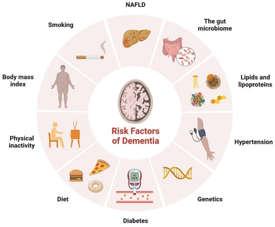
Figure 1.
Representation of various risk factors involved in the development of dementia.
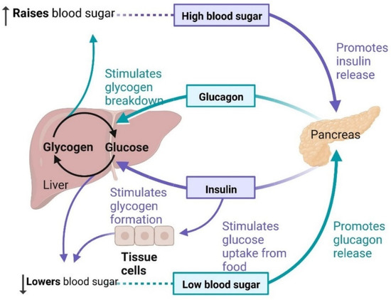
Figure 2.
Regulation of blood glucose occurs through insulin.
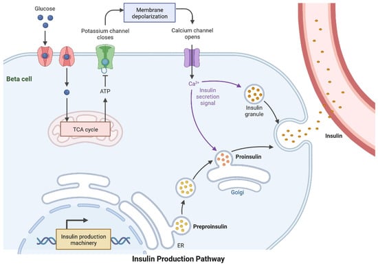
Figure 3.
Proinsulin is transformed into insulin and C-peptide and stored in secretory vesicles, where it can be released when needed. Insulin is secreted by pancreatic beta cells to regulate glucose homeostasis. Through the GLUT transporters, glucose is freely taken in by the beta cell and metabolized to produce ATP. This triggers a series of signals inside the beta cell that are required for glucose-induced insulin production.
The uptake of glucose in brain cells increased after activation of the ERK (extracellular signal-regulated kinases) and AKT (protein kinase B) pathways [8]. The P13K/AKT signaling pathway plays an important role in memory-encoding mechanisms and neuromodulation [9]. The insulin level in neuronal cells is determined by the rate at which it crosses the BBB by receptor-mediated transport and diffusion mechanisms. GLP-1 (glucagon-like peptide-1) regulates blood glucose levels by reducing glucagon secretion and decreasing food intake [10]. In T2DM, the GLP-1 receptor (GLP-1R) is responsible for the genes’ regulatory elements involved in neuronal survival and function. As GLP-1 agonists, they are used as targets in neurological disorders [10]. Some studies have shown that GLP-R is activated via the cAMP (cellular levels of cyclic AMP)/PKA (Protein Kinase A) pathways and is involved in neuroprotective action. The GLP-1R analog crosses the BBB and provides neuroprotection via cAMP/PKA signaling [11]. In this article, we will discuss the role of insulin signaling in the development of dementia and other neurological disorders. The discussion on GLP-1 activators and biomarkers linked to the development of T2DM and dementia revealed some remarkable points.
2. T2DM and Dementia
T2DM patients are 1.5–2.5-times more likely to develop neurological complications than people without diabetes. Insulin resistance (IR) is linked with Alzheimer’s disease (AD), and dysregulation in the molecular mechanism of insulin secretion may lead to histopathological lesions in AD. Hyperglycemia is a major risk factor for cognitive impairment and dementia. Cognitive function may also be impacted negatively by hypoglycemia. IR is the major problem contributing to the emergence of clinical complications. The abnormalities caused by raised glucose levels were identified using the mechanisms of the insulin signaling network, including protein and lipid levels, which may cause IR. In major tissues, such as liver muscles and adipose tissues, insulin activity and its receptor regulate signaling via gene expression, phosphorylation, and vascular trafficking, increasing the consumption of nutrition, and reducing the catabolic reaction. Insulin receptors are expressed in the brain and regulate energy consumption, diet intake, behavior, and vascular function. In the last few years, molecular mechanisms and the identification and characterization of genes and proteins, circulating lipids, exosome micro-RNA, and metabolites have provided significant outcomes for IR in T2DM. AD is more common in diabetic patients than in nondiabetic patients and it is associated with a higher incidence or mortality rate [12]. The development of dementia and other neurological disorders is common in T2DM, although a sporadic form of dementia is more common (Table 1) [13]. The pancreatic islets contain alpha and beta cells, which regulate glucagon and insulin, respectively. Insulin lowers the effects of glucose uptake in the skeletal muscles, liver, and brain. Blood glucose is increased by glucagon during the gluconeogenesis and lipolysis processes. The energy level is maintained by the brain in various parts of the body with a glucose homeostasis mechanism. Neuronal control of peripheral insulin sensitivity and glucose is shown in Figure 4. Glucose intolerance has been investigated in up to 80% of dementia patients [14]. In more than 11 years of study, researchers observed a higher prevalence of dementia and AD, and a 50–100% higher risk of developing dementia in diabetic patients [15,16].The common cause of dementia is AD, an irreversible disorder, which develops slowly (Figure 4). Common signs and symptoms of dementia are loss of memory, difficulty concentrating, difficulty with familiar tasks, altered behavior, and confusion in time and place. There are various types of dementia, such as vascular dementia, Lewy body dementia, frontotemporal dementia, mixed dementia, Huntington’s disease-related dementia, and Parkinson-related dementia [17]. An epidemiological study showed that more than 55 million people are suffering from dementia and, every year, more than 10 million cases are diagnosed [18]. Dementia affects various events, such as memory, thinking, orientation, comprehension, calculation, learning capacity, language, and judgment. The brain cells affected by tau protein accumulation and plaque formation, which may cause dementia and lead to irreversible neuronal cell damage, are more common in the frontal and temporal lobes [16]. Lewy body dementia is caused by an abnormal accumulation of alpha-synuclein (-Syn) in the neurons of the substantia nigra in Parkinson’s disease [12]. Vascular dementia is caused by deformities in brain tissue, blood clots, and abnormalities of blood vessels [7]. It can be used as a component or as an interface. T2DM and dementia are associated with age and affect millions of people around the world. Patients with dementia show abnormal blood glucose levels. The regulation of signal transduction pathways depends on the signaling of extracellular chain reactions; each response is based on the course of signaling requirements. In adipose tissue and muscles, glucose enters through the GLUT 2 receptors in the beta cells of the pancreas and in the liver cells (uptake of glucose in muscle and adipose tissue via enhanced diffusion at GLUT4 receptors). GLUT 1 and GLUT allow glucose to reach cells, including the brain, retina, kidney, RBC, and other parts of the body (Figure 3, Figure 4 and Figure 5).

Table 1.
The common occurrence of neurological complications in insulin-resistant patients.
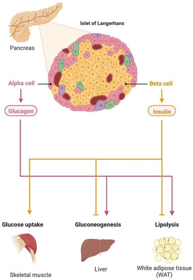
Figure 4.
Neuronal control of peripheral insulin sensitivity and glucose metabolism. The pancreatic islets contain alpha and beta cells, which regulate glucagon and insulin, respectively. Insulin lowers the effects of glucose uptake in the skeletal muscles, liver, and brain. Blood glucose is increased by glucagon during the gluconeogenesis and lipolysis processes. The energy level is maintained by the brain in various parts of the body with a glucose homeostasis mechanism.
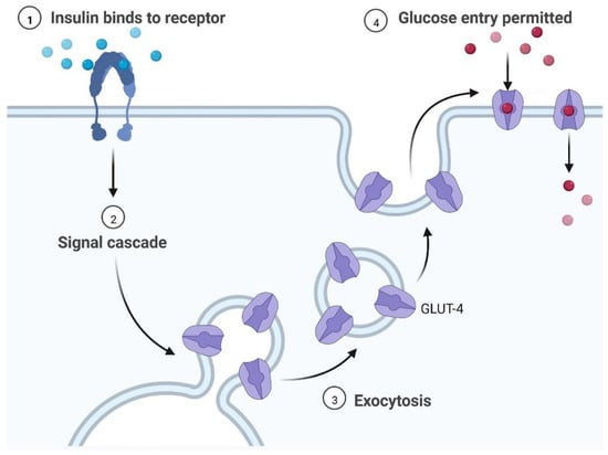
Figure 5.
Key steps of the insulin signaling pathway, including insulin binding, signal cascade, exocytosis, and glucose entry. The regulation of signal transduction pathways depends on the signaling of extracellular chain reactions; each response is based on the course of signaling requirements. Glucose is taken up in adipose tissue and muscles via the GLUT 2 receptors in pancreatic beta cells and liver cells, with enhanced diffusion at GLUT 4 receptors. GLUT 1 and GLUT 2 allow glucose to reach cells, including the brain, retina, kidney, RBC, and other parts of the body.
2.1. The Glucose Transporter’s Function in Cognition
T2DM increases the risk of cognitive impairment and IR is linked to a more rapid reduction in the memory-encoding mechanism and thinking skills [31]. Various types of genes, including SLC2A1–SLC2A14 and molecules (GLUT1–GLUT14), are actively involved in the transport of glucose in various regions of the brain and each cell type expresses multiple proteins [32]. GLUT proteins are classified into three classes. Proteins GLUT 1–GLUT 4 and GLUT 14 are classified as group I, GLUT 5, GLUT 7, GLUT 9, and GLUT 11 are classified as group 2, and GLUT 6, GLUT 8, GLUT 10, GLUT 12, and GLUT 13 are classified as group 3 [33,34]. GLUT 2 controls energy regulation, neurotransmitter release, and glucose release in glial cells. Brain stem nuclei and tanycytic, vagus motor nucleus, astrocytes, hypothalamus, arcuate nucleus, olfactory bulbs, nucleus tractus solitarius, paraventricular hypothalamic nucleus, lateral hypothalamic area, and neurons all contain GLUT 2 [34]. GLUT 3 is found in cell bodies, neurons, and dendrites; brain micro vessels; and brain astroglia cells. It has been observed that insulin accelerates the translocation of GLUT 3 and increases glucose uptake by neurons [35]. Glucose entry into the cells is carried out by GLUT 4, which is mainly found in the hippocampal region and maintains insulin regulation and improves cognitive development. It also acts as an insulin-sensitive glucose transporter [36]. There is evidence of GLUT 5 in microglial cells. The brain has a low fructose concentration, and glucose transport activity is substantially lower than that of fructose. Studies on animal models have demonstrated that fructose can pass across the BBB and be used as an energy source by brain cells [31]. The importance of GLUT 5 in the brain remains unclear and requires further investigation. GLUT 6 is involved in the nervous system’s physiological activity and transports hexoses across the membrane [34].
2.2. Insulin Signaling and Neuro-Complications
IR in dementia is caused by the amyloid precursor protein GSK3 (Glycogen synthase kinase 3) enzyme, involved in glycogen metabolism, oxidative stress, mitochondrial dysfunction, brain inflammation, ion channel activation, and the Shc family of signaling adaptor proteins. IR causes cell breakdown and destruction as well as increased glucose uptake, metabolism, and intake, all of which contribute to abnormal tau aggregation, inhibit lipolysis, and inhibit gluconeogenesis (Figure 6) [6]. If brain cells becoming too resistant to insulin leads to elevated blood glucose, impaired synaptic plasticity, microglial overactivation, mitochondrial dysfunction, neuronal apoptosis, nutrient deprivation, TAU phosphorylation, and cholinergic dysfunction, dementia is a group of symptoms affecting memory-encoding mechanisms, including difficulty in visual and spatial abilities, problem-solving, handling complex tasks, planning and organizing, coordination and motor functions, loss of memory, and changes in cognitive functions. Advanced dementia may develop into Alzheimer’s disease (AD). Dementia patients have synuclein aggregates and plaque accumulation, blood–brain barrier leakage, and neuroinflammation (Figure 7). Disintegrating microtubules and amyloid beta plaques are formed in AD (Figure 8). Sporadic forms of dementia are more common; both semantic and episodic memory are caused by cognitive impairment and visuospatial impairment. Motor coordination is also affected in severe cases of disease (Figure 5). [7]. IRs affect intellectual ability, increase the generation of excitability, and promote memory consolidation. IR in cognitive impairment is signified by mitochondrial dysfunction, which is involved in neurodegeneration by reducing glucose transport and inducing the formation of phosphorylated tau protein (Figure 6) [37]. The activation of IR autophosphorylation (IRP) leads to the tyrosine phosphorylation of IRS-1 (insulin receptor substrate 1), which activates PI3K (phosphoinositide 3-kinase) and decreases synaptic plasticity and memory [37,38]. The NMDARs (N-methyl-D-aspartate receptors) are activated by calcium ion channels and activated signaling is linked to the development of neurological complications. NMDAR and IR-dependent signaling molecular mechanisms resulted in amyloid oligomer accumulation, increased TNF-α release, and increased concentrations of stress-induced JNK (Jun N-terminal kinase), resulting in IRS-1 inhibitory phosphorylation. The amyloid β oligomers activate further extracellular exclusion of IRs from the cell surface. All these events block the neuronal regulation of insulin, leading to impaired synaptic plasticity (Figure 9) [37,38]. Dementia is a group of symptoms affecting memory-encoding mechanisms, including difficulty in visual and spatial abilities, problem-solving, handling complex tasks, planning, and organizing, coordination and motor functions, loss of memory, and changes in cognitive functions. Advanced dementia can progress to Alzheimer’s disease [36]. Dementia patients have synuclein aggregates and plaque accumulation, blood–brain barrier leakage, and neuroinflammation, as shown in Figure 10. Disintegrating microtubules and amyloid beta plaques are formed in AD, as shown in Figure 7.
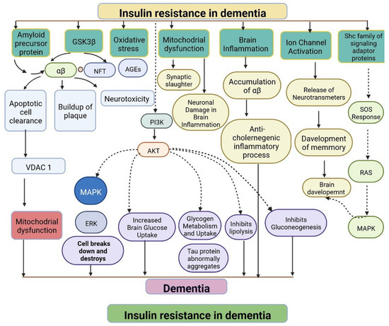
Figure 6.
Representation of IR in dementia: resistance to insulin leads to mitochondrial dysfunction, damage to brain cells, altered brain glucose, aggregation of tau protein, inhibition of the lipolysis process, and an anticholinergic inflammatory process. The dotted lines represent inhibitory mechanisms.
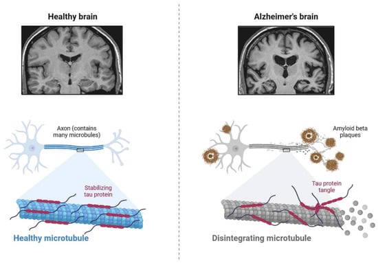
Figure 7.
The pathological properties of the Alzheimer’s disease brain (involving tau protein) compared with that of the healthy brain, on various magnification levels. It can be adapted as a component.
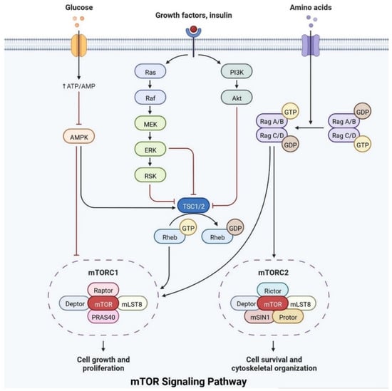
Figure 8.
The mTOR pathway integrates growth factor signaling to regulate cellular metabolism, growth, and survival.
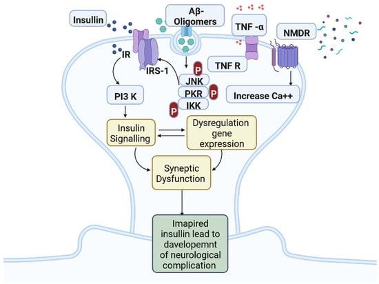
Figure 9.
Insulin resistance and development of neuro-complications: the activation of IR autophosphorylation (IRP) leads to the tyrosine phosphorylation of IRS-1 (insulin receptor substrate 1), which activates PI3K (phosphoinositide 3-kinase) and decreases synaptic plasticity and memory.
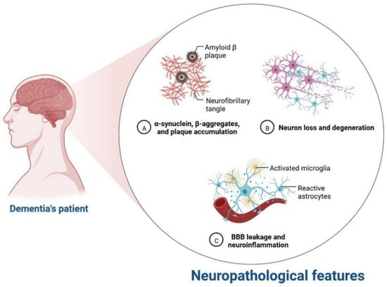
Figure 10.
Neurological complication in dementia patients.
2.3. Relationship between Insulin Receptor m-TOR Pathway
Insulin plays an important role in cell growth, repair, activation, dendritic development, synaptic maintenance, and neuroprotection and is actively involved in learning and memory. Various investigations have shown that changes in insulin levels in the brain lead to the development of neurological complications [39]. Insulin also activates the N-methyl-D-aspartate receptor on the cell membrane, cortical cerebral glucose metabolism, acetylcholine, and norepinephrine, which are actively involved in memory-encoding mechanisms [40]. In the cerebral cortex, IRS (insulin receptor substrate 1) is abundant in the cerebral cortex, hippocampus, hypothalamus, olfactory bulb, septum, and amygdala. Insulin signaling is also involved in regulating synaptic remodeling, which is involved in memory consolidation [9]. T2DM patients have lower brain insulin receptor sensitivity, downregulate the IRS-1, and have lower levels of insulin-like growth factors, as well as lower insulin levels in CSF [30]. Insulin signaling is controlled by mTOR pathways, as shown in Figure 8.
IRs are actively involved in memory and tau phosphorylation, due to the loss of IR receptor activity and downregulated Aβ (Amyloid beta) oligomer binding sites in the synapse [29]. The accumulation of Aβ leads to the development of AD. IGF signaling activation promotes amyloid-protein precursor (APP-A) trafficking, as well as the accumulation of amyloid processing, tau phosphorylation, and a reduction in cerebral blood flow [41]. IRs are transmembrane receptors and are made with alpha and beta subunits. Both the subunits are activated by insulin, which then activates tyrosine kinase enzymes for phosphorylation, which leads to conformational changes in their structure [38]. Changes in their structures favor binding with PI3K (phosphoinositide 3-kinases) and binding with the IRS [9]. After interaction with IRs, the inactive form of PI3K becomes active. The active PI3K enzyme is generated by PIP2 (phosphorylate phosphatidylinositol (4,5)-bisphosphate) in the cell membrane, which then causes the creation of phosphatidylinositol (3,4,5)-trisphosphate (PIP3), which activates AKT/PKB (Figure 8) [42]. Studies on animal models have revealed that IR inhibitors prevent the memory-encoding mechanism. Injecting insulin in an animal model increases the memory-encoding process [42]. IR decreases AKT activity, which inhibits GSK-3 and causes tau protein to be hyperphosphorylated [43]. AKT signaling regulates various responses at the cellular level, such as glycogen synthase kinase-3 beta (GSK3) neuronal survival and TAU phosphorylation [41]. Increasing the synthesis of GSK3β may alter the post-translational modifications in MAPs (microtubule-associated proteins), such as tau protein [41]. Dementia is also caused by mutations in APP (amyloid precursor protein). Synaptic signaling is interrupted by Aβ fibrils infiltrating into synaptic clefts [42]. Polymers of Aβ also play an important role in the development of dementia [38]. The collection of APP alters the ion channel mechanism and disrupts the altered glucose homeostasis, leading to neuronal integrity degradation and cell death [44].
2.4. Development of Dementia due to Genetic Modifications
mRNA expression and the downregulation of associated receptors are linked to dementia [45]. The oxidative-phosphorylation-related genes are expressed in dementia patients. The mtDNA irregularities are associated with phenotypic variability [46]. Genetic abnormalities are caused by chromosomal defects and damage from neuronal oxidation. The development of dementia due to T2DM is linked with genetic variability [47]. APO E (apolipoprotein E) is expressed by chromosome 19 and exists in three isoforms: apo e2, apo e3, and polymorphic in nature [48]. More than 75 loci have been identified for the development of disease traits [49]. ADAM17 (A disintegrin and metalloprotease 17), ICA1 (Islet Cell Autoantigen 1), DOC2A (Double C2 Domain Alpha), DGKQ (Diacylglycerol Kinase Theta), and ICA1L (Islet Cell Autoantigen 1 Like) are the genes responsible for the regulation of APP metabolism via non-amyloid pathways. Cognitive impairment is caused by HMGB1 (High-mobility group box protein 1), RAGE (Receptor for Advanced Glycation End Products), and TLR4 (Toll-like receptor 4) in hyperglycemic conditions [50]. All these genes impair endothelial cell function and may disrupt various signaling pathways, resulting in an accumulation [51]. Dementia is also associated with the APP, PS1 (presenilin 1), and PS2 genes. Mutations in these genes cause IR in astrocytes and microglial cells [52].
2.5. Progression of Dementia due to Dopamine Dysregulation in Substantia Nigra
Insulin secretion and glucose homeostasis serve as a basis for neural modulation. Degenerative and functional disorders of the central nervous system are directly related to dementia. Dopamine is a neurotransmitter that plays a major role in neurological complications, as shown in Figure 11.
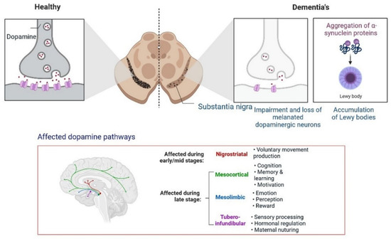
Figure 11.
Progression of dementia due to dopamine dysregulation in substantia nigra.
Poor insulin activity in the brain is linked with a high level of cholinergic action, leading to the development of dementia [53].
Furthermore, the development of dementia also occurs due to alterations in the dopamine pathway in the substantia nigra. A PPAR agonist causes memory loss by increasing intracellular glucose oxidation uptake in neurons [54]. Dysregulation in cholinergic neurotransmission alters the performance of the hippocampus area, the recollection of memory, acetylcholine-induced responses, and increased ChE activity in the brain during cognitive deficits. In low amounts, choline acetyltransferase (ChAT) is observed in patients with dementia and neuronal dysfunction [55]. All these findings can be used for identification and the development of etiology and treatment options.
2.6. Neuronal Apoptosis in Dementia
IR and T2DM result in neuronal death. Different neurological conditions, such as Huntington’s disease, amyotrophic lateral sclerosis and dementia, Parkinson’s disease, and Alzheimer’s disease, may occur because of apoptosis [56], IGF (insulin-like growth factor) [57], increased Bax/Bcl-x ratio, hippocampal neuronal death and L caspase-3 activity, mitochondrial dysfunction, cerebral blood vessel dysfunction, myelin and axon damage, intracytoplasmic calcium deposition, and Purkinje cell damage; IGF-I, IGF-IR, and IR activity; endoplasmic dysfunction, BBB degradation; ependymal [58].
2.7. Significance of Ketone Bodies in Diabetes-Related Dementia
Brain cells use ketone bodies as a source of energy in situations of nutrient deprivation, after exercise, or low carbohydrates. Regulation of ketone bodies is also linked with gluconeogenesis, the tricarboxylic acid cycle, and fatty acid b-oxidation. Antioxidant responses to ketone bodies are increased [59]. Hyperglycemic conditions can reduce the activity of GABA and glutamate neurotransmitters. Cholinergic transmission was found to be dysregulated in the brain hippocampus [60]. Another neurotransmitter, dopamine, is also associated with behavior, cognition, and emotions. Reduced levels of dopamine receptors have been observed in patients with type 2 diabetes [61]. The 5-HT (5-hydroxytryptamine) neurotransmitters are associated with neuronal cell regeneration and synaptic plasticity. Glucagon-like peptide-1 hormone (GLP-1H) inhibits IR and neuroinflammation in the brain under oxidative stress. GLP1H is also involved in the regulation of synaptic plasticity and neurogenesis [62]. Ketosis increases the levels of GABA (gamma-aminobutyric acid) and excitatory glutamate and regulates the levels of serotonin and dopamine, which are linked with depression and anxiety [63]. Ketone bodies in the hydroxybutyrate form are involved in neuronal anti-apoptosis pathways and cell survival [63]. In the mouse model, fat-rich animals induce APP/PS1xdb/db deposition, whereas ketone bodies improve cognitive impairment function [60]. Ketone bodies regulate neural signaling, increase the sensitivity of insulin, reduce the effects of oxidative stress, increase synaptic activity, and maintain the level of neurotransmitter activity. Low ketone body levels may cause pathological conditions in T2DM [64]. More research is needed to understand the regulation of ketone bodies at low levels in neuroprotection and neurotoxicity at high levels in diabetes-induced dementia treatment options.
2.8. The Function of Mitochondria in Diabetes-Related Cognitive Impairment
Cell signaling molecules and transcription factors play a very important role in intracellular energy metabolism in the mitochondria. Brain disorders linked with diabetes are caused by abnormalities in mitochondrial functions [65]. IR is also caused by mitochondrial dysfunction, oxidative stress, neuronal damage, decreased mETC (mitochondrial electron transport chain) activity and ATP synthesis, apoptosis, lipid peroxide accumulation, decreased glutathione peroxidase activity, ferroptosis, and An accumulation, all of which can lead to cognitive impairment [66]. Mitophagy is regulated by PINK1 (PTEN-induced kinase 1) and protects the neurons, as has been observed in animal models. PINK1-dependent mitophagy via MT2/Akt/NF-κB is achieved by melatonin, which prevents ROS (reactive oxygen species) accumulation and apoptosis (Figure 12) [67].
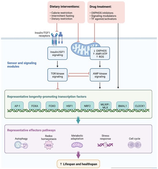
Figure 12.
Representation of sensor and signaling molecule dietary intervention and treatment option regulated by effector pathways.
In diabetic mice, increased levels of LC3-II (microtubule-associated protein 1A/1B-light chain 3) and p62 (nucleoporin p62) and decreased levels of PINK1 have been observed, which leads to blocked autophagy [68]. Dephosphorylation of FUNDC1 was also observed in T2DM mice. Cognitive impairment in T2DM mice occurs due to homeostasis, impaired mitophagy, proteostasis disorder, and damage to multiple mechanisms [69]. Although various mechanisms are not completely known, we need to explore new mechanisms and pathways for the prevention and treatment of dementia. Figure 7 depicts the roles of sensor and signaling molecules, as well as transcription factors, in dietary intervention and treatment options regulated by effector pathways for early diagnosis and treatment, regulated by effector pathways for early diagnosis and treatment.
2.9. Progressive Dementia Due to Microglial Overactivation
Microglia cells are expressed by the AAβ protein and increase brain insulin levels due to Aβ accumulation. Protein synthesis is being studied as a potential target for drug development and dementia development. Apoptosis occurs due to the generation of ROS and AGE products [70]. Neuroinflammation is also observed due to plaque and tangle formation, oxidative stress markers, oxidized lipids and proteins, and ROS, which is insulin resistant and causes dementia. Microglial cell stimulation and increased levels of proinflammatory cytokines, such as interleukin-1, IL-6, and tumor necrosis factor, inhibit neurogenesis and cause cognitive deficits (Figure 13) [71]. More research is needed to investigate the potential of neuroinflammation treatment, and the cognitive impairment caused by diseases in their early stages.
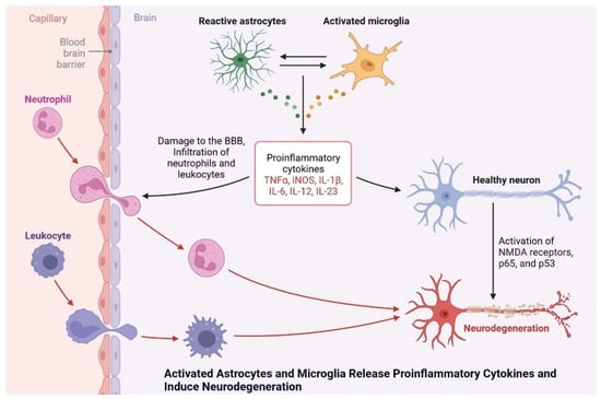
Figure 13.
Activated astrocytes and microglia release proinflammatory cytokines and induce neurodegeneration.
3. Antidiabetic Drug Development
Various anti-diabetic drugs are available for patients with T2DM as monotherapy or combination therapy [72]. Most of the drugs mainly target neurological, cognitive, and cardiovascular clinical complications. Antidiabetic drugs are classified based on their mechanism of action [73]. The clinical evaluation of anti-diabetic drugs in diabetic patients with cognitive dysfunction was investigated in various studies (Table 2).

Table 2.
Antidiabetic drug list, including mechanisms of action, significance, and risk factors.
3.1. Glucose–Sodium Co-transporter 2
SGLT2 (sodium–glucose co-transporter 2) is an inhibitor of the reabsorption of glucose in the renal tubules. In a mouse model, the efficacy of empagliflozin and dapagliflozin was investigated. In treated mice, plaque burden and neuronal deactivation have been observed [73]. In another study, improved mitochondrial function and cognitive impairment were observed in rats treated with dapagliflozin [74].
3.2. Pioglitazone
Pioglitazone has been shown to improve cognitive impairment and reduce tau protein deposition in triple-transgenic mice [75]. Pioglitazone has also been shown to activate microglia in another model [76]. Pioglitazone treatment also normalizes metabolic and vascular functions, reduces IR, and maintains ROS (reactive oxygen species) in the hippocampus and cerebral cortex for learning and memory [77].
3.3. Rosiglitazone
In various investigations, the clinical efficacy of rosiglitazone was observed. A randomized double-blind study showed improved cognitive impairment in early-stage AD patients without the ApoE4 allele [78]. Treatment with rosiglitazone has shown mitochondrial biogenesis in the mouse brain [79] and combination therapy with metformin has shown stable results [80].
3.4. Metformin
Metformin is an oral hypoglycemia drug; various clinical investigations are carried out on cognitive dysfunction in T2DM. Metformin in combination with sulfonylureas has reduced dementia by up to 35% [81]. Long-term metformin treatment has been linked to the development of Alzheimer’s disease. No dementia or neurological complications have been observed with other drugs, such as thiazolidinediones, sulfonylureas, or insulin.
3.5. Thiazolidinediones
Thiazolidinediones are PPAR agonists that increase insulin secretion in T2DM patients. Two drugs, namely rosiglitazone and pioglitazone, belong to the thiazolidinediones. Clinical efficacy has been shown to improve cognitive impairment [82].
3.6. GLP-1-Based Therapies
It has been observed that the expression of GLP-1 in mice improves cognitive function. Glp-1-expressing mice also had severely impaired LTP in the CA1 area of the hippocampus. GLP-1 plays an important role in memory formation [83].
3.7. GLP-1 Analogs
Exendin-4 reduced HbA1c and cerebrovascular A in a 16-week trial in 5XFAD mice (3xTgAD). Exendin-4 also reduces neuronal apoptosis, activates the CREB transcription factor, and induces BDNF activity. Another 16-week study in 5XFAD mice looked at maintaining mitochondrial functioning and cognitive impairment [84].
3.8. Liraglutide
Improved cognitive impairment has been observed after a 3-month administration of liraglutide in a mouse model. Another study analyzed a 28-day liraglutide treatment in 3xTg-AD female mice and observed that it prevented memory impairment [85]. In treated liraglutide HFD-fed mice or ob/ob mice, neurogenesis and hippocampal synaptic plasticity were significantly improved [86].
3.9. Dulaglutide and Lisisenatide
The clinical efficacy of dulaglutide was investigated in 371 sites in 24 countries and reduced cognitive impairment in T2DM by 14%. Lixisenatide was found to be effective against plaque deposition and neuroinflammation in the brains of tau mice after 2 months of treatment. In HFD-fed mice, lixisenatide has been shown to reduce IR and neurogenesis [87].
3.10. New GLP-1 Analogs
GLP-1 and gastric inhibitory peptide (GIP) receptor agonists may be more effective in the treatment of Alzheimer’s disease [88]. In an intracerebroventricular STZ-induced IR AD rat model, working memory and spatial memory deficits were reversed by DA5-CH, a new dual GLP-1/GIP receptor agonist [89]. In the 3xTg mouse model, a new GLP-1/GIP, and GIP were found to enhance working memory.
3.11. DPP-4 Inhibitors
Current diabetic treatments, such as sitagliptin, vildagliptin, alogliptin, and linagliptin, all use different forms of DPP-4 inhibitors to maintain the levels of endogenous GLP-1. DPP-4 inhibitors have been shown to greatly prevent dementia with metformin and thiazolidinedione [90].
3.12. Linagliptin A
In a 3xTg-AD mouse model, linagliptin treatment for 8 weeks resulted in a notable improvement in cognitive impairment. Linagliptin also lessens the development of amyloid beta-amyloid and neuroinflammation [91]. Furthermore, linagliptin also increases cerebral blood flow and cognitive decline in a tauopathy mouse model. The DPP-4 inhibitor also increased the levels of the tight-junction protein claudin-5 and prevented neuronal loss in the hippocampus and cortex of these mice.
3.13. Alogliptin
Alogliptin mainly activates CREB in insulin-positive β cells of the islets and increases anti-apoptotic bcl-2. Another study carried out in Zucker diabetic rats for 10 weeks showed neuroprotective proteins in the brain and reduced the level of inflammatory markers [92].
3.14. Vildagliptin
A 6-month clinical trial with vildagliptin improved HbA1c and cognitive impairment in T2DM patients [93]. In HFD rats, combined therapy with pioglitazone improved dendritic spines in the CA1 hippocampus and reduced apoptosis of hippocampal neurons [93].
3.15. Sitagliptin
The efficacy of sitagliptin has been studied in animal models. Improved cognitive impairment and reduced plaque deposition have been observed in APP/PS1 mice after oral administration of sitagliptin for 8 weeks [94]. Furthermore, they reduced the activity of inflammatory markers and suppressed white-matter lesions.
4. Conclusions
Determining the mechanisms associated with T2DM can lead to new approaches to dementia’s early detection and treatment. The link between T2DM and cognitive dysfunction is mainly reliable and a study on humans and animals can explore the most effective mechanism that could be useful. There is a very tough puzzle to solve for T2DM and the degeneration of neurons. Neurofibrillary tangles and neurotic plaques, cerebrovascular abnormalities, decreased brain volume, markers of white-matter injury, retinal measures, retinal nerve fiber, retinal vascular tortuosity, and fluid markers for gliosis and neurodegeneration are all targets for cognitive dysfunction. The development of glycoproteomic-based tools may be useful for the identification of novel biomarkers for early diagnosis and treatment. The development of novel treatment options for T2DM in dementia IR is developed due to altered insulin signaling, which is essential for energy metabolism, neuronal growth, neuroprotection, and synaptic plasticity. Peripheral and central IR are common in neurological disorders and can be used as potential targets for intervention in and treatment of dementia. Various clinical trials have improved cognition. As a result, insulin treatment must be tested in a variety of contexts, including known resistance or patients with T2DM, screening, and including patients at all stages. We can reduce the global burden of dementia by applying these promising areas of research and novel approaches for patients with T2DM and dementia.
Author Contributions
D.D.S. and D.K.Y. conceived and designed the project and collected data from the literature. D.D.S., A.A.S., M.Y.A., S.E.I.E., I.H., E.-H.C. and D.K.Y. analyzed the data. D.D.S. and D.K.Y. wrote the manuscript. All authors have read and approved the final version of the manuscript.
Funding
The authors are thankful to the Basic Science Research Program of the National Research Foundation of Korea (NRF) funded by the Ministry of Education (2021R1A6A1A03038785 and 2020R1I1A1A01073071).
Acknowledgments
D.D.S. thanks the Amity Institute of Biotechnology, Amity University Rajasthan, Jaipur, India, and D.K.Y. thanks Gachon University, Republic of Korea, for providing the necessary computational and journal subscriptions for the needed literature search. The authors appreciate Biorender.com’s graphics assistance. The authors extend their appreciation to the Deanship of Scientific Research at King Khalid University for funding this work through large groups (project under grant number R.G.P.2/78/43).
Conflicts of Interest
The authors declare that the research was conducted in the absence of any commercial or financial relationships that could be construed as a potential conflict of interest.
References
- Chatterjee, S.; Khunti, K.; Davies, M.J. Type 2 Diabetes. Lancet 2017, 389, 2239–2251. [Google Scholar] [CrossRef]
- Feldman, E.L.; Callaghan, B.C.; Pop-Busui, R.; Zochodne, D.W.; Wright, D.E.; Bennett, D.L.; Bril, V.; Russell, J.W.; Viswanathan, V. Diabetic Neuropathy. Nat. Rev. Dis. Primers 2019, 5, 41. [Google Scholar] [CrossRef]
- Bose, K.S.; Sarma, R.H. Delineation of the Intimate Details of the Backbone Conformation of Pyridine Nucleotide Coenzymes in Aqueous Solution. Biochem. Biophys. Res. Commun. 1975, 66, 1173–1179. [Google Scholar] [CrossRef]
- Tomic, D.; Shaw, J.E.; Magliano, D.J. The Burden and Risks of Emerging Complications of Diabetes Mellitus. Nat. Rev. Endocrinol. 2022, 18, 525–539. [Google Scholar] [CrossRef] [PubMed]
- Bunn, F.; Goodman, C.; Reece Jones, P.; Russell, B.; Trivedi, D.; Sinclair, A.; Bayer, A.; Rait, G.; Rycroft-Malone, J.; Burton, C. What Works for Whom in the Management of Diabetes in People Living with Dementia: A Realist Review. BMC Med. 2017, 15, 141. [Google Scholar] [CrossRef] [PubMed]
- Thomassen, J.Q.; Tolstrup, J.S.; Benn, M.; Frikke-Schmidt, R. Type-2 Diabetes and Risk of Dementia: Observational and Mendelian Randomisation Studies in 1 Million Individuals. Epidemiol. Psychiatr. Sci. 2020, 29, e118. [Google Scholar] [CrossRef]
- Lyu, F.; Wu, D.; Wei, C.; Wu, A. Vascular Cognitive Impairment and Dementia in Type 2 Diabetes Mellitus: An Overview. Life Sci. 2020, 254, 117771. [Google Scholar] [CrossRef] [PubMed]
- McNay, E.C.; Pearson-Leary, J. GluT4: A Central Player in Hippocampal Memory and Brain Insulin Resistance. Exp. Neurol. 2020, 323, 113076. [Google Scholar] [CrossRef] [PubMed]
- Zheng, M.; Wang, P. Role of Insulin Receptor Substance-1 Modulating PI3K/Akt Insulin Signaling Pathway in Alzheimer’s Disease. 3 Biotech 2021, 11, 179. [Google Scholar] [CrossRef] [PubMed]
- Monti, G.; Gomes Moreira, D.; Richner, M.; Mutsaers, H.A.M.; Ferreira, N.; Jan, A. GLP-1 Receptor Agonists in Neurodegeneration: Neurovascular Unit in the Spotlight. Cells 2022, 11, 2023. [Google Scholar] [CrossRef]
- Yang, X.; Qiang, Q.; Li, N.; Feng, P.; Wei, W.; Hölscher, C. Neuroprotective Mechanisms of Glucagon-Like Peptide-1-Based Therapies in Ischemic Stroke: An Update Based on Preclinical Research. Front. Neurol. 2022, 13, 844697. [Google Scholar] [CrossRef] [PubMed]
- Chau, A.C.M.; Cheung, E.Y.W.; Chan, K.H.; Chow, W.S.; Shea, Y.F.; Chiu, P.K.C.; Mak, H.K.F. Impaired Cerebral Blood Flow in Type 2 Diabetes Mellitus—A Comparative Study with Subjective Cognitive Decline, Vascular Dementia and Alzheimer’s Disease Subjects. NeuroImage Clin. 2020, 27, 102302. [Google Scholar] [CrossRef]
- Ehtewish, H.; Arredouani, A.; El-Agnaf, O. Diagnostic, Prognostic, and Mechanistic Biomarkers of Diabetes Mellitus-Associated Cognitive Decline. Int. J. Mol. Sci. 2022, 23, 6144. [Google Scholar] [CrossRef]
- Janson, J.; Laedtke, T.; Parisi, J.E.; O’Brien, P.; Petersen, R.C.; Butler, P.C. Increased Risk of Type 2 Diabetes in Alzheimer Disease. Diabetes 2004, 53, 474–481. [Google Scholar] [CrossRef] [PubMed]
- Huang, C.-C.; Chung, C.-M.; Leu, H.-B.; Lin, L.-Y.; Chiu, C.-C.; Hsu, C.-Y.; Chiang, C.-H.; Huang, P.-H.; Chen, T.-J.; Lin, S.-J.; et al. Diabetes Mellitus and the Risk of Alzheimer’s Disease: A Nationwide Population-Based Study. PLoS ONE 2014, 9, e87095. [Google Scholar] [CrossRef]
- Biessels, G.J.; Staekenborg, S.; Brunner, E.; Brayne, C.; Scheltens, P. Risk of Dementia in Diabetes Mellitus: A Systematic Review. Lancet Neurol. 2006, 5, 64–74. [Google Scholar] [CrossRef] [PubMed]
- Biessels, G.J.; Despa, F. Cognitive Decline and Dementia in Diabetes Mellitus: Mechanisms and Clinical Implications. Nat. Rev. Endocrinol. 2018, 14, 591–604. [Google Scholar] [CrossRef] [PubMed]
- Nichols, E.; Steinmetz, J.D.; Vollset, S.E.; Fukutaki, K.; Chalek, J.; Abd-Allah, F.; Abdoli, A.; Abualhasan, A.; Abu-Gharbieh, E.; Akram, T.T.; et al. Estimation of the Global Prevalence of Dementia in 2019 and Forecasted Prevalence in 2050: An Analysis for the Global Burden of Disease Study 2019. Lancet Public Health 2022, 7, e105–e125. [Google Scholar] [CrossRef] [PubMed]
- Ott, A.; Stolk, R.P.; van Harskamp, F.; Pols, H.A.P.; Hofman, A.; Breteler, M.M.B. Diabetes Mellitus and the Risk of Dementia: The Rotterdam Study. Neurology 1999, 53, 1937. [Google Scholar] [CrossRef] [PubMed]
- MacKnight, C.; Rockwood, K.; Awalt, E.; McDowell, I. Diabetes Mellitus and the Risk of Dementia, Alzheimer’s Disease and Vascular Cognitive Impairment in the Canadian Study of Health and Aging. Dement. Geriatr. Cogn. Disord. 2002, 14, 77–83. [Google Scholar] [CrossRef] [PubMed]
- Craft, S.; Peskind, E.; Schwartz, M.W.; Schellenberg, G.D.; Raskind, M.; Porte, D. Cerebrospinal Fluid and Plasma Insulin Levels in Alzheimer’s Disease: Relationship to Severity of Dementia and Apolipoprotein E Genotype. Neurology 1998, 50, 164–168. [Google Scholar] [CrossRef] [PubMed]
- Xu, W.L.; Qiu, C.X.; Wahlin, A.; Winblad, B.; Fratiglioni, L. Diabetes Mellitus and Risk of Dementia in the Kungsholmen Project: A 6-Year Follow-up Study. Neurology 2004, 63, 1181–1186. [Google Scholar] [CrossRef]
- Schnaider Beeri, M.; Goldbourt, U.; Silverman, J.M.; Noy, S.; Schmeidler, J.; Ravona-Springer, R.; Sverdlick, A.; Davidson, M. Diabetes Mellitus in Midlife and the Risk of Dementia Three Decades Later. Neurology 2004, 63, 1902–1907. [Google Scholar] [CrossRef]
- Ronnemaa, E.; Zethelius, B.; Sundelof, J.; Sundstrom, J.; Degerman-Gunnarsson, M.; Berne, C.; Lannfelt, L.; Kilander, L. Impaired Insulin Secretion Increases the Risk of Alzheimer Disease. Neurology 2008, 71, 1065–1071. [Google Scholar] [CrossRef] [PubMed]
- Lin, Z.; Tian, H.; Lam, K.S.L.; Lin, S.; Hoo, R.C.L.; Konishi, M.; Itoh, N.; Wang, Y.; Bornstein, S.R.; Xu, A.; et al. Adiponectin Mediates the Metabolic Effects of FGF21 on Glucose Homeostasis and Insulin Sensitivity in Mice. Cell Metab. 2013, 17, 779–789. [Google Scholar] [CrossRef] [PubMed]
- Mehta, H.B.; Mehta, V.; Goodwin, J.S. Association of Hypoglycemia with Subsequent Dementia in Older Patients with Type 2 Diabetes Mellitus. J. Gerontol. Ser. A 2016, 72, 1110–1116. [Google Scholar] [CrossRef] [PubMed]
- Cheng, G.; Huang, C.; Deng, H.; Wang, H. Diabetes as a Risk Factor for Dementia and Mild Cognitive Impairment: A Meta-Analysis of Longitudinal Studies: Diabetes and Cognitive Function. Intern. Med. J. 2012, 42, 484–491. [Google Scholar] [CrossRef] [PubMed]
- Feinkohl, I.; Aung, P.P.; Keller, M.; Robertson, C.M.; Morling, J.R.; McLachlan, S.; Deary, I.J.; Frier, B.M.; Strachan, M.W.J.; Price, J.F.; et al. Severe Hypoglycemia and Cognitive Decline in Older People with Type 2 Diabetes: The Edinburgh Type 2 Diabetes Study. Diabetes Care 2014, 37, 507–515. [Google Scholar] [CrossRef] [PubMed]
- Smolina, K.; Wotton, C.J.; Goldacre, M.J. Risk of Dementia in Patients Hospitalised with Type 1 and Type 2 Diabetes in England, 1998–2011: A Retrospective National Record Linkage Cohort Study. Diabetologia 2015, 58, 942–950. [Google Scholar] [CrossRef] [PubMed]
- Matsuzaki, T.; Sasaki, K.; Tanizaki, Y.; Hata, J.; Fujimi, K.; Matsui, Y.; Sekita, A.; Suzuki, S.O.; Kanba, S.; Kiyohara, Y.; et al. Insulin Resistance Is Associated with the Pathology of Alzheimer Disease: The Hisayama Study. Neurology 2010, 75, 764–770. [Google Scholar] [CrossRef]
- Shah, K.; DeSilva, S.; Abbruscato, T. The Role of Glucose Transporters in Brain Disease: Diabetes and Alzheimer’s Disease. Int. J. Mol. Sci. 2012, 13, 12629–12655. [Google Scholar] [CrossRef] [PubMed]
- Vulturar, R.; Chiș, A.; Pintilie, S.; Farcaș, I.M.; Botezatu, A.; Login, C.C.; Sitar-Taut, A.-V.; Orasan, O.H.; Stan, A.; Lazea, C.; et al. One Molecule for Mental Nourishment and More: Glucose Transporter Type 1—Biology and Deficiency Syndrome. Biomedicines 2022, 10, 1249. [Google Scholar] [CrossRef]
- Holman, G.D. Structure, Function and Regulation of Mammalian Glucose Transporters of the SLC2 Family. Pflugers Arch. Eur. J. Physiol. 2020, 472, 1155–1175. [Google Scholar] [CrossRef] [PubMed]
- Koepsell, H. Glucose Transporters in Brain in Health and Disease. Pflugers Arch. Eur. J. Physiol. 2020, 472, 1299–1343. [Google Scholar] [CrossRef] [PubMed]
- Frazier, H.N.; Ghoweri, A.O.; Anderson, K.L.; Lin, R.-L.; Popa, G.J.; Mendenhall, M.D.; Reagan, L.P.; Craven, R.J.; Thibault, O. Elevating Insulin Signaling Using a Constitutively Active Insulin Receptor Increases Glucose Metabolism and Expression of GLUT3 in Hippocampal Neurons. Front. Neurosci. 2020, 14, 668. [Google Scholar] [CrossRef]
- Jurcovicova, J. Glucose Transport in Brain—Effect of Inflammation. Endocr. Regul. 2014, 48, 35–48. [Google Scholar] [CrossRef] [PubMed]
- Bell, S.M.; Barnes, K.; de Marco, M.; Shaw, P.J.; Ferraiuolo, L.; Blackburn, D.J.; Venneri, A.; Mortiboys, H. Mitochondrial Dysfunction in Alzheimer’s Disease: A Biomarker of the Future? Biomedicines 2021, 9, 63. [Google Scholar] [CrossRef]
- Oddo, S. The Role of MTOR Signaling in Alzheimer Disease. Front. Biosci. 2012, S4, 941–952. [Google Scholar] [CrossRef]
- Shaughness, M.; Acs, D.; Brabazon, F.; Hockenbury, N.; Byrnes, K.R. Role of Insulin in Neurotrauma and Neurodegeneration: A Review. Front. Neurosci. 2020, 14, 547175. [Google Scholar] [CrossRef] [PubMed]
- Chen, Y.; Deng, Y.; Zhang, B.; Gong, C.-X. Deregulation of Brain Insulin Signaling in Alzheimer’s Disease. Neurosci. Bull. 2014, 30, 282–294. [Google Scholar] [CrossRef]
- Hobday, A.L.; Parmar, M.S. The Link Between Diabetes Mellitus and Tau Hyperphosphorylation: Implications for Risk of Alzheimer’s Disease. Cureus 2021, 13, e18362. [Google Scholar] [CrossRef]
- Heras-Sandoval, D.; Avila-Muñoz, E.; Arias, C. The Phosphatidylinositol 3-Kinase/MTor Pathway as a Therapeutic Target for Brain Aging and Neurodegeneration. Pharmaceuticals 2011, 4, 1070–1087. [Google Scholar] [CrossRef]
- Yang, L.; Wang, H.; Liu, L.; Xie, A. The Role of Insulin/IGF-1/PI3K/Akt/GSK3β Signaling in Parkinson’s Disease Dementia. Front. Neurosci. 2018, 12, 73. [Google Scholar] [CrossRef] [PubMed]
- Hu, Z.; Jiao, R.; Wang, P.; Zhu, Y.; Zhao, J.; de Jager, P.; Bennett, D.A.; Jin, L.; Xiong, M. Shared Causal Paths Underlying Alzheimer’s Dementia and Type 2 Diabetes. Sci. Rep. 2020, 10, 4107. [Google Scholar] [CrossRef] [PubMed]
- Canchi, S.; Raao, B.; Masliah, D.; Rosenthal, S.B.; Sasik, R.; Fisch, K.M.; de Jager, P.L.; Bennett, D.A.; Rissman, R.A. Integrating Gene and Protein Expression Reveals Perturbed Functional Networks in Alzheimer’s Disease. Cell Rep. 2019, 28, 1103–1116.e4. [Google Scholar] [CrossRef]
- Ramos-Campoy, O.; Lladó, A.; Bosch, B.; Ferrer, M.; Pérez-Millan, A.; Vergara, M.; Molina-Porcel, L.; Fort-Aznar, L.; Gonzalo, R.; Moreno-Izco, F.; et al. Differential Gene Expression in Sporadic and Genetic Forms of Alzheimer’s Disease and Frontotemporal Dementia in Brain Tissue and Lymphoblastoid Cell Lines. Mol. Neurobiol. 2022, 59, 6411–6428. [Google Scholar] [CrossRef]
- Wang, X.; Lopez, O.L.; Sweet, R.A.; Becker, J.T.; DeKosky, S.T.; Barmada, M.M.; Demirci, F.Y.; Kamboh, M.I. Genetic Determinants of Disease Progression in Alzheimer’s Disease. J. Alzheimer’s Dis. 2014, 43, 649–655. [Google Scholar] [CrossRef]
- Liu, C.-C.; Kanekiyo, T.; Xu, H.; Bu, G. Apolipoprotein E and Alzheimer Disease: Risk, Mechanisms and Therapy. Nat. Rev. Neurol. 2013, 9, 106–118. [Google Scholar] [CrossRef] [PubMed]
- Dove, A.; Shang, Y.; Xu, W.; Grande, G.; Laukka, E.J.; Fratiglioni, L.; Marseglia, A. The Impact of Diabetes on Cognitive Impairment and Its Progression to Dementia. Alzheimer’s Dement. 2021, 17, 1769–1778. [Google Scholar] [CrossRef] [PubMed]
- Bottero, V.; Potashkin, J.A. Meta-Analysis of Gene Expression Changes in the Blood of Patients with Mild Cognitive Impairment and Alzheimer’s Disease Dementia. Int. J. Mol. Sci. 2019, 20, 5403. [Google Scholar] [CrossRef] [PubMed]
- Newcombe, E.A.; Camats-Perna, J.; Silva, M.L.; Valmas, N.; Huat, T.J.; Medeiros, R. Inflammation: The Link between Comorbidities, Genetics, and Alzheimer’s Disease. J. Neuroinflammation 2018, 15, 276. [Google Scholar] [CrossRef] [PubMed]
- Hiltunen, M.; Khandelwal, V.K.M.; Yaluri, N.; Tiilikainen, T.; Tusa, M.; Koivisto, H.; Krzisch, M.; Vepsäläinen, S.; Mäkinen, P.; Kemppainen, S.; et al. Contribution of Genetic and Dietary Insulin Resistance to Alzheimer Phenotype in APP/PS1 Transgenic Mice. J. Cell. Mol. Med. 2012, 16, 1206–1222. [Google Scholar] [CrossRef] [PubMed]
- Duarte, A.I.; Moreira, P.I.; Oliveira, C.R. Insulin in Central Nervous System: More than Just a Peripheral Hormone. J. Aging Res. 2012, 2012, 384017. [Google Scholar] [CrossRef] [PubMed]
- Iannotti, F.A.; Vitale, R.M. The Endocannabinoid System and PPARs: Focus on Their Signaling Crosstalk, Action and Transcriptional Regulation. Cells 2021, 10, 586. [Google Scholar] [CrossRef] [PubMed]
- Chen, Z.-R.; Huang, J.-B.; Yang, S.-L.; Hong, F.-F. Role of Cholinergic Signaling in Alzheimer’s Disease. Molecules 2022, 27, 1816. [Google Scholar] [CrossRef] [PubMed]
- De Mello, N.P.; Orellana, A.M.; Mazucanti, C.H.; de Morais Lima, G.; Scavone, C.; Kawamoto, E.M. Insulin and Autophagy in Neurodegeneration. Front. Neurosci. 2019, 13, 491. [Google Scholar] [CrossRef] [PubMed]
- Al-Samerria, S.; Radovick, S. The Role of Insulin-like Growth Factor-1 (IGF-1) in the Control of Neuroendocrine Regulation of Growth. Cells 2021, 10, 2664. [Google Scholar] [CrossRef] [PubMed]
- O’Kusky, J.; Ye, P. Neurodevelopmental Effects of Insulin-like Growth Factor Signaling. Front. Neuroendocrinol. 2012, 33, 230–251. [Google Scholar] [CrossRef] [PubMed]
- Jensen, N.J.; Wodschow, H.Z.; Nilsson, M.; Rungby, J. Effects of Ketone Bodies on Brain Metabolism and Function in Neurodegenerative Diseases. Int. J. Mol. Sci. 2020, 21, 8767. [Google Scholar] [CrossRef]
- Chung, J.Y.; Kim, O.Y.; Song, J. Role of Ketone Bodies in Diabetes-Induced Dementia: Sirtuins, Insulin Resistance, Synaptic Plasticity, Mitochondrial Dysfunction, and Neurotransmitter. Nutr. Rev. 2022, 80, 774–785. [Google Scholar] [CrossRef] [PubMed]
- Juárez Olguín, H.; Calderón Guzmán, D.; Hernández García, E.; Barragán Mejía, G. The Role of Dopamine and Its Dysfunction as a Consequence of Oxidative Stress. Oxidative Med. Cell. Longev. 2016, 2016, 9730467. [Google Scholar] [CrossRef]
- Kim, Y.-K.; Kim, O.Y.; Song, J. Alleviation of Depression by Glucagon-Like Peptide 1 Through the Regulation of Neuroinflammation, Neurotransmitters, Neurogenesis, and Synaptic Function. Front. Pharmacol. 2020, 11, 1270. [Google Scholar] [CrossRef] [PubMed]
- McGrath, T.; Baskerville, R.; Rogero, M.; Castell, L. Emerging Evidence for the Widespread Role of Glutamatergic Dysfunction in Neuropsychiatric Diseases. Nutrients 2022, 14, 917. [Google Scholar] [CrossRef]
- Dilliraj, L.N.; Schiuma, G.; Lara, D.; Strazzabosco, G.; Clement, J.; Giovannini, P.; Trapella, C.; Narducci, M.; Rizzo, R. The Evolution of Ketosis: Potential Impact on Clinical Conditions. Nutrients 2022, 14, 3613. [Google Scholar] [CrossRef] [PubMed]
- Galizzi, G.; Di Carlo, M. Insulin and Its Key Role for Mitochondrial Function/Dysfunction and Quality Control: A Shared Link between Dysmetabolism and Neurodegeneration. Biology 2022, 11, 943. [Google Scholar] [CrossRef]
- De Felice, F.G.; Ferreira, S.T. Inflammation, Defective Insulin Signaling, and Mitochondrial Dysfunction as Common Molecular Denominators Connecting Type 2 Diabetes to Alzheimer Disease. Diabetes 2014, 63, 2262–2272. [Google Scholar] [CrossRef]
- Shan, Z.; Fa, W.H.; Tian, C.R.; Yuan, C.S.; Jie, N. Mitophagy and Mitochondrial Dynamics in Type 2 Diabetes Mellitus Treatment. Aging 2022, 14, 2902–2919. [Google Scholar] [CrossRef] [PubMed]
- Potenza, M.A.; Sgarra, L.; Desantis, V.; Nacci, C.; Montagnani, M. Diabetes and Alzheimer’s Disease: Might Mitochondrial Dysfunction Help Deciphering the Common Path? Antioxidants 2021, 10, 1257. [Google Scholar] [CrossRef] [PubMed]
- Luo, J.-S.; Ning, J.-Q.; Chen, Z.-Y.; Li, W.-J.; Zhou, R.-L.; Yan, R.-Y.; Chen, M.-J.; Ding, L.-L. The Role of Mitochondrial Quality Control in Cognitive Dysfunction in Diabetes. Neurochem. Res. 2022, 47, 2158–2172. [Google Scholar] [CrossRef]
- Ahmad, R.; Chowdhury, K.; Kumar, S.; Irfan, M.; Reddy, G.S.; Akter, F.; Jahan, D.; Haque, M. Diabetes Mellitus: A Path to Amnesia, Personality, and Behavior Change. Biology 2022, 11, 382. [Google Scholar] [CrossRef]
- Selman, A.; Burns, S.; Reddy, A.P.; Culberson, J.; Reddy, P.H. The Role of Obesity and Diabetes in Dementia. Int. J. Mol. Sci. 2022, 23, 9267. [Google Scholar] [CrossRef]
- Chaudhury, A.; Duvoor, C.; Reddy Dendi, V.S.; Kraleti, S.; Chada, A.; Ravilla, R.; Marco, A.; Shekhawat, N.S.; Montales, M.T.; Kuriakose, K.; et al. Clinical Review of Antidiabetic Drugs: Implications for Type 2 Diabetes Mellitus Management. Front. Endocrinol. 2017, 8, 6. [Google Scholar] [CrossRef] [PubMed]
- LeRoith, D.; Biessels, G.J.; Braithwaite, S.S.; Casanueva, F.F.; Draznin, B.; Halter, J.B.; Hirsch, I.B.; McDonnell, M.E.; Molitch, M.E.; Murad, M.H.; et al. Treatment of Diabetes in Older Adults: An Endocrine Society* Clinical Practice Guideline. J. Clin. Endocrinol. Metab. 2019, 104, 1520–1574. [Google Scholar] [CrossRef]
- Sa-nguanmoo, P.; Tanajak, P.; Kerdphoo, S.; Jaiwongkam, T.; Pratchayasakul, W.; Chattipakorn, N.; Chattipakorn, S.C. SGLT2-Inhibitor and DPP-4 Inhibitor Improve Brain Function via Attenuating Mitochondrial Dysfunction, Insulin Resistance, Inflammation, and Apoptosis in HFD-Induced Obese Rats. Toxicol. Appl. Pharmacol. 2017, 333, 43–50. [Google Scholar] [CrossRef]
- Searcy, J.L.; Phelps, J.T.; Pancani, T.; Kadish, I.; Popovic, J.; Anderson, K.L.; Beckett, T.L.; Murphy, M.P.; Chen, K.-C.; Blalock, E.M.; et al. Long-Term Pioglitazone Treatment Improves Learning and Attenuates Pathological Markers in a Mouse Model of Alzheimer’s Disease. J. Alzheimer’s Dis. 2012, 30, 943–961. [Google Scholar] [CrossRef]
- Dybjer, E.; Engström, G.; Helmer, C.; Nägga, K.; Rorsman, P.; Nilsson, P.M. Incretin hormones, insulin, glucagon and advanced glycation end products in relation to cognitive function in older people with and without diabetes, a population-based study. Diabet. Med. 2020, 37, 1157–1166. [Google Scholar] [CrossRef]
- Risner, M.E.; Saunders, A.M.; Altman, J.F.B.; Ormandy, G.C.; Craft, S.; Foley, I.M.; Zvartau-Hind, M.E.; Hosford, D.A.; Roses, A.D.; for the Rosiglitazone in Alzheimer’s Disease Study Group. Efficacy of Rosiglitazone in a Genetically Defined Population with Mild-to-Moderate Alzheimer’s Disease. Pharm. J. 2006, 6, 246–254. [Google Scholar] [CrossRef]
- Strum, J.C.; Shehee, R.; Virley, D.; Richardson, J.; Mattie, M.; Selley, P.; Ghosh, S.; Nock, C.; Saunders, A.; Roses, A. Rosiglitazone Induces Mitochondrial Biogenesis in Mouse Brain. J. Alzheimer’s Dis. 2007, 11, 45–51. [Google Scholar] [CrossRef] [PubMed]
- Gold, M.; Alderton, C.; Zvartau-Hind, M.; Egginton, S.; Saunders, A.M.; Irizarry, M.; Craft, S.; Landreth, G.; Linnamägi, Ü.; Sawchak, S. Rosiglitazone Monotherapy in Mild-to-Moderate Alzheimer’s Disease: Results from a Randomized, Double-Blind, Placebo-Controlled Phase III Study. Dement. Geriatr. Cogn. Disord. 2010, 30, 131–146. [Google Scholar] [CrossRef]
- Hsu, C.-C.; Wahlqvist, M.L.; Lee, M.-S.; Tsai, H.-N. Incidence of Dementia Is Increased in Type 2 Diabetes and Reduced by the Use of Sulfonylureas and Metformin. J. Alzheimer’s Dis 2011, 24, 485–493. [Google Scholar] [CrossRef]
- Nanjan, M.J.; Mohammed, M.; Prashantha Kumar, B.R.; Chandrasekar, M.J.N. Thiazolidinediones as Antidiabetic Agents: A Critical Review. Bioorganic Chem. 2018, 77, 548–567. [Google Scholar] [CrossRef] [PubMed]
- Cheng, D.; Yang, S.; Zhao, X.; Wang, G. The Role of Glucagon-Like Peptide-1 Receptor Agonists (GLP-1 RA) in Diabetes-Related Neurodegenerative Diseases. Drug Des. Dev. Ther. 2022, 16, 665–684. [Google Scholar] [CrossRef] [PubMed]
- An, J.; Zhou, Y.; Zhang, M.; Xie, Y.; Ke, S.; Liu, L.; Pan, X.; Chen, Z. Exenatide Alleviates Mitochondrial Dysfunction and Cognitive Impairment in the 5×FAD Mouse Model of Alzheimer’s Disease. Behav. Brain Res. 2019, 370, 111932. [Google Scholar] [CrossRef]
- Duarte, A.I.; Candeias, E.; Alves, I.N.; Mena, D.; Silva, D.F.; Machado, N.J.; Campos, E.J.; Santos, M.S.; Oliveira, C.R.; Moreira, P.I. Liraglutide Protects Against Brain Amyloid-Β1–42 Accumulation in Female Mice with Early Alzheimer’s Disease-Like Pathology by Partially Rescuing Oxidative/Nitrosative Stress and Inflammation. Int. J. Mol. Sci. 2020, 21, 1746. [Google Scholar] [CrossRef] [PubMed]
- Porter, W.D.; Flatt, P.R.; Hölscher, C.; Gault, V.A. Liraglutide Improves Hippocampal Synaptic Plasticity Associated with Increased Expression of Mash1 in Ob/Ob Mice. Int. J. Obes. 2013, 37, 678–684. [Google Scholar] [CrossRef]
- Cai, H.-Y.; Yang, J.-T.; Wang, Z.-J.; Zhang, J.; Yang, W.; Wu, M.-N.; Qi, J.-S. Lixisenatide Reduces Amyloid Plaques, Neurofibrillary Tangles and Neuroinflammation in an APP/PS1/Tau Mouse Model of Alzheimer’s Disease. Biochem. Biophys. Res. Commun. 2018, 495, 1034–1040. [Google Scholar] [CrossRef]
- Hölscher, C. Brain Insulin Resistance: Role in Neurodegenerative Disease and Potential for Targeting. Expert Opin. Investig. Drugs 2020, 29, 333–348. [Google Scholar] [CrossRef]
- Li, C.; Liu, W.; Li, X.; Zhang, Z.; Qi, H.; Liu, S.; Yan, N.; Xing, Y.; Hölscher, C.; Wang, Z. The Novel GLP-1/GIP Analogue DA5-CH Reduces Tau Phosphorylation and Normalizes Theta Rhythm in the Icv. STZ Rat Model of AD. Brain Behav. 2020, 10, e01505. [Google Scholar] [CrossRef]
- Makrilakis, K. The Role of DPP-4 Inhibitors in the Treatment Algorithm of Type 2 Diabetes Mellitus: When to Select, What to Expect. Int. J. Environ. Res. Public Health 2019, 16, 2720. [Google Scholar] [CrossRef]
- Kosaraju, J.; Holsinger, R.M.D.; Guo, L.; Tam, K.Y. Linagliptin, a Dipeptidyl Peptidase-4 Inhibitor, Mitigates Cognitive Deficits and Pathology in the 3xTg-AD Mouse Model of Alzheimer’s Disease. Mol. Neurobiol. 2017, 54, 6074–6084. [Google Scholar] [CrossRef]
- Qin, L.; Chong, T.; Rodriguez, R.; Pugazhenthi, S. Glucagon-Like Peptide-1-Mediated Modulation of Inflammatory Pathways in the Diabetic Brain: Relevance to Alzheimer’s Disease. Curr. Alzheimer Res. 2016, 13, 1346–1355. [Google Scholar] [CrossRef]
- Rizzo, M.R.; Barbieri, M.; Boccardi, V.; Angellotti, E.; Marfella, R.; Paolisso, G. Dipeptidyl Peptidase-4 Inhibitors Have Protective Effect on Cognitive Impairment in Aged Diabetic Patients with Mild Cognitive Impairment. J. Gerontol. Ser. A Biol. Sci. Med. Sci. 2014, 69, 1122–1131. [Google Scholar] [CrossRef]
- Pipatpiboon, N.; Pintana, H.; Pratchayasakul, W.; Chattipakorn, N.; Chattipakorn, S.C. DPP4-Inhibitor Improves Neuronal Insulin Receptor Function, Brain Mitochondrial Function and Cognitive Function in Rats with Insulin Resistance Induced by High-Fat Diet Consumption. Eur. J. Neurosci. 2013, 37, 839–849. [Google Scholar] [CrossRef]
- Dong, Q.; Teng, S.-W.; Wang, Y.; Qin, F.; Li, Y.; Ai, L.-L.; Yu, H. Sitagliptin Protects the Cognition Function of the Alzheimer’s Disease Mice through Activating Glucagon-like Peptide-1 and BDNF-TrkB Signalings. Neurosci. Lett. 2019, 696, 184–190. [Google Scholar] [CrossRef]
Publisher’s Note: MDPI stays neutral with regard to jurisdictional claims in published maps and institutional affiliations. |
© 2022 by the authors. Licensee MDPI, Basel, Switzerland. This article is an open access article distributed under the terms and conditions of the Creative Commons Attribution (CC BY) license (https://creativecommons.org/licenses/by/4.0/).