Review

Integrin Conformational Dynamics and Mechanotransduction

by
Reza Kolasangiani
1,2,
Tamara C. Bidone
1,2 and
Martin A. Schwartz
3,4,5,*
1
Department of Biomedical Engineering, University of Utah, Salt Lake City, UT 84112, USA
2
Scientific Computing and Imaging Institute, University of Utah, Salt Lake City, UT 84112, USA
3
Yale Cardiovascular Research Center, Department of Cardiovascular Medicine, Yale University, New Haven, CT 06520, USA
4
Department of Cell Biology, Yale University, New Haven, CT 06520, USA
5
Department of Biomedical Engineering, School of Engineering and Applied Science, Yale University, New Haven, CT 06520, USA
*
Author to whom correspondence should be addressed.
Cells 2022, 11(22), 3584; https://doi.org/10.3390/cells11223584
Submission received: 17 October 2022 / Revised: 4 November 2022 / Accepted: 10 November 2022 / Published: 12 November 2022
(This article belongs to the Special Issue Integrin Activation and Signal Transduction)

Abstract

:
The function of the integrin family of receptors as central mediators of cell-extracellular matrix (ECM) and cell–cell adhesion requires a remarkable convergence of interactions and influences. Integrins must be anchored to the cytoskeleton and bound to extracellular ligands in order to provide firm adhesion, with force transmission across this linkage conferring tissue integrity. Integrin affinity to ligands is highly regulated by cell signaling pathways, altering affinity constants by 1000-fold or more, via a series of long-range conformational transitions. In this review, we first summarize basic, well-known features of integrin conformational states and then focus on new information concerning the impact of mechanical forces on these states and interstate transitions. We also discuss how these effects may impact mechansensitive cell functions and identify unanswered questions for future studies.

1. Introduction

Over three decades after receiving its name, the integrin family of transmembrane adhesion receptors is now recognized as the core component of the most studied cellular mechanotransduction pathways to date [1,2,3,4,5]. Mechanotransduction is the process by which a cell converts mechanical stimuli into biochemical signals that regulate cell activity. It occurs in a vast range of physiological processes, including embryonic morphogenesis [6], wound healing [7], and tissue regeneration [8], and in pathologies such as cancer [9,10], fibrosis of many organs [11,12], and cardiovascular diseases [13]. Well-studied, physiologically important instances include the functional adaptation of osteoblasts and osteoclasts to mechanical loading during the formation, repair, and remodeling of bone tissue, and the response of endothelial cells to fluid shear stress that determines local susceptibility to atherosclerosis. Integrins have been implicated in most of these processes, both physiological and pathological.
Integrins are noncovalent heterodimers formed by an α and a β subunit. Both subunits consist of an extracellular globular ‘head’ with a rod-like ‘leg’, a membrane-spanning helix, and a short cytoplasmic tail (Figure 1A). The main cytoskeletal anchorage of integrin is through the β cytoplasmic tail, which binds accessory proteins such as talin, integrin-linked kinase, and filamin, to connect to actin filaments. There are 18 α-subunits and 8 β-subunits, forming 24 distinct αβ integrin combinations [14]. The combination of α- and β-subunits defines its cell type specificity and the affinity for different ligands, such as collagen, fibronectin, and laminin [15,16]. Integrins were named to indicate their roles as integral membrane proteins that physically connect the cell cytoskeleton to the extracellular matrix (ECM), thus integrating internal and external structures [17]. Subsequent work demonstrated that integrins are not only structural but also signaling receptors (reviewed in [18,19]), thus introducing the possibility that force across the integrins might also regulate signaling pathways. The growth of mechanobiology as a discipline has thus fueled growing interest in understanding integrin not only as a structural macromolecule, but also as a mechanotransducer that is able to sense forces, stresses, and deformations to elicit adaptive cell responses.
Mechanotransduction requires that integrins bear mechanical load. These are transmitted via noncovalent binding to extracellular ligands and cytoplasmic adapters that link the extracellular matrix and the contractile actin cytoskeleton. Force can originate either from intracellular contractile acto-myosin or from extracellular deformations but with similar consequences. Integrins respond to forces through changes in adhesion assembly, and in the organization and function of the entire structure, from extracellular matrix fibrils that require tension to assemble [20], to the intracellular proteins that associate with integrin and link to actin filaments [21,22], to the contractile acto-myosin bundles that generate force [23]. All of these are important in cellular responses to force; however, in this review we focus on the integrins themselves and their conformational dynamics.
Figure 1. Schematics of integrin in bent and extended conformations. (A). Integrins are non-covalent heterodimers comprised of an α subunit (blue) and a β subunit (orange). Both subunits include an extracellular region, or ectodomain, which includes the headpiece and legs, a transmembrane helix, and a cytoplasmic domain. (B). Bent and extended conformations of integrin. The α subunit (blue) consists of an N-terminal, a β-propeller, a thigh domain, two calf domains, transmembrane α helix, and cytoplasmic α tail. The α A domain is represented in gray. The β subunit (orange) consists of an N-terminal β-I domain followed by the hybrid, the plexin-semaphorin-integrin domain (PSI), four cysteine-rich epidermal growth factor (EGF) modules (I-EGF) 1-4, β-Tail domain (BTD), transmembrane β helix, and cytoplasmic β tail. (C). Ribbon representation of extended-open αIIbβ3 from cryo-EM reconstructions [24]. Secondary structure elements of the α (blue) and β (orange) subunit are shown. The α subunit seven-bladed β-propeller domain is represented in the zoomed section.
Cells 11 03584 g001
Integrins exist in different types of adhesions that can be classified as nascent adhesions, focal complexes, and mature focal adhesions [25]. During directional cell migration, the polymerization of actin filaments against the cell leading edge establishes a protrusion that transport integrins to the leading edge. As integrins bind the ECM, the recruitment of intracellular adaptors leads to the sequestration of more integrins, with the formation of small (10–100 nm in diameter), short lived (1–2 min) integrin clusters named nascent adhesions. Nascent adhesions either disassemble or stabilize and elongate into focal complexes (1–2 µm, lifetimes of a few minutes), with recruitment of myosin, which provides higher contractile forces [3,26]; recruitment of adapters, such as talin, vinculin, and α-actinin; and activation of tyrosine kinases [27,28,29,30,31,32,33,34]. A subset of focal complexes further mature into longer and more stable focal adhesions (several µm long, lifetimes of 10’s of minutes) that anchor large actin stress fibers to the ECM matrix [32,35]. In migrating cells, these events form a cycle, whereafter focal adhesion disassembly, integrins, and other components subsequently recycle to the cell’s leading edge to create new nascent adhesions [3,31,36,37].
During the assembly of adhesions, from nascent to focal complexes and mature focal adhesions, integrins undergo dynamic transitions from the bent (inactive) to the extended (active) conformations [3,36,38]. These transitions involve long-range motions of secondary structure elements and functional domains, including significant reorientation of the ligand binding interface, which increases affinity for ECM ligands (>1000-fold in some cases) [39,40,41], and the lifetime of the ligand-bound state (~5-fold) [42].
How these conformational transitions impact integrin functions as a structural linker, a mechanotransducer, and a signaling receptor is the subject of this review. We will focus on the current evidence supporting a role for integrin conformation in mechanotransduction by discussing how integrins change conformation under force and identifying prevailing hypotheses for future studies. We will start by focusing on how conformational changes of integrins are implicated in cell adhesiveness. Then, we will summarize the experimentally detected ranges of force on adhesion complexes and on single adhesion molecules and present the characteristics of integrin conformation and molecular rearrangements underlying its interconversion from inactive to active conformations. Lastly, we will identify important questions for future research needed to better understand integrin conformational dynamics in physiology and disease.

2. Integrin Conformation in Cell Adhesiveness

Changes in integrin conformation tightly regulate its affinity for ligand binding, thus determining cellular adhesion to ligands in the ECM or on other cells. Electron microscopy and X-ray crystallography studies using ECM ligands or activating and inhibitory antibodies have shown that integrins adopt a range of conformations [24,42,43,44,45,46,47,48,49,50]. For β1, β2, and β3 integrins, three main conformations have been visualized: bent-closed, extended-closed, and extended-open [43,44,45,46,47,48,49,50,51,52]. For these integrins, high concentrations of ligands induce the extended-open conformation, which binds ligands with higher affinity than the closed conformations, as reported from several techniques including electron microscopy (EM) combined with cell-surface affinity measurements and conformation-specific Fabs [47,48,49,50,53,54,55].
Attachment and spreading of cells on ligand-coated substrates to assess cell adhesiveness indicate a role for mechanics in these processes. Cells adhere and spread less well on soft substrates, which impacts a range of signaling and gene expression pathways, a process termed stiffness sensing [56]. The differences in spread area of cells on stiff versus soft substrates correlate with changes in the morphology and turnover of focal adhesions. On compliant substrates, focal adhesions present irregular shapes and are highly dynamic, whereas those on more rigid substrates have regular shapes and are more stable [57].
Studies of cell spreading in different mechanical environments have revealed that integrin conformational activation depends on ECM stiffness and cytoskeletal forces [58,59]. Analysis of a collection of equivalently activated αVβ3 mutants on elastic substrates of varying stiffness showed that these mutants shift cell traction and spreading towards lower stiffnesses [59]. Multiscale modeling based on atomistic simulations demonstrated that these mutants present different conformational flexibilities and shift the force required for integrin extension towards lower values, indicating a critical role for force-induced conformational deformations of integrin in stiffness sensing [59]. Similarly, activating exogenous integrins with manganese ions stabilized nascent adhesions and increases cell spreading on soft surfaces [58]. Collectively, these studies support a picture in which force impacts integrin conformational activation to regulate cell adhesiveness and spreading.
This concept is valid also in other types of cell adhesiveness measurements. For example, measures of platelet aggregation have implicated force-dependent integrin conformations that are intermediate between bent and extended conformations [42]. Using fluorescence dual biomembrane force probe, microfluidics, and cone-and-plate rheometry, αIIBβ3 integrins under various mechanical stimuli were found in conformational intermediates with ectodomain extensions and affinities for binding ECM ligands that are intermediate between those of the bent and extended conformations [42]. The conformational intermediates presented lifetimes of ligand-bound states that are intermediate between those of inactive and active conformations [42]. Interestingly, these integrin force-dependent intermediates were sustainably expressed on live platelets and promoted platelet aggregation. The variations in the spread area of cells on different substrates and in the amount of cell aggregation from the different integrin conformations indicate a functional role of intermediates, in which conformational changes of integrins are correlated with gradual changes in the activity of the receptor.
Taken together, these studies on the relation between integrin conformation, adhesion dynamics, and cell adhesiveness revealed the existence of functionally important integrin conformations that are different from the well-known bent and extended conformations. These conformations are mechanosensitive and depend both structurally and functionally on force, further suggesting that they can underlie distinct signaling mechanisms.

3. Ranges of Mechanical Force on Integrins

Force on integrin adhesions can be measured in bulk, for the whole adhesion or whole cell, or per molecule. Bulk measurements use deformable substrates, such as polymer films or gels, or cantilevers to determine traction stresses [60,61,62,63,64,65]; per molecule measurements use fluorescent sensor modules containing molecular springs of defined properties [66,67]. Some per molecule measurements use substrates to which adhesive ligands are attached via double stranded DNA linkages that break under defined loads to report maximal rather than average force per molecule [68,69,70]. Average forces on single talin molecules that transmit force between integrins and actin filaments are in the 5 pN range, with considerable variation both within the same adhesions and in different adhesions within the same cell [67,71]. However, measurements of maximal force per integrin report higher levels, up to 40 pN [69,70]. These values may be somewhat biased by the high loading rate used for calibration; when exerted for longer times, bonds break at lower forces [37]. However, a tension sensor that reversibly unfolds at 7–11 pN also shows that a significant fraction of talin molecules reached this higher force level [72].
Traction force microscopy with smooth muscle cells and mouse fibroblasts combined with estimates of numbers of engaged integrins have also suggested that the average force per integrin within focal adhesions is in the single pN range [73,74]. These results are thus comparable to the average force measured with the talin tension sensor [67]. As talin is the main link between integrins and F-actin, it is a reasonable surrogate for force/integrin. Average forces per integrin are thus likely to be in the mid-single pN range but with a fraction at substantially higher levels.
Using elastic micropatterned substrates, stationary fibroblasts, and cardiac myocytes on elastic gels exert adhesion forces that positively correlate with the area of the adhesion, which varies between 2–5 μm2 and corresponds to forces between 1–25 nN [73]. The stresses, calculated as force per unit area of adhesion, average ~5 nN/μm2 [73]. Forces at cell edges are always directed inward, which correlates with the direction of adhesion elongation. Force-induced elongation is typically a reversible process such that reducing cytoskeletal force leads to adhesion shrinkage [73].
It is generally considered that forces at a few pN are sufficient to regulate protein conformation and thus local molecular interactions [56]. This sensitivity of integrin to force allows cells to readily distinguish ligands that are fixed on the ECM versus presented in soluble form. Moreover, the large distance change and energetics associated with integrin activation suggest that a constant force as low as 2 pN applied across a single integrin-ligand bond should be enough to induce a conformational change from the bent to the extended-open conformation [40].
Single molecule sensors have reported differences in the magnitude of force on adhesion molecules during the initial assembly, during the “sliding” towards the cell center, which involves disassembly at the rear and assembly at the front, and in mature adhesions [71]. Generally, high tension on focal adhesion molecules is associated with either actin alignment [71] or with adhesion assembly and enlargement [66]. Conversely, low tension is associated with disassembly [66,75]. These results, again, make it plausible that effects of force on integrin conformation contribute to the observed adhesion dynamics. However, in the absence of appropriate methods to assess integrin conformation in situ at high resolution, this remains an open question.

4. How Force Affects Integrin Kinetics

Force strengthens integrin-mediated adhesions by promoting their growth, maturation, and recruitment of new cytoskeletal proteins that help resist the applied force. There is extensive evidence that force-dependent activation and stabilization of the talin-vinculin connection contributes to force-induced strengthening and maturation of adhesions [37,56,76,77,78]. By contrast, the involvement of integrin conformation in these processes is attractive but less well documented. Elegant biophysical studies have showed that forces on integrins increase their ligand-bound lifetimes, termed catch bond behavior, although, of course, very high forces decrease bond lifetime [79]. Applying force to a single αLβ2 integrin in living cells via its ligand ICAM-1 increases the lifetime of the ligand-bound state [80,81]. Under rapid or/and cyclic forces, the increase of binding affinity is even more efficient, with bond stabilization persisting well after the force is removed [82], indicating a form of molecular memory.
Experiments and simulations on adhesion proteins that show affinity shifts have revealed that integrin catch bond behavior is coupled to long range conformational rearrangements of the receptor far from the binding site [83,84,85]. Binding to ECM ligands is an essential prerequisite for force application on integrins, with the force itself deriving from some combination of internal actomyosin contractility and/or externally applied strain. Force can deform the binding site and propagate through the protein structure. Ample evidence currently suggests that integrin can bind ligands in bent or intermediate conformations [24,86,87,88]. For example, αvβ8 integrins with closed legs can bind ligands [89]. Additionally, closed conformations of β1 integrins present on-rates for ligand binding up to ~20-fold higher than the extended-open conformation [90]. These results suggest that in some cases conformational extension occurs after ligand binding [91]. Taken together, these studies indicate that ligand binding may precede inside-out signaling and that the conformational transitions of integrin plausibly contribute to cell responses to forces.
In regard to ECM stiffness sensing, cells in or on soft ECM substrates not only spread less but also reduce their contractility and exert lower force on the matrix, compared to cells in or on stiff materials [92,93]. According to a molecular clutch mechanism, the critical variable in stiffness sensing is loading rate, such that on soft substrates, the deformation of the substrate buffers the applied load to decrease the rate [94,95]. This is an active mechanosensing process whereby cells exert force on the ECM and “measure” the resultant displacement or force building/loading rate, which determines the lifetime of bonds within the ECM-integrin-cytoskeleton linkage and thus determines adhesion stability and signaling outputs. Combining the focal adhesion clutch model with knowledge of integrin catch bond behavior thus strongly implies that force on integrins that stabilize the ligand-bound, cytoskeletal-anchored conformation contributes to focal adhesion growth and stability under tension.

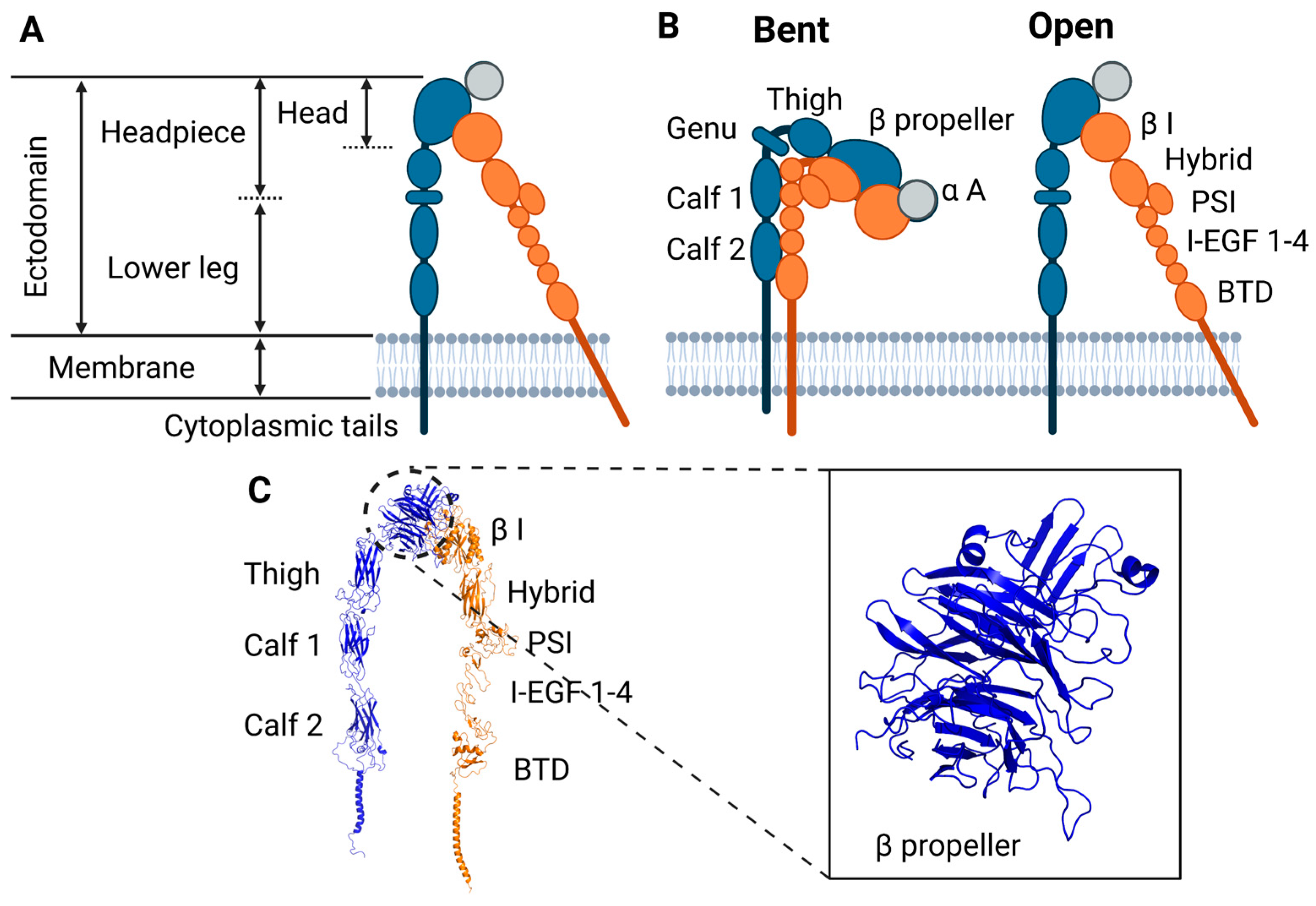
5. Integrin Conformation and Conformational Dynamics

Integrin conformational transitions are thought to contribute to its interrelated but distinct functions of mechanotransducer, force transmitter, and signal transducer. Integrin α and β subunits consist of consecutive domains connected by loops and flexible linkers (Figure 1B). The extracellular region of the α subunit contains an N-terminal domain followed by a large seven-bladed β-propeller domain, followed by the thigh domain, and two calf domains (Figure 1B,C). Nine of 18 α subunits have an α-A domain (I or inserted domain), consisting of approximately 200 amino acids, located between blades 2 and 3 of the β-propeller (gray domain in Figure 1B) [96]. The α-I domain assumes a Rossman fold with five β-sheets surrounded by seven α helices, similar to von Willebrand A domains [97,98]. The β subunit extracellular region consists of an N-terminal β-I domain (that resembles the α-I domain) followed by the hybrid domain, the plexin-semaphorin-integrin domain (PSI), and four cysteine-rich epidermal growth factor (EGF) modules (I-EGF) 1-4 and β-T domains (Figure 1B). Both α and β subunits have a single membrane spanning helix that ends with a C-terminal cytoplasmic tail. While β cytoplasmic domains show substantial sequence homology consistent with conserved binding to talin and other adapters, α cytoplasmic domains show little homology. The transmembrane domains (TMD) of the α and β subunits (α-TMD and β-TMD, respectively) associate with each other, which helps stabilize the closed, low affinity conformation. During conformational activation from bent to extended conformations, the domains reorient and change relative positions (Figure 1B), including separation of the transmembrane and cytoplasmic domains that is critical in relaying long-range conformational changes to the extracellular domains [99,100]. Thus, mutations that disrupt cytoplasmic domain noncovalent interaction can activate integrins [101,102,103,104].
Integrin αIIBβ3 has been resolved in bent, intermediate, and extended conformations (Figure 2), in which the intermediate conformations present closed subunits and varying degrees of flattening between the headpiece and the legs, resulting in different degrees of vertical extension [24,105]. In the bent conformation, the α and β chains are close together, with the headpiece bent against the lower legs and two extremely bent ‘knees’ at the α genu and between I-EGF 1 and I-EGF 2 domains of the β subunit [47,48]. The ligand binding interface is situated between the α subunit β-propeller domain and the β subunit β-I domain, which is responsible for coordinating interactions with charged residues in ligands [98]. In bent integrin, this interface is close to and oriented towards the plasma membrane (Figure 2A). Bent integrin is ~10-11 nm high on the extracellular side. Although the molecular interfaces buried in the bent conformation of integrin are extensive, they have low shape complementarity and are hydrophilic and readily replaced by water molecules, so that destabilization of the bent conformation promotes the open conformation [47,48]. Upon extension, the headpiece can remain closed (Figure 2B), or can transition into an open conformation (Figure 2C), with the ligand binding interface oriented away, rather than towards the plasma membrane. When extended, the ligand binding affinity of integrin is high for large biological ligands, such as fibronectin and fibrinogen [47]. In the open, fully extended conformation, the head is separated from the legs and the α and β legs are apart (Figure 2C). In the extended-open conformation, the extracellular domains span a total vertical distance of about 15–20 nm from the membrane, for a total length of integrin up of ~28 nm from the cytoplasmic tails to the distal ligand binding interface [44,106,107].
Before ligand binding, three metal ion binding sites in the β subunit β-I domain are typically occupied by Ca2+ and Mg2+ [108]. The Mg2+ bound to the central, metal ion-dependent adhesion site (MIDAS) directly coordinates with the acidic sidechain shared by all integrin ligands [109]. The site adjacent to MIDAS (ADMIDAS) binds the Mn2+ ion that triggers activation of low-affinity integrins [110]. By competing with Ca2+ at the ADMIDAS, Mn2+ shifts integrin conformational equilibrium towards the open conformation [111]. On the other side of the MIDAS (Figure 3), the synergistic metal ion binding site (SyMBS), also termed ligand-associated metal ion binding site (LIMBS), binds the Mn2+. SyMBS was found bound to the ion simultaneously with the AMIDAS in the ligated crystal structure of αVβ3 integrin. On the lower side of the β–propeller blades facing away from the ligand-binding surface, the last three or four blades of the β -propeller domain in the α subunit contain EF-hand domains that also bind Ca2+, to affect ligand binding allosterically [110,112].
In the ligand binding interface of integrin, the forces between ions and the chains of the ligand can drive changes in the positions of the aminoacids of the β-I and β-propeller domains of integrin, resulting in increased ligand binding affinity. Conversely, the changes in positions of these aminoacids upon integrin activation change the distances between ions and ligand chains, thus increasing electrostatic interaction potentials and integrin affinity for ligand binding. Therefore, ligand binding affects integrin conformation and integrin conformation affects ligand binding, creating a reciprocal relationship between structure and ligand-bound state of integrin.

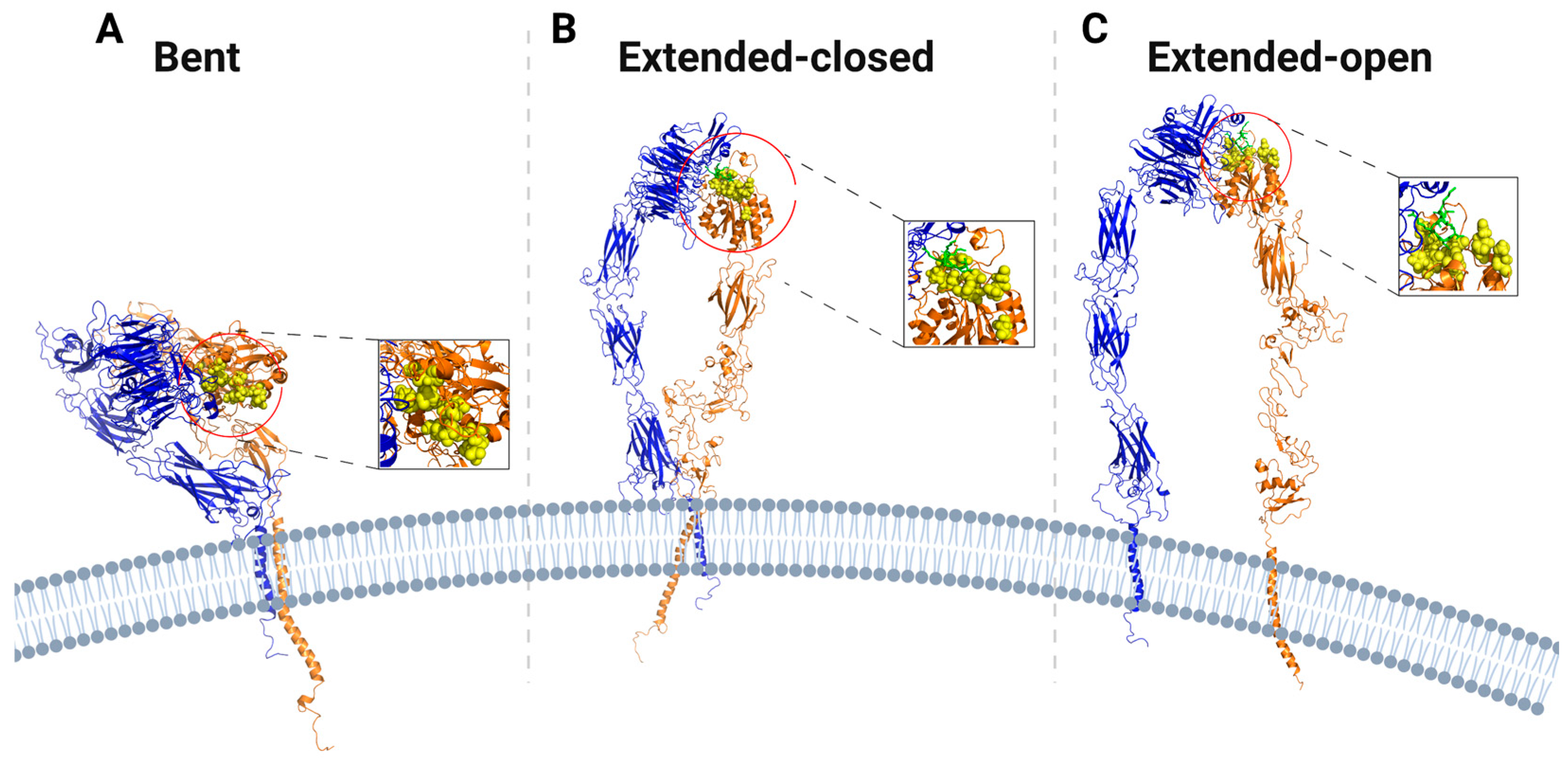
6. The Effects of Force on Integrin Conformation

Several studies have reported that force promotes integrin conformational activation by governing the transition from bent to extended conformations [44,50,108,113,114,115,116]. The main structural modifications that integrins undergo during conformational activation have been identified; however, the exact mechanisms by which force promotes the changes in integrin conformation are poorly understood.
The two main conformational rearrangements during integrin activation are hinging of the legs in the transition from bent to extended and swing out of the β subunit hybrid domain. SMD analysis and single-molecule experiments have indicated a role for mechanical force in hybrid domain swing-out and integrin extension [81,85,117,118]. These motions are accompanied by the opening of the highly flexible interface between the β subunit domains I-EGF1 and I-EGF2, and extension of the α-genu in the α subunit. While several different structures of αXβ2 in different crystal lattices show that the β subunit appears to be more flexible than the α subunit [45], α subunits contain two regions of high flexibility: the linker between the β–propeller and thigh domain and the knee at the bend between the thigh and the calf-1 domain, which allows extension of this subunit by a hinging at the α knee, or “genu” [98]. For the β subunit, the region around the EGF domain is almost plastic, including the linker between EGF1 and EGF2 the PSI/hybrid and hybrid/I-EGF1 junctions [98]. In the β-I/hybrid region, there is evidence of significant conformational changes both in the absence and in the presence of force [47,108,119,120,121,122,123,124,125]. During conformational activation of αIIBβ3, the displacement of the α7-helix in the hybrid domain leads to a ~60° reorientation between the β-I and hybrid domain, and the α and β leg knees become separated by 70° due to the transmission through the firmly linked PSI domain in the upper β leg [108].
One hypothesis for how force induces these conformational changes is that force physically propagates from the ligand binding site and the intracellular β tail across the integrin structure, with sequential stretching and deformations of the interconnections between its rigid domains. In this case, rapid force transmission across the protein leads to rigid-body domain displacements without affecting secondary structures. Force, propagated along the linkers between domains, provides the energy for overcoming the barrier(s) between bent and extended conformations. This concept of rigid body-based structure, in which domains act as rigid units with articulation points located between them, was proposed from high-resolution reconstructions of αIIBβ3 and αvβ8 integrins, and of a β2 subunit fragment [126,127,128]. A second hypothesis is that physical force propagation across the structure sequentially stretches and deforms the individual domains, so that the more flexible domains also undergo conformational changes. The presence of flexible domains within integrins has been reported for α5β1 and αXβ2 integrins [51,129]. These integrins have also been shown to bind ligands in conformations that are different from the extended conformation [90,91]; thus force may alter domains structure in addition to their relative positions. The flexible domains include leg domains, such as the thigh, PSI and EGF domains, that are distant from the ligand binding site and can alter allosteric equilibria during the conformational transition [130]. A third hypothesis is that force does not directly propagate across the entire structure but is absorbed at the site of application. In this case, the fast extension or dynamic rearrangement of a flexible domain buffers this force by directly converting it into local intramolecular adjustments that more slowly drive the global change in conformation of the protein. In support of this picture, steered molecular dynamics simulations of integrin conformational activation have shown that an inward displacement of the MIDAD metal ion is coupled to the movement of the α-7 helix from the up to middle and down positions, and that this movement correlates to different degrees of integrin conformational activation [131]. This finding has also confirmed previous crystallography and biomembrane force probe experiments [132,133]. The main difference between these three possible pathways relies on how exactly force propagates (or does not) from the ECM or cytoskeletal binding sites to allosterically drive the whole structure into a new conformation.
Both closed and open conformations of integrin can bind ligands [125]. Integrin ligand binding may be favored kinetically for the extended-closed conformation, whose ligand-binding site is more accessible to ligands than the bent closed conformations (Figure 2) [125]. With the extended-closed integrin anchored to the cytoskeleton and bound to an extracellular ligand, if force is normal to the plasma membrane, integrin conformation may be shifted to the extended-open conformation (Figure 4). In this case, ECM ligand pulling of the β subunit promotes lateral extension of the hybrid domain away from the ligand binding interface. This extended-open conformation of integrin presents slow ligand unbinding rate under force, following catch bond behavior [79,114].
It is important to note that, although this pathway is generally shared across several β3 integrins (e.g., αvβ3 and αIIBβ3), whether or how it applies to other integrins is uncertain, in particular, α-I domain containing integrins such as αXβ2, or α4β1, and α5β1. Truncated integrin α5β1 ectodomains do not undergo the large conformational change in the presence of the RGD peptide alone [53]. In these cases, the closed conformations of β1 integrins bind ligands with higher on-rates and higher ligand binding affinity than the extended-open conformation [90,91]. α4β1 was shown to be easier to activate, but its high-affinity conformation binds fibronectin ~100- to 1000-fold lower affinity than α5β1 [41]. Moreover, negative-stain electron microscopy (EM) has hinted that the α5β1 ectodomain may not reach a fully bent conformation [134]. Thus, for these integrins, the conformational transition from bent closed to extended-open may occur mainly after force is applied. In α-I containing integrins, the high flexibility of the α-I domain itself is tightly coupled with the β-I domain to fine-tune ligand binding affinity. Initial coupling between the β-I and αI domains might stabilize a partially open state of the α-I domain with a closed MIDAS, which on ligand binding converts to the open α-I state with an open MIDAS [45,132]. These observations challenge the view that bent or partially bent integrins present an occluded ligand-binding site and suggest that the receptor response to force varies across families.
It is also hypothesized that force from the actin cytoskeleton is exerted on extended-closed integrin, with the force parallel to the plasma membrane [135,136]. This lateral force then reorients the β helix relative to the α helix to induce the extended-open conformation (Figure 5). In this model, lateral force on the β tail is transmitted through the lower β-leg domains to the hybrid domain, which swings out and assumes an orientation like that in the open headpiece.

7. Conclusions and New Areas of Research

To summarize the above discussion, integrin conformational dynamics in which bent, low affinity conformations convert to extended, open, high affinity conformations are modulated by cytoplasmic adapter and ECM ligand binding, and then further modified by tension applied through this linkage. Accelerated conversion to or stabilization of high affinity conformations contributes to cellular mechanotransduction, such as stiffness sensing or responses to applied tension. However, major questions remain about the exact conformational paths that mediate these transitions, how forces are transmitted through the integrin structure, and how they impact these transitions. It is also unclear whether integrins under tension might access conformations that are rare in unloaded conditions.
In the absence of experimental techniques to obtain dynamic, atomic-level insights into the integrin activation pathway particularly under tension, addressing these questions will remain limited to computational modeling based on Xray or cryo-EM structures of the unliganded closed-hinge and the ligand-bound open-hinge β3-integrin headpiece domains [43]. Molecular dynamics (MD) simulations of the β3-integrin headpiece domains have illustrated the Ångstrom-level structural pathway of ligand-induced hinge-angle opening [114]. Incorporating force into these calculations and then experimental verification of predictions is the current state of the art.
Lastly, how integrin conformational dynamics impact cell functions is a major unexplored area. Integrin-based adhesions of platelets and leukocytes are “off” in the vasculature and “on” when these cells are activated. Directional cell migration requires local control of integrin activation and adhesion strengthening at the cell front and deactivation and detachment at the rear [135]. Indeed, adhesion strengthening is reportedly confined to the cell front [3]. Cell adhesions through integrins must also be coordinated with cytoskeletal dynamics to enable integrins to connect to the force-exerting actin fibers and provide traction and propulsive forces for cell migration [39,76,135]. These processes appear to differ for different integrins. For example, α4β1 is reportedly activated by lower forces or at a lower concentration of adaptor proteins compared to α5β1, perhaps in keeping with α4β1 mediating leukocytes adhesion and migration that occurs under low tension [41]. Thus, understanding how force-dependent conformational dynamics underlie adhesion dynamics is an important area for future study. Addressing these questions likely requires new methods in structural and mechanobiology but will be foundational for understanding integrin functions in physiology and disease.

Author Contributions

Conceptualization, M.A.S.; methodology, T.C.B. and M.A.S.; data curation, R.K.; writing—original draft preparation, R.K.; writing—review and editing, R.K., T.C.B. and M.A.S.; visualization, R.K.; supervision, T.C.B.; funding acquisition, T.C.B. and M.A.S. All authors have read and agreed to the published version of the manuscript.

Funding

This research was funded by the National Science Foundation, grant number: 2044394.

Institutional Review Board Statement

Not applicable.

Informed Consent Statement

Not applicable.

Data Availability Statement

Not applicable.

Conflicts of Interest

The authors declare no conflict of interest.

References

  1. Chen, C.S. Mechanotransduction—A field pulling together? J. Cell Sci. 2008, 121, 3285–3292. [Google Scholar] [CrossRef] [PubMed] [Green Version]
  2. Paszek, M.J.; Weaver, V.M. The tension mounts: Mechanics meets morphogenesis and malignancy. J. Mammary Gland. Biol. Neoplasia 2004, 9, 325–342. [Google Scholar] [CrossRef] [PubMed]
  3. Puklin-Faucher, E.; Sheetz, M.P. The mechanical integrin cycle. J. Cell Sci. 2009, 122, 179–186. [Google Scholar] [CrossRef] [PubMed] [Green Version]
  4. Bouvard, D.; Pouwels, J.; De Franceschi, N.; Ivaska, J. Integrin inactivators: Balancing cellular functions in vitro and in vivo. Nat. Rev. Mol. Cell Biol. 2013, 14, 430–442. [Google Scholar] [CrossRef] [PubMed]
  5. Hynes, R.O. Integrins: Bidirectional, allosteric signaling machines. Cell 2002, 110, 673–687. [Google Scholar] [CrossRef] [Green Version]
  6. Shih, J.; Keller, R. Patterns of cell motility in the organizer and dorsal mesoderm of Xenopus laevis. Development 1992, 116, 915–930. [Google Scholar] [CrossRef]
  7. Barnes, L.A.; Marshall, C.D.; Leavitt, T.; Hu, M.S.; Moore, A.L.; Gonzalez, J.G.; Longaker, M.T.; Gurtner, G.C. Mechanical Forces in Cutaneous Wound Healing: Emerging Therapies to Minimize Scar Formation. Adv. Wound Care 2018, 7, 47–56. [Google Scholar] [CrossRef] [Green Version]
  8. Shu, C.; Chien, S.; Li, S.; Shiu, Y.-T.; Li, Y.-S. Molecular basis of mechanical modulation of endothelial cell migration. Front. Biosci. 2005, 10, 1985–2000. [Google Scholar]
  9. Zaman, M.H.; Trapani, L.M.; Sieminski, A.L.; MacKellar, D.; Gong, H.; Kamm, R.D.; Wells, A.; Lauffenburger, D.A.; Matsudaira, P. Migration of tumor cells in 3D matrices is governed by matrix stiffness along with cell-matrix adhesion and proteolysis. Proc. Natl. Acad. Sci. USA 2006, 103, 10889–10894. [Google Scholar] [CrossRef] [Green Version]
  10. Hamidi, H.; Ivaska, J. Every step of the way: Integrins in cancer progression and metastasis. Nat. Rev. Cancer 2018, 18, 533–548. [Google Scholar] [CrossRef] [Green Version]
  11. Duscher, D.; Maan, Z.; Wong, V.W.; Rennert, R.C.; Januszyk, M.; Rodrigues, M.; Hu, M.; Whitmore, A.J.; Whittam, A.J.; Longaker, M.T.; et al. Mechanotransduction and fibrosis. J. Biomech. 2014, 47, 1997–2005. [Google Scholar] [CrossRef] [PubMed]
  12. Yeh, C.F.; Chou, C.; Yang, K.C. Mechanotransduction in fibrosis: Mechanisms and treatment targets. Curr. Top. Membr. 2021, 87, 279–314. [Google Scholar] [PubMed]
  13. Conway, D.E.; Schwartz, M.A. Flow-dependent cellular mechanotransduction in atherosclerosis. J. Cell Sci. 2013, 126 Pt 22, 5101–5109. [Google Scholar] [CrossRef] [PubMed] [Green Version]
  14. Humphries, J.D.; Byron, A.; Humphries, M.J. Integrin ligands at a glance. J. Cell Sci. 2006, 119, 3901–3903. [Google Scholar] [CrossRef] [PubMed] [Green Version]
  15. Plow, E.F.; Haas, T.A.; Zhang, L.; Loftus, J.; Smith, J.W. Ligand binding to integrins. J. Biol. Chem. 2000, 275, 21785–21788. [Google Scholar] [CrossRef] [Green Version]
  16. Van der Flier, A.; Sonnenberg, A. Function and interactions of integrins. Cell Tissue Res. 2001, 305, 285–298. [Google Scholar] [CrossRef]
  17. Tamkun, J.W.; DeSimone, D.W.; Fonda, D.; Patel, R.S.; Buck, C.; Horwitz, A.F.; Hynes, R.O. Structure of integrin, a glycoprotein involved in the transmembrane linkage between fibronectin and actin. Cell 1986, 46, 271–282. [Google Scholar] [CrossRef]
  18. Schwartz, M.A.; Schaller, M.D.; Ginsberg, M.H. Integrins: Emerging paradigms of signal transduction. Annu. Rev. Cell Dev. Biol. 1995, 11, 549–599. [Google Scholar] [CrossRef]
  19. Kechagia, J.Z.; Ivaska, J.; Roca-Cusachs, P. Integrins as biomechanical sensors of the microenvironment. Nat. Rev. Mol. Cell Biol. 2019, 20, 457–473. [Google Scholar] [CrossRef]
  20. Zhong, C.; Chrzanowska-Wodnicka, M.; Brown, J.; Shaub, A.; Belkin, A.M.; Burridge, K. Rho-mediated contractility exposes a cryptic site in fibronectin and induces fibronectin matrix assembly. J. Cell Biol. 1998, 141, 539–551. [Google Scholar] [CrossRef] [Green Version]
  21. Kanchanawong, P.; Shtengel, G.; Pasapera, A.M.; Ramko, E.B.; Davidson, M.W.; Hess, H.F.; Waterman, C.M. Nanoscale architecture of integrin-based cell adhesions. Nature 2010, 468, 580–584. [Google Scholar] [CrossRef] [PubMed]
  22. Critchley, D.R.; Holt, M.R.; Barry, S.T.; Priddle, H.; Hemmings, L.; Norman, J. Integrin-mediated cell adhesion: The cytoskeletal connection. Biochem. Soc. Symp. 1999, 65, 79–99. [Google Scholar] [PubMed]
  23. Schwarz, U.S.; Gardel, M.L. United we stand: Integrating the actin cytoskeleton and cell-matrix adhesions in cellular mechanotransduction. J. Cell Sci. 2012, 125 Pt 13, 3051–3060. [Google Scholar] [CrossRef] [PubMed] [Green Version]
  24. Xu, X.P.; Kim, E.; Swift, M.; Smith, J.W.; Volkmann, N.; Hanein, D. Three-Dimensional Structures of Full-Length, Membrane-Embedded Human alpha(IIb)beta(3) Integrin Complexes. Biophys. J. 2016, 110, 798–809. [Google Scholar] [CrossRef] [PubMed] [Green Version]
  25. Case, L.B.; Waterman, C.M. Integration of actin dynamics and cell adhesion by a three-dimensional, mechanosensitive molecular clutch. Nat. Cell Biol. 2015, 17, 955–963. [Google Scholar] [CrossRef] [PubMed]
  26. Worth, D.C.; Parsons, M. Adhesion dynamics: Mechanisms and measurements. Int. J. Biochem. Cell Biol. 2008, 40, 2397–2409. [Google Scholar] [CrossRef]
  27. Zhang, X.; Jiang, G.; Cai, Y.; Monkley, S.J.; Critchley, D.R.; Sheetz, M.P. Talin depletion reveals independence of initial cell spreading from integrin activation and traction. Nat. Cell Biol. 2008, 10, 1062–1068. [Google Scholar] [CrossRef] [Green Version]
  28. Jiang, G.; Giannone, G.; Critchley, D.R.; Fukumoto, E.; Sheetz, M.P. Two-piconewton slip bond between fibronectin and the cytoskeleton depends on talin. Nature 2003, 424, 334–337. [Google Scholar] [CrossRef]
  29. Zaidel-Bar, R.; Milo, R.; Kam, Z.; Geiger, B. A paxillin tyrosine phosphorylation switch regulates the assembly and form of cell-matrix adhesions. J. Cell Sci. 2007, 120, 137–148. [Google Scholar] [CrossRef] [Green Version]
  30. Ballestrem, C.; Erez, N.; Kirchner, J.; Kam, Z.; Bershadsky, A.; Geiger, B. Molecular mapping of tyrosine-phosphorylated proteins in focal adhesions using fluorescence resonance energy transfer. J. Cell Sci. 2006, 119, 866–875. [Google Scholar] [CrossRef] [Green Version]
  31. Zaidel-Bar, R.; Ballestrem, C.; Kam, Z.; Geiger, B. Early molecular events in the assembly of matrix adhesions at the leading edge of migrating cells. J. Cell Sci. 2003, 116, 4605–4613. [Google Scholar] [CrossRef] [PubMed]
  32. Beningo, K.A.; Dembo, M.; Kaverina, I.; Small, J.V.; Wang, Y.-L. Nascent focal adhesions are responsible for the generation of strong propulsive forces in migrating fibroblasts. J. Cell Biol. 2001, 153, 881–888. [Google Scholar] [CrossRef] [PubMed] [Green Version]
  33. Zimerman, B.; Volberg, T.; Geiger, B. Early molecular events in the assembly of the focal adhesion-stress fiber complex during fibroblast spreading. Cell Motil. Cytoskelet. 2004, 58, 143–159. [Google Scholar] [CrossRef] [PubMed]
  34. Sun, H.; Lagarrigue, F.; Ginsberg, M.H. The Connection between Rap1 and Talin1 in the Activation of Integrins in Blood Cells. Front. Cell Dev. Biol. 2022, 10, 908622. [Google Scholar] [CrossRef] [PubMed]
  35. Schwartz, M.A. Integrins and extracellular matrix in mechanotransduction. Cold Spring Harb. Perspect. Biol. 2010, 2, a005066. [Google Scholar] [CrossRef] [PubMed]
  36. Lauffenburger, D.A.; Horwitz, A.F. Cell migration: A physically integrated molecular process. Cell 1996, 84, 359–369. [Google Scholar] [CrossRef] [Green Version]
  37. Vogel, V.; Sheetz, M. Local force and geometry sensing regulate cell functions. Nat. Rev. Mol. Cell Biol. 2006, 7, 265–275. [Google Scholar] [CrossRef]
  38. Lock, J.G.; Wehrle-Haller, B.; Strömblad, S. Cell–matrix adhesion complexes: Master control machinery of cell migration. In Seminars in Cancer Biology; Elsevier: Amsterdam, The Netherlands, 2008. [Google Scholar]
  39. Springer, T.A.; Dustin, M.L. Integrin inside-out signaling and the immunological synapse. Curr. Opin. Cell Biol. 2012, 24, 107–115. [Google Scholar] [CrossRef] [Green Version]
  40. Li, J.; Su, Y.; Xia, W.; Qin, Y.; Humphries, M.J.; Vestweber, D.; Cabañas, C.; Lu, C.; Springer, T.A. Conformational equilibria and intrinsic affinities define integrin activation. EMBO J. 2017, 36, 629–645. [Google Scholar] [CrossRef]
  41. Li, J.; Springer, T.A. Energy landscape differences among integrins establish the framework for understanding activation. J. Cell Biol. 2018, 217, 397–412. [Google Scholar] [CrossRef] [Green Version]
  42. Chen, Y.; Ju, L.A.; Zhou, F.; Liao, J.; Xue, L.; Su, Q.P.; Jin, D.; Yuan, Y.; Lu, H.; Jackson, S.P.; et al. An integrin alphaIIbbeta3 intermediate affinity state mediates biomechanical platelet aggregation. Nat. Mater. 2019, 18, 760–769. [Google Scholar] [CrossRef] [PubMed]
  43. Xiong, J.-P.; Stehle, T.; Diefenbach, B.; Zhang, R.; Dunker, R.; Scott, D.L.; Joachimiak, A.; Goodman, S.L.; Arnaout, M.A. Crystal structure of the extracellular segment of integrin alpha Vbeta3. Science 2001, 294, 339–345. [Google Scholar] [CrossRef] [PubMed] [Green Version]
  44. Zhu, J.; Luo, B.-H.; Xiao, T.; Zhang, C.; Nishida, N.; Springer, T.A. Structure of a complete integrin ectodomain in a physiologic resting state and activation and deactivation by applied forces. Mol. Cell 2008, 32, 849–861. [Google Scholar] [CrossRef] [PubMed] [Green Version]
  45. Xie, C.; Zhu, J.; Chen, X.; Mi, L.-Z.; Nishida, N.; Springer, T.A. Structure of an integrin with an alphaI domain, complement receptor type 4. EMBO J. 2010, 29, 666–679. [Google Scholar] [CrossRef]
  46. Dong, X.; Mi, L.-Z.; Zhu, J.; Wang, W.; Hu, P.; Luo, B.-H.; Springer, T.A. alpha(V)beta(3) integrin crystal structures and their functional implications. Biochemistry 2012, 51, 8814–8828. [Google Scholar] [CrossRef] [Green Version]
  47. Takagi, J.; Petre, B.M.; Walz, T.; Springer, T.A. Global conformational rearrangements in integrin extracellular domains in outside-in and inside-out signaling. Cell 2002, 110, 599–611. [Google Scholar] [CrossRef] [Green Version]
  48. Nishida, N.; Xie, C.; Shimaoka, M.; Cheng, Y.; Walz, T.; Springer, T.A. Activation of leukocyte beta2 integrins by conversion from bent to extended conformations. Immunity 2006, 25, 583–594. [Google Scholar] [CrossRef] [Green Version]
  49. Chen, X.; Xie, C.; Nishida, N.; Li, Z.; Walz, T.; Springer, T.A. Requirement of open headpiece conformation for activation of leukocyte integrin alphaXbeta2. Proc. Natl. Acad. Sci. USA 2010, 107, 14727–14732. [Google Scholar] [CrossRef] [Green Version]
  50. Eng, E.T.; Smagghe, B.J.; Walz, T.; Springer, T.A. Intact alphaIIbbeta3 integrin is extended after activation as measured by solution X-ray scattering and electron microscopy. J. Biol. Chem. 2011, 286, 35218–35226. [Google Scholar] [CrossRef] [Green Version]
  51. Xia, W.; Springer, T.A. Metal ion and ligand binding of integrin alpha5beta1. Proc. Natl. Acad. Sci. USA 2014, 111, 17863–17868. [Google Scholar] [CrossRef] [Green Version]
  52. Su, Y.; Xia, W.; Li, J.; Walz, T.; Humphries, M.J.; Vestweber, D.; Cabañas, C.; Lu, C.; Springer, T.A. Relating conformation to function in integrin alpha5beta1. Proc. Natl. Acad. Sci. USA 2016, 113, E3872–E3881. [Google Scholar] [CrossRef] [PubMed]
  53. Takagi, J.; Strokovich, K.; Springer, T.A.; Walz, T. Structure of integrin alpha5beta1 in complex with fibronectin. EMBO J. 2003, 22, 4607–4615. [Google Scholar] [CrossRef] [PubMed] [Green Version]
  54. Zhu, J.; Zhu, J.; Springer, T.A. Complete integrin headpiece opening in eight steps. J. Cell Biol. 2013, 201, 1053–1068. [Google Scholar] [CrossRef] [PubMed] [Green Version]
  55. Schürpf, T.; Chen, Q.; Liu, J.; Wang, R.; Springer, T.A.; Wang, J. The RGD finger of Del-1 is a unique structural feature critical for integrin binding. FASEB J. 2012, 26, 3412–3420. [Google Scholar] [CrossRef] [PubMed] [Green Version]
  56. Moore, S.W.; Roca-Cusachs, P.; Sheetz, M.P. Stretchy proteins on stretchy substrates: The important elements of integrin-mediated rigidity sensing. Dev. Cell 2010, 19, 194–206. [Google Scholar] [CrossRef] [PubMed] [Green Version]
  57. Pelham, R.J., Jr.; Wang, Y. Cell locomotion and focal adhesions are regulated by substrate flexibility. Proc. Natl. Acad. Sci. USA 1997, 94, 13661–13665. [Google Scholar] [CrossRef] [PubMed] [Green Version]
  58. Oakes, P.W.; Bidone, T.C.; Beckham, Y.; Skeeters, A.V.; Juan, G.R.R.-S.; Winter, S.P.; Voth, G.A.; Gardel, M.L. Lamellipodium is a myosin-independent mechanosensor. Proc. Natl. Acad. Sci. USA 2018, 115, 2646–2651. [Google Scholar] [CrossRef] [Green Version]
  59. Driscoll, T.P.; Bidone, T.C.; Ahn, S.J.; Yu, A.; Groisman, A.; Voth, G.A.; Schwartz, M.A. Integrin-based mechanosensing through conformational deformation. Biophys. J. 2021, 120, 4349–4359. [Google Scholar] [CrossRef]
  60. Harris, A.K.; Wild, P.; Stopak, D. Silicone rubber substrata: A new wrinkle in the study of cell locomotion. Science 1980, 208, 177–179. [Google Scholar] [CrossRef]
  61. Lee, J.; Leonard, M.; Oliver, T.; Ishihara, A.; Jacobson, K. Traction forces generated by locomoting keratocytes. J. Cell Biol. 1994, 127 Pt 2, 1957–1964. [Google Scholar] [CrossRef] [Green Version]
  62. Galbraith, C.G.; Sheetz, M.P. A micromachined device provides a new bend on fibroblast traction forces. Proc. Natl. Acad. Sci. USA 1997, 94, 9114–9118. [Google Scholar] [CrossRef] [PubMed]
  63. Dembo, M.; Wang, Y.L. Stresses at the cell-to-substrate interface during locomotion of fibroblasts. Biophys. J. 1999, 76, 2307–2316. [Google Scholar] [CrossRef] [Green Version]
  64. Burton, K.; Park, J.H.; Taylor, D.L. Keratocytes generate traction forces in two phases. Mol. Biol. Cell 1999, 10, 3745–3769. [Google Scholar] [CrossRef]
  65. Sabass, B.; Gardel, M.L.; Waterman, C.M.; Schwarz, U.S. High resolution traction force microscopy based on experimental and computational advances. Biophys. J. 2008, 94, 207–220. [Google Scholar] [CrossRef] [PubMed] [Green Version]
  66. Grashoff, C.; Hoffman, B.D.; Brenner, M.D.; Zhou, R.; Parsons, M.; Yang, M.T.; McLean, M.A.; Sligar, S.G.; Chen, C.S.; Ha, T.; et al. Measuring mechanical tension across vinculin reveals regulation of focal adhesion dynamics. Nature 2010, 466, 263–266. [Google Scholar] [CrossRef] [PubMed] [Green Version]
  67. Kumar, A.; Ouyang, M.; Van den Dries, K.; McGhee, E.J.; Tanaka, K.; Anderson, M.D.; Groisman, A.; Goult, B.T.; Anderson, K.I.; Schwartz, M.A. Talin tension sensor reveals novel features of focal adhesion force transmission and mechanosensitivity. J. Cell Biol. 2016, 213, 371–383. [Google Scholar] [CrossRef] [Green Version]
  68. Lee, M.K.; Park, J.; Wang, X.; Roein-Peikar, M.; Ko, E.; Qin, E.; Lee, J.; Ha, T.; Kong, H. Rupture force of cell adhesion ligand tethers modulates biological activities of a cell-laden hydrogel. Chem. Commun. 2016, 52, 4757–4760. [Google Scholar] [CrossRef] [Green Version]
  69. Wang, X.; Ha, T. Defining single molecular forces required to activate integrin and notch signaling. Science 2013, 340, 991–994. [Google Scholar] [CrossRef] [Green Version]
  70. Zhang, Y.; Ge, C.; Zhu, C.; Salaita, K. DNA-based digital tension probes reveal integrin forces during early cell adhesion. Nat. Commun. 2014, 5, 5167. [Google Scholar] [CrossRef] [Green Version]
  71. Kumar, A.; Anderson, K.L.; Swift, M.F.; Hanein, D.; Volkmann, N.; Schwartz, M.A. Local Tension on Talin in Focal Adhesions Correlates with F-Actin Alignment at the Nanometer Scale. Biophys. J. 2018, 115, 1569–1579. [Google Scholar] [CrossRef] [Green Version]
  72. Austen, K.; Ringer, P.; Mehlich, A.; Chrostek-Grashoff, A.; Kluger, C.; Klingner, C.; Sabass, B.; Zent, R.; Rief, M.; Grashoff, C. Extracellular rigidity sensing by talin isoform-specific mechanical linkages. Nat. Cell Biol. 2015, 17, 1597–1606. [Google Scholar] [CrossRef] [PubMed]
  73. Balaban, N.Q.; Schwarz, U.; Riveline, D.; Goichberg, P.; Tzur, G.; Sabanay, I.; Mahalu, D.; Safran, S.; Bershadsky, A.; Addadiand, L.; et al. Force and focal adhesion assembly: A close relationship studied using elastic micropatterned substrates. Nat. Cell Biol. 2001, 3, 466–472. [Google Scholar] [CrossRef] [PubMed]
  74. Tan, J.L.; Tien, J.; Pirone, D.M.; Gray, D.S.; Bhadriraju, K.; Chen, C.S. Cells lying on a bed of microneedles: An approach to isolate mechanical force. Proc. Natl. Acad. Sci. USA 2003, 100, 1484–1489. [Google Scholar] [CrossRef] [PubMed] [Green Version]
  75. Chrzanowska-Wodnicka, M.; Burridge, K. Rho-stimulated contractility drives the formation of stress fibers and focal adhesions. J. Cell Biol. 1996, 133, 1403–1415. [Google Scholar] [CrossRef] [Green Version]
  76. Sun, Z.; Guo, S.S.; Fassler, R. Integrin-mediated mechanotransduction. J. Cell Biol. 2016, 215, 445–456. [Google Scholar] [CrossRef] [Green Version]
  77. Goult, B.T.; Brown, N.H.; Schwartz, M.A. Talin in mechanotransduction and mechanomemory at a glance. J. Cell Sci. 2021, 134, jcs258749. [Google Scholar] [CrossRef]
  78. Zhao, Y.; Lykov, N.; Tzeng, C. Talin1 interaction network in cellular mechanotransduction (Review). Int. J. Mol. Med. 2022, 49, 1–12. [Google Scholar] [CrossRef]
  79. Kong, F.; García, A.J.; Mould, A.P.; Humphries, M.J.; Zhu, C. Demonstration of catch bonds between an integrin and its ligand. J. Cell Biol. 2009, 185, 1275–1284. [Google Scholar] [CrossRef] [Green Version]
  80. Li, Z.; Lee, H.; Zhu, C. Molecular mechanisms of mechanotransduction in integrin-mediated cell-matrix adhesion. Exp. Cell Res. 2016, 349, 85–94. [Google Scholar] [CrossRef] [Green Version]
  81. Chen, W.; Lou, J.; Evans, E.A.; Zhu, C. Observing force-regulated conformational changes and ligand dissociation from a single integrin on cells. J. Cell Biol. 2012, 199, 497–512. [Google Scholar] [CrossRef]
  82. Kong, F.; Li, Z.; Parks, W.M.; Dumbauld, D.W.; García, A.J.; Mould, A.P.; Humphries, M.J.; Zhu, C. Cyclic mechanical reinforcement of integrin–ligand interactions. Mol. Cell 2013, 49, 1060–1068. [Google Scholar] [CrossRef]
  83. Tchesnokova, V.; Aprikian, P.; Yakovenko, O.; LaRock, C.; Kidd, B.; Vogel, V.; Thomas, W.; Sokurenko, E. Integrin-like allosteric properties of the catch bond-forming FimH adhesin of Escherichia coli. J. Biol. Chem. 2008, 283, 7823–7833. [Google Scholar] [CrossRef] [PubMed] [Green Version]
  84. Thomas, W.; Forero, M.; Yakovenko, O.; Nilsson, L.; Vicini, P.; Sokurenko, E.; Vogel, V. Catch-bond model derived from allostery explains force-activated bacterial adhesion. Biophys. J. 2006, 90, 753–764. [Google Scholar] [CrossRef] [PubMed] [Green Version]
  85. Yakovenko, O.; Sharma, S.; Forero, M.; Tchesnokova, V.; Aprikian, P.; Kidd, B.; Mach, A.; Vogel, V.; Sokurenko, E.; Thomas, W.E. FimH forms catch bonds that are enhanced by mechanical force due to allosteric regulation. J. Biol. Chem. 2008, 283, 11596–11605. [Google Scholar] [CrossRef] [PubMed] [Green Version]
  86. Li, J.; Springer, T.A. Integrin extension enables ultrasensitive regulation by cytoskeletal force. Proc. Natl. Acad. Sci. USA 2017, 114, 4685–4690. [Google Scholar] [CrossRef] [Green Version]
  87. Coutinho, A.; Garcia, C.; González-Rodríguez, J.; Lillo, M.P. Conformational changes in human integrin alphaIIbbeta3 after platelet activation, monitored by FRET. Biophys. Chem. 2007, 130, 76–87. [Google Scholar] [CrossRef] [Green Version]
  88. Ye, F.; Liu, J.; Winkler, H.; Taylor, K.A. Integrin alpha IIb beta 3 in a membrane environment remains the same height after Mn2+ activation when observed by cryoelectron tomography. J. Mol. Biol. 2008, 378, 976–986. [Google Scholar] [CrossRef] [Green Version]
  89. Song, G.; Luo, B.H. Effects of the association of the alphav beta8 lower legs on integrin ligand binding. J. Cell Biochem. 2021, 122, 801–813. [Google Scholar] [CrossRef]
  90. Li, J.; Yan, J.; Springer, T.A. Low-affinity integrin states have faster ligand-binding kinetics than the high-affinity state. eLife 2021, 10, e73359. [Google Scholar] [CrossRef]
  91. Schumacher, S.; Dedden, D.; Nunez, R.V.; Matoba, K.; Takagi, J.; Biertümpfel, C.; Mizuno, N. Structural insights into integrin alpha5beta1 opening by fibronectin ligand. Sci. Adv. 2021, 7, eabe9716. [Google Scholar] [CrossRef]
  92. Choquet, D.; Felsenfeld, D.P.; Sheetz, M.P. Extracellular matrix rigidity causes strengthening of integrin-cytoskeleton linkages. Cell 1997, 88, 39–48. [Google Scholar] [CrossRef]
  93. Saez, A.; Buguin, A.; Silberzan, P.; Ladoux, B. Is the mechanical activity of epithelial cells controlled by deformations or forces? Biophys. J. 2005, 89, L52–L54. [Google Scholar] [CrossRef] [PubMed] [Green Version]
  94. Chan, C.E.; Odde, D.J. Traction dynamics of filopodia on compliant substrates. Science 2008, 322, 1687–1691. [Google Scholar] [CrossRef] [PubMed]
  95. Elosegui-Artola, A.; Oria, R.; Chen, Y.; Kosmalska, A.; Pérez-González, C.; Castro, N.; Zhu, C.; Trepat, X.; Roca-Cusachs, P. Mechanical regulation of a molecular clutch defines force transmission and transduction in response to matrix rigidity. Nat. Cell Biol. 2016, 18, 540–548. [Google Scholar] [CrossRef] [PubMed] [Green Version]
  96. Larson, R.S.; Corbí, A.L.; Berman, L.; Springer, A. Primary structure of the leukocyte function-associated molecule-1 alpha subunit: An integrin with an embedded domain defining a protein superfamily. J. Cell Biol. 1989, 108, 703–712. [Google Scholar] [CrossRef]
  97. Barczyk, M.; Carracedo, S.; Gullberg, D. Integrins. Cell Tissue Res. 2010, 339, 269–280. [Google Scholar] [CrossRef] [Green Version]
  98. Campbell, I.D.; Humphries, M.J. Integrin structure, activation, and interactions. Cold Spring Harb. Perspect. Biol. 2011, 3, a004994. [Google Scholar] [CrossRef] [Green Version]
  99. Kadry, Y.A.; Calderwood, D.A. Structural and signaling functions of integrins. Biochim. Biophys. Acta (BBA)-Biomembr. 2020, 1862, 183206. [Google Scholar] [CrossRef]
  100. Sun, H.; Fan, Z.; Gingras, A.R.; Lopez-Ramirez, M.A.; Ginsberg, M.H.; Ley, K. Frontline Science: A flexible kink in the transmembrane domain impairs beta2 integrin extension and cell arrest from rolling. J. Leukoc. Biol. 2020, 107, 175–183. [Google Scholar] [CrossRef]
  101. Drugan, J.K.; Khosravi-Far, R.; White, M.A.; Der, C.J.; Sung, Y.-J.; Hwang, Y.-W.; Campbell, S.L. Ras Interaction with Two Distinct Binding Domains in Raf-1 5 Be Required for Ras Transformation (∗). J. Biol. Chem. 1996, 271, 233–237. [Google Scholar] [CrossRef] [Green Version]
  102. Luo, B.-H.; Springer, T.A.; Takagi, J. A specific interface between integrin transmembrane helices and affinity for ligand. PLoS Biol. 2004, 2, e153. [Google Scholar] [CrossRef] [PubMed]
  103. Li, R.; Mitra, N.; Gratkowski, H.; Vilaire, G.; Litvinov, R.; Nagasami, C.; Weisel, J.W.; Lear, J.D.; DeGrado, W.F.; Bennett, J.S. Activation of integrin αIIbß3 by modulation of transmembrane helix associations. Science 2003, 300, 795–798. [Google Scholar] [CrossRef] [PubMed]
  104. Ginsberg, M.H. Integrin activation. BMB Rep. 2014, 47, 655. [Google Scholar] [CrossRef] [PubMed]
  105. Xiong, J.-P.; Stehle, T.; Goodman, S.L.; Arnaout, M.A. A novel adaptation of the integrin PSI domain revealed from its crystal structure. J. Biol. Chem. 2004, 279, 40252–40254. [Google Scholar] [CrossRef] [Green Version]
  106. Ye, F.; Hu, G.; Taylor, D.; Ratnikov, B.; Bobkov, A.A.; McLean, M.A.; Sligar, S.G.; Taylor, K.A.; Ginsberg, M.H. Recreation of the terminal events in physiological integrin activation. J. Cell Biol. 2010, 188, 157–173. [Google Scholar] [CrossRef] [Green Version]
  107. Moore, T.I.; Aaron, J.; Chew, T.-L.; Springer, T.A. Measuring Integrin Conformational Change on the Cell Surface with Super-Resolution Microscopy. Cell Rep. 2018, 22, 1903–1912. [Google Scholar] [CrossRef] [Green Version]
  108. Xiao, T.; Takagi, J.; Coller, B.S.; Wang, J.-H.; Springer, T.A. Structural basis for allostery in integrins and binding to fibrinogen-mimetic therapeutics. Nature 2004, 432, 59–67. [Google Scholar] [CrossRef] [Green Version]
  109. Lee, J.-O.; Bankston, L.A.; Liddington, M.A.A.R.C. Two conformations of the integrin A-domain (I-domain): A pathway for activation? Structure 1995, 3, 1333–1340. [Google Scholar] [CrossRef] [Green Version]
  110. Humphries, M.J.; Symonds, E.J.; Mould, A.P. Mapping functional residues onto integrin crystal structures. Curr. Opin. Struct. Biol. 2003, 13, 236–243. [Google Scholar] [CrossRef]
  111. Anderson, J.M.; Li, J.; Springer, T.A. Regulation of integrin alpha5beta1 conformational states and intrinsic affinities by metal ions and the ADMIDAS. Mol. Biol. Cell 2022, 33, ar56. [Google Scholar] [CrossRef]
  112. Oxvig, C.; Springer, T.A. Experimental support for a β-propeller domain in integrin α-subunits and a calcium binding site on its lower surface. Proc. Natl. Acad. Sci. USA 1998, 95, 4870–4875. [Google Scholar] [CrossRef] [PubMed]
  113. Friedland, J.C.; Lee, M.H.; Boettiger, D. Mechanically activated integrin switch controls alpha5beta1 function. Science 2009, 323, 642–644. [Google Scholar] [CrossRef] [PubMed]
  114. Puklin-Faucher, E.; Vogel, V. Integrin activation dynamics between the RGD-binding site and the headpiece hinge. J. Biol. Chem. 2009, 284, 36557–36568. [Google Scholar] [CrossRef] [PubMed] [Green Version]
  115. Mao, D.; Lü, S.; Zhang, X.; Long, M. Mechanically Regulated Outside-In Activation of an I-Domain-Containing Integrin. Biophys. J. 2020, 119, 966–977. [Google Scholar] [CrossRef]
  116. Luo, B.H.; Carman, C.V.; Springer, T.A. Structural basis of integrin regulation and signaling. Annu. Rev. Immunol. 2007, 25, 619–647. [Google Scholar] [CrossRef] [Green Version]
  117. Yao, M.; Goult, B.; Chen, H.; Cong, P.; Sheetz, M.P.; Yan, J. Mechanical activation of vinculin binding to talin locks talin in an unfolded conformation. Sci. Rep. 2014, 4, 4610. [Google Scholar] [CrossRef] [Green Version]
  118. Shang, H.; Lee, G.U. Magnetic Tweezers Measurement of the Bond Lifetime−Force Behavior of the IgG−Protein A Specific Molecular Interaction. J. Am. Chem. Soc. 2007, 129, 6640–6646. [Google Scholar] [CrossRef]
  119. Luo, B.H.; Springer, T.A.; Takagi, J. Stabilizing the open conformation of the integrin headpiece with a glycan wedge increases affinity for ligand. Proc. Natl. Acad. Sci. USA 2003, 100, 2403–2408. [Google Scholar] [CrossRef] [Green Version]
  120. Smagghe, B.J.; Huang, P.; Ban, Y.-E.A.; Baker, D.; Springer, T.A. Modulation of integrin activation by an entropic spring in the [137]-knee. J. Biol. Chem. 2010, 285, 32954–32966. [Google Scholar] [CrossRef] [Green Version]
  121. Kamata, T.; Handa, M.; Ito, S.; Sato, Y.; Ohtani, T.; Kawai, Y.; Ikeda, Y.; Aiso, S. Structural requirements for activation in alphaIIb beta3 integrin. J. Biol. Chem. 2010, 285, 38428–38437. [Google Scholar] [CrossRef] [Green Version]
  122. Provasi, D.; Murcia, M.; Coller, B.S.; Filizola, M. Targeted molecular dynamics reveals overall common conformational changes upon hybrid domain swing-out in beta3 integrins. Proteins 2009, 77, 477–489. [Google Scholar] [CrossRef] [PubMed]
  123. Cheng, M.; Li, J.; Negri, A.; Coller, B.S. Swing-out of the beta3 hybrid domain is required for alphaIIbbeta3 priming and normal cytoskeletal reorganization, but not adhesion to immobilized fibrinogen. PLoS ONE 2013, 8, e81609. [Google Scholar] [CrossRef] [PubMed] [Green Version]
  124. Dong, X.; Zhao, B.; Iacob, R.E.; Zhu, J.; Koksal, A.C.; Lu, C.; Engen, J.R.; Springer, T.A. Force interacts with macromolecular structure in activation of TGF-beta. Nature 2017, 542, 55–59. [Google Scholar] [CrossRef] [PubMed] [Green Version]
  125. Dong, X.; Zhao, B.; Lin, F.-Y.; Lu, C.; Rogers, B.N.; Springer, T.A. High integrin alphaVbeta6 affinity reached by hybrid domain deletion slows ligand-binding on-rate. Proc. Natl. Acad. Sci. USA 2018, 115, E1429–E1436. [Google Scholar] [CrossRef] [PubMed] [Green Version]
  126. Adair, B.D.; Yeager, M. Three-dimensional model of the human platelet integrin alpha IIbbeta 3 based on electron cryomicroscopy and x-ray crystallography. Proc. Natl. Acad. Sci. USA 2002, 99, 14059–14064. [Google Scholar] [CrossRef] [Green Version]
  127. Shi, M.; Sundramurthy, K.; Liu, B.; Tan, S.-M.; Law, S.K.A.; Lescar, J. The crystal structure of the plexin-semaphorin-integrin domain/hybrid domain/I-EGF1 segment from the human integrin beta2 subunit at 1.8-A resolution. J. Biol. Chem. 2005, 280, 30586–30593. [Google Scholar] [CrossRef] [Green Version]
  128. Cormier, A.; Campbell, M.G.; Ito, S.; Wu, S.; Lou, J.; Marks, J.; Baron, J.L.; Nishimura, S.L.; Cheng, Y. Cryo-EM structure of the alphavbeta8 integrin reveals a mechanism for stabilizing integrin extension. Nat. Struct. Mol. Biol. 2018, 25, 698–704. [Google Scholar] [CrossRef]
  129. Sen, M.; Yuki, K.; Springer, T.A. An internal ligand-bound, metastable state of a leukocyte integrin, alphaXbeta2. J. Cell Biol. 2013, 203, 629–642. [Google Scholar] [CrossRef]
  130. Motlagh, H.N.; Wrabl, J.O.; Li, J.; Hilser, V.J. The ensemble nature of allostery. Nature 2014, 508, 331–339. [Google Scholar] [CrossRef] [Green Version]
  131. Xiang, X.; Lee, C.-Y.; Li, T.; Chen, W.; Lou, J.; Zhu, C. Structural basis and kinetics of force-induced conformational changes of an alphaA domain-containing integrin. PLoS ONE 2011, 6, e27946. [Google Scholar] [CrossRef] [Green Version]
  132. Shimaoka, M.; Xiao, T.; Liu, J.-H.; Yang, Y.; Dong, Y.; Jun, C.-D.; McCormack, A.; Zhang, R.; Joachimiak, A.; Takagi, J.; et al. Structures of the alpha L I domain and its complex with ICAM-1 reveal a shape-shifting pathway for integrin regulation. Cell 2003, 112, 99–111. [Google Scholar] [CrossRef]
  133. Chen, W.; Lou, J.; Zhu, C. Forcing switch from short- to intermediate- and long-lived states of the alphaA domain generates LFA-1/ICAM-1 catch bonds. J. Biol. Chem. 2010, 285, 35967–35978. [Google Scholar] [CrossRef] [PubMed] [Green Version]
  134. Miyazaki, N.; Iwasaki, K.; Takagi, J. A systematic survey of conformational states in beta1 and beta4 integrins using negative-stain electron microscopy. J. Cell Sci. 2018, 131, jcs216754. [Google Scholar] [CrossRef] [PubMed] [Green Version]
  135. Chang, A.C.; Mekhdjian, A.H.; Morimatsu, M.; Denisin, A.K.; Pruitt, B.L.; Dunn, A.R. Single Molecule Force Measurements in Living Cells Reveal a Minimally Tensioned Integrin State. ACS Nano 2016, 10, 10745–10752. [Google Scholar] [CrossRef] [PubMed] [Green Version]
  136. Nordenfelt, P.; Elliott, H.L.; Springer, T.A. Coordinated integrin activation by actin-dependent force during T-cell migration. Nat. Commun. 2016, 7, 13119. [Google Scholar] [CrossRef]
Figure 2. Reorientation of the ligand-binding interface during conformational activation of integrin αIIbβ3. (A). Ribbon representation of the platelet αIIbβ3 integrin in the bent conformation, with the ligand-binding interface (yellow, vdW representation) oriented towards the lower legs. (B,C). Ribbon representation of integrin in intermediate and extended-open conformations, with the ligand-binding interface (yellow, vdW representation) oriented away from the lower legs. The α subunit and the β subunit are represented in blue and orange, respectively. All structures are acquired from cryo-EM reconstructions of αIIbβ3 integrin [24].
Cells 11 03584 g002
Figure 3. Structure of the metal ion-binding sites of bent αIIbβ3. The metal ion binding sites correspond to the following residues: MIDAS (in pink) includes E 220, S 121, S 123, D 119, D 251; SyMBS (in cyan) includes D 217, N 215, D 158, P 219; ADMIDAS (in grey) includes D 126, D 127, M 335.
Cells 11 03584 g003
Figure 4. Effect of ECM ligand binding and pulling on integrin conformation. (A). Schematics of talin-bound integrin in extended-closed conformation. (B). Once the extended-closed conformation of integrin attaches to an ECM ligand, a membrane-normal force is exerted. (C). ECM pulling may shift the conformation to the extended-open with swing-out of the hybrid domain. A single integrin is shown here; note, however, that integrins usually function in clusters.
Cells 11 03584 g004
Figure 5. Effect of talin binding and pulling on integrin conformation. (A). Schematics of ECM-bound integrin in extended-closed conformation. (B). Once the extended-closed conformation of integrin attaches to intracellular talin, a membrane-parallel force is exerted on the β tail. (C). The connection of talin to the actin cytoskeleton provides lateral pulling of the β tail, which reorients the transmembrane β helix relative to the α helix, to induce the extended-open conformation. A single integrin is shown here; note, however, that integrins usually function in clusters.
Cells 11 03584 g005
Publisher’s Note: MDPI stays neutral with regard to jurisdictional claims in published maps and institutional affiliations.

