Review

Mechanisms Controlling MicroRNA Expression in Tumor

1
Special Key Laboratory of Gene Detection and Therapy & Base for Talents in Biotherapy of Guizhou Province, Zunyi 563000, China
2
Department of Immunology, Zunyi Medical University, Zunyi 563000, China
3
The Second Affiliated Hospital of Zunyi Medical University, Zunyi 563000, China
4
Department of Medical Physics, Zunyi Medical University, Zunyi 563000, China
5
Department of Cardiovascular Surgery, Affiliated Hospital of Guizhou Medical University, Guiyang 550031, China
6
Department of Cardiovascular Surgery, Affiliated Hospital of Zunyi Medical University, Zunyi 563000, China
*
Authors to whom correspondence should be addressed.
Cells 2022, 11(18), 2852; https://doi.org/10.3390/cells11182852
Submission received: 2 August 2022 / Revised: 6 September 2022 / Accepted: 9 September 2022 / Published: 13 September 2022
(This article belongs to the Special Issue MicroRNAs in Cancer: Biomarkers, Functions and Therapies)

Abstract

:
MicroRNAs (miRNAs) are widely present in many organisms and regulate the expression of genes in various biological processes such as cell differentiation, metabolism, and development. Numerous studies have shown that miRNAs are abnormally expressed in tumor tissues and are closely associated with tumorigenesis. MiRNA-based cancer gene therapy has consistently shown promising anti-tumor effects and is recognized as a new field in cancer treatment. So far, some clinical trials involving the treatment of malignancies have been carried out; however, studies of miRNA-based cancer gene therapy are still proceeding slowly. Therefore, furthering our understanding of the regulatory mechanisms of miRNA can bring substantial benefits to the development of miRNA-based gene therapy or other combination therapies and the clinical outcome of patients with cancer. Recent studies have revealed that the aberrant expression of miRNA in tumors is associated with promoter sequence mutation, epigenetic alteration, aberrant RNA modification, etc., showing the complexity of aberrant expression mechanisms of miRNA in tumors. In this paper, we systematically summarized the regulation mechanisms of miRNA expression in tumors, with the aim of providing assistance in the subsequent elucidation of the role of miRNA in tumorigenesis and the development of new strategies for tumor prevention and treatment.

1. Introduction

MicroRNA (miRNA) is a group of endogenous non-coding single-stranded small RNA that is widely found in eukaryotic organisms. It completely or partially combines with the 3′untranslated region (3′-UTR) of the target mRNA through base complementary pairing, which promotes the degradation of target mRNA or translational inhibition at the post-transcription level [1]. The first miRNA (lin-4) was discovered in Caenorhabditis elegans in 1993 [2], and so far, 38,589 human mature miRNA sequences have been annotated in the miRBase database. Under the appropriate time and environment, these diverse and abundant miRNAs are involved in regulating the normal expression of various functional genes to maintain the body’s homeostasis [3]. Recent studies have shown that the expression of miRNA is significantly different between tumor tissue and normal tissue-derived cells, and abnormal expression of miRNA is closely related to the occurrence and development of tumors [4]. Importantly, the restoration of miRNA expression is beneficial to the treatment of tumor patients, suggesting that it can be used as an important target molecule for tumor therapy [5]. Although most studies have focused on the regulation of downstream target genes by miRNA, the mechanisms of how tumor-suppressor/tumor-promoting miRNA is downregulated/upregulated in tumors are largely unexplained. Therefore, further elucidation of the regulatory mechanisms of miRNA expression in tumors is of great significance for the treatment and prognosis of tumor patients.
In this paper, we systematically expounded on the regulation mechanism of miRNA expression in tumors from the aspects of genetic mutation of miRNA genome, epigenetic change, aberrant RNA modification, abnormal splicing of processing enzymes, and regulation of long non-coding RNA (lncRNA), as well as extracellular secretion, so as to provide a basis for the development of clinical cancer therapy targeting miRNA.

2. The Production Process of miRNA

The classical pathway for the generation of mature miRNA mainly undergoes two sequential processes at different sites by the ribonuclease (RNase) type III enzymes DROSHA and DICER. In the nucleus, the miRNA gene is first transcribed into primary miRNA (pri-miRNA) by RNA polymerase II (Pol II) and undergoes capping, splicing, and polyadenylation [6]. Since then, the RNase III enzyme DROSHA and its cofactor double-stranded RNA binding protein DGCR8 participate in the formation of a so-called microprocessor complex to cleave pri-miRNA [7]. After processing in the nucleus, a stem-loop precursor miRNA (pre-miRNA) of about 70 nucleotides is generated and exported to the cytoplasm with the help of exportin 5 (XPO5) along with the guanosine-5′-triphosphate ras-related GTP-binding nucleoprotein [8]. After being released by XPO5, the RNase type III enzyme DICER performs a second shearing of pre-miRNA to generate mature miRNA duplexes [9]. Finally, such duplexes are loaded onto argonaute (AGO) proteins to form effectors called RNA-induced silencing complexes (RISC), and a strand is selected within them to become mature miRNA. Mature miRNA can bind to the 3′-UTR of specific mRNA to mediate target mRNA degradation, destabilization, or translational repression [10,11]. On the other hand, in addition to this classical way of production, miRNA can also be produced by some non-classical ways. For example, pri-miRNA can produce hairpin structures similar to pre-miRNA without the cleavage of DROSHA, indicating that the sources of miRNA are diverse [12].
Furthermore, newly published studies have shown that the transcription of miRNA is not completely carried out in the way of gene coding. The integrity of upstream regulatory sequences, the modified state of histones, the shearing of various processing enzymes, and long non-coding RNAs (lncRNAs) can all affect their normal expression and play the role in cancer inhibition or carcinogenesis, which shows the complexity of abnormal expression mechanisms of miRNA in tumors, as shown in Figure 1. Therefore, it is of great significance to further clarify the regulation mechanisms of miRNA expression in tumors.

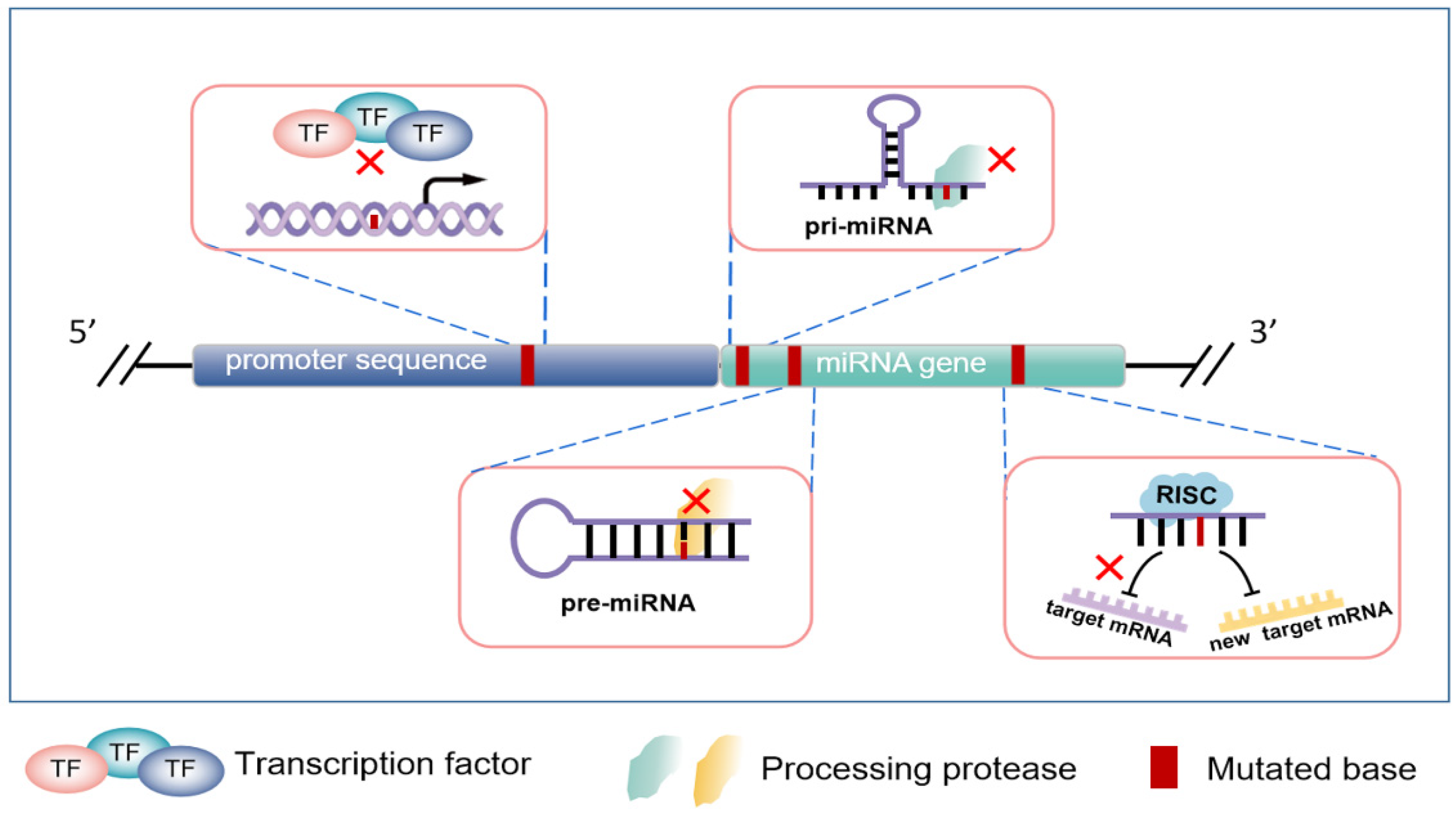
3. MicroRNA Genomic Variation

3.1. Mutations in Promoter Regions of miRNA Encoding Genes

A promoter is a necessary sequence element for the initiation of gene transcription, which can combine with related transcription factors through specific sites and promote the transcription of target genes with the participation of Pol II [13]. In a variety of human malignant tumors, a major genetic variation type, SNP, has been observed in a large number of miRNA promoter regions and leads to abnormal expression of miRNA. For instance, SNP rs4938723 is located in the promoter region of pri-miR-34b/c, and this genetic variation increases the risk of rectal cancer [14] and renal cancer [15]. Further studies showed that in hepatocellular carcinoma (HCC), SNP rs4938723, which appears in the promoter of tumor suppressor gene miR-34b/c, may affect the binding ability with putative transcription factor GATA-X, thereby reducing the transcriptional activity of miR-34b/c [16,17]. In addition, the minor allele of SNP rs4705342 in the miR-143 promoter region can significantly enhance the binding ability of the transcription factor NF-κB and promote gene transcription, ultimately causing the upregulation of miR-143 expression. This leads to the downregulation of its target gene ORP8 and becomes an important factor for HCC in HBV-positive patients [18].
So far, many studies have demonstrated that miR-7 is a tumor suppressor, and the downregulation of miR-7 is usually closely related to the occurrence and development of tumors [19,20,21]. Based on the previous research progress, our team found that the -604 and -617 sites of the miR-7-2 promoter region in lung cancer tissues were mutated, and miR-7 expression with promoter site mutation was lower than that with non-mutated in tumor tissues. Besides, we further proved that mutation in the promoter region could significantly reduce the expression level of mature miR-7 [22]. These studies suggest that the genetic integrity of miRNA promoter sequences may induce the change of miRNA expression in tumors.
At present, these studies mainly focus on the single SNP in miRNA promoter. However, whether multiple SNPs on the same sequence have different regulatory mechanisms or through coordinated regulation needs to be further elucidated in the future.

3.2. Variations of miRNA Coding Sequences

SNP in miRNA coding sequence (including pri-miRNA, pre-miRNA, and mature miRNA) can change miRNA processing, expression, or regulatory activity, thus leading to cancer. Le et al. [23] found that SNP in the pri-mir-146a sequence could affect the expression of miR-146a. This SNP could induce a new mGHG sequence at the apical junction of pri-mir-146a to interact with the double-stranded RNA-binding domain of DROSHA. Surprisingly, the orientation of the new mGHG sequence on pri-mir-146a was switched from basolateral to apical, causing the microprocessor to unproductively cut SNP-pri-mir-146a at its non-splicing site, eventually leading to the downregulation of miRNA.
In the Chinese population, SNP rs11614913 in pre-mir-196a2 and SNP rs3746444 in pre-mir-499 are associated with a significantly increased risk of breast cancer [24]. In addition, SNP rs3746444 in pre-mir-499 and SNP rs2910164 in pre-mir-146a are also significantly associated with the occurrence of cervical squamous cell carcinoma (CSCC) [25]. These carcinogenesis mechanisms may be achieved by altering the expression or processing maturation of related miRNAs. Importantly, for different environmental pressures, SNP at the same site may also regulate miRNA production through different processing mechanisms. Xu et al. [26] showed that the G allele of SNP rs2910164 in the stem structure corresponding to the pre-miR-146a sequence could cause the increased expression of mature miR-146a and was closely related to the risk of hepatocellular carcinoma. In contrast, Kogo et al. [27] reported that in gastric cancer, patients with GG genotype of the same SNP (rs2910164) had significantly lower expression levels of miR-146a than patients with CC genotype, and caused the upregulation of EGFR and IRAK1, which might be due to the diversity of molecular functions and processing patterns in different tumor types.
In addition, it has also been reported that, albeit rarely, mutations in mature miRNAs can lead to impaired tight regulation of target mRNA expression and contribute to poor patient outcomes. For example, SNP rs11614913, located in the 3p mature miRNA region of hsa-mir-196a2, is closely associated with the survival time of individuals with non-small cell lung cancer (NSCLC) [28]. Interestingly, this SNP also contributes to cancer susceptibility in a variety of cancers by impairing the ability of hsa-mir-196a2-3p to regulate downstream target genes, or by changing the type of regulated target genes [29]. Analogously, this phenomenon also was confirmed by Kawahara et al. [30], who found that variation in the mature miR-376 sequence caused by RNA editing was sufficient to redirect miR-376 to silence a new set of target genes.
In summary, the above studies have shown that the variation of the miRNA coding sequence in tumors can significantly affect the ability of miRNA processing, expression, and regulation through different mechanisms, which is depicted in Figure 2. However, the exact mechanisms by which these mutations occur in tumor cells need to be further elucidated by turning to genetics, environmental pressures, and tumor types.

4. Regulation of miRNA by Epigenetic Modifications

4.1. The Influence of DNA Methylation on miRNA Expression

DNA methylation is a DNA chemical modification that catalyzes the binding of methyl groups to the 5th carbon of cytosine by DNA methyltransferases, which is essential for regulating gene expression [31]. Two DNA methylation patterns were observed in cancer cells, including the increase in CpG island methylation in gene promoters and the decrease of global DNA methylation patterns [32]. Recent studies have shown that the methylation level in the CpG island of the miRNA promoter in tumor cells is significantly increased, especially in tumor suppressor genes, as shown in Figure 3A. Multiple miRNA loci, including miR-9-1, miR-193a, miR-137, miR-342, miR-203, and miR-34, were found to be abnormally elevated in methylation levels in human cancers [33,34]. Among them, miR-34a is involved in regulating the expression of genes related to cell cycle, differentiation, and apoptosis, and exerts a tumor suppressor function by reducing cancer stemness and increasing drug sensitivity [35]. The CpG island of the miR-34a promoter is hypermethylated, resulting in its expression being silenced in a variety of tumors, including breast, colon, lung, and bladder cancers [36]. Importantly, when the expression of miR-34a is restored in tumor models, many classic proto-oncogenes such as MYC, KIT, BCL2, and SIRT1 are strongly downregulated [37]. Similarly, Hashimoto et al. [38] observed the hypermethylation in the upstream region of miR-181c in gastric cancer tissues and cells. Treatment with 5-Aza-CdR, a methyltransferase inhibitor, could restore the expression of miR-181c and inhibit the growth and proliferation of gastric cancer cells, suggesting that miR-181c has a tumor suppressive function in gastric cancer cells and its expression is downregulated by DNA methylation.
Global DNA hypomethylation in tumors is considered the companion of genomic CpG island hypermethylation, but it usually appears in different sequences [39]. In fact, tumor cells generally exhibit more global hypomethylation of DNA than CpG island hypermethylation, resulting in a net reduction in genomic 5-methylcytosine content, which provides the possibility for tumor development [40,41]. Because global DNA hypomethylation can lead to genomic instability and induce aneuploidy, chromosome translocation, and copy number change [42,43]. It promotes tumor progression through abnormal gene expression, including miRNA, oncogenes, and so on [44,45,46]. For example, the hypomethylation of miR-106b, miR-25, miR-93, miR-23a, and miR-27a promoter in HCC leads to upregulation of their expression and facilitates the growth of hepatocellular carcinoma cells through various pathways [47].
For the mechanism of miRNA differential methylation in tumors and tissues, studies have shown that it may be related to DNA motif background and different cellular internal factors. DNA methylation depends on the local context of gene sequence, which is reflected in differences in the distribution of methylated motifs: hypermethylated motifs are mainly located in promoter regions rich in CG or CpG islands, whereas hypomethylated motifs are mainly found in gene bodies [48]. In addition, the expression of each isoform of DNMTs in tumors is regulated by multiple factors and they have different affinities for intrinsic DNA motifs, resulting in complex and diverse modifications and regulation of DNA methylation in the genome [49]. Thus, CpG islands that are unmethylated in normal tissues are methylated in tumor cells, and this flexible regulatory mechanism is exploited by tumor cells to meet their own needs. Furthermore, another regulatory mechanism that causes differential methylation modification may arise from the preference of specific transcription factors for methylation motifs in tumor cells [50]. A typical example is Krüppel-like factor 4 (KLF4), which binds preferentially to methylated sequences and activates rather than represses transcription of related genes and tumor development [51,52]. This specific selectivity can have a major impact on gene transcription, especially cancer-promoting miRNAs that are hypermethylated in tumors.
Interestingly, recent studies have shown that there is a feedback loop between miRNA and DNA methylation. MicroRNA can reverse regulate DNA methylation by targeting DNA methyltransferases or methylation-related proteins [53]. In short, abnormal expression of miRNA caused by the altered methylation level is widespread in various tumors, and the crosstalk between miRNA and DNA methylation, as well as the specific methylation regulators in tumors, may helpfully provide some new therapeutic targets.

4.2. The Influence of Histone Modification on miRNA Expression

Histone modification is a plasticity regulatory mechanism. During normal biological development, histone modifications need to be precisely regulated at each developmental stage. Once some stimulus-induced dysregulation of the regulatory program favors cell survival, this adaptive program will be infinitely amplified with the growth of the dominant cells [54]. The dysregulated landscape of histone epigenetic modifications in tumor cells is shown in Figure 3B.

4.2.1. Histone Acetylation

As one of the most common epigenetic regulators, histone acetylation is involved in the expression of miRNA in tumors by regulating the transcriptional activity of genes. Studies have shown that multiple lysine residues in the histone tail can be modified with acetyl groups, which weakens the binding degree between negatively charged DNA and histone by neutralizing the alkaline charges at lysine residues [55]. An abnormal acetylation pattern of histone has been reported as a common feature of human tumor cells [56]. Due to the overall activation of DNA in tumor cells, this provides a large number of modification sites for acetylases, which promote gene transcription through histone acetylation. Such as, it has been shown that in the breast cancer cell line SKBr3, treatment by histone deacetylase (HDAC) inhibitors caused a rapid change in the expression levels of 27 miRNAs, indicating that histones are extensively regulated by acetylation modifications in tumor cells [57].
Notably, some transcription factors can also regulate miRNA expression through histone modifications. For example, Myc can directly or indirectly regulate the target genes of cell growth and proliferation to promote tumorigenesis [58]. Zhang et al. [59] reported that Myc induces histone deacetylation and histone trimethylation to inhibit miR-29 expression in B-cell lymphoma (BCL), as evidenced by Myc can recruit histone deacetylase 3 (HDAC3) and zeste homologue 2 (EZH2) to the miR-29 promoter to form C-Myc/HDAC3/EZH2 co-repressor complex, which leads to miR-29 transcriptional silencing [59]. In ovarian cancer, the cancer suppressor miR-99a reduces cell proliferation through the AKT/mTOR pathway and downregulates the expression of its target gene HOXA1 to inhibit epithelial–mesenchymal transition (EMT). However, the transcription factor YY1 can attenuate the anti-tumor function of miR-99a and promote the dryness of ovarian cancer (OC) cells [60]. Mechanistically, YY1 reduces the acetylation level of miR-99a by recruiting HDAC5 (histone deacetylase 5) to the miR-99a promoter, and finally inhibits the expression of miR-99a [60,61].
These studies have shown that the altered expression of miRNA is partially regulated by histone acetylation in tumors. However, the exact cause remains elusive.

4.2.2. Histone Methylation

Accumulated evidence suggests that the disruption of the balance of histone methylation and demethylation can lead to abnormal expression of related genes, including miRNA, and ultimately contribute to tumor progression [62,63]. Lysine at different sites of the N-terminal tail of histone can be methylated and produce different biological effects, which indicates the complexity of the regulation mechanism of histone methylation. For example, H3K4me3 and H3K36me3 are involved in the transcription and expression of active genes. While H3K9me3, H4K20me3, and H3K27me3 contribute to gene silencing [64]. In diffuse large B-cell lymphoma, upregulated miR-193b and miR-365 are associated with the deletion of H3K27me3 and enrichment of H3K4me3. Conversely, the downregulated miRNAs, including miR-223, miR-150, and miR-451 are correlated with the enrichment of H3K4me3, indicating that histone methylation is the epigenetic mechanism of these differentially expressed miRNAs [65]. In addition, miR-139 is downregulated as a tumor inhibitor in many tumor types. In NSCLC, miR-139 and its host gene PDE2A are apparently silenced by H3K27me3, which is independent of promoter DNA methylation [66]. Knockout of EZH2 or treatment with HDAC can restore miR-139 expression and promote metastasis of lung cancer [66]. Consistently, miR-133a is silenced by epigenetic factors in lung cancer cells, leading to upregulation of its target gene PTBP1 and accelerated lung cancer cell growth and metastasis. Mechanistically, KDM5C, as a specific demethylase of histone H3K4, causes histone demethylation of miR-133a promoter to inhibit transcriptional activation of miR-133a [67].
Up to now, a large number of different types of histone methylating and demethylating proteases have been discovered. Furthermore, the methylation status of histones is highly dependent on the regulation of these different proteins. In mammalian chromatin, Suv39h histone methyltransferases (HMTases) selectively monomethylate H3K27 and trimethylate H3K9 [68], while KMT2A (also known as MLL1) is responsible for catalyzing the dimethylation and trimethylation of H3K4 [69,70,71]. Therefore, dysregulation of specific modifiers may directionally alter the transcriptional activity of some miRNAs. For example, EZh2 dysregulation in lymphoma causes an increase in H3K27me3 labeling [72,73]. This will lead to epigenetic silencing of a large number of miRNAs. Furthermore, in tumor cells, this dysregulatory mechanism may create a positive feedback to further promote tumor progression.

4.2.3. Other Modifications of Histones

In addition to these well-known modifications, histones can be modified in other ways and jointly determine the structure of chromatin. These modifications include phosphorylation, ubiquitin, citrullination, butyrylation, hydroxylation, formylation, propionylation, and crotonylation [74,75,76]. To some extent, the regulatory role of these histone-modified markers in various cancers has not been fully elucidated. Recently, however, there is still some evidence that they participate in tumor progression by regulating miRNA expression. For example, the phosphorylated histone H2AX binds to the miR-3196 promoter, which leads to the transcriptional silencing of miR-3196 by increasing H3K27 trimethyl in the promoter region of miR-3196 and inhibiting the binding of RNA Pol II [77]. In lung cancer cells, decreased phosphorylation of H2AX leads to high expression of miR-3196, which inhibits lung cancer cell apoptosis by targeting the p53-upregulated apoptosis regulator (PUMA) [77]. Furthermore, E3 ligase HectH9, which mediates ubiquitin modification, induces K63 polyubiquitin modification of DDX17 under anoxic conditions and promotes its dissociation from the pri-miRNA-Drosha-DCGR8 complex. This will hinder the processing and maturation of a large number of tumor suppressor miRNAs, including miR-16 and miR-34a, thus favoring the expression of cancer stemness genes [78]. All of this evidence suggests that miRNA expression is regulated by multiple histone modifications, and further exploration of the combinatorial effects of different histone modifications might be a key work in this field.

5. Regulation of miRNA by RNA Modification

Recent studies have revealed that alterations in the ncRNA transcriptome may be a novel mechanism of tumorigenesis, especially targeting the modification regulation of miRNA. Although the regulation of miRNA by chemical modifications such as m5C, hm5C, N6-methyladenosine (m6A), m1A, and uridylation has not been fully elucidated, RNA modification still provides researchers with a broad research prospect [79]. Most current studies have shown that m6A and uridylation modifications have a significant impact on the expression of miRNA and determine its fate in the occurrence pathway.

5.1. The Influence of m6A Modification on miRNA Expression

At the transcriptome level, as the most abundant RNA modification in eukaryotes, the role of m6A modification in regulating miRNA expression has become increasingly prominent [80]. The process of m6A modification is mainly coordinated by methyltransferase (m6A writer), demethylase (m6A eraser), and m6A binding protein (m6A reader) [81]. Direct evidence showed that in breast cancer cells, m6A methylation motifs are abundantly enriched in pri-miRNA sequences but absent in pre-miRNA sequences [82]. The m6A methylation motif provides a site for methyltransferase (METTL) to mark pri-miRNA with m6A, which facilitates the recognition and processing of DGCR8, suggesting that the regulation of miRNA expression by m6A modification occurs primarily during pri-miRNA processing and exhibits m6A dependence [82], as shown in Figure 4.
Most of the proteins involved in m6A modification belong to oncogenes, and they are frequently upregulated in tumor tissues, thereby increasing the processing and expression of oncogenes and promoting the malignant development of cells [83]. For example, the expression of METTL3 is significantly upregulated in bladder cancer, which enhances DGCR8 processing and promotes its expression by increasing the cancer-promoting miR-221/222 m6A modification [84]. Consistently, cigarette smoke induces METTL3 overexpression, which promotes the processing and maturation of miR-25-3p by increasing the level of m6A modification of pri-mir-25. Subsequently, high levels of mature miR-25/miR-25-3p inhibit the expression of PH structural domain leucine-rich repeat protein phosphatase 2 (PHLPP2) and induce activation of oncogenic AKT-p70S6K signaling to promote pancreatic cancer progression [85].
Although tumor suppressor genes processing may also be increased, differences in gene transcription levels already dictate that more oncogenes undergo m6A-modified processing than suppressor genes. In addition, another accepted mechanism may be that specific regulators of m6A modification act by blocking m6A modification of suppressor genes, which have been reported. After knockdown of the m6A demethylase FTO in HEK293 cells, several mature miRNAs were nevertheless downregulated [86]. In addition, the m6A reader HNRNPA2B1 has been shown to recruit DGCR8 after recognition of m6A to facilitate the shearing process of pri-miRNA by DROSHA. However, HNRNPA2B1 overexpression shows opposite effects on miR-222, miR-29a-3p and miR-29b-3p expression and leads to endocrine resistance in breast cancer cells [87]. All of this evidence fully demonstrates the complexity and diversity of m6A modification and the regulation of subsequent miRNA processing.
Current studies mainly focus on the effects of aberrant m6A modification, but the mechanisms of how various functional components involved in miRNA m6A modification are altered are not fully understood. Furthermore, how m6A modification differentially regulates miRNA processing and expression in tumor cells is unclear. Therefore, further studies on these molecules are necessary in the future to refine the mechanisms of miRNA expression regulation.

5.2. The Influence of Uridylation on miRNA Expression

The role of uridylation on miRNA was first identified in the precursor sequence of let-7 [88]. Uridylation of the 3′ end of pre-let-7 mediated by Lin28 evades DICER processing and induces pre-let-7 degradation [88]. Conversely, in human cells, modification of pre-let-7 monouridine by the terminal uridylyltransferase TUT7/4/2 can switch the optimal structure from a 1 nt 3′ protruding end to a 2 nt 3′ protruding end, thereby enhancing processing shear of DICER [89]. Interestingly, TUT, a broad RNA modifying enzyme, can also switch specific pre-miRNA from degradation mode to processing mode to promote miRNA expression, a switch that is determined by the cellular environment [90]. For example, in TUT4/7-depleted prostate and OC cells, some specific miRNAs showed differential expression patterns [91]. In particular, miR-200c-3p and miR-141-3p, which are diagnostic markers of OC, are significantly downregulated and can impair the migration ability in OC cells, suggesting that uridylation modifications regulating miRNA expression may have specific regulatory mechanisms in different tumor cells [91].

6. Abnormal Cleavage by Processing Enzymes

Although numerous studies have demonstrated that controlling the expression of individual miRNA has oncogenic or pro-cancer effects, the overall altered expression of miRNA in tumor cells compared to normal cells strongly suggests that the processing components of miRNA may be dysregulated in tumors [92]. Table 1 shows the outcome of dysregulation of some miRNA processing components in tumors.

6.1. Microprocessor Abnormalities and miRNA

In the nucleus, a heterotrimeric complex (called a microprocessor) consisting of one DROSHA and two DGCR8 molecules mediates the splicing of pri-miRNA. Among them, DGCR8 interacts with the stem and apical elements of pri-miRNA through its double-stranded RNA (dsRNA) binding domain and RNA-binding heme domain, respectively. Meanwhile, DROSHA acts as a “ruler” for measuring the 11-base distance of the single-stranded to double-stranded RNA (ssRNA-dsRNA) junction and cleaves the stem-loop of pri-miRNA to release pre-miRNA [112,113]. Besides, the microprocessor contains several functional cofactors, including DEAD-box, RNA helicase, p72 (DDX17), and the Ewing’s sarcoma family of proteins, which together promote the fidelity and activity of DROSHA processing [114]. However, abnormal expression or function of microprocessors and related components play important roles in tumors by altering miRNA processing and maturation to set the stage for cellular transformation and tumorigenesis in vivo [115,116,117]. One of its upstream mechanisms may be due to genetic mutation or transcriptional silencing/activation of a large number of processing components in tumors that disrupt the normal processing of miRNA.

6.2. XPO5 Abnormalities and miRNA

XPO5, as a “vehicle” of miRNA, shows paralysis or overload of transportation in cancer. There has been evidence that genetic mutations in XPO5 lead to defects in the C-terminal region of the pre-miRNA/XPO5/Ran-GTP ternary complex and retain the pre-miRNA in the nucleus [100]. In hepatocellular carcinoma, XPO5 is phosphorylated by ERK, leading to its conformational change by the prolyl isomerase Pin1, which ultimately reduces the ability of XPO5 to export pre-miRNA [118]. These studies suggest that the output mechanism of nucleoplasm may be damaged in tumor cells.

6.3. DICER Abnormalities and miRNA

When the pre-miRNA is transported to the cytoplasm, DICER participates in the final cutting process. DICER recognizes dsRNA and acts as a second molecular “ruler” by cleaving it at a specific distance from the end of the helix [119]. Consistently, mutations in the DICER gene are also frequently present in tumors and lead to impaired miRNA biogenesis and processing [120]. In addition, many regulatory factors regulate cancer progression by targeting the expression of DICER. For example, activated HIF-1α in tumors downregulates Dicer expression by inducing ubiquitination of the E3 ligase Parkin, and further reduces the expression of tumor suppressor miRNAs, including let-7 and miR-200b, which promotes EMT and metastasis in tumor-bearing mice [121].
In short, various processing enzymes and their components not only determine the correct production steps of miRNA but also maintain the normal expression of miRNA. Notably, the regulatory mechanisms of these processing steps are often different in various tumor types, and the expression patterns or functions of the same molecules may differ, so future perspectives are necessary to focus on tumor-specific targets for further exploration.

7. Regulation of miRNA by lncRNA

LncRNAs, including long intergenic noncoding RNAs (lincRNAs), circular RNAs (circRNAs), and pseudogenes, can regulate tumor biological behaviors through a competitive endogenous RNA (ceRNA) mechanism [122,123]. They act as a “sponge” for miRNA by sharing miRNA response element (MRE), thereby reducing the number of miRNAs that target mRNA, as shown in Figure 5.

7.1. Regulation of miRNA by lincRNA

LincRNA is an RNAi molecule consisting of hundreds to thousands of nucleotides that has been shown to play an important role in gene regulation. Although lincRNA has diverse regulatory mechanisms in tumors, their role as decoys for miRNA in the ceRNA regulatory network has become increasingly prominent. linc-ROR is upregulated in tumor tissues and promotes tumor progression by inducing EMT and promoting malignant abilities such as proliferation, migration, etc. [124,125]. Mechanistically, linc-ROR acts as a ceRNA of miR-205, inhibiting its expression and indirectly promoting the expression of miR-205 target genes [124]. Furthermore, linc-ROR also acts as a ceRNA of miR-145 and promotes the invasion of triple-negative breast cancer through the linc-ROR/miR-145/ARF6 pathway [126]. In contrast, lincRNA-p21 has oncogenic effects and can play a role in tumors by targeting different miRNAs, including miR-9 [127], miR-17-5p [128] and miR-181b [129]. Notably, some lincRNAs can also serve as feedstock for miRNAs that are processed to form mature miRNAs to participate in the biological behavior of tumors. For example, lincRNA H19 serves as a major source of precursors and a regulatable reservoir for miR-675 by regulating its processing and expression in response to cellular stress or oncogenic signals [130,131]. These studies suggest that the expression and function of multiple miRNAs in tumors are regulated by lincRNA and lead to altered expression of related target genes.

7.2. Regulation of miRNA by circRNA

CircRNAs are a class of single-stranded closed RNA molecules, usually formed by the reverse connection of exonic precursor mRNA, which indirectly regulate the activity of miRNA target genes by acting as miRNA sponges through abundant miRNA binding sites [132]. Recent studies have shown that the expression of circRNA is mainly expressed in specific cell types and tissues, and is dynamically regulated by various environments, suggesting that circRNA may play an important role in tissue development [133]. Moreover, its dysregulation can strongly affect the expression of miRNA and participate in tumorigenesis. The most representative of them is CIRS-7 (also known as CDR1as), which contains more than 70 conserved miR-7 target sites and is a super-sponge of miR-7 [134,135]. MiR-7 has been proven to inhibit cancer cell growth and promote apoptosis by directly targeting and downregulating key oncogenic factors in cancer-related signaling pathways [136,137]. In addition, our group further revealed that miR-7 plays an important role in alleviating the ConA-induced acute autoimmune liver injury model in mice by regulating immune cells. MiR-7 negatively regulates the activation and function of CD4+ T cells through the MAPK4 pathway, thereby reducing the pathological changes of autoimmune hepatitis [138]. This study enriches the regulatory function of miR-7 and provides researchers with a new perspective on the involvement of miR-7 in immune-related diseases including tumors. Notably, miR-7 was frequently downregulated in various tumor tissues, and the expression levels of miR-7 and CIRS-7 were negatively correlated. In tumor cells, upregulated CIRS-7 exerts its sponge effect by targeting miR-7 and upregulates its key target genes, including EGFR, CCNE1, and PIK3CD, to induce tumor cell proliferation [139,140].
The latest literature has documented that circ-Foxo3 has binding sites for multiple miRNAs and can act as a ceRNA to inhibit or promote tumor growth [141]. Specifically, circ-Foxo3 upregulates the expression of NFAT5 nuclear factor through a mechanism of sponge miR-138-5p/miR-432-5p to promote the proliferation and invasion of glioblastoma [142]. In gastric cancer, circ-Foxo3 upregulates the expression of USP44 by targeting miR-143-3p, thereby exerting a tumor-promoting effect [143].
In all, the above current studies have shown that circRNA, as a powerful miRNA inhibitor in tumors, is a novel marker for cancer diagnosis and potential therapeutic targets.

7.3. Regulation of miRNA by Pseudogene

Pseudogenes are a special group of lncRNA that develop from protein-coding genes and have been regarded as “evolutionary garbage” due to the loss of their ability to encode proteins [144]. However, recent studies have found that pseudogenes and their corresponding coding genes can bind to the same miRNA and thus act as a ceRNA, suggesting the importance of pseudogenes in the regulation of miRNA expression [145]. PTEN P1 is the first pseudogene found to regulate its parental gene PTEN through a ceRNA mechanism [146]. In tumors, PTEN P1 acts as a bait to adsorb and degrade miRNA targeting PTEN, and actively regulates the expression of miRNA targeting PTEN, such as miR-17, miR-21, miR-214, miR-19, and miR-26 families, resulting in upregulation of PTEN gene expression and inhibition of tumor growth [147,148,149]. On the other hand, pseudogenes can also act as oncogenes in the body. Karreth et al. [150] reported that BRAF P1, a pseudogene of the oncogene BRAF, is mainly expressed in tumor cells. Importantly, BRAF P1, with multiple miRNA binding sites, can regulate the expression of its parental gene by sequestering specific miRNAs through a competitive endogenous RNA mechanism, and this specificity depends on the tumor type. Hao et al. [151] showed that the pseudogene AKR1B10P1 can promote the growth and motility of HCC cells in vitro and in vivo. Further studies found that the pseudogene AKR1B10P1 effectively abolished miR-138-induced SOX4 mRNA degradation and enhanced EMT in HCC by directly sponge miR-138.
To date, although substantial evidence suggests that pseudogenes play an important role in cancer, their functions and underlying mechanisms remain largely undetermined. One of the main functions of pseudogenes has been found to act as miRNA decoys, competing with miRNA that may target parental genes and regulating their activities, thereby playing a role in promoting or suppressing tumors.

8. Extracellular Secretion and Endocytosis of miRNA

Notably, extracellular vesicles (EVs) as communication mediators with neighboring or distant cells undoubtedly provide a new pathway for differential expression and functional regulation of miRNA in the tumor and tumor microenvironment. These cells rely on the active transport system of EVs to deliver their own synthesized miRNAs to recipient cells, where they act as endogenous miRNAs to regulate multiple target genes or signaling pathways [152,153]. For example, in a hypoxic environment, lung cancer cells receive EV-derived miR-31-5p to activate MEK/ERK signaling, thereby contributing to their development and metastasis [154]. Besides, tumor cells secrete produced miRNAs to promote their own growth and create a tumor microenvironment that evades surveillance and attack by the immune system [155,156]. In particular, miR-1298-5p secreted by glioma cells not only reduces the level of endogenous miR-1298-5p to promote their proliferative ability but also activates myeloid-derived suppressor cells (MDSCs) through the NF-κB pathway to suppress the immune system [157]. These studies have shown that miRNA expression is highly regulated by the cell’s own secretion, and when the host receives the entry of exogenous miRNA, it can also cause significant changes.
Despite the abundance of miRNA species, some miRNAs involved in cancer proliferation, growth, and metastasis were significantly enriched in extracellular vesicles, suggesting the existence of a mechanism that regulates the loading of specific miRNAs into vesicles for exocrine expression [158,159]. Given the variety of miRNAs in EVs and the specificity of different tumor cell biological behaviors and states, it remains mysterious whether these “miRNA weapons” employed by tumor cells have their specific screening and secretion mechanisms. Fortunately, some recent studies have reported that the molecular mechanism of miRNA screening and loading in tumor cells may be related to different heterogeneous nuclear ribonucleoproteins (hnRNPs). First of all, in lung cancer cells, hnRNPA2B1 binds the tumor suppressor miR-122-5p through the EXO motif to selectively sort and transfer it into secreted EVs [160]. Meanwhile, the delivery of lung cancer EVs-miR-122-5p promoted hepatocyte migration by increasing the expression of N-cadherin and vimentin, which plays an important role in the establishment of the pre-metastatic microenvironment in lung cancer and liver metastasis [160]. Furthermore, in highly metastatic cancers, another heterogeneous nuclear ribonucleoprotein (hnRNPK), driven by non-caveolin-1 (CAV1), assists the EV loading and secretion of miR-148a-3p, which favors migration of prostate and colorectal cancer cells [161].
At present, these studies expand the understanding of the expression regulation of tumor miRNA, including changes in endogenous miRNA caused by active cellular secretion and host acceptance of miRNA. However, whether or not the extracellular secretion of miRNA is highly selective and related underlying mechanism remains to be fully elucidated.

9. Conclusions and Perspectives

Up to now, many studies have demonstrated that the abnormal expression of miRNA in tumors is closely related to the occurrence and development of tumors. Therefore, the detailed elucidation of miRNA expression regulation mechanisms is of great significance for the diagnosis, prognosis, and treatment of cancer patients.
Current research on the regulation of miRNA expression mainly focuses on multiple processes ranging from gene transcription to post-transcriptional modification regulation. However, the mechanisms underlying the uncontrolled expression of miRNA in tumors are far from clear. We propose there are three main facets of the regulatory mechanism of miRNAs in tumors that still need to be further elucidated. Firstly, recent studies have shown that the gut microbiota can regulate the expression of host miRNAs to participate in tumorigenesis and development [162,163]. In addition, plant-derived miRNAs in food can be taken up by mammals and participate in the regulation of miRNA composition and function in vivo [164,165]. More importantly, intermittent fasting can also affect the expression of related miRNAs and shows amazing anticancer effects [166,167,168]. These studies suggested that human diet, living habits, and other aspects might can affect the expression of body-related miRNAs and jointly promote or inhibit tumorigenesis. Secondarily, in fact, miRNA, as a kind of powerful, numerous, and abundant small RNA, has complex and diverse regulatory factors involved in its production. Therefore, how to outline the underlying connections among these different factors at distinct levels is still urgent and important for understanding the regulatory mechanisms of miRNA. Thirdly, the expression of miRNA has temporal and spatial specificity in different tissues or cells, and its expression regulation mechanisms are often different. Therefore, in the future, it is necessary to use advanced technologies such as single-cell multi-omics sequencing and high-resolution spatial omics to further explore the specific regulatory mechanisms of miRNA in specific tumors.
In conclusion, it is necessary to comprehensively study the complex network covering the expression of miRNAs and their regulatory mechanisms, so as to open up a new way for human beings to deeply understand the expression and regulation mechanisms of tumors and to apply small molecular means such as miRNA to prevent and treat tumors.

Author Contributions

Conceptualization, S.C. and L.X.; writing—original draft preparation, S.C., Y.W., D.L. and H.W.; writing—review and editing, S.C., X.Z., J.Y., L.C. and L.X.; supervision, S.C., M.G., J.Z., C.C. and L.X.; funding acquisition, L.X.; supervision, Y.Z., G.L. and L.X. All authors have read and agreed to the published version of the manuscript.

Funding

This work was supported by the Program for High-level Innovative Talents in Guizhou Province (QKH-RC-2016-4031), the National Natural Science Foundation of China (32160178, 82160503, 81960509), the Project of the Guizhou Provincial Department of Science and Technology (QKH-JC-2018-1428, QKHZC-2020-4Y156), the Program for New Century Excellent Talents in University, Ministry of Education of China (NCET-12-0661), Collaborative Innovation Center of Chinese Ministry of Education (2020-39), the Project of the Zunyi Science and Technology Bureau (ZKH-HZ-2021-193) and the Program for Excellent Young Talents of Zunyi Medical University (15ZY-001).

Institutional Review Board Statement

Not applicable.

Informed Consent Statement

Not applicable.

Data Availability Statement

Not applicable.

Acknowledgments

We sincerely gratitude to the professional organization American Journal Experts (AJE) for the polish of this article. In addition, we sincerely thank Figdraw for providing the graphic material for Figure 1 and Figure 3B in this paper.

Conflicts of Interest

No potential conflicts of interest are disclosed.

Abbreviations

3′-UTR3′-untranslated region
lncRNALong non-coding RNA
pri-miRNAPrimary miRNA
RNaseRibonuclease
Pol IIPolymerase II
pre-miRNAPrecursor miRNA
XPO5Exportin 5
AGOArgonaute
RISCRNA-induced silencing complexes
HCCHepatocellular carcinoma
CSCCCervical squamous cell carcinoma
NSCLCNon-small cell lung cancer
DNMTDNA methyltransferase
HDACHistone deacetylase
BCLB-cell lymphoma
EZH2Zeste homologue 2
EMTEpithelial–mesenchymal transition
m6AN6-methyladenosine
METTLMethyltransferase
OCOvarian cancer
lincRNALong intergenic noncoding RNA
circRNACircular RNA
ceRNACompetitive endogenous RNA
EVExtracellular vesicle
hnRNPHeterogeneous nuclear ribonucleoproteins

References

  1. Gerlach, D.; Kriventseva, E.V.; Rahman, N.; Vejnar, C.E.; Zdobnov, E.M. miROrtho: Computational survey of microRNA genes. Nucleic Acids Res. 2009, 37, D111–D117. [Google Scholar] [CrossRef] [PubMed]
  2. Lee, R.C.; Feinbaum, R.L.; Ambros, V. The C. Elegans heterochronic gene lin-4 encodes small RNAs with antisense complementarity to lin-14. Cell 1993, 75, 843–854. [Google Scholar] [CrossRef]
  3. Sayed, D.; Abdellatif, M. MicroRNAs in development and disease. Physiol. Rev. 2011, 91, 827–887. [Google Scholar] [CrossRef] [PubMed]
  4. Lujambio, A.; Lowe, S.W. The microcosmos of cancer. Nature 2012, 482, 347–355. [Google Scholar] [CrossRef]
  5. Garzon, R.; Marcucci, G.; Croce, C.M. Targeting microRNAs in cancer: Rationale, strategies and challenges. Nat. Rev. Drug Discov 2010, 9, 775–789. [Google Scholar] [CrossRef]
  6. Lee, Y.; Kim, M.; Han, J.; Yeom, K.H.; Lee, S.; Baek, S.H.; Kim, V.N. MicroRNA genes are transcribed by RNA polymerase II. EMBO J. 2004, 23, 4051–4060. [Google Scholar] [CrossRef]
  7. Denli, A.M.; Tops, B.B.; Plasterk, R.H.; Ketting, R.F.; Hannon, G.J. Processing of primary microRNAs by the Microprocessor complex. Nature 2004, 432, 231–235. [Google Scholar] [CrossRef]
  8. Bohnsack, M.T.; Czaplinski, K.; Gorlich, D. Exportin 5 is a RanGTP-dependent dsRNA-binding protein that mediates nuclear export of pre-miRNAs. RNA 2004, 10, 185–191. [Google Scholar] [CrossRef]
  9. Hutvágner, G.; McLachlan, J.; Pasquinelli, A.E.; Bálint, E.; Tuschl, T.; Zamore, P.D. A cellular function for the RNA-interference enzyme Dicer in the maturation of the let-7 small temporal RNA. Science 2001, 293, 834–838. [Google Scholar] [CrossRef]
  10. Desvignes, T.; Batzel, P.; Berezikov, E.; Eilbeck, K.; Eppig, J.T.; McAndrews, M.S.; Singer, A.; Postlethwait, J.H. miRNA Nomenclature: A View Incorporating Genetic Origins, Biosynthetic Pathways, and Sequence Variants. Trends Genet. 2015, 31, 613–626. [Google Scholar] [CrossRef] [Green Version]
  11. Liu, J.; Carmell, M.A.; Rivas, F.V.; Marsden, C.G.; Thomson, J.M.; Hammond, S.M.; Joshua-Tor, L.; Hannon, G.J. Argonaute2 is the catalytic engine of mammalian RNAi. Science 2004, 305, 1437–1441. [Google Scholar] [CrossRef]
  12. Yang, J.S.; Lai, E.C. Alternative miRNA biogenesis pathways and the interpretation of core miRNA pathway mutants. Mol. Cell 2011, 43, 892–903. [Google Scholar] [CrossRef]
  13. Perera, D.; Poulos, R.C.; Shah, A.; Beck, D.; Pimanda, J.E.; Wong, J.W. Differential DNA repair underlies mutation hotspots at active promoters in cancer genomes. Nature 2016, 532, 259–263. [Google Scholar] [CrossRef]
  14. Gao, L.B.; Li, L.J.; Pan, X.M.; Li, Z.H.; Liang, W.B.; Bai, P.; Zhu, Y.H.; Zhang, L. A genetic variant in the promoter region of miR-34b/c is associated with a reduced risk of colorectal cancer. Biol. Chem. 2013, 394, 415–420. [Google Scholar] [CrossRef]
  15. Zhang, S.; Qian, J.; Cao, Q.; Li, P.; Wang, M.; Wang, J.; Ju, X.; Meng, X.; Lu, Q.; Shao, P.; et al. A potentially functional polymorphism in the promoter region of miR-34b/c is associated with renal cell cancer risk in a Chinese population. Mutagenesis 2014, 29, 149–154. [Google Scholar] [CrossRef]
  16. Xu, Y.; Liu, L.; Liu, J.; Zhang, Y.; Zhu, J.; Chen, J.; Liu, S.; Liu, Z.; Shi, H.; Shen, H.; et al. A potentially functional polymorphism in the promoter region of miR-34b/c is associated with an increased risk for primary hepatocellular carcinoma. Int. J. Cancer 2011, 128, 412–417. [Google Scholar] [CrossRef]
  17. Son, M.S.; Jang, M.J.; Jeon, Y.J.; Kim, W.H.; Kwon, C.I.; Ko, K.H.; Park, P.W.; Hong, S.P.; Rim, K.S.; Kwon, S.W.; et al. Promoter polymorphisms of pri-miR-34b/c are associated with hepatocellular carcinoma. Gene 2013, 524, 156–160. [Google Scholar] [CrossRef]
  18. Yin, X.; Sun, S.; Zhao, J.; Yang, J.; Lei, X.; Xu, C.; Li, K. Rs4705342 polymorphism is involved in the tumorigenesis of HBV positive HCC by altering the binding affinity of HBV induced NF-kB with the promoter region of microRNA-143. J. Cell Biochem. 2018, 119, 5233–5242. [Google Scholar] [CrossRef]
  19. Saydam, O.; Senol, O.; Würdinger, T.; Mizrak, A.; Ozdener, G.B.; Stemmer-Rachamimov, A.O.; Yi, M.; Stephens, R.M.; Krichevsky, A.M.; Saydam, N.; et al. miRNA-7 attenuation in Schwannoma tumors stimulates growth by upregulating three oncogenic signaling pathways. Cancer Res. 2011, 71, 852–861. [Google Scholar] [CrossRef]
  20. Zhang, N.; Li, X.; Wu, C.W.; Dong, Y.; Cai, M.; Mok, M.T.; Wang, H.; Chen, J.; Ng, S.S.; Chen, M.; et al. microRNA-7 is a novel inhibitor of YY1 contributing to colorectal tumorigenesis. Oncogene 2013, 32, 5078–5088. [Google Scholar] [CrossRef] [Green Version]
  21. Gajda, E.; Grzanka, M.; Godlewska, M.; Gawel, D. The Role of miRNA-7 in the Biology of Cancer and Modulation of Drug Resistance. Pharmaceuticals 2021, 14, 149. [Google Scholar] [CrossRef]
  22. Zhao, J.; Wang, K.; Liao, Z.; Li, Y.; Yang, H.; Chen, C.; Zhou, Y.A.; Tao, Y.; Guo, M.; Ren, T.; et al. Promoter mutation of tumor suppressor microRNA-7 is associated with poor prognosis of lung cancer. Mol. Clin. Oncol. 2015, 3, 1329–1336. [Google Scholar] [CrossRef]
  23. Le, C.T.; Nguyen, T.L.; Nguyen, T.D.; Nguyen, T.A. Human disease-associated single nucleotide polymorphism changes the orientation of DROSHA on pri-mir-146a. RNA 2020, 26, 1777–1786. [Google Scholar] [CrossRef]
  24. Hu, Z.; Liang, J.; Wang, Z.; Tian, T.; Zhou, X.; Chen, J.; Miao, R.; Wang, Y.; Wang, X.; Shen, H. Common genetic variants in pre-microRNAs were associated with increased risk of breast cancer in Chinese women. Hum. Mutat. 2009, 30, 79–84. [Google Scholar] [CrossRef]
  25. Zhou, B.; Wang, K.; Wang, Y.; Xi, M.; Zhang, Z.; Song, Y.; Zhang, L. Common genetic polymorphisms in pre-microRNAs and risk of cervical squamous cell carcinoma. Mol. Carcinog. 2011, 50, 499–505. [Google Scholar] [CrossRef]
  26. Xu, T.; Zhu, Y.; Wei, Q.K.; Yuan, Y.; Zhou, F.; Ge, Y.Y.; Yang, J.R.; Su, H.; Zhuang, S.M. A functional polymorphism in the miR-146a gene is associated with the risk for hepatocellular carcinoma. Carcinogenesis 2008, 29, 2126–2131. [Google Scholar] [CrossRef]
  27. Kogo, R.; Mimori, K.; Tanaka, F.; Komune, S.; Mori, M. Clinical significance of miR-146a in gastric cancer cases. Clin. Cancer Res. 2011, 17, 4277–4284. [Google Scholar] [CrossRef]
  28. Hu, Z.; Chen, J.; Tian, T.; Zhou, X.; Gu, H.; Xu, L.; Zeng, Y.; Miao, R.; Jin, G.; Ma, H.; et al. Genetic variants of miRNA sequences and non-small cell lung cancer survival. J. Clin. Investig. 2008, 118, 2600–2608. [Google Scholar] [CrossRef] [PubMed]
  29. Chu, H.; Wang, M.; Shi, D.; Ma, L.; Zhang, Z.; Tong, N.; Huo, X.; Wang, W.; Luo, D.; Gao, Y.; et al. Hsa-miR-196a2 Rs11614913 polymorphism contributes to cancer susceptibility: Evidence from 15 case-control studies. PLoS ONE 2011, 6, e18108. [Google Scholar] [CrossRef] [PubMed]
  30. Kawahara, Y.; Zinshteyn, B.; Sethupathy, P.; Iizasa, H.; Hatzigeorgiou, A.G.; Nishikura, K. Redirection of silencing targets by adenosine-to-inosine editing of miRNAs. Science 2007, 315, 1137–1140. [Google Scholar] [CrossRef] [PubMed] [Green Version]
  31. Sriraman, A.; Debnath, T.K.; Xhemalce, B.; Miller, K.M. Making it or breaking it: DNA methylation and genome integrity. Essays Biochem. 2020, 64, 687–703. [Google Scholar]
  32. Jones, P.A.; Baylin, S.B. The epigenomics of cancer. Cell 2007, 128, 683–692. [Google Scholar] [CrossRef]
  33. Lujambio, A.; Esteller, M. How epigenetics can explain human metastasis: A new role for microRNAs. Cell Cycle 2009, 8, 377–382. [Google Scholar] [CrossRef]
  34. Lujambio, A.; Calin, G.A.; Villanueva, A.; Ropero, S.; Sánchez-Céspedes, M.; Blanco, D.; Montuenga, L.M.; Rossi, S.; Nicoloso, M.S.; Faller, W.J.; et al. A microRNA DNA methylation signature for human cancer metastasis. Proc. Natl. Acad. Sci. USA 2008, 105, 13556–13561. [Google Scholar] [CrossRef]
  35. Farooqi, A.A.; Tabassum, S.; Ahmad, A. MicroRNA-34a: A Versatile Regulator of Myriads of Targets in Different Cancers. Int. J. Mol. Sci. 2017, 18, 2089. [Google Scholar] [CrossRef]
  36. Lodygin, D.; Tarasov, V.; Epanchintsev, A.; Berking, C.; Knyazeva, T.; Körner, H.; Knyazev, P.; Diebold, J.; Hermeking, H. Inactivation of miR-34a by aberrant CpG methylation in multiple types of cancer. Cell Cycle 2008, 7, 2591–2600. [Google Scholar] [CrossRef]
  37. Misso, G.; Di Martino, M.T.; De Rosa, G.; Farooqi, A.A.; Lombardi, A.; Campani, V.; Zarone, M.R.; Gullà, A.; Tagliaferri, P.; Tassone, P.; et al. Mir-34: A new weapon against cancer? Mol. Ther. Nucleic. Acids 2014, 3, e194. [Google Scholar] [CrossRef]
  38. Hashimoto, Y.; Akiyama, Y.; Otsubo, T.; Shimada, S.; Yuasa, Y. Involvement of epigenetically silenced microRNA-181c in gastric carcinogenesis. Carcinogenesis 2010, 31, 777–784. [Google Scholar] [CrossRef]
  39. Ehrlich, M. DNA hypomethylation in cancer cells. Epigenomics 2009, 1, 239–259. [Google Scholar] [CrossRef]
  40. Ehrlich, M. Cancer-linked DNA hypomethylation and its relationship to hypermethylation. Curr. Top Microbiol. Immunol. 2006, 310, 251–274. [Google Scholar]
  41. Ehrlich, M. DNA methylation in cancer: Too much, but also too little. Oncogene 2002, 21, 5400–5413. [Google Scholar] [CrossRef]
  42. Karpf, A.R.; Matsui, S. Genetic disruption of cytosine DNA methyltransferase enzymes induces chromosomal instability in human cancer cells. Cancer Res. 2005, 65, 8635–8639. [Google Scholar] [CrossRef] [Green Version]
  43. Eden, A.; Gaudet, F.; Waghmare, A.; Jaenisch, R. Chromosomal instability and tumors promoted by DNA hypomethylation. Science 2003, 300, 455. [Google Scholar] [CrossRef]
  44. Baer, C.; Claus, R.; Frenzel, L.P.; Zucknick, M.; Park, Y.J.; Gu, L.; Weichenhan, D.; Fischer, M.; Pallasch, C.P.; Herpel, E.; et al. Extensive promoter DNA hypermethylation and hypomethylation is associated with aberrant microRNA expression in chronic lymphocytic leukemia. Cancer Res. 2012, 72, 3775–3785. [Google Scholar] [CrossRef]
  45. Nojima, M.; Matsui, T.; Tamori, A.; Kubo, S.; Shirabe, K.; Kimura, K.; Shimada, M.; Utsunomiya, T.; Kondo, Y.; Iio, E.; et al. Global, cancer-specific microRNA cluster hypomethylation was functionally associated with the development of non-B non-C hepatocellular carcinoma. Mol. Cancer 2016, 15, 31. [Google Scholar] [CrossRef]
  46. Karpf, A.R.; Bai, S.; James, S.R.; Mohler, J.L.; Wilson, E.M. Increased expression of androgen receptor coregulator MAGE-11 in prostate cancer by DNA hypomethylation and cyclic AMP. Mol. Cancer Res. 2009, 7, 523–535. [Google Scholar] [CrossRef]
  47. He, X.X.; Kuang, S.Z.; Liao, J.Z.; Xu, C.R.; Chang, Y.; Wu, Y.L.; Gong, J.; Tian, D.A.; Guo, A.Y.; Lin, J.S. The regulation of microRNA expression by DNA methylation in hepatocellular carcinoma. Mol. Biosyst. 2015, 11, 532–539. [Google Scholar] [CrossRef]
  48. Scala, G.; Federico, A.; Palumbo, D.; Cocozza, S.; Greco, D. DNA sequence context as a marker of CpG methylation instability in normal and cancer tissues. Sci. Rep. 2020, 10, 1721. [Google Scholar] [CrossRef]
  49. Mao, S.Q.; Cuesta, S.M.; Tannahill, D.; Balasubramanian, S. Genome-wide DNA Methylation Signatures Are Determined by DNMT3A/B Sequence Preferences. Biochemistry 2020, 59, 2541–2550. [Google Scholar] [CrossRef]
  50. Yin, Y.; Morgunova, E.; Jolma, A.; Kaasinen, E.; Sahu, B.; Khund-Sayeed, S.; Das, P.K.; Kivioja, T.; Dave, K.; Zhong, F.; et al. Impact of cytosine methylation on DNA binding specificities of human transcription factors. Science 2017, 356, eaaj2239. [Google Scholar] [CrossRef]
  51. Wan, J.; Su, Y.; Song, Q.; Tung, B.; Oyinlade, O.; Liu, S.; Ying, M.; Ming, G.L.; Song, H.; Qian, J.; et al. Methylated cis-regulatory elements mediate KLF4-dependent gene transactivation and cell migration. eLife 2017, 6, e20068. [Google Scholar] [CrossRef] [PubMed]
  52. Oyinlade, O.; Wei, S.; Kammers, K.; Liu, S.; Wang, S.; Ma, D.; Huang, Z.Y.; Qian, J.; Zhu, H.; Wan, J.; et al. Analysis of KLF4 regulated genes in cancer cells reveals a role of DNA methylation in promoter- enhancer interactions. Epigenetics 2018, 13, 751–768. [Google Scholar] [CrossRef] [PubMed]
  53. Karimzadeh, M.R.; Pourdavoud, P.; Ehtesham, N.; Qadbeigi, M.; Asl, M.M.; Alani, B.; Mosallaei, M.; Pakzad, B. Regulation of DNA methylation machinery by epi-miRNAs in human cancer: Emerging new targets in cancer therapy. Cancer Gene Ther. 2021, 28, 157–174. [Google Scholar] [CrossRef] [PubMed]
  54. Flavahan, W.A.; Gaskell, E.; Bernstein, B.E. Epigenetic plasticity and the hallmarks of cancer. Science 2017, 357, eaal2380. [Google Scholar] [CrossRef]
  55. Kouzarides, T. Chromatin modifications and their function. Cell 2007, 128, 693–705. [Google Scholar] [CrossRef]
  56. Fraga, M.F.; Ballestar, E.; Villar-Garea, A.; Boix-Chornet, M.; Espada, J.; Schotta, G.; Bonaldi, T.; Haydon, C.; Ropero, S.; Petrie, K.; et al. Loss of acetylation at Lys16 and trimethylation at Lys20 of histone H4 is a common hallmark of human cancer. Nat. Genet. 2005, 37, 391–400. [Google Scholar] [CrossRef]
  57. Scott, G.K.; Mattie, M.D.; Berger, C.E.; Benz, S.C.; Benz, C.C. Rapid alteration of microRNA levels by histone deacetylase inhibition. Cancer Res. 2006, 66, 1277–1281. [Google Scholar] [CrossRef]
  58. Nilsson, J.A.; Cleveland, J.L. Myc pathways provoking cell suicide and cancer. Oncogene 2003, 22, 9007–9021. [Google Scholar] [CrossRef]
  59. Zhang, X.; Zhao, X.; Fiskus, W.; Lin, J.; Lwin, T.; Rao, R.; Zhang, Y.; Chan, J.C.; Fu, K.; Marquez, V.E.; et al. Coordinated silencing of MYC-mediated miR-29 by HDAC3 and EZH2 as a therapeutic target of histone modification in aggressive B-Cell lymphomas. Cancer Cell 2012, 22, 506–523. [Google Scholar] [CrossRef]
  60. Qian, S.; Wang, W.; Li, M. Transcriptional factor Yin Yang 1 facilitates the stemness of ovarian cancer via suppressing miR-99a activity through enhancing its deacetylation level. Biomed. Pharmacother. 2020, 126, 110085. [Google Scholar] [CrossRef]
  61. Zhang, L.; Liu, X.L.; Yuan, Z.; Cui, J.; Zhang, H. MiR-99a suppressed cell proliferation and invasion by directly targeting HOXA1 through regulation of the AKT/mTOR signaling pathway and EMT in ovarian cancer. Eur. Rev. Med. Pharmacol. Sci. 2019, 23, 4663–4672. [Google Scholar]
  62. Chi, P.; Allis, C.D.; Wang, G.G. Covalent histone modifications--miswritten, misinterpreted and mis-erased in human cancers. Nat. Rev. Cancer 2010, 10, 457–469. [Google Scholar] [CrossRef]
  63. Hyun, K.; Jeon, J.; Park, K.; Kim, J. Writing, erasing and reading histone lysine methylations. Exp. Mol. Med. 2017, 49, e324. [Google Scholar] [CrossRef] [Green Version]
  64. Vermeulen, M.; Eberl, H.C.; Matarese, F.; Marks, H.; Denissov, S.; Butter, F.; Lee, K.K.; Olsen, J.V.; Hyman, A.A.; Stunnenberg, H.G.; et al. Quantitative interaction proteomics and genome-wide profiling of epigenetic histone marks and their readers. Cell 2010, 142, 967–980. [Google Scholar] [CrossRef]
  65. Vento-Tormo, R.; Rodríguez-Ubreva, J.; Lisio, L.D.; Islam, A.B.; Urquiza, J.M.; Hernando, H.; López-Bigas, N.; Shannon-Lowe, C.; Martínez, N.; Montes-Moreno, S.; et al. NF-κB directly mediates epigenetic deregulation of common microRNAs in Epstein-Barr virus-mediated transformation of B-cells and in lymphomas. Nucleic Acids Res. 2014, 42, 11025–11039. [Google Scholar] [CrossRef]
  66. Watanabe, K.; Amano, Y.; Ishikawa, R.; Sunohara, M.; Kage, H.; Ichinose, J.; Sano, A.; Nakajima, J.; Fukayama, M.; Yatomi, Y.; et al. Histone methylation-mediated silencing of miR-139 enhances invasion of non-small-cell lung cancer. Cancer Med. 2015, 4, 1573–1582. [Google Scholar] [CrossRef]
  67. Zhang, Q.; Xu, L.; Wang, J.; Zhu, X.; Ma, Z.; Yang, J.; Li, J.; Jia, X.; Wei, L. KDM5C Expedites Lung Cancer Growth and Metastasis Through Epigenetic Regulation of MicroRNA-133a. OncoTargets Ther. 2021, 14, 1187–1204. [Google Scholar] [CrossRef]
  68. Peters, A.H.; Kubicek, S.; Mechtler, K.; O’Sullivan, R.J.; Derijck, A.A.; Perez-Burgos, L.; Kohlmaier, A.; Opravil, S.; Tachibana, M.; Shinkai, Y.; et al. Partitioning and plasticity of repressive histone methylation states in mammalian chromatin. Mol. Cell 2003, 12, 1577–1589. [Google Scholar] [CrossRef]
  69. Milne, T.A.; Briggs, S.D.; Brock, H.W.; Martin, M.E.; Gibbs, D.; Allis, C.D.; Hess, J.L. MLL targets SET domain methyltransferase activity to Hox gene promoters. Mol. Cell 2002, 10, 1107–1117. [Google Scholar] [CrossRef]
  70. Dou, Y.; Milne, T.A.; Ruthenburg, A.J.; Lee, S.; Lee, J.W.; Verdine, G.L.; Allis, C.D.; Roeder, R.G. Regulation of MLL1 H3K4 methyltransferase activity by its core components. Nat. Struct. Mol. Biol. 2006, 13, 713–719. [Google Scholar] [CrossRef]
  71. Schneider, J.; Wood, A.; Lee, J.S.; Schuster, R.; Dueker, J.; Maguire, C.; Swanson, S.K.; Florens, L.; Washburn, M.P.; Shilatifard, A. Molecular regulation of histone H3 trimethylation by COMPASS and the regulation of gene expression. Mol. Cell 2005, 19, 849–856. [Google Scholar] [CrossRef]
  72. Pawlyn, C.; Bright, M.D.; Buros, A.F.; Stein, C.K.; Walters, Z.; Aronson, L.I.; Mirabella, F.; Jones, J.R.; Kaiser, M.F.; Walker, B.A.; et al. Overexpression of EZH2 in multiple myeloma is associated with poor prognosis and dysregulation of cell cycle control. Blood Cancer J. 2017, 7, e549. [Google Scholar] [CrossRef]
  73. Nienstedt, J.C.; Schroeder, C.; Clauditz, T.; Simon, R.; Sauter, G.; Muenscher, A.; Blessmann, M.; Hanken, H.; Pflug, C. EZH2 overexpression in head and neck cancer is related to lymph node metastasis. J. Oral Pathol. Med. 2018, 47, 240–245. [Google Scholar] [CrossRef]
  74. Nowak, S.J.; Corces, V.G. Phosphorylation of histone H3: A balancing act between chromosome condensation and transcriptional activation. Trends Genet. 2004, 20, 214–220. [Google Scholar] [CrossRef]
  75. Swatek, K.N.; Komander, D. Ubiquitin modifications. Cell Res. 2016, 26, 399–422. [Google Scholar] [CrossRef]
  76. Zhao, Y.; Garcia, B.A. Comprehensive Catalog of Currently Documented Histone Modifications. Cold Spring Harb. Perspect. Biol. 2015, 7, a025064. [Google Scholar] [CrossRef]
  77. Xu, C.; Zhang, L.; Duan, L.; Lu, C. MicroRNA-3196 is inhibited by H2AX phosphorylation and attenuates lung cancer cell apoptosis by downregulating PUMA. Oncotarget 2016, 7, 77764–77776. [Google Scholar] [CrossRef]
  78. Kao, S.H.; Cheng, W.C.; Wang, Y.T.; Wu, H.T.; Yeh, H.Y.; Chen, Y.J.; Tsai, M.H.; Wu, K.J. Regulation of miRNA Biogenesis and Histone Modification by K63-Polyubiquitinated DDX17 Controls Cancer Stem-like Features. Cancer Res. 2019, 79, 2549–2563. [Google Scholar] [CrossRef]
  79. Torsin, L.I.; Petrescu, G.E.D.; Sabo, A.A.; Chen, B.; Brehar, F.M.; Dragomir, M.P.; Calin, G.A. Editing and Chemical Modifications on Non-Coding RNAs in Cancer: A New Tale with Clinical Significance. Int. J. Mol. Sci. 2021, 22, 581. [Google Scholar] [CrossRef]
  80. Chen, Y.; Lin, Y.; Shu, Y.; He, J.; Gao, W. Interaction between N6-methyladenosine (m6A) modification and noncoding RNAs in cancer. Mol. Cancer 2020, 19, 94. [Google Scholar] [CrossRef]
  81. Han, X.; Guo, J.; Fan, Z. Interactions between m6A modification and miRNAs in malignant tumors. Cell Death Dis. 2021, 12, 598. [Google Scholar] [CrossRef] [PubMed]
  82. Alarcón, C.R.; Lee, H.; Goodarzi, H.; Halberg, N.; Tavazoie, S.F. N6-methyladenosine marks primary microRNAs for processing. Nature 2015, 519, 482–485. [Google Scholar] [CrossRef] [PubMed]
  83. Jia, J.; Wu, S.; Jia, Z.; Wang, C.; Ju, C.; Sheng, J.; He, F.; Zhou, M.; He, J. Novel insights into m6A modification of coding and non-coding RNAs in tumor biology: From molecular mechanisms to therapeutic significance. Int. J. Biol. Sci. 2022, 18, 4432–4451. [Google Scholar] [CrossRef] [PubMed]
  84. Han, J.; Wang, J.Z.; Yang, X.; Yu, H.; Zhou, R.; Lu, H.C.; Yuan, W.B.; Lu, J.C.; Zhou, Z.J.; Lu, Q.; et al. METTL3 promote tumor proliferation of bladder cancer by accelerating pri-miR221/222 maturation in m6A-dependent manner. Mol. Cancer 2019, 18, 110. [Google Scholar] [CrossRef] [PubMed]
  85. Zhang, J.; Bai, R.; Li, M.; Ye, H.; Wu, C.; Wang, C.; Li, S.; Tan, L.; Mai, D.; Li, G.; et al. Excessive miR-25-3p maturation via N6-methyladenosine stimulated by cigarette smoke promotes pancreatic cancer progression. Nat. Commun. 2019, 10, 1858. [Google Scholar] [CrossRef]
  86. Berulava, T.; Rahmann, S.; Rademacher, K.; Klein-Hitpass, L.; Horsthemke, B. N6-adenosine methylation in MiRNAs. PLoS ONE 2015, 10, e0118438. [Google Scholar] [CrossRef]
  87. Klinge, C.M.; Piell, K.M.; Tooley, C.S.; Rouchka, E.C. HNRNPA2/B1 is upregulated in endocrine-resistant LCC9 breast cancer cells and alters the miRNA transcriptome when overexpressed in MCF-7 cells. Sci. Rep. 2019, 9, 9430. [Google Scholar] [CrossRef]
  88. Heo, I.; Joo, C.; Cho, J.; Ha, M.; Han, J.; Kim, V.N. Lin28 mediates the terminal uridylation of let-7 precursor MicroRNA. Mol. Cell 2008, 32, 276–284. [Google Scholar] [CrossRef]
  89. Heo, I.; Ha, M.; Lim, J.; Yoon, M.J.; Park, J.E.; Kwon, S.C.; Chang, H.; Kim, V.N. Mono-uridylation of pre-microRNA as a key step in the biogenesis of group II let-7 microRNAs. Cell 2012, 151, 521–532. [Google Scholar] [CrossRef]
  90. De Almeida, C.; Scheer, H.; Zuber, H.; Gagliardi, D. RNA uridylation: A key posttranscriptional modification shaping the coding and noncoding transcriptome. Wiley Interdiscip. Rev. RNA 2018, 9, e1440. [Google Scholar] [CrossRef]
  91. Medhi, R.; Price, J.; Furlan, G.; Gorges, B.; Sapetschnig, A.; Miska, E.A. RNA uridyl transferases TUT4/7 differentially regulate miRNA variants depending on the cancer cell type. RNA 2022, 28, 353–370. [Google Scholar] [CrossRef]
  92. Thomson, J.M.; Newman, M.; Parker, J.S.; Morin-Kensicki, E.M.; Wright, T.; Hammond, S.M. Extensive post-transcriptional regulation of microRNAs and its implications for cancer. Genes Dev. 2006, 20, 2202–2207. [Google Scholar] [CrossRef]
  93. Muralidhar, B.; Winder, D.; Murray, M.; Palmer, R.; Barbosa-Morais, N.; Saini, H.; Roberts, I.; Pett, M.; Coleman, N. Functional evidence that Drosha overexpression in cervical squamous cell carcinoma affects cell phenotype and microRNA profiles. J. Pathol. 2011, 224, 496–507. [Google Scholar] [CrossRef]
  94. Lønvik, K.; Sørbye, S.W.; Nilsen, M.N.; Paulssen, R.H. Prognostic value of the MicroRNA regulators Dicer and Drosha in non-small-cell lung cancer: Co-expression of Drosha and miR-126 predicts poor survival. BMC Clin. Pathol. 2014, 14, 45. [Google Scholar] [CrossRef]
  95. Poursadegh Zonouzi, A.A.; Shekari, M.; Nejatizadeh, A.; Shakerizadeh, S.; Fardmanesh, H.; Poursadegh Zonouzi, A.; Rahmati-Yamchi, M.; Tozihi, M. Impaired expression of Drosha in breast cancer. Breast Dis. 2017, 37, 55–62. [Google Scholar] [CrossRef]
  96. Rakheja, D.; Chen, K.S.; Liu, Y.; Shukla, A.A.; Schmid, V.; Chang, T.C.; Khokhar, S.; Wickiser, J.E.; Karandikar, N.J.; Malter, J.S.; et al. Somatic mutations in DROSHA and DICER1 impair microRNA biogenesis through distinct mechanisms in Wilms tumours. Nat. Commun. 2014, 2, 4802. [Google Scholar] [CrossRef]
  97. Walz, A.L.; Ooms, A.; Gadd, S.; Gerhard, D.S.; Smith, M.A.; Guidry Auvil, J.M.; Meerzaman, D.; Chen, Q.R.; Hsu, C.H.; Yan, C.; et al. Recurrent DGCR8, DROSHA, and SIX homeodomain mutations in favorable histology Wilms tumors. Cancer Cell 2015, 27, 286–297. [Google Scholar] [CrossRef]
  98. Shigeyasu, K.; Okugawa, Y.; Toden, S.; Boland, C.R.; Goel, A. Exportin-5 Functions as an Oncogene and a Potential Therapeutic Target in Colorectal Cancer. Clin. Cancer Res. 2017, 23, 1312–1322. [Google Scholar] [CrossRef]
  99. Mansini, A.P.; Lorenzo Pisarello, M.J.; Thelen, K.M.; Cruz-Reyes, M.; Peixoto, E.; Jin, S.; Howard, B.N.; Trussoni, C.E.; Gajdos, G.B.; LaRusso, N.F.; et al. MicroRNA (miR)-433 and miR-22 dysregulations induce histone-deacetylase-6 overexpression and ciliary loss in cholangiocarcinoma. Hepatology 2018, 68, 561–573. [Google Scholar] [CrossRef]
  100. Melo, S.A.; Moutinho, C.; Ropero, S.; Calin, G.A.; Rossi, S.; Spizzo, R.; Fernandez, A.F.; Davalos, V.; Villanueva, A.; Montoya, G.; et al. A genetic defect in exportin-5 traps precursor microRNAs in the nucleus of cancer cells. Cancer Cell 2010, 18, 303–315. [Google Scholar] [CrossRef]
  101. Chiosea, S.; Jelezcova, E.; Chandran, U.; Acquafondata, M.; McHale, T.; Sobol, R.W.; Dhir, R. Up-regulation of dicer, a component of the MicroRNA machinery, in prostate adenocarcinoma. Am. J. Pathol. 2006, 169, 1812–1820. [Google Scholar] [CrossRef]
  102. Karube, Y.; Tanaka, H.; Osada, H.; Tomida, S.; Tatematsu, Y.; Yanagisawa, K.; Yatabe, Y.; Takamizawa, J.; Miyoshi, S.; Mitsudomi, T.; et al. Reduced expression of Dicer associated with poor prognosis in lung cancer patients. Cancer Sci. 2005, 96, 111–115. [Google Scholar] [CrossRef]
  103. Qi, Y.; Wang, D.; Huang, W.; Wang, B.; Huang, D.; Xiong, F.; Chen, X.; Chen, Y. CyclinD1 inhibits dicer and crucial miRNA expression by chromatin modification to promote the progression of intrahepatic cholangiocarcinoma. J. Exp. Clin. Cancer Res. 2019, 38, 413. [Google Scholar] [CrossRef] [Green Version]
  104. Wang, Y.; Chen, J.; Yang, W.; Mo, F.; Senz, J.; Yap, D.; Anglesio, M.S.; Gilks, B.; Morin, G.B.; Huntsman, D.G. The oncogenic roles of DICER1 RNase IIIb domain mutations in ovarian Sertoli-Leydig cell tumors. Neoplasia 2015, 17, 650–660. [Google Scholar] [CrossRef]
  105. Wu, Y.; Gu, W.; Han, X.; Jin, Z. LncRNA PVT1 promotes the progression of ovarian cancer by activating TGF-β pathway via miR-148a-3p/AGO1 axis. J. Cell Mol. Med. 2021, 25, 8229–8243. [Google Scholar] [CrossRef] [PubMed]
  106. Liu, X.; Meng, X.; Peng, X.; Yao, Q.; Zhu, F.; Ding, Z.; Sun, H.; Liu, X.; Li, D.; Lu, Y.; et al. Impaired AGO2/miR-185-3p/NRP1 axis promotes colorectal cancer metastasis. Cell Death Dis. 2021, 12, 390. [Google Scholar] [CrossRef]
  107. Kulkarni, R.P.; Elmi, A.; Alcantara-Adap, E.; Hubrack, S.; Nader, N.; Yu, F.; Dib, M.; Ramachandran, V.; Najafi Shoushtari, H.; Machaca, K. miRNA-dependent regulation of STIM1 expression in breast cancer. Sci. Rep. 2019, 9, 13076. [Google Scholar] [CrossRef]
  108. Guo, Y.; Tian, P.; Yang, C.; Liang, Z.; Li, M.; Sims, M.; Lu, L.; Zhang, Z.; Li, H.; Pfeffer, L.M.; et al. Silencing the double-stranded RNA binding protein DGCR8 inhibits ovarian cancer cell proliferation, migration, and invasion. Pharm. Res. 2015, 32, 769–778. [Google Scholar] [CrossRef] [PubMed]
  109. Ambs, S.; Prueitt, R.L.; Yi, M.; Hudson, R.S.; Howe, T.M.; Petrocca, F.; Wallace, T.A.; Liu, C.G.; Volinia, S.; Calin, G.A.; et al. Genomic profiling of microRNA and messenger RNA reveals deregulated microRNA expression in prostate cancer. Cancer Res. 2008, 68, 6162–6170. [Google Scholar] [CrossRef] [PubMed]
  110. Rivera, B.; Nadaf, J.; Fahiminiya, S.; Apellaniz-Ruiz, M.; Saskin, A.; Chong, A.S.; Sharma, S.; Wagener, R.; Revil, T.; Condello, V.; et al. DGCR8 microprocessor defect characterizes familial multinodular goiter with schwannomatosis. J. Clin. Investig. 2020, 130, 1479–1490. [Google Scholar] [CrossRef] [PubMed]
  111. Paulsson, J.O.; Rafati, N.; DiLorenzo, S.; Chen, Y.; Haglund, F.; Zedenius, J.; Juhlin, C.C. Whole-genome Sequencing of Follicular Thyroid Carcinomas Reveal Recurrent Mutations in MicroRNA Processing Subunit DGCR8. J. Clin. Endocrinol. Metab. 2021, 106, 3265–3282. [Google Scholar] [CrossRef]
  112. Nguyen, T.A.; Jo, M.H.; Choi, Y.G.; Park, J.; Kwon, S.C.; Hohng, S.; Kim, V.N.; Woo, J.S. Functional Anatomy of the Human Microprocessor. Cell 2015, 161, 1374–1387. [Google Scholar] [CrossRef]
  113. Han, J.; Lee, Y.; Yeom, K.H.; Kim, Y.K.; Jin, H.; Kim, V.N. The Drosha-DGCR8 complex in primary microRNA processing. Genes Dev. 2004, 18, 3016–3027. [Google Scholar] [CrossRef] [Green Version]
  114. Gregory, R.I.; Yan, K.P.; Amuthan, G.; Chendrimada, T.; Doratotaj, B.; Cooch, N.; Shiekhattar, R. The Microprocessor complex mediates the genesis of microRNAs. Nature 2004, 432, 235–240. [Google Scholar] [CrossRef]
  115. Wegert, J.; Ishaque, N.; Vardapour, R.; Geörg, C.; Gu, Z.; Bieg, M.; Ziegler, B.; Bausenwein, S.; Nourkami, N.; Ludwig, N.; et al. Mutations in the SIX1/2 pathway and the DROSHA/DGCR8 miRNA microprocessor complex underlie high-risk blastemal type Wilms tumors. Cancer Cell 2015, 27, 298–311. [Google Scholar] [CrossRef]
  116. Fuller-Pace, F.V.; Moore, H.C. RNA helicases p68 and p72: Multifunctional proteins with important implications for cancer development. Future Oncol. 2011, 7, 239–251. [Google Scholar] [CrossRef]
  117. Gurtner, A.; Falcone, E.; Garibaldi, F.; Piaggio, G. Dysregulation of microRNA biogenesis in cancer: The impact of mutant p53 on Drosha complex activity. J. Exp. Clin. Cancer Res. 2016, 35, 45. [Google Scholar] [CrossRef]
  118. Sun, H.L.; Cui, R.; Zhou, J.; Teng, K.Y.; Hsiao, Y.H.; Nakanishi, K.; Fassan, M.; Luo, Z.; Shi, G.; Tili, E.; et al. ERK Activation Globally Downregulates miRNAs through Phosphorylating Exportin-5. Cancer Cell 2016, 30, 723–736. [Google Scholar] [CrossRef]
  119. Macrae, I.J.; Zhou, K.; Li, F.; Repic, A.; Brooks, A.N.; Cande, W.Z.; Adams, P.D.; Doudna, J.A. Structural basis for double-stranded RNA processing by Dicer. Science 2006, 311, 195–198. [Google Scholar] [CrossRef]
  120. Robertson, J.C.; Jorcyk, C.L.; Oxford, J.T. DICER1 Syndrome: DICER1 Mutations in Rare Cancers. Cancers 2018, 10, 143. [Google Scholar] [CrossRef]
  121. Lai, H.H.; Li, J.N.; Wang, M.Y.; Huang, H.Y.; Croce, C.M.; Sun, H.L.; Lyu, Y.J.; Kang, J.W.; Chiu, C.F.; Hung, M.C.; et al. HIF-1α promotes autophagic proteolysis of Dicer and enhances tumor metastasis. J. Clin. Investig. 2018, 128, 625–643. [Google Scholar] [CrossRef]
  122. Tay, Y.; Rinn, J.; Pandolfi, P.P. The multilayered complexity of ceRNA crosstalk and competition. Nature 2014, 505, 344–352. [Google Scholar] [CrossRef]
  123. Paci, P.; Colombo, T.; Farina, L. Computational analysis identifies a sponge interaction network between long non-coding RNAs and messenger RNAs in human breast cancer. BMC Syst. Biol. 2014, 8, 83. [Google Scholar] [CrossRef]
  124. Hou, P.; Zhao, Y.; Li, Z.; Yao, R.; Ma, M.; Gao, Y.; Zhao, L.; Zhang, Y.; Huang, B.; Lu, J. LincRNA-ROR induces epithelial-to-mesenchymal transition and contributes to breast cancer tumorigenesis and metastasis. Cell Death Dis. 2014, 5, e1287. [Google Scholar] [CrossRef]
  125. Zhan, H.X.; Wang, Y.; Li, C.; Xu, J.W.; Zhou, B.; Zhu, J.K.; Han, H.F.; Wang, L.; Wang, Y.S.; Hu, S.Y. LincRNA-ROR promotes invasion, metastasis and tumor growth in pancreatic cancer through activating ZEB1 pathway. Cancer Lett. 2016, 374, 261–271. [Google Scholar] [CrossRef]
  126. Eades, G.; Wolfson, B.; Zhang, Y.; Li, Q.; Yao, Y.; Zhou, Q. lincRNA-RoR and miR-145 regulate invasion in triple-negative breast cancer via targeting ARF6. Mol. Cancer Res. 2015, 13, 330–338. [Google Scholar] [CrossRef]
  127. Ding, G.; Peng, Z.; Shang, J.; Kang, Y.; Ning, H.; Mao, C. LincRNA-p21 inhibits invasion and metastasis of hepatocellular carcinoma through miR-9/E-cadherin cascade signaling pathway molecular mechanism. OncoTargets Ther. 2017, 10, 3241–3247. [Google Scholar] [CrossRef]
  128. Ao, X.; Jiang, M.; Zhou, J.; Liang, H.; Xia, H.; Chen, G. lincRNA-p21 inhibits the progression of non-small cell lung cancer via targeting miR-17-5p. Oncol. Rep. 2019, 41, 789–800. [Google Scholar] [CrossRef]
  129. Zhu, D.; Shi, C.; Jiang, Y.; Zhu, K.; Wang, X.; Feng, W. Cisatracurium inhibits the growth and induces apoptosis of ovarian cancer cells by promoting lincRNA-p21. Bioengineered 2021, 12, 1505–1516. [Google Scholar] [CrossRef]
  130. Keniry, A.; Oxley, D.; Monnier, P.; Kyba, M.; Dandolo, L.; Smits, G.; Reik, W. The H19 lincRNA is a developmental reservoir of miR-675 that suppresses growth and Igf1r. Nat. Cell Biol. 2012, 14, 659–665. [Google Scholar] [CrossRef] [PubMed]
  131. Cai, X.; Cullen, B.R. The imprinted H19 noncoding RNA is a primary microRNA precursor. RNA 2007, 13, 313–316. [Google Scholar] [CrossRef] [PubMed]
  132. Zhang, X.O.; Wang, H.B.; Zhang, Y.; Lu, X.; Chen, L.L.; Yang, L. Complementary sequence-mediated exon circularization. Cell 2014, 159, 134–147. [Google Scholar] [CrossRef] [PubMed]
  133. Barrett, S.P.; Salzman, J. Circular RNAs: Analysis, expression and potential functions. Development 2016, 143, 1838–1847. [Google Scholar] [CrossRef] [PubMed] [Green Version]
  134. Hansen, T.B.; Jensen, T.I.; Clausen, B.H.; Bramsen, J.B.; Finsen, B.; Damgaard, C.K.; Kjems, J. Natural RNA circles function as efficient microRNA sponges. Nature 2013, 495, 384–388. [Google Scholar] [CrossRef]
  135. Memczak, S.; Jens, M.; Elefsinioti, A.; Torti, F.; Krueger, J.; Rybak, A.; Maier, L.; Mackowiak, S.D.; Gregersen, L.H.; Munschauer, M.; et al. Circular RNAs are a large class of animal RNAs with regulatory potency. Nature 2013, 495, 333–338. [Google Scholar] [CrossRef]
  136. Reddy, S.D.; Ohshiro, K.; Rayala, S.K.; Kumar, R. MicroRNA-7, a homeobox D10 target, inhibits p21-activated kinase 1 and regulates its functions. Cancer Res. 2008, 68, 8195–8200. [Google Scholar] [CrossRef]
  137. Yue, K.; Wang, X.; Wu, Y.; Zhou, X.; He, Q.; Duan, Y. microRNA-7 regulates cell growth, migration and invasion via direct targeting of PAK1 in thyroid cancer. Mol. Med. Rep. 2016, 14, 2127–2134. [Google Scholar] [CrossRef]
  138. Zhao, J.; Chu, F.; Xu, H.; Guo, M.; Shan, S.; Zheng, W.; Tao, Y.; Zhou, Y.; Hu, Y.; Chen, C.; et al. C/EBPα/miR-7 Controls CD4+ T-Cell Activation and Function and Orchestrates Experimental Autoimmune Hepatitis in Mice. Hepatology 2021, 74, 379–396. [Google Scholar] [CrossRef]
  139. Zhang, X.; Yang, D.; Wei, Y. Overexpressed CDR1as functions as an oncogene to promote the tumor progression via miR-7 in non-small-cell lung cancer. OncoTargets Ther. 2018, 11, 3979–3987. [Google Scholar] [CrossRef]
  140. Yu, L.; Gong, X.; Sun, L.; Zhou, Q.; Lu, B.; Zhu, L. The Circular RNA Cdr1as Act as an Oncogene in Hepatocellular Carcinoma through Targeting miR-7 Expression. PLoS ONE 2016, 11, e0158347. [Google Scholar]
  141. Yang, T.; Li, Y.; Zhao, F.; Zhou, L.; Jia, R. Circular RNA Foxo3: A Promising Cancer-Associated Biomarker. Front. Genet. 2021, 12, 652995. [Google Scholar] [CrossRef]
  142. Zhang, S.; Liao, K.; Miao, Z.; Wang, Q.; Miao, Y.; Guo, Z.; Qiu, Y.; Chen, B.; Ren, L.; Wei, Z.; et al. CircFOXO3 promotes glioblastoma progression by acting as a competing endogenous RNA for NFAT5. Neuro. Oncol. 2019, 21, 1284–1296. [Google Scholar] [CrossRef]
  143. Xiang, T.; Jiang, H.S.; Zhang, B.T.; Liu, G. CircFOXO3 functions as a molecular sponge for miR-143-3p to promote the progression of gastric carcinoma via upregulating USP44. Gene 2020, 753, 144798. [Google Scholar] [CrossRef]
  144. Balakirev, E.S.; Ayala, F.J. Psevdogeny: Konservatsiia struktury, ékspressiia i funktsiia [Pseudogenes: Structure conservation, expression, and functions]. Z. Obs. Biol. 2004, 65, 306–321. [Google Scholar]
  145. Poliseno, L. Pseudogenes: Newly discovered players in human cancer. Sci. Signal 2012, 5, re5. [Google Scholar] [CrossRef]
  146. Poliseno, L.; Salmena, L.; Zhang, J.; Carver, B.; Haveman, W.J.; Pandolfi, P.P. A coding-independent function of gene and pseudogene mRNAs regulates tumour biology. Nature 2010, 465, 1033–1038. [Google Scholar] [CrossRef]
  147. Poliseno, L.; Haimovic, A.; Christos, P.J.; Vega, Y.; Saenz de Miera, E.C.; Shapiro, R.; Pavlick, A.; Berman, R.S.; Darvishian, F.; Osman, I. Deletion of PTENP1 pseudogene in human melanoma. J. Investig. Dermatol. 2011, 131, 2497–2500. [Google Scholar] [CrossRef]
  148. Gao, L.; Ren, W.; Zhang, L.; Li, S.; Kong, X.; Zhang, H.; Dong, J.; Cai, G.; Jin, C.; Zheng, D.; et al. PTENp1, a natural sponge of miR-21, mediates PTEN expression to inhibit the proliferation of oral squamous cell carcinoma. Mol. Carcinog. 2017, 56, 1322–1334. [Google Scholar] [CrossRef]
  149. Gong, T.; Zheng, S.; Huang, S.; Fu, S.; Zhang, X.; Pan, S.; Yang, T.; Sun, Y.; Wang, Y.; Hui, B.; et al. PTENP1 inhibits the growth of esophageal squamous cell carcinoma by regulating SOCS6 expression and correlates with disease prognosis. Mol. Carcinog. 2017, 56, 2610–2619. [Google Scholar] [CrossRef]
  150. Karreth, F.A.; Reschke, M.; Ruocco, A.; Ng, C.; Chapuy, B.; Léopold, V.; Sjoberg, M.; Keane, T.M.; Verma, A.; Ala, U.; et al. The BRAF pseudogene functions as a competitive endogenous RNA and induces lymphoma in vivo. Cell 2015, 161, 319–332. [Google Scholar] [CrossRef]
  151. Hao, F.; Fei, X.; Ren, X.; Xi Xiao, J.; Chen, Y.; Wang, J. Pseudogene AKR1B10P1 enhances tumorigenicity and regulates epithelial-mesenchymal transition in hepatocellular carcinoma via stabilizing SOX4. J. Cell Mol. Med. 2020, 24, 11779–11790. [Google Scholar] [CrossRef]
  152. Chen, X.; Liang, H.; Zhang, J.; Zen, K.; Zhang, C.Y. Secreted microRNAs: A new form of intercellular communication. Trends Cell Biol. 2012, 22, 125–132. [Google Scholar] [CrossRef]
  153. Chen, C.; Liu, J.M.; Luo, Y.P. MicroRNAs in tumor immunity: Functional regulation in tumor-associated macrophages. J. Zhejiang Univ. Sci. B 2020, 21, 12–28. [Google Scholar] [CrossRef]
  154. Yu, F.; Liang, M.; Huang, Y.; Wu, W.; Zheng, B.; Chen, C. Hypoxic tumor-derived exosomal miR-31-5p promotes lung adenocarcinoma metastasis by negatively regulating SATB2-reversed EMT and activating MEK/ERK signaling. J. Exp. Clin. Cancer Res. 2021, 40, 179. [Google Scholar] [CrossRef]
  155. Zhou, C.; Wei, W.; Ma, J.; Yang, Y.; Liang, L.; Zhang, Y.; Wang, Z.; Chen, X.; Huang, L.; Wang, W.; et al. Cancer-secreted exosomal miR-1468-5p promotes tumor immune escape via the immunosuppressive reprogramming of lymphatic vessels. Mol. Ther. 2021, 29, 1512–1528. [Google Scholar] [CrossRef]
  156. Tan, S.; Xia, L.; Yi, P.; Han, Y.; Tang, L.; Pan, Q.; Tian, Y.; Rao, S.; Oyang, L.; Liang, J.; et al. Exosomal miRNAs in tumor microenvironment. J. Exp. Clin. Cancer Res. 2020, 39, 67. [Google Scholar] [CrossRef]
  157. Qi, Y.; Jin, C.; Qiu, W.; Zhao, R.; Wang, S.; Li, B.; Zhang, Z.; Guo, Q.; Zhang, S.; Gao, Z.; et al. The dual role of glioma exosomal microRNAs: Glioma eliminates tumor suppressor miR-1298-5p via exosomes to promote immunosuppressive effects of MDSCs. Cell Death Dis. 2022, 13, 426. [Google Scholar] [CrossRef] [PubMed]
  158. Ohshima, K.; Inoue, K.; Fujiwara, A.; Hatakeyama, K.; Kanto, K.; Watanabe, Y.; Muramatsu, K.; Fukuda, Y.; Ogura, S.; Yamaguchi, K.; et al. Let-7 microRNA family is selectively secreted into the extracellular environment via exosomes in a metastatic gastric cancer cell line. PLoS ONE 2010, 5, e13247. [Google Scholar] [CrossRef] [PubMed]
  159. Sun, H.; Meng, Q.; Shi, C.; Yang, H.; Li, X.; Wu, S.; Familiari, G.; Relucenti, M.; Aschner, M.; Wang, X.; et al. Hypoxia-Inducible Exosomes Facilitate Liver-Tropic Premetastatic Niche in Colorectal Cancer. Hepatology 2021, 74, 2633–2651. [Google Scholar] [CrossRef]
  160. Li, C.; Qin, F.; Wang, W.; Ni, Y.; Gao, M.; Guo, M.; Sun, G. hnRNPA2B1-Mediated Extracellular Vesicles Sorting of miR-122-5p Potentially Promotes Lung Cancer Progression. Int. J. Mol. Sci. 2021, 22, 12866. [Google Scholar] [CrossRef]
  161. Robinson, H.; Ruelcke, J.E.; Lewis, A.; Bond, C.S.; Fox, A.H.; Bharti, V.; Wani, S.; Cloonan, N.; Lai, A.; Margolin, D.; et al. Caveolin-1-driven membrane remodelling regulates hnRNPK-mediated exosomal microRNA sorting in cancer. Clin. Transl. Med. 2021, 11, e381. [Google Scholar] [CrossRef] [PubMed]
  162. Dong, J.; Tai, J.W.; Lu, L.F. miRNA-Microbiota Interaction in Gut Homeostasis and Colorectal Cancer. Trends Cancer 2019, 5, 666–669. [Google Scholar] [CrossRef] [PubMed]
  163. Li, M.; Chen, W.D.; Wang, Y.D. The roles of the gut microbiota-miRNA interaction in the host pathophysiology. Mol. Med. 2020, 26, 101. [Google Scholar] [CrossRef]
  164. Marzano, F.; Caratozzolo, M.F.; Consiglio, A.; Licciulli, F.; Liuni, S.; Sbisà, E.; D’Elia, D.; Tullo, A.; Catalano, D. Plant miRNAs Reduce Cancer Cell Proliferation by Targeting MALAT1 and NEAT1: A Beneficial Cross-Kingdom Interaction. Front. Genet. 2020, 11, 552490. [Google Scholar] [CrossRef]
  165. Liu, J.; Wang, F.; Song, H.; Weng, Z.; Bao, Y.; Fang, Y.; Tang, X.; Shen, X. Soybean-derived gma-miR159a alleviates colon tumorigenesis by suppressing TCF7/MYC in mice. J. Nutr. Biochem. 2021, 92, 108627. [Google Scholar] [CrossRef]
  166. Zhao, R.; Cao, B.; Li, H.; Li, T.; Xu, X.; Cui, H.; Deng, H.; Wei, B. Glucose starvation suppresses gastric cancer through targeting miR-216a-5p/Farnesyl-Diphosphate Farnesyltransferase 1 axis. Cancer Cell Int. 2021, 21, 704. [Google Scholar] [CrossRef]
  167. Zhao, X.; Yang, J.; Huang, R.; Guo, M.; Zhou, Y.; Xu, L. The role and its mechanism of intermittent fasting in tumors: Friend or foe? Cancer Biol. Med. 2021, 18, 63–73. [Google Scholar] [CrossRef]
  168. Lopez, S.; Bermudez, B.; Montserrat-de la Paz, S.; Abia, R.; Muriana, F.J.G. A microRNA expression signature of the postprandial state in response to a high-saturated-fat challenge. J. Nutr. Biochem. 2018, 57, 45–55. [Google Scholar] [CrossRef] [Green Version]
Figure 1. Summary of miRNA expression regulation mechanism in tumors. The expression of miRNAs in tumors is complexly regulated by miRNA genome mutations, epigenetic changes, RNA modifications, abnormal splicing by processing enzymes, and lncRNAs.
Cells 11 02852 g001
Figure 2. The effect of miRNA genomic mutation in tumors. Base mutation in promoter sequence can affect the binding ability of related transcription factors or change their types. In addition, base mutation in the initial or precursor sequence of miRNA can lead to mislocation and shearing of splicing enzymes. More importantly, base mutation presents in mature miRNA sequence may weaken their inhibitory effect or retarget a new set of mRNAs.
Cells 11 02852 g002
Figure 3. Epigenetic modification of miRNA genomes in tumors. (A) Global loss of DNA methylation in tumor cells and selective hypermethylation of CPG islands in tumor suppressor miRNA promoters. (B) Histones near the tumor suppressor miRNA in tumor cells show an enrichment of numerous transcriptional silencing-related markers (e.g., H3K9me3, H4K20me3, and H3K27me3) and a significant decrease of transcriptional activation markers (e.g., acetylation of H3 and H4 lysine residues, H3K4me3 and H3K36me3), while histones near cancer-promoting miRNAs showed the opposite labeling state. Moreover, histone modifications involve a large number of underappreciated types that together regulate the tightness of histones.
Cells 11 02852 g003
Figure 4. The m6A modification regulation of miRNAs in tumors. The m6A modification process of miRNA is coordinated by m6A writers, readers, and erasers and determines its maturation and expression. Purple circle indicates m6A modification performed by m6A writers at the pri-miRNA motif, which facilitates processing by associated proteases and favors the production of pre-miRNA. However, tumor suppressor miRNA cannot be efficiently labeled by m6A due to lack of m6A writers or readers, or over-processing by demethylases, which results in a large number of pri-miRNAs remaining in the nucleus and unable to undergo normal processing steps. On the contrary, oncogenic miRNA is more easily modified by m6A to promote its expression.
Cells 11 02852 g004
Figure 5. Regulation of miRNA by LncRNAs. LincRNAs, circRNAs, and pseudogenes can act as “sponges” of miRNA through the ceRNA mechanism, thereby participating in the regulation of specific miRNA targeting mRNA in different tumor cells.
Cells 11 02852 g005
Table 1. The effect of abnormalities in miRNA processing-related proteins.
ChangesChanged Target miRNAsCancer TypesOutcomeReferences
DROSHA
UpregulationMiR-31; miR-126, etc.Squamous cell carcinoma; NSCLCIncreased tumor cell viability and invasion; positive association with poor prognosis [93,94]
DownregulationGlobal miRNA expressionBreast cancerPositive association with older age at diagnosis, higher histological grade, higher tumor size, and metastasis [95]
DROSHA RNase domain mutationLet-7 family; miR-200 familyWilms tumorPositive association with a higher rate of relapse and death [96,97]
XPO5
UpregulationmiR-21, miR-10b, miR-27a, miR-92a, miR-182, etc.Colorectal cancerPositive association with worse clinicopathological features, and poor survival [98]
DownregulationMiR-433, miR-22, etc.CholangiocarcinomaIncreased cell proliferation and shorter cilia [99]
Frameshift mutationsMiR-200 family, let-7a, miR-26a, etc.Endometrial cancer, colorectal cancer, stomach cancerIncreased tumor cell growth and colony-forming capacity [100]
DICER
UpregulationGlobal miRNA expressionProstate cancerPositive association with clinical stage, lymph node status, and Gleason score [101]
DownregulationLet-7; miR-1914-5p and miR-541-5pLung cancer; cholangiocarcinomaPositive association with patient survival; increased tumor cell proliferation and invasion [102,103]
DICER RNase domain mutationLet-7 familyWilms tumors; ovarian Sertoli-Leydig cell tumorsDefine a distinct subclass of Wilms tumors; increased ovarian oncogenic transformation [96,104]
Argonaute
UpregulationMiR-148a-3p, miR-361-5p, miR-15b-5p, etc.OCPositive association with advanced FIGO stage, lymph-node metastasis, poor survival rate [105]
DownregulationMiR-185-3p, miR-223, miR-150, etc.CRC, aggressive breast cancersElevated metastatic capacity of CRC and breast cancer [106,107]
DGCR8
UpregulationMiR-27b, miR-32, miR-106b/25 cluster, miR-30c-1, miR-15b, miR-16-2 and miR-153-2OC, prostate cancerIncreased cell proliferation, migration, invasion, and drug resistance; increased prostate tumor cell proliferation [108,109]
DGCR8 domain mutationMiR-29c, miR-30e, miR-100, miR-221, miR-125a, etc.Schwannoma, follicular thyroid carcinomasPositive association with poor prognosis [110,111]
Publisher’s Note: MDPI stays neutral with regard to jurisdictional claims in published maps and institutional affiliations.

