Review

Role of Intestinal Dysbiosis and Nutrition in Rheumatoid Arthritis

1
Drexel University College of Medicine, Drexel University, Philadelphia, PA 19129, USA
2
Division of Rheumatology, Department of Medicine, NYU Grossman School of Medicine, NYU Langone Health, New York, NY 10003, USA
*
Author to whom correspondence should be addressed.
Cells 2022, 11(15), 2436; https://doi.org/10.3390/cells11152436
Submission received: 13 June 2022 / Revised: 2 August 2022 / Accepted: 4 August 2022 / Published: 5 August 2022

Abstract

:
Rheumatoid arthritis is a chronic systemic immune-mediated disease caused by genetic and environmental factors. It is often characterized by the generation of autoantibodies that lead to synovial inflammation and eventual multi-joint destruction. A growing number of studies have shown significant differences in the gut microbiota composition of rheumatoid arthritis (RA) patients compared to healthy controls. Environmental factors, and changes in diet and nutrition are thought to play a role in developing this dysbiosis. This review aims to summarize the current knowledge of intestinal dysbiosis, the role of nutritional factors, and its implications in the pathogenesis of rheumatoid arthritis and autoimmunity. The future direction focuses on developing microbiome manipulation therapeutics for RA disease management.

1. Introduction

Rheumatoid arthritis (RA) is a chronic autoimmune disease found in roughly 1% of the population worldwide and that affects women more than men. It is characterized by polyarticular joint swelling, tenderness, and systemic inflammation due to the production of self-antigens (anti-rheumatoid factor or anti-citrullinated protein antibodies) in joint spaces. Substantial insights into RA pathophysiology suggest that various inflammatory pathways lead to an altered immune system and disease onset. The inflammatory pathway in RA is characterized by an imbalance in the anti-inflammatory—pro-inflammatory cytokine ratio, induced by alterations in the Th1 cell profile. There is release of IL-1, IL-6, and TNF-α from inflammatory cells in affected joint spaces, with concurrent decreases in IL-11, IL-13, and IL-10 [1]. Previously having high mortality and morbidity, the prognosis of RA has improved significantly in recent decades due to the development of more effective treatment options, including conventional disease-modifying anti-rheumatic drugs (DMARDs) and biologics that allow affected patients to achieve low disease activity or even clinical remission.
While the exact etiopathogenesis of RA is not well understood, it is currently thought that clinical presentation of RA results from genetically susceptible individuals coming into contact with environmental factors that induce targeted activation of the immune system, leading to disease. This theory is backed by monozygotic twin population studies showing low concordance rates between twins but high heritability rates [2,3,4]. Smoking is the environmental factor most consistently associated with RA development, but other factors include infections, diet, and perturbations in the oral and gut microbiota [5]. While significant research has focused on genetic factors, there is a growing realization that the host microbiota, especially the gut microbiota, play a crucial role in the development and progression of RA. In concert with the gut-associated lymphoid tissue, the gut microbiome is involved in maintaining immune homeostasis and acts as an indicator of the host’s health status. Perturbation of this interaction can affect mucosal and systemic immunity, and promote various inflammatory and autoimmune diseases [6,7,8].
Research in the microbiome field has exploded in recent decades. Now considered our “last organ” [9], it is clear that the micro-organisms in the human body are heavily tied to human health. Recent work has shown an increasing breadth of research connecting changes in the disturbances in the function of the microbiome, or dysbiosis, to the pathogenesis of RA. Consequently, attempts have been made to define the beneficial microbes and their metabolites for treating various diseases. Studies have also looked at nutritional modifications and their effect on the gut microbiota. This review will showcase current research regarding the nutrition–microbiome–disease axis and future research endeavors.

2. What Is the Gut Microbiome?

The microbiome is a term first coined by Whipps et al. in their work on rhizosphere micro-organisms, where they defined it as a “characteristic microbial community in a reasonably well-defined habitat that has distinct physiochemical properties as their theater of activity” [10]. Joshua Lederberg evolved this understanding and defined the microbiome as constituting the ecological communities of commensal, symbiotic, and pathogenic micro-organisms that share our body space. This interpretation allowed bacteria, fungi, protista, archaea, phages, viruses, and plasmids to all be members of a microbiome. Additionally, it is essential to distinguish between the terms microbiome and microbiota. While the microbiota is the collection of micro-organisms in a specified environment, the microbiome is the summation of all genetic material of the microbiota and their metabolic byproducts.

2.1. Development of the Gut Microbiome

The development of the microbiome starts at birth. The current consensus is that prior to birth, humans are sterile. While some studies contest this idea, noting the physiologic placenta and amniotic fluid colonization [11,12], more research must be done to validate the claim. In the immediate neonatal period, the infant’s intestine comes into contact with the maternal and external environment and is colonized. Depending on the mode of delivery, the microbiome’s composition will differ. Infants that are vaginally delivered are colonized with maternal vaginal Lactobacillus, Prevotella, and Sneathia on their skin and oral mucosa [13], and Enterobacteriaceae and breast milk Bifidobacteria colonize their intestine. Those delivered by C-section are colonized by more common skin microbes such as Staphylococcus [12]. Interestingly, a pilot study has shown that infants born via C-section and exposed to vaginal fluids at birth had microbiome compositions similar to those delivered vaginally, showcasing the importance of environmental exposure in the initial development of the microbiome.
The microbiome continues to change with time as the host ages. Development of the gastrointestinal system and the transition to an adult diet allows it to fully mature, becoming populated with the phyla Firmicutes, Actinobacteria, Fusobacteria, Proteobacteria, and Bacteroidetes, with the Firmicutes and Bacteroidetes representing over 90% of the population [14]. Table 1 further notes the common bacterial populations found in the gut microbiome. It is important to note that, even in healthy individuals, taxonomical variations exist due to differences in diet, geography, and lifetime medication and antibiotic use [15]. Despite these differences, the composition of the adult gut microbiome can be classified into three clusters, or enterotypes, based on species composition [14].

2.2. Gut Microbiome Function

The gut microbiota carries out a variety of physiological functions that help maintain health homeostasis, most of which revolve around the production of metabolites. With the consumption of food products, the microbial populations work to break down and derive energy from complex molecules, such as polysaccharides, lipids, and proteins. For example, Akkermansia is a bacterium commonly found in the human gut [17] that turns mucin into short-chain fatty acids (SCFAs), including acetate. Other beneficial bacteria such as Firmicutes use acetate to produce butyrate, a vital energy source for cells lining the gut. A recent study by Sun et al. reported a higher prevalence of the mucin-degrading Akkermansia in healthy subjects than in RA patients in the Chinese population [18]. Akkermansia is also inversely associated with obesity, diabetes, inflammation, and metabolic disorders. Response to fecal microbiota transplantation in treating irritable bowel syndrome also correlated with an increased relative abundance of Akkermansia. Reduction in the Ruminococcus, Lachnospira, and Blautia genera, as well as in the Akkermansia muciniphila species, is also reported in psoriasis [19,20,21], psoriatic arthritis, and spondyloarthritis [22], and negatively correlated with the serum levels of C3 in SLE patients [23].
A significant flow of carbon from dietary fibers to the host is through the transformation of non-digestible carbohydrates (e.g., cellulose and resistant starch), and through the fermentation process by the co-operative activity of commensal bacteria, into short chain fatty acids (SCFA) [24,25,26,27]. The major SCFA products are formate, acetate, propionate, and butyrate. Fermentation of protein-derived branched-chain amino acids also contributes to acetate and propionate production. Locally, SCFAs affect the intestine function via activation of Treg cells and can exert systemic effects following transport to other organs. A deficiency in SCFA production has been related to immune insufficiency and dysregulation in RA [28]. SCFA has a variety of functions. They activate signaling pathways via G-protein coupled receptors (GPCR), with three important ones being GPR41, GPR43, and GPR109A. Activation of GPR109A found on intestinal macrophages promotes anti-inflammatory properties, including the production of IL-10, and allows them to induce differentiation of T-regulatory cells [29]. The binding of acetate to GPR43 on intestinal epithelial cells (IEC) can cause hyperpolarization through potassium efflux, leading to the activation of the NLRP3 inflammasome complex [30]. The function of GPR41 was studied in GPR41/GPR43 KO mice, showing that exposure to oral infection, gut barrier disruption induced by ethanol, or TNBS-induced colitis led to increased expression of pro-inflammatory genes [31].
SCFA are also integral in aiding local cell proliferation. Germ-free (GF) or specific pathogen-free (SPF) mice showed a lower IEC proliferation ability than healthy controls. However, the proliferation rate improved when treated with SCFA and commensal bacterial populations. Conversely, SCFAs have been shown to suppress cancer cell proliferation and induce autophagy instead of apoptosis in colon cancer lines, to protect the local environment [32]. SCFA also functions in the stabilization of the gut epithelial barrier. Butyrate induces genes in the encoding of tight-junction proteins and stabilization of hypoxia-inducible factor-1 (HIF-1), which is important in maintaining barrier stability [33].
The gut microbiota also plays an essential role in nutritional homeostasis by producing essential vitamins, amino acids, and bile acids. For example, Bifidobacteria and Lactobacillus have been shown to be associated with increased intestinal barrier protection, and Faecalibacterium prausnitzii, Roseburia, Bacteroides vulgatus, and Akkermansia muciniphila are butyrate-producing bacteria. Vitamin K and the B group vitamins biotin, folate, niacin, pantothenate, pyridoxine, riboflavin, thiamine, and cobalamin are known to be produced by the microbiota. Vitamin K is essential for the normal function of the clotting cascade. GF mice grown without external vitamin supplementation were shown to have low PT levels and increased rates of hemorrhage events [34]. Many of the B vitamins are important in the stimulation of the immune system. Folate (B9) promotes the survival of T-regulatory cells through the downregulation of NK cell activity [35], while pyridoxine (B6) promotes the growth of Bacteroides, a key fighter of Salmonella typhimurium infections [36]. In addition, the production of vitamins such as menadione and coenzymes Q1-Q3 and 1,4-naphthoquinone helps inhibit pathogen growth and reduce exotoxin production by infecting Gram-positive bacteria [35]. While most of the amino acids in the intestines are derived from dietary sources, a small percentage are synthesized de novo by the gut microbiome; from 1 to 20% of circulating lysine originates from the gut microbiota [37]. Additionally, the production of D-amino acids is important in forming bacterial cell walls and inhibiting biofilm formation. D-serine can accumulate intracellularly during EHEC infections and suppress their type III secretion system, restricting colonization [38].

3. Microbiota in Rheumatoid Arthritis

Rheumatoid arthritis (RA) is a systemic autoimmune disease caused by genetic and environmental factors. Genetic research into rheumatoid arthritis is substantial; over 300 single-nucleotide polymorphisms (SNP) or risk loci have been found across all chromosomes in genome-wide association studies (GWAS), with roughly 100 being replicated in further meta-analyses [39]. However, the findings from these studies only explain a fraction of disease variance in patients and are not considered sufficient to guarantee disease development. The investigation was expanded and began to look at environmental factors, particularly the gut microbiome, as a critical factor in the etiology of RA. Hypotheses connecting the microbiome and RA are not novel. Andrews and Hoke associated “albuminous putrefaction” in the intestinal tract with the development of symptoms, and Hunter proposed the “oral sepsis hypothesis” that dental infections were correlated to RA [40]. Albuminous putrefaction (AP) is defined as the fermentation of water-soluble proteins present in food through the diversity of chemical reactions by bacteria in the intestine, thus leading to the formation of ptomaine ammonia compounds and hydrocarbons, which are known to alter the intestine integrity and gut microbiome.
Interestingly, Andrew and Hoke reported that the process of AP was present in rheumatoid patients and could be changed by correcting the type of food uptake. However, they were unclear whether the rheumatic condition occurred due to putrefaction or secondarily to the disease [41]. Regardless, a potential causal relationship is proposed in RA patients. With laboratory technological advances, research moved to animal models.

3.1. Experimental Animal Models

In one of the first studies done, Kohashi et al. found that in an adjuvant-induced arthritis model, GF rats developed severe arthritis, with a 100% incidence compared to SPF and conventional (CV) F344 rats. The study concluded that the presence of bacterial flora in the CV group modulated the development of the immune system, leading to a suppressed immune response with the induction of bacterial PG [42]. Other studies were done on the three main experimental animal models of arthritis: CIA, collagen-induced arthritis; K/BxN, mice expressing both the T cell receptor (TCR) transgene KRN and the MHC class II molecule A(g7); and SKG mice, which harbor a point mutation in the ZAP-70 gene, yielding reduced T-cell receptor (TCR) signaling.

3.2. CIA Model

Some studies utilized the CIA model, where injection of a type II collagen and Freund’s adjuvant emulsion leads to an inflammatory arthritic condition. In the initial stages of disease in affected mice, there are critical changes in the gut microbial populations. There is a decrease in Bacteroidetes and an increase in Firmicutes and Proteobacteria, all of which cause perturbations in metabolite production [43]. It is thought that the reduction in Bacteroidetes phylum leads to a reduction of CD4+ differentiation into T-regulatory cells, resulting in an overall pro-inflammatory state. Interestingly, CIA mice grown in a GF environment that subsequently have Bacteroides introduced to the intestine showed an increase in T regulatory (T-reg) cell population through stimulation of CD4+ cells by polysaccharide A, an immunomodulatory molecule released by Bacteroides [44]. A later study focused on the function of the intestinal barrier in this model. CIA mice exhibiting clinical disease had higher levels of zonulin, a protein essential in the modulation of tight junction permeability and previously shown to be more highly expressed in autoimmune conditions. Pre-treatment of mice with larazotide acetate, a zonulin antagonist, reduced arthritis onset [45].

3.3. SKG Model

In the SKG model, a point mutation in the Zap-70 gene leads to reduced T-cell signaling and altered thymic T-cell selection, causing spontaneous development of an inflammatory arthritic state [46]. When grown under GF conditions, the disease was not present. However, inoculation with Prevotella (discussed later) induced inflammatory arthritis [47].

3.4. K/BxN Model

Other studies utilized the K/BxN model, where mice expressed both the T-cell receptor (TCR) transgene KRN and MCH class II molecule A (g7). This led to the development of autoantibodies to glucose-6-phosphate and resultant severe inflammatory arthritis [48]. When grown in GF conditions, arthritis severity was significantly reduced, due to a decrease in autoantibody production and Th17 cells. The introduction of segmented filamentous bacteria (SFB) upregulates amyloid output in the ileum, inducing differentiation of Th17 cells and level reinstation, leading to arthritis [49]. SFB colonization generally induces a pro-inflammatory state. Small populations have been found in the human gut and are associated with increased IgA [50]. Flak et al. found increased gut permeability due to reduced numbers of tight junctions compared to healthy controls [51].

3.5. IL1rn -/- Model
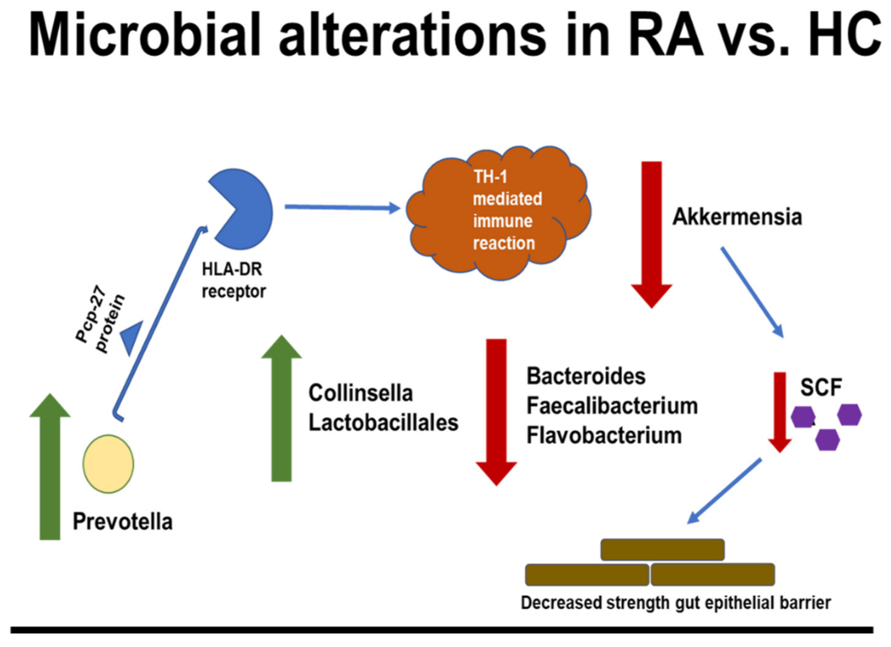
Interleukin-1 receptor antagonist knock-out mice (il1rn -/-) spontaneously developed an autoimmune T-cell mediated arthritis, due to excessive IL-1 signaling. They did not develop the autoimmune disease when raised in a germ-free environment. Lack of IL-1RA reduces intestinal microbial diversity, specifically causing a decrease in Ruminococcus and Prevotella populations. This change causes an increase in Th17 cells, which are implicated in RA pathogenesis [52,53]. Interestingly, when the GF mice were colonized with Lactobacillus bifidus, they developed an arthritic state similar to the initially mentioned group, due to bacteria-induced activation of toll-like receptors (TLR) and a resultant imbalance in T-regulatory–Th17 homeostasis [54].

4. Human RA Studies

More recently, research has investigated the role of the gut microbiome flora and function in human RA. The 16S next-generation sequencing (NGS) allows for rapid comparison of gut flora from healthy controls to RA patients. Prevotella copri (P. copri) is patients’ main bacterial population associated with dysbiosis [55,56]. A first-degree human gut microbiome study from Rodriguez et al. showed that patients with preclinical auto-antibody positive RA had marked differences in gut microbial populations compared to healthy controls, namely an increase in Prevotellacaea, specifically Prevotella spp. [57]. Scher et al. found that P. copri was most abundant in new-onset RA compared to chronic RA, PsA, and healthy controls, and correlated with decreased Bacteroides fragilis [58]. Functional genomic analyses of these patients suggested a decrease in vitamin production due to the inability of Prevotella spp. to engage in vitamin metabolism. Intriguingly, the decrease in gut folate metabolism suggests an MTX treatment-resistant phenotype. Further studies may shed light on the drug metabolism capabilities of the gut microbiome, potentially affecting decision-making in disease treatment [59].
In addition, a later study found that Prevotella copri produced the Pc-p27 protein, which could trigger a Th1-mediated immune response by binding to HLA-DR in RA patients. This claim was supported by the IgA-like antibody response in acute and chronic RA patients to Pc-p27 and correlated with the production of TH17 cytokines and anti-citrullinated protein antibodies (ACPA) [60]. Another cross-sectional study from Chen et al. found lower gut microbial diversity in RA patients compared to healthy controls. Notably, RA patients on DMARDs such as methotrexate (MTX) and hydroxychloroquine (HCQ) had higher diversity than treatment-free individuals [8]. Other studies expanded on these findings, noting a decrease in Faecalibacterium and increases in Collinsella [61].

5. Rheumatoid Arthritis and Nutrition

As previously mentioned, environmental factors are thought to play a substantial role in the development of RA, with nutrition being one. Looking at worldwide dietary patterns, the heterogeneity in food choices, and an overall decrease in plant-based foods, with increased meat consumption and food processing [62], these dietary changes have resulted in observable changes in the gut microbiome function. One study comparing the microbiota of hunter-gatherers in Tanzania to healthy controls noted significantly higher levels of CAZymes, responsible for the breakdown and modification of oligo and polysaccharides [63]. Changes in the produced metabolites and enzymes showed that diet differences affect gut microbiome function. The conventional treatment of RA depends on using steroids, DMARD, and biological regimens along with appropriate healthy changes such as smoking cessation and weight reduction. As the field of research building on the relationship between the gut microbiome, diet, and autoimmune diseases increased, questions arose regarding the efficacy of dietary changes aimed at restoring health homeostasis. The Mediterranean diet has been the main target of study.

5.1. Mediterranean Diet

The Mediterranean diet, which utilizes olive oil as the principal source of fat in cooking, is characterized by a high consumption of vegetables, legumes, alcohol, and white meat (fish and chicken) and low salt. The diet excludes red meats, which are often processed with carcinogenic nitrites and pro-inflammatory nitrates. Studies have shown that long-term adherence to the diet leads to observable changes in the gut microbiome.
The Mediterranean diet (MED) has been associated with an over-representation of species such as Faecalibacterium prausnitzii, Eubacterium, and Roseburia [64], as well as Collinsella aerofaciens, Dorea formicigenerans, and Clostridium ramosum. MED also increased the abundance of Prevotella and branched-chain amino acids (BCAA) degrading pathways and decreased Bifidobacterium and BCAA biosynthesis pathways in the gut microbiome [65,66]. Other studies have reported an increased abundance of Proteobacteria, Bacteroides uniformis, and Prevotella stercorea. In contrast, western diets (characterized by high meat and fat intake) were associated with a decrease in gut microbiota diversity and an increase in the Firmicutes/Bacteroidetes ratio, which are related to many inflammatory-adjacent disorders such as type 2 diabetes and obesity [67]. Meslier et al. found that adopting the Mediterranean diet in obese and overweight patients for two months reduced cholesterol, increased insulin sensitivity, and enhanced gut microbial populations [68]. Other studies saw an upregulation in butyrate production, integral in immunological homeostasis [69,70].
As a result, the Mediterranean diet is frequently clinically recommended to patients due to its antioxidant [71,72] and immune-modulatory properties, stemming from the production of monounsaturated and polyunsaturated fatty acids (MUFA, PUFA) and polyphenols. Polyphenols’ production improves the intestinal barrier’s strengths through the upregulation of tight junction protein production [73]. Linoleic acid (LA) and alfa-linolenic acid (ALA) are essential fatty acids derived from the diet. Derived from n-6 and n-3 PUFA precursors, the ratios in which these compounds are found dictate their profound effects on immune modulation [74,75]. LA and n-6 PUFA come from animal products and are pro-inflammatory, while ALA and n-3 PUFA are found in fish, oil, nuts, and berries, and are anti-inflammatory [76]. Due to the composition of the Mediterranean diet, the n-6/n-3 ratio is balanced, unlike more western diets (heavy in fats, red meats, and food processing), where there is an excess of n-6 PUFA and a deficiency of n-3 PUFA metabolites [77]. Several case reports and cohort studies have suggested that the higher levels of n-3 PUFA have a protective modulatory role in RA-susceptible patients. However, further research is necessary [78,79].

5.2. Dietary Regimens

Other diets have been studied for the potential management of RA. Fasting has been shown to improve RA disease activity scores. Small cohort studies were initially performed and found that short periods of fasting led to improvements in disease activity scores [74,75]. This was further validated by Kragh et al., who placed patients on a 7–10 day-long fast that was transitioned into a lactovegetarian diet and found improvements in the number of tender and swollen joints, morning stiffness, pain scores, as well as ESR and CRP, with these changes lasting for up to a year post-study completion [76]. The elemental diet, a liquid meal replacement diet that holds its nutritional value as “dietary building blocks” (amino acids, vitamins, monosaccharides), was compared in a small study to 2-week oral prednisolone in RA patients and was found to be similarly effective in disease management [78]. Short-term high-fiber diet supplementation increased SCFA levels and decreased the synthesis of pro-inflammatory mediators, MCP-1, IL-18, and IL-33 [79].

5.3. Other Dietary Factors

Several studies have looked at more specific dietary choices and their effects. CIA mice models given small amounts of alcohol had the onset of arthritis delayed [80]. In a small cohort study, low to moderate consumption of alcohol in affected patients was found to be protective [81]. Regular consumption of sugary beverages was correlated with an increased risk of RA development, independent of obesity. High decaf coffee consumption, defined as >4 cups per day, was associated with increased RA onset, while tea consumption was inversely associated [82]. High salt consumption has been a risk factor, particularly in smokers. A study from Sweden showed that high sodium intake doubled RA risk in smokers [83], and a separate cohort study showed high daily sodium intake (>4.5 g per day) increased RA risk with adjustment for confounders such as HTN, CV disease, diabetes, and cancer [84,85].

6. Therapeutics Targeted toward Dysbiosis

As the microbiome–nutrition–disease axis becomes more evident in RA, attention has turned toward the potential of therapeutics for clinical management. Numerous emerging technologies aim to manipulate the gut microbiome, to induce a healthy state. Fecal material therapy (FMT) is one such technique. It involves the transfer of fecal material from a healthy donor to the gastrointestinal tract of a patient with dysbiosis. The fecal material can be administered via capsule, enema, colonoscopy, or nasoduodenal tube [86]. While FMT is best known for its treatment of chronic, recurrent Clostridioides difficle infection (C. diff), research is being conducted into its usage for other autoimmune disorders. Few studies exist looking at FMT usage in RA patients. A case report from Zeng et al. showed successful treatment of RA with FMT via a decrease in RF, DAS-28, and HAQ scoring immediately after a treatment lasting 78 days.

6.1. Probiotics

Probiotics, or live organisms intended to have health benefits with consumption, are also being researched [87]. Simply described, their intended function is to reduce harmful bacterial populations and increase beneficial ones, while maintaining an appropriate equilibrium. The most well-known probiotics include Lactobacillus and Bifidobacterium, populations that produce SCFA and that work to maintain gut mucosal stability [88]. Several studies have reported on the beneficial effects they had on animal models of arthritis. One study found that oral administration of L. casei in CIA mice delayed the onset and reduced the severity of CIA [89]. Fan et al. looked at the preventative vs. therapeutic administration of B. adolescentis in CIA mice [90]. Early administration showed a better promotion of SCFA production and reduction of clinical symptoms. Another study found that early administration of L. helveticus SBT2171 significantly reduced joint tenderness and swelling. These findings suggest prophylactic treatment may be more effective in disease management [91].
Unlike animal studies, research on the effect of probiotics on human disease states is limited. Hatakka et al. looked at the L. rhamnosus supplementation in stable RA patients not on DMARD mediations compared to healthy controls. Patients were given the intervention for 12 months, twice daily, in addition to their regular mediation regimen. While there were no significant differences in inflammatory markers or clinical disease compared to the placebo group, patients in the supplementation group noted a subjective health improvement [92]. Ramini et al. investigated the effect of probiotic supplementation on RA patients. Participants in the active arm were given an eight-week course of probiotics containing L. acidophilus, L. casei, and B. bifidum. Compared to the control group, they showed improvements in DAS28 and high sensitivity CRP (hs-CRP) levels. Other studies found similar improvements in clinical disease markers with Lactobacillus probiotic supplementation [93,94]. A recent study tested the effect of a combined probiotic pill containing L. casei LC-11, L. acidophilus LA-14, Lactococcus lactis LL-23, B. lactis BL-04, and B. bifidum BB-06 in active RA patients, compared to healthy controls. The test group showed significant reductions in WBC, TNF, and IL-6. However, no changes were noted in IL-10, adiponectin, CRP, and ESR levels [95].

6.2. Fermented Food

Fermented foods consist of foods or beverages made using the conversion of food components through microbial enzymes [96]. Dedicated research in the field is currently limited. However, observational studies have suggested that fermented foods positively modulate the gut immune system by forming bioactive metabolites in the present microbial populations [97]. Bifidobacterium and Lactobacillus are common microbial populations used in food preparation [98].

6.3. Microbiome-Derived Metabolites

The gut microbiome produces many bioactive metabolites, including vitamins, butyrate, and bile acids. As described earlier in the paper, these molecules work to maintain healthy homeostasis. Little research has been done to assess their potential use in microbiome modulation. Pena-Rodriguez et al. studied the effect of butyrate on the intestinal inflammatory response in a cholestasis model. Administration of butyrate was found to reduce pro-inflammatory cytokines and upregulate the production of tight junction proteins [99]. Berberine, a therapeutic option in treating type-2 diabetes, increased SCFA-producing bacteria and lowered gut inflammation [100].

7. Conclusions

Recent years have shown an explosion in the research supporting the gut microbiome’s role in RA pathogenesis. In a nutshell, the host–microbiota interactions and dysbiosis in rheumatoid arthritis are shown in Figure 1.
Experimental animal and human models indicate that dysbiosis occurs far before the clinical onset of disease and continues to shift during disease progression. However, to gain a mechanistic understanding of how the gut microbiota affects the host metabolism, comprehensive analyses of high-throughput multi-omics data, including metagenomics and metabolomics data, and measures of host physiology hold potential in the identification of possible molecular mechanisms in the host–microbiome association. Furthermore, we also increasingly understand how diet, through microbes or metabolites, modifies health. Clinical evidence needs to be translated into practice, using consistent matrices of prebiotics or probiotics or fecal microbiota transplantation to assess changes in gut microbiota composition and health outcomes. Connecting this knowledge to the importance of nutrition and diet in the modulation of the microbiome and the immune system has opened avenues of research into the therapeutic usage of interventions such as probiotics and microbial-derived metabolites in disease management. However, it is clear that further studies are needed before interventions can be considered for clinical use. One challenge is that many microbiome studies are more association studies than correlational ones.
Furthermore, the majority of studies are derived from sample repositories where the US is overrepresented [101]. As discussed, geography plays a prominent role in diet and lifestyle, affecting microbiome populations. More extensive and iterative clinical trials are necessary for study findings to have global generalizability. Another challenge involves the uncertainty regarding the future patentability of interventions. The patenting of natural products is controversial. A recent ruling in 2013 of the Association for Molecular Pathology vs. Myriad Genetics declared genes unpatentable due to their being products of nature [102]. How that affects bacterial strains’ patentability, or microbial metabolites remains unclear. Additionally, studies evaluating the host–microbiome relationship in RA are necessary to elucidate possible triggering factors and close the gap in our knowledge regarding the role of gene–environment interactions.

Author Contributions

M.A (Malavikalakshmi Attur), J.U.S., S.B.A. and M.A. (Mukundan Attur), wrote, edited, and approved the published manuscript. All authors have read and agreed to the published version of the manuscript.

Funding

This work is supported by National Institute of Health/National Institute of Arthritis and Musculoskeletal and Skin Disease (R01AR074500 and 1UC2AR081034-01 to J.U.S.) and The Beatrice Snyder Foundation.

Institutional Review Board Statement

Not applicable.

Informed Consent Statement

Not applicable.

Data Availability Statement

Not applicable.

Conflicts of Interest

The authors declare no conflict of interest.

References

  1. Peroumal, D.; Abimannan, T.; Tagirasa, R.; Parida, J.R.; Singh, S.K.; Padhan, P.; Devadas, S. Inherent low Erk and p38 activity reduce Fas Ligand expression and degranulation in T helper 17 cells leading to activation induced cell death resistance. Oncotarget 2016, 7, 54339–54359. [Google Scholar] [CrossRef] [PubMed] [Green Version]
  2. Kim, K.; Bang, S.Y.; Lee, H.S.; Bae, S.C. Update on the genetic architecture of rheumatoid arthritis. Nat. Rev. Rheumatol. 2017, 13, 13–24. [Google Scholar] [CrossRef]
  3. MacGregor, A.J.; Snieder, H.; Rigby, A.S.; Koskenvuo, M.; Kaprio, J.; Aho, K.; Silman, A.J. Characterizing the quantitative genetic contribution to rheumatoid arthritis using data from twins. Arthritis Rheum. 2000, 43, 30–37. [Google Scholar] [CrossRef]
  4. Silman, A.J.; MacGregor, A.J.; Thomson, W.; Holligan, S.; Carthy, D.; Farhan, A.; Ollier, W.E. Twin concordance rates for rheumatoid arthritis: Results from a nationwide study. Br. J. Rheumatol. 1993, 32, 903–907. [Google Scholar] [CrossRef] [PubMed]
  5. McInnes, I.B.; Schett, G. The pathogenesis of rheumatoid arthritis. N. Engl. J. Med. 2011, 365, 2205–2219. [Google Scholar] [CrossRef] [PubMed] [Green Version]
  6. Mazmanian, S.K.; Liu, C.H.; Tzianabos, A.O.; Kasper, D.L. An immunomodulatory molecule of symbiotic bacteria directs maturation of the host immune system. Cell 2005, 122, 107–118. [Google Scholar] [CrossRef] [PubMed] [Green Version]
  7. Rakoff-Nahoum, S.; Paglino, J.; Eslami-Varzaneh, F.; Edberg, S.; Medzhitov, R. Recognition of commensal microflora by toll-like receptors is required for intestinal homeostasis. Cell 2004, 118, 229–241. [Google Scholar] [CrossRef] [Green Version]
  8. Bodkhe, R.; Balakrishnan, B.; Taneja, V. The role of microbiome in rheumatoid arthritis treatment. Ther. Adv. Musculoskelet. Dis. 2019, 11, 1759720x19844632. [Google Scholar] [CrossRef]
  9. Baquero, F.; Nombela, C. The microbiome as a human organ. Clin. Microbiol. Infect. 2012, 18 (Suppl. 4), 2–4. [Google Scholar] [CrossRef] [Green Version]
  10. Berg, G.; Rybakova, D.; Fischer, D.; Cernava, T.; Verges, M.C.; Charles, T.; Chen, X.; Cocolin, L.; Eversole, K.; Corral, G.H.; et al. Microbiome definition re-visited: Old concepts and new challenges. Microbiome 2020, 8, 103. [Google Scholar]
  11. Wassenaar, T.M.; Panigrahi, P. Is a foetus developing in a sterile environment? Lett. Appl. Microbiol. 2014, 59, 572–579. [Google Scholar] [CrossRef] [PubMed]
  12. Ardissone, A.N.; de la Cruz, D.M.; Davis-Richardson, A.G.; Rechcigl, K.T.; Li, N.; Drew, J.C.; Murgas-Torrazza, R.; Sharma, R.; Hudak, M.L.; Triplett, E.W.; et al. Meconium microbiome analysis identifies bacteria correlated with premature birth. PLoS ONE 2014, 9, e90784. [Google Scholar] [CrossRef] [PubMed] [Green Version]
  13. Dominguez-Bello, M.G.; Costello, E.K.; Contreras, M.; Magris, M.; Hidalgo, G.; Fierer, N.; Knight, R. Delivery mode shapes the acquisition and structure of the initial microbiota across multiple body habitats in newborns. Proc. Natl. Acad. Sci. USA 2010, 107, 11971–11975. [Google Scholar] [CrossRef] [Green Version]
  14. Arumugam, M.; Raes, J.; Pelletier, E.; Le Paslier, D.; Yamada, T.; Mende, D.R.; Fernandes, G.R.; Tap, J.; Bruls, T.; Batto, J.M.; et al. Enterotypes of the human gut microbiome. Nature 2011, 473, 174–180. [Google Scholar] [CrossRef]
  15. Rinninella, E.; Raoul, P.; Cintoni, M.; Franceschi, F.; Miggiano, G.A.D.; Gasbarrini, A.; Mele, M.C. What is the Healthy Gut Microbiota Composition? A Changing Ecosystem across Age, Environment, Diet, and Diseases. Microorganisms 2019, 7, 14. [Google Scholar] [CrossRef] [PubMed] [Green Version]
  16. Barko, P.C.; McMichael, M.A.; Swanson, K.S.; Williams, D.A. The Gastrointestinal Microbiome: A Review. J. Vet. Intern. Med. 2018, 32, 9–25. [Google Scholar] [CrossRef] [PubMed] [Green Version]
  17. Zhou, K. Strategies to promote abundance of Akkermansia muciniphila, an emerging probiotics in the gut, evidence from dietary intervention studies. J. Funct. Foods 2017, 33, 194–201. [Google Scholar] [CrossRef]
  18. Sun, Y.; Chen, Q.; Lin, P.; Xu, R.; He, D.; Ji, W.; Bian, Y.; Shen, Y.; Li, Q.; Liu, C.; et al. Characteristics of Gut Microbiota in Patients with Rheumatoid Arthritis in Shanghai, China. Front. Cell. Infect. Microbiol. 2019, 9, 369. [Google Scholar] [CrossRef] [Green Version]
  19. Schade, L.; Mesa, D.; Faria, A.R.; Santamaria, J.R.; Xavier, C.A.; Ribeiro, D.; Hajar, F.N.; Azevedo, V.F. The gut microbiota profile in psoriasis: A Brazilian case-control study. Lett. Appl. Microbiol. 2022, 74, 498–504. [Google Scholar] [CrossRef]
  20. Scher, J.U.; Ubeda, C.; Artacho, A.; Attur, M.; Isaac, S.; Reddy, S.M.; Marmon, S.; Neimann, A.; Brusca, S.; Patel, T.; et al. Decreased bacterial diversity characterizes the altered gut microbiota in patients with psoriatic arthritis, resembling dysbiosis in inflammatory bowel disease. Arthritis Rheumatol. 2015, 67, 128–139. [Google Scholar] [CrossRef] [Green Version]
  21. Hamazaki, M.; Sawada, T.; Yamamura, T.; Maeda, K.; Mizutani, Y.; Ishikawa, E.; Furune, S.; Yamamoto, K.; Ishikawa, T.; Kakushima, N.; et al. Fecal microbiota transplantation in the treatment of irritable bowel syndrome: A single-center prospective study in Japan. BMC Gastroenterol. 2022, 22, 342. [Google Scholar] [CrossRef]
  22. Wang, L.; Wang, Y.; Zhang, P.; Song, C.; Pan, F.; Li, G.; Peng, L.; Yang, Y.; Wei, Z.; Huang, F. Gut microbiota changes in patients with spondyloarthritis: A systematic review. Semin. Arthritis Rheum. 2022, 52, 151925. [Google Scholar] [CrossRef] [PubMed]
  23. Liu, F.; Ren, T.; Li, X.; Zhai, Q.; Xu, X.; Zhang, N.; Jiang, P.; Niu, Y.; Lv, L.; Shi, G.; et al. Distinct Microbiomes of Gut and Saliva in Patients with Systemic Lupus Erythematous and Clinical Associations. Front. Immunol. 2021, 12, 626217. [Google Scholar] [CrossRef] [PubMed]
  24. den Besten, G.; van Eunen, K.; Groen, A.K.; Venema, K.; Reijngoud, D.J.; Bakker, B.M. The role of short-chain fatty acids in the interplay between diet, gut microbiota, and host energy metabolism. J. Lipid. Res. 2013, 54, 2325–2340. [Google Scholar] [CrossRef] [Green Version]
  25. Morrison, D.J.; Preston, T. Formation of short chain fatty acids by the gut microbiota and their impact on human metabolism. Gut Microbes 2016, 7, 189–200. [Google Scholar] [CrossRef] [PubMed] [Green Version]
  26. Ohland, C.L.; Jobin, C. Microbial Activities and Intestinal Homeostasis: A Delicate Balance between Health and Disease. Cell. Mol. Gastroenterol. Hepatol. 2015, 1, 28–40. [Google Scholar] [CrossRef] [Green Version]
  27. Belkaid, Y.; Harrison, O.J. Homeostatic Immunity and the Microbiota. Immunity 2017, 46, 562–576. [Google Scholar] [CrossRef] [Green Version]
  28. Park, J.; Kim, M.; Kang, S.G.; Jannasch, A.H.; Cooper, B.; Patterson, J.; Kim, C.H. Short-chain fatty acids induce both effector and regulatory T cells by suppression of histone deacetylases and regulation of the mTOR–S6K pathway. Mucosal Immunol. 2015, 8, 80–93. [Google Scholar] [CrossRef] [Green Version]
  29. Singh, N.; Gurav, A.; Sivaprakasam, S.; Brady, E.; Padia, R.; Shi, H.; Thangaraju, M.; Prasad, P.D.; Manicassamy, S.; Munn, D.H.; et al. Activation of Gpr109a, receptor for niacin and the commensal metabolite butyrate, suppresses colonic inflammation and carcinogenesis. Immunity 2014, 40, 128–139. [Google Scholar] [CrossRef] [Green Version]
  30. Macia, L.; Tan, J.; Vieira, A.T.; Leach, K.; Stanley, D.; Luong, S.; Maruya, M.; McKenzie, C.I.; Hijikata, A.; Wong, C.; et al. Metabolite-sensing receptors GPR43 and GPR109A facilitate dietary fibre-induced gut homeostasis through regulation of the inflammasome. Nat. Commun. 2015, 6, 6734. [Google Scholar] [CrossRef] [Green Version]
  31. Kim, M.H.; Kang, S.G.; Park, J.H.; Yanagisawa, M.; Kim, C.H. Short-chain fatty acids activate GPR41 and GPR43 on intestinal epithelial cells to promote inflammatory responses in mice. Gastroenterology 2013, 145, 396–406.e1–10. [Google Scholar] [CrossRef] [PubMed]
  32. Matthews, G.M.; Howarth, G.S.; Butler, R.N. Short-chain fatty acids induce apoptosis in colon cancer cells associated with changes to intracellular redox state and glucose metabolism. Chemotherapy 2012, 58, 102–109. [Google Scholar] [CrossRef] [PubMed]
  33. Venegas, D.P.; De la Fuente, M.K.; Landskron, G.; Gonzalez, M.J.; Quera, R.; Dijkstra, G.; Harmsen, H.J.M.; Faber, K.N.; Hermoso, M.A. Short Chain Fatty Acids (SCFAs)-Mediated Gut Epithelial and Immune Regulation and Its Relevance for Inflammatory Bowel Diseases. Front. Immunol. 2019, 10, 277. [Google Scholar]
  34. Gustafsson, B.E.; Daft, F.S.; McDaniel, E.G.; Smith, J.C.; Fitzgerald, R.J. Effects of vitamin K-active compounds and intestinal microorganisms in vitamin K-deficient germfree rats. J. Nutr. 1962, 78, 461–468. [Google Scholar] [CrossRef] [PubMed]
  35. Courtemanche, C.; Elson-Schwab, I.; Mashiyama, S.T.; Kerry, N.; Ames, B.N. Folate deficiency inhibits the proliferation of primary human CD8+ T lymphocytes in vitro. J. Immunol. 2004, 173, 3186–3192. [Google Scholar] [CrossRef] [Green Version]
  36. Sperandio, V. Take Your Pick: Vitamins and Microbiota Facilitate Pathogen Clearance. Cell Host Microbe 2017, 21, 130–131. [Google Scholar] [CrossRef] [Green Version]
  37. Li, Z.; Quan, G.; Jiang, X.; Yang, Y.; Ding, X.; Zhang, D.; Wang, X.; Hardwidge, P.R.; Ren, W.; Zhu, G. Effects of Metabolites Derived from Gut Microbiota and Hosts on Pathogens. Front. Cell. Infect. Microbiol. 2018, 8, 314. [Google Scholar] [CrossRef] [Green Version]
  38. Connolly, J.P.; Gabrielsen, M.; Goldstone, R.J.; Grinter, R.; Wang, D.; Cogdell, R.J.; Walker, D.; Smith, D.G.; Roe, A.J. A Highly Conserved Bacterial D-Serine Uptake System Links Host Metabolism and Virulence. PLoS Pathog. 2016, 12, e1005359. [Google Scholar] [CrossRef] [Green Version]
  39. Wells, P.M.; Williams, F.M.K.; Matey-Hernandez, M.L.; Menni, C.; Steves, C.J. ‘RA and the microbiome: Do host genetic factors provide the link? J. Autoimmun. 2019, 99, 104–115. [Google Scholar] [CrossRef]
  40. Scher, J.U.; Bretz, W.A.; Abramson, S.B. Periodontal disease and subgingival microbiota as contributors for rheumatoid arthritis pathogenesis: Modifiable risk factors? Curr. Opin. Rheumatol. 2014, 26, 424–429. [Google Scholar] [CrossRef] [Green Version]
  41. Andrews, C.R.; Hoke, M. A preliminary report on the relation of albuminous putrefaction in the intestines to arthritis deformans (rheumatoid arthritis, osteo-arthritis); its influence upon treatment. Am. J. Orthop. Surg. 1907, 2, 61–72. [Google Scholar]
  42. Kohashi, O.; Kuwata, J.; Umehara, K.; Uemura, F.; Takahashi, T.; Ozawa, A. Susceptibility to adjuvant-induced arthritis among germfree, specific-pathogen-free, and conventional rats. Infect. Immun. 1979, 26, 791–794. [Google Scholar] [CrossRef] [PubMed] [Green Version]
  43. Aa, L.X.; Fei, F.; Qi, Q.; Sun, R.B.; Gu, S.H.; Di, Z.Z.; Aa, J.Y.; Wang, G.J.; Liu, C.X. Rebalancing of the gut flora and microbial metabolism is responsible for the anti-arthritis effect of kaempferol. Acta Pharmacol. Sin. 2020, 41, 73–81. [Google Scholar] [CrossRef] [PubMed]
  44. Round, J.L.; Mazmanian, S.K. Inducible Foxp3+ regulatory T-cell development by a commensal bacterium of the intestinal microbiota. Proc. Natl. Acad. Sci. USA 2010, 107, 12204–12209. [Google Scholar] [CrossRef] [Green Version]
  45. Tajik, N.; Frech, M.; Schulz, O.; Schalter, F.; Lucas, S.; Azizov, V.; Durholz, K.; Steffen, F.; Omata, Y.; Rings, A.; et al. Targeting zonulin and intestinal epithelial barrier function to prevent onset of arthritis. Nat. Commun. 2020, 11, 1995. [Google Scholar] [CrossRef] [Green Version]
  46. Sakaguchi, N.; Takahashi, T.; Hata, H.; Nomura, T.; Tagami, T.; Yamazaki, S.; Sakihama, T.; Matsutani, T.; Negishi, I.; Nakatsuru, S.; et al. Altered thymic T-cell selection due to a mutation of the ZAP-70 gene causes autoimmune arthritis in mice. Nature 2003, 426, 454–460. [Google Scholar] [CrossRef]
  47. Maeda, Y.; Kurakawa, T.; Umemoto, E.; Motooka, D.; Ito, Y.; Gotoh, K.; Hirota, K.; Matsushita, M.; Furuta, Y.; Narazaki, M.; et al. Dysbiosis Contributes to Arthritis Development via Activation of Autoreactive T Cells in the Intestine. Arthritis Rheumatol. 2016, 68, 2646–2661. [Google Scholar] [CrossRef]
  48. Monach, P.A.; Mathis, D.; Benoist, C. The K/BxN arthritis model. Curr. Protoc. Immunol. 2008, 81, 15–22. [Google Scholar] [CrossRef]
  49. Abdollahi-Roodsaz, S.; Joosten, L.A.; Koenders, M.I.; Devesa, I.; Roelofs, M.F.; Radstake, T.R.; Heuvelmans-Jacobs, M.; Akira, S.; Nicklin, M.J.; Ribeiro-Dias, F.; et al. Stimulation of TLR2 and TLR4 differentially skews the balance of T cells in a mouse model of arthritis. J. Clin. Investig. 2008, 118, 205–216. [Google Scholar] [CrossRef] [Green Version]
  50. Chen, B.; Chen, H.; Shu, X.; Yin, Y.; Li, J.; Qin, J.; Chen, L.; Peng, K.; Xu, F.; Gu, W.; et al. Presence of Segmented Filamentous Bacteria in Human Children and Its Potential Role in the Modulation of Human Gut Immunity. Front. Microbiol. 2018, 9, 1403. [Google Scholar] [CrossRef]
  51. Flak, M.B.; Colas, R.A.; Munoz-Atienza, E.; Curtis, M.A.; Dalli, J.; Pitzalis, C. Inflammatory arthritis disrupts gut resolution mechanisms, promoting barrier breakdown by Porphyromonas gingivalis. JCI Insight 2019, 4, e125191. [Google Scholar] [CrossRef] [PubMed]
  52. Rogier, R.; Ederveen, T.H.A.; Boekhorst, J.; Wopereis, H.; Scher, J.U.; Manasson, J.; Frambach, S.; Knol, J.; Garssen, J.; van der Kraan, P.M.; et al. Aberrant intestinal microbiota due to IL-1 receptor antagonist deficiency promotes IL-17- and TLR4-dependent arthritis. Microbiome 2017, 5, 63. [Google Scholar] [CrossRef] [PubMed]
  53. Alunno, A.; Manetti, M.; Caterbi, S.; Ibba-Manneschi, L.; Bistoni, O.; Bartoloni, E.; Valentini, V.; Terenzi, R.; Gerli, R. Altered immunoregulation in rheumatoid arthritis: The role of regulatory T cells and proinflammatory Th17 cells and therapeutic implications. Mediat. Inflamm. 2015, 2015, 751793. [Google Scholar] [CrossRef] [PubMed]
  54. Yuichi, M.; Takeda, K. Role of Gut Microbiota in Rheumatoid Arthritis. J. Clin. Med. 2017, 6, 60. [Google Scholar]
  55. Kishikawa, T.; Maeda, Y.; Nii, T.; Motooka, D.; Matsumoto, Y.; Matsushita, M.; Matsuoka, H.; Yoshimura, M.; Kawada, S.; Teshigawara, S.; et al. Metagenome-wide association study of gut microbiome revealed novel aetiology of rheumatoid arthritis in the Japanese population. Ann. Rheum. Dis. 2020, 79, 103–111. [Google Scholar] [CrossRef]
  56. Tong, Y.; Tang, H.; Li, Y.; Su, L.; Wu, Y.; Bozec, A.; Zaiss, M.; Qing, P.; Zhao, H.; Tan, C.; et al. Gut Microbiota Dysbiosis in High-Risk Individuals for Rheumatoid Arthritis Triggers Mucosal Immunity Perturbation and Promotes Arthritis in Mice. Res. Sq. 2020. [Google Scholar] [CrossRef]
  57. Alpizar-Rodriguez, D.; Lesker, T.R.; Gronow, A.; Gilbert, B.; Raemy, E.; Lamacchia, C.; Gabay, C.; Finckh, A.; Strowig, T. Prevotella copri in individuals at risk for rheumatoid arthritis. Ann. Rheum. Dis. 2019, 78, 590–593. [Google Scholar] [CrossRef]
  58. Scher, J.U.; Sczesnak, A.; Longman, R.S.; Segata, N.; Ubeda, C.; Bielski, C.; Rostron, T.; Cerundolo, V.; Pamer, E.G.; Abramson, S.B.; et al. Expansion of intestinal Prevotella copri correlates with enhanced susceptibility to arthritis. eLife 2013, 2, e01202. [Google Scholar] [CrossRef]
  59. Han, M.; Zhang, N.; Mao, Y.; Huang, B.; Ren, M.; Peng, Z.; Bai, Z.; Chen, L.; Liu, Y.; Wang, S.; et al. The Potential of Gut Microbiota Metabolic Capability to Detect Drug Response in Rheumatoid Arthritis Patients. Front. Microbiol. 2022, 13, 839015. [Google Scholar] [CrossRef]
  60. Pianta, A.; Arvikar, S.; Strle, K.; Drouin, E.E.; Wang, Q.; Costello, C.E.; Steere, A.C. Evidence of the Immune Relevance of Prevotella copri, a Gut Microbe, in Patients with Rheumatoid Arthritis. Arthritis Rheumatol. 2017, 69, 964–975. [Google Scholar] [CrossRef] [Green Version]
  61. Chen, J.; Wright, K.; Davis, J.M.; Jeraldo, P.; Marietta, E.V.; Murray, J.; Nelson, H.; Matteson, E.L.; Taneja, V. An expansion of rare lineage intestinal microbes characterizes rheumatoid arthritis. Genome Med. 2016, 8, 43. [Google Scholar] [CrossRef] [PubMed] [Green Version]
  62. Cordain, L.; Eaton, S.B.; Sebastian, A.; Mann, N.; Lindeberg, S.; Watkins, B.A.; O’Keefe, J.H.; Brand-Miller, J. Origins and evolution of the Western diet: Health implications for the 21st century. Am. J. Clin. Nutr. 2005, 81, 341–354. [Google Scholar] [CrossRef] [PubMed]
  63. Smits, S.A.; Leach, J.; Sonnenburg, E.D.; Gonzalez, C.G.; Lichtman, J.S.; Reid, G.; Knight, R.; Manjurano, A.; Changalucha, J.; Elias, J.E.; et al. Seasonal cycling in the gut microbiome of the Hadza hunter-gatherers of Tanzania. Science 2017, 357, 802–806. [Google Scholar] [CrossRef] [PubMed] [Green Version]
  64. Ghosh, T.S.; Rampelli, S.; Jeffery, I.B.; Santoro, A.; Neto, M.; Capri, M.; Giampieri, E.; Jennings, A.; Candela, M.; Turroni, S.; et al. Mediterranean diet intervention alters the gut microbiome in older people reducing frailty and improving health status: The NU-AGE 1-year dietary intervention across five European countries. Gut 2020, 69, 1218–1228. [Google Scholar] [CrossRef] [PubMed] [Green Version]
  65. Rinott, E.; Meir, A.Y.; Tsaban, G.; Zelicha, H.; Kaplan, A.; Knights, D.; Tuohy, K.; Scholz, M.U.; Koren, O.; Stampfer, M.J.; et al. The effects of the Green-Mediterranean diet on cardiometabolic health are linked to gut microbiome modifications: A randomized controlled trial. Genome Med. 2022, 14, 29. [Google Scholar] [CrossRef]
  66. Nagpal, R.; Shively, C.A.; Register, T.C.; Craft, S.; Yadav, H. Gut microbiome-Mediterranean diet interactions in improving host health. F1000Res 2019, 8, 699. [Google Scholar] [CrossRef] [Green Version]
  67. Tsigalou, C.; Paraschaki, A.; Karvelas, A.; Kantartzi, K.; Gagali, K.; Tsairidis, D.; Bezirtzoglou, E. Gut microbiome and Mediterranean diet in the context of obesity. Current knowledge, perspectives and potential therapeutic targets. Metab. Open 2021, 9, 100081. [Google Scholar] [CrossRef]
  68. Meslier, V.; Laiola, M.; Roager, H.M.; De Filippis, F.; Roume, H.; Quinquis, B.; Giacco, R.; Mennella, I.; Ferracane, R.; Pons, N.; et al. Mediterranean diet intervention in overweight and obese subjects lowers plasma cholesterol and causes changes in the gut microbiome and metabolome independently of energy intake. Gut 2020, 69, 1258–1268. [Google Scholar] [CrossRef] [Green Version]
  69. Gutierrez-Diaz, I.; Fernandez-Navarro, T.; Sanchez, B.; Margolles, A.; Gonzalez, S. Mediterranean diet and faecal microbiota: A transversal study. Food Funct. 2016, 7, 2347–2356. [Google Scholar] [CrossRef]
  70. So, D.; Whelan, K.; Rossi, M.; Morrison, M.; Holtmann, G.; Kelly, J.T.; Shanahan, E.R.; Staudacher, H.M.; Campbell, K.L. Dietary fiber intervention on gut microbiota composition in healthy adults: A systematic review and meta-analysis. Am. J. Clin. Nutr. 2018, 107, 965–983. [Google Scholar] [CrossRef] [Green Version]
  71. Philippou, E.; Nikiphorou, E. Are we really what we eat? Nutrition and its role in the onset of rheumatoid arthritis. Autoimmun. Rev. 2018, 17, 1074–1077. [Google Scholar] [CrossRef] [PubMed] [Green Version]
  72. Santangelo, C.; Vari, R.; Scazzocchio, B.; De Sanctis, P.; Giovannini, C.; D’Archivio, M.; Masella, R. Anti-inflammatory Activity of Extra Virgin Olive Oil Polyphenols: Which Role in the Prevention and Treatment of Immune-Mediated Inflammatory Diseases? Endocr. Metab. Immune Disord. Drug Targets 2018, 18, 36–50. [Google Scholar] [CrossRef] [PubMed]
  73. Ghosh, S.; Whitley, C.S.; Haribabu, B.; Jala, V.R. Regulation of Intestinal Barrier Function by Microbial Metabolites. Cell. Mol. Gastroenterol. Hepatol. 2021, 11, 1463–1482. [Google Scholar] [CrossRef] [PubMed]
  74. Sundqvist, T.; Lindstrom, F.; Magnusson, K.E.; Skoldstam, L.; Stjernstrom, I.; Tagesson, C. Influence of fasting on intestinal permeability and disease activity in patients with rheumatoid arthritis. Scand. J. Rheumatol. 1982, 11, 33–38. [Google Scholar] [CrossRef]
  75. Skoldstam, L.; Larsson, L.; Lindstrom, F.D. Effect of fasting and lactovegetarian diet on rheumatoid arthritis. Scand. J. Rheumatol. 1979, 8, 249–255. [Google Scholar] [CrossRef]
  76. Kjeldsen-Kragh, J.; Haugen, M.; Borchgrevink, C.F.; Laerum, E.; Eek, M.; Mowinkel, P.; Hovi, K.; Forre, O. Controlled trial of fasting and one-year vegetarian diet in rheumatoid arthritis. Lancet 1991, 338, 899–902. [Google Scholar] [CrossRef]
  77. Scaioli, E.; Liverani, E.; Belluzzi, A. The Imbalance between n-6/n-3 Polyunsaturated Fatty Acids and Inflammatory Bowel Disease: A Comprehensive Review and Future Therapeutic Perspectives. Int. J. Mol. Sci. 2017, 18, 2619. [Google Scholar] [CrossRef] [Green Version]
  78. Podas, T.; Nightingale, J.M.; Oldham, R.; Roy, S.; Sheehan, N.J.; Mayberry, J.F. Is rheumatoid arthritis a disease that starts in the intestine? A pilot study comparing an elemental diet with oral prednisolone. Postgrad. Med. J. 2007, 83, 128–131. [Google Scholar] [CrossRef] [Green Version]
  79. Durholz, K.; Hofmann, J.; Iljazovic, A.; Hager, J.; Lucas, S.; Sarter, K.; Strowig, T.; Bang, H.; Rech, J.; Schett, G.; et al. Dietary Short-Term Fiber Interventions in Arthritis Patients Increase Systemic SCFA Levels and Regulate Inflammation. Nutrients 2020, 12, 3207. [Google Scholar] [CrossRef]
  80. Jonsson, I.M.; Verdrengh, M.; Brisslert, M.; Lindblad, S.; Bokarewa, M.; Islander, U.; Carlsten, H.; Ohlsson, C.; Nandakumar, K.S.; Holmdahl, R.; et al. Ethanol prevents development of destructive arthritis. Proc. Natl. Acad. Sci. USA 2007, 104, 258–263. [Google Scholar] [CrossRef] [Green Version]
  81. Kallberg, H.; Jacobsen, S.; Bengtsson, C.; Pedersen, M.; Padyukov, L.; Garred, P.; Frisch, M.; Karlson, E.W.; Klareskog, L.; Alfredsson, L. Alcohol consumption is associated with decreased risk of rheumatoid arthritis: Results from two Scandinavian case-control studies. Ann. Rheum. Dis. 2009, 68, 222–227. [Google Scholar] [CrossRef] [PubMed]
  82. Mikuls, T.R.; Cerhan, J.R.; Criswell, L.A.; Merlino, L.; Mudano, A.S.; Burma, M.; Folsom, A.R.; Saag, K.G. Coffee, tea, and caffeine consumption and risk of rheumatoid arthritis: Results from the Iowa Women’s Health Study. Arthritis Rheum. 2002, 46, 83–91. [Google Scholar] [CrossRef]
  83. Sundström, B.; Johansson, I.; Rantapää-Dahlqvist, S. Interaction between dietary sodium and smoking increases the risk for rheumatoid arthritis: Results from a nested case-control study. Rheumatology 2015, 54, 487–493. [Google Scholar] [CrossRef] [PubMed] [Green Version]
  84. Alpízar-Rodríguez, D.; Finckh, A.; Gilbert, B. The Role of Nutritional Factors and Intestinal Microbiota in Rheumatoid Arthritis Development. Nutrients 2020, 13, 96. [Google Scholar] [CrossRef] [PubMed]
  85. Bae, J.H.; Shin, M.Y.; Kang, E.H.; Lee, Y.J.; Ha, Y.J. Association of rheumatoid arthritis and high sodium intake with major adverse cardiovascular events: A cross-sectional study from the seventh Korean National Health and Nutrition Examination Survey. BMJ Open 2021, 11, e056255. [Google Scholar] [CrossRef]
  86. Choi, H.H.; Cho, Y.S. Fecal Microbiota Transplantation: Current Applications, Effectiveness, and Future Perspectives. Clin. Endosc. 2016, 49, 257–265. [Google Scholar] [CrossRef]
  87. Hill, C.; Guarner, F.; Reid, G.; Gibson, G.R.; Merenstein, D.J.; Pot, B.; Morelli, L.; Canani, R.B.; Flint, H.J.; Salminen, S.; et al. Expert consensus document. The International Scientific Association for Probiotics and Prebiotics consensus statement on the scope and appropriate use of the term probiotic. Nat. Rev. Gastroenterol. Hepatol. 2014, 11, 506–514. [Google Scholar] [CrossRef] [Green Version]
  88. Bungau, S.G.; Behl, T.; Singh, A.; Sehgal, A.; Singh, S.; Chigurupati, S.; Vijayabalan, S.; Das, S.; Palanimuthu, V.R. Targeting Probiotics in Rheumatoid Arthritis. Nutrients 2021, 13, 3376. [Google Scholar] [CrossRef]
  89. Kato, I.; Endo-Tanaka, K.; Yokokura, T. Suppressive effects of the oral administration of Lactobacillus casei on type II collagen-induced arthritis in DBA/1 mice. Life Sci. 1998, 63, 635–644. [Google Scholar] [CrossRef]
  90. Fan, Z.; Yang, B.; Ross, R.P.; Stanton, C.; Shi, G.; Zhao, J.; Zhang, H.; Chen, W. Protective effects of Bifidobacterium adolescentis on collagen-induced arthritis in rats depend on timing of administration. Food Funct. 2020, 11, 4499–4511. [Google Scholar] [CrossRef]
  91. Yamashita, M.; Matsumoto, K.; Endo, T.; Ukibe, K.; Hosoya, T.; Matsubara, Y.; Nakagawa, H.; Sakai, F.; Miyazaki, T. Preventive Effect of Lactobacillus helveticus SBT2171 on Collagen-Induced Arthritis in Mice. Front. Microbiol. 2017, 8, 1159. [Google Scholar] [CrossRef]
  92. Hatakka, K.; Martio, J.; Korpela, M.; Herranen, M.; Poussa, T.; Laasanen, T.; Saxelin, M.; Vapaatalo, H.; Moilanen, E.; Korpela, R. Effects of probiotic therapy on the activity and activation of mild rheumatoid arthritis—A pilot study. Scand. J. Rheumatol. 2003, 32, 211–215. [Google Scholar] [CrossRef] [PubMed]
  93. Vaghef-Mehrabany, E.; Alipour, B.; Homayouni-Rad, A.; Sharif, S.K.; Asghari-Jafarabadi, M.; Zavvari, S. Probiotic supplementation improves inflammatory status in patients with rheumatoid arthritis. Nutrition 2014, 30, 430–435. [Google Scholar] [CrossRef] [PubMed]
  94. Alipour, B.; Homayouni-Rad, A.; Vaghef-Mehrabany, E.; Sharif, S.K.; Vaghef-Mehrabany, L.; Asghari-Jafarabadi, M.; Nakhjavani, M.R.; Mohtadi-Nia, J. Effects of Lactobacillus casei supplementation on disease activity and inflammatory cytokines in rheumatoid arthritis patients: A randomized double-blind clinical trial. Int. J. Rheum. Dis. 2014, 17, 519–527. [Google Scholar]
  95. Cannarella, L.A.T.; Mari, N.L.; Alcantara, C.C.; Iryioda, T.M.V.; Costa, N.T.; Oliveira, S.R.; Lozovoy, M.A.B.; Reiche, E.M.V.; Dichi, I.; Simao, A.N.C. Mixture of probiotics reduces inflammatory biomarkers and improves the oxidative/nitrosative profile in people with rheumatoid arthritis. Nutrition 2021, 89, 111282. [Google Scholar] [CrossRef] [PubMed]
  96. Dimidi, E.; Cox, S.R.; Rossi, M.; Whelan, K. Fermented Foods: Definitions and Characteristics, Impact on the Gut Microbiota and Effects on Gastrointestinal Health and Disease. Nutrients 2019, 11, 1806. [Google Scholar] [CrossRef] [Green Version]
  97. Fijan, S. Microorganisms with claimed probiotic properties: An overview of recent literature. Int. J. Environ. Res. Public Health 2014, 11, 4745–4767. [Google Scholar] [CrossRef]
  98. Terpou, A.; Papadaki, A.; Lappa, I.K.; Kachrimanidou, V.; Bosnea, L.A.; Kopsahelis, N. Probiotics in Food Systems: Significance and Emerging Strategies Towards Improved Viability and Delivery of Enhanced Beneficial Value. Nutrients 2019, 11, 1591. [Google Scholar] [CrossRef] [Green Version]
  99. Pena-Rodriguez, M.; Vega-Magana, N.; Garcia-Benavides, L.; Zepeda-Nuno, J.S.; Gutierrez-Silerio, G.Y.; Gonzalez-Hernandez, L.A.; Andrade-Villanueva, J.F.; Del Toro-Arreola, S.; Pereira-Suarez, A.L.; Bueno-Topete, M.R. Butyrate administration strengthens the intestinal epithelium and improves intestinal dysbiosis in a cholestasis fibrosis model. J. Appl. Microbiol. 2022, 132, 571–583. [Google Scholar] [CrossRef]
  100. Zhang, Q.; Li, Y.; Chen, L. Effect of berberine in treating type 2 diabetes mellitus and complications and its relevant mechanisms. Zhongguo Zhong Yao Za Zhi 2015, 40, 1660–1665. [Google Scholar]
  101. Abdill, R.J.; Adamowicz, E.M.; Blekhman, R. Public human microbiome data are dominated by highly developed countries. PLoS Biol. 2022, 20, e3001536. [Google Scholar] [CrossRef] [PubMed]
  102. Bakshi, A.M. Gene patents at the Supreme Court: Association for Molecular Pathology v. Myriad Genetics. J. Law Biosci. 2014, 1, 183–189. [Google Scholar] [CrossRef] [PubMed]
Figure 1. Microbial dysbiosis and gut short-chain fatty acid (SCFA) dysregulation lead to immune activation and inflammation in RA.
Cells 11 02436 g001
Table 1. Taxonomic data of most common gut microbial populations [16].
PhylumClassOrder
FirmicutesClostridiaClostridiales
BacilliLactobacillales
ErysipheoltrichiaErisophelotrichales
NegativicutesSelenomonadales
Veillonellales
BacteroidetesBacteroidiaBacteroidales
ActinobacteriaCoriobacteriaCoriobacteriales
Eggerthellales
ActinobacteriaBifidobacteriales
FusobacteriaFusobacteriaFusobacteriales
ProteobacteriaGammaproteobacteriaEnterobacteriales
Aeromonadales
Publisher’s Note: MDPI stays neutral with regard to jurisdictional claims in published maps and institutional affiliations.

