Review

Roles of Epigenetics in Cardiac Fibroblast Activation and Fibrosis

1
The Province and Ministry Co-Sponsored Collaborative Innovation Center for Medical Epigenetics, Tianjin Key Laboratory on Technologies Enabling Development of Clinical Therapeutics and Diagnostics, School of Pharmacy, Tianjin Medical University, Tianjin 300070, China
2
Tianjin Key Laboratory of Inflammatory Biology, Department of Pharmacology, School of Basic Medical Sciences, Tianjin Medical University, Tianjin 300070, China
*
Author to whom correspondence should be addressed.
Cells 2022, 11(15), 2347; https://doi.org/10.3390/cells11152347
Submission received: 28 June 2022 / Revised: 22 July 2022 / Accepted: 27 July 2022 / Published: 30 July 2022
(This article belongs to the Special Issue Cardiac Fibroblasts, Fibrosis and Cardiovascular Disease)

Abstract

:
Cardiac fibrosis is a common pathophysiologic process associated with numerous cardiovascular diseases, resulting in cardiac dysfunction. Cardiac fibroblasts (CFs) play an important role in the production of the extracellular matrix and are the essential cell type in a quiescent state in a healthy heart. In response to diverse pathologic stress and environmental stress, resident CFs convert to activated fibroblasts, referred to as myofibroblasts, which produce more extracellular matrix, contributing to cardiac fibrosis. Although multiple molecular mechanisms are implicated in CFs activation and cardiac fibrosis, there is increasing evidence that epigenetic regulation plays a key role in this process. Epigenetics is a rapidly growing field in biology, and provides a modulated link between pathological stimuli and gene expression profiles, ultimately leading to corresponding pathological changes. Epigenetic modifications are mainly composed of three main categories: DNA methylation, histone modifications, and non-coding RNAs. This review focuses on recent advances regarding epigenetic regulation in cardiac fibrosis and highlights the effects of epigenetic modifications on CFs activation. Finally, we provide some perspectives and prospects for the study of epigenetic modifications and cardiac fibrosis.

1. Introduction

Cardiovascular disease continues to be the leading cause of morbidity and mortality worldwide [1]. Fibrosis, characterized by excessive deposition of extracellular matrix (ECM) proteins, occurs in many organ injuries and is related to many cardiovascular diseases [2]. Cardiac fibrosis, a common pathophysiological change in cardiac remodeling-related diseases, is a common pathophysiologic process in many cardiovascular diseases [3,4]. The main mechanisms of cardiac fibrosis are the excessive activation and proliferation of cardiac fibroblasts (CFs), which is a process that accompanies the progressive deposition of ECM [5,6]. The activated CFs, also known as myofibroblasts, are the major contributors to ECM production and major effector cells for fibrosis in the heart. Aberrant activation and proliferation of CFs are the cell biological basis of cardiac fibrosis [7]. Under physiological conditions, resident CFs are relatively quiescent and primarily responsible for maintaining extracellular matrix homeostasis, cardiac structure, and mediating electrophysiological conduction [8]. However, in the event of cardiac injury, such as ischemic injury and myocardial infarction (MI), myofibroblasts secrete more ECM and cause cardiac fibrosis [9,10] (Figure 1). Although there have been many studies on CFs activation, the molecular mechanism of fibrosis is still not well-understood.
Cardiac fibrosis is a very complex process regulated by multiple factors. A variety of studies show that epigenetic modifications have well-documented effects on the activation of CFs and the expression of fibrosis-associated genes [11,12,13]. Epigenetics differs from the realm of classical genetics by referring mainly to changes in DNA modifications (e.g., DNA methylation), protein modifications (e.g., post-translational histone modification), and RNA (e.g., non-coding RNAs and RNA methylation), without involving changes in genomic DNA base sequence [14,15,16]. Meanwhile, epigenetics also bridges genetics and environmental factors to explain genetic phenomena that genetics cannot. Actually, epigenetic modifications controlled by a series of writers (storing them), readers (interpreting them), and erasers (deleting them) are reversible [15,17]. The reversibility of epigenetics makes it easier for drugs to correct aberrant gene expression by modulating such regulators than by genetic alteration [11]. Meanwhile, diverse stimuli and stresses lead to changes in the epigenetic characteristics of CFs, which mediates the expression of pro-fibrotic and anti-fibrotic genes. Therefore, exploring the etiology of cardiac fibrosis from an epigenetic perspective will provide novel insight into fibrosis-targeting therapies. In this review, we summarized the function and underlying mechanisms of epigenetic modifications in CFs activation and cardiac fibrosis and highlighted epigenetic modifications as potential therapeutic targets for cardiac fibrosis-related diseases.

2. DNA Methylation in CFs Activation and Cardiac Fibrosis

2.1. DNA Methylation

One of the first and most extensively studied epigenetic modifications is DNA methylation, which is extensively involved in many biological regulatory processes such as gene expression regulations and chromatin structural changes [18]. In mammals, DNA methyltransferases (DNMTs) and demethylases cooperate to control DNA methylation levels; DNMTs transfer the methyl provided by S-adenosyl-L-methionine (SAM) to the fifth carbon atom of cytosine (C) to form 5-methylcytosine(5Mc) [18,19]. In general, DNA methylation is mostly modified on cytosine–phosphate–guanine (CPG) dinucleotides, which are mainly located in the promoter and exon regions of human genes [20]. DNA methylation is often associated with gene silencing, as it inhibits the interaction of chromatin to DNA-binding proteins or transcription factors (TFs) required for gene expression [21,22]. Growing evidence suggests that DNA methylation is involved in the cardiac fibrosis process through multiple pathways [11,23] (Table 1).

2.2. DNMTs in CFs Activation and Cardiac Fibrosis

DNA methylation modification consists of three phases: establishment, maintenance, and demethylation [18]. In mammals, DNMTs are responsible for catalyzing DNA methylation as writers. Although five DNMT proteins are found, only DNMT1, DNMT3a, and DNMT3b have catalytic methyltransferase activity. DNMT3a and DNMT3b are responsible for the de novo synthesis of methylation, and DNMT1 is responsible for the maintenance of the methylation state [37,38].
A recent study shows that DNMT3a inhibition prevents hypoxia-induced CFs activation and cardiac fibrosis [26]. In isoprenaline (ISO)-caused rat cardiac fi brosis model, DNMT3a promotes CFs activation and fibrosis through the ERK1/2 pathway [27]. Hedgehog (Shh) signaling is critical for the proliferation of CFs, and Patched1 is a negative regulator of the Hedgehog signaling pathway [39]. Interestingly, as the target gene of miR-369-5p, DNMT3a suppresses the Patched1 pathway by hypermethylation, promoting CFs proliferation in a cardiac fibrosis model of abdominal aortic stenosis [29]. Meanwhile, the pro-fibrotic effect of DNMT3a is also found to be associated with CFs activation and proliferation in the ISO-induced fibrosis model [28]. In addition, DNMT3a is also discovered to mediate fibroblast autophagy by regulation of miR-200b, which provides a new therapeutic strategy for cardiac fibrosis [30,40].
Like DNMT3a, DNMT3b is also thought to inhibit the hypoxia-induced CFs activation and fibrosis-related protein synthesis [31]. Methyl-seq analysis reveals aberrant methylation of the Ras protein activator like-1 (Rasal1) and Ras-association domain family 1 (Rassf1) promoters may be involved in the development of transverse aortic constriction (TAC)-induced cardiac fibrosis. Salvia miltiorrhiza Burge (Danshen) and Carthamus tinctorius are the medicinal herbs used for the treatment of cardiovascular diseases in Asian countries [41,42]. That Danhong injection (DHI), a Chinese herbal medicine composed of Salvia miltiorrhiza and Carthamus tinctorius, is found to prevent the hypermethylation of Rasal1 and Rassf1 through the downregulation of DNMT3b in CFs, which could reverse cardiac fibrosis and improve cardiac function [32,36].
Accumulating evidence has demonstrated that DNMT1 is involved in fibrotic disease [43,44]. In diabetic cardiac fibrosis, DNMT1 expression is increased in CFs. DNMT1-mediated SOCS3 promoter hypermethylation leading to SOCS3 axis silencing is a driver of CFs activation [24]. Furthermore, in ISO-induced cardiac fibrosis, DNMT1 methylation promotes the activation and proliferation of CFs by inhibiting the microRNA-152-3p via the Wnt1/β-catenin pathway [25]. Actually, methylation of DNA is not an isolated event but instead forges crosstalk. During myocardial hypoxia, HIF-1α-mediated ROS upregulates DNMT1 and DNMT3b expression and decreases Rassf1A expression through activation of Snail, which in turn increases the synthesis of related fibrosis markers [33].
In summary, DNA methylation modifiers are implicated in the regulation of the critical processes of CFs activation and cardiac fibrosis [23,45]. Moreover, it is demonstrated that cardiac fibrosis is attenuated by modulating the readers or erasers influencing the DNA methylation process, such as DNMTs or the Ten-eleven translocation (TETs) [34,35,36], indicating that targeting such modulators may attenuate or reverse cardiac fibrosis. Nevertheless, as described above, different isoforms of DNMTs have different mechanisms in different models of cardiac fibrosis, making it impossible to precisely target therapy, and the regulatory process involves intricate signaling pathways and molecules with more side effects. Therefore, it is urgently needed to investigate new mechanisms of DNA methylation regulating cardiac fibrosis and to discover new regulatory pathways, such as mitochondrial metabolism [46], which may provide new strategies for the diagnosis and treatment of cardiac fibrosis (Figure 2).

3. Histone Modification in CFs Activation and Cardiac Fibrosis

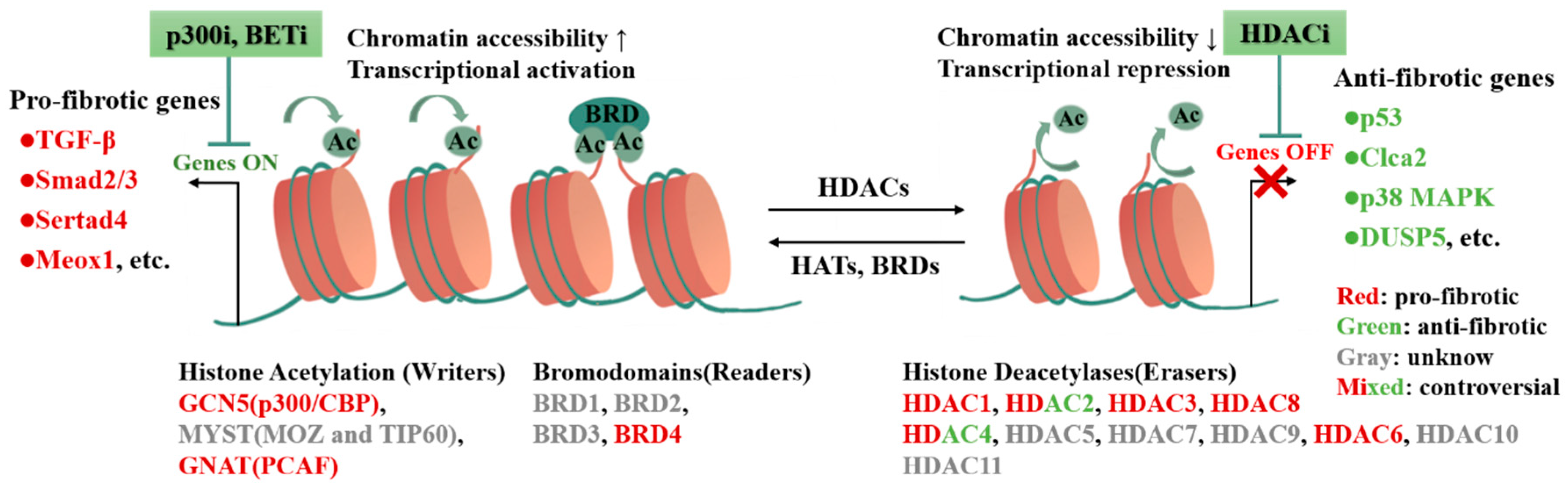
3.1. Histone Modification

Histones are basic proteins with highly conserved sequences in the nucleus with functions in maintaining DNA structure, protecting genetic information, and regulating gene expression. Two copies each containing four core histones (H2A, H2B, H3, and H4) are organized as a histone octamer that wraps ~145 base pairs of DNA to form the basic unit of chromatin [47]. Histone modifications are an important component in epigenetic regulation as well as an important post-translational modification (PTM) pathway. Multiple histone modifications such as methylation, acetylation, phosphorylation, and monoubiquitination are performed at the NH2-terminal tails of different histones protruding from the nucleosome [11,48]. These modifications of histone amino acid residues not only affect the interaction of histones and other proteins, but also influence chromatin structure and gene expression [48,49]. Histone acetylation and histone methylation are the most deeply studied so far. Here, we will focus our review on the roles of these two epigenetic regulatory mechanisms in CFs activation and cardiac fibrosis.

3.2. Histone Acetylation in CFs Activation and Cardiac Fibrosis

Histone acetylation modifications mainly occur at the amino-terminal site of lysine, and the addition or removal of histone tail acetylation modification is a dynamic and reversible process, which is mediated by a family of histone acetylation “writers” and “erasers” including histone acetyltransferases (HATs) and histone deacetylases (HDACs) [50,51,52]. Histone acetylation “writers” and “erasers” modulate the sparseness of chromatin structure through site-specific acetylation modifications, which can precisely regulate gene expression. Specifically, HATs-mediated acetylation causes structural loosening of chromatin nucleosomes, promotes binding of TFs to genes, and enhances gene transcriptional activity; while HDACs-mediated deacetylation causes tight binding of histones to genes, inhibits transcription factors binding to promoters, and silences or suppresses transcription and expression of related genes [53,54]. HATs and HDACs work in concert to maintain the relative balance of histone acetylation modifications. HATs and HDACs are the nexus of multiple pro-fibrotic signaling networks that mediate the cardiac fibrosis process in multiple ways [55] (Table 2).
Epigenetic alterations caused by histone acetylation are closely associated with CFs activation, proliferation, excessive ECM deposition and cardiac pathological remodeling [72]. Acetyltransferase p300, a primary epigenetic writer of histone acetylation, is activated and promotes cardiac fibrosis via Smad2 in a high glucose-induced cardiac fibrosis model [56]. Type I collagen is epigenetically regulated by the acetyltransferase activity of p300 and its subsequent interaction with Smad3, and profibrotic cytokine TGF-β is unable to induce collagen synthesis in fibroblasts in absence of p300 [73,74,75]. Importantly, p300 inhibitors L002 and C646 could both reverse hypertension-induced cardiac hypertrophy and fibrosis in vivo [57,73]. In addition, curcumin, a natural product of p300-histone acetyltransferase inhibitor, suppresses the development of heart failure in vivo [76]. Moreover, curcumin analogues GO-Y030, a specific inhibitor of p300, is also reported to attenuate cardiac fibrosis in a mouse model of pressure overload [58]. Consequently, p300 holds promise as a specific target for anti-fibrosis. In addition, p300/CBP-associated factor (PCAF), a histone acetyltransferase, is also elucidated to be required for the activation of CFs in response to TGF-β1 [59]. Intriguingly, miR-134-5p knockdown protected myocardial remodeling and cardiac fibrosis in the rat model of MI by upregulating lysine acetyltransferase 7 expression and histone H3K14 acetylation [77].
Histone deacetylases HDACs are enzymes that catalyze the deacetylation of histone or non-histone lysine residues, known as “erasers”, which are necessary for the removal of specific PTMs from histones. HDACs are classified into four classes (I, II, III, and IV), of which classes I, II, and IV HDACs are deacetylated by a zinc-dependent mechanism, while the deacetylation of class III HDACs is mainly dependent on nicotinamide adenine dinucleotide (NAD+) [78]. Class I HDACs include HDAC1, HDAC2, HDAC3, and HDAC8. Evidences from in vitro and in vivo studies indicate that class I HDACs are important regulators of cardiac fibrosis by CFs activation [79,80]. In-depth studies reveal that TGF-β may recruit HDAC1 to the Chloride channel accessory 2 (Clca2) promoter via Twist1, which inhibits Clca2 transcription by erasing histone H3/H4 acetylation. Subsequently, it is found that silencing of Clca2 promotes the activation of CFs into myofibroblasts and induces cardiac fibrosis, and Clca2 over-expression reverses this process and alleviates TGF-β-induced cardiac fibrosis [60]. Besides activating CFs, HDAC1 also functions to promote the proliferation and migration of CFs. A gene expression profiling study reveals peptidase inhibitor 16 (PI16) attenuates Ang-II-induced cardiac fibrosis by inhibiting HDAC1 expression [81]. Mechanically, PI16 attenuates Ang-II-induced elevated HDAC1 levels through the HDAC1/p53 signaling pathway, increasing H3K18 and H3K27 acetylation, thus inhibiting CFs proliferation and fibrosis-related protein expression, as well as significantly attenuating Ang-II-induced cardiac fibrosis [61]. Furthermore, inhibition of HDAC1 attenuates Ang-II-induced fibrosis by reducing mitochondrial overactivity and calcium overload [62].
In addition, HDAC2, HDAC3, and HDAC8 are also associated with cardiac fibrosis. HDAC2 may play a driving role in the development of cardiac fibrosis. HDAC2 activity is correlated with the phosphorylation status of HDAC2 S394 [82] and PPP2CA, the catalytic subunit of PP2A, which can bind to HDAC2 and inhibit HDAC2 function by preventing HDAC2 phosphorylation. Isoproterenol and pressure overload can induce cardiac fibrosis together with the process of PPP2CA and HDAC2 dissociation. Interestingly, ISO-induced cardiac hypertrophy and fibrosis are attenuated in PPP2CA transgenic mice with HDAC2 function inhibited [63]. It is noted that the depletion and inactivation of HDAC2 reduces Col1A1 and α-SMA expression in CFs [64]. Plantamajoside, an active component extracted from traditional Chinese herbal medicine Plantaginis, protects the myocardium from ISO-induced injury by inhibiting HDAC2 and AKT/GSK-3β signaling pathways [65]. Selective HDAC3 inhibitor RGFP966 significantly improves diabetes-induced cardiac dysfunction and fibrosis [66]. Furthermore, ISO treatment induces cardiac fibrosis by increasing the expression of HDAC8, p38 MAPK, and fibrosis-related genes. Using both the selective HDAC8 inhibitor PCI34051 [83] and the HDAC8 knockdown alleviate ISO-induced cardiac fibrosis by inhibiting the HDAC8-regulated p38 MAPK pathway [67].
Rhein, a Class I/II HDAC inhibitor, regulates fibrosis-related pathways and the cell cycle of CFs by inhibiting HDAC-dependent p53 protein stability under normoxic conditions [84]. The class II HDAC shuttles between the cytoplasm and the nucleus and is further divided into two subgroups: class IIa (HDAC 4, 5, 7, and 9) and class IIb (HDAC 6, 10). In myocyte-specific activated HDAC4 transgenic mice, activated HDAC4 promotes cardiac hypertrophy and fibrosis and exacerbates cardiac dysfunction in infarcted myocardium [68]. Moreover, sodium butyrate (NaBu) significantly attenuates Ang-II-induced cardiac hypertrophy and fibrosis, and inflammation by inhibiting the activation of the COX2/PGE2 pathway in HDAC5/HDAC6-dependent manner [69]. The other members of Class II HDACs have been sparsely studied in cardiac fibrosis, but have been discovered to be associated with other organ fibrosis models [85,86,87,88].
HDAC inhibitors (HDACIs) have shown strong promising potential for the treatment of cardiac fibrosis by inhibiting HDAC function [89]. The first-generation HDACIs (TSA, SAHA) have been shown to have anti-fibrosis properties; however, their molecular mechanisms remain to be elucidated. Currently, there are four HDACIs approved by the United States Food and Drug Administration (US FDA), all of which are pan-HDACIs, but none is applied to the treatment of fibrotic disease. Therefore, a comprehensive analysis suggests that there are still more barriers to the application of HDACIs in the clinical treatment of fibrotic diseases [90]. Firstly, during the development of fibrosis, there may be two or more HDACs exerting opposite effects simultaneously, pan-HDAC inhibitors may not achieve the expected effect or even have stronger side effects, and subtype-specific inhibitors should be developed to reduce adverse drug reactions. However, class I, II, and IV HDACs share a conserved catalytic domain and have very high homology among the same type of HDACs, making it difficult to develop subtype-specific inhibitors [91]. Secondly, both histones and non-histones are targets of HDACs, and many of them are key regulators of important pathways. Therefore, the regulation of fibrosis-related histone acetylation modifications may also cause strong toxicity [92]. Finally, the role of HDACs may differ at different stages of fibrosis development [93]. Consequently, a deeper understanding of the etiology of fibrosis is needed for more precise drug delivery to improve drug efficacy while reducing unwanted side effects.
As highly conserved proteins recognizing and reading histone acetylation lysine modifications, the bromodomain-containing proteins (BRDs) play important roles in regulating gene expression [94]. Based on the structural or sequence similarity, BRDs are divided into eight families, among which the bromodomain and extra terminal domain (BET) protein families are most intensively and extensively studied [95]. BET inhibitor JQ1 reduces cardiac hypertrophy and fibrosis induced by phenylephrine or thoracic aortic fasciculation and heart failure after MI [96,97,98]. Moreover, BET inhibitor JQ1 also inhibits the expression of pro-inflammatory genes in CFs and suppresses CFs activation and proliferation in the DCM model. Inhibition of BET proteins abrogates adverse cardiac remodeling, reduces cardiac fibrosis, and prolongs survival. Mechanistically, BRD4, a member of the BET family, is a central regulator of the pro-fibrotic CFs phenotype through p38-dependent signaling. Inhibition of BRD4 suppresses the activation of CFs and alleviates cardiac fibrosis by silencing Sertad4 (SERTA domain-containing protein 4) and periostin (myofibroblast maker gene) [70]. In addition, the promising molecule C-34 exhibits effective BRD4 inhibitory activity. The novel BRD4 inhibitor C-34 silences the downstream target c-MYC and further inhibits the TGF-β1/Smad2/3 signaling pathway, alleviating CFs activation in vitro and cardiac fibrosis in vivo [71]. The anti-fibrotic effects of BET inhibitors are promising for alleviating cardiac dysfunction and fibrosis [99,100]. However, BET proteins also play an important role in maintaining myocardial contractile function and cellular mitochondrial homeostasis [101], and pan-BET inhibitors are likely to disrupt many other critical cellular functions. Therefore, a more in-depth dissecting of the precise molecular mechanisms of BET inhibitor regulation will facilitate precise targeting and reduce drug side effects. It is found that modulation of the transcriptional switch regulates CFs activation. In fibroblasts, the TF Mesenchyme homebox 1 (MEOX1) is a central regulator of stress-induced CFs activation and cardiac fibrosis. Blocking MEOX1 turn-on inhibits fibroblast activation, which plays similar roles with BET inhibitors. Mechanistically, BET inhibitors act as a reversible switch for fibroblast activation to precisely regulate MEOX1 turn-on in disease states, while selective CRISPR inhibition of the single most dynamic cis-element of the enhancer blocks TGF-β-induced MEOX1 activation and ameliorates cardiac fibrosis [9]. This study provides new perspectives on the plasticity and specificity of BET-dependent regulation in tissue fibroblasts and also provides new trans- and cis-targets for the treatment of fibrotic diseases [100]. Consequently, the development of drugs targeting BET will hopefully bring new light to the treatment of cardiac fibrosis [71] (Figure 3).

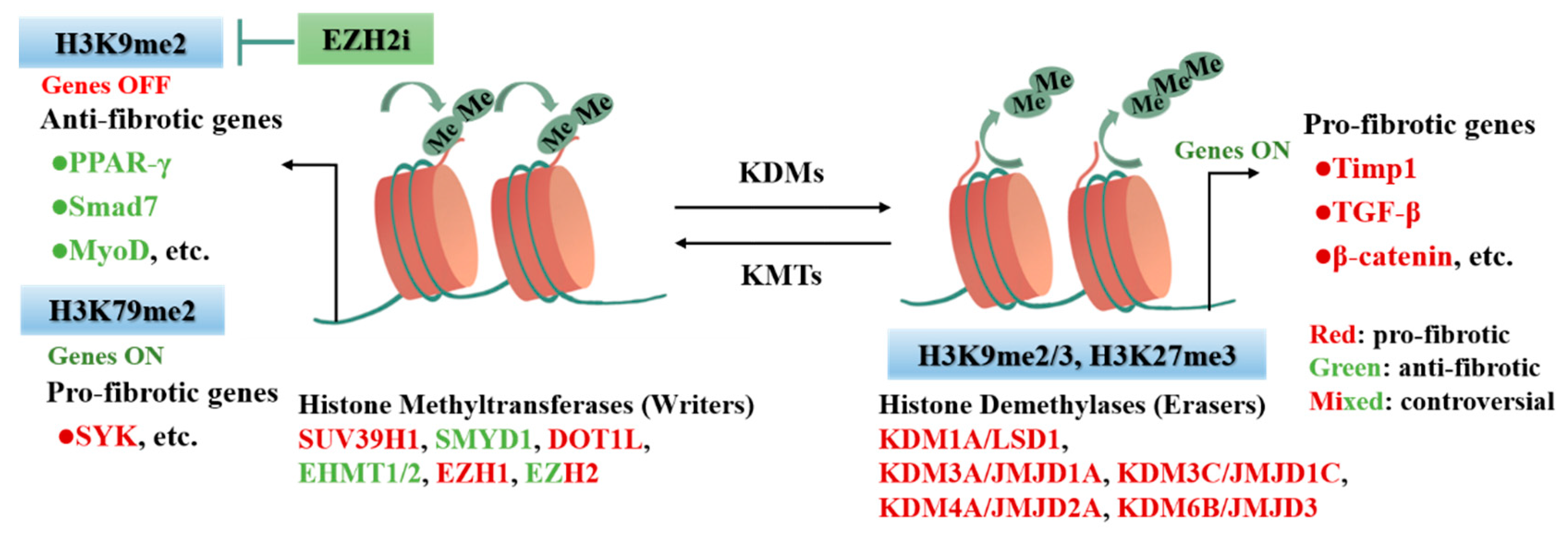
3.3. Histone Methylation in CFs Activation and Cardiac Fibrosis

Epigenetic regulation, especially histone methylation modification, is reported to be related to cardiac fibrosis [23]. Histone methylation modification is an important intracellular epigenetic modality that can regulate the transcriptional expression of specific genes by influencing chromosome conformation and the specific recruitment of downstream effector molecules. Histone methylation usually occurs at lysine (K) or arginine (R) residues in the tail of histone H3 and H4, and the modification site and number of methyl group all determine the transcriptional activity of the relevant genes [102,103]. Histone methylation can alter chromatin condensation to an open state that promotes chromatin transcription or to a closed state associated with decreased chromatin transcription. Histone methylation is catalyzed by histone methyltransferases (HMTs), which can be divided into protein lysine methyltransferases (KMTs) and protein arginine methyltransferases (PRMTs); and removed by histone lysine demethylase (KDMs), which are broadly divided into two families, Lysine-specific demethylase (LSD) and JMJC domain-containing family (JMJD). Methylation of histone lysine can exist in three states: monomethylated (me1), dimethylated (me2), and trimethylated (me3), which serve as markers of active or repressed gene expression [102]. Generally, H3K4me1/2/3 (short for mono/di/tri-methylation of lysine 4 of histone H3, similar below), H3K9me1, H3K27me1, H3K36me1/2/3, and H3k79me1/2/3 are considered to be active markers occupying actively transcribed gene regions in chromatin, while H3K9me3, H3K27me3, and H4K20me2/3 are known repressive markers that are usually associated with gene silencing and chromatin condensation [103,104]. KMTs and KDMs played important roles in cardiac fibrosis-related regulation (Table 3).
Based on the sequence of catalytic domains, KMTs can be classified into SET-containing domains and non-SET domains [116]. The SET domain is common to most transferases and is mainly responsible for the enzymatic activity of methyltransferases, including SUV39, EHMT1/2, EZH1/2, and RIZ (PRDM, SMYD, SUV420), and other families. Among them, enhancer of zeste homologue 2 (EZH2), a member of the EZH family, is most widely studied. EZH2 acts as the main catalytic subunit of the Polycomb repressive complex 2 (PRC2), which specifically catalyzes the methylation of histone H3 at lysine-27 (H3K27me1/me2/me3) through the SET structural domain [117]. EZH2 acts as an important epigenetic regulator during cardiac development and disease progression [117,118,119]. Interestingly, a high-fat diet during gestation or lactation in the mother induces cardiac reprogramming with reduced expression of EZH2 and DNMT3B and downregulates level of H3K27me2/3, which promotes the expression of pro-fibrotic and pro-hypertrophic genes [105]. In addition, EZH2 increases susceptibility to fibrosis in pathological processes [118], and that inhibition of EZH2 has the potential to reverse the fibrotic phenotype in cardiac disease. Both EZH2 inhibitor GSK126 and molecular silencing of EZH2 can block Ang-II-induced atrial fibroblast activation, migration, and ECM production in mice, attenuate Ang-II-induced atrial enlargement and fibrosis, and reduce atrial fibrillation vulnerability [106]. MiR-101a-3p might also prevent the development of atrial fibrillation in rats by targeting EZH2 to inhibit collagen synthesis and atrial fibrosis [120]. Similarly, miR-214-3p binds to EZH1/2 and represses EZH1/2 expression at the transcriptional level, which increases the peroxisome proliferator-activated receptor-γ (PPAR-γ) expression and inhibits Col1a1 and Col3a1 expression in myofibroblasts [109]. lncRNA-ANRIL alters the expression of fibronectin (FN), type IV collagen (Col1α4), and VEGF by interacting with multiple epigenetic regulators, including EZH2, p300, and regulates cardiac fibrosis in diabetic mice [107]. Furthermore, LncRNA NEAT1 inhibits Smad7 expression by recruiting EZH2 to the Smad7 promoter region, which ultimately aggravates the progression of cardiac fibrosis, while silencing NEAT1 significantly ameliorates TAC surgery-induced cardiac fibrosis and dysfunction in mice [108]. Moreover, in spontaneously hypertensive rats (SHRs), SUV39H1 is recruited by lncRNA MALAT1, resulting in H3K9me3 of MyoD-binding loci to mediate cardiac fibrosis [110]. Similarly, disruptor of telomeric silencing 1-like (DOT1L), the only non-SET domain KMT, promotes spleen tyrosine kinase (SYK) expression via increasing H3K79me2 modification of the SYK promoter, and upregulation of SYK activates TGF-β1/Smad3 signaling to mediate CFs proliferation and cardiac fibrosis. Knockdown of DOT1L significantly ameliorated the cardiac injury and fibrosis caused by MI [111].
Histone lysine demethylase (KDMs) is an emerging regulator of transcriptional reprogramming, though the potential mechanism in cardiac fibrosis is unclear. Currently, histone KDM is comprised of two subfamilies, Lysine-specific demethylase (LSD) and JMJC domain-containing family (JMJD) [121]. LSD family demethylases specifically remove mono- and di-methylated modifications of histone H3K4 and H3K9, while JMJD family demethylases catalyze the removal of three methylation modifications. LSD1/KDM1 has been recognized as an important target of cardiac fibrosis, whose direct substrates H3K4me1/2 and H3K9me1/2 play a critical role in cardiac remodeling [122,123]. Huo et al. reported that myofibroblast-specific LSD1 inducible knockout effectively alleviates cardiac hypertrophy, fibrosis, and pressure overload-induced heart failure by inhibiting the TGF-β signaling pathway [112]. KDM3A could remove H3K9me2/3 modifications, and the level of H3K9 methylation is downregulated in hypertrophic and failing hearts in mice and humans [124,125]. Tissue metalloproteinase inhibitor 1 (Timp1) is commonly used as a cardiac fibrosis biomarker, whose levels are associated with cardiac fibrosis in patients with heart disease [126]. In TAC-induced cardiac fibrosis, KDM3A binds the Timp1-promoter to remove methylation modifications and promotes TIMP1 transcriptional activation, which then activates CFs and enhances cardiac fibrosis [113]. Furthermore, TIMP1 is also identified as a potential target for KDM3C-mediated pro-fibrotic function. In the Ang-II-induced mouse model, KDM3C removes the methyl from the H3K9me2 on the Timp1 promoter, promotes Timp1 transcriptional activation, activates resident CFs, and induces cardiac fibrosis [114]. Furthermore, the H3K27me3 demethylase KDM6B removes the suppressive mark H3K27me3 from the β-catenin promoter in CFs, which activates CFs and significantly upregulates the expression of fibrogenic genes [115]. More studies are necessary to determine the effects of histone methylation regulators on the cardiac fibrosis process in pathophysiological situations [127]. Furtherly, we also need to understand the molecular mechanisms that include governing substrate specificity and the downstream effects of their catalytic activity (Figure 4).

4. RNA in CFs Activation and Cardiac Fibrosis

Epigenetic regulation occurs not only at the genomic level but also at the transcriptome level and plays an important role in post-transcriptional regulation [128]. RNA acts as the “bridge” between DNA and cellular protein production processes. Non-coding RNAs (ncRNAs) could regulate gene expression directly as epigenetic regulators or indirectly by modifying mRNAs and ncRNAs depending on regulators such as writers, erasers, and readers. Consequently, epigenetic modifications occurring at the transcriptomic level have critical regulatory roles in biological activities, and studies demonstrate that epigenetic modifications are closely associated with many diseases, including cardiovascular diseases (Table 4).

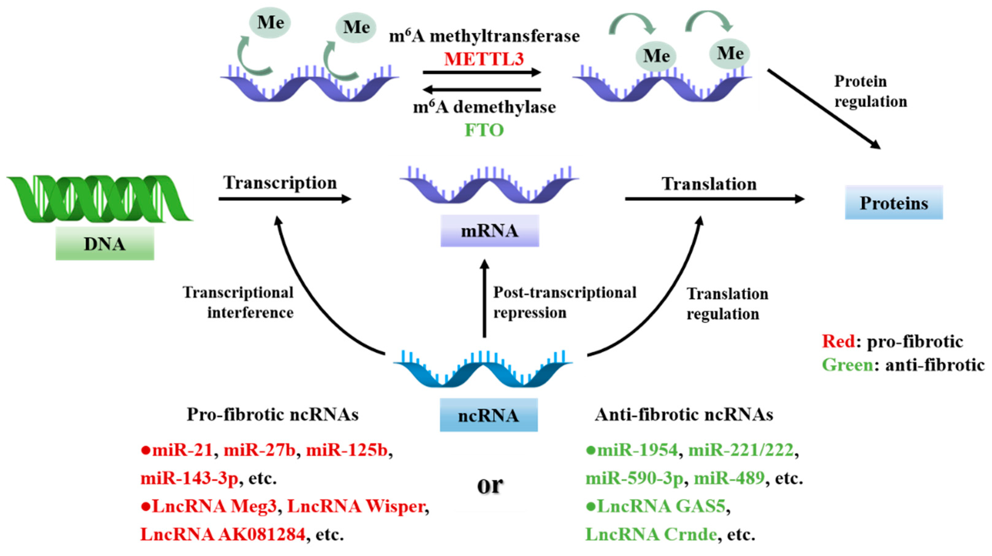
4.1. Non-Coding RNAs in CFs Activation and Cardiac Fibrosis

NcRNAs mainly include microRNAs (miRNAs), long non-coding RNAs (lncRNAs), and circular RNAs (circ RNAs), which are RNA transcripts with biological functions without protein-coding ability. Recently, there are growing evidences that ncRNAs can be involved in the development of cardiac fibrosis by influencing post-transcriptional levels of regulation of gene expression.
MiRNAs are short ncRNAs (<200 nucleotides) whose biological functions depend on complementary binding to the 3 ‘ or 5 ‘ -untranslated regions (UTRs) sequences of target mRNAs, inhibiting protein translation or targeting mRNA degradation to mediate post-transcriptional gene silencing. MicroRNA-369-5p (miR-369-5p) promotes fibroblast proliferation by inhibiting the Patched1 pathway through modulation of DNMT3A methylation modification [29]. Similarly, miR-489 also alleviates cardiac fibrosis by regulating HDAC2 to inhibit fibroblast activation and proliferation [64]. Interestingly, miRNAs not only act as upstream factors to regulate DNA methylation and histone modifications but also as downstream effectors to directly promote or inhibit fibrosis. It was found that miR-27b [129], miR-125b [130], miR-99b-3p [131], miR-143-3p [132], and other miRNAs regulate the activation, proliferation, and migration process of CFs by regulating the downstream fibrosis-related genes FBW7, Apelin, p53, GSK-3β, SPRY3, etc., facilitating the fibrosis development. The miR-21 in macrophages also regulates macrophage–fibroblast communication and promotes activation of quiescent fibroblasts into myofibroblasts, promoting cardiac fibrosis [133]. MiR-27b-3p suppresses mitochondrial oxidative phosphorylation (OXPHOS) through the target gene FGF1, and inhibition of FGF1 alleviates TAC or Ang-II-induced cardiac fibrosis by enhancing OXPHOS [134]. In addition, miRNAs such as miR-590-3p [135], miR-221/222 [136], and miR-1954 [137] alleviate cardiac fibrosis by regulating ZEB1, Smad2, and THBS1. SKI, a TGF-β negative regulator, promotes cardiac fibroblast deactivation and alleviates cardiac fibrosis [150]. It has been demonstrated that the epigenetic regulator miR-34a/miR-93 can affect both TGF-β and SKI and alleviate cardiac fibrosis. Mechanically, miR-34a/miR-93 affects TGF-β1-induced fibroblast proliferation and ECM deposition via SKI [151]. In addition, apigenin, a flavonoid isolated from Apium graveolens, shows promising protective effects against ISO-induced cardiac fibrosis. Apigenin alleviates cardiac fibrosis by upregulating miR-122-5p, c-Ski, and Smad7 and downregulating miR-155-5p, TGF-β1, hypoxia-inducible factor-1α (HIF-1α), Smad2/3, etc. [152].
LncRNAs as long noncoding RNAs (>200 nucleotides) are involved in the development of cardiac fibrosis mainly through directly binding to DNA, RNA, and proteins to regulate gene expression [128]. lncRNA AK081284 [138], lncRNA Meg3 [139], lncRNA Wisper [140], and lncRNA AK048087 [143] promote the activation and proliferation of CFs by regulating fibrosis-related genes such as IL-17, MMP-2, and COTL1. Meanwhile, lncRNA AK137033 regulates the stability of Sfrp2 mRNA [141], and lncRNA PCFL interacts with miR-378 [142], which together regulates cardiac fibrosis after MI. Furthermore, lncRNAs act as inhibitors to alleviate the development of cardiac fibrosis. LncRNA GAS5 overexpression can reduce TGF-β1-induced α-SMA and Col1A1 levels in CFs by inhibiting miR-21 expression to alleviate cardiac fibrosis [144]. LncRNA Crnde also suppresses CFs activation and alleviates DCM-associated cardiac fibrosis through negative feedback regulation of Smad3 transcriptional activation [145]. Dioscin, a glucoside saponin isolated from Dioscorea nipponica Makino, reduced MI-induced cardiac fibrosis by upregulating lncRNA MANTIS to promote the expression of angiogenesis-related genes such as SOX18, SMAD6, and COUP-TFII [146]. Meanwhile, histone demethylase JARID1B could also regulate lncRNA MANTIS and restrict the expression of angiogenesis-related genes such as SMAD6 [153]. However, whether JARID1B regulates lncRNA MANTIS through methylation modification and thus affects cardiac fibrosis remains to be further investigated.

4.2. RNA Modifications in CFs Activation and Cardiac Fibrosis

RNA modifications directly affect the biological functions of RNA. N6-methylated adenosine (m6A) is the most abundant epigenetic modification of eukaryotic mRNA, which is mainly involved in the pathophysiological process of cardiac fibrosis by regulating the transport, degradation, and translation of mRNA. m6A is regulated by three kinds of effector proteins, the m6A methyltransferase METTL3-METTL14 complex, m6A demethylases FTO and ALKBH5, and the m6A reader and the YTH structural domain proteins [154]. Overexpression of m6A methyltransferase METTL3 improves the level of m6A modification, increases collagen synthesis, and promotes cardiac fibrosis [147]. However, the expression level of m6A demethylase FTO is low in cardiac fibrosis induced by MI [155], hypoxia, and heart failure [149], and overexpression of FTO inhibits the activation, proliferation, and migration of cardiac fibroblasts and alleviates the development of cardiac fibrosis [148]. At present, the studies on m6A methylation in cardiac fibrosis are mainly focused on METTL3 and FTO. Future studies should investigate how other writers and erasers regulate the expression of downstream proteins, how m6A readers mediate the activity and mechanism of m6A methylation in CFs and further explore m6A methylation may provide new therapeutic strategies for cardiac fibrosis (Figure 5).

5. Conclusions and Future Prospects

In this review, we focus on the recent progress between epigenetic regulation and activation of CFs during cardiac fibrosis based on the main categories of epigenetic modifications. As key nodes, epigenetic-modifying enzymes can transmit upstream signaling through gene transcriptional profiling reprogramming to mediate cardiac fibrogenesis, providing a large number of potential drug targets for preclinical studies of cardiac fibrosis, and also providing a new perspective for the development of the clinical protocol in cardiac fibrosis [12,23]. It is important to elucidate the role of epigenetics in the mechanism of endogenous fibrosis, while it is equally necessary to focus on the studies of anti-fibrotic factors, especially those that involve the deactivation of cardiac myofibroblasts. Although there is currently rare research on the relationship between epigenetics and deactivation of cardiac myofibroblasts, future research in this area may yield some important breakthroughs due to the reversibility of epigenetic modifications. SKI, a negative regulator of the TGFβ1/Smad signaling, has a powerful ability to inhibit fibrosis and deactivate activated fibroblasts [150,156]. Studying the regulation mechanism of myofibroblasts deactivation, especially in epigenetic regulation, will also provide a new direction for the treatment of cardiac fibrosis. However, we must recognize that the regulatory processes of epigenetic modulators are extensive and complex. There is a crosstalk between each epigenetic modification, which can interact and influence each other. Meanwhile, complex feedback regulatory loops make it difficult to draw general conclusions about homogeneous epigenetic modifications, which increases the challenge of our epigenetics-based studies on the mechanisms of cardiac fibrosis.
Compared to genetic research, no genome-wide analysis of epigenetic modifications in CFs has been reported to provide a comprehensive and systematic understanding of the biological significance and catalytic activity regulation mechanisms of each epigenetic regulator. Nevertheless, with the development of gene sequencing technologies in recent years, the integration of single-cell RNA sequencing, single-cell epigenomics, high-resolution ChIP-seq, and HITS-CLIP (high-throughput sequencing with cross-linked immunoprecipitation) may provide a new approach to reveal the precise regulatory mechanisms, while the use of CRISPR-Cas9 gene-editing technology may also provide a more comprehensive resolution of epigenetic regulation. Meanwhile, several epigenetic modulator inhibitors, such as HDACIs, have been demonstrated to prevent and alleviate cardiac fibrosis. However, most inhibitors are not specific and may impair other key cellular functions during administration. Hence, more elaborate and comprehensive studies of epigenetic mechanisms are necessary to target more precise targets, thereby reducing the occurrence of adverse outcomes due to unnecessary inhibition or activation by drugs. In this process, it is also necessary to investigate whether and how epigenetic regulators exert cell-specific activities, which of course relies on further studies on the activation of CFs in cardiac fibrosis.
Growing evidences suggest that CFs activation is actually more complex than previously thought during cardiac fibrosis, and epigenetic modifications are very important for CFs activation and cardiac fibrosis. While many mechanisms need to be explored and conflicting evidence exists, future breakthroughs in the epigenetic field may provide avenues for the diagnosis and treatment of cardiac fibrosis. For example, chimeric antigen receptor T cell (CAR-T) therapy targeting FAP alleviates the degree of cardiac fibrosis and abnormal increased ventricular mass in model mice as well as improved cardiac systolic function, although the safety of this therapy needs to be further validated [157]. To better apply CAR-T to the treatment of cardiac fibrosis, researchers have also developed a way to generate transient CAR-T cells in vivo with mRNA molecules, which allows T cells to gain the ability to specifically target CFs for a short period to be fully effective and avoid affecting other cells [158]. Meanwhile, various means can reprogram CFs into cardiomyocytes, and epigenetics plays a key role in this process [159,160]; these advances will provide an alternative approach for clinical trials and treatments for patients with cardiac fibrosis in the future.

Author Contributions

J.S. and S.Z. jointly decided on the idea of this review. J.S. wrote the first entire draft, J.L. and S.Z. revised the manuscript. All authors have read and agreed to the published version of the manuscript.

Funding

This work was funded by the National Natural Science Foundation of China (82171792), and the National Key R&D Program of China (2021YFF0702103).

Institutional Review Board Statement

Not applicable.

Informed Consent Statement

Not applicable.

Data Availability Statement

Not applicable.

Conflicts of Interest

The authors declare no conflict of interest.

References

  1. Heimlich, J.B.; Bick, A.G. Somatic Mutations in Cardiovascular Disease. Circ. Res. 2022, 130, 149–161. [Google Scholar] [CrossRef] [PubMed]
  2. Travers, J.G.; Kamal, F.A.; Robbins, J.; Yutzey, K.E.; Blaxall, B.C. Cardiac Fibrosis: The Fibroblast Awakens. Circ. Res. 2016, 118, 1021–1040. [Google Scholar] [CrossRef] [PubMed] [Green Version]
  3. Frangogiannis, N.G. Cardiac fibrosis. Cardiovasc. Res. 2021, 117, 1450–1488. [Google Scholar] [CrossRef] [PubMed]
  4. Hinderer, S.; Schenke-Layland, K. Cardiac fibrosis—A short review of causes and therapeutic strategies. Adv. Drug Deliv. Rev. 2019, 146, 77–82. [Google Scholar] [CrossRef] [PubMed]
  5. Liu, M.; López de Juan Abad, B.; Cheng, K. Cardiac fibrosis: Myofibroblast-mediated pathological regulation and drug delivery strategies. Adv. Drug Deliv. Rev. 2021, 173, 504–519. [Google Scholar] [CrossRef]
  6. Burke, R.M.; Dirkx, R.A., Jr.; Quijada, P.; Lighthouse, J.K.; Mohan, A.; O’Brien, M.; Wojciechowski, W.; Woeller, C.F.; Phipps, R.P.; Alexis, J.D.; et al. Prevention of Fibrosis and Pathological Cardiac Remodeling by Salinomycin. Circ. Res. 2021, 128, 1663–1678. [Google Scholar] [CrossRef]
  7. Tarbit, E.; Singh, I.; Peart, J.N.; Rose’Meyer, R.B. Biomarkers for the identification of cardiac fibroblast and myofibroblast cells. Heart Fail. Rev. 2019, 24, 1–15. [Google Scholar] [CrossRef]
  8. Plikus, M.V.; Wang, X.; Sinha, S.; Forte, E.; Thompson, S.M.; Herzog, E.L.; Driskell, R.R.; Rosenthal, N.; Biernaskie, J.; Horsley, V. Fibroblasts: Origins, definitions, and functions in health and disease. Cell 2021, 184, 3852–3872. [Google Scholar] [CrossRef] [PubMed]
  9. Alexanian, M.; Przytycki, P.F.; Micheletti, R.; Padmanabhan, A.; Ye, L.; Travers, J.G.; Gonzalez-Teran, B.; Silva, A.C.; Duan, Q.; Ranade, S.S.; et al. A transcriptional switch governs fibroblast activation in heart disease. Nature 2021, 595, 438–443. [Google Scholar] [CrossRef]
  10. Gibb, A.A.; Lazaropoulos, M.P.; Elrod, J.W. Myofibroblasts and Fibrosis: Mitochondrial and Metabolic Control of Cellular Differentiation. Circ. Res. 2020, 127, 427–447. [Google Scholar] [CrossRef] [PubMed]
  11. Papait, R.; Serio, S.; Condorelli, G. Role of the Epigenome in Heart Failure. Physiol. Rev. 2020, 100, 1753–1777. [Google Scholar] [CrossRef] [PubMed]
  12. Xue, T.; Qiu, X.; Liu, H.; Gan, C.; Tan, Z.; Xie, Y.; Wang, Y.; Ye, T. Epigenetic regulation in fibrosis progress. Pharmacol. Res. 2021, 173, 105910. [Google Scholar] [CrossRef] [PubMed]
  13. Travers, J.G.; Tharp, C.A.; Rubino, M.; McKinsey, T.A. Therapeutic targets for cardiac fibrosis: From old school to next-gen. J. Clin. Investig. 2022, 132, e148554. [Google Scholar] [CrossRef]
  14. van der Harst, P.; de Windt, L.J.; Chambers, J.C. Translational Perspective on Epigenetics in Cardiovascular Disease. J. Am. Coll. Cardiol. 2017, 70, 590–606. [Google Scholar] [CrossRef] [PubMed]
  15. Cavalli, G.; Heard, E. Advances in epigenetics link genetics to the environment and disease. Nature 2019, 571, 489–499. [Google Scholar] [CrossRef] [Green Version]
  16. Park, S.; Nguyen, N.B.; Pezhouman, A.; Ardehali, R. Cardiac fibrosis: Potential therapeutic targets. Transl. Res. J. Lab. Clin. Med. 2019, 209, 121–137. [Google Scholar] [CrossRef]
  17. Berdasco, M.; Esteller, M. Clinical epigenetics: Seizing opportunities for translation. Nat. Rev. Genet. 2019, 20, 109–127. [Google Scholar] [CrossRef]
  18. Greenberg, M.V.C.; Bourc’his, D. The diverse roles of DNA methylation in mammalian development and disease. Nat. Rev. Mol. Cell Biol. 2019, 20, 590–607. [Google Scholar] [CrossRef]
  19. Parry, A.; Rulands, S.; Reik, W. Active turnover of DNA methylation during cell fate decisions. Nat. Rev. Genet. 2021, 22, 59–66. [Google Scholar] [CrossRef]
  20. Deaton, A.M.; Bird, A. CpG islands and the regulation of transcription. Genes Dev. 2011, 25, 1010–1022. [Google Scholar] [CrossRef] [PubMed] [Green Version]
  21. Zhu, H.; Wang, G.; Qian, J. Transcription factors as readers and effectors of DNA methylation. Nat. Rev. Genet. 2016, 17, 551–565. [Google Scholar] [CrossRef] [PubMed] [Green Version]
  22. Neri, F.; Rapelli, S.; Krepelova, A.; Incarnato, D.; Parlato, C.; Basile, G.; Maldotti, M.; Anselmi, F.; Oliviero, S. Intragenic DNA methylation prevents spurious transcription initiation. Nature 2017, 543, 72–77. [Google Scholar] [CrossRef] [PubMed]
  23. Tao, H.; Song, Z.Y.; Ding, X.S.; Yang, J.J.; Shi, K.H.; Li, J. Epigenetic signatures in cardiac fibrosis, special emphasis on DNA methylation and histone modification. Heart Fail. Rev. 2018, 23, 789–799. [Google Scholar] [CrossRef] [PubMed]
  24. Tao, H.; Shi, P.; Zhao, X.D.; Xuan, H.Y.; Gong, W.H.; Ding, X.S. DNMT1 deregulation of SOCS3 axis drives cardiac fibroblast activation in diabetic cardiac fibrosis. J. Cell. Physiol. 2021, 236, 3481–3494. [Google Scholar] [CrossRef]
  25. Xu, S.S.; Ding, J.F.; Shi, P.; Shi, K.H.; Tao, H. DNMT1-Induced miR-152-3p Suppression Facilitates Cardiac Fibroblast Activation in Cardiac Fibrosis. Cardiovasc. Toxicol. 2021, 21, 984–999. [Google Scholar] [CrossRef]
  26. Zhao, K.; Weng, L.; Xu, T.; Yang, C.; Zhang, J.; Ni, G.; Guo, X.; Tu, J.; Zhang, D.; Sun, W.; et al. Low-intensity pulsed ultrasound prevents prolonged hypoxia-induced cardiac fibrosis through HIF-1alpha/DNMT3a pathway via a TRAAK-dependent manner. Clin. Exp. Pharmacol. Physiol. 2021, 48, 1500–1514. [Google Scholar] [CrossRef]
  27. Tao, H.; Yang, J.J.; Chen, Z.W.; Xu, S.S.; Zhou, X.; Zhan, H.Y.; Shi, K.H. DNMT3A silencing RASSF1A promotes cardiac fibrosis through upregulation of ERK1/2. Toxicology 2014, 323, 42–50. [Google Scholar] [CrossRef]
  28. Qin, R.H.; Tao, H.; Ni, S.H.; Shi, P.; Dai, C.; Shi, K.H. microRNA-29a inhibits cardiac fibrosis in Sprague-Dawley rats by downregulating the expression of DNMT3A. Anatol. J. Cardiol. 2018, 20, 198–205. [Google Scholar] [CrossRef]
  29. Tao, H.; Dai, C.; Ding, J.F.; Yang, J.J.; Ding, X.S.; Xu, S.S.; Shi, K.H. Epigenetic aberrations of miR-369-5p and DNMT3A control Patched1 signal pathway in cardiac fibrosis. Toxicology 2018, 410, 182–192. [Google Scholar] [CrossRef]
  30. Zhao, X.D.; Qin, R.H.; Yang, J.J.; Xu, S.S.; Tao, H.; Ding, X.S.; Shi, K.H. DNMT3A controls miR-200b in cardiac fibroblast autophagy and cardiac fibrosis. Inflamm. Res. 2018, 67, 681–690. [Google Scholar] [CrossRef]
  31. Watson, C.J.; Collier, P.; Tea, I.; Neary, R.; Watson, J.A.; Robinson, C.; Phelan, D.; Ledwidge, M.T.; McDonald, K.M.; McCann, A.; et al. Hypoxia-induced epigenetic modifications are associated with cardiac tissue fibrosis and the development of a myofibroblast-like phenotype. Hum. Mol. Genet. 2014, 23, 2176–2188. [Google Scholar] [CrossRef] [PubMed] [Green Version]
  32. Li, S.; Li, P.; Liu, W.; Shang, J.; Qiu, S.; Li, X.; Liu, W.; Shi, H.; Zhou, M.; Liu, H. Danhong Injection Alleviates Cardiac Fibrosis via Preventing the Hypermethylation of Rasal1 and Rassf1 in TAC Mice. Oxidative Med. Cell. Longev. 2020, 2020, 3158108. [Google Scholar] [CrossRef] [PubMed]
  33. Rajgarhia, A.; Ayasolla, K.R.; Zaghloul, N.; Lopez Da Re, J.M.; Miller, E.J.; Ahmed, M. Extracellular Superoxide Dismutase (EC-SOD) Regulates Gene Methylation and Cardiac Fibrosis During Chronic Hypoxic Stress. Front. Cardiovasc. Med. 2021, 8, 669975. [Google Scholar] [CrossRef] [PubMed]
  34. Sano, S.; Oshima, K.; Wang, Y.; Katanasaka, Y.; Sano, M.; Walsh, K. CRISPR-Mediated Gene Editing to Assess the Roles of Tet2 and Dnmt3a in Clonal Hematopoiesis and Cardiovascular Disease. Circ. Res. 2018, 123, 335–341. [Google Scholar] [CrossRef] [PubMed]
  35. Tao, H.; Xu, W.; Qu, W.; Gao, H.; Zhang, J.; Cheng, X.; Liu, N.; Chen, J.; Xu, G.L.; Li, X.; et al. Loss of ten-eleven translocation 2 induces cardiac hypertrophy and fibrosis through modulating ERK signaling pathway. Hum. Mol. Genet. 2021, 30, 865–879. [Google Scholar] [CrossRef] [PubMed]
  36. Xu, X.; Tan, X.; Tampe, B.; Nyamsuren, G.; Liu, X.; Maier, L.S.; Sossalla, S.; Kalluri, R.; Zeisberg, M.; Hasenfuss, G.; et al. Epigenetic balance of aberrant Rasal1 promoter methylation and hydroxymethylation regulates cardiac fibrosis. Cardiovasc. Res. 2015, 105, 279–291. [Google Scholar] [CrossRef] [PubMed] [Green Version]
  37. Lyko, F. The DNA methyltransferase family: A versatile toolkit for epigenetic regulation. Nat. Rev. Genet. 2018, 19, 81–92. [Google Scholar] [CrossRef] [PubMed]
  38. Chen, Z.; Zhang, Y. Role of Mammalian DNA Methyltransferases in Development. Annu. Rev. Biochem. 2020, 89, 135–158. [Google Scholar] [CrossRef]
  39. Kusano, K.F.; Pola, R.; Murayama, T.; Curry, C.; Kawamoto, A.; Iwakura, A.; Shintani, S.; Ii, M.; Asai, J.; Tkebuchava, T.; et al. Sonic hedgehog myocardial gene therapy: Tissue repair through transient reconstitution of embryonic signaling. Nat. Med. 2005, 11, 1197–1204. [Google Scholar] [CrossRef]
  40. Wu, R.N.; Yu, T.Y.; Zhou, J.C.; Li, M.; Gao, H.K.; Zhao, C.; Dong, R.Q.; Peng, D.; Hu, Z.W.; Zhang, X.W.; et al. Targeting HMGB1 ameliorates cardiac fibrosis through restoring TLR2-mediated autophagy suppression in myocardial fibroblasts. Int. J. Cardiol. 2018, 267, 156–162. [Google Scholar] [CrossRef]
  41. Li, Z.M.; Xu, S.W.; Liu, P.Q. Salvia miltiorrhizaBurge (Danshen): A golden herbal medicine in cardiovascular therapeutics. Acta Pharmacol. Sin. 2018, 39, 802–824. [Google Scholar] [CrossRef] [Green Version]
  42. Yang, G.S.; Zheng, B.; Qin, Y.; Zhou, J.; Yang, Z.; Zhang, X.H.; Zhao, H.Y.; Yang, H.J.; Wen, J.K. Salvia miltiorrhiza-derived miRNAs suppress vascular remodeling through regulating OTUD7B/KLF4/NMHC IIA axis. Theranostics 2020, 10, 7787–7811. [Google Scholar] [CrossRef] [PubMed]
  43. Barcena-Varela, M.; Paish, H.; Alvarez, L.; Uriarte, I.; Latasa, M.U.; Santamaria, E.; Recalde, M.; Garate, M.; Claveria, A.; Colyn, L.; et al. Epigenetic mechanisms and metabolic reprogramming in fibrogenesis: Dual targeting of G9a and DNMT1 for the inhibition of liver fibrosis. Gut 2021, 70, 388–400. [Google Scholar] [CrossRef] [PubMed]
  44. She, Q.; Shi, P.; Xu, S.S.; Xuan, H.Y.; Tao, H.; Shi, K.H.; Yang, Y. DNMT1 Methylation of LncRNA GAS5 Leads to Cardiac Fibroblast Pyroptosis via Affecting NLRP3 Axis. Inflammation 2020, 43, 1065–1076. [Google Scholar] [CrossRef]
  45. Li, X.; Yang, Y.; Chen, S.; Zhou, J.; Li, J.; Cheng, Y. Epigenetics-based therapeutics for myocardial fibrosis. Life Sci. 2021, 271, 119186. [Google Scholar] [CrossRef] [PubMed]
  46. Tian, L.; Wu, D.; Dasgupta, A.; Chen, K.H.; Mewburn, J.; Potus, F.; Lima, P.D.A.; Hong, Z.; Zhao, Y.Y.; Hindmarch, C.C.T.; et al. Epigenetic Metabolic Reprogramming of Right Ventricular Fibroblasts in Pulmonary Arterial Hypertension: A Pyruvate Dehydrogenase Kinase-Dependent Shift in Mitochondrial Metabolism Promotes Right Ventricular Fibrosis. Circ. Res. 2020, 126, 1723–1745. [Google Scholar] [CrossRef]
  47. Kornberg, R.D.; Lorch, Y. Twenty-five years of the nucleosome, fundamental particle of the eukaryote chromosome. Cell 1999, 98, 285–294. [Google Scholar] [CrossRef] [Green Version]
  48. Mersfelder, E.L.; Parthun, M.R. The tale beyond the tail: Histone core domain modifications and the regulation of chromatin structure. Nucleic Acids Res. 2006, 34, 2653–2662. [Google Scholar] [CrossRef] [Green Version]
  49. Spyropoulou, A.; Piperi, C.; Adamopoulos, C.; Papavassiliou, A.G. Deregulated chromatin remodeling in the pathobiology of brain tumors. Neuromolecular Med. 2013, 15, 1–24. [Google Scholar] [CrossRef]
  50. Yun, M.; Wu, J.; Workman, J.L.; Li, B. Readers of histone modifications. Cell Res. 2011, 21, 564–578. [Google Scholar] [CrossRef] [Green Version]
  51. Strahl, B.D.; Allis, C.D. The language of covalent histone modifications. Nature 2000, 403, 41–45. [Google Scholar] [CrossRef] [PubMed]
  52. Shvedunova, M.; Akhtar, A. Modulation of cellular processes by histone and non-histone protein acetylation. Nature Rev. Mol. Cell Biol. 2022, 23, 329–349. [Google Scholar] [CrossRef] [PubMed]
  53. Gates, L.A.; Foulds, C.E.; O’Malley, B.W. Histone Marks in the ‘Driver’s Seat’: Functional Roles in Steering the Transcription Cycle. Trends Biochem. Sci. 2017, 42, 977–989. [Google Scholar] [CrossRef]
  54. Wiesel-Motiuk, N.; Assaraf, Y.G. The key roles of the lysine acetyltransferases KAT6A and KAT6B in physiology and pathology. Drug Resist. Updates Rev. Comment. Antimicrob. Anticancer. Chemother. 2020, 53, 100729. [Google Scholar] [CrossRef]
  55. Pang, M.; Zhuang, S. Histone deacetylase: A potential therapeutic target for fibrotic disorders. J. Pharmacol. Exp. Ther. 2010, 335, 266–272. [Google Scholar] [CrossRef] [PubMed] [Green Version]
  56. Bugyei-Twum, A.; Advani, A.; Advani, S.L.; Zhang, Y.; Thai, K.; Kelly, D.J.; Connelly, K.A. High glucose induces Smad activation via the transcriptional coregulator p300 and contributes to cardiac fibrosis and hypertrophy. Cardiovasc. Diabetol. 2014, 13, 89. [Google Scholar] [CrossRef] [Green Version]
  57. Rai, R.; Sun, T.; Ramirez, V.; Lux, E.; Eren, M.; Vaughan, D.E.; Ghosh, A.K. Acetyltransferase p300 inhibitor reverses hypertension-induced cardiac fibrosis. J. Cell. Mol. Med. 2019, 23, 3026–3031. [Google Scholar] [CrossRef] [Green Version]
  58. Shimizu, K.; Sunagawa, Y.; Funamoto, M.; Wakabayashi, H.; Genpei, M.; Miyazaki, Y.; Katanasaka, Y.; Sari, N.; Shimizu, S.; Katayama, A.; et al. The Synthetic Curcumin Analogue GO-Y030 Effectively Suppresses the Development of Pressure Overload-induced Heart Failure in Mice. Sci. Rep. 2020, 10, 7172. [Google Scholar] [CrossRef]
  59. Lim, Y.; Jeong, A.; Kwon, D.H.; Lee, Y.U.; Kim, Y.K.; Ahn, Y.; Kook, T.; Park, W.J.; Kook, H. P300/CBP-Associated Factor Activates Cardiac Fibroblasts by SMAD2 Acetylation. Int. J. Mol. Sci. 2021, 22, 9944. [Google Scholar] [CrossRef]
  60. Shao, T.; Xue, Y.; Fang, M. Epigenetic Repression of Chloride Channel Accessory 2 Transcription in Cardiac Fibroblast: Implication in Cardiac Fibrosis. Front. Cell Dev. Biol. 2021, 9, 771466. [Google Scholar] [CrossRef]
  61. Deng, M.; Yang, S.; Ji, Y.; Lu, Y.; Qiu, M.; Sheng, Y.; Sun, W.; Kong, X. Overexpression of peptidase inhibitor 16 attenuates angiotensin II-induced cardiac fibrosis via regulating HDAC1 of cardiac fibroblasts. J. Cell. Mol. Med. 2020, 24, 5249–5259. [Google Scholar] [CrossRef] [PubMed] [Green Version]
  62. Huynh, T.V.; Rethi, L.; Chung, C.C.; Yeh, Y.H.; Kao, Y.H.; Chen, Y.J. Class I HDAC modulates angiotensin II-induced fibroblast migration and mitochondrial overactivity. Eur. J. Clin. Investig. 2022, 52, e13712. [Google Scholar] [CrossRef] [PubMed]
  63. Yoon, S.; Kook, T.; Min, H.K.; Kwon, D.H.; Cho, Y.K.; Kim, M.; Shin, S.; Joung, H.; Jeong, S.H.; Lee, S.; et al. PP2A negatively regulates the hypertrophic response by dephosphorylating HDAC2 S394 in the heart. Exp. Mol. Med. 2018, 50, 1–14. [Google Scholar] [CrossRef]
  64. Yang, X.; Yu, T.; Zhang, S. MicroRNA-489 suppresses isoproterenol-induced cardiac fibrosis by downregulating histone deacetylase 2. Exp. Ther. Med. 2020, 19, 2229–2235. [Google Scholar] [CrossRef] [PubMed] [Green Version]
  65. Shang, L.; Pin, L.; Zhu, S.; Zhong, X.; Zhang, Y.; Shun, M.; Liu, Y.; Hou, M. Plantamajoside attenuates isoproterenol-induced cardiac hypertrophy associated with the HDAC2 and AKT/GSK-3β signaling pathway. Chem. -Biol. Interact. 2019, 307, 21–28. [Google Scholar] [CrossRef] [PubMed]
  66. Xu, Z.; Tong, Q.; Zhang, Z.; Wang, S.; Zheng, Y.; Liu, Q.; Qian, L.B.; Chen, S.Y.; Sun, J.; Cai, L. Inhibition of HDAC3 prevents diabetic cardiomyopathy in OVE26 mice via epigenetic regulation of DUSP5-ERK1/2 pathway. Clin. Sci. 2017, 131, 1841–1857. [Google Scholar] [CrossRef] [Green Version]
  67. Zhao, T.; Kee, H.J.; Bai, L.; Kim, M.K.; Kee, S.J.; Jeong, M.H. Selective HDAC8 Inhibition Attenuates Isoproterenol-Induced Cardiac Hypertrophy and Fibrosis via p38 MAPK Pathway. Front. Pharmacol. 2021, 12, 677757. [Google Scholar] [CrossRef]
  68. Zhang, L.X.; Du, J.; Zhao, Y.T.; Wang, J.; Zhang, S.; Dubielecka, P.M.; Wei, L.; Zhuang, S.; Qin, G.; Chin, Y.E.; et al. Transgenic overexpression of active HDAC4 in the heart attenuates cardiac function and exacerbates remodeling in infarcted myocardium. J. Appl. Physiol. 2018, 125, 1968–1978. [Google Scholar] [CrossRef] [Green Version]
  69. Zhang, L.; Deng, M.; Lu, A.; Chen, Y.; Chen, Y.; Wu, C.; Tan, Z.; Boini, K.M.; Yang, T.; Zhu, Q.; et al. Sodium butyrate attenuates angiotensin II-induced cardiac hypertrophy by inhibiting COX2/PGE2 pathway via a HDAC5/HDAC6-dependent mechanism. J. Cell. Mol. Med. 2019, 23, 8139–8150. [Google Scholar] [CrossRef] [Green Version]
  70. Stratton, M.S.; Bagchi, R.A.; Felisbino, M.B.; Hirsch, R.A.; Smith, H.E.; Riching, A.S.; Enyart, B.Y.; Koch, K.A.; Cavasin, M.A.; Alexanian, M.; et al. Dynamic Chromatin Targeting of BRD4 Stimulates Cardiac Fibroblast Activation. Circ. Res. 2019, 125, 662–677. [Google Scholar] [CrossRef]
  71. He, Z.; Jiao, H.; An, Q.; Zhang, X.; Zengyangzong, D.; Xu, J.; Liu, H.; Ma, L.; Zhao, W. Discovery of novel 4-phenylquinazoline-based BRD4 inhibitors for cardiac fibrosis. Acta Pharm. Sin. B 2022, 12, 291–307. [Google Scholar] [CrossRef] [PubMed]
  72. Yang, M.; Zhang, Y.; Ren, J. Acetylation in cardiovascular diseases: Molecular mechanisms and clinical implications. Biochim. Biophy. Acta—Mol. Basis Dis. 2020, 1866, 165836. [Google Scholar] [CrossRef] [PubMed]
  73. Ghosh, A.K. FAT-free p300 is good for scar-free tissue repair. J. Cell. Biochem. 2014, 115, 1486–1489. [Google Scholar] [CrossRef]
  74. Ghosh, A.K. Acetyltransferase p300 Is a Putative Epidrug Target for Amelioration of Cellular Aging-Related Cardiovascular Disease. Cells 2021, 10, 2839. [Google Scholar] [CrossRef]
  75. Ghosh, A.K.; Varga, J. The transcriptional coactivator and acetyltransferase p300 in fibroblast biology and fibrosis. J. Cell. Physiol. 2007, 213, 663–671. [Google Scholar] [CrossRef] [PubMed]
  76. Balasubramanyam, K.; Varier, R.A.; Altaf, M.; Swaminathan, V.; Siddappa, N.B.; Ranga, U.; Kundu, T.K. Curcumin, a novel p300/CREB-binding protein-specific inhibitor of acetyltransferase, represses the acetylation of histone/nonhistone proteins and histone acetyltransferase-dependent chromatin transcription. J. Biol. Chem. 2004, 279, 51163–51171. [Google Scholar] [CrossRef] [PubMed] [Green Version]
  77. Hao, S.; Sui, X.; Wang, J.; Zhang, J.; Pei, Y.; Guo, L.; Liang, Z. Secretory products from epicardial adipose tissue induce adverse myocardial remodeling after myocardial infarction by promoting reactive oxygen species accumulation. Cell Death Dis. 2021, 12, 848. [Google Scholar] [CrossRef] [PubMed]
  78. Seto, E.; Yoshida, M. Erasers of histone acetylation: The histone deacetylase enzymes. Cold Spring Harb. Perspect. Biol. 2014, 6, a018713. [Google Scholar] [CrossRef] [Green Version]
  79. Williams, S.M.; Golden-Mason, L.; Ferguson, B.S.; Schuetze, K.B.; Cavasin, M.A.; Demos-Davies, K.; Yeager, M.E.; Stenmark, K.R.; McKinsey, T.A. Class I HDACs regulate angiotensin II-dependent cardiac fibrosis via fibroblasts and circulating fibrocytes. J. Mol. Cell. Cardiol. 2014, 67, 112–125. [Google Scholar] [CrossRef] [Green Version]
  80. Nural-Guvener, H.F.; Zakharova, L.; Nimlos, J.; Popovic, S.; Mastroeni, D.; Gaballa, M.A. HDAC class I inhibitor, Mocetinostat, reverses cardiac fibrosis in heart failure and diminishes CD90+ cardiac myofibroblast activation. Fibrogenesis Tissue Repair 2014, 7, 10. [Google Scholar] [CrossRef] [Green Version]
  81. Ghosh, A.K.; Murphy, S.B.; Kishore, R.; Vaughan, D.E. Global gene expression profiling in PAI-1 knockout murine heart and kidney: Molecular basis of cardiac-selective fibrosis. PLoS ONE 2013, 8, e63825. [Google Scholar] [CrossRef] [Green Version]
  82. Eom, G.H.; Cho, Y.K.; Ko, J.H.; Shin, S.; Choe, N.; Kim, Y.; Joung, H.; Kim, H.S.; Nam, K.I.; Kee, H.J.; et al. Casein kinase-2α1 induces hypertrophic response by phosphorylation of histone deacetylase 2 S394 and its activation in the heart. Circulation 2011, 123, 2392–2403. [Google Scholar] [CrossRef] [PubMed] [Green Version]
  83. Kee, H.J.; Ryu, Y.; Seok, Y.M.; Choi, S.Y.; Sun, S.; Kim, G.R.; Jeong, M.H. Selective inhibition of histone deacetylase 8 improves vascular hypertrophy, relaxation, and inflammation in angiotensin II hypertensive mice. Clin. Hypertens. 2019, 25, 13. [Google Scholar] [CrossRef]
  84. Barbosa, D.M.; Fahlbusch, P.; Herzfeld de Wiza, D.; Jacob, S.; Kettel, U.; Al-Hasani, H.; Krüger, M.; Ouwens, D.M.; Hartwig, S.; Lehr, S.; et al. Rhein, a novel Histone Deacetylase (HDAC) inhibitor with antifibrotic potency in human myocardial fibrosis. Sci. Rep. 2020, 10, 4888. [Google Scholar] [CrossRef] [PubMed]
  85. Chen, X.; Yu, C.; Hou, X.; Li, J.; Li, T.; Qiu, A.; Liu, N.; Zhuang, S. Histone deacetylase 6 inhibition mitigates renal fibrosis by suppressing TGF-β and EGFR signaling pathways in obstructive nephropathy. Am. J. Physiol. Ren. Physiol. 2020, 319, F1003–F1014. [Google Scholar] [CrossRef]
  86. Yang, Y.; Bae, M.; Park, Y.K.; Lee, Y.; Pham, T.X.; Rudraiah, S.; Manautou, J.; Koo, S.I.; Lee, J.Y. Histone deacetylase 9 plays a role in the antifibrogenic effect of astaxanthin in hepatic stellate cells. J. Nutr. Biochem. 2017, 40, 172–177. [Google Scholar] [CrossRef] [Green Version]
  87. Jones, D.L.; Haak, A.J.; Caporarello, N.; Choi, K.M.; Ye, Z.; Yan, H.; Varelas, X.; Ordog, T.; Ligresti, G.; Tschumperlin, D.J. TGFβ-induced fibroblast activation requires persistent and targeted HDAC-mediated gene repression. J. Cell Sci. 2019, 132, jcs233486. [Google Scholar] [CrossRef] [PubMed] [Green Version]
  88. Hua, H.S.; Wen, H.C.; Weng, C.M.; Lee, H.S.; Chen, B.C.; Lin, C.H. Histone deacetylase 7 mediates endothelin-1-induced connective tissue growth factor expression in human lung fibroblasts through p300 and activator protein-1 activation. J. Biomed. Sci. 2021, 28, 38. [Google Scholar] [CrossRef] [PubMed]
  89. Schuetze, K.B.; McKinsey, T.A.; Long, C.S. Targeting cardiac fibroblasts to treat fibrosis of the heart: Focus on HDACs. J. Mol. Cell. Cardiol. 2014, 70, 100–107. [Google Scholar] [CrossRef] [Green Version]
  90. Yoon, S.; Kang, G.; Eom, G.H. HDAC Inhibitors: Therapeutic Potential in Fibrosis-Associated Human Diseases. Int. J. Mol. Sci. 2019, 20, 1329. [Google Scholar] [CrossRef] [Green Version]
  91. Yoon, S.; Eom, G.H. HDAC and HDAC Inhibitor: From Cancer to Cardiovascular Diseases. Chonnam Med. J. 2016, 52, 1–11. [Google Scholar] [CrossRef] [PubMed] [Green Version]
  92. Kwon, D.H.; Eom, G.H.; Ko, J.H.; Shin, S.; Joung, H.; Choe, N.; Nam, Y.S.; Min, H.K.; Kook, T.; Yoon, S.; et al. MDM2 E3 ligase-mediated ubiquitination and degradation of HDAC1 in vascular calcification. Nat. Commun. 2016, 7, 10492. [Google Scholar] [CrossRef] [PubMed] [Green Version]
  93. Lyu, X.; Hu, M.; Peng, J.; Zhang, X.; Sanders, Y.Y. HDAC inhibitors as antifibrotic drugs in cardiac and pulmonary fibrosis. Ther. Adv. Chronic Dis. 2019, 10, 2040622319862697. [Google Scholar] [CrossRef] [PubMed] [Green Version]
  94. Fujisawa, T.; Filippakopoulos, P. Functions of bromodomain-containing proteins and their roles in homeostasis and cancer. Nat. Rev. Mol. Cell Biol. 2017, 18, 246–262. [Google Scholar] [CrossRef]
  95. Filippakopoulos, P.; Picaud, S.; Mangos, M.; Keates, T.; Lambert, J.P.; Barsyte-Lovejoy, D.; Felletar, I.; Volkmer, R.; Müller, S.; Pawson, T.; et al. Histone recognition and large-scale structural analysis of the human bromodomain family. Cell 2012, 149, 214–231. [Google Scholar] [CrossRef] [PubMed] [Green Version]
  96. Anand, P.; Brown, J.D.; Lin, C.Y.; Qi, J.; Zhang, R.; Artero, P.C.; Alaiti, M.A.; Bullard, J.; Alazem, K.; Margulies, K.B.; et al. BET bromodomains mediate transcriptional pause release in heart failure. Cell 2013, 154, 569–582. [Google Scholar] [CrossRef] [Green Version]
  97. Spiltoir, J.I.; Stratton, M.S.; Cavasin, M.A.; Demos-Davies, K.; Reid, B.G.; Qi, J.; Bradner, J.E.; McKinsey, T.A. BET acetyl-lysine binding proteins control pathological cardiac hypertrophy. J. Mol. Cell. Cardiol. 2013, 63, 175–179. [Google Scholar] [CrossRef] [Green Version]
  98. Duan, Q.; McMahon, S.; Anand, P.; Shah, H.; Thomas, S.; Salunga, H.T.; Huang, Y.; Zhang, R.; Sahadevan, A.; Lemieux, M.E.; et al. BET bromodomain inhibition suppresses innate inflammatory and profibrotic transcriptional networks in heart failure. Sci. Transl. Med. 2017, 9, eaah5084. [Google Scholar] [CrossRef] [Green Version]
  99. Nicholls, S.J.; Schwartz, G.G.; Buhr, K.A.; Ginsberg, H.N.; Johansson, J.O.; Kalantar-Zadeh, K.; Kulikowski, E.; Toth, P.P.; Wong, N.; Sweeney, M.; et al. Apabetalone and hospitalization for heart failure in patients following an acute coronary syndrome: A prespecified analysis of the BETonMACE study. Cardiovasc. Diabetol. 2021, 20, 13. [Google Scholar] [CrossRef]
  100. Schumacher, D.; Peisker, F.; Kramann, R. MEOX1: A novel druggable target that orchestrates the activation of fibroblasts in cardiac fibrosis. Signal Transduct. Target. Ther. 2021, 6, 440. [Google Scholar] [CrossRef]
  101. Kim, S.Y.; Zhang, X.; Schiattarella, G.G.; Altamirano, F.; Ramos, T.A.R.; French, K.M.; Jiang, N.; Szweda, P.A.; Evers, B.M.; May, H.I.; et al. Epigenetic Reader BRD4 (Bromodomain-Containing Protein 4) Governs Nucleus-Encoded Mitochondrial Transcriptome to Regulate Cardiac Function. Circulation 2020, 142, 2356–2370. [Google Scholar] [CrossRef] [PubMed]
  102. Jambhekar, A.; Dhall, A.; Shi, Y. Roles and regulation of histone methylation in animal development. Nat. Rev. Mol. Cell Biol. 2019, 20, 625–641. [Google Scholar] [CrossRef] [PubMed]
  103. Zhang, Y.; Sun, Z.; Jia, J.; Du, T.; Zhang, N.; Tang, Y.; Fang, Y.; Fang, D. Overview of Histone Modification. Adv. Exp. Med. Biol. 2021, 1283, 32. [Google Scholar] [CrossRef]
  104. Black, J.C.; Van Rechem, C.; Whetstine, J.R. Histone lysine methylation dynamics: Establishment, regulation, and biological impact. Mol. Cell 2012, 48, 491–507. [Google Scholar] [CrossRef] [Green Version]
  105. Blin, G.; Liand, M.; Mauduit, C.; Chehade, H.; Benahmed, M.; Simeoni, U.; Siddeek, B. Maternal Exposure to High-Fat Diet Induces Long-Term Derepressive Chromatin Marks in the Heart. Nutrients 2020, 12, 181. [Google Scholar] [CrossRef] [PubMed] [Green Version]
  106. Song, S.; Zhang, R.; Mo, B.; Chen, L.; Liu, L.; Yu, Y.; Cao, W.; Fang, G.; Wan, Y.; Gu, Y.; et al. EZH2 as a novel therapeutic target for atrial fibrosis and atrial fibrillation. J. Mol. Cell. Cardiol. 2019, 135, 119–133. [Google Scholar] [CrossRef] [PubMed]
  107. Thomas, A.A.; Feng, B.; Chakrabarti, S. ANRIL regulates production of extracellular matrix proteins and vasoactive factors in diabetic complications. Am. J. Physiol. Endocrinol. Metab. 2018, 314, E191–E200. [Google Scholar] [CrossRef]
  108. Ge, Z.; Yin, C.; Li, Y.; Tian, D.; Xiang, Y.; Li, Q.; Tang, Y.; Zhang, Y. Long noncoding RNA NEAT1 promotes cardiac fibrosis in heart failure through increased recruitment of EZH2 to the Smad7 promoter region. J. Transl. Med. 2022, 20, 7. [Google Scholar] [CrossRef]
  109. Zhu, W.S.; Tang, C.M.; Xiao, Z.; Zhu, J.N.; Lin, Q.X.; Fu, Y.H.; Hu, Z.Q.; Zhang, Z.; Yang, M.; Zheng, X.L.; et al. Targeting EZH1 and EZH2 contributes to the suppression of fibrosis-associated genes by miR-214-3p in cardiac myofibroblasts. Oncotarget 2016, 7, 78331–78342. [Google Scholar] [CrossRef] [Green Version]
  110. Li, D.; Zhang, C.; Li, J.; Che, J.; Yang, X.; Xian, Y.; Li, X.; Cao, C. Long non-coding RNA MALAT1 promotes cardiac remodeling in hypertensive rats by inhibiting the transcription of MyoD. Aging 2019, 11, 8792–8809. [Google Scholar] [CrossRef]
  111. Li, F.; Li, L.; Zhang, J.; Yang, X.; Liu, Y. Histone methyltransferase DOT1L mediates the TGF-β1/Smad3 signaling pathway through epigenetic modification of SYK in myocardial infarction. Hum. Cell 2022, 35, 98–110. [Google Scholar] [CrossRef]
  112. Huo, J.L.; Jiao, L.; An, Q.; Chen, X.; Qi, Y.; Wei, B.; Zheng, Y.; Shi, X.; Gao, E.; Liu, H.M.; et al. Myofibroblast Deficiency of LSD1 Alleviates TAC-Induced Heart Failure. Circ. Res. 2021, 129, 400–413. [Google Scholar] [CrossRef] [PubMed]
  113. Zhang, Q.J.; Tran, T.A.T.; Wang, M.; Ranek, M.J.; Kokkonen-Simon, K.M.; Gao, J.; Luo, X.; Tan, W.; Kyrychenko, V.; Liao, L.; et al. Histone lysine dimethyl-demethylase KDM3A controls pathological cardiac hypertrophy and fibrosis. Nat. Commun. 2018, 9, 5230. [Google Scholar] [CrossRef] [Green Version]
  114. Zhang, S.; Lu, Y.; Jiang, C. Inhibition of histone demethylase JMJD1C attenuates cardiac hypertrophy and fibrosis induced by angiotensin II. J. Recept. Signal Transduct. Res. 2020, 40, 339–347. [Google Scholar] [CrossRef] [PubMed]
  115. Long, F.; Wang, Q.; Yang, D.; Zhu, M.; Wang, J.; Zhu, Y.; Liu, X. Targeting JMJD3 histone demethylase mediates cardiac fibrosis and cardiac function following myocardial infarction. Biochem. Biophys. Res. Commun. 2020, 528, 671–677. [Google Scholar] [CrossRef]
  116. Luo, M. Chemical and Biochemical Perspectives of Protein Lysine Methylation. Chem. Rev. 2018, 118, 6656–6705. [Google Scholar] [CrossRef]
  117. Yuan, J.L.; Yin, C.Y.; Li, Y.Z.; Song, S.; Fang, G.J.; Wang, Q.S. EZH2 as an Epigenetic Regulator of Cardiovascular Development and Diseases. J. Cardiovasc. Pharmacol. 2021, 78, 192–201. [Google Scholar] [CrossRef]
  118. Jiang, Y.; Xiang, C.; Zhong, F.; Zhang, Y.; Wang, L.; Zhao, Y.; Wang, J.; Ding, C.; Jin, L.; He, F.; et al. Histone H3K27 methyltransferase EZH2 and demethylase JMJD3 regulate hepatic stellate cells activation and liver fibrosis. Theranostics 2021, 11, 361–378. [Google Scholar] [CrossRef]
  119. Delgado-Olguín, P.; Huang, Y.; Li, X.; Christodoulou, D.; Seidman, C.E.; Seidman, J.G.; Tarakhovsky, A.; Bruneau, B.G. Epigenetic repression of cardiac progenitor gene expression by Ezh2 is required for postnatal cardiac homeostasis. Nat. Genet. 2012, 44, 343–347. [Google Scholar] [CrossRef] [Green Version]
  120. Zhu, J.; Zhu, N.; Xu, J. miR-101a-3p overexpression prevents acetylcholine-CaCl(2)-induced atrial fibrillation in rats via reduction of atrial tissue fibrosis, involving inhibition of EZH2. Mol. Med. Rep. 2021, 24, 740. [Google Scholar] [CrossRef]
  121. Kooistra, S.M.; Helin, K. Molecular mechanisms and potential functions of histone demethylases. Nat. Rev. Mol. Cell Biol. 2012, 13, 297–311. [Google Scholar] [CrossRef]
  122. Zhang, Q.J.; Liu, Z.P. Histone methylations in heart development, congenital and adult heart diseases. Epigenomics 2015, 7, 321–330. [Google Scholar] [CrossRef] [Green Version]
  123. Guénantin, A.C.; Jebeniani, I.; Leschik, J.; Watrin, E.; Bonne, G.; Vignier, N.; Pucéat, M. Targeting the histone demethylase LSD1 prevents cardiomyopathy in a mouse model of laminopathy. J. Clin. Investig. 2021, 131, e136488. [Google Scholar] [CrossRef]
  124. Zhang, Q.J.; Chen, H.Z.; Wang, L.; Liu, D.P.; Hill, J.A.; Liu, Z.P. The histone trimethyllysine demethylase JMJD2A promotes cardiac hypertrophy in response to hypertrophic stimuli in mice. J. Clin. Investig. 2011, 121, 2447–2456. [Google Scholar] [CrossRef] [Green Version]
  125. Thienpont, B.; Aronsen, J.M.; Robinson, E.L.; Okkenhaug, H.; Loche, E.; Ferrini, A.; Brien, P.; Alkass, K.; Tomasso, A.; Agrawal, A.; et al. The H3K9 dimethyltransferases EHMT1/2 protect against pathological cardiac hypertrophy. J. Clin. Investig. 2017, 127, 335–348. [Google Scholar] [CrossRef] [PubMed] [Green Version]
  126. López, B.; González, A.; Díez, J. Circulating biomarkers of collagen metabolism in cardiac diseases. Circulation 2010, 121, 1645–1654. [Google Scholar] [CrossRef] [Green Version]
  127. Szulik, M.W.; Davis, K.; Bakhtina, A.; Azarcon, P.; Bia, R.; Horiuchi, E.; Franklin, S. Transcriptional regulation by methyltransferases and their role in the heart: Highlighting novel emerging functionality. Am. J. Physiol. Heart Circ. Physiol. 2020, 319, H847–H865. [Google Scholar] [CrossRef]
  128. Yang, Z.; Jiang, S.; Shang, J.; Jiang, Y.; Dai, Y.; Xu, B.; Yu, Y.; Liang, Z.; Yang, Y. LncRNA: Shedding light on mechanisms and opportunities in fibrosis and aging. Ageing Res. Rev. 2019, 52, 17–31. [Google Scholar] [CrossRef]
  129. Fu, Q.; Lu, Z.; Fu, X.; Ma, S.; Lu, X. MicroRNA 27b promotes cardiac fibrosis by targeting the FBW7/Snail pathway. Aging 2019, 11, 11865–11879. [Google Scholar] [CrossRef]
  130. Nagpal, V.; Rai, R.; Place, A.T.; Murphy, S.B.; Verma, S.K.; Ghosh, A.K.; Vaughan, D.E. MiR-125b Is Critical for Fibroblast-to-Myofibroblast Transition and Cardiac Fibrosis. Circulation 2016, 133, 291–301. [Google Scholar] [CrossRef] [Green Version]
  131. Yu, Y.H.; Zhang, Y.H.; Ding, Y.Q.; Bi, X.Y.; Yuan, J.; Zhou, H.; Wang, P.X.; Zhang, L.L.; Ye, J.T. MicroRNA-99b-3p promotes angiotensin II-induced cardiac fibrosis in mice by targeting GSK-3β. Acta Pharmacol. Sin. 2021, 42, 715–725. [Google Scholar] [CrossRef] [PubMed]
  132. Li, C.; Li, J.; Xue, K.; Zhang, J.; Wang, C.; Zhang, Q.; Chen, X.; Gao, C.; Yu, X.; Sun, L. MicroRNA-143-3p promotes human cardiac fibrosis via targeting sprouty3 after myocardial infarction. J. Mol. Cell. Cardiol. 2019, 129, 281–292. [Google Scholar] [CrossRef]
  133. Ramanujam, D.; Schön, A.P.; Beck, C.; Vaccarello, P.; Felician, G.; Dueck, A.; Esfandyari, D.; Meister, G.; Meitinger, T.; Schulz, C.; et al. MicroRNA-21-Dependent Macrophage-to-Fibroblast Signaling Determines the Cardiac Response to Pressure Overload. Circulation 2021, 143, 1513–1525. [Google Scholar] [CrossRef]
  134. Li, G.; Shao, Y.; Guo, H.C.; Zhi, Y.; Qiao, B.; Ma, K.; Lai, Y.Q.; Du, J.; Li, Y. MicroRNA-27b-3p downregulates FGF1 and aggravates pathological cardiac remodelling. Cardiovasc. Res. 2021, 118, cvab248. [Google Scholar] [CrossRef]
  135. Yuan, X.; Pan, J.; Wen, L.; Gong, B.; Li, J.; Gao, H.; Tan, W.; Liang, S.; Zhang, H.; Wang, X. MiR-590-3p regulates proliferation, migration and collagen synthesis of cardiac fibroblast by targeting ZEB1. J. Cell. Mol. Med. 2020, 24, 227–237. [Google Scholar] [CrossRef] [Green Version]
  136. Verjans, R.; Peters, T.; Beaumont, F.J.; van Leeuwen, R.; van Herwaarden, T.; Verhesen, W.; Munts, C.; Bijnen, M.; Henkens, M.; Diez, J.; et al. MicroRNA-221/222 Family Counteracts Myocardial Fibrosis in Pressure Overload-Induced Heart Failure. Hypertension 2018, 71, 280–288. [Google Scholar] [CrossRef] [PubMed]
  137. Chiasson, V.; Takano, A.P.C.; Guleria, R.S.; Gupta, S. Deficiency of MicroRNA miR-1954 Promotes Cardiac Remodeling and Fibrosis. J. Am. Heart Assoc. 2019, 8, e012880. [Google Scholar] [CrossRef]
  138. Zhang, Y.; Zhang, Y.Y.; Li, T.T.; Wang, J.; Jiang, Y.; Zhao, Y.; Jin, X.X.; Xue, G.L.; Yang, Y.; Zhang, X.F.; et al. Ablation of interleukin-17 alleviated cardiac interstitial fibrosis and improved cardiac function via inhibiting long non-coding RNA-AK081284 in diabetic mice. J. Mol. Cell. Cardiol. 2018, 115, 64–72. [Google Scholar] [CrossRef]
  139. Piccoli, M.T.; Gupta, S.K.; Viereck, J.; Foinquinos, A.; Samolovac, S.; Kramer, F.L.; Garg, A.; Remke, J.; Zimmer, K.; Batkai, S.; et al. Inhibition of the Cardiac Fibroblast-Enriched lncRNA Meg3 Prevents Cardiac Fibrosis and Diastolic Dysfunction. Circ. Res. 2017, 121, 575–583. [Google Scholar] [CrossRef]
  140. Micheletti, R.; Plaisance, I.; Abraham, B.J.; Sarre, A.; Ting, C.C.; Alexanian, M.; Maric, D.; Maison, D.; Nemir, M.; Young, R.A.; et al. The long noncoding RNA Wisper controls cardiac fibrosis and remodeling. Sci. Transl. Med. 2017, 9, eaai9118. [Google Scholar] [CrossRef] [Green Version]
  141. Hao, K.; Lei, W.; Wu, H.; Wu, J.; Yang, Z.; Yan, S.; Lu, X.A.; Li, J.; Xia, X.; Han, X.; et al. LncRNA-Safe contributes to cardiac fibrosis through Safe-Sfrp2-HuR complex in mouse myocardial infarction. Theranostics 2019, 9, 7282–7297. [Google Scholar] [CrossRef] [PubMed]
  142. Sun, F.; Zhuang, Y.; Zhu, H.; Wu, H.; Li, D.; Zhan, L.; Yang, W.; Yuan, Y.; Xie, Y.; Yang, S.; et al. LncRNA PCFL promotes cardiac fibrosis via miR-378/GRB2 pathway following myocardial infarction. J. Mol. Cell. Cardiol. 2019, 133, 188–198. [Google Scholar] [CrossRef] [PubMed]
  143. Zhang, F.; Fu, X.; Kataoka, M.; Liu, N.; Wang, Y.; Gao, F.; Liang, T.; Dong, X.; Pei, J.; Hu, X.; et al. Long noncoding RNA Cfast regulates cardiac fibrosis. Mol. Therapy. Nucleic Acids 2021, 23, 377–392. [Google Scholar] [CrossRef] [PubMed]
  144. Tao, H.; Zhang, J.G.; Qin, R.H.; Dai, C.; Shi, P.; Yang, J.J.; Deng, Z.Y.; Shi, K.H. LncRNA GAS5 controls cardiac fibroblast activation and fibrosis by targeting miR-21 via PTEN/MMP-2 signaling pathway. Toxicology 2017, 386, 11–18. [Google Scholar] [CrossRef]
  145. Zheng, D.; Zhang, Y.; Hu, Y.; Guan, J.; Xu, L.; Xiao, W.; Zhong, Q.; Ren, C.; Lu, J.; Liang, J.; et al. Long noncoding RNA Crnde attenuates cardiac fibrosis via Smad3-Crnde negative feedback in diabetic cardiomyopathy. FEBS J. 2019, 286, 1645–1655. [Google Scholar] [CrossRef] [PubMed] [Green Version]
  146. Kong, C.; Lyu, D.; He, C.; Li, R.; Lu, Q. Dioscin elevates lncRNA MANTIS in therapeutic angiogenesis for heart diseases. Aging Cell 2021, 20, e13392. [Google Scholar] [CrossRef] [PubMed]
  147. Li, T.; Zhuang, Y.; Yang, W.; Xie, Y.; Shang, W.; Su, S.; Dong, X.; Wu, J.; Jiang, W.; Zhou, Y.; et al. Silencing of METTL3 attenuates cardiac fibrosis induced by myocardial infarction via inhibiting the activation of cardiac fibroblasts. FASEB J. Off. Publ. Fed. Am. Soc. Exp. Biol. 2021, 35, e21162. [Google Scholar] [CrossRef]
  148. Ju, W.; Liu, K.; Ouyang, S.; Liu, Z.; He, F.; Wu, J. Changes in N6-Methyladenosine Modification Modulate Diabetic Cardiomyopathy by Reducing Myocardial Fibrosis and Myocyte Hypertrophy. Front. Cell Dev. Biol. 2021, 9, 702579. [Google Scholar] [CrossRef]
  149. Mathiyalagan, P.; Adamiak, M.; Mayourian, J.; Sassi, Y.; Liang, Y.; Agarwal, N.; Jha, D.; Zhang, S.; Kohlbrenner, E.; Chepurko, E.; et al. FTO-Dependent N(6)-Methyladenosine Regulates Cardiac Function During Remodeling and Repair. Circulation 2019, 139, 518–532. [Google Scholar] [CrossRef]
  150. Landry, N.M.; Rattan, S.G.; Filomeno, K.L.; Meier, T.W.; Meier, S.C.; Foran, S.J.; Meier, C.F.; Koleini, N.; Fandrich, R.R.; Kardami, E.; et al. SKI activates the Hippo pathway via LIMD1 to inhibit cardiac fibroblast activation. Basic Res. Cardiol. 2021, 116, 25. [Google Scholar] [CrossRef]
  151. Zhang, C.; Zhang, Y.; Zhu, H.; Hu, J.; Xie, Z. MiR-34a/miR-93 target c-Ski to modulate the proliferaton of rat cardiac fibroblasts and extracellular matrix deposition in vivo and in vitro. Cell. Signal. 2018, 46, 145–153. [Google Scholar] [CrossRef] [PubMed]
  152. Wang, F.; Zhang, J.; Niu, G.; Weng, J.; Zhang, Q.; Xie, M.; Li, C.; Sun, K. Apigenin inhibits isoproterenol-induced myocardial fibrosis and Smad pathway in mice by regulating oxidative stress and miR-122-5p/155-5p expressions. Drug Dev. Res. 2022, 83, 1003–1015. [Google Scholar] [CrossRef] [PubMed]
  153. Leisegang, M.S.; Fork, C.; Josipovic, I.; Richter, F.M.; Preussner, J.; Hu, J.; Miller, M.J.; Epah, J.; Hofmann, P.; Günther, S.; et al. Long Noncoding RNA MANTIS Facilitates Endothelial Angiogenic Function. Circulation 2017, 136, 65–79. [Google Scholar] [CrossRef]
  154. Zhao, B.S.; Roundtree, I.A.; He, C. Post-transcriptional gene regulation by mRNA modifications. Nat. Rev. Mol. Cell Biol. 2017, 18, 31–42. [Google Scholar] [CrossRef] [PubMed] [Green Version]
  155. Li, X.X.; Mu, B.; Li, X.; Bie, Z.D. circCELF1 Inhibits Myocardial Fibrosis by Regulating the Expression of DKK2 Through FTO/m(6)A and miR-636. J. Cardiovasc. Transl. Res. 2022. [CrossRef]
  156. de Oliveira Camargo, R.; Abual’anaz, B.; Rattan, S.G.; Filomeno, K.L.; Dixon, I.M.C. Novel factors that activate and deactivate cardiac fibroblasts: A new perspective for treatment of cardiac fibrosis. Wound Repair Regen. Off. Publ. Wound Health Soc. Eur. Tissue Repair Soc. 2021, 29, 667–677. [Google Scholar] [CrossRef]
  157. Hill, J.A. When the CAR Targets Scar. N. Engl. J. Med. 2019, 381, 2475–2476. [Google Scholar] [CrossRef] [PubMed]
  158. Rurik, J.G.; Tombácz, I.; Yadegari, A.; Méndez Fernández, P.O.; Shewale, S.V.; Li, L.; Kimura, T.; Soliman, O.Y.; Papp, T.E.; Tam, Y.K.; et al. CAR T cells produced in vivo to treat cardiac injury. Science 2022, 375, 91–96. [Google Scholar] [CrossRef]
  159. Paoletti, C.; Chiono, V. Bioengineering Methods in MicroRNA-Mediated Direct Reprogramming of Fibroblasts Into Cardiomyocytes. Front. Cardiovasc. Med. 2021, 8, 750438. [Google Scholar] [CrossRef]
  160. Pascale, E.; Caiazza, C.; Paladino, M.; Parisi, S.; Passaro, F.; Caiazzo, M. MicroRNA Roles in Cell Reprogramming Mechanisms. Cells 2022, 11, 940. [Google Scholar] [CrossRef]
Figure 1. Cardiac fibroblasts activation and cardiac fibrosis. Activation of CFs into myofibroblasts is the cellular biological basis of cardiac fibrosis. Resident fibroblasts contribute to the maintenance of cardiac structure and homeostasis in a healthy heart. Under diverse pathologic stress and environmental stress, resident CFs in the injured heart are activated as myofibroblasts, infiltrate and proliferate in the injured area, synthesize and deposit excessive ECM, and eventually lead to the occurrence of cardiac fibrosis.
Cells 11 02347 g001
Figure 2. DNA methylation modifications regulate fibrosis-related genes involved in CFs activation and cardiac fibrosis. DNMTs and TETs cooperate to regulate DNA methylation levels, which determines the transcriptional activity of fibrotic genes as well as induces cardiac fibrosis. DNMTs as writers drive DNA methylation, repress transcription, and silence anti-fibrotic genes. Conversely, erasers’ TETs-mediated DNA demethylation activate transcription and involve in the activation of pro-fibrotic genes. As shown in the figure, different colors suggest that different genes and epigenetic-modifying enzymes play different roles in cardiac fibrosis. The application of DNMTi can target to alleviate DNA methylation-regulated cardiac fibrosis.
Cells 11 02347 g002
Figure 3. Histone acetylation modifications regulate fibrosis-related genes involved in CFs activation and cardiac fibrosis. The processes of histone acetylation and deacetylation are dynamically reversible, and histone acetylation modification states determine the chromatin accessibility as well as further transcriptional activity of fibrotic genes. HATs as writers and BRD4 as readers enhance chromatin accessibility and activate transcription, promoting pro-fibrotic gene expression. Conversely, HDACs mediate deacetylation as erasers, which reduce chromatin accessibility, repress transcription and participate in the epigenetic silencing of anti-fibrotic genes. As shown in the figure, different colors suggest different genes, and epigenetic-modifying enzymes play different roles in cardiac fibrosis. The application of p300i, BETi, and HDACi can target to alleviate histone acetylation-regulated cardiac fibrosis.
Cells 11 02347 g003
Figure 4. Histone methylation modifications regulate fibrosis-related genes involved in CFs activation and cardiac fibrosis. Histone methylation and demethylation are dynamic and reversible, and histone methylation modification sites and status determine chromosome accessibility and further transcriptional activity of fibrotic genes. Not only do writers KMTs drive H3K9me2, reducing chromatin accessibility and repressing transcription to silence anti-fibrotic genes, but they also drive H3K79me2, increasing chromatin accessibility, activating transcription, and promoting pro-fibrotic gene expression. Conversely, KDMs remove methyl from H3K9me2/3, which increases chromatin accessibility, promotes transcription, and is involved in the activation of pro-fibrotic genes. As shown in the figure, different colors suggest different genes, and epigenetic-modifying enzymes play different roles in cardiac fibrosis. The application of EZH2i can target the alleviation of histone methylation-regulated cardiac fibrosis.
Cells 11 02347 g004
Figure 5. RNA regulates transcriptional and translational processes through multi-level ncRNA and RNA modifications involved in CFs activation and cardiac fibrosis. Multi-level of ncRNAs (e.g., miRNAs and lncRNAs) play cardiac pro-fibrotic and anti-fibrotic roles. In addition, m6A affects the expression of fiber-associated proteins by regulating the level of mRNA methylation. m6A modification process is dynamically reversible and co-regulated by METTL3 and FTO in CFs. As shown in the figure, different colors suggest that different ncRNA and epigenetic-modifying enzymes play different roles in cardiac fibrosis.
Cells 11 02347 g005
Table 1. The roles of DNA methylation modifiers in CFs activation and cardiac fibrosis.
SubclassModifierFibrosis ModelTargetOutputRefs.
DNA methylation
DNMTsDNMT1DCMSOCS3Pro-fibrotic, CFs activation[24]
ISOmicroRNA-
152-3p
Pro-fibrotic, CFs activation and proliferation[25]
DNMT3aTACTRAAKPro-fibrotic, CFs activation[26]
ISORASSF1A, ERK1/2Pro-fibrotic, CFs activation[27]
Ras/ERK1/2Pro-fibrotic, CFs activation and proliferation[28]
AACPatched1Pro-fibrotic, CFs proliferation[29]
miR-200bPro-fibrotic, CFs autophagy[30]
DNMT3bHypoxiaHIF-1αPro-fibrotic, CFs activation[31]
TACRasal1,
Rassf1
Pro-fibrotic. CFs activation[32]
DNMT1,
DNMT3b
HypoxiaRASSF1A, ERK1/2Pro-fibrotic, CFs proliferation[33]
DNA demethylation
TETsTET2Ang-IIIL-6Anti-fibrotic, suppression of inflammatory response[34]
TET2 KOHspa1bAnti-fibrotic, protection of
cardiomyocytes
[35]
TET3TACRasal1Anti-fibrotic, EndMT[36]
DCM, diabetic cardiomyopathy; ISO, isoproterenol; TAC, thoracic aortic constriction; AAC, abdominal aortic constriction; Ang-II, angiotensin II; CFs, cardiac fibroblasts.
Table 2. The roles of histone acetylation modifiers in CFs activation and cardiac fibrosis.
SubclassModifierFibrosis ModelTargetOutputRefs.
Histone acetylation
HATsp300high glucoseSmad2Pro-fibrotic, collagen production[56]
Ang-IIH3K9Pro-fibrotic, CFs activation and type I collagen synthesis[57]
TACGATA4Pro-fibrotic, collagen production[58]
PCAFISOSmad2Pro-fibrotic, CFs activation[59]
Histone deacetylation
HDACs
Class I
HDAC1LAD, TACClca2Pro-fibrotic, CFs activation[60]
Ang-IIp53Pro-fibrotic, CFs activation and proliferation[61]
mitochondriaPro-fibrotic, CFs migration[62]
HDAC2ISOPPP2CAPro-fibrotic, α-SMA synthesis[63]
α-SMAPro-fibrotic, CFs activation[64]
AKT/
GSK-3β
Pro-fibrotic, collagen production[65]
HDAC3DCMDUSP5Pro-fibrotic, fibrosis markers and collagen accumulation[66]
HDAC8ISOp38 MAPKPro-fibrotic, markers of fibrosis[67]
Class IIHDAC4MIN.A.Pro-fibrotic, cardiokines
reduction
[68]
HDAC5/
HDAC6
Ang-IICOX2/
PGE2
Pro-fibrotic, cardiac hypertrophy[69]
BRDsBRD4TACSertad4Pro-fibrotic, CFs activation and proliferation[70]
TACMeox1Pro-fibrotic, CFs activation[9]
c-MYCPro-fibrotic, CFs activation[71]
Ang-II, angiotensin II; ISO, isoproterenol; LAD, left-anterior descending; TAC, thoracic aortic constriction; DCM, diabetic cardiomyopathy; MI, myocardial infarction; N.A., not applicable. CFs, cardiac fibroblasts.
Table 3. The roles of histone methylation modifiers in CFs activation and cardiac fibrosis.
SubclassModifierFibrosis ModelTargetOutputRefs.
Histone methylation
KMTsEZH2High-FatH3K27me2/3Anti-fibrotic, suppression of pro-fibrotic genes[105]
Ang-IIACTA2Pro-fibrotic, CFs activation and migration[106]
DCMlncRNA-
ANRIL
Pro-fibrotic, increased expression of FN, Col1α4[107]
TACSmad7Pro-fibrotic, CFs activation[108]
EZH1/2Ang-IIPPAR-γPro-fibrotic, Col1a1 and Col3a1 synthesis[109]
SUV39H1SHRsMyoDPro-fibrotic, CFs proliferation and collagen accumulation[110]
DOT1LMISYKPro-fibrotic, CFs activation[111]
Histone demethylation
KDMsLSD1TACTGF-βPro-fibrotic, CFs activation and collagen secretion[112]
KDM3ATimp1Pro-fibrotic, CFs activation[113]
KDM3CAng-IITimp1Pro-fibrotic, CFs activation[114]
KDM6Bβ-cateninPro-fibrotic, ECM deposition[115]
Ang-II, angiotensin II; DCM, diabetic cardiomyopathy; TAC, thoracic aortic constriction; SHRs, spontaneously hypertensive rats; MI, myocardial infarction; CFs, cardiac fibroblasts.
Table 4. The roles of RNA in CFs activation and cardiac fibrosis.
SubclassModifierFibrosis ModelTargetOutputRefs.
Non-coding RNAs
miRNAsmiR-27bAng-IIFBW7pro-fibrotic, CFs proliferation and collagen production[129]
miR-125bApelin, p53Pro-fibrotic, CFs proliferation[130]
miR-99b
-3p
GSK-3βPro-fibrotic, CFs proliferation and migration[131]
miR-143
-3p
MISPRY3Pro-fibrotic, CFs activation,
proliferation, and migration
[132]
miR-21TACN.A.Pro-fibrotic, CFs activation[133]
miR-27b
-3p
TAC/
Ang-II
FGF1Pro-fibrotic, mitochondrial
oxidative phosphorylation
[134]
miR-590
-3p
MIZEB1Anti-fibrotic, CFs activation,
proliferation, and migration
[135]
miR-221/
222
Ang-IISMAD2Anti-fibrotic, CFs activation,
and proliferation
[136]
miR-1954THBS1Anti-fibrotic, attenuation
inflammation
[137]
lncRNAslncRNA AK081284DCMIL-17Pro-fibrotic, CFs proliferation, and collagen production[138]
lncRNA
Meg3
TACMMP-2Pro-fibrotic, ECM deposition[139]
lncRNA WisperMITIA1-related proteinPro-fibrotic, CFs proliferation[140]
lncRNA AK137033Sfrp2Pro-fibrotic, CFs activation,
and proliferation
[141]
lncRNA
PCFL
miR-378Pro-fibrotic, CFs proliferation, and collagen production[142]
lncRNA AK048087MI/
Ang-II
COTL1Pro-fibrotic, CFs activation,
and proliferation
[143]
lncRNA
GAS5
ISOmiR-21Anti-fibrotic, CFs proliferation[144]
lncRNA CrndeDCMSmad3Anti-fibrotic, CFs activation[145]
lncRNA MANTISMISox18,
Smad6, etc.
Anti-fibrotic, vascular neogenesis[146]
RNA modifications
m6AMETTL3MIFibrosis-related genesPro-fibrotic, CFs activation,
and proliferation
[147]
FTODCMCD36,
Slc5a33
Anti-fibrotic, collagen deposition suppression[148]
MISerca2aAnti-fibrotic, CFs activation,
proliferation, and migration
[149]
N.A., not applicable. DCM, diabetic cardiomyopathy; ISO, isoproterenol; TAC, thoracic aortic constriction; Ang-II, angiotensin II; MI, myocardial infarction; CFs, cardiac fibroblasts.
Publisher’s Note: MDPI stays neutral with regard to jurisdictional claims in published maps and institutional affiliations.

