Review

Thyroid and Corticosteroid Signaling in Amphibian Metamorphosis

1
Department of Biological Sciences, University of Cincinnati, Cincinnati, OH 45221, USA
2
Section on Molecular Morphogenesis, National Institute of Child Health and Human Development, National Institutes of Health, Bethesda, MD 20814, USA
3
UMR 7221 Molecular Physiology and Adaption, CNRS, Museum National d’Histoire Naturelle, 57 Rue Cuvier, CEDEX 05, 75231 Paris, France
*
Author to whom correspondence should be addressed.
Cells 2022, 11(10), 1595; https://doi.org/10.3390/cells11101595
Submission received: 28 March 2022 / Revised: 28 April 2022 / Accepted: 5 May 2022 / Published: 10 May 2022

Abstract

:
In multicellular organisms, development is based in part on the integration of communication systems. Two neuroendocrine axes, the hypothalamic–pituitary–thyroid and the hypothalamic–pituitary–adrenal/interrenal axes, are central players in orchestrating body morphogenesis. In all vertebrates, the hypothalamic–pituitary–thyroid axis controls thyroid hormone production and release, whereas the hypothalamic–pituitary–adrenal/interrenal axis regulates the production and release of corticosteroids. One of the most salient effects of thyroid hormones and corticosteroids in post-embryonic developmental processes is their critical role in metamorphosis in anuran amphibians. Metamorphosis involves modifications to the morphological and biochemical characteristics of all larval tissues to enable the transition from one life stage to the next life stage that coincides with an ecological niche switch. This transition in amphibians is an example of a widespread phenomenon among vertebrates, where thyroid hormones and corticosteroids coordinate a post-embryonic developmental transition. The review addresses the functions and interactions of thyroid hormone and corticosteroid signaling in amphibian development (metamorphosis) as well as the developmental roles of these two pathways in vertebrate evolution.

1. Introduction

Thyroid hormones (THs) play essential roles in vertebrate growth and development by controlling cell differentiation, migration, and homeostasis. The hypothalamic–pituitary–thyroid (HPT) is the neuroendocrine axis (Figure 1, Blue color) composed of brain thyrotropin-releasing hormone (TRH) neurons, pituitary thyrotropes (cells producing thyroid stimulating hormone or TSH), and the thyroid gland [1]. TRH acts via its receptor expressed by the pituitary thyrotropes to stimulate the synthesis of pituitary TSH. Following its release, TSH binds its receptor expressed on the thyroid gland to induce the production and release of THs, mainly thyroxine (T4) and to a lesser extent triiodothyronine (T3). T4, often considered to be the precursor for T3, is converted to T3 by monodeiodinases at target tissues. Both THs bind to TH nuclear receptors (TRs) in various tissues. Finally, THs negatively feedback on the brain (hypothalamic TRH neurons, thyrotropes).
Corticosteroids (CSs) are stress hormones controlling development, cellular energy metabolism, skeletal growth, cardiac functions, immune functions, reproduction, and cognition. The hypothalamic–pituitary–interrenal axis (HPI) in amphibians is the neuroendocrine axis (Figure 1, Bordeaux color) where the neurohormone, corticotropin-releasing hormone (CRH), controls at the pituitary level the production and release of corticotropin (adrenocorticotropic hormone, ACTH) [1]. ACTH leads to production and release of CSs from the adrenal cortex in amniotes or interrenal cells in fish and amphibians. CSs can act on various tissues via two receptors, mineralocorticoid receptor (MR) and glucocorticoid receptor (GR). Similar to THs, CSs negatively feedback on the brain (hypothalamic CRH neurons, corticotropes).
In some amphibians, the main action controlled by the HPT and HPI axes is the developmental body change corresponding to metamorphosis (from the Greek meta- (change) and morph (form)) [2]. This post-embryonic developmental process is strongly linked to a need to adapt to a change in habitat for complex life cycles with different ecophases. This period is also a developmental window sensitive to diverse biotic and abiotic factors that can induce an organismal response by modulating hormone production by the HPT and HPI neuroendocrine axes that then profoundly influence behavior, morphology, growth, and development [3]. Stressors, such as pond desiccation, predation, or limited food availability, can cause activation of the HPI axis [4]. In normal and stressed conditions, the synthesis and release of TSH is under the control of CRH in tadpoles [3,5] (Figure 1, Green arrow). The classical regulation of TSH by TRH seen in other vertebrates occurs only after metamorphosis [6] (Figure 1, purple arrow). CRH injection into tadpoles is able to accelerate metamorphosis by increasing TSH and ACTH levels [7,8]. Other interactions between the HPT and HPI axes can be involved in the control of the timing and the rate of metamorphosis (Figure 1, red arrows). CSs can repress the expression of inactivating deiodinases and promote the expression of activating deiodinases, thus contributing to T3 availability in target cells [9]. Further, TH can act in synergy with CSs at the level of their respective receptors. CSs increase the amount of TR transcripts [10] and the binding capacity of T3 in nucleus [11]. Conversely, T3 can increase the expression of GR in the tail and decrease its expression in the brain [12]. Finally, some genes are synergistically regulated by T3 and CSs during metamorphosis [13,14,15] and some through direct transcriptional regulation by TR and GR [16]. In the present review, we will highlight the hormonal control of metamorphic changes by THs, with increasing evidence pointing to prominent roles by CSs. We will also highlight the conservation during vertebrate evolution of both hormones signaling and their interaction.

2. T3 and TR in Xenopus Development

Over a century ago, Gudernatsch discovered that one or more substances in the thyroid could accelerate anuran metamorphosis [17]. Subsequent studies have shown that T3 is the causative agent of amphibian metamorphosis [2,17,18,19,20]. First, plasma concentrations of T3 and T4 in the anuran Xenopus laevis correlate with metamorphosis [21] (Figure 2). Second, blocking the synthesis of endogenous T3 prevents natural metamorphosis, leading to the formation of giant tadpoles that remain as tadpoles well past the time needed to complete natural metamorphosis but will resume metamorphosis once T3 is available [18]. Third, T3 treatment of premetamorphic tadpoles as early as at the onset of feeding leads to precocious metamorphosis [2,18,22]. Finally, T3 can even induce metamorphic transformations in organ cultures of premetamorphic tadpoles [23,24,25], suggesting that individual organs autonomously undergo T3-dependent, organ-specific metamorphosis. This section on THs reviews the evidence for the importance of THs in Xenopus metamorphosis.

2.1. Establishing a Dual Function Model for TR during Development

T3 functions mainly by binding to its TRs to regulate target gene transcription, although T3 can also bind to cytoplasmic and plasma membrane proteins to exert non-genomic effects on cells, at least in cultured mammalian cells [26], although no non-genomic roles have been studied in amphibians [27,28,29,30,31,32,33,34]. There are two highly conserved TR subtypes, encoded by TRα and TRβ genes, respectively, in vertebrates [28]. There is only a single gene for each TR subtype in the diploid anuran Xenopus tropicalis, although the pseudo-tetraploid anuran Xenopus laevis has two duplicated TRα and TRβ genes [35,36,37].
Extensive biochemical and molecular studies have shown that for T3-induced genes, TRs function mainly as heterodimers with 9-cis retinoic acid receptors (RXRs) and constitutively bind to T3 response elements (TREs) in target genes [28,30,31,38,39]. They repress the target genes in the absence of T3 but activate them when T3 is present by recruiting corepressor complexes and coactivator complexes, respectively, to target promoters [40,41,42,43,44,45]. These T3-dependent dual effects of TR on transcription, coupled with the developmental expression profiles of the TR genes in Xenopus laevis [35,46,47,48,49], have led to the proposal of a dual function model for TR during Xenopus development (Figure 2) [50]. According to this model, T3-inducible genes are expressed at basal levels during embryogenesis when there is little TR or T3 present. By the end of embryogenesis when a feeding tadpole is formed (stage NF45 according to the Xenopus developmental table, [51]), the expression of these genes is no longer needed and is thus repressed, in part due to the activation of the expression of TR and RXR genes, especially TRα and RXRα. As there is little T3 present in premetamorphic tadpoles, TR/RXR heterodimers binds to TREs in these target genes to recruit corepressors and repress their expression. Such gene repression helps to prevent premature initiation of metamorphosis and allows the tadpole to grow sufficiently before metamorphosis. When the endogenous T3 level rises after stage NF54 due to the activation of T3 synthesis and secretion, T3 binds to TRs, and the resulting liganded TR/RXR heterodimers in turn recruit coactivator complexes to activate the expression of target genes, leading to metamorphosis. While this model was proposed largely based on available information for Xenopus laevis, it is likely applicable toward at least other anurans, particularly the diploid Xenopus tropicalis, which is highly related to the pseudo-tetraploid Xenopus laevis, due to the conservation in TR function and temporal expression profiles of the TR genes [37].
Many published studies have provided strong support for this model of TR function during development. First, transient and transgenic overexpression of wild-type and mutant TRs in Xenopus laevis have shown that TR is both necessary and sufficient to mediate the metamorphic effects of T3 [52,53,54,55,56,57,58,59]. More recently, with the development of gene editing technologies, both TRα and TRβ genes have been knocked out individually or together in the diploid Xenopus tropicalis [60,61,62,63,64,65,66,67,68,69,70,71,72,73,74]. Consistent with the model and the lack of TR expression during embryogenesis, knockout of either TRα or TRβ or both TR genes has little effect on embryogenesis (Table 1). As expected from the fact that there are only two TR genes in Xenopus tropicalis, knocking out both TRα and TRβ abolishes premetamorphic tadpole responses to exogenous T3 treatment [71]. Importantly, TR double-knockout animals initiate metamorphosis precociously compared to the wild-type siblings, i.e., they reach the onset of metamorphosis (stage NF54) at a younger age (Table 1) [71]. Thus, unliganded TR controls metamorphic timing, as predicted from the dual function model. Once metamorphosis begins, the TR double-knockout tadpoles develop more slowly, taking a much longer time to develop from stage NF54 to stage NF58 (prometamorphosis) or from stage NF58 to stage NF61 (early metamorphic climax) (Table 1) [71]. These findings indicate that liganded TR regulates the rate of metamorphic progression. The TR double-knockout tadpoles are subsequently stalled at stage NF61 for up to 2 weeks before eventual death. Thus, liganded TR is not only important for a proper rate of metamorphic progression but is also essential for the completion of metamorphosis and tadpole survival (Table 1) [71], supporting the dual function model.
Mechanistically, chromatin immunoprecipitation (ChIP) assays have shown that TRs bind to TREs in target genes constitutively, both in premetamorphic tadpoles when there is little T3 and during metamorphosis when T3 levels are high [75]. Furthermore, as predicted by the model, unliganded TR in premetamorphic tadpoles recruits corepressor complexes, and disrupting corepressor complex recruitment via transgenic expression of a dominant negative form of the corepressor N-CoR that can bind to unliganded TR but cannot form a functional corepressor complex leads to derepression of T3-target genes in premetamorphic tadpoles and premature initiation of metamorphosis in Xenopus laevis [57,76,77,78]. Thus, unliganded TR recruits corepressor complexes to repress gene expression to control metamorphic timing. ChIP assays have also shown that coactivators, such as the histone acetyltransferases SRC3 and p300 and the arginine methyltransferase PRMT1, are recruited to T3 target genes during metamorphosis when T3 is present [79,80,81,82,83]. More importantly, transgenic expression of a dominant negative SRC3 or p300 that disrupts coactivator complex formation in Xenopus laevis or knocking out SRC3 in Xenopus tropicalis inhibits T3 signaling and metamorphic progression [79,81,82,84], demonstrating an important role of coactivator complex recruitment by liganded TR for metamorphosis.

2.2. Temporal Correlation of TR Subtype Expression with Organ-Specific Metamorphosis

Anuran metamorphosis transforms essentially every organ/tissue in a tadpole. However, different organs undergo distinct changes at different developmental stages. Temporal coordination of the changes in different organs are essential to ensure the formation of a frog and survival of the animal. For example, while treatment of tadpoles with low levels of exogenous T3 can induce premature metamorphosis or accelerate metamorphosis, T3, even at levels similar to peak levels at the climax of metamorphosis, can cause lethality when added to rearing water of premetamorphic tadpoles, likely due to discoordinated metamorphic changes in different organs. The limbs, intestine, and the tail represent three types of organs with distinct metamorphic changes at different developmental stages (Figure 3A) [2,51]. The limbs are adult-specific organs and are formed early during metamorphosis through de novo growth and differentiation. The initiation of digit formation in the hindlimbs at stage NF54 is considered at the onset of metamorphosis, and limb morphogenesis is largely complete by stage NF58, the beginning of metamorphic climax. The tail, however, is the last organ to complete metamorphosis, with tail length reduction taking place after stage NF61/NF62 and completion of tail resorption through cell death by the end of metamorphosis at stage NF66. The intestine remodels in between limb development and tail resorption, involving both degeneration of the larval epithelium through programmed cell death and de novo formation of adult epithelial stem cells, followed by their proliferation and differentiation.
The mechanism for the temporal regulation of metamorphosis of different organs is not fully understood. There is a TR subtype-dependent temporal correlation of TR expression with organ specific changes, at least in the limb, tail, and intestine in both Xenopus laevis and Xenopus tropicalis (Figure 3B) [37,49]. In the hindlimbs, the TRα mRNA level is high, while TRβ is low during limb morphogenesis (stages NF54-NF58). However, in the tail, the expression of both TR genes is low prior to any significant tail length reduction before stage NF60. During tail resorption (stages NF60-NF66), TRα expression is moderately increased, while TRβ expression is strongly upregulated, especially during stages NF62-NF64 when rapid tail length reduction occurs. Similarly, during intestinal remodeling, mainly between stages NF58-NF66, TRβ expression is strongly upregulated while TRα expression is increased slightly. Furthermore, there is also a temporal correlation, although somewhat weaker, of RXR expression with metamorphosis in these three organs (Figure 3B). Thus, TR may have subtype-dependent roles in the temporal control of organ-specific metamorphosis, most likely as TR/RXR heterodimers.
Recent gene knockout studies have provided genetic support for the subtype-specific roles of TR in different organs. Consistent with the early and high levels of TRα but not TRβ expression in the hindlimb during development, TRα but not TRβ knockout leads to premature initiation of metamorphosis, i.e., accelerated limb development (note that limb morphology is typically used to stage tadpoles from stages NF50-NF58 [51]) (Figure 2 and Figure 3, Table 1) [60,61,62,63,64,65,70]. Limb development is similarly accelerated in premetamorphic TRα knockout and TR double-knockout tadpoles, suggesting that TRβ does not play any significant role in regulating limb development during premetamorphosis. Furthermore, during metamorphosis between stages NF54-NF58 when the T3 plasma level rises, TRα knockout delays limb development, while TRβ knockout has little effect. Limb development between stages NF54-NF58 is further delayed in TR double-knockout animals compared to TRα knockout ones (Table 1), suggesting that liganded TRβ also contributes to limb development during this period. As limb development is essentially complete by stage NF58, there are no detectible effects of any TR knockout on the limb after stage NF58.
The subtype-specific effects of TR knockout on the tail are the opposite of those for the limb, but again agree with the temporal expression profiles of the TR genes (Figure 3 and Table 1). Here, TRα knockout has little effect on tail resorption, which occurs at metamorphic climax stages (Table 1) [60,61,63,65], while TRβ knockout delays tail resorption, particularly the notochord, (Table 1) [62,63], likely due to the much more dramatic increase in TRβ, especially in the notochord, but not TRα expression in the tail during tail resorption (Figure 3) [69]. Tadpoles lacking both TRα and TRβ can develop up to stage NF61 but are then stalled for up to two weeks before death without any significantly shortening of the tail (Table 1) [71], suggesting that both TRα and TRβ contribute to tail resorption and can compensate for each other to different degrees.
A different picture emerges for the intestine, where knockout of either TRα or TRβ delays intestinal remodeling (Figure 2) [60,61,62,63,64,65,70], although TRβ is upregulated much more dramatically than TRα (Figure 2). Since the absolute levels of TRs, especially TR proteins, are unknown, it is possible that TRα and TRβ are expressed at comparable levels during intestinal remodeling despite the difference in their developmental regulation patterns. Furthermore, TR double knockout has much more severe effects on intestinal remodeling, preventing both larval epithelial cell death and adult intestinal stem cell development [71,85], supporting important but compensatory roles of TRα and TRβ in the intestine.

2.3. TR Is Essential for Completion of Metamorphosis but Derepression of Gene Expression due to TR Double-knockout Allows for Precocious Development of Many Adult Organs

While knockout of TRα and TRβ individually leads to significant developmental changes in different organs/tissues, it does not prevent the completion of metamorphosis nor the formation of reproductive adults, which have normal external morphology, indicating that TRα and TRβ can compensate for the loss of each other to ensure completion of development, although with some alternations in developmental rate and timing in different organs. However, when both TR genes are knocked out, the tadpoles are developmentally stalled at the climax stage NF61 before eventual death (Table 1), while even the presence of a single copy of wild-type TRβ gene allows for development to reproductive adults [71]. Many adult organs/tissues are formed by stage NF61 in the TR double-knockout animals [71,74,85]. This contrasts with tadpoles devoid of T3, which do not develop past stages NF51/NF52 [2,18]. Thus, the derepression of TR target genes due to the double-knockout appears to be sufficient for the formation of many adult organs/tissues.
The most noticeable effect of TR double-knockout is the lack of tail resorption as the knockout animals are stalled at stage NF61 for up to two weeks before death, a period longer than the one week or so that is needed for the wild-type tadpoles at stage NF61 to complete tail resorption (Table 1) [71]. The resorption of the gills, which are also larval specific organs, and the degeneration of the larval intestinal epithelium are also inhibited in the TR double-knockout tadpoles [71,85]. Thus, it is likely that the formation of most adult organs/tissues merely require the derepression of TR target genes (although additional activation by liganded TR may speed up the process), while larval tissue degeneration requires liganded TR. The failure of the larval tissue degeneration in TR double-knockout tadpoles may contribute to their lethality at the climax of metamorphosis.

3. Corticosteroids (CSs) and Their Receptors in Xenopus Development

More than twenty-five years after it was discovered that a substance from the thyroid gland induces metamorphosis [17], treatment of tadpoles with adrenal steroids was found not only to increase the rate of metamorphosis induced by T3, but also to have no developmental effect in the absence of T3 except to prevent or delay the onset of metamorphosis [86,87,88,89]. These foundational observations reflect the two accepted developmental roles of CSs in frog metamorphosis: (1) direct actions on tissues to increase their sensitivity/responsivity to T3 [10,90] and (2) direct and perhaps indirect action on the HPI/HPT axes to influence plasma levels of TH and CSs [91]. By increasing tissue sensitivity/responsivity to T3, CSs accelerate T3-dependent organ transformations, and by influencing the shape of the developmental profile of plasma hormone levels, CSs regulate the initiation and overall rate of metamorphosis. Compared to TH, it has been difficult to study CSs in metamorphosis because surgical removal of interrenal tissue has not been successful [92]. In addition, corticosteroid synthesis inhibitors have not been able to overcome the counteracting effect of the HPI axis to partially restore the tissue content of CSs [4,93]. Recent gene disruption studies have shed new light on CSs and their receptors in development [94,95]. This section on CSs will review the evidence for the importance of CSs in Xenopus metamorphosis.

3.1. Production of CSs during Development

The predominant CSs synthesized in tadpole interrenal cells are corticosterone (CORT, a glucocorticoid) and aldosterone (ALDO, a mineralocorticoid), and some cortisol is also made in some species [96]. An important role in natural metamorphosis for CSs was first indicated by histological studies showing increased interrenal cell activity during metamorphosis [97,98]. Later, the output of hormones by the interrenals was quantified by radioimmunoassay showing CORT and ALDO plasma levels peak at the climax of metamorphosis [14]. The synthesis of both CORT and ALDO in interrenal tissue is stimulated by the pituitary hormone adrenocorticotropin (ACTH) encoded by the gene pomc (CRH) [91]. The stimulation of ALDO synthesis by the renin–angiotensin system known in frogs and other vertebrates has not been established in tadpoles [99]. All known effects of exogenous CSs in development can be accomplished by both CORT and ALDO or related steroids and agonists, such as dexamethasone, desoxycortiocsterone, and hydrocortisone [14]. CSs have several known physiological roles in tadpoles in addition to affecting development, including carbohydrate and lipid catabolism, immune function, and morphological and behavioral response to predators [100,101,102]. CSs are also known to regulate salt/water homeostasis in adults, but no such action has been observed in tadpoles until adult skin starts to form during metamorphosis [92]. The presence of two CSs, CORT and ALDO, with likely distinct physiological and perhaps developmental roles in tadpoles, complicates the analysis of the endogenous actions of how these two CSs may regulate development.

3.2. Nuclear Receptors for Corticosteroid Signaling during Development

CSs function by binding to nuclear receptors in target cells to regulate transcription, and cell surface receptors for CSs can carry out non-genomic effects as well [103,104]. There are two nuclear receptors for CSs, the glucocorticoid receptor (GR) and the mineralocorticoid receptor (MR), present in all vertebrates [105]. Both GR and MR have nearly universal expression among tissues in the adult (Xenbase) and are both expressed in all tadpole tissues examined [106,107]. GR and MR are sequestered in the cytoplasm in the absence of ligand and thus, unlike TH receptors, do not regulate gene expression unless ligand is present [104]. MR binds both CORT and ALDO with equal and high affinity, whereas GR binds only CORT and at a lower affinity compared to CORT/MR binding [108]. At supraphysiological concentrations, ALDO can activate GR, which has confounded efforts to identify distinct developmental roles for GR versus MR [107,108].

3.3. Requirement of CSs for Metamorphosis

The fact that CSs per se have no known developmental role suggests the possibility that metamorphosis does not require signaling by CSs, where T3-dependent metamorphosis could still occur but without the modulation in rate. Early experiments with receptor antagonists and steroidogenic enzyme inhibitors supported this suggestion. The inhibitors, metyrapone and amphenone B, delayed but did not prohibit T3-induced metamorphosis in vivo [4,93,109]. Similarly, treatment of tadpoles with the GR receptor antagonist RU486 in vivo delayed but did not prevent survival through metamorphosis [102]. However, neither of these experimental approaches rule out a requirement for CSs because inhibition of corticosteroid signaling was incomplete. In addition, in-vivo T3 treatment alone without CORT often causes death, but this death is attributed to dysregulated organ transformations rather than lack of the presence of CSs. Further, T3 treatment in culture allowed for complete tail resorption in the absence of CSs [108,110,111], indicating that CORT is not necessary, but the tail tip results do not necessarily reflect the in vivo situation. Thus, the above experiments (1) do not give definitive insight into the need for CSs in metamorphosis, (2) do not rule out if CSs have developmental effects independent of T3, and (3) give little insight into which corticosteroid (CORT versus ALDO) or which receptor (GR versus MR) may influence metamorphic events.
Disruption of genes involved in corticosteroid production and signaling are starting to shed new light on the essential components and their basic roles during metamorphosis (Table 2). In contrast to prior expectations, pomc mutants, which lack ACTH to stimulate interrrenal production of CSs, die at NF64 (near the end of metamorphosis halfway through tail resorption) [94]. In addition, these mutants did not show a significant peak of CORT at NF 62 (metamorphic climax) and could be rescued to survive to adulthood by continuous exogenous CORT treatment (Figure 4A). Similarly, GR mutants die but at an earlier stage (NF61-62), likely because lack of GR signaling is more severe than in ACTH mutants where basal levels of CORT are still present to provide some corticosteroid signaling [95]. These mutants show that corticosteroid signaling through GR is required for survival through metamorphosis.
Gene disruption was also carried out for cyp21a2 to more fully eliminate CSs compared to ACTH mutants (unpublished data). The gene cyp21a2 encodes 21-hydroxylase, which functions at the end steps in CORT and ALDO synthesis. Surprisingly, cyp21a2 mutants survived metamorphosis, despite having low plasma CORT levels, similar to ACTH mutants (Figure 4A). The persistent, although low, CORT levels in cyp21a2 mutants were accompanied by high levels of steroid synthesis precursors or intermediates. To explain cyp21a2 survival, cyp21a2 mutants had, unlike in ACTH mutants, maximal stimulus for adrenal steroid production by virtue of lack of negative feedback, allowing for high ACTH levels to stimulate the production of high levels of CORT and ALDO precursors. Despite their lower affinity, the agonist activity of such precursors at heightened concentrations may signal enough through GR and MR for survival through metamorphosis. These results from cyp21a2 mutants are consistent with results from GR and ACTH mutants, indicating that a sufficient level of corticosteroid signaling through GR is required for survival through metamorphosis.

3.4. Necessity of Corticosteroid-Mediated Enhancement of Thyroid Hormone Signaling

The cause of death at metamorphic climax in GR and ACTH mutant animals is not precisely known. In these mutants, TH signaling was impaired, as indicated by reduced expression of T3-response genes, likely due to loss of corticosteroid signaling that enhances tissue sensitivity and responsivity to T3 in wild-type animals (Figure 4B) [9,10,94,95]. Importantly, both mutants can be rescued from death and allowed to proceed completely through metamorphosis if treated with exogenous T3 [94,112]. Survival of GR and ACTH mutants through metamorphosis by use of exogenous T3 alone suggests that these mutants had insufficient TH signaling to progress beyond metamorphic climax. These results support that endogenous corticosteroid signaling in wild-type animals is required to achieve sufficient T3 signaling in end-stage metamorphic events that need the highest amount of T3 signaling. This result with GR and ACTH mutants is consistent with the death at metamorphic climax in TR mutant tadpoles, which are also not able to achieve metamorphic events that need high T3 signaling. Sufficient T3 signaling requires high enough expression of TR in combination with high levels of plasma and intracellular T3, none of which obtain in ACTH, GR, or TRα/β double knockouts.

3.5. Developmental Roles of Corticosteroids Independent of TH

Frog GR and ACTH mutants seem to die from reduced T3 signaling rather than from lack of a vital action of CSs on development [94,95]. In contrast, mice mutant for GR or MR die at birth and weaning from impaired lung development and salt wasting, respectively, presumably due to direct actions of CSs on these processes [113]. Nevertheless, rescue of GR and ACTH mutant tadpoles with exogenous T3 does not rule out the possibility that corticosteroid signaling may be vital for development independent of T3 action. In the case of ACTH mutants, basal levels of CORT and perhaps ALDO were present to activate the wild-type GR and/or MR enough to stimulate a putative vital role. Similarly, wild-type MR was present in GR mutants to conduct possible vital roles. For both GR and ACTH mutants, the animals died before the end of metamorphosis, precluding observation of a potential vital role of corticosteroid signaling during that period. Future studies with MR mutants and double GR/MR mutants are required to rule out vital actions of CSs through these receptors. Similarly, cyp11a1 mutants (P450scc, required for synthesis of all steroid hormones from cholesterol) are required to rule out vital roles of CSs independent of T3. In light of the critical interactions between CSs and T3 identified in amphibian development in combination with the conserved nature of hormonal control of development among vertebrates detailed in the next section, the vital actions attributed solely to CSs, e.g., the role of CSs in mammalian lung development may have heretofore undocumented critical involvement of T3.

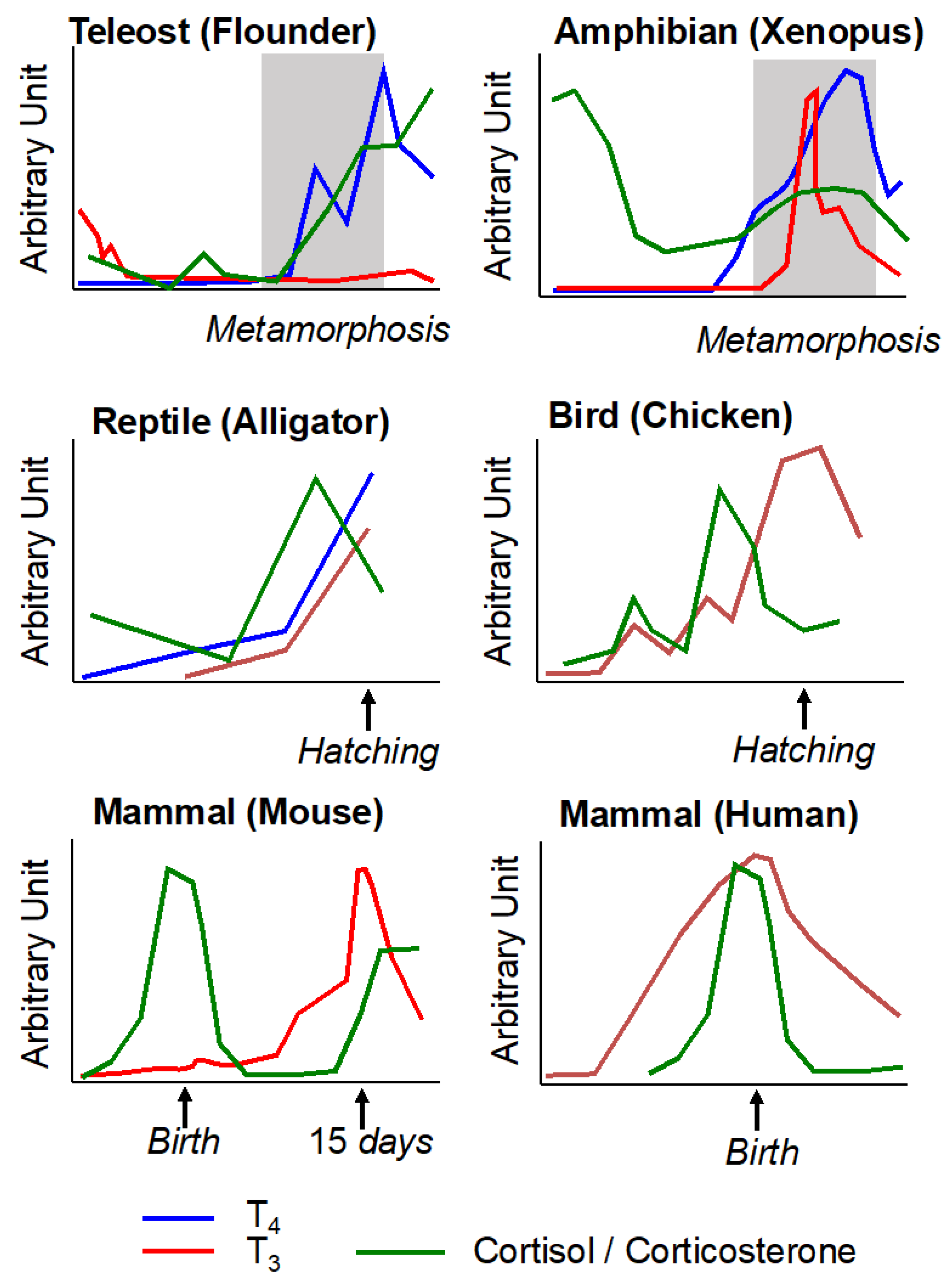
4. Conservation during Vertebrate Evolution of Thyroid Hormone and Corticosteroid Action in Post-Embryonic Development

Metamorphosis is an emblematic type of post-embryonic developmental process. This life stage transition from one ecological niche to a different ecological niche is also present in various metazoan phylogenetic groups. Here, we will present the contributions of TH and CSs in metamorphosis and developmental transitions in other vertebrates, including larval and secondary metamorphosis in teleost fishes, egg hatching in sauropsids, and birth in mammals. Two neuroendocrine axes, the hypothalamic–pituitary–thyroid and the hypothalamic–pituitary–adrenal (mammals and sauropsids)/interrenal (amphibians and teleost fishes) axes, are central players in the regulation of these life transitions. To highlight the evolutionary conservation of these two axes, all vertebrates have both axes (Figure 1) with a peak of T3 and a peak of CSs that co-occur during post-embryonic development (Figure 5). In addition, similar T3 and CS concentration profiles are observed in certain birds and reptiles at hatching and during molting, as well as in certain teleost fishes at hatching and during larval metamorphosis and smoltification [114].

4.1. Metamorphosis in Teleost Fishes

T3 has an important role in fish egg hatching [119], but it will not be considered in this review. Several studies also report developmental changes controlled by T3 during larval to juvenile transition in various teleosts such as grouper Epinephelus coioides [120], seabream [121], gobiid Sicyopterus lagocephalus [122] or clownfish Amphiprion percula [123]. Our review will focus on the two types of metamorphosis involving morphological and physiological modifications leading to niche/habitat changes. One occurs at the larval to juvenile transition in elopomorphs and pleuronectiforms, and the second one occurs in juveniles of some diadromic migratory teleosts, i.e., smoltification.
Metamorphosis of the flatfish represents the classical larval metamorphosis in teleosts, with spectacular morphological changes such as the migration of one eye to the opposite side of the head [124]. These changes are also accompanied by physiological (such as development of gastrointestinal tract to adapt to the novel food resources in a new habitat) and behavioral (pelagic to benthic life and locomotion) changes. Larval tissue T4 and T3 concentrations peak at metamorphic climax in the Japanese flounder Paralichthys olivaceus [125], in spotted halibut, Verasper variegatus [126], Atlantic halibut Hippoglossus hippoglossus [127], and summer flounder, Paralichthys dentatus [128]. T3 is involved in many metamorphic changes, including shortening of fin rays, eye migration, and asymmetry [124]. Treatments of flatfish with T3 or anti-thyroid drugs lead respectively to acceleration or delay of these classical changes [129,130,131,132]. The developmental profiles of deiodinases, which convert T4 to T3 or inactivate T3, coincide with the rise of TH levels. During sole metamorphosis, DIO2 activity, which converts T4 to T3, increases during metamorphosis, while DIO3 activity, which inactivates T3, declines at the mid-late metamorphic period [133]. As during amphibian metamorphosis, the expression of TR increases at metamorphic climax [134,135].
As seen in amphibians, CS concentration also reaches a peak at climax in Japanese flounder [115]. CS treatment of larval fishes does not trigger the changes observed during metamorphosis, as in eye migration and settling (benthic) behavior, but it does induce shortening of the second fin ray [130]. Furthermore, in spotted halibut, CS treatment increases the development of adult-type pigment cells [136]. With respect to interactions between T3 and CS signaling, CS treatment leads to enhanced hepatic and renal DIO2 activities in juvenile Senegalese sole [137], but this CS effect on extrathyroidal T3 production has not yet been confirmed during larval metamorphosis. In the Japanese flounder, a permissive effect of CSs on T3 action is observed in vitro but not in vivo during metamorphosis [130]. Thus, future studies are required to investigate the potential CS/T3 interactions in teleost metamorphoses as observed in amphibians.
A secondary metamorphosis in teleosts is exemplified by smoltification in salmonids, which allows for the transition from freshwater to the ocean. Smoltification involves morphological changes, such as body silvering and fin darkening, as well as behavioral changes, with swimming activity in open space and migration, and finally physiological changes related to adaptation to seawater and imprinting [138,139]. Many parallel features exist between flatfish metamorphosis and salmonid smoltification [140], the first one being the control by T3. Here again, plasma T4 and/or T3 levels rise at the time of smoltification [140]. Manipulation of T3 levels supports a major role of T3 in adaptation to a new environment, such as change in visual pigments, olfaction, metabolism, swimming behavior, and migration [139,141]. A peak of expression of tshβ1b was recently measured at the time of initiation of the downstream migration in Atlantic salmon [142]. In all the tissues involved in smoltification-related changes, expression of TR has been detected [143,144,145,146] but does not change between the different stages [146]. The HPI axis is also involved during smoltification with a peak in plasma cortisol levels [147,148,149,150]. Manipulation of the HPI axis can affect pigmentation [151] and osmoregulation [152]. The involvement of GR was supported by data showing a stimulatory effect of cortisol in gill tissue blocked by the GR antagonist, mifepristone [153] and an increase in GR concentration and gr expression [154,155,156,157]. Few studies report interactions between T3 and CS signaling during smoltification. T3 can increase CRH neurogenesis [158], but CS treatment can lower plasma T3 and reduce the increases of TR mRNA and protein expression [159,160], suggesting some antagonistic/negative effects of CSs on T3 action.

4.2. Egg Hatching in Sauropsids

Hatching in birds and reptiles is characterized by a transition from an aqueous environment to a terrestrial environment. For birds, it is important to differentiate between precocial and altricial species [161,162]. In precocial species where hatchlings are immediately mobile and independent after hatching, plasma T3 concentrations of the embryos increase during the peri-hatch period [161]. In contrast, hatchling altricial birds need to be fed and thermoregulated by their parents in the nest and have low circulating concentrations of T3 that increase only after hatching [163,164]. Manipulation of egg T3 levels influences avian hatching time [165,166,167]. T3 availability is controlled by deiodinase activity in chicken with high levels of DIO3 during embryogenesis [117,168,169,170] and DIO1, another enzyme that can convert T4 to T3, whereas DIO2 levels increase to a peak around hatching [117,169,170]. T3 effects are driven by TR expressed in many embryonic tissues as observed in chicken [171,172].
Around the time of hatching, an increase in avian embryonic CSs is observed in plasma [173,174,175,176]. Injection of CORT in the turkey or chicken yolk sac can increase the incubation period when injected at early stages of embryonic development, but injection of CORT in the same species can shorten incubation period when injected at late stages [175,177,178]. CSs act on chicken lung prior to hatching to stimulate surfactant synthesis [179]. The CS effects in chicken are mediated by GR expressed during the pre-hatching period [176]. Interactions between T3 and CS signaling have also been observed in birds. Again, in chicken embryos, hypothyroidism leads to medullar hypertrophy in the adrenal gland [180], and CS or ACTH injection leads to increased plasma T3 concentrations via increased DIO2 activity [181] and decreased DIO3 activity [182].
In reptiles, T3 signaling is associated with hatching, as exemplified by an increase in T3 levels in saltwater crocodile Crocodylus porosus, [116] and with thyroid exhibiting the features of a fully active gland in the grass snake Natrix natrix [183]. In turtle, injection of an antithyroid drug delays hatching time [184], and treatment with T3 shortens incubation duration [185]. T3 treatment also increases DIO1 and DIO2 activity in crocodile [116] as well as the secretion of pulmonary surfactant in crocodile [186] and turtle [187]. CSs also stimulate pulmonary surfactant in turtle [187], crocodile [186] and lizard [188]. An increase in CSs is observed around hatching in alligator [189], lizard [190], and turtle [191], but a decrease is observed in crocodile [116]. In lizard, CS injection accelerates egg hatching [192]. In alligator, GR expression during embryonic development suggests a potential role for CSs in tissue maturation before hatching [193]. Finally, note that both T3 and CSs are involved in the regulation of surfactant production in reptiles, similar to that observed in other amniotes [194].

4.3. Birth in Mammals

During mammalian development, T3 availability is tightly controlled by developmental changes in endogenous synthesis in the thyroid and tissue deiodinase activity, leading to a rise in plasma T3 concentration near term [195] or shortly after delivery [118,196,197]. T3 has a major regulatory role on fetal growth and development [195], including maturation of the central nervous system [198], maturation of the respiratory system including synthesis of the components of surfactant [199,200,201], maturation of the skeleton [202], and maturation of the gastro-intestinal tract [203]. These post-embryonic developmental maturations are crucial to cope with the change of environment (from aquatic to terrestrial) and of nutrition (from parenteral to enteral). An increase in plasma fetal CSs is detected near parturition in many mammals [196,204,205,206]. Two striking examples of exogenous CS effects are: first, the injection of ACTH or CSs into the ovine fetus leads to premature delivery [207,208], and second, CSs are routinely injected to favor lung maturation in human preterm birth [209]. As observed for T3 signaling, many roles of CSs on organ maturation, including brain, lung, digestive tract, and skeleton, have been demonstrated and are the subjects of recent reviews [210]. Before birth, T3 and CSs synergize for the maturation of these organs. This synergy is partly the result of CS-induced increases in circulating T3 and local T3 bioavailability [211,212].

5. Conclusions

In all of the developmental transitions described here, a common involvement of HPT and HPA/HPI axes is observed, including the cross-talk between these two (Figure 1). The hormone-dependent developmental processes are diverse with morphological, behavioral, and physiological changes essential to adapt to a new environment and/or new way of life. The conservation along vertebrate evolution of the mechanisms underlying these major biological processes is striking from fish and amphibian metamorphoses to egg hatching in sauropsids and birth in mammals. The synchronized increase in T3 and CS production and release, coupled with the local control of hormone availability and expression of receptors, allow tissues to fine-tune the two hormone responses. The multiple cross-talks between the HPT and HPA/HPI axes represent critical mechanisms by which vertebrates modulate their perinatal/post-embryonic development as well as their responses to a changing environment. Of all these interactions, one differs between mammals and non-mammalian vertebrates. In amphibian, sauropsids, and teleost fishes, the CRH appears to be a coordinator of activation of both HPT and HPA/HPI axes at the time of developmental transitions involving stimulation of TSH production and release. It is not the case in mammals, where TRH and CRH respectively control the HPT and HPA axes early during development. Mechanisms of integration of environmental and internal cues, and regulation/operation of these neuroendocrine axes represent key questions in an “eco–evo–devo” perspective of metamorphosis and more generally post-embryonic developmental processes. The roles played by developmental transitions in the innovation, adaptation, and plasticity of life cycles throughout vertebrates are then essential, especially within the current context of global anthropogenic disturbance. The impact of environmental factors, such as global warming, habitat destruction, and endocrine disruptors on the HPT and HPA/HPI axes, and regulation of developmental transitions must be seen as a threat for biodiversity. Humans are not spared. In preterm birth, which is the leading cause of perinatal morbidity and mortality in developed countries, the affected infants often experience HPT and HPA dysfunctions [213,214,215]. Antenatal CS administration reduces the complications after preterm birth, while T3 supplementation has not been shown to be beneficial. However, the benefit of a therapy is still a controversy [209]. Thus, key questions regarding HPT and HPA/HPI functions during post-embryonic developmental transition still remain to be answered to protect biodiversity and human health.

Author Contributions

Conceptualization, Y.-B.S., D.R.B. and L.M.S.; writing—original draft preparation, B.P., Z.R.S., Y.-B.S., D.R.B. and L.M.S.; writing—review and editing, Y.-B.S., D.R.B. and L.M.S.; supervision, L.M.S.; project administration, L.M.S.; funding acquisition, Y.-B.S., D.R.B. and L.M.S. All authors have read and agreed to the published version of the manuscript.

Funding

L.M.S.’s work was supported by “ERGO”, a European Union’s Horizon 2020 research and innovation program, under grant agreement n° 825753. L.M.S. is supported by the “Centre National de la Recherche Scientifique” and the “Muséum National d’Histoire Naturelle”. Grant NSF IOS 2035732 supported the work by D.R.B. and Y.B.S. was supported by the Intramural Research Program of NICHD, National Institutes of Health, USA.

Institutional Review Board Statement

Not applicable.

Informed Consent Statement

Not applicable.

Conflicts of Interest

The authors declare that the research was conducted in the absence of any commercial or financial relationships that could be construed as a potential conflict of interest.

References

  1. Rousseau, K.; Dufour, S.; Sachs, L.M. Interdependence of Thyroid and Corticosteroid Signaling in Vertebrate Developmental Transitions. Front. Ecol. Evol. 2021, 9, 735487. [Google Scholar] [CrossRef]
  2. Shi, Y.-B. Amphibian Metamorphosis: From Morphology to Molecular Biology; John Wiley & Sons: New York, NY, USA, 1999. [Google Scholar]
  3. Denver, R.J. Stress hormones mediate developmental plasticity in vertebrates with complex life cycles. Neurobiol. Stress 2021, 14, 100301. [Google Scholar] [CrossRef] [PubMed]
  4. Glennemeier, K.A.; Denver, R.J. Small changes in whole-body corticosterone content affect larval Rana pipiens fitness components. Gen. Comp. Endocrinol. 2002, 127, 16–25. [Google Scholar] [CrossRef]
  5. Denver, R.J. Acceleration of anuran amphibian metamorphosis by corticotropin-releasing hormone-like peptides. Gen. Comp. Endocrinol. 1993, 91, 38–51. [Google Scholar] [CrossRef] [PubMed]
  6. De Groef, B.; Van Der Geyten, S.; Darras, V.M.; Kühn, E.R. Role of corticotropin-releasing hormone as a thyrotropin-releasing factor in non-mammalian vertebrates. Gen. Comp. Endocrinol. 2006, 146, 62–68. [Google Scholar] [CrossRef] [PubMed]
  7. Miranda, L.A.; Affanni, J.M.; Paz, D.A. Corticotropin-releasing factor accelerates metamorphosis in Bufo arenarum: Effect on pituitary ACTH and TSH cells. J. Exp. Zool. 2000, 286, 473–480. [Google Scholar] [CrossRef]
  8. Denver, R.J. Environmental stress as a developmental cue: Corticotropin-releasing hormone is a proximate mediator of adaptive phenotypic plasticity in amphibian metamorphosis. Horm. Behav. 1997, 31, 169–179. [Google Scholar] [CrossRef]
  9. Galton, V.A. Mechanisms underlying the acceleration of thyroid hormone-induced tadpole metamorphosis by corticosterone. Endocrinology 1990, 127, 2997–3002. [Google Scholar] [CrossRef]
  10. Bonett, R.M.; Hoopfer, E.D.; Denver, R.J. Molecular mechanisms of corticosteroid synergy with thyroid hormone during tadpole metamorphosis. Gen. Comp. Endocrinol. 2010, 168, 209–219. [Google Scholar] [CrossRef] [Green Version]
  11. Suzuki, M.R.; Kikuyama, S. Corticoids augment nuclear binding capacity for triiodothyronine in bullfrog tadpole tail fins. Gen. Comp. Endocrinol. 1983, 52, 272–278. [Google Scholar] [CrossRef]
  12. Krain, L.P.; Denver, R.J. Developmental expression and hormonal regulation of glucocorticoid and thyroid hormone receptors during metamorphosis in Xenopus laevis. J. Endocrinol. 2004, 181, 91–104. [Google Scholar] [CrossRef] [PubMed] [Green Version]
  13. Kulkarni, S.S.; Buchholz, D.R. Beyond synergy: Corticosterone andthyroid hormone have numerous interaction effects on gene regulation in Xenopus tropicalis tadpoles. Endocrinology 2012, 153, 5309–5324. [Google Scholar] [CrossRef] [PubMed] [Green Version]
  14. Kulkarni, S.S.; Buchholz, D.R. Corticosteroid signaling in frog metamorphosis. Gen. Comp. Endocrinol. 2014, 203, 225–231. [Google Scholar] [CrossRef] [PubMed]
  15. Buisine, N.; Grimaldi, A.; Jonchere, V.; Rigolet, M.; Blugeon, C.; Hamroun, J.; Sachs, L.M. Transcriptome and Methylome Analysis Reveal Complex Cross-Talks between Thyroid Hormone and Glucocrticoid Signaling at Xenopus Metamorphosis. Cells 2021, 10, 2375. [Google Scholar] [CrossRef] [PubMed]
  16. Bagamasbad, P.D.; Bonett, R.M.; Sachs, L.; Buisine, N.; Raj, S.; Knoedler, J.; Kyono, Y.; Ruan, Y.; Ruan, X.; Denver, R.J. Deciphering the regulatory logic of an ancient, ultraconserved nuclear receptor enhancer module. Mol. Endocrinol. 2015, 29, 856–872. [Google Scholar] [CrossRef] [Green Version]
  17. Gudernatsch, J.F. Feeding experiments on tadpoles. I. The influence of specific organs given as food on growth and differentiation: A contribution to the knowledge of organs with internal secretion. Arch. Entwickl. Org. 1912, 35, 457–483. [Google Scholar] [CrossRef] [Green Version]
  18. Dodd, M.H.I.; Dodd, J.M. The biology of metamorphosis. In Physiology of the Amphibia; Lofts, B., Ed.; Academic Press: New York, NY, USA, 1976; pp. 467–599. [Google Scholar]
  19. Gilbert, L.I.; Tata, J.R.; Atkinson, B.G. Metamorphosis: Post-Embryonic Reprogramming of Gene Expression in Amphibian and Insect Cells; Academic Press: New York, NY, USA, 1996. [Google Scholar]
  20. Tata, J.R. Gene expression during metamorphosis: An ideal model for post-embryonic development. Bioessays 1993, 15, 239–248. [Google Scholar] [CrossRef]
  21. Leloup, J.; Buscaglia, M. La triiodothyronine: Hormone de la métamorphose des amphibiens. CR Acad. Sci. 1977, 284, 2261–2263. [Google Scholar]
  22. Tata, J.R. Early metamorphic competence of Xenopus larvae. Dev. Biol. 1968, 18, 415–440. [Google Scholar] [CrossRef]
  23. Tata, J.R. Requirement for RNA and protein synthesis for induced regression of the tadpole tail in organ culture. Dev. Biol. 1966, 13, 77–94. [Google Scholar] [CrossRef]
  24. Ishizuya-Oka, A.; Shimozawa, A. Induction of metamorphosis by thyroid hormone in anuran small intestine cultured organotypically in vitro. In Vitro Cell. Dev. Biol. 1991, 27, 853–857. [Google Scholar] [CrossRef] [PubMed]
  25. Tata, J.R.; Kawahara, A.; Baker, B.S. Prolactin inhibits both thyroid hormone-induced morphogenesis and cell death in cultured amphibian larval tissues. Dev. Biol. 1991, 146, 72–80. [Google Scholar] [CrossRef]
  26. Flamant, F.; Cheng, S.-Y.; Hollenberg, A.N.; Moeller, L.C.; Samarut, J.; Wondisford, F.E.; Yen, P.M.; Refetoff, S. Thyroid Hormone Signaling Pathways: Time for a More Precise Nomeclature. Endocrinology 2017, 158, 2052–2057. [Google Scholar] [CrossRef] [PubMed]
  27. Shi, Y.-B.; Wong, J.; Puzianowska-Kuznicka, M.; Stolow, M.A. Tadpole competence and tissue-specific temporal regulation of amphibian metamorphosis: Roles of thyroid hormone and its receptors. Bioessays 1996, 18, 391–399. [Google Scholar] [CrossRef] [PubMed]
  28. Laudet, V.; Gronemeyer, H. The Nuclear Receptor FactsBook; Academic Press: San Diego, CA, USA, 2002. [Google Scholar]
  29. Forrest, D. The erbA/thyroid hormone receptor genes in development of the central nervous system. Semin. Cancer Biol. 1994, 5, 167–176. [Google Scholar]
  30. Lazar, M.A. Thyroid hormone receptors: Multiple forms, multiple possibilities. Endocr. Rev. 1993, 14, 184–193. [Google Scholar] [CrossRef]
  31. Yen, P.M. Physiological and molecular basis of thyroid hormone action. Physiol. Rev. 2001, 81, 1097–1142. [Google Scholar] [CrossRef] [Green Version]
  32. Davis, P.J.; Davis, F.B. Nongenomic actions of thyroid hormone. Thyroid 1996, 6, 497–504. [Google Scholar] [CrossRef]
  33. Davis, P.J.; Davis, F.B. Nongenomic actions of thyroid hormone on the heart. Thyroid 2002, 12, 459–466. [Google Scholar] [CrossRef]
  34. Parkison, C.; Ashizawa, K.; McPhie, P.; Lin, K.H.; Cheng, S.Y. The monomer of pyruvate kinase, subtype M1, is both a kinase and a cytosolic thyroid hormone binding protein. Biochem. Biophys. Res. Commun. 1991, 179, 668–674. [Google Scholar] [CrossRef]
  35. Shi, Y.-B.; Yaoita, Y.; Brown, D.D. Genomic organization and alternative promoter usage of the two thyroid hormone receptor ß genes in Xenopus laevis. J. Biol. Chem. 1992, 267, 733–788. [Google Scholar] [CrossRef]
  36. Yaoita, Y.; Shi, Y.B.; Brown, D.D. Xenopus laevis alpha and beta thyroid hormone receptors. Proc. Natl. Acad. Sci. USA 1990, 87, 7090–7094. [Google Scholar] [CrossRef] [PubMed] [Green Version]
  37. Wang, X.; Matsuda, H.; Shi, Y.-B. Developmental regulation and function of thyroid hormone receptors and 9-cis retinoic acid receptors during Xenopus tropicalis metamorphosis. Endocrinology 2008, 149, 5610–5618. [Google Scholar] [CrossRef] [PubMed] [Green Version]
  38. Mangelsdorf, D.J.; Thummel, C.; Beato, M.; Herrlich, P.; Schutz, G.; Umesono, K.; Blumberg, B.; Kastner, P.; Mark, M.; Chambon, P. The nuclear receptor superfamily: The second decade. Cell 1995, 83, 835–839. [Google Scholar] [CrossRef] [Green Version]
  39. Tsai, M.J.; O’Malley, B.W. Molecular mechanisms of action of steroid/thyroid receptor superfamily members. Ann. Rev. Biochem. 1994, 63, 451–486. [Google Scholar] [CrossRef] [PubMed]
  40. O’Malley, B.W.; Malovannaya, A.; Qin, J. Minireview: Nuclear receptor and coregulator proteomics—2012 and beyond. Mol. Endocrinol. 2012, 26, 1646–1650. [Google Scholar] [CrossRef] [Green Version]
  41. Bulynko, Y.A.; O’Malley, B.W. Nuclear receptor coactivators: Structural and functional biochemistry. Biochemistry 2011, 50, 313–328. [Google Scholar] [CrossRef] [Green Version]
  42. McKenna, N.J.; Cooney, A.J.; DeMayo, F.J.; Downes, M.; Glass, C.K.; Lanz, R.B.; Lazar, M.A.; Mangelsdorf, D.J.; Moore, D.D.; Qin, J.; et al. Minireview: Evolution of NURSA, the Nuclear Receptor Signaling Atlas. Mol. Endocrinol. 2009, 23, 740–746. [Google Scholar] [CrossRef]
  43. Grimaldi, A.; Buisine, N.; Miller, T.; Shi, Y.B.; Sachs, L.M. Mechanisms of thyroid hormone receptor action during development: Lessons from amphibian studies. Biochim. Biophys. Acta 2013, 1830, 3882–3892. [Google Scholar] [CrossRef]
  44. Shi, Y.B.; Matsuura, K.; Fujimoto, K.; Wen, L.; Fu, L. Thyroid hormone receptor actions on transcription in amphibia: The roles of histone modification and chromatin disruption. Cell Biosci. 2012, 2, 42. [Google Scholar] [CrossRef] [Green Version]
  45. Perissi, V.; Jepsen, K.; Glass, C.K.; Rosenfeld, M.G. Deconstructing repression: Evolving models of co-repressor action. Nat. Rev. Genet. 2010, 11, 109–123. [Google Scholar] [CrossRef] [PubMed]
  46. Yaoita, Y.; Brown, D.D. A correlation of thyroid hormone receptor gene expression with amphibian metamorphosis. Genes Dev. 1990, 4, 1917–1924. [Google Scholar] [CrossRef] [PubMed] [Green Version]
  47. Kanamori, A.; Brown, D.D. The regulation of thyroid hormone receptor beta genes by thyroid hormone in Xenopus laevis. J. Biol. Chem. 1992, 267, 739–745. [Google Scholar] [CrossRef]
  48. Wang, Z.; Brown, D.D. Thyroid hormone-induced gene expression program for amphibian tail resorption. J. Biol. Chem. 1993, 268, 16270–16278. [Google Scholar] [CrossRef]
  49. Wong, J.; Shi, Y.-B. Coordinated regulation of and transcriptional activation by Xenopus thyroid hormone and retinoid X receptors. J. Biol. Chem. 1995, 270, 18479–18483. [Google Scholar] [CrossRef] [Green Version]
  50. Sachs, L.M.; Damjanovski, S.; Jones, P.L.; Li, Q.; Amano, T.; Ueda, S.; Shi, Y.B.; Ishizuya-Oka, A. Dual functions of thyroid hormone receptors during Xenopus development. Comp. Biochem. Physiol. B Biochem. Mol. Biol. 2000, 126, 199–211. [Google Scholar] [CrossRef]
  51. Nieuwkoop, P.D.; Faber, J. Normal Table of Xenopus Laevis (Daudin); Garland Publishing Inc.: New York, NY, USA, 1994. [Google Scholar]
  52. Puzianowska-Kuznicka, M.; Damjanovski, S.; Shi, Y.-B. Both thyroid Hormone and 9-cis Retinoic Acid receptors are Required to Efficiently mediate the Effects of Thyroid Hormone on Embryonic Development and Specific Gene Regulation in Xenopus laevis. Mol. Cell. Biol. 1997, 17, 4738–4749. [Google Scholar] [CrossRef] [Green Version]
  53. Nakajima, K.; Yaoita, Y. Dual mechanisms governing muscle cell death in tadpole tail during amphibian metamorphosis. Dev. Dyn. 2003, 227, 246–255. [Google Scholar] [CrossRef]
  54. Schreiber, A.M.; Brown, D.D. Tadpole skin dies autonomously in response to thyroid hormone at metamorphosis. Proc. Natl. Acad. Sci. USA 2003, 100, 1769–1774. [Google Scholar] [CrossRef] [Green Version]
  55. Das, B.; Schreiber, A.M.; Huang, H.; Brown, D.D. Multiple thyroid hormone-induced muscle growth and death programs during metamorphosis in Xenopus laevis. Proc. Natl. Acad. Sci. USA 2002, 99, 12230–12235. [Google Scholar] [CrossRef] [Green Version]
  56. Schreiber, A.M.; Das, B.; Huang, H.; Marsh-Armstrong, N.; Brown, D.D. Diverse developmental programs of Xenopus laevis metamorphosis are inhibited by a dominant negative thyroid hormone receptor. Proc. Natl. Acad. Sci. USA 2001, 98, 10739–10744. [Google Scholar] [CrossRef] [PubMed] [Green Version]
  57. Buchholz, D.R.; Hsia, V.S.-C.; Fu, L.; Shi, Y.-B. A dominant negative thyroid hormone receptor blocks amphibian metamorphosis by retaining corepressors at target genes. Mol. Cell. Biol. 2003, 23, 6750–6758. [Google Scholar] [CrossRef] [PubMed] [Green Version]
  58. Buchholz, D.R.; Tomita, A.; Fu, L.; Paul, B.D.; Shi, Y.-B. Transgenic analysis reveals that thyroid hormone receptor is sufficient to mediate the thyroid hormone signal in frog metamorphosis. Mol. Cell. Biol. 2004, 24, 9026–9037. [Google Scholar] [CrossRef] [PubMed] [Green Version]
  59. Buchholz, D.R.; Paul, B.D.; Fu, L.; Shi, Y.B. Molecular and developmental analyses of thyroid hormone receptor function in Xenopus laevis, the African clawed frog. Gen. Comp. Endocrinol. 2006, 145, 1–19. [Google Scholar] [CrossRef]
  60. Choi, J.; Ishizuya-Oka, A.; Buchholz, D.R. Growth, development, and intestinal remodeling occurs in the absence of thyroid hormone receptor alpha in tadpoles of Xenopus tropicalis. Endocrinology 2017, 158, 1623–1633. [Google Scholar] [CrossRef]
  61. Choi, J.; Suzuki, K.I.; Sakuma, T.; Shewade, L.; Yamamoto, T.; Buchholz, D.R. Unliganded thyroid hormone receptor alpha regulates developmental timing via gene repression as revealed by gene disruption in Xenopus tropicalis. Endocrinology 2015, 156, 735–744. [Google Scholar] [CrossRef] [Green Version]
  62. Sakane, Y.; Iida, M.; Hasebe, T.; Fujii, S.; Buchholz, D.R.; Ishizuya-Oka, A.; Yamamoto, T.; Suzuki, K.T. Functional analysis of thyroid hormone receptor beta in Xenopus tropicalis founders using CRISPR-Cas. Biol. Open 2018, 7, bio030338. [Google Scholar] [CrossRef] [Green Version]
  63. Nakajima, K.; Tazawa, I.; Yaoita, Y. Thyroid Hormone Receptor alpha- and beta-Knockout Xenopus tropicalis Tadpoles Reveal Subtype-Specific Roles During Development. Endocrinology 2018, 159, 733–743. [Google Scholar] [CrossRef]
  64. Wen, L.; Shi, Y.B. Unliganded thyroid hormone receptor alpha controls developmental timing in Xenopus tropicalis. Endocrinology 2015, 156, 721–734. [Google Scholar] [CrossRef] [Green Version]
  65. Wen, L.; Shibata, Y.; Su, D.; Fu, L.; Luu, N.; Shi, Y.-B. Thyroid hormone receptor α controls developmental timing and regulates the rate and coordination of tissue specific metamorphosis in Xenopus tropicalis. Endocrinology 2017, 158, 1985–1998. [Google Scholar] [CrossRef]
  66. Wen, L.; Shi, Y.B. Regulation of growth rate and developmental timing by Xenopus thyroid hormone receptor alpha. Dev. Growth. Differ. 2016, 58, 106–115. [Google Scholar] [CrossRef] [PubMed]
  67. Sachs, L.M. Unliganded thyroid hormone receptor function: Amphibian metamorphosis got TALENs. Endocrinology 2015, 156, 409–410. [Google Scholar] [CrossRef] [PubMed]
  68. Yen, P.M. Unliganded TRs regulate growth and developmental timing during early embryogenesis: Evidence for a dual function mechanism of TR action. Cell Biosci. 2015, 5, 8. [Google Scholar] [CrossRef] [PubMed] [Green Version]
  69. Nakajima, K.; Tazawa, I.; Shi, Y.B. A unique role of thyroid hormone receptor beta in regulating notochord resorption during Xenopus metamorphosis. Gen. Comp. Endocrinol. 2019, 277, 66–72. [Google Scholar] [CrossRef]
  70. Shibata, Y.; Tanizaki, Y.; Shi, Y.B. Thyroid hormone receptor beta is critical for intestinal remodeling during Xenopus tropicalis metamorphosis. Cell Biosci. 2020, 10, 46. [Google Scholar] [CrossRef] [Green Version]
  71. Shibata, Y.; Wen, L.; Okada, M.; Shi, Y.B. Organ-Specific Requirements for Thyroid Hormone Receptor Ensure Temporal Coordination of Tissue-Specific Transformations and Completion of Xenopus Metamorphosis. Thyroid 2020, 30, 300–313. [Google Scholar] [CrossRef]
  72. Buchholz, D.R.; Shi, Y.B. Dual function model revised by thyroid hormone receptor alpha knockout frogs. Gen. Comp. Endocrinol. 2018, 265, 214–218. [Google Scholar] [CrossRef]
  73. Nakajima, K.; Tanizaki, Y.; Luu, N.; Zhang, H.; Shi, Y.B. Comprehensive RNA-Seq analysis of notochord-enriched genes induced during Xenopus tropicalis tail resorption. Gen. Comp. Endocrinol. 2020, 287, 113349. [Google Scholar] [CrossRef]
  74. Shi, Y.B. Life without thyroid hormone receptor. Endocrinology 2021, 162, bqab028. [Google Scholar] [CrossRef]
  75. Sachs, L.M.; Shi, Y.-B. Targeted chromatin binding and histone acetylation in vivo by thyroid hormone receptor during amphibian development. Proc. Natl. Acad. Sci. USA 2000, 97, 13138–13143. [Google Scholar] [CrossRef] [Green Version]
  76. Tomita, A.; Buchholz, D.R.; Shi, Y.-B. Recruitment of N-CoR/SMRT-TBLR1 corepressor complex by unliganded thyroid hormone receptor for gene repression during frog development. Mol. Cell. Biol. 2004, 24, 3337–3346. [Google Scholar] [CrossRef] [PubMed] [Green Version]
  77. Sachs, L.M.; Jones, P.L.; Havis, E.; Rouse, N.; Demeneix, B.A.; Shi, Y.-B. N-CoR recruitment by unliganded thyroid hormone receptor in gene repression during Xenopus laevis development. Mol. Cell. Biol. 2002, 22, 8527–8538. [Google Scholar] [CrossRef] [PubMed] [Green Version]
  78. Sato, Y.; Buchholz, D.R.; Paul, B.D.; Shi, Y.-B. A role of unliganded thyroid hormone receptor in postembryonic development in Xenopus laevis. Mech. Dev. 2007, 124, 476–488. [Google Scholar] [CrossRef] [PubMed] [Green Version]
  79. Matsuda, H.; Paul, B.D.; Choi, C.Y.; Hasebe, T.; Shi, Y.-B. Novel functions of protein arginine methyltransferase 1 in thyroid hormone receptor-mediated transcription and in the regulation of metamorphic rate in Xenopus laevis. Mol. Cell. Biol. 2009, 29, 745–757. [Google Scholar] [CrossRef] [PubMed] [Green Version]
  80. Paul, B.D.; Buchholz, D.R.; Fu, L.; Shi, Y.-B. Tissue- and gene-specific recruitment of steroid receptor coactivator-3 by thyroid hormone receptor during development. J. Biol. Chem. 2005, 280, 27165–27172. [Google Scholar] [CrossRef] [Green Version]
  81. Paul, B.D.; Fu, L.; Buchholz, D.R.; Shi, Y.-B. Coactivator recruitment is essential for liganded thyroid hormone receptor to initiate amphibian metamorphosis. Mol. Cell. Biol. 2005, 25, 5712–5724. [Google Scholar] [CrossRef] [Green Version]
  82. Paul, B.D.; Buchholz, D.R.; Fu, L.; Shi, Y.-B. SRC-p300 coactivator complex is required for thyroid hormone induced amphibian metamorphosis. J. Biol. Chem. 2007, 282, 7472–7481. [Google Scholar] [CrossRef] [Green Version]
  83. Havis, E.; Sachs, L.M.; Demeneix, B.A. Metamorphic T3-response genes have specific co-regulator requirements. EMBO Rep. 2003, 4, 883–888. [Google Scholar] [CrossRef] [Green Version]
  84. Tanizaki, Y.; Bao, L.; Shi, B.; Shi, Y.-B. A role of endogenous histone acetyltransferase steroid hormone receptor coactivator (SRC) 3 in thyroid hormone signaling during Xenopus intestinal metamorphosis. Thyroid 2021, 31, 692–702. [Google Scholar] [CrossRef]
  85. Shibata, Y.; Tanizaki, Y.; Zhang, H.; Lee, H.; Dasso, M.; Shi, Y.-B. Thyroid hormone receptor is essential for larval epithelial apoptosis and adult epithelial stem cell development but not adult intestinal morphogenesis during Xenopus tropicalis metamorphosis. Cells 2021, 10, 536. [Google Scholar] [CrossRef]
  86. Bock, K.A. Die einwirkung von nebennierenrinden-extract auf den ablauf der thyroxin-metamorphose bei froschlarven und beim axolotl. Klin. Woschschr. 1938, 17, 1311–1314. [Google Scholar] [CrossRef]
  87. Frieden, E.; Naile, B. Biochemistry of amphibian metamorphosi: 1. Enhancement of induced metamorphosis by glucocorticoids. Science 1955, 121, 37–39. [Google Scholar] [CrossRef] [PubMed]
  88. Gasche, P. Effect of Desoxycorticosterone acetate on larvae of Xenopus laevis in their various stages of metamorphosis. Helv. Physiol. Pharmacol. 1945, Acta 3, C10. [Google Scholar]
  89. Kobayashi, H. Effects of desoxycorticosterone acetate on metamorphosis induced by thyroxine in anuran tadpoles. Endocrinology 1958, 62, 371–377. [Google Scholar] [CrossRef] [PubMed]
  90. Hu, F.; Knoedler, J.R.; Denver, R.J. A mechanism to enhance cellular responsivity to hormone action: Krüppel-like factor 9 promotes thyroid hormone receptor-β autoinduction during postembryonic brain development. Endocrinology 2016, 157, 1683–1693. [Google Scholar] [CrossRef] [Green Version]
  91. Denver, R.J. Endocrinology of Complex Life Cycles: Amphibians. In Anonymous Hormones, Brain, and Behavior, 3rd ed.; Elsevier: Amsterdam, The Netherlands, 2017; Volume 2, Chapter 2.09; pp. 145–168. [Google Scholar]
  92. Hanke, W. The adrenal cortex in amphibia. In General, Comparative, and Clinical Endocrinology of the Adrenal Cortex; Jones, I.C., Henderson, I.W., Eds.; Academic Press, Inc.: London, UK, 1978; Volume 2, Chapter 5; pp. 419–495. [Google Scholar]
  93. Hayes, T.B.; Wu, T.H. Interdependence of corticosterone and thyroid hormones in toad larvae (Bufo boreas). II. Regulation of corticosterone and thyroid hormones. J. Exp. Zool. 1995, 271, 103–111. [Google Scholar] [CrossRef]
  94. Shewade, L.H.; Schoephoerster, J.A.; Patmann, M.D.; Kulkarni, S.S.; Buchholz, D.R. Corticosterone is Essential for Survival Through Frog Metamorphosis. Endocrinology 2020, 161, bqaa193. [Google Scholar] [CrossRef]
  95. Sterner, Z.R.; Shewade, L.H.; Mertz, K.M.; Sturgeon, S.M.; Buchholz, D.R. Glucocorticoid receptor is required to survive through metamorphosis in the frog Xenopus tropicalis. Gen. Comp. Endocrinol. 2020, 291, 113419. [Google Scholar] [CrossRef]
  96. Jolivet-Jaudet, G.; Leloup-Hatey, J. Interrenal function during Amphibia metamorphosis: In vitro biosynthesis of radioacdtive corticosteroids from (4-14C)-progesterone by interrenal in Xenopus laevis tadpoles. Comp. Biochem. Physiol. Part B 1984, 79, 239–244. [Google Scholar] [CrossRef]
  97. Leist, K.H.; Bergerhoff, K.; Pehlemann, F.W.; Hanke, W. Histopathological investigations of the development of the interrenal organ in the clawed toad (Xenopus laevis Daudin). Z. Zellforsch. Mikrosk. Anat. 1969, 93, 105–125. [Google Scholar] [CrossRef]
  98. Rapola, J. Development of the amphibian adrenal cortex: Morphological and physiological studies on Xenopus laevis Daudin. Ann. Acad. Sci. Fenn. Ser. 1962, A64, 1–81. [Google Scholar]
  99. Uchiyama, M.; Konno, N. Hormonal regulation of ion and water transport in anuran amphibians. Gen. Comp. Endocrinol. 2006, 147, 54–61. [Google Scholar] [CrossRef] [PubMed]
  100. Hanke, W.; Leist, K.H. The effect of ACTH and corticosteroids on carbohydrate metabolism during the metamorphosis of Xenopus laevis. Gen. Comp. Endocrinol. 1971, 16, 137–148. [Google Scholar] [CrossRef]
  101. Maher, J.M.; Werner, E.E.; Denver, R.J. Stress hormones mediate predator- induced phenotypic plasticity in amphibian tadpoles. Proc. Biol. Sci. 2013, 280, 20123075. [Google Scholar] [CrossRef] [Green Version]
  102. Rollins-Smith, L.A.; Barker, K.S.; Davis, A.T. Involvement of glucocorticoids in the reorganization of the amphibian immune system at metamorphosis. Dev. Immunol. 1997, 5, 145–152. [Google Scholar] [CrossRef] [PubMed] [Green Version]
  103. Patel, R.; Williams-Dautovich, J.; Cummins, C.L. Minireview: New Molecular Mediators of Glucocorticoid Receptor Activity in Metabolic Tissues. Mol. Endocrinol. 2014, 28, 999–1011. [Google Scholar] [CrossRef] [Green Version]
  104. Weikum, E.R.; Knuesel, M.T.; Ortlund, E.A.; Yamamoto, K.R. Glucocorticoid receptor control of transcription: Precision and plasticity via allostery. Nat. Rev. Mol. Cell. Bio. 2017, 18, 159–174. [Google Scholar] [CrossRef]
  105. Bridgham, J.T.; Carroll, S.M.; Thornton, J.W. Evolution of Hormone-Receptor Complexity by Molecular Exploitation. Science 2006, 312, 97–101. [Google Scholar] [CrossRef] [Green Version]
  106. James-Zorn, C.; Ponferrada, V.G.; Burns, K.A.; Fortriede, J.D.; Lotay, V.S.; Liu, Y.; Karpinka, B.J.; Karimi, K.; Zorn, A.M.; Vize, P.D. Xenbase: Core features, data acquisition, and data processing. Genesis 2015, 53, 486–497. [Google Scholar] [CrossRef] [Green Version]
  107. Shewade, L.H.; Schneider, K.A.; Brown, A.C.; Buchholz, D.R. In-vivo regulation of Krüppel-like factor 9 by corticosteroids and their receptors across tissues in tadpoles of Xenopus tropicalis. Gen. Comp. Endocrinol. 2017, 248, 79–86. [Google Scholar] [CrossRef]
  108. Gomez-Sanchez, E.; Gomez-Sanchez, C.E. The Multifaceted Mineralocorticoid Receptor. Compr. Physiol. 2014, 4, 965–994. [Google Scholar] [CrossRef] [Green Version]
  109. Kikuyama, S.; Niki, K.; Mayumi, M.; Kawamura, K. Retardation of thyroxine-induced metamorphosis by Amphenone B in toad tadpoles. Endocrinol. Jpn. 1982, 29, 659–662. [Google Scholar] [CrossRef] [PubMed] [Green Version]
  110. Buchholz, D.R.; Hayes, T.B. Variation in thyroid hormone action and tissue content underlies species differences in the timing of metamorphosis in desert frogs. Evol. Dev. 2005, 7, 458–467. [Google Scholar] [CrossRef] [PubMed]
  111. Derby, A. An in vitro quantitative analysis of the response of tadpole tissue to thyroxine. J. Exp. Zool. 1968, 168, 147–156. [Google Scholar] [CrossRef] [PubMed]
  112. Sterner, Z.R.; Buchholz, D.R. Glucocorticoid receptor mediates corticosterone-thyroid hormone synergy essential for metamorphosis in Xenopus tropicalis tadpoles. Gen. Comp. Endocrinol. 2022, 315, 113942. [Google Scholar] [CrossRef]
  113. Kolber, B.J.; Wieczorek, L.; Muglia, L.J. Hypothalamic–pituitary–adrenal axis dysregulation and behavioral analysis of mouse mutants with altered glucocorticoid or mineralocorticoid receptor function. Stress 2008, 11, 321–338. [Google Scholar] [CrossRef] [Green Version]
  114. Wada, H. Glucocorticoids: Mediators of vertebrate ontogenetic transitions. Gen. Comp. Endocrinol. 2008, 156, 441–453. [Google Scholar] [CrossRef]
  115. De Jesus, E.; Hirano, T.; Inui, Y. Changes in cortisol and thyroid hormone concentrations during early development and metamorphosis in the Japanese flounder, Paralichthys olivaceus. Gen. Comp. Endocrinol. 1991, 82, 369–376. [Google Scholar] [CrossRef]
  116. Shepherdley, C.A.; Daniels, C.B.; Orgeig, S.; Richardson, S.J.; Evans, B.K.; Darras, V.M. Glucocorticoids, thyroid hormones, and iodothyronine deiodinases in embryonic saltwater crocodiles. Am. J. Physiol. 2002, 283, 1155–1163. [Google Scholar] [CrossRef] [Green Version]
  117. Reyns, G.E.; Venken, K.; De Escobar, G.M.; Kühn, E.R.; Darras, V.M. Dynamics and regulation of intracellular thyroid hormone concentrations in embryonic chicken liver, kidney, brain, and blood. Gen. Comp. Endocrinol. 2003, 134, 80–87. [Google Scholar] [CrossRef]
  118. Hadj-Sahraoui, N.; Seugnet, I.; Ghorbel, M.; Demeneix, B. Hypothyroidism prolongs miotic acivity in the post-natal mouse brain. Neurosci. Lett. 2000, 280, 79–82. [Google Scholar] [CrossRef]
  119. Brown, C.; Urbinati, E.; Zhang, W.; Brown, S.; McComb-Kobza, M. Maternal thyroid and glucocorticoid hormone interactions in larval fish development, and their applications in aquaculture. Rev. Fish. Sci. Aquac. 2014, 22, 207–220. [Google Scholar] [CrossRef]
  120. De Jesus, E.G.T.; Toledo, J.D.; Simpas, M.S. Thyroid hormones promote early metamorphosis in grouper (Epinephelus coioides) larvae. Gen. Comp. Endocrinol. 1998, 112, 10–16. [Google Scholar] [CrossRef] [PubMed]
  121. Campinho, M.A.; Galay-Burgos, M.; Sweeney, G.E.; Power, D.M. Coordination of deiodinase and thyroid hormone receptor expression during the larval to juvenile transition in sea bream (Sparus aurata, Linnaeus). Gen. Comp. Endocrinol. 2010, 165, 181–194. [Google Scholar] [CrossRef] [PubMed]
  122. Taillebois, L.; Keith, P.; Valade, P.; Torres, P.; Baloche, S.; Dufour, S.; Rousseau, K. Involvement of thyroid hormones in the control of larval metamorphosis in Sicyopterus lagocephalus (Teleostei: Gobioidei) at the time of river recruitment. Gen. Comp. Endocrinol. 2011, 173, 281–288. [Google Scholar] [CrossRef]
  123. Salis, P.; Roux, N.; Huang, D.; Marcionetti, A.; Mouginot, P.; Reynaud, M.; Salles, O.; Salamin, N.; Pujol, B.; Parichy, D.M.; et al. Thyroid hormones regulate the formation and environmental plasticity of white bars in clownfishes. Proc. Natl. Acad. Sci. USA 2021, 118, e2101634118. [Google Scholar] [CrossRef]
  124. Inui, Y.; Miwa, S. Metamorphosis of flatfish (Pleuronectiformes). In Metamorphosis in Fish; Dufour, S., Rousseau, K., Kapoor, B., Eds.; CRC Press: Boca Raton, FL, USA, 2012; pp. 107–153. [Google Scholar]
  125. Miwa, S.; Tagawa, M.; Inui, Y.; Hirano, T. Thyroxine surge in metamorphosing flounder larvae. Gen. Comp. Endocrinol. 1988, 70, 158–163. [Google Scholar] [CrossRef]
  126. Hotta, Y.; Aritaki, M.; Tagawa, M.; Tanaka, M. Changes in tissue thyroid hormone levels of metamorphosing spotted halibut Verasper variegatus reared at different temperatures. Fish. Sci. 2001, 67, 1119–1124. [Google Scholar] [CrossRef]
  127. Einarsdóttir, I.E.; Silva, N.; Power, D.M.; Smáradóttir, H.; Björnsson, B.T. Thyroid and pituitary gland development from hatching through metamorphosis of a teleost flatfish, the Atlantic halibut. Anat. Embryol. 2006, 211, 47–60. [Google Scholar] [CrossRef] [Green Version]
  128. Schreiber, A.M.; Specker, J.L. Metamorphosis in the summer flounder, Paralichthys dentatus: Stage-specific developmental response to altered thyroid status. Gen. Comp. Endocrinol. 1998, 111, 156–166. [Google Scholar] [CrossRef]
  129. Inui, Y.; Miwa, S. Thyroid hormone induces metamorphosis of flounder larvae. Gen. Comp. Endocrinol. 1985, 60, 450–454. [Google Scholar] [CrossRef]
  130. De Jesus, E.G.; Inui, Y.; Hirano, T. Cortisol enhances the stimulating action of thyroid hormones on dorsal fin-ray resorption of flounder larvae in vitro. Gen. Comp. Endocrinol. 1990, 79, 167–173. [Google Scholar] [CrossRef]
  131. Solbakken, J.S.; Norberg, B.; Watanabe, K.; Pittman, K. Thyroxine as a mediator of metamorphosis of Atlantic halibut, Hippoglossus hippoglossus. Environ. Biol. Fishes 1999, 56, 53–65. [Google Scholar] [CrossRef]
  132. Campinho, M.A.; Silva, N.; Martins, G.G.; Anjos, L.; Florindo, C.; Roman-Padilla, J.; Garcia-Cegarra, A.; Louro, B.; Manchado, M.; Power, D.M. A thyroid hormone regulated asymmetric responsive centre is correlated with eye migration during flatfish metamorphosis. Sci. Rep. 2018, 8, 12267. [Google Scholar] [CrossRef] [Green Version]
  133. Isorna, E.; Obregon, M.J.; Calvo, R.M.; Vázquez, R.; Pendón, C.; Falcón, J.; Muñoz-Cueto, J.A. Iodothyronine deiodinases and thyroid hormone receptors regulation during flatfish (solea senegalensis) metamorphosis. J. Exp. Zool. 2009, 312, 231–246. [Google Scholar] [CrossRef]
  134. Yamano, K.; Miwa, S. Differential gene expression of thyroid hormone receptor α and β in fish development. Gen. Comp. Endocrinol. 1998, 109, 75–85. [Google Scholar] [CrossRef]
  135. Marchand, O.; Duffraisse, M.; Triqueneaux, G.; Safi, R.; Laudet, V. Molecular cloning and developmental expression patterns of thyroid hormone receptors and T3 target genes in the turbot (Scophtalmus maximus) during post-embryonic development. Gen. Comp. Endocrinol. 2004, 135, 345–357. [Google Scholar] [CrossRef]
  136. Yamada, T.; Donai, H.; Okauchi, M.; Tagawa, M.; Araki, K. Induction of ambicoloration by exogenous cortisol during metamorphosis of spotted halibut Verasper variegatus. Comp. Biochem. Physiol. B 2011, 160, 174–180. [Google Scholar] [CrossRef]
  137. Arjona, F.J.; de Vroeze, E.; Visser, T.J.; Flik, G.; Klaren, P.H.M. Idenification and functional characterization of Zebrafish Solute Carrier Slc16a2 (Mct8) as a Thyroid Hormone Membrane Transporter. Endocrinology 2011, 152, 5065–5073. [Google Scholar] [CrossRef] [Green Version]
  138. Hoar, W.S. The physiology of smolting salmonids. Fish Physiol. 1988, 11, 275–343. [Google Scholar] [CrossRef]
  139. Rousseau, K.; Martin, P.; Boeuf, G.; Dufour, S. Salmonid secondary metamorphosis: Smoltification. In Metamorphosis in Fish; Dufour, S.K., Rousseau, K., Kappor, B., Eds.; CRC Press: Boca Raton, FL, USA, 2012; pp. 167–215. [Google Scholar]
  140. Björnsson, B.T.; Einarsdottir, I.E.; Power, D. Is salmon smoltification an example of vertebrate metamorphosis? Lessons learnt from work on flatfish larval development. Aquaculture 2012, 362–363, 264–272. [Google Scholar] [CrossRef]
  141. McBride, J.; Higgs, D.; Fagerlund, U.; Buckley, J. Thyroid hormones and steroid hormones: Potential for control of growth and smoltification of salmonids. Aquaculture 1982, 28, 201–210. [Google Scholar] [CrossRef]
  142. Fleming, M.S.; Maugars, G.; Lafont, A.-G.; Rancon, J.; Fontaine, R.; Nourizadeh-Lillabadi, R.; Weltzien, F.-A.; Yebra-Pimentel, E.S.; Dirks, R.; McCormick, S.D.; et al. Functional divergence of thyrotropin beta-subunit paralogs gives new insights into salmon smoltification metamorphosis. Sci. Rep. 2019, 9, 4561. [Google Scholar] [CrossRef] [PubMed]
  143. Marchand, O.; Safi, R.; Escriva, H.; Van Rompaey, E.; Prunet, P.; Laudet, V. Molecular cloning and characterization of thyroid hormone receptors in teleost fish. J. Mol. Endocrinol. 2001, 26, 51–65. [Google Scholar] [CrossRef] [PubMed] [Green Version]
  144. Jones, I.; Rogers, S.A.; Kille, P.; Sweeney, G.E. Molecular cloning and expression of thyroid hormone receptor alpha during salmonid development. Gen. Comp. Endocrinol. 2002, 125, 226–235. [Google Scholar] [CrossRef] [PubMed]
  145. Raine, J.C.; Cameron, C.; Vijayan, M.M.; MacKenzie, D.S.; Leatherland, J.F. Effect of fasting on thyroid hormone levels, and TRα and TRβ mRNA accumulation in late-stage embryo and juvenile rainbow trout, Oncorhynchus mykiss. Comp. Biochem. Physiol. 2005, 140, 452–459. [Google Scholar] [CrossRef]
  146. Harada, M.; Yoshinaga, T.; Ojima, D.; Iwata, M. cDNA cloning and expression analysis of thyroid hormone receptor in the coho salmon Oncorhynchus kisutch during smoltification. Gen. Comp. Endocrinol. 2008, 155, 658–667. [Google Scholar] [CrossRef]
  147. Specker, J.; Schreck, C. Changes in plasma corticosteroids during smoltification of coho salmon Oncorhynchus kisutch. Gen. Comp. Endocrinol. 1982, 46, 53–58. [Google Scholar] [CrossRef]
  148. Langhorne, P.; Simpson, T.H. The interrelationship of cortisol, Gill (Na + K) ATPase, and homeostasis during the Parr-Smolt transformation of atlantic salmon (Salmo salar L.). Gen. Comp. Endocrinol. 1986, 61, 203–213. [Google Scholar] [CrossRef]
  149. Young, G.; Björnsson, B.; Prunet, P.; Lin, R.; Bern, H. Smoltification and seawater adaptation in coho salmon (Oncorhynchus kisutch): Plasma prolactin, growth hormone, thyroid hormones, and cortisol. Gen. Comp. Endocrinol. 1989, 74, 335–345. [Google Scholar] [CrossRef] [Green Version]
  150. Nagae, M.; Fuda, H.; Hara, A.; Saneyoshi, M.; Yamauchi, K. Changes in Serum Concentrations of Immunoglobulin M (IgM), Cortisol and Thyroxine (T4) during Smoltification in the Masu Salmon Oncorhynchus masou. Fish. Sci. 1994, 60, 241–242. [Google Scholar] [CrossRef] [Green Version]
  151. Langdon, J.S.; Thorpe, J.E.; Roberts, R.J. Effects of cortisol and acth on gill Na+/K+-ATPase, SDH and chloride cells in juvenile atlantic salmon Salmo salar L. Comp. Biochem. Physiol. 1984, 77, 9–12. [Google Scholar] [CrossRef]
  152. Richman, N.H.; de Diaz, S.T.; Nishioka, R.S.; Prunet, P.; Bern, H.A. Osmoregulatory and endocrine relationships with chloride cell morphology and density during smoltification in coho salmon (Oncorhynchus kisutch). Aquaculture 1987, 60, 265–285. [Google Scholar] [CrossRef] [Green Version]
  153. Tipsmark, C.K.; Jørgensen, C.; Brande-Lavridsen, N.; Engelund, M.; Olesen, J.H.; Madsen, S.S. Effects of cortisol, growth hormone and prolactin on gill claudin expression in Atlantic salmon. Gen. Comp. Endocrinol. 2009, 163, 270–277. [Google Scholar] [CrossRef]
  154. McLeese, J.M.; Johnsson, J.; Huntley, T.F.M.; Clarke, W.C.; Weisbart, M. Seasonal changes in osmoregulation, cortisol, and cortisol receptor activity in the gills of parr/smolt of steelhead trout and steelhead-rainbow trout hybrids, Oncorhynchus mykiss. Gen. Comp. Endocrinol. 1994, 93, 103–113. [Google Scholar] [CrossRef]
  155. Mazurais, D.; Ducouret, B.; Tujague, M.; Valotaire, Y.; D’Cotta, H.; Gallais, C.; Prunet, P. Regulation of the glucocorticoid receptor mRNA levels in the gills of Atlantic salmon (Salmo salar) during smoltification. Bull. Fr. Pêche Piscic. 1998, 350–351, 499–510. [Google Scholar] [CrossRef]
  156. Mizuno, S.; Ura, K.; Onodera, Y.; Fukada, H.; Misaka, N.; Hara, A.; Adachi, S.; Yamauchi, K. Changes in transcript levels of gill cortisol receptor during smoltification in wild masu salmon, oncorhynchus masou. Zoolog. Sci. 2001, 18, 853–860. [Google Scholar] [CrossRef] [Green Version]
  157. Kiilerich, P.; Kristiansen, K.; Madsen, S.S. Hormone receptors in gills of smolting Atlantic salmon, Salmo salar: Expression of growth hormone, prolactin, mineralocorticoid and glucocorticoid receptors and 11β-hydroxysteroid dehydrogenase type 2. Gen. Comp. Endocrinol. 2007, 152, 295–303. [Google Scholar] [CrossRef]
  158. Ebbesson, L.O.E.; Nilsen, T.O.; Helvik, J.V.; Tronci, V.; Stefansson, S.O. Corticotropin-releasing factor neurogenesis during midlife development in salmon: Genetic, environmental and thyroid hormone regulation. J. Neuroendocrinol. 2011, 23, 733–741. [Google Scholar] [CrossRef]
  159. Redding, J.M.; Patiño, R.; Schreck, C.B. Cortisol effects on plasma electrolytes and thyroid hormones during smoltification in coho salmon Oncorhynchus kisutch. Gen. Comp. Endocrinol. 1991, 81, 373–382. [Google Scholar] [CrossRef]
  160. Shin, H.S.; Choi, Y.J.; Kim, N.N.; Lee, J.; Ueda, H.; Choi, C.Y. Effects of exogenous cortisol and seawater adaptation on thyroid hormone receptors in the smolt stage of the sockeye salmon, Oncorhynchus nerka. Ichthyol. Res. 2014, 61, 9–16. [Google Scholar] [CrossRef]
  161. McNabb, F.M.A. Avian thyroid development and adaptive plasticity. Gen. Comp. Endocrinol. 2006, 147, 93–101. [Google Scholar] [CrossRef] [PubMed]
  162. De Groef, B.; Grommen, S.V.H.; Darras, V.M. Hatching the cleidoic egg: The role of thyroid hormones. Front. Endocrinol. 2013, 4, 63. [Google Scholar] [CrossRef] [PubMed] [Green Version]
  163. McNabb, F.M.A.; Stanton, F.W.; Dicken, S.G. Post-hatching thyroid development and body growth in precocial vs altricial birds. Comp. Biochem. Physiol. 1984, 78, 629–635. [Google Scholar] [CrossRef]
  164. Výboh, P.; Zeman, M.; Juráni, M.; Buyse, J.; Decuypere, E. Plasma thyroid hormone and growth hormone patterns in precocial Japanese quail and altricial European starlings during postnatal development. Comp. Biochem. Physiol. 1996, 114, 23–27. [Google Scholar] [CrossRef]
  165. Grossowicz, N. Influence of thiourea on development of the chick embryo. Proc. Soc. Exp. Biol. Med. 1946, 63, 151–152. [Google Scholar] [CrossRef]
  166. Romanoff, A.; Laufer, H. The effect of injected thiourea on the development of some organs of the chick embryo. Endocrinology 1956, 59, 611–619. [Google Scholar] [CrossRef]
  167. Haba, G.; Nishigori, H.; Tezuka, Y.; Kagami, K.; Sugiyama, T.; Nishigori, H. Effect of antithyroid drug on chick embryos during the last week of development: Delayed hatching and decreased cerebellar acetylcholinesterase activity. J. Obstet. Gynaecol. Res. 2011, 37, 1549–1556. [Google Scholar] [CrossRef]
  168. Galton, V.A.; Hiebert, A. The ontogeny of the enzyme systems for the 5- and 5-deiodination of thyroid hormones in chick embryo liver. Endocrinology 1987, 120, 2604–2610. [Google Scholar] [CrossRef]
  169. Darras, V.M.; Visser, T.J.; Berghman, L.R.; Kühn, E.R. Ontogeny of type I and type III deiodinase activities in embryonic and posthatch chicks: Relationship with changes in plasma triiodothyronine and growth hormone levels. Comp. Biochem. Physiol. 1992, 103, 131–136. [Google Scholar] [CrossRef]
  170. Van Herck, S.L.J.; Delbaere, J.; Bourgeois, N.M.A.; McAllan, B.M.; Richardson, S.J.; Darras, V.M. Expression of thyroid hormone transporters and deiodinases at the brain barriers in the embryonic chicken: Insights into the regulation of thyroid hormone availability during neurodevelopment. Gen. Comp. Endocrinol. 2015, 214, 30–39. [Google Scholar] [CrossRef] [PubMed]
  171. Forrest, D.; Sjoberg, M.; Vennstrom, B. Contrasting developmental and tissue-specific expression of α and β thyroid hormone receptor genes. EMBO J. 1990, 9, 1519–1528. [Google Scholar] [CrossRef] [PubMed]
  172. Sjöberg, M.; Vennström, B.; Forrest, D. Thyroid hormone receptors in chick retinal development: Differential expression of mRNAs for α and N-terminal variant β receptors. Development 1992, 114, 39–47. [Google Scholar] [CrossRef] [PubMed]
  173. Wise, P.M.; Frye, B.E. Functional development of the hypothalamo-hypophyseal-adrenal cortex axis in the chick embryo, Gallus domesticus. J. Exp. Zool. 1973, 185, 277–291. [Google Scholar] [CrossRef] [Green Version]
  174. Scott, T.R.; Johnson, W.A.; Satterlee, D.G.; Gildersleeve, R.P. Circulating levels of corticosterone in the serum of developing chick embryos and newly hatched chicks. Poult. Sci. 1981, 60, 1314–1320. [Google Scholar] [CrossRef] [PubMed]
  175. Wentworth, B.C.; Hussein, M.O. Serum corticosterone levels in embryos, newly hatched, and young turkey poults. Poult. Sci. 1985, 64, 2195–2201. [Google Scholar] [CrossRef]
  176. Porter, T.E.; Ghavam, S.; Muchow, M.; Bossis, I.; Ellestad, L. Cloning of partial cDNAs for the chicken glucocorticoid and mineralocorticoid receptors and characterization of mRNA levels in the anterior pituitary gland during chick embryonic development. Domest. Anim. Endocrinol. 2007, 33, 226–239. [Google Scholar] [CrossRef]
  177. Mashaly, M.M. Effect of exogenous corticosterone on chicken embryonic development. Poult. Sci. 1991, 70, 371–374. [Google Scholar] [CrossRef]
  178. Tona, K.; Onagbesan, O.; Bruggeman, V.; De Smit, L.; Figueiredo, D.; Decuypere, E. Non-ventilation during early incubation in combination with dexamethasone administration during late incubation: 1. Effects on physiological hormone levels, incubation duration and hatching events. Domest. Anim. Endocrinol. 2007, 33, 32–46. [Google Scholar] [CrossRef]
  179. Hylka, V.W.; Doneen, B.A. Ontogeny of embryonic chicken lung: Effects of pituitary gland, corticosterone, and other hormones upon pulmonary growth and synthesis of surfactant phospholipids. Gen. Comp. Endocrinol. 1983, 52, 108–120. [Google Scholar] [CrossRef] [Green Version]
  180. Kingsbury, J.; Emery, S.; Adams, A. Effects of thiourea on the adrenal glands of chick embryos. Endocrinology 1955, 56, 299–304. [Google Scholar] [CrossRef] [PubMed]
  181. Decuypere, E.; Scanes, C.; Kühn, E. Effects of Glucocorticoids on Circulating Concentrations of Thyroxine (T4) and Triiodothyronine (T3) and on Peripheral Monodeiodination in Pre- and Post-Hatching Chicken. Horm. Metab. Res. 1983, 15, 233–236. [Google Scholar] [CrossRef] [PubMed]
  182. Darras, V.M.; Kotanen, S.P.; Geris, K.L.; Berghman, L.R.; Kühn, E.R. Plasma thyroid hormone levels and iodothyronine deiodinase activity following an acute glucocorticoid challenge in embryonic compared with posthatch chickens. Gen. Comp. Endocrinol. 1996, 104, 203–212. [Google Scholar] [CrossRef] [PubMed]
  183. Rupik, W. Structural and ultrastructural differentiation of the thyroid gland during embryogenesis in the grass snake Natrix natrix L. (Lepidosauria, Serpentes). Zoology 2011, 114, 284–297. [Google Scholar] [CrossRef] [PubMed]
  184. Dimond, M.-T. The reactions of developing snapping turtles, Chelydra serpentina serpentina (Linné), to thiourea. J. Exp. Zool. 1954, 127, 93–117. [Google Scholar] [CrossRef]
  185. McGlashan, J.K.; Thompson, M.B.; Van Dyke, J.U.; Spencer, R.J. Thyroid hormones reduce incubation period without developmental or metabolic costs in murray river short-necked turtles (Emydura macquarii). Physiol. Biochem. Zool. 2017, 90, 34–46. [Google Scholar] [CrossRef] [Green Version]
  186. Sullivan, L.C.; Orgeig, S.; Daniels, C.B. Control of the development of the pulmonary surfactant system in the saltwater crocodile, Crocodylus porosus. Am. J. Physiol. 2002, 283, 1164–1176. [Google Scholar] [CrossRef] [Green Version]
  187. Sullivan, L.C.; Orgeig, S.; Wood, P.G.; Daniels, C.B. The ontogeny of pulmonary surfactant secretion in the embryonic green sea turtle (Chelonia mydas). Physiol. Biochem. Zool. 2001, 74, 493–501. [Google Scholar] [CrossRef]
  188. Sullivan, L.C.; Orgeig, S.; Daniels, C.B. Regulation of pulmonary surfactant secretion in the developing lizard, Pogona vitticeps. Comp. Biochem. Physiol. 2002, 133, 539–546. [Google Scholar] [CrossRef]
  189. Medler, K.F.; Lance, V.A. Sex differences in plasma corticosterone levels in alligator (Alligator mississippiensis) embryos. J. Exp. Zool. 1998, 280, 238–244. [Google Scholar] [CrossRef]
  190. Jennings, D.H.; Moore, M.C.; Knapp, R.; Matthews, L.; Orchinik, M. Plasma steroid-binding globulin mediation of differences in stress reactivity in alternative male phenotypes in tree lizards, Urosaurus ornatus. Gen. Comp. Endocrinol. 2000, 120, 289–299. [Google Scholar] [CrossRef] [PubMed]
  191. Owens, D.; Morris, Y. The comparative endocrinology of sea turtles. Copeia 1985, 3, 723–735. [Google Scholar] [CrossRef]
  192. Weiss, S.L.; Johnston, G.; Moore, M.C. Corticosterone stimulates hatching of late-term tree lizard embryos. Comp. Biochem. Physiol. 2007, 146, 360–365. [Google Scholar] [CrossRef] [Green Version]
  193. Izaz, A.; Pan, T.; Wang, L.; Zhang, H.; Duan, S.; Li, E.; Yan, P.; Wu, X. Molecular cloning, characterization, and gene expression behavior of glucocorticoid and mineralocorticoid receptors from the Chinese alligator (Alligator sinensis). J. Exp. Zool. 2021, 336, 50–72. [Google Scholar] [CrossRef] [PubMed]
  194. Sullivan, L.; Orgeig, S.; Daniels, C. The Role of Extrinsic and Intrinsic Factors in the Evolution of the Control of Pulmonary Surfactant Maturation during Development in the Amniotes. Physiol. Biochem. Zool. 2003, 76, 281–295. [Google Scholar] [CrossRef] [PubMed]
  195. Forhead, A.J.; Fowden, A.L. Thyroid hormones in fetal growth and prepartum maturation. J. Endocrinol. 2014, 221, R87–R103. [Google Scholar] [CrossRef] [PubMed] [Green Version]
  196. Dubois, J.; Dussault, J. Ontogenesis of thyroid function in the neonatal rat. Thyroxine and triiodothyronine production rates. Endocrinology 1977, 101, 435–441. [Google Scholar] [CrossRef] [PubMed]
  197. Lamers, W.H.; Mooren, P.G.; Griep, H.; Endert, E.; Degenhart, H.J.; Charles, R. Hormones in perinatal rat and spiny mouse: Relation to altricial and precocial timing of birth. Am. J. Physiol. 1986, 251, E78–E85. [Google Scholar] [CrossRef] [PubMed]
  198. Patel, J.; Landers, K.; Li, H.; Mortimer, R.H.; Richard, K. Thyroid hormones and fetal neurological development. J. Endocrinol. 2011, 209, 1–8. [Google Scholar] [CrossRef] [PubMed] [Green Version]
  199. Das, D.; Ayromlooi, J.; Bandyopadhyay, D.; Bandyopadhyay, S.; Neogi, A.; Steinberg, H. Potentiation of surfactant release in fetal lung by thyroid hormone action. J. Appl. Physiol. 1984, 56, 1621–1626. [Google Scholar] [CrossRef]
  200. Warburton, D.; Parton, L.; Buckley, S.; Cosico, L.; Enns, G.; Saluna, T. Combined effects of corticosteroid, thyroid hormones, and (β-agonist on surfactant, pulmonary mechanics, and β-receptor binding in fetal lamb lung. Pediatr. Res. 1988, 24, 166–170. [Google Scholar] [CrossRef] [PubMed] [Green Version]
  201. Van Tuyl, M.; Blommaart, P.E.; De Boer, P.A.J.; Wert, S.E.; Ruijter, J.M.; Islam, S.; Schnitzer, J.; Ellison, A.R.; Tibboel, D.; Moorman, A.F.M.; et al. Prenatal exposure to thyroid hormone is necessary for normal postnatal development of murine heart and lungs. Dev. Biol. 2004, 272, 104–117. [Google Scholar] [CrossRef] [PubMed] [Green Version]
  202. Bassett, J.H.; Williams, G.R. Role of Thyroid Hormones in Skeletal Development and Bone Maintenance. Endocr. Rev. 2016, 37, 135–187. [Google Scholar] [CrossRef] [Green Version]
  203. Sirakov, M.; Plateroti, M. The thyroid hormones and their nuclear receptors in the gut: From developmental biology to cancer. Biochim. Biophys. Acta 2011, 1812, 938–946. [Google Scholar] [CrossRef] [PubMed]
  204. Alexander, D.P.; Britton, H.G.; James, V.H.T.; Nixon, D.A.; Parker, R.A.; Wintour, E.M.; Wright, R.D. Steroid secretion by the adrenal gland of foetal and neonatal sheep. J. Endocrinol. 1968, 40, 1–13. [Google Scholar] [CrossRef] [PubMed]
  205. Mulay, S.; Giannopoulos, G.; Solomon, S. Corticosteroid levels in mother and fetus of the rabbit during gestation. Endocrinology 1973, 93, 1342–1348. [Google Scholar] [CrossRef]
  206. Yoon, B.H.; Romero, R.; Jun, J.K.; Maymon, E.; Mazor, M.; Park, J.S. An increase in fetal plasma cortisol but not dehydroepiandrosterone sulfate is followed by the onset of preterm labor in patients with preterm premature rupture of the membranes. Am. J. Obstet. Gynecol. 1998, 179, 1107–1114. [Google Scholar] [CrossRef]
  207. Van Rensburg, S.J. Gestation in sheep after foetal adrenalectomy and cortisol acetate administration. J. Endocrinol. 1967, 38, 83–84. [Google Scholar] [CrossRef]
  208. Liggins, G.C. Premature delivery of foetal lambs infused with glucocorticoids. J. Endocrinol. 1069, 45, 515–523. [Google Scholar] [CrossRef] [Green Version]
  209. Shanks, A.L.; Grasch, J.L.; Quinney, S.K.; Haas, D.M. Controversies in antenatal corticosteroids. Semin. Fetal Neonatal Med. 2019, 24, 182–188. [Google Scholar] [CrossRef]
  210. Fowden, A.L.; Forhead, A.J. Glucocorticoids as regulatory signals during intrauterine development. Exp. Physiol. 2015, 100, 1477–1487. [Google Scholar] [CrossRef] [PubMed]
  211. Forhead, A.J.; Curtis, K.; Kaptein, E.; Visser, T.J.; Fowden, A.L. Developmental control of iodothyronine deiodinases by cortisol in the ovine fetus and placenta near term. Endocrinology 2006, 147, 5988–5994. [Google Scholar] [CrossRef] [PubMed]
  212. Fowden, A.L.; Forhead, A.J. Hormones as epigenetic signals in developmental programming. Exp. Physiol. 2009, 94, 607–625. [Google Scholar] [CrossRef] [PubMed]
  213. Zdraveska, N.; Kocova, M. Thyroid function and dysfunction in preterm infants-Challenges in evaluation, diagnosis and therapy. Clin. Endocrinol. 2021, 95, 556–570. [Google Scholar] [CrossRef]
  214. LaFranchi, S.H. Thyroid Function in Preterm/Low Birth Weight Infants: Impact on Diagnosis and Management of Thyroid Dysfunction. Front. Endocrinol. 2021, 12, 666207. [Google Scholar] [CrossRef]
  215. Cole, T.J.; Short, K.L.; Hooper, S.B. The science of steroids. Semin. Fetal Neonatal Med. 2019, 24, 170–175. [Google Scholar] [CrossRef]
Figure 1. Hypothalamus–pituitary–thyroid and hypothalamus–pituitary–interrenal neuroendocrine axes and their interactions. The main actors of the hypothalamic–pituitary–thyroid (HPT) axis are in blue. Abbreviations: dio, deiodinase; T4, thyroxine; T3, tri-iodothyronine; rT3, reverse triiodothyronine; T2, diiodothyronine; TR, thyroid hormone receptor; TRH, thyrotropin releasing hormone; TRHR, thyrotropin releasing hormone receptor; TSH, thyrotropin; TSHR, thyrotropin receptor. The action of TRH on TSH in amphibians is observed only in juvenile and adult frogs (purple arrow). In tadpoles, TSH is controlled by CRH (green arrow). The main actors of the corticotropic (hypothalamic–pituitary–adrenal/interrenal, HPA/HPI) axis are in Bordeaux. Abbreviations: ACTH, adrenocorticotropin; CRH, corticotropin-releasing hormone; CRHR1, corticotropin-releasing hormone receptor 1; CRHR2, corticotropin-releasing hormone receptor 2; GR, glucocorticoid receptor; MC2R, melanocortin receptor 2.
Cells 11 01595 g001
Figure 2. TR function during frog development. (Top). Correlations of plasma T3 and T4 and mRNA levels of TRα, TRβ, and RXRα with Xenopus laevis development. The embryos hatch around stage NF35, and a feeding stage tadpole is formed by stage NF45. Note that the expression of TRα (solid line) and RXRα (bold line) is activated to high levels when tadpole feeding begins, while high levels of T3/T4 and TRβ mRNA (broken line) are present only during metamorphosis. (Bottom). Regulation of T3-inducible genes in frog development. During embryogenesis, there is little T3/T4 or TR expression, and TR target genes are expressed at basal levels independent of TRs or T3/T4. During premetamorphosis between stage NF45 but before stage NF54 when there is little T3/T4 present, the expression of TRs, mainly TRα and RXRs, causes repression of the genes due to the binding of the TR target genes by unliganded TR/RXR heterodimers that recruit corepressor complexes. The synthesis of endogenous T3/T4 after stage NF54 leads to formation of liganded TRs that recruit coactivator complexes for the activation of target genes and metamorphosis.
Cells 11 01595 g002
Figure 3. The expression of TR and RXR genes temporally correlates with organ-specific metamorphosis. (A). Temporal regulation of metamorphic organ transformations in Xenopus laevis. Tadpole developmental stages and ages are based on [51]. The tails at stages NF62-NF66 are drawn to the same scale to show resorption (no tail is left by stage NF66), while the tadpoles, intestinal cross-sections (stages NF54-NF66), and the hindlimbs (stages NF52-NF58) at different stages are not to scale in order to show stage-dependent morphological changes during metamorphosis. Tadpole small intestines have a single epithelial fold, where connective tissue is abundant, while frogs have a multiple-folded intestinal epithelium, with thick connective tissue and muscles. Black dots, proliferating adult intestinal epithelial cells; circles, apoptotic larval intestinal epithelial cells; L, intestinal lumen. (BD). TR-subtype-dependent temporal regulation of TR and RXR expression in the hindlimb, intestine, and tail. Note that in general, the mRNA levels are high when the organs undergo metamorphosis. During tail resorption (stages NF62-NF66), TRβ expression is strongly upregulated, while TRα expression is only moderately increased (B). In contrast, during limb morphogenesis (stages NF54-NF58), TRα mRNA level is high, while TRβ is low (C). During intestinal remodeling, TRβ expression is strongly upregulated, while TRα mRNA level is slightly increased (D). Adapted from [27,49].
Cells 11 01595 g003
Figure 4. Levels of plasma CORT and klf9 expression in corticosteroid mutant frogs. (A). Compared to wild-type (WT) tadpoles, glucocorticoid receptor knockout (GRKO) tadpoles have high corticosterone (CORT) levels from lack of negative feedback, adrenocorticotropic hormone knockout (ACTHKO) tadpoles have low CORT levels from lack of adrenal stimulation by ACTH, and cyp21a2, encoding 21-hydroxylase, knockout (21OHKO) tadpoles have low CORT levels from lack of a steroidogenic enzyme to make CORT. (B). The expression of klf9 (Krüppel-like factor 9) is directly induced by CORT and by T3. The expression of klf9 is also indirectly increased by CORT via CORT’s action to increase tissue responsivity to T3. Thus, compared to WT tadpoles, 21OHKO tadpoles have reduced klf9 levels from reduced CORT signaling caused by lack of CORT but abundant CORT synthesis precursors able to bind CORT receptors with lower affinity. ACTHKO and GRKO have low klf9 levels from minimal to no contribution of CORT/GR signaling to induce klf9 via CORT signaling per se or via an increase in tissue responsivity to T3.
Cells 11 01595 g004
Figure 5. Schematic representation of changes in baseline thyroid hormones and corticosteroids during post-embryonic development. Blue line—T4, thyroxine; red line—T3, triiodothyronine; green line—cortisol/corticosterone, corticosteroids. Thyroid hormone levels are from flounder (teleost fish, [115]), Xenopus (amphibian, [21]), alligator (reptile, [116]), chicken (bird, [117]), and mammals (mouse, [118] and human, [20]). Corticosteroid concentrations are from Wada 2008 [114].
Cells 11 01595 g005
Table 1. Organ-dependent effects in single and double TRα/β knockout Xenopus tropicalis.
EmbryogenesisPremetamorphosisPrometamorphosisEarly Metamorphic ClimaxLate Climax
Stages 1–4545–5454–5858–6262–66
Rate of developmentTRα KONo effect 1AccelerationDelayNo effect 1No effect 1
TRβ KONo effect 1No effect 1No effect 1Minor delayDelay
TRα + β KONo effect 1AccelerationFurther DelayDelay, then deathN/A 2
LimbTRα KONo effect 1AccelerationDelayNo effect 1No effect 1
TRβ KONo effect 1No effect 1No effect 1No effect 1No effect 1
TRα + β KONo effect 1AccelerationFurther DelayNo effect 1N/A 2
IntestineTRα KONo effect 1No effect 1No effect 1DelayDelay
TRβ KONo effect 1No effect 1No effect 1DelayDelay
TRα + β KONo effect 1No effect 1No effect 1Complex effects 3N/A 2
TailTRα KONo effect 1No effect 1No effect 1No effect 1No effect 1
TRβ KONo effect 1No effect 1No effect 1DelayDelay 4
TRα + β KONo effect 1No effect 1No effect 1InhibitionN/A 2
1. “No effect” refers to little or no obvious effect. 2. Development is stalled at stage 61 for up to 2 weeks or so before eventual death, while wild-type tadpoles begin tail resorption around stage 61 and complete the process in a week or so. 3. Inhibition of both larval epithelial cell death and adult epithelial stem cell development, but premature formation of adult epithelial folds. 4. Especially notochord.
Table 2. Effects of corticosteroid mutants on hormone signaling.
ACTH KOGR KOcyp21a2 KO
GRExpressedAbsentExpressed
MRExpressedExpressedExpressed
CORTlowhighlow
ALDONot measuredhighNot measured
steroid precursorsNot measuredhighhigh
CORT responsivityNot measuredAbsentNot measured
TH responsivityvery lowvery lowlow
development rateslowfast then slow slow
survivalnonoyes
Publisher’s Note: MDPI stays neutral with regard to jurisdictional claims in published maps and institutional affiliations.

