
Heterochromatin Networks: Topology, Dynamics, and Function (a Working Hypothesis)

 ,
,  , ,
, ,  ,
,  and
and 
Abstract
:“Dissipative structures appear in fact as giant fluctuations, stabilized through matter and energy exchanges with the outer world”[1]
1. Introduction
2. Visualization of the Supra-Chromosomal Networks
3. Dynamics of the Heterochromatin Network: Boundaries and Radial Gradient
4. Phase Transitions of the CHR Network and Its Determinants in Early Embryo Development
5. The Radial-Concentric Order of PADs Networks, Their Relationship with Nucleoli and Nuclear Speckles
6. Flexible Heterochromatin, Transcription Regulation, and Related Mechanobiology of the Actomyosin Network
7. Nucleolar Pulsing and Nucleolus-Associated Domains (NADs)
8. Transcriptional Pulsing and Synchronization in a Cell Population
9. Replication Timing and the Tissue-Specific Heterochromatin Code as Address in a Coordinate System
10. Conclusions
Funding
Acknowledgments
Conflicts of Interest
References
- The Nobel Prize in Chemistry. 1977. Available online: https://www.nobelprize.org/prizes/chemistry/1977/prigogine/lecture/ (accessed on 28 May 2021).
- Schroedinger, E. What Is Life? The Physical Aspect of the Living Cell, Etc; Cambridge University Press: Cambridge, UK, 1944. [Google Scholar]
- Cavalli, G.; Misteli, T. Functional Implications of Genome Topology. Nat. Struct. Mol. Biol. 2013, 20, 290–299. [Google Scholar] [CrossRef]
- Misteli, T. The Self-Organizing Genome: Principles of Genome Architecture and Function. Cell 2020, 183, 28–45. [Google Scholar] [CrossRef] [PubMed]
- Knoch, T.A. A consistent systems mechanics model of the 3D architecture and dynamics of genomes. In Chromatin and Epigenetics; IntechOpen: London, UK, 2020; ISBN 9781789844924. [Google Scholar]
- Zidovska, A. The Rich Inner Life of the Cell Nucleus: Dynamic Organization, Active Flows, and Emergent Rheology. Biophys. Rev. 2020, 12, 1093–1106. [Google Scholar] [CrossRef] [PubMed]
- Nicolis, G.; Prigogine, I. Self-Organization in Nonequilibrium Systems: From Dissipative Structures to Order Through Fluctuations; John Wiley & Sons Incorporated: Hoboken, NJ, USA, 1977. [Google Scholar]
- Erenpreisa, J.; Giuliani, A. Resolution of Complex Issues in Genome Regulation and Cancer Requires Non-Linear and Network-Based Thermodynamics. Int. J. Mol. Sci. 2019, 21, 240. [Google Scholar] [CrossRef] [PubMed] [Green Version]
- Tsuchiya, M.; Wong, S.T.; Yeo, Z.X.; Colosimo, A.; Palumbo, M.C.; Farina, L.; Crescenzi, M.; Mazzola, A.; Negri, R.; Bianchi, M.M.; et al. Gene Expression Waves. Cell Cycle Independent Collective Dynamics in Cultured Cells. FEBS J. 2007, 274, 2878–2886. [Google Scholar] [CrossRef] [PubMed]
- Dekker, J.; Mirny, L. The 3D Genome as Moderator of Chromosomal Communication. Cell 2016, 164, 1110–1121. [Google Scholar] [CrossRef] [Green Version]
- Lanctôt, C.; Cheutin, T.; Cremer, M.; Cavalli, G.; Cremer, T. Dynamic Genome Architecture in the Nuclear Space: Regulation of Gene Expression in Three Dimensions. Nat. Rev. Genet. 2007, 8, 104–115. [Google Scholar] [CrossRef]
- Larsson, A.J.M.; Johnsson, P.; Hagemann-Jensen, M.; Hartmanis, L.; Faridani, O.R.; Reinius, B.; Segerstolpe, Å.; Rivera, C.M.; Ren, B.; Sandberg, R. Genomic Encoding of Transcriptional Burst Kinetics. Nature 2019, 565, 251–254. [Google Scholar] [CrossRef]
- Erenpreisa, J.; Budylin, A. Related Changes in RNA Synthesis and DNA Superhelicity during Starvation of Ehrlich Ascites Tumour Cells. Proc. Latv. Acad. Sci. Ser. B 1990, 10, 90–94. [Google Scholar]
- Lieberman-Aiden, E.; van Berkum, N.L.; Williams, L.; Imakaev, M.; Ragoczy, T.; Telling, A.; Amit, I.; Lajoie, B.R.; Sabo, P.J.; Dorschner, M.O.; et al. Comprehensive Mapping of Long-Range Interactions Reveals Folding Principles of the Human Genome. Science 2009, 326, 289–293. [Google Scholar] [CrossRef] [Green Version]
- Nurick, I.; Shamir, R.; Elkon, R. Genomic Meta-Analysis of the Interplay between 3D Chromatin Organization and Gene Expression Programs under Basal and Stress Conditions. Epigenetics Chromatin 2018, 11, 49. [Google Scholar] [CrossRef] [Green Version]
- Kumar, D.; Jothi, R. Bivalent Chromatin Protects Reversibly Repressed Genes from Irreversible Silencing. bioRxiv 2020. [Google Scholar] [CrossRef]
- Blanco, E.; González-Ramírez, M.; Alcaine-Colet, A.; Aranda, S.; Di Croce, L. The Bivalent Genome: Characterization, Structure, and Regulation. Trends Genet. 2020, 36, 118–131. [Google Scholar] [CrossRef] [PubMed]
- Anatskaya, O.V.; Vinogradov, A.E.; Vainshelbaum, N.M.; Giuliani, A.; Erenpreisa, J. Phylostratic Shift of Whole-Genome Duplications in Normal Mammalian Tissues towards Unicellularity Is Driven by Developmental Bivalent Genes and Reveals a Link to Cancer. Int. J. Mol. Sci. 2020, 21, 8759. [Google Scholar] [CrossRef] [PubMed]
- Erenpreisa, J.; Salmina, K.; Anatskaya, O.; Cragg, M.S. Paradoxes of Cancer: Survival at the Brink. Semin. Cancer Biol. 2020, in press. [Google Scholar] [CrossRef]
- Janssen, A.; Colmenares, S.U.; Karpen, G.H. Heterochromatin: Guardian of the Genome. Annu. Rev. Cell Dev. Biol. 2018, 34, 265–288. [Google Scholar] [CrossRef] [PubMed] [Green Version]
- Falk, M.; Feodorova, Y.; Naumova, N.; Imakaev, M.; Lajoie, B.R.; Leonhardt, H.; Joffe, B.; Dekker, J.; Fudenberg, G.; Solovei, I.; et al. Heterochromatin Drives Compartmentalization of Inverted and Conventional Nuclei. Nature 2019, 570, 395–399. [Google Scholar] [CrossRef]
- Strom, A.R.; Biggs, R.J.; Banigan, E.J.; Wang, X.; Chiu, K.; Herman, C.; Collado, J.; Yue, F.; Politz, J.C.R.; Tait, L.J.; et al. HP1α Is a Chromatin Crosslinker That Controls Nuclear and Mitotic Chromosome Mechanics. bioRxiv 2020. [Google Scholar] [CrossRef]
- Girard, M.; de la Olvera Cruz, M.; Marko, J.F.; Erbaş, A. Heterochromatin Flexibility Contributes to Chromosome Segregation in the Cell Nucleus. bioRxiv 2020. [Google Scholar] [CrossRef]
- Gibson, B.A.; Doolittle, L.K.; Jensen, L.E.; Gamarra, N.; Redding, S.; Rosen, M.K. Organization and Regulation of Chromatin by Liquid-Liquid Phase Separation. bioRxiv 2019. [Google Scholar] [CrossRef] [Green Version]
- Erenpreisa, J.; Freivalds, T. Anisotropic Staining of Apurinic Acid with Toluidine Blue. Histochemistry 1979, 60, 321–325. [Google Scholar] [CrossRef]
- Erenpreisa, J. Two Mechanisms of Chromatin Compaction. Acta Histochem. 1989, 86, 129–135. [Google Scholar] [CrossRef]
- Strickfaden, H.; Tolsma, T.; Sharma, A.; Alan Underhill, D.; Hansen, J.C.; Hendzel, M.J. Condensed Chromatin Behaves like a Solid on the Mesoscale in Vitro and in Living Cells. Cell 2020, 183, 1772–1784.e13. [Google Scholar] [CrossRef] [PubMed]
- Fourel, G.; Magdinier, F.; Gilson, E. Insulator Dynamics and the Setting of Chromatin Domains. Bioessays 2004, 26, 523–532. [Google Scholar] [CrossRef] [PubMed] [Green Version]
- Thorn, G.J.; Clarkson, C.T.; Rademacher, A.; Mamayusupova, H.; Schotta, G.; Rippe, K.; Teif, V.B. DNA Sequence-Dependent Formation of Heterochromatin Nanodomains. bioRxiv 2020. [Google Scholar] [CrossRef]
- Hoencamp, C.; Dudchenko, O.; Elbatsh, A.M.O.; Brahmachari, S.; Raaijmakers, J.A.; van Schaik, T.; Sedeño Cacciatore, Á.; Contessoto, V.G.; van Heesbeen, R.G.H.P.; van den Broek, B.; et al. 3D Genomics across the Tree of Life Reveals Condensin II as a Determinant of Architecture Type. Science 2021, 372, 984–989. [Google Scholar] [CrossRef]
- Bierhoff, H.; Postepska-Igielska, A.; Grummt, I. Noisy Silence: Non-Coding RNA and Heterochromatin Formation at Repetitive Elements. Epigenetics 2014, 9, 53–61. [Google Scholar] [CrossRef] [PubMed] [Green Version]
- Marcand, S.; Gasser, S.M.; Gilson, E. Chromatin: A Sticky Silence. Curr. Biol. 1996, 6, 1222–1225. [Google Scholar] [CrossRef] [Green Version]
- Branco, M.R.; Pombo, A. Chromosome Organization: New Facts, New Models. Trends Cell Biol. 2007, 17, 127–134. [Google Scholar] [CrossRef]
- Gáspár, M.E.; Csermely, P. Rigidity and Flexibility of Biological Networks. Brief. Funct. Genom. 2012, 11, 443–456. [Google Scholar] [CrossRef] [PubMed]
- Scherrer, K. A Unified Matrix Hypothesis of DNA-Directed Morphogenesis, Protodynamism and Growth Control. Biosci. Rep. 1989, 9, 157–188. [Google Scholar] [CrossRef]
- Nagl, W. Condensed chromatin: Species-specificity, tissue-specificity, and cell cycle-specificity, as monitored by scanning cytometry. In Cell Growth; Springer: Boston, MA, USA, 1982; pp. 171–218. ISBN 9781468440485. [Google Scholar]
- Nagl, W.; Popp, F.A. A Physical (electromagnetic) Model of Differentiation. 1. Basic Considerations. Cytobios 1983, 37, 45–62. [Google Scholar]
- Ērenpreisa, J.; Zhukotsky, A. Interphase Genome as the Active Space: Chromatin Dynamics during Chick Embryo Chondrogenesis. Mech. Ageing Dev. 1993, 67, 21–32. [Google Scholar] [CrossRef]
- Bártová, E.; Kozubek, S.; Jirsová, P.; Kozubek, M.; Gajová, H.; Lukášová, E.; Skalníková, M.; Gaňová, A.; Koutná, I.; Hausmann, M. Nuclear Structure and Gene Activity in Human Differentiated Cells. J. Struct. Biol. 2002, 139, 76–89. [Google Scholar] [CrossRef]
- Zhang, Y.; Máté, G.; Müller, P.; Hillebrandt, S.; Krufczik, M.; Bach, M.; Kaufmann, R.; Hausmann, M.; Heermann, D.W. Radiation Induced Chromatin Conformation Changes Analysed by Fluorescent Localization Microscopy, Statistical Physics, and Graph Theory. PLoS ONE 2015, 10, e0128555. [Google Scholar] [CrossRef] [Green Version]
- Winick-Ng, W.; Kukalev, A.; Harabula, I.; Redondo, L.Z.; Meijer, M.; Serebreni, L.; Bianco, S.; Szabo, D.; Chiariello, A.M.; Azcarate, I.I.; et al. Cell-Type Specialization in the Brain Is Encoded by Specific Long-Range Chromatin Topologies. bioRxiv 2020. [Google Scholar] [CrossRef] [Green Version]
- Erenpreisa, J.; Zhukotsky, A. The Genome Field. Proc. Latv. Acad. Sci. Ser. B 1992, 536, 64–68. [Google Scholar]
- Singh, P.B.; Huskisson, N.S. Chromatin Complexes as Aperiodic Microcrystalline Arrays That Regulate Genome Organisation and Expression. Dev. Genet. 1998, 22, 85–99. [Google Scholar] [CrossRef]
- Krigerts, J.; Salmina, K.; Freivalds, T.; Zayakin, P.; Rumnieks, F.; Inashkina, I.; Giuliani, A.; Hausmann, M.; Erenpreisa, J. Differentiating Cancer Cells Reveal Early Large-Scale Genome Regulation by Pericentric Domains. Biophys. J. 2021, 120, 711–724. [Google Scholar] [CrossRef] [PubMed]
- Elgin, S.C.R.; Reuter, G. Position-Effect Variegation, Heterochromatin Formation, and Gene Silencing in Drosophila. Cold Spring Harb. Perspect. Biol. 2013, 5, a017780. [Google Scholar] [CrossRef]
- Batista, C.A.S.; Batista, A.M.; de Pontes, J.C.A.; Lopes, S.R.; Viana, R.L. Bursting Synchronization in Scale-Free Networks. Chaos Solitons Fractals 2009, 41, 2220–2225. [Google Scholar] [CrossRef]
- Giuliani, A. The Statistical Mechanics of Life: Comment on “Dynamic and Thermodynamic Models of Adaptation” by A.N. Gorban et al. Phys. Life Rev. 2021, 37, 100–102. [Google Scholar] [CrossRef] [PubMed]
- Wolpert, L. Positional Information and the Spatial Pattern of Cellular Differentiation. J. Theor. Biol. 1969, 25, 1–47. [Google Scholar] [CrossRef]
- Wolpert, L. Positional Information and Patterning Revisited. J. Theor. Biol. 2011, 269, 359–365. [Google Scholar] [CrossRef] [Green Version]
- Cavalli, G. Chromosome Kissing. Curr. Opin. Genet. Dev. 2007, 17, 443–450. [Google Scholar] [CrossRef]
- Misteli, T. Beyond the Sequence: Cellular Organization of Genome Function. Cell 2007, 128, 787–800. [Google Scholar] [CrossRef] [Green Version]
- Erenpreisa, J. Large Rosettes—The Element of the Suprachromonemal Organisation of Interphase Cell Nucleus. Proc. Latv. Acad. Sci. 1989, 7, 68–71. [Google Scholar]
- Erenpreisa, J. Organization of the Chromatin in Interphase Cell Nucleus; Zinatne Publishers: Riga, Latvia, 1990. [Google Scholar]
- Cremer, T.; Cremer, M.; Hübner, B.; Silahtaroglu, A.; Hendzel, M.; Lanctôt, C.; Strickfaden, H.; Cremer, C. The Interchromatin Compartment Participates in the Structural and Functional Organization of the Cell Nucleus. Bioessays 2020, 42, e1900132. [Google Scholar] [CrossRef] [Green Version]
- Erenpreisa, J. Lysis of Cell Nuclei on Perforated Electron Microscopic Supports. Microsc. Acta 1982, 85, 369–380. [Google Scholar] [PubMed]
- Zhukotsky, A.; Erenpreisa, J. The Chromatin Network: Image Processing and Verification. Proc. Latv. Acad. Sci. Ser. B 1992, 74–75. [Google Scholar]
- Erenpreisa, J.; Zhukotsky, A.; Kozlov, A. The Chromatin Network: Image Analysis of Differentiating Chick Embryo Chondrocytes. Eur. J. Histochem. 1993, 37, 139–147. [Google Scholar] [PubMed]
- Hausmann, M.; Ilić, N.; Pilarczyk, G.; Lee, J.-H.; Logeswaran, A.; Borroni, A.P.; Krufczik, M.; Theda, F.; Waltrich, N.; Bestvater, F.; et al. Challenges for Super-Resolution Localization Microscopy and Biomolecular Fluorescent Nano-Probing in Cancer Research. Int. J. Mol. Sci. 2017, 18, 2066. [Google Scholar] [CrossRef] [PubMed] [Green Version]
- Hausmann, M.; Falk, M.; Neitzel, C.; Hofmann, A.; Biswas, A.; Gier, T.; Falkova, I.; Heermann, D.W.; Hildenbrand, G. Elucidation of the Clustered Nano-Architecture of Radiation-Induced DNA Damage Sites and Surrounding Chromatin in Cancer Cells: A Single Molecule Localization Microscopy Approach. Int. J. Mol. Sci. 2021, 22, 3636. [Google Scholar] [CrossRef] [PubMed]
- Marković, D.; Gros, C. Power Laws and Self-Organized Criticality in Theory and Nature. Phys. Rep. 2014, 536, 41–74. [Google Scholar] [CrossRef] [Green Version]
- Erenpreisa, J. Elongated Basophilic Lumps in Cell Nuclei after Short Acid Hydrolysis. Indian J. Exp. Biol. 1982, 20, 507–512. [Google Scholar]
- Noble, R.; Tasaki, K.; Noble, P.J.; Noble, D. Biological Relativity Requires Circular Causality but Not Symmetry of Causation: So, Where, What and When Are the Boundaries? Front. Physiol. 2019, 10, 827. [Google Scholar] [CrossRef] [Green Version]
- Sumino, Y.; Magome, N.; Hamada, T.; Yoshikawa, K. Self-Running Droplet: Emergence of Regular Motion from Nonequilibrium Noise. Phys. Rev. Lett. 2005, 94, 068301. [Google Scholar] [CrossRef] [Green Version]
- Gilbert, D.M. Nuclear Position Leaves Its Mark on Replication Timing. J. Cell Biol. 2001, 152, F11–F16. [Google Scholar] [CrossRef] [Green Version]
- Solovei, I.; Thanisch, K.; Feodorova, Y. How to Rule the Nucleus: Divide et Impera. Curr. Opin. Cell Biol. 2016, 40, 47–59. [Google Scholar] [CrossRef] [Green Version]
- Shevelyov, Y.Y.; Ulianov, S.V. The Nuclear Lamina as an Organizer of Chromosome Architecture. Cells 2019, 8, 136. [Google Scholar] [CrossRef] [Green Version]
- Xu, P.; Mahamid, J.; Dombrowski, M.; Baumeister, W.; Olins, A.L.; Olins, D.E. Interphase Epichromatin: Last Refuge for the 30-Nm Chromatin Fiber? Chromosoma 2021, 1–12. [Google Scholar] [CrossRef]
- Erenpreisa, J.; Zhukotsky, A.; Butusova, N.; Erenpreiss, J.; Arshavskaya, T. Accumulation of DNA within Chromocentres of Terminally Differentiating Chick Embryo Chondrocytes. Acta Histochem. 1991, 90, 113–119. [Google Scholar] [CrossRef]
- Erenpreisa, J.; Roach, H.I. Aberrant Death in Dark Chondrocytes of the Avian Growth Plate. Cell Death Differ. 1998, 5, 60–66. [Google Scholar] [CrossRef] [PubMed] [Green Version]
- Branco, M.R.; Branco, T.; Ramirez, F.; Pombo, A. Changes in Chromosome Organization during PHA-Activation of Resting Human Lymphocytes Measured by Cryo-FISH. Chromosome Res. 2008, 16, 413–426. [Google Scholar] [CrossRef] [PubMed]
- Dillinger, S.; Straub, T.; Németh, A. Nucleolus Association of Chromosomal Domains Is Largely Maintained in Cellular Senescence despite Massive Nuclear Reorganisation. PLoS ONE 2017, 12, e0178821. [Google Scholar] [CrossRef] [Green Version]
- Savić, N.; Bär, D.; Leone, S.; Frommel, S.C.; Weber, F.A.; Vollenweider, E.; Ferrari, E.; Ziegler, U.; Kaech, A.; Shakhova, O.; et al. lncRNA Maturation to Initiate Heterochromatin Formation in the Nucleolus Is Required for Exit from Pluripotency in ESCs. Cell Stem Cell 2014, 15, 720–734. [Google Scholar] [CrossRef] [Green Version]
- Bizhanova, A.; Kaufman, P.D. Close to the Edge: Heterochromatin at the Nucleolar and Nuclear Peripheries. Biochim. Biophys. Acta (BBA) Gene Regul. Mech. 2021, 1864, 194666. [Google Scholar] [CrossRef]
- Sullivan, G.J.; Bridger, J.M.; Cuthbert, A.P.; Newbold, R.F.; Bickmore, W.A.; McStay, B. Human Acrocentric Chromosomes with Transcriptionally Silent Nucleolar Organizer Regions Associate with Nucleoli. EMBO J. 2001, 20, 2867–2874. [Google Scholar] [CrossRef] [Green Version]
- Tchelidze, P.; Benassarou, A.; Kaplan, H.; O’Donohue, M.-F.; Lucas, L.; Terryn, C.; Rusishvili, L.; Mosidze, G.; Lalun, N.; Ploton, D. Nucleolar Sub-Compartments in Motion during rRNA Synthesis Inhibition: Contraction of Nucleolar Condensed Chromatin and Gathering of Fibrillar Centers Are Concomitant. PLoS ONE 2017, 12, e0187977. [Google Scholar] [CrossRef] [Green Version]
- Chubb, J.R.; Boyle, S.; Perry, P.; Bickmore, W.A. Chromatin Motion Is Constrained by Association with Nuclear Compartments in Human Cells. Curr. Biol. 2002, 12, 439–445. [Google Scholar] [CrossRef] [Green Version]
- Cremer, T.; Cremer, C. Chromosome Territories, Nuclear Architecture and Gene Regulation in Mammalian Cells. Nat. Rev. Genet. 2001, 2, 292–301. [Google Scholar] [CrossRef] [PubMed]
- Laubichler, M.D.; Davidson, E.H. Boveri’s Long Experiment: Sea Urchin Merogones and the Establishment of the Role of Nuclear Chromosomes in Development. Dev. Biol. 2008, 314, 1–11. [Google Scholar] [CrossRef] [Green Version]
- Rabl, C. Uber Zelltheilung. Morphol. Jahrb. 1885, 10, 214–330. [Google Scholar]
- Manuelidis, L.; Borden, J. Reproducible Compartmentalization of Individual Chromosome Domains in Human CNS Cells Revealed by in Situ Hybridization and Three-Dimensional Reconstruction. Chromosoma 1988, 96, 397–410. [Google Scholar] [CrossRef] [PubMed]
- Comings, D.E. Mammalian Chromosome Structure. Chromosomes Today 1977, 6, 19–26. [Google Scholar]
- Padeken, J.; Heun, P. Nucleolus and Nuclear Periphery: Velcro for Heterochromatin. Curr. Opin. Cell Biol. 2014, 28, 54–60. [Google Scholar] [CrossRef] [PubMed]
- Liao, X.; Xia, Q.; Qian, Y.; Zhang, L.; Hu, G.; Mi, Y. Pattern Formation in Oscillatory Complex Networks Consisting of Excitable Nodes. Phys. Rev. E Stat. Nonlin. Soft Matter Phys. 2011, 83, 056204. [Google Scholar] [CrossRef] [Green Version]
- Braun, E.; Keren, K. From DNA to Transistors. Adv. Phys. 2004, 53, 441–496. [Google Scholar] [CrossRef]
- Levin, M. Morphogenetic Fields in Embryogenesis, Regeneration, and Cancer: Non-Local Control of Complex Patterning. Biosystems 2012, 109, 243–261. [Google Scholar] [CrossRef] [Green Version]
- Gurwitsch, A.A. A Biological Field Theory; Nauka: Moscow, Russia, 1944. [Google Scholar]
- Sjakste, N.; Riekstiņa, U. DNA Damage and Repair in Differentiation of Stem Cells and Cells of Connective Cell Lineages: A Trigger or a Complication? Eur. J. Histochem. 2021, 65, 3206. [Google Scholar] [CrossRef]
- Sjakste, N.I.; Sjakste, T.G. Possible Involvement of DNA Breaks in Epigenetic Regulation of Cell Differentiation. Russ. J. Genet. 2007, 43, 467–484. [Google Scholar] [CrossRef]
- Sjakste, N.I. Role of DNA Breaks in the Regulation of Processes of Cell Proliferation, Differentiation and Aging. Sov. J. Dev. Biol. 1987, 18, 139–146. [Google Scholar]
- Hedley, J.G.; Teif, V.B.; Kornyshev, A.A. Nucleosome-Induced Homology Recognition in Chromatin. J. R. Soc. Interface 2021, 18, 20210147. [Google Scholar] [CrossRef] [PubMed]
- Racko, D.; Benedetti, F.; Dorier, J.; Stasiak, A. Transcription-Induced Supercoiling as the Driving Force of Chromatin Loop Extrusion during Formation of TADs in Interphase Chromosomes. Nucleic Acids Res. 2018, 46, 1648–1660. [Google Scholar] [CrossRef] [PubMed] [Green Version]
- Arsuaga, J.; Jayasinghe, R.G.; Scharein, R.G.; Segal, M.R.; Stolz, R.H.; Vazquez, M. Current Theoretical Models Fail to Predict the Topological Complexity of the Human Genome. Front. Mol Biosci. 2015, 2, 48. [Google Scholar] [CrossRef] [PubMed] [Green Version]
- Vallot, A.; Tachibana, K. The Emergence of Genome Architecture and Zygotic Genome Activation. Curr. Opin. Cell Biol. 2020, 64, 50–57. [Google Scholar] [CrossRef] [PubMed]
- Wang, Q.; Sun, Q.; Czajkowsky, D.M.; Shao, Z. Sub-Kb Hi-C in D. Melanogaster Reveals Conserved Characteristics of TADs between Insect and Mammalian Cells. Nat. Commun. 2018, 9, 188. [Google Scholar] [CrossRef] [PubMed] [Green Version]
- Liao, Y.; Zhang, X.; Chakraborty, M.; Emerson, J.J. Topologically Associating Domains and Their Role in the Evolution of Genome Structure and Function in. Genome Res. 2021, 31, 397–410. [Google Scholar] [CrossRef]
- Zheng, X.; Kim, Y.; Zheng, Y. Identification of Lamin B-Regulated Chromatin Regions Based on Chromatin Landscapes. Mol. Biol. Cell 2015, 26, 2685–2697. [Google Scholar] [CrossRef]
- Jachowicz, J.W.; Santenard, A.; Bender, A.; Muller, J.; Torres-Padilla, M.-E. Heterochromatin Establishment at Pericentromeres Depends on Nuclear Position. Genes Dev. 2013, 27, 2427–2432. [Google Scholar] [CrossRef] [Green Version]
- George, P.; Kinney, N.A.; Liang, J.; Onufriev, A.V.; Sharakhov, I.V. Three-Dimensional Organization of Polytene Chromosomes in Somatic and Germline Tissues of Malaria Mosquitoes. Cells 2020, 9, 339. [Google Scholar] [CrossRef] [Green Version]
- Ulianov, S.V.; Doronin, S.A.; Khrameeva, E.E.; Kos, P.I.; Luzhin, A.V.; Starikov, S.S.; Galitsyna, A.A.; Nenasheva, V.V.; Ilyin, A.A.; Flyamer, I.M.; et al. Nuclear Lamina Integrity Is Required for Proper Spatial Organization of Chromatin in Drosophila. Nat. Commun. 2019, 10, 1176. [Google Scholar] [CrossRef] [Green Version]
- Hancock, R.; Jeon, K.W. New Models of the Cell Nucleus: Crowding, Entropic Forces, Phase Separation, and Fractals; Academic Press: San Diego, CA, USA, 2014; ISBN 9780128000465. [Google Scholar]
- Lebeaupin, T.; Smith, R.; Huet, S. The multiple effects of molecular crowding in the cell nucleus. In Nuclear Architecture and Dynamics; Elsevier B.V.: Amsterdam, The Netherlands, 2018; pp. 209–232. ISBN 9780128034804. [Google Scholar]
- Chatron-Colliet, A.; Lalun, N.; Terryn, C.; Kurdykowski, S.; Lorenzato, M.; Rusciani, A.; Ploton, D.; Duca, L.; Bobichon, H. The Elastin Peptide (VGVAPG)3 Induces the 3D Reorganisation of PML-NBs and SC35 Speckles Architecture, and Accelerates Proliferation of Fibroblasts and Melanoma Cells. Histochem. Cell Biol. 2015, 143, 245–258. [Google Scholar] [CrossRef]
- Kim, J.; Han, K.Y.; Khanna, N.; Ha, T.; Belmont, A.S. Nuclear Speckle Fusion via Long-Range Directional Motion Regulates the Number and Size of Speckles. bioRxiv 2018. [Google Scholar] [CrossRef] [Green Version]
- Fáberová, V.; Kalasová, I.; Krausová, A.; Hozák, P. Super-Resolution Localisation of Nuclear PI(4)P and Identification of Its Interacting Proteome. Cells 2020, 9, 1191. [Google Scholar] [CrossRef]
- Haaf, T.; Ward, D.C. Inhibition of RNA Polymerase II Transcription Causes Chromatin Decondensation, Loss of Nucleolar Structure, and Dispersion of Chromosomal Domains. Exp. Cell Res. 1996, 224, 163–173. [Google Scholar] [CrossRef] [PubMed]
- van Steensel, B.; Belmont, A.S. Lamina-Associated Domains: Links with Chromosome Architecture, Heterochromatin, and Gene Repression. Cell 2017, 169, 780–791. [Google Scholar] [CrossRef] [Green Version]
- Salmina, K.; Kurg, R.; Krigerts, J.; Erenpreisa, J. The Topological Relationship between Ribogenesis, mRNA Transcription/splicing and the Tension of Actin Cytoskeleton. Biopolym. Cell 2019, 35, 231–232. [Google Scholar] [CrossRef]
- Jalal, S.; Shi, S.; Acharya, V.; Huang, R.Y.-J.; Viasnoff, V.; Bershadsky, A.D.; Tee, Y.H. Actin Cytoskeleton Self-Organization in Single Epithelial Cells and Fibroblasts under Isotropic Confinement. J. Cell Sci. 2019, 132, jcs220780. [Google Scholar] [CrossRef] [PubMed] [Green Version]
- Xie, X.; Percipalle, P. An Actin-Based Nucleoskeleton Involved in Gene Regulation and Genome Organization. Biochem. Biophys. Res. Commun. 2018, 506, 378–386. [Google Scholar] [CrossRef]
- de Lanerolle, P. Nuclear Actin and Myosins at a Glance. J. Cell Sci. 2012, 125, 4945–4949. [Google Scholar] [CrossRef] [PubMed] [Green Version]
- Venit, T.; Mahmood, S.R.; Endara-Coll, M.; Percipalle, P. Nuclear Actin and Myosin in Chromatin Regulation and Maintenance of Genome Integrity. Int. Rev. Cell Mol. Biol. 2020, 355, 67–108. [Google Scholar]
- Pegoraro, A.F.; Janmey, P.; Weitz, D.A. Mechanical Properties of the Cytoskeleton and Cells. Cold Spring Harb. Perspect. Biol. 2017, 9, a022038. [Google Scholar] [CrossRef] [PubMed]
- Bohn, M.; Diesinger, P.; Kaufmann, R.; Weiland, Y.; Müller, P.; Gunkel, M.; von Ketteler, A.; Lemmer, P.; Hausmann, M.; Heermann, D.W.; et al. Localization Microscopy Reveals Expression-Dependent Parameters of Chromatin Nanostructure. Biophys. J. 2010, 99, 1358–1367. [Google Scholar] [CrossRef] [PubMed] [Green Version]
- Hofmann, W.A.; Vargas, G.M.; Ramchandran, R.; Stojiljkovic, L.; Goodrich, J.A.; de Lanerolle, P. Nuclear Myosin I Is Necessary for the Formation of the First Phosphodiester Bond during Transcription Initiation by RNA Polymerase II. J. Cell. Biochem. 2006, 99, 1001–1009. [Google Scholar] [CrossRef] [PubMed]
- Hu, P.; Wu, S.; Hernandez, N. A Role for Beta-Actin in RNA Polymerase III Transcription. Genes Dev. 2004, 18, 3010–3015. [Google Scholar] [CrossRef] [PubMed] [Green Version]
- Philimonenko, V.V.; Zhao, J.; Iben, S.; Dingová, H.; Kyselá, K.; Kahle, M.; Zentgraf, H.; Hofmann, W.A.; de Lanerolle, P.; Hozák, P.; et al. Nuclear Actin and Myosin I Are Required for RNA Polymerase I Transcription. Nat. Cell Biol. 2004, 6, 1165–1172. [Google Scholar] [CrossRef] [PubMed]
- Yamazaki, S.; Yamamoto, K.; Harata, M. Contribution of Nuclear Actin to Transcription Regulation. Genom Data 2015, 4, 127–129. [Google Scholar] [CrossRef] [Green Version]
- Wei, M.; Fan, X.; Ding, M.; Li, R.; Shao, S.; Hou, Y.; Meng, S.; Tang, F.; Li, C.; Sun, Y. Nuclear Actin Regulates Inducible Transcription by Enhancing RNA Polymerase II Clustering. Sci. Adv. 2020, 6, eaay6515. [Google Scholar] [CrossRef] [PubMed] [Green Version]
- Ye, J.; Zhao, J.; Hoffmann-Rohrer, U.; Grummt, I. Nuclear Myosin I Acts in Concert with Polymeric Actin to Drive RNA Polymerase I Transcription. Genes Dev. 2008, 22, 322–330. [Google Scholar] [CrossRef] [Green Version]
- Pranchevicius, M.C.S.; Baqui, M.M.A.; Ishikawa-Ankerhold, H.C.; Lourenço, E.V.; Leão, R.M.; Banzi, S.R.; dos Santos, C.T.; Roque-Barreira, M.C.; Espreafico, E.M.; Larson, R.E. Myosin Va Phosphorylated on Ser1650 Is Found in Nuclear Speckles and Redistributes to Nucleoli upon Inhibition of Transcription. Cell Motil. Cytoskelet. 2008, 65, 441–456. [Google Scholar] [CrossRef]
- Negishi, M.; Sakaue, T.; Takiguchi, K.; Yoshikawa, K. Cooperation between Giant DNA Molecules and Actin Filaments in a Microsphere. Phys. Rev. E Stat. Nonlin. Soft Matter Phys. 2010, 81, 051921. [Google Scholar] [CrossRef] [PubMed]
- Percipalle, P.; Jonsson, A.; Nashchekin, D.; Karlsson, C.; Bergman, T.; Guialis, A.; Daneholt, B. Nuclear Actin Is Associated with a Specific Subset of hnRNP A/B-Type Proteins. Nucleic Acids Res. 2002, 30, 1725–1734. [Google Scholar] [CrossRef] [Green Version]
- Percipalle, P.; Fomproix, N.; Kylberg, K.; Miralles, F.; Bjorkroth, B.; Daneholt, B.; Visa, N. An Actin-Ribonucleoprotein Interaction Is Involved in Transcription by RNA Polymerase II. Proc. Natl. Acad. Sci. USA 2003, 100, 6475–6480. [Google Scholar] [CrossRef] [Green Version]
- Hofmann, E. Betriebswirtschaftslehre Als Anwendungsorientierte Wissenschaftsdisziplin. In Netzkompetenz Supply Chains; Springer: Berlin/Heidelberg, Germany, 2004; pp. 285–297. [Google Scholar]
- Viita, T.; Kyheröinen, S.; Prajapati, B.; Virtanen, J.; Frilander, M.J.; Varjosalo, M.; Vartiainen, M.K. Nuclear Actin Interactome Analysis Links Actin to KAT14 Histone Acetyl Transferase and mRNA Splicing. J. Cell Sci. 2019, 132, jcs226852. [Google Scholar] [CrossRef] [Green Version]
- Balaban, C.; Sztacho, M.; Blažíková, M.; Hozák, P. The F-Actin-Binding MPRIP Forms Phase-Separated Condensates and Associates with PI(4,5)P2 and Active RNA Polymerase II in the Cell Nucleus. Cells 2021, 10, 848. [Google Scholar] [CrossRef]
- Alexander, K.A.; Coté, A.; Nguyen, S.C.; Zhang, L.; Gholamalamdari, O.; Agudelo-Garcia, P.; Lin-Shiao, E.; Tanim, K.M.A.; Lim, J.; Biddle, N.; et al. p53 Mediates Target Gene Association with Nuclear Speckles for Amplified RNA Expression. Mol. Cell 2021, 81, 1666–1681.e6. [Google Scholar] [CrossRef]
- Spector, D.L.; Lamond, A.I. Nuclear Speckles. Cold Spring Harb. Perspect. Biol. 2011, 3, a005074. [Google Scholar] [CrossRef] [PubMed] [Green Version]
- Galganski, L.; Urbanek, M.O.; Krzyzosiak, W.J. Nuclear Speckles: Molecular Organization, Biological Function and Role in Disease. Nucleic Acids Res. 2017, 45, 10350–10368. [Google Scholar] [CrossRef] [PubMed] [Green Version]
- Balbiani, E.G. Sur Les Mouvements Qui Se Manifestent Dans Ia Tache Germinative Chez Quelques Animaux. C. R. Soc. Biol. 1864, 4, 64–68. [Google Scholar]
- Barlow, P. Vacuoles in the Nucleoli of Zea Mays Root Apices and Their Possible Significance in Nucleolar Physiology. Caryologia G Cytol. Cytosystematics Cytogenet. 1970, 23, 61–70. [Google Scholar]
- Maszewski, J.; Kwiatkowska, M. Number, Size, and Transcriptional Activity of Nucleoli during Different Periods of Interphase in Antheridial Filaments of Chara Vulgaris L. Folia Histochem. Cytobiol. 1984, 22, 9–19. [Google Scholar] [PubMed]
- Zybina, E.V. The structure of nucleus and nucleolus during ovogenesis of mice. Tsitologiia 1968, 10, 36–42. [Google Scholar]
- Pliss, A.; Kuzmin, A.N.; Kachynski, A.V.; Baev, A.; Berezney, R.; Prasad, P.N. Fluctuations and Synchrony of RNA Synthesis in Nucleoli. Integr. Biol. 2015, 7, 681–692. [Google Scholar] [CrossRef] [PubMed]
- Caragine, C.M.; Haley, S.C.; Zidovska, A. Nucleolar Dynamics and Interactions with Nucleoplasm in Living Cells. Elife 2019, 8, e47533. [Google Scholar] [CrossRef]
- Hausnerova, V.V.; Krizek, P.; Hagen, G.M.; Lanctot, C. Transcriptional Pulsing of a Nucleolar Transgene. bioRxiv 2016. [Google Scholar] [CrossRef]
- Kobayashi, T. A New Role of the rDNA and Nucleolus in the Nucleus—rDNA Instability Maintains Genome Integrity. Bioessays 2008, 30, 267–272. [Google Scholar] [CrossRef]
- Henderson, A.S.; Warburton, D.; Atwood, K.C. Location of Ribosomal DNA in the Human Chromosome Complement. Proc. Natl. Acad. Sci. USA 1972, 69, 3394–3398. [Google Scholar] [CrossRef] [Green Version]
- van Koningsbruggen, S.; Gierlinski, M.; Schofield, P.; Martin, D.; Barton, G.J.; Ariyurek, Y.; den Dunnen, J.T.; Lamond, A.I. High-Resolution Whole-Genome Sequencing Reveals That Specific Chromatin Domains from Most Human Chromosomes Associate with Nucleoli. Mol. Biol. Cell 2010, 21, 3735–3748. [Google Scholar] [CrossRef] [Green Version]
- Németh, A.; Conesa, A.; Santoyo-Lopez, J.; Medina, I.; Montaner, D.; Péterfia, B.; Solovei, I.; Cremer, T.; Dopazo, J.; Längst, G. Initial Genomics of the Human Nucleolus. PLoS Genet. 2010, 6, e1000889. [Google Scholar] [CrossRef] [Green Version]
- Floutsakou, I.; Agrawal, S.; Nguyen, T.T.; Seoighe, C.; Ganley, A.R.D.; McStay, B. The Shared Genomic Architecture of Human Nucleolar Organizer Regions. Genome Res. 2013, 23, 2003–2012. [Google Scholar] [CrossRef] [Green Version]
- Pombo, A.; Dillon, N. Three-Dimensional Genome Architecture: Players and Mechanisms. Nat. Rev. Mol. Cell Biol. 2015, 16, 245–257. [Google Scholar] [CrossRef] [PubMed]
- Belaghzal, H.; Borrman, T.; Stephens, A.D.; Lafontaine, D.L. Compartment-Dependent Chromatin Interaction Dynamics Revealed by Liquid Chromatin Hi-C. bioRxiv 2019. [Google Scholar] [CrossRef] [Green Version]
- Vertii, A.; Ou, J.; Yu, J.; Yan, A.; Pagès, H.; Liu, H.; Zhu, L.J.; Kaufman, P.D. Two Contrasting Classes of Nucleolus-Associated Domains in Mouse Fibroblast Heterochromatin. Genome Res. 2019, 29, 1235–1249. [Google Scholar] [CrossRef] [PubMed] [Green Version]
- Bizhanova, A.; Yan, A.; Yu, J.; Zhu, L.J.; Kaufman, P.D. Distinct Features of Nucleolus-Associated Domains in Mouse Embryonic Stem Cells. Chromosoma 2020, 129, 121–139. [Google Scholar] [CrossRef] [Green Version]
- Wong, L.H.; Brettingham-Moore, K.H.; Chan, L.; Quach, J.M.; Anderson, M.A.; Northrop, E.L.; Hannan, R.; Saffery, R.; Shaw, M.L.; Williams, E.; et al. Centromere RNA Is a Key Component for the Assembly of Nucleoproteins at the Nucleolus and Centromere. Genome Res. 2007, 17, 1146–1160. [Google Scholar] [CrossRef] [PubMed] [Green Version]
- Porokhovnik, L.N.; Lyapunova, N.A. Dosage Effects of Human Ribosomal Genes (rDNA) in Health and Disease. Chromosome Res. 2019, 27, 5–17. [Google Scholar] [CrossRef] [PubMed]
- Roberts, P.; Moshitch-Moshkovitz, S.; Kvam, E.; O’Toole, E.; Winey, M.; Goldfarb, D.S. Piecemeal Microautophagy of Nucleus in Saccharomyces Cerevisiae. Mol. Biol. Cell 2003, 14, 129–141. [Google Scholar] [CrossRef] [PubMed] [Green Version]
- Smirnov, E.; Hornáček, M.; Vacík, T.; Cmarko, D.; Raška, I. Discontinuous Transcription. Nucleus 2018, 9, 149–160. [Google Scholar] [CrossRef] [Green Version]
- Budylin, A.; Erenpreisa, J.; Zhukotsky, A.; Zaleskaya, N. Fluctuations of the Reparable DNA Strand Breaks in Starving Tumour Cells. Proc. Latv. Acad. Sci. 1993, 5, 72–73. [Google Scholar]
- Beckman, W.F.; Jiménez, M.Á.L.; Moerland, P.D.; Westerhoff, H.V.; Verschure, P.J. 4sUDRB-Sequencing for Genome-Wide Transcription Bursting Quantification in Breast Cancer Cells. bioRxiv 2020. [Google Scholar] [CrossRef]
- Zhukotsky, A. A vector model of structural regulation in the hepatocyte nuclei following exposure of the rat to phenobarbital. Biofizika 1985, 30, 177. [Google Scholar]
- Tsuchiya, M.; Giuliani, A.; Hashimoto, M.; Erenpreisa, J.; Yoshikawa, K. Self-Organizing Global Gene Expression Regulated through Criticality: Mechanism of the Cell-Fate Change. PLoS ONE 2016, 11, e0167912. [Google Scholar] [CrossRef] [PubMed] [Green Version]
- Rhind, N.; Gilbert, D.M. DNA Replication Timing. Cold Spring Harb. Perspect. Biol. 2013, 5, a010132. [Google Scholar] [CrossRef] [Green Version]
- Takahashi, S.; Miura, H.; Shibata, T.; Nagao, K.; Okumura, K.; Ogata, M.; Obuse, C.; Takebayashi, S.-I.; Hiratani, I. Genome-Wide Stability of the DNA Replication Program in Single Mammalian Cells. Nat. Genet. 2019, 51, 529–540. [Google Scholar] [CrossRef]
- Kolesnikova, T.D. Regulation of DNA replication timing. Mol. Biol. 2013, 47, 12–37. [Google Scholar] [CrossRef] [PubMed]
- Cherstvy, A.G.; Teif, V.B. Structure-Driven Homology Pairing of Chromatin Fibers: The Role of Electrostatics and Protein-Induced Bridging. J. Biol. Phys. 2013, 39, 363–385. [Google Scholar] [CrossRef] [Green Version]
- Klein, K.N.; Zhao, P.A.; Lyu, X.; Sasaki, T.; Bartlett, D.A.; Singh, A.M.; Tasan, I.; Zhang, M.; Watts, L.P.; Hiraga, S.-I.; et al. Replication Timing Maintains the Global Epigenetic State in Human Cells. Science 2021, 372, 371–378. [Google Scholar] [CrossRef]
- Shipony, Z.; Mukamel, Z.; Cohen, N.M.; Landan, G.; Chomsky, E.; Zeliger, S.R.; Fried, Y.C.; Ainbinder, E.; Friedman, N.; Tanay, A. Dynamic and Static Maintenance of Epigenetic Memory in Pluripotent and Somatic Cells. Nature 2014, 513, 115–119. [Google Scholar] [CrossRef] [PubMed]
- Meir, Z.; Mukamel, Z.; Chomsky, E.; Lifshitz, A.; Tanay, A. Single-Cell Analysis of Clonal Maintenance of Transcriptional and Epigenetic States in Cancer Cells. Nat. Genet. 2020, 52, 709–718. [Google Scholar] [CrossRef]


| Species | Cell Type | Method of Heterochromatin Network Discrimination and Alveoli Diameter Determination | Diameter of Alveoli (µm) | Source |
|---|---|---|---|---|
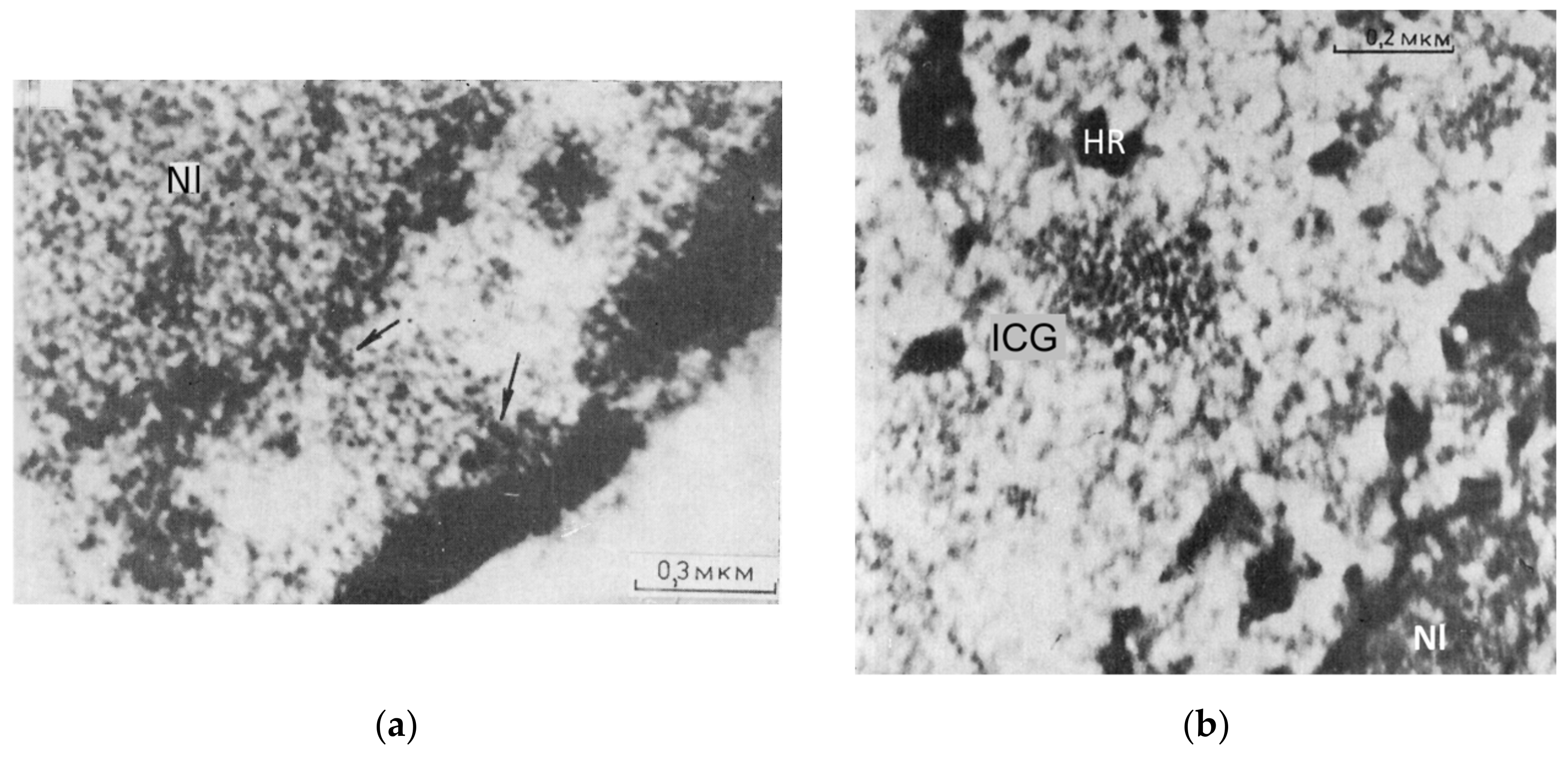
| Rat | Thymocytes | Treatment with heparin of isolated nuclei on EM supports; interactively determined | ~2 | [52] |
| Human | Breast cancer line SkBr3 | H3K9me3, localization microscopy; semi-automated | 1.7 ± 0.4 | own data, unpublished |
| Human | Breast cancer line SkBr3 (30 min post 4 Gy) | H3K9me3, localization microscopy; semi-automated | 1.9 ± 0.5 | own data, unpublished |
| Chicken | Embryonal chondrocytes | Specific Feulgen-type DNA staining of cell imprints; mathematic skeletonization | 1.8 ± 0.7 | [57] |
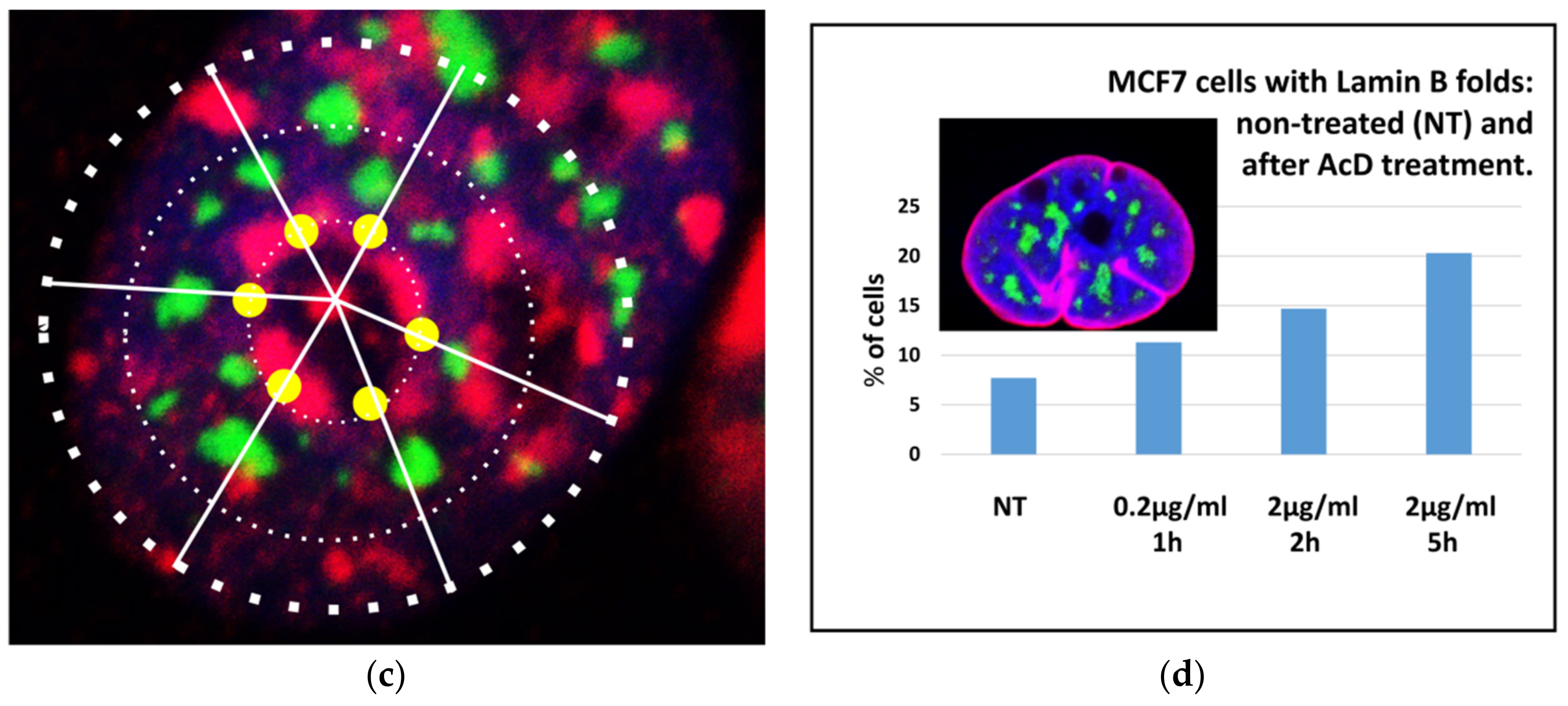
| Human | Breast cancer line MCF7 | Acridine orange DNA staining (after RNAse); epifluorescence, deconvolution, image analysis | 1.87 ± 0.74 | current |
| Human | Breast cancer line MCF7 (AcD 2 µg/mL, 5 h) | Acridine orange DNA staining (after RNAse); epifluorescence, deconvolution, image analysis | 2.12 ± 0.67 | current |
| Human | Breast cancer line MCF7 | Affinity stain for heterochromatin with toluidine blue; image analysis | 1.7 ± 0.4 | current |
Publisher’s Note: MDPI stays neutral with regard to jurisdictional claims in published maps and institutional affiliations. |
© 2021 by the authors. Licensee MDPI, Basel, Switzerland. This article is an open access article distributed under the terms and conditions of the Creative Commons Attribution (CC BY) license (https://creativecommons.org/licenses/by/4.0/).
Share and Cite
Erenpreisa, J.; Krigerts, J.; Salmina, K.; Gerashchenko, B.I.; Freivalds, T.; Kurg, R.; Winter, R.; Krufczik, M.; Zayakin, P.; Hausmann, M.; et al. Heterochromatin Networks: Topology, Dynamics, and Function (a Working Hypothesis). Cells 2021, 10, 1582. https://doi.org/10.3390/cells10071582
Erenpreisa J, Krigerts J, Salmina K, Gerashchenko BI, Freivalds T, Kurg R, Winter R, Krufczik M, Zayakin P, Hausmann M, et al. Heterochromatin Networks: Topology, Dynamics, and Function (a Working Hypothesis). Cells. 2021; 10(7):1582. https://doi.org/10.3390/cells10071582
Chicago/Turabian StyleErenpreisa, Jekaterina, Jekabs Krigerts, Kristine Salmina, Bogdan I. Gerashchenko, Talivaldis Freivalds, Reet Kurg, Ruth Winter, Matthias Krufczik, Pawel Zayakin, Michael Hausmann, and et al. 2021. "Heterochromatin Networks: Topology, Dynamics, and Function (a Working Hypothesis)" Cells 10, no. 7: 1582. https://doi.org/10.3390/cells10071582
APA StyleErenpreisa, J., Krigerts, J., Salmina, K., Gerashchenko, B. I., Freivalds, T., Kurg, R., Winter, R., Krufczik, M., Zayakin, P., Hausmann, M., & Giuliani, A. (2021). Heterochromatin Networks: Topology, Dynamics, and Function (a Working Hypothesis). Cells, 10(7), 1582. https://doi.org/10.3390/cells10071582