Review

Adoptive T Cell Therapy for Solid Tumors: Pathway to Personalized Standard of Care

1
Department of Microbiology & Immunology, University of Rochester School of Medicine & Dentistry, Rochester, NY 14642, USA
2
Department of Surgery, University of Rochester Medical Center, Rochester, NY 14642, USA
*
Author to whom correspondence should be addressed.
Cells 2021, 10(4), 808; https://doi.org/10.3390/cells10040808
Submission received: 1 March 2021 / Revised: 25 March 2021 / Accepted: 29 March 2021 / Published: 5 April 2021

Abstract

:
Adoptive cell therapy (ACT) with tumor-infiltrating T cells (TILs) has emerged as a promising therapy for the treatment of unresectable or metastatic solid tumors. One challenge to finding a universal anticancer treatment is the heterogeneity present between different tumors as a result of genetic instability associated with tumorigenesis. As the epitome of personalized medicine, TIL-ACT bypasses the issue of intertumoral heterogeneity by utilizing the patient’s existing antitumor immune response. Despite being one of the few therapies capable of inducing durable, complete tumor regression, many patients fail to respond. Recent research has focused on increasing therapeutic efficacy by refining various aspects of the TIL protocol, which includes the isolation, ex vivo expansion, and subsequent infusion of tumor specific lymphocytes. This review will explore how the therapy has evolved with time by highlighting various resistance mechanisms to TIL therapy and the novel strategies to overcome them.

1. Introduction

Solid malignancies encompass a wide range of diseases from lymphomas to carcinomas that affect nearly every organ in the body. The malignant transformation from normal cells is accompanied by progressive accumulation of genetic mutations. As a result, patients diagnosed with the same histological tumor type exhibit a wide range of genetic mutations and tumor microenvironments (TME). This inherent heterogeneity in tumors often leads to variations in patient-specific responses. Precision medicine, which refers to the tailoring of treatments to tumor-specific cellular or molecular characteristics, has recently became the mainstay of oncological therapy. The epitome of personalized therapy is adoptive cell transfer (ACT) with autologous tumor-infiltrating lymphocytes (TIL). This process involves tumor excision, isolation of TILs, ex vivo selection for autologous tumor reactivity, rapid expansion, and reinfusion of the T cell product back to the host [1] (Figure 1).
The process of tumorigenesis produces nonsynonymous somatic mutations that lead to the generation of neoantigens. Neoantigens may be derived from (1) translation of mutated genes, (2) aberrant expression of non-mutated genes, and/or (3) the untimely expression of embryonic or cell-lineage specific genes [2,3]. In a similar stochastic process, every T cell expresses a unique T cell receptor (TCR) specific for one antigenic peptide sequence. Recognition of the cognate peptide presented on major histocompatibility complex (MHC) molecules lead to T cell activation and cytolytic target killing [4,5]. This diversity in potential neoantigens and endogenous TCRs poses a significant challenge for the effective immune recognition of tumors. As TIL-ACT specifically utilizes endogenous lymphocytes found within tumors, it increases the probability of enriching for tumor-specific T cells. Other ACT protocols have experimented with the transfer of different lymphocyte populations, including, but not limited to, genetically modified lymphocytes isolated from peripheral blood. In contrast to TILs, which contain endogenous tumor-recognition abilities, these lymphocytes are altered ex vivo to express tumor-specific TCRs and/or to have increased effector functions. [6]. A notable example, which is frequently used to treat hematological malignancies, is the transfer of genetically modified T cells with chimeric antigen receptors (CAR-T) that are specific for previously identified tumor-specific neoantigens [7].
The first evidence that ACT of autologous TILs leads to tumor-specific cytolysis and metastatic tumor control emerged in the 1980 s from murine models of sarcoma, colon adenocarcinoma, melanoma, and bladder carcinoma [8]. In the initial clinical study demonstrating the curative potential of TIL-ACT, 29 metastatic melanoma patients were treated with autologous TILs accompanied by high-dose interleukin-2 (IL-2), with a 31% objective response rate (ORR, complete response (CR) + partial response (PR)), and four patients achieving complete tumor regression [9]. A seminal clinical trial conducted by the Surgery Branch of the National Cancer Institute (NCI) in the early 2000 s demonstrated that the ORR for metastatic melanoma patients can be as high as 72%, with complete response rate (CRR) of 22% following one TIL infusion [10]. Therapeutic response to TIL-ACT is restricted by numerous factors, including, but not limited to, the initial amount of tumor-infiltrating T cells, the presence of immunosuppressive cell types in the TME, and the failure of adoptively transferred cells to persist in the host.
Advances in tumor immunology have led to the gradual optimization of the TIL-ACT protocol from lymphocyte selection to pre-treatment conditioning, and to combination with adjunctive treatment modalities. In this review, we will provide an overview of recent preclinical and clinical advances that have led to the continual improvement of TIL-ACT. We will highlight common mechanisms of resistance as well as novel therapeutic strategies that target these pathways to overcome resistance in the treatment of solid tumor malignancies.

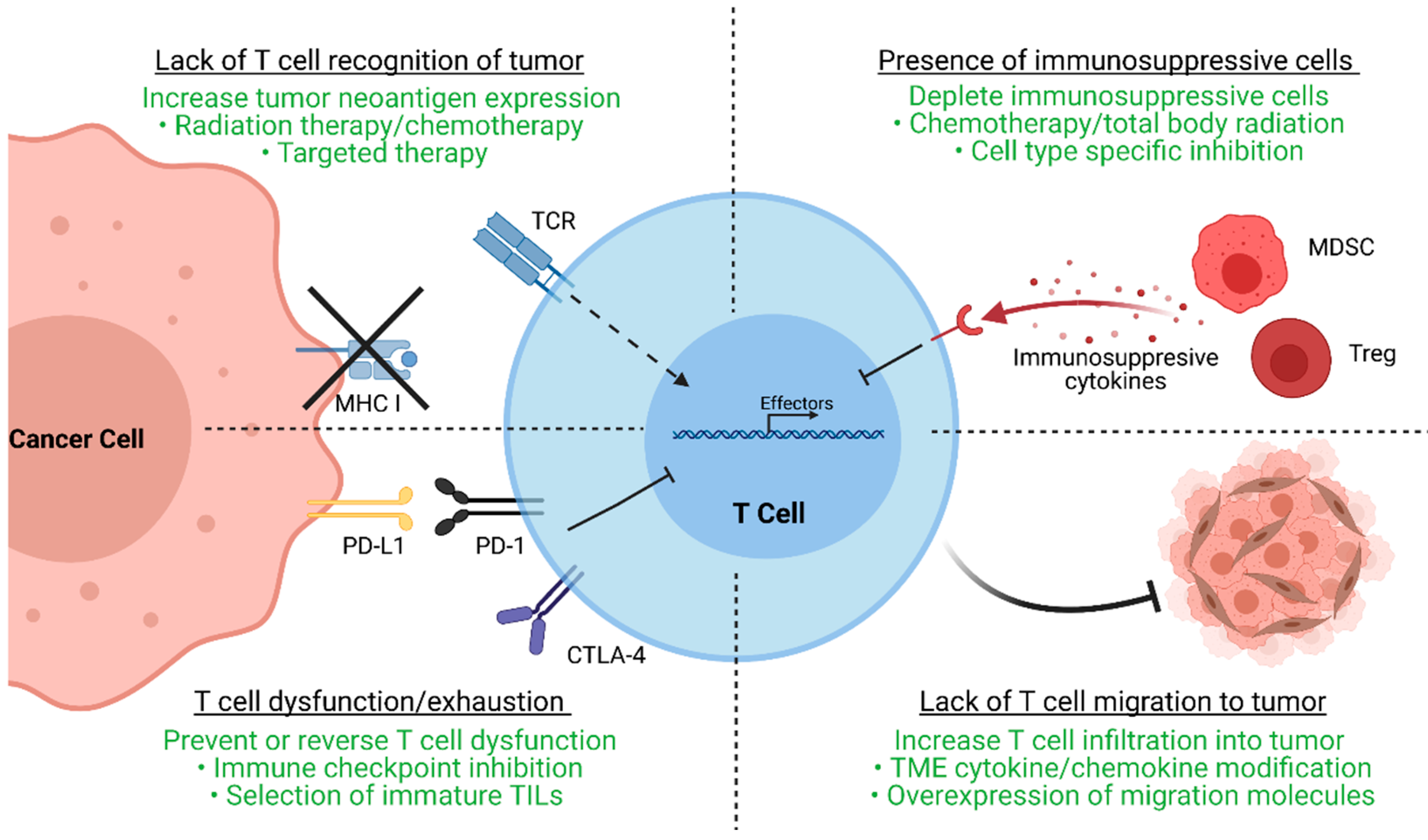
2. Mechanisms of ACT-TIL Resistance

After the initial clinical trials in metastatic melanoma conducted by the NCI, TIL-ACT has been adopted by numerous other institutions around the world for the treatment of a variety of solid tumors [11,12,13,14]. While objective responses have been replicated in subsequent clinical trials, a significant percentage of patients experience therapy failure by displaying innate or acquired therapy resistance. Innate resistance occurs in patients unresponsive to initial therapy, whereas acquired resistance occurs in patients who were initially responsive but eventually become unresponsive. Regardless of when resistance occurs, the underlying mechanisms result in the lack of generation of functional T cells, or the suppression an otherwise productive T cell response (Figure 2).
As ACT is based on the premise of delivering antitumor T cells to the tumor, the lack of functional, tumor-specific T cells within tumor can abrogate therapeutic efficacy [12]. Effective TILs recognize tumor-specific neoantigens, which are results of stochastic somatic mutation accumulation during tumorigenesis. Thus, cancers with high mutational burdens, such as melanoma and lung cancer, have increased chances of generating neoantigens and are more likely to respond to T cell-based therapies [15,16,17]. In contrast, cancers with lower mutational burden, such as cervical cancer, exhibit decreased response to TIL- ACT (ORR 28%) [18]. However, cytolysis of neoantigen containing tumor clones may lead to the eventual outgrowth of antigen-negative tumor clones that are unrecognizable by TILs. Furthermore, tumor cells may also downregulate MHC molecules or other components of antigen presentation to evade immunosurveillance [19]. Aside from tumor cells, the TME can physically prevent TIL infiltration via generation of dense extracellular matrix, secretion of chemo-repulsive chemokines, or aberrant angiogenesis [20]. The TME can also contain a multitude of immunosuppressive cell types such as myeloid-derived suppressor cells (MDSCs) and regulatory T cells (Tregs). These cells can secrete anti-inflammatory cytokines (IL-10 and TGF-β), deplete essential T cell nutrients from the microenvironment, and suppress antigen presenting cells (APCs) to further inhibit T cell-mediated immunity [21,22]. Ultimately, different TME factors converge to generate a microenvironment conducive to T cell exhaustion, a state in which T cells upregulate checkpoint molecules as they progressively lose the ability to proliferative and perform effector functions [23]. In reality, numerous distinct escape mechanisms contribute to the immunosuppression and therapy resistance of TILs.
Given that every tumor is unique as a result of intra- and inter-tumoral heterogeneity, one must identify and target non-redundant resistance pathways present in each patient to optimize therapeutic benefit. With this knowledge, multiple clinical trials have focused on improving TIL-ACT responses by altering the original protocol based on four main components: (1) ex vivo selection and expansion of tumor-specific T lymphocytes; (2) patient pre-conditioning regimens; (3) post-infusion in vivo TIL support; and (4) potential adjuvant therapies. A partial list of current clinical trials involving TIL-ACT is summarized in Table 1.

3. TIL-ACT Protocol Refinement

3.1. TIL Cell Type Selection

ACT is the process by which endogenous cells are isolated, selected, expanded, and re-infused back into the patient; however, the type of cells transferred ultimately determines the therapeutic effects. Although TILs and genetically modified T cells both derive from autologous lymphocytes and are eventually transferred back to the patient, there exist significant differences between the two (Figure 3). Although a thorough dissection of non-TIL based ACT is beyond the scope for this review, we will briefly compare the use of CAR-T with TIL in the treatment of solid tumors.
TILs typically refer to unmodified T cells with endogenous TCRs, whereas CAR-T cells contain synthetic, modified TCRs. Endogenous TCRs consist of heterodimers that recognize MHC restricted short peptide chains but cannot initiate downstream signaling without the recruitment of additional accessory molecules [24]. In contrast, CAR-T cells express synthetic single-chain receptors derived from monoclonal antibodies that are not MHC restricted and are coupled with T cell signaling domains. First generation CARs consist of a single CD3ζ TCR activation domain, whereas subsequent generations of CARs contain one or more costimulatory molecules to further augment T cell signaling and activation [25,26,27,28]. CAR-T cells can thus automatically activate downstream signaling pathways upon binding to cognate neoantigen without the requirement for engagement of external co-stimulatory molecules or co-receptors. The differences between TCR and CAR dictate the functionality of these T cells in immunotherapy. The diversity of the TCRs allows endogenous TILs to recognize a wider range of unknown tumor neoantigens, while the specificity of the synthetic monoclonal receptor allows high affinity CAR binding to known neoantigens [6,29]. However, this high affinity CAR-T binding to a single neoantigen leads to increased probability of off-target toxicities secondary to antigen presence on normal cells [7]. Whereas the toxicities for ACT-TIL are predominantly attributed to lymphodepletion and IL-2 regimens with rare, minor autoimmunity (vitiligo and uveitis) secondary to TIL infusion, CAR-T infusions have frequently been linked to cytokine release syndrome (CRS) and CAR T-cell-related encephalopathy syndrome (CRES) in addition to toxicities from preparative regimens [7,30]. As a result, CAR-T is preferentially used in the treatment of hematological malignancies with well-characterized mutations, such as CD19 CAR-T for B cell leukemias, while a more diverse set of tumor-reactive TILs is preferred in the ACT of heterogeneous solid tumors [31]. As the generation of CAR-T is limited by the knowledge of receptor sequences targeting specific neoantigens, continued efforts in identifying a wide range of neoantigens unique to solid tumors and subsequent reverse engineering of cognate chimeric receptors is necessary to improve CAR-T cell usage and efficacy.
The generation of a diverse set of tumor-specific TILs is both complex and time-consuming. After initial ex vivo isolation and expansion from a resected tumor, multiple T cell clones are selected against autologous tumor cell lines for IFN-γ activity, which acts as a surrogate marker for tumor-reactivity. Clones negative for tumor-specificity are eliminated while the rest undergo rapid expansion to generate the ultimate infusion product which typically contains at minimum 1 × 10¹⁰ cells [32]. The entire process from tumor resection to cell administration normally takes 6–8 weeks [33]. However, objective clinical response to TIL-ACT has been associated with increased average telomere length in transferred TILs (6.7 kb in CR, 6.2 kb in PR vs. 5.1 kb in no response (NR), p = 0.006), which is inversely associated with the ex vivo culture time [14,34]. To reduce culturing time, the Surgery Branch at the NCI subsequently developed a TIL-ACT protocol with “young” TILs that are enriched for CD8+ T cell but not additionally selected for tumor reactivity. Patients treated with “young” TILs exhibited similar ORR as those treated with traditional TILs. However, as the new protocol accelerated and increased the success rate for TIL generation, it has since been adopted by other institutions [13,35,36].
While other short-term selection methods have been attempted, inherent intra- and intertumoral heterogeneity complicates the selection process. The NCI has recently developed an unbiased high-throughput TIL screen against autologous neoantigens to circumvent this issue. In this novel protocol, TILs are selected against autologous antigen presenting dendritic cells (DC) pulsed with peptide pools or tandem minigenes derived from nonsynonymous mutations identified by whole-exome sequencing of the tumor [37,38,39]. This highly sensitive screen requires less time than traditional autologous tumor cell co-cultures. Furthermore, it is capable of isolating multiple tumor-specific T cell clones when the traditional screen failed in a patient that otherwise would have been excluded from receiving TIL-ACT [31,40]. However, as whole exome sequencing remains costly, the widespread usage of this selection method is currently limited.
Multiple clinical trials have associated objective treatment responses to increasing total number of infused TIL, and more specifically of CD8+ T cells [14,35,41,42]. Tumors isolated from patients with low lymphocytic infiltration often fail to generate sufficient numbers of T cells required for re-infusion (median of 8% lymphocytic infiltration for failed cultures vs. ~50% successful cultures, p = 5 × 10⁻⁸) [36]. In contrast, no correlation has been identified between the number of CD4+ T cells and treatment response. The heterogeneity within overall CD4+ T cell population may contribute to its ambiguous role in antitumor immunity. CD4+ T cells can be characterized into helper T cells (TH1, TH2, and TH17) and Tregs. Tregs secrete anti-inflammatory cytokines that suppress ongoing immune responses, and their presence is associated with poor clinical prognosis [43,44]. In contrast, helper T cells secrete pro-inflammatory cytokines and chemokines that enhance antitumor responses and mediate tumor regression in preclinical models [44,45,46]. Case reports have shown the potential of adoptively transferred tumor-infiltrating TH1 and of bulk CD4+ T cells in mediating transient tumor regression in cholangiocarcinoma and melanoma, respectively, via tumor antigen-specific secretion of IFN-γ [47,48]. However, the adoptive transfer of CD8+ T cell enriched TIL products containing minimal CD4+ T cells results in ORRs similar to that of bulk TILs, indicating that CD4+ T cells do not significantly contribute to the observed therapeutic response [35,49].
Another characteristic that has been positively correlated with objective treatment response is the persistent survival of transferred, functional TILs in the patient [10,11,42,50]. Tumor-specific TILs have been detected within peripheral blood of responsive patients for up to 34 months post infusion [11]. Furthermore, in one clinical study, increasing response to therapy has been correlated with increasing half-lives of tumor-specific TIL clonotypes (132–173 days for CR vs. 31–53 days for PR and 13–15 days for NR, p < 0.05) [51]. Often, in vivo persistence and clinical response are dependent on the type of TIL transferred. In fact, effector CD8+ T cells derived from less differentiated precursors, such as central memory and naïve T cells, demonstrate increased secretion of effector molecules and proliferation over those derived from the heterogeneous bulk TIL populations [52]. Conversely, the transfer of more differentiated T cells leads to impaired antitumor efficacy and decreased overall survival in preclinical models [53,54]. Single cell analysis of different TIL clonotypes in a patient with metastatic colorectal cancer exhibiting partial response revealed a set of genetic signatures associated with T cell persistence that resembles less differentiated T cells. Of note, compared to non-persistent cells, persistent cells express decreased levels of EOMES (transcription factor upregulated in terminally exhausted T cells), and elevated levels of IL7R (receptor for homeostatic proliferation cytokine IL-7) [55,56,57]. Thus, the selection for CD8+ T cells with more progenitor-like phenotypes may increase the ORR of TIL-ACT.

3.2. Role of Cytokine Support

Cytokines play an indispensable role in the generation, activation, and proliferation of lymphocytes. As objective responses to TIL-ACT have been linked to the continual persistence of adoptively transferred lymphocytes in vivo, the cytokines used for the ex vivo expansion and post-infusion support of TIL become important determinants of treatment efficacy.
IL-2, IL-7, IL-12, and IL-15 all affect T cell differentiation and, thus, are optimal candidates for the ex vivo expansion and differentiation of TILs used for therapy. IL-2 is a pleiotropic cytokine that induces cell-type specific responses. For example, IL-2 is not only involved in the differentiation and homeostasis of Tregs but it also promotes the differentiation of effector CD8+ T cells and increased synthesis of effector molecules [44,58,59]. Since the initial clinical trials in the 1980s, TIL-ACT protocols have incorporated the use of IL-2 in the ex vivo expansion phase [9,32]. Thus far, this has led to a highly reproducible and largely successful generation of sufficient numbers of TIL for subsequent adoptive transfer. With increasing understanding of how different lymphocytic subsets mediate tumor regression, other cytokines are currently being considered to enrich for more effective T cells. IL-12 is a potential antitumor cytokine that can both directly augment T cell cytolytic potential and increase antigen presentation [60]. Murine models indicate that IL-12 mediated activation of naïve T cells generate highly proliferative progenies with elevated cytolytic function and increased resistance to exhaustion [61,62,63]. Furthermore, the adoptive transfer of IL-2 + IL-12 primed T cells lead to enhanced tumor regression in multiple studies [61,62,64]. In contrast, IL-7 and IL-15 have been identified as critical factors for maintaining in vivo homeostatic T cell proliferation and function, particularly of memory T cells [57,65,66]. However, the combination of IL-7 + IL-15 does not result in superior ex vivo generation of effector T cell for ACT than when compared to IL-2 [67].
Beyond ex vivo expansion, TIL protocols also use high-dose (HD) IL-2 as an adjuvant to support lymphocyte proliferation in vivo after transfer [32,68]. However, systemic IL-2 treatment is associated with severe toxicities that restrict its widespread clinical usage [14,69,70]. In fact, most patients do not complete the entire post-transfer IL-2 regimen due to overwhelming adverse reactions, including, but not limited to, vascular leak syndromes and neurological symptoms [71]. Many clinical trials have explored the use of attenuated or low-dose (LD) IL-2. This dosage reduction leads to better tolerance of the therapy and less severe, more transient adverse events [72,73,74,75]. A meta-analysis of the efficacy of HD (n = 332) vs. LD IL-2 (n = 78) with TIL-ACT showed that the pooled ORR (CRR) was estimated to be 43% (14%) and 35% (7%), respectively [71]. Hence, LD IL-2 regimens have the potential to elicit durable responses while minimizing associated adverse effects. However, at this point, clinical trials involving LD IL-2 have limited patient enrollment with no trial directly comparing the efficacy of HD vs. LD IL-2 as adjuvants to TIL-ACT.

3.3. Role of Pre-Conditioning Regimens

The necessity of modifying the host immune environment prior to TIL transfer has been long established as therapeutic effects were only achieved with the addition of cyclophosphamide [8,12,76]. Currently, the main preparative conditioning regimens for TIL-ACT consist of non-myeloablative chemotherapy (NMA) with cyclophosphamide + fludarabine and/or total body irradiation (TBI). Both methods result in the depletion of host lymphocyte-mediated immunosuppressive factors that potentially hinder the functions of transferred T cells [77,78]. Further research indicates that lymphodepletion can eliminate homeostatic cytokine sinks, inhibit immunosuppressive Tregs, and increase neoantigen presentation [53,79,80]. The eradication of endogenous cells that compete for homeostatic cytokines IL-7 and IL-15 leads to an increase in serum levels that is thought to be beneficial for TIL persistence [42,69,81]. The removal of host tumor-infiltrating and peripheral CD4+ CD25+ FoxP3+ Tregs liberates adoptively transferred TILs from extrinsic immunosuppression [43,44,82]. Lastly, preconditioning may induce tumor cells to undergo immunogenic cell death, resulting in the release of pro-inflammatory cytokines and tumor neoantigens that further augments the antitumor immune response [83].
The association of increased lymphodepletion intensity to increasing TIL-ACT efficacy further demonstrates the importance of preparative regimens [84]. In a clinical trial conducted by the NCI, the addition of 2 or 12 Gy irradiation to preconditioning NMA chemotherapy increased the ORR from 49% (21/43 patients) to 52% (13/25 patients) and 72% (18/25 patients), respectively, in metastatic melanoma patients treated with TIL-ACT [69]. Subsequently, a randomized trial of NMA chemotherapy vs. NMA + 12 Gy TBI showed similar complete response rates (24% in both), but latter group had more partial responders (22% vs. 38%) [42]. While patients typically recover endogenous bone marrow function accompanied by a return of peripheral hematological values to normal limits within 2 to 3 weeks after lymphodepletion, the majority does experience severe treatment-related toxicities requiring advanced medical support [69,85,86]. Thus, the optimization of preconditioning regimens that minimize adverse events is necessary to increase the clinical utility of TIL-ACT.

4. Combination Treatments

4.1. Immune Checkpoint Inhibitors

A common immunotherapy resistance mechanism exhibited by various types of tumors is the upregulation of negative T cell checkpoint pathways, such as programmed cell death protein 1 (PD-1) and cytotoxic T-lymphocyte-associated protein 4 (CTLA-4), in the TME. Chronic co-inhibitory signaling induces T cell exhaustion, a functional state defined by the loss of effector functions and proliferative capability [87,88,89]. Accumulation of exhausted T cells eventually lead to tumor progression and immune evasion. Without extensive TME changes, any adoptively transferred TILs would be subjected to the same external suppression mechanisms, nullifying the therapy. However, blocking these pathways with immune checkpoint inhibitors (ICIs) can re-invigorate previously exhausted T cells to promote metastatic tumor regression [90]. Recently, ICIs against PD-1 and CTLA-4 have been approved by the Food and Drug Administration (FDA) for the treatment of numerous solid malignancies [91].
Although the characterization of TIL products demonstrate increased PD-1 expression on tumor-reactive T cells [11,92,93,94], chronic persistence of PD-1 expression on post-transfer T cells is associated with therapeutic resistance [72]. The existence of PD-1+ TIL cells may indicate potential sensitivity to ICI. Preclinical data suggest that the combination of ICI with TIL-ACT can synergistically increase T cell cytolytic ability, effector molecule synthesis, and tumor infiltration, leading to greater tumor control and improved survival [95,96,97,98]. In one case study, combinatory treatment of TIL and a PD-1 inhibitor led to complete regression of all six metastases in a patient with metastatic breast cancer [99]. Furthermore, multiple metastatic melanoma patients who previously failed ICI treatments were able to achieve partial response with TIL and went on to have durable complete responses with subsequent ICI treatment [14,75]. Patients treated with ACT of peripheral blood-derived, antigen-specific T cells in combination with CTLA-4 blockade also achieved complete response after prior progression on salvage CTLA-4 therapy [100]. These results suggest the possibility of ICI potentiating adoptively transferred TIL to elicit complete tumor regressions in vivo. A small pilot trial for the combination of TIL-ACT with nivolumab (anti-PD-1) in ovarian cancer demonstrated the safety and feasibility of this combination with two out of six patients achieving objective response [101]. Other on-going clinical trials aiming to assess the efficacy of TIL-ACT and ICI in solid tumors are listed in Table 1.

4.2. Targeted Therapy

The advent of genetic sequencing has led to the identification of common driver mutations in solid tumor malignancies. Targeted therapies are designed to specifically kill tumor cells expressing these oncogenes. Increased tumor lysis generates a more pro-inflammatory TME as well as releases additional tumor neoantigens to be recognized by TILs. Although preliminary experiments exploring targeted therapy combined with TIL-ACT have only been conducted in melanoma at this point, there exists potential for the extrapolation of this combination in the treatment of other solid tumors with identifiable driver mutations.
Roughly 50% of melanomas carry the BRAFV600E or BRAFV600K driver mutations that are targetable by BRAF inhibitors (BRAFi) such as vemurafenib and dabrafenib [102]. BRAFi mediated apoptosis of BRAF-mutant cells results in transient tumor regression [103]. Preclinical studies suggest that the addition of BRAF inhibition to TIL-ACT can increase T cell tumor infiltration, induce antigen presentation, and sensitize BRAF mutant melanoma to T cell mediated cytolysis [104,105,106,107]. A clinical trial investigated the combined efficacy of BRAFi with TIL-ACT where patients with BRAFV600 mutations underwent metastastectomy for TIL growth, followed by two weeks of vemurafenib, another metastastectomy, standard TIL infusion, and resumption of vemurafenib for up to two years. Patients on combination therapy demonstrated an ORR of 64% (n = 11 patients), which was similar to the 60% ORR (n = 15 patients) of TIL-ACT monotherapy [108]. However, metastases isolated from patients following combination treatment do exhibit increased T cell infiltration compared to monotherapy, suggesting the existence of potential synergistic effects of BRAFi pretreatment with TIL-ACT.
Another targeted therapy commonly used in the treatment of malignant melanoma is MEK inhibitors (MEKi). MEKi are typically added to BRAFi to prevent acquired resistance and patients on this combination experience drastic but transient tumor regression, with median progression-free survival of less than 10 months [109]. The mechanism by which MEKi affects the T cell mediated immunity is relatively unknown due to contradictory preclinical data. While some murine studies demonstrated that neoadjuvant MEK inhibition generate memory T cells with increased effector functions, others have shown that MEKi impairs T cell proliferation and function [110]. Given the lack of concrete evidence, further investigation is needed to determine whether MEKi offers additional clinical benefits when used in combination with TIL-ACT or BRAFi + TIL-ACT.

4.3. Other Investigative Therapies

Other adjuvant therapies aimed at (1) increasing tumor infiltration of TILs, (2) enhancing T cell antitumor potentials, or (3) alleviating TME immunosuppression have been explored in both preclinical and small clinical settings. Some strategies for increasing tumor infiltration of adoptively transferred TIL include, but are not limited to, overexpression of adhesion molecule CD62L or chemokine receptor CXCR2 on TILs [95,111], along with intratumoral overexpression of chemokine CCL21 [112]. Alternatively, cytokines such as IL-12 and tumor antigen-specific vaccinations have also been shown to augment transferred TIL effector function and prolong their survival [113,114,115,116]. In a proof-of-concept clinical trial, patients with tumor neoantigen NY-ESO-1 positive advanced sarcoma or melanoma were treated with ACT of NY-ESO-1 specific T cells in combination with NY-ESO-1 DC vaccination. Two out of six patients experienced objective response, with one attaining complete tumor regression [117]. This study suggests the potential of combining tumor vaccination with ACT.
Recent advances in identifying immunosuppressive MDSCs within the TME indicate potential for modifying the immune microenvironment to augment TIL efficacy. Additional activation of antigen presenting cells and macrophages by agonist CD40 monoclonal antibodies leads to enhanced T cell antitumor activity and proliferation [118,119]. Inhibition of MDSC migration by CXCR1/2 inhibitors resulted in increased accumulation of adoptively transferred TILs in murine models of melanoma, lung, and oral carcinomas [120]. Furthermore, MDSC depletion via doxorubicin and docetaxel in combination with TIL-ACT increased T cell tumor-infiltration and enhanced tumor control in preclinical models of melanoma and breast cancer [121,122]. However, due to the preliminary nature of these studies, further investigation is necessary to determine if these strategies could provide additional benefits to TIL-ACT and be safely translated into the clinical setting.

5. The Future of TIL-ACT

Although the clinical success of TIL-ACT has been replicated across multiple institutions, its availability has so far been limited to large academic medical centers. These centers have the expertise to successfully grow TILs and the capability to adequately manage adverse events patients experience [123]. The ex vivo TIL expansion protocol is labor-intensive and the requirement to use fresh TIL products for infusion impose further restrictions on the implantation. Outsourcing TIL production to dedicated manufacturing centers is necessary to increase the cost-effectiveness and expand the availability of this therapy [124]. Iovance Biotherapeutics has established a centralized, high-throughput manufacturing facility for the production of cryopreserved TILs (brand named Lifleucel). Iovance’s model involve receiving surgically resected tumors shipped by different medical centers, expanding TILs to therapeutic numbers in their central facility, and shipping cryopreserved TIL products back to the medical centers [125] (Figure 4). This alleviates the burdens on medical centers and theoretically allows TIL-ACT to be administered anywhere that has the capacity to surgically excise tumors and medically monitor patients during the reinfusion. Furthermore, cryopreserved TIL products offer more flexibility in scheduling as opposed to the rigid timeline dictated by the use fresh TILs. Despite the fact that TIL-ACT has yet to be FDA approved for the treatment of solid malignancies, strong preclinical and clinical results support its potential in eliciting durable tumor regression.

6. Conclusions

Preclinical studies and early phase clinical trials of TIL-ACT have shown promising results in the treatment of solid tumors, including the potential for eliciting durable complete tumor regression. TIL-ACT takes advantage of the body’s own immune system and intrinsic antitumor immunity. Autologous TILs are isolated, expanded, and selected for tumor reactivity in this epitome of personalized medicine. Despite the intimate nature of the therapy, a significant amount of patients develop resistance and fail to objectively respond to TIL-ACT. With increasing knowledge of tumor immunology, TIL research has been focused on refining all parts of TIL-ACT protocol to overcome various resistance mechanisms and improve overall therapeutic efficacy. From the initial trial conducted by Rosenberg et al. in the 1980s, the ORR for unresectable melanoma patients treated with TIL-ACT has improved from 31% to as much as 72%, and its usage has been extended to the treatment of other solid tumor [10,32]. The addition of other adjuvant therapies, including, but not limited to, targeted therapy, immune checkpoint inhibitors, monoclonal antibody therapy, and other biologics, may further enhance TIL efficacy via non-redundant pathways and prevent subsequent tumor immune escape. Continual protocol refinement and larger clinical trials of these new avenues can facilitate the widespread adaptation of this personalized immunotherapy for the treatment of malignant solid tumors.

Author Contributions

Conceptualization, S.S.Q. and P.A.P.; writing—original draft preparation, S.S.Q.; writing—review and editing, S.S.Q., A.D.M., A.C.C., and P.A.P.; figure generation-S.S.Q. and A.D.M.; literature search, S.S.Q., A.D.M., and A.C.C. All authors have read and agreed to the published version of the manuscript.

Funding

This work is supported in part by the Wilmot Cancer Institute (SSQ).

Institutional Review Board Statement

Not applicable.

Informed Consent Statement

Not applicable.

Data Availability Statement

Data sharing not applicable.

Acknowledgments

We would like to thank Scott A. Gerber for his expertise and assistance in editing the manuscript. All schematic figures were generated with BioRender.

Conflicts of Interest

The authors declare no conflict of interest.

References

  1. Rohaan, M.W.; van den Berg, J.H.; Kvistborg, P.; Haanen, J. Adoptive transfer of tumor-infiltrating lymphocytes in melanoma: A viable treatment option. J. Immunother. Cancer 2018, 6, 102. [Google Scholar] [CrossRef]
  2. Schumacher, T.N.; Scheper, W.; Kvistborg, P. Cancer Neoantigens. Annu. Rev. Immunol. 2019, 37, 173–200. [Google Scholar] [CrossRef]
  3. Yarchoan, M.; Johnson, B.A., 3rd; Lutz, E.R.; Laheru, D.A.; Jaffee, E.M. Targeting neoantigens to augment antitumour immunity. Nat. Rev. Cancer 2017, 17, 569. [Google Scholar] [CrossRef] [Green Version]
  4. Groscurth, P.; Filgueira, L. Killing Mechanisms of Cytotoxic T Lymphocytes. News Physiol. Sci 1998, 13, 17–21. [Google Scholar] [CrossRef]
  5. Halle, S.; Halle, O.; Forster, R. Mechanisms and Dynamics of T Cell-Mediated Cytotoxicity In Vivo. Trends Immunol. 2017, 38, 432–443. [Google Scholar] [CrossRef]
  6. Chandran, S.S.; Klebanoff, C.A. T cell receptor-based cancer immunotherapy: Emerging efficacy and pathways of resistance. Immunol. Rev. 2019, 290, 127–147. [Google Scholar] [CrossRef] [PubMed]
  7. Rohaan, M.W.; Wilgenhof, S.; Haanen, J. Adoptive cellular therapies: The current landscape. Virchows Arch. 2019, 474, 449–461. [Google Scholar] [CrossRef] [PubMed] [Green Version]
  8. Rosenberg, S.A.; Spiess, P.; Lafreniere, R. A new approach to the adoptive immunotherapy of cancer with tumor-infiltrating lymphocytes. Science 1986, 233, 1318–1321. [Google Scholar] [CrossRef]
  9. Rosenberg, S.A.; Yannelli, J.R.; Yang, J.C.; Topalian, S.L.; Schwartzentruber, D.J.; Weber, J.S.; Parkinson, D.R.; Seipp, C.A.; Einhorn, J.H.; White, D.E. Treatment of patients with metastatic melanoma with autologous tumor-infiltrating lymphocytes and interleukin 2. J. Natl. Cancer Inst. 1994, 86, 1159–1166. [Google Scholar] [CrossRef]
  10. Rosenberg, S.A.; Yang, J.C.; Sherry, R.M.; Kammula, U.S.; Hughes, M.S.; Phan, G.Q.; Citrin, D.E.; Restifo, N.P.; Robbins, P.F.; Wunderlich, J.R.; et al. Durable complete responses in heavily pretreated patients with metastatic melanoma using T-cell transfer immunotherapy. Clin. Cancer Res. 2011, 17, 4550–4557. [Google Scholar] [CrossRef] [Green Version]
  11. van den Berg, J.H.; Heemskerk, B.; van Rooij, N.; Gomez-Eerland, R.; Michels, S.; van Zon, M.; de Boer, R.; Bakker, N.A.M.; Jorritsma-Smit, A.; van Buuren, M.M.; et al. Tumor infiltrating lymphocytes (TIL) therapy in metastatic melanoma: Boosting of neoantigen-specific T cell reactivity and long-term follow-up. J. Immunother. Cancer 2020, 8, e000848. [Google Scholar] [CrossRef] [PubMed]
  12. Pockaj, B.A.; Sherry, R.M.; Wei, J.P.; Yannelli, J.R.; Carter, C.S.; Leitman, S.F.; Carasquillo, J.A.; Steinberg, S.M.; Rosenberg, S.A.; Yang, J.C. Localization of 111indium-labeled tumor infiltrating lymphocytes to tumor in patients receiving adoptive immunotherapy. Augmentation with cyclophosphamide and correlation with response. Cancer 1994, 73, 1731–1737. [Google Scholar] [CrossRef]
  13. Itzhaki, O.; Hovav, E.; Ziporen, Y.; Levy, D.; Kubi, A.; Zikich, D.; Hershkovitz, L.; Treves, A.J.; Shalmon, B.; Zippel, D.; et al. Establishment and large-scale expansion of minimally cultured “young” tumor infiltrating lymphocytes for adoptive transfer therapy. J. Immunother. 2011, 34, 212–220. [Google Scholar] [CrossRef] [PubMed]
  14. Besser, M.J.; Shapira-Frommer, R.; Itzhaki, O.; Treves, A.J.; Zippel, D.B.; Levy, D.; Kubi, A.; Shoshani, N.; Zikich, D.; Ohayon, Y.; et al. Adoptive transfer of tumor-infiltrating lymphocytes in patients with metastatic melanoma: Intent-to-treat analysis and efficacy after failure to prior immunotherapies. Clin. Cancer Res. 2013, 19, 4792–4800. [Google Scholar] [CrossRef] [Green Version]
  15. Tran, E.; Robbins, P.F.; Rosenberg, S.A. ‘Final common pathway’ of human cancer immunotherapy: Targeting random somatic mutations. Nat. Immunol. 2017, 18, 255–262. [Google Scholar] [CrossRef]
  16. Martincorena, I.; Campbell, P.J. Somatic mutation in cancer and normal cells. Science 2015, 349, 1483–1489. [Google Scholar] [CrossRef]
  17. Alexandrov, L.B.; Stratton, M.R. Mutational signatures: The patterns of somatic mutations hidden in cancer genomes. Curr. Opin. Genet. Dev. 2014, 24, 52–60. [Google Scholar] [CrossRef] [Green Version]
  18. Stevanovic, S.; Helman, S.R.; Wunderlich, J.R.; Langhan, M.M.; Doran, S.L.; Kwong, M.L.M.; Somerville, R.P.T.; Klebanoff, C.A.; Kammula, U.S.; Sherry, R.M.; et al. A Phase II Study of Tumor-infiltrating Lymphocyte Therapy for Human Papillomavirus-associated Epithelial Cancers. Clin. Cancer Res. 2019, 25, 1486–1493. [Google Scholar] [CrossRef]
  19. Beatty, G.L.; Gladney, W.L. Immune escape mechanisms as a guide for cancer immunotherapy. Clin. Cancer Res. 2015, 21, 687–692. [Google Scholar] [CrossRef] [Green Version]
  20. van der Woude, L.L.; Gorris, M.A.J.; Halilovic, A.; Figdor, C.G.; de Vries, I.J.M. Migrating into the Tumor: A Roadmap for T Cells. Trends Cancer 2017, 3, 797–808. [Google Scholar] [CrossRef]
  21. Gabrilovich, D.I.; Ostrand-Rosenberg, S.; Bronte, V. Coordinated regulation of myeloid cells by tumours. Nat. Rev. Immunol. 2012, 12, 253–268. [Google Scholar] [CrossRef] [PubMed] [Green Version]
  22. Ohue, Y.; Nishikawa, H. Regulatory T (Treg) cells in cancer: Can Treg cells be a new therapeutic target? Cancer Sci. 2019, 110, 2080–2089. [Google Scholar] [CrossRef] [PubMed]
  23. van der Leun, A.M.; Thommen, D.S.; Schumacher, T.N. CD8(+) T cell states in human cancer: Insights from single-cell analysis. Nat. Rev. Cancer 2020, 20, 218–232. [Google Scholar] [CrossRef] [PubMed]
  24. Kuhns, M.S.; Davis, M.M.; Garcia, K.C. Deconstructing the form and function of the TCR/CD3 complex. Immunity 2006, 24, 133–139. [Google Scholar] [CrossRef] [PubMed] [Green Version]
  25. Kagoya, Y.; Tanaka, S.; Guo, T.; Anczurowski, M.; Wang, C.H.; Saso, K.; Butler, M.O.; Minden, M.D.; Hirano, N. A novel chimeric antigen receptor containing a JAK-STAT signaling domain mediates superior antitumor effects. Nat. Med. 2018, 24, 352–359. [Google Scholar] [CrossRef] [PubMed]
  26. Maher, J.; Brentjens, R.J.; Gunset, G.; Riviere, I.; Sadelain, M. Human T-lymphocyte cytotoxicity and proliferation directed by a single chimeric TCRzeta /CD28 receptor. Nat. Biotechnol. 2002, 20, 70–75. [Google Scholar] [CrossRef]
  27. Krause, A.; Guo, H.F.; Latouche, J.B.; Tan, C.; Cheung, N.K.; Sadelain, M. Antigen-dependent CD28 signaling selectively enhances survival and proliferation in genetically modified activated human primary T lymphocytes. J. Exp. Med. 1998, 188, 619–626. [Google Scholar] [CrossRef] [Green Version]
  28. Eshhar, Z.; Bach, N.; Fitzer-Attas, C.J.; Gross, G.; Lustgarten, J.; Waks, T.; Schindler, D.G. The T-body approach: Potential for cancer immunotherapy. Springer Semin Immunopathol. 1996, 18, 199–209. [Google Scholar] [CrossRef]
  29. Mohanty, R.; Chowdhury, C.R.; Arega, S.; Sen, P.; Ganguly, P.; Ganguly, N. CAR T cell therapy: A new era for cancer treatment (Review). Oncol. Rep. 2019, 42, 2183–2195. [Google Scholar] [CrossRef]
  30. Wolf, B.; Zimmermann, S.; Arber, C.; Irving, M.; Trueb, L.; Coukos, G. Safety and Tolerability of Adoptive Cell Therapy in Cancer. Drug Saf. 2019, 42, 315–334. [Google Scholar] [CrossRef] [Green Version]
  31. Parkhurst, M.R.; Robbins, P.F.; Tran, E.; Prickett, T.D.; Gartner, J.J.; Jia, L.; Ivey, G.; Li, Y.F.; El-Gamil, M.; Lalani, A.; et al. Unique Neoantigens Arise from Somatic Mutations in Patients with Gastrointestinal Cancers. Cancer Discov. 2019, 9, 1022–1035. [Google Scholar] [CrossRef] [PubMed]
  32. Rosenberg, S.A.; Packard, B.S.; Aebersold, P.M.; Solomon, D.; Topalian, S.L.; Toy, S.T.; Simon, P.; Lotze, M.T.; Yang, J.C.; Seipp, C.A.; et al. Use of tumor-infiltrating lymphocytes and interleukin-2 in the immunotherapy of patients with metastatic melanoma. A preliminary report. N. Engl. J. Med. 1988, 319, 1676–1680. [Google Scholar] [CrossRef]
  33. Dudley, M.E.; Wunderlich, J.R.; Shelton, T.E.; Even, J.; Rosenberg, S.A. Generation of tumor-infiltrating lymphocyte cultures for use in adoptive transfer therapy for melanoma patients. J. Immunother. 2003, 26, 332–342. [Google Scholar] [CrossRef]
  34. Zhou, J.; Shen, X.; Huang, J.; Hodes, R.J.; Rosenberg, S.A.; Robbins, P.F. Telomere length of transferred lymphocytes correlates with in vivo persistence and tumor regression in melanoma patients receiving cell transfer therapy. J. Immunol. 2005, 175, 7046–7052. [Google Scholar] [CrossRef] [Green Version]
  35. Dudley, M.E.; Gross, C.A.; Langhan, M.M.; Garcia, M.R.; Sherry, R.M.; Yang, J.C.; Phan, G.Q.; Kammula, U.S.; Hughes, M.S.; Citrin, D.E.; et al. CD8+ enriched “young” tumor infiltrating lymphocytes can mediate regression of metastatic melanoma. Clin. Cancer Res. 2010, 16, 6122–6131. [Google Scholar] [CrossRef] [PubMed] [Green Version]
  36. Dudley, M.E.; Gross, C.A.; Somerville, R.P.; Hong, Y.; Schaub, N.P.; Rosati, S.F.; White, D.E.; Nathan, D.; Restifo, N.P.; Steinberg, S.M.; et al. Randomized selection design trial evaluating CD8+-enriched versus unselected tumor-infiltrating lymphocytes for adoptive cell therapy for patients with melanoma. J. Clin. Oncol. 2013, 31, 2152–2159. [Google Scholar] [CrossRef] [Green Version]
  37. Lu, Y.C.; Yao, X.; Crystal, J.S.; Li, Y.F.; El-Gamil, M.; Gross, C.; Davis, L.; Dudley, M.E.; Yang, J.C.; Samuels, Y.; et al. Efficient identification of mutated cancer antigens recognized by T cells associated with durable tumor regressions. Clin. Cancer Res. 2014, 20, 3401–3410. [Google Scholar] [CrossRef] [Green Version]
  38. Malekzadeh, P.; Pasetto, A.; Robbins, P.F.; Parkhurst, M.R.; Paria, B.C.; Jia, L.; Gartner, J.J.; Hill, V.; Yu, Z.; Restifo, N.P.; et al. Neoantigen screening identifies broad TP53 mutant immunogenicity in patients with epithelial cancers. J. Clin. Investig. 2019, 129, 1109–1114. [Google Scholar] [CrossRef]
  39. Lu, Y.C.; Zheng, Z.; Robbins, P.F.; Tran, E.; Prickett, T.D.; Gartner, J.J.; Li, Y.F.; Ray, S.; Franco, Z.; Bliskovsky, V.; et al. An Efficient Single-Cell RNA-Seq Approach to Identify Neoantigen-Specific T Cell Receptors. Mol. Ther. 2018, 26, 379–389. [Google Scholar] [CrossRef] [Green Version]
  40. Yossef, R.; Tran, E.; Deniger, D.C.; Gros, A.; Pasetto, A.; Parkhurst, M.R.; Gartner, J.J.; Prickett, T.D.; Cafri, G.; Robbins, P.F.; et al. Enhanced detection of neoantigen-reactive T cells targeting unique and shared oncogenes for personalized cancer immunotherapy. JCI Insight 2018, 3, e122467. [Google Scholar] [CrossRef] [Green Version]
  41. Radvanyi, L.G.; Bernatchez, C.; Zhang, M.; Fox, P.S.; Miller, P.; Chacon, J.; Wu, R.; Lizee, G.; Mahoney, S.; Alvarado, G.; et al. Specific lymphocyte subsets predict response to adoptive cell therapy using expanded autologous tumor-infiltrating lymphocytes in metastatic melanoma patients. Clin. Cancer Res. 2012, 18, 6758–6770. [Google Scholar] [CrossRef] [Green Version]
  42. Goff, S.L.; Dudley, M.E.; Citrin, D.E.; Somerville, R.P.; Wunderlich, J.R.; Danforth, D.N.; Zlott, D.A.; Yang, J.C.; Sherry, R.M.; Kammula, U.S.; et al. Randomized, Prospective Evaluation Comparing Intensity of Lymphodepletion Before Adoptive Transfer of Tumor-Infiltrating Lymphocytes for Patients With Metastatic Melanoma. J. Clin. Oncol. 2016, 34, 2389–2397. [Google Scholar] [CrossRef]
  43. Yao, X.; Ahmadzadeh, M.; Lu, Y.C.; Liewehr, D.J.; Dudley, M.E.; Liu, F.; Schrump, D.S.; Steinberg, S.M.; Rosenberg, S.A.; Robbins, P.F. Levels of peripheral CD4(+)FoxP3(+) regulatory T cells are negatively associated with clinical response to adoptive immunotherapy of human cancer. Blood 2012, 119, 5688–5696. [Google Scholar] [CrossRef]
  44. Antony, P.A.; Piccirillo, C.A.; Akpinarli, A.; Finkelstein, S.E.; Speiss, P.J.; Surman, D.R.; Palmer, D.C.; Chan, C.C.; Klebanoff, C.A.; Overwijk, W.W.; et al. CD8+ T cell immunity against a tumor/self-antigen is augmented by CD4+ T helper cells and hindered by naturally occurring T regulatory cells. J. Immunol. 2005, 174, 2591–2601. [Google Scholar] [CrossRef] [Green Version]
  45. Li, K.; Donaldson, B.; Young, V.; Ward, V.; Jackson, C.; Baird, M.; Young, S. Adoptive cell therapy with CD4(+) T helper 1 cells and CD8(+) cytotoxic T cells enhances complete rejection of an established tumour, leading to generation of endogenous memory responses to non-targeted tumour epitopes. Clin. Transl. Immunol. 2017, 6, e160. [Google Scholar] [CrossRef]
  46. Imai, N.; Tawara, I.; Yamane, M.; Muraoka, D.; Shiku, H.; Ikeda, H. CD4(+) T cells support polyfunctionality of cytotoxic CD8(+) T cells with memory potential in immunological control of tumor. Cancer Sci. 2020, 111, 1958–1968. [Google Scholar] [CrossRef]
  47. Friedman, K.M.; Prieto, P.A.; Devillier, L.E.; Gross, C.A.; Yang, J.C.; Wunderlich, J.R.; Rosenberg, S.A.; Dudley, M.E. Tumor-specific CD4+ melanoma tumor-infiltrating lymphocytes. J. Immunother. 2012, 35, 400–408. [Google Scholar] [CrossRef]
  48. Tran, E.; Turcotte, S.; Gros, A.; Robbins, P.F.; Lu, Y.C.; Dudley, M.E.; Wunderlich, J.R.; Somerville, R.P.; Hogan, K.; Hinrichs, C.S.; et al. Cancer immunotherapy based on mutation-specific CD4+ T cells in a patient with epithelial cancer. Science 2014, 344, 641–645. [Google Scholar] [CrossRef]
  49. Prieto, P.A.; Durflinger, K.H.; Wunderlich, J.R.; Rosenberg, S.A.; Dudley, M.E. Enrichment of CD8+ cells from melanoma tumor-infiltrating lymphocyte cultures reveals tumor reactivity for use in adoptive cell therapy. J. Immunother. 2010, 33, 547–556. [Google Scholar] [CrossRef]
  50. Robbins, P.F.; Dudley, M.E.; Wunderlich, J.; El-Gamil, M.; Li, Y.F.; Zhou, J.; Huang, J.; Powell, D.J., Jr.; Rosenberg, S.A. Cutting edge: Persistence of transferred lymphocyte clonotypes correlates with cancer regression in patients receiving cell transfer therapy. J. Immunol. 2004, 173, 7125–7130. [Google Scholar] [CrossRef] [Green Version]
  51. Chapuis, A.G.; Desmarais, C.; Emerson, R.; Schmitt, T.M.; Shibuya, K.; Lai, I.; Wagener, F.; Chou, J.; Roberts, I.M.; Coffey, D.G.; et al. Tracking the Fate and Origin of Clinically Relevant Adoptively Transferred CD8(+) T Cells In Vivo. Sci. Immunol. 2017, 2, eaal2568. [Google Scholar] [CrossRef] [PubMed] [Green Version]
  52. Wu, F.; Zhang, W.; Shao, H.; Bo, H.; Shen, H.; Li, J.; Liu, Y.; Wang, T.; Ma, W.; Huang, S. Human effector T cells derived from central memory cells rather than CD8(+)T cells modified by tumor-specific TCR gene transfer possess superior traits for adoptive immunotherapy. Cancer Lett. 2013, 339, 195–207. [Google Scholar] [CrossRef] [PubMed]
  53. Klebanoff, C.A.; Scott, C.D.; Leonardi, A.J.; Yamamoto, T.N.; Cruz, A.C.; Ouyang, C.; Ramaswamy, M.; Roychoudhuri, R.; Ji, Y.; Eil, R.L.; et al. Memory T cell-driven differentiation of naive cells impairs adoptive immunotherapy. J. Clin. Investig. 2016, 126, 318–334. [Google Scholar] [CrossRef] [PubMed] [Green Version]
  54. Hinrichs, C.S.; Borman, Z.A.; Cassard, L.; Gattinoni, L.; Spolski, R.; Yu, Z.; Sanchez-Perez, L.; Muranski, P.; Kern, S.J.; Logun, C.; et al. Adoptively transferred effector cells derived from naive rather than central memory CD8+ T cells mediate superior antitumor immunity. Proc. Natl. Acad. Sci. USA 2009, 106, 17469–17474. [Google Scholar] [CrossRef] [PubMed] [Green Version]
  55. Lu, Y.C.; Jia, L.; Zheng, Z.; Tran, E.; Robbins, P.F.; Rosenberg, S.A. Single-Cell Transcriptome Analysis Reveals Gene Signatures Associated with T-cell Persistence Following Adoptive Cell Therapy. Cancer Immunol. Res. 2019, 7, 1824–1836. [Google Scholar] [CrossRef] [PubMed]
  56. Paley, M.A.; Kroy, D.C.; Odorizzi, P.M.; Johnnidis, J.B.; Dolfi, D.V.; Barnett, B.E.; Bikoff, E.K.; Robertson, E.J.; Lauer, G.M.; Reiner, S.L.; et al. Progenitor and terminal subsets of CD8+ T cells cooperate to contain chronic viral infection. Science 2012, 338, 1220–1225. [Google Scholar] [CrossRef] [Green Version]
  57. Sprent, J.; Surh, C.D. Normal T cell homeostasis: The conversion of naive cells into memory-phenotype cells. Nat. Immunol. 2011, 12, 478–484. [Google Scholar] [CrossRef]
  58. Furlan, S.N.; Singh, K.; Lopez, C.; Tkachev, V.; Hunt, D.J.; Hibbard, J.; Betz, K.M.; Blazar, B.R.; Trapnell, C.; Kean, L.S. IL-2 enhances ex vivo-expanded regulatory T-cell persistence after adoptive transfer. Blood Adv. 2020, 4, 1594–1605. [Google Scholar] [CrossRef] [Green Version]
  59. Ross, S.H.; Cantrell, D.A. Signaling and Function of Interleukin-2 in T Lymphocytes. Annu. Rev. Immunol. 2018, 36, 411–433. [Google Scholar] [CrossRef]
  60. Liu, J.; Cao, S.; Kim, S.; Chung, E.Y.; Homma, Y.; Guan, X.; Jimenez, V.; Ma, X. Interleukin-12: An update on its immunological activities, signaling and regulation of gene expression. Curr. Immunol. Rev. 2005, 1, 119–137. [Google Scholar] [CrossRef]
  61. Tucker, C.G.; Mitchell, J.S.; Martinov, T.; Burbach, B.J.; Beura, L.K.; Wilson, J.C.; Dwyer, A.J.; Singh, L.M.; Mescher, M.F.; Fife, B.T. Adoptive T Cell Therapy with IL-12-Preconditioned Low-Avidity T Cells Prevents Exhaustion and Results in Enhanced T Cell Activation, Enhanced Tumor Clearance, and Decreased Risk for Autoimmunity. J. Immunol. 2020, 205, 1449–1460. [Google Scholar] [CrossRef]
  62. Diaz-Montero, C.M.; Naga, O.; Zidan, A.A.; Salem, M.L.; Pallin, M.; Parmigiani, A.; Walker, G.; Wieder, E.; Komanduri, K.; Cole, D.J.; et al. Synergy of brief activation of CD8 T-cells in the presence of IL-12 and adoptive transfer into lymphopenic hosts promotes tumor clearance and anti-tumor memory. Am. J. Cancer Res. 2011, 1, 882–896. [Google Scholar] [PubMed]
  63. Curtsinger, J.M.; Johnson, C.M.; Mescher, M.F. CD8 T cell clonal expansion and development of effector function require prolonged exposure to antigen, costimulation, and signal 3 cytokine. J. Immunol. 2003, 171, 5165–5171. [Google Scholar] [CrossRef] [PubMed] [Green Version]
  64. Johnson, C.B.; Riesenberg, B.P.; May, B.R.; Gilreath, S.C.; Li, G.; Staveley-O’Carroll, K.F.; Garrett-Mayer, E.; Mehrotra, S.; Cole, D.J.; Rubinstein, M.P. Effector CD8+ T-cell Engraftment and Antitumor Immunity in Lymphodepleted Hosts Is IL7Ralpha Dependent. Cancer Immunol. Res. 2015, 3, 1364–1374. [Google Scholar] [CrossRef] [PubMed] [Green Version]
  65. Drake, A.; Kaur, M.; Iliopoulou, B.P.; Phennicie, R.; Hanson, A.; Chen, J. Interleukins 7 and 15 Maintain Human T Cell Proliferative Capacity through STAT5 Signaling. PLoS ONE 2016, 11, e0166280. [Google Scholar] [CrossRef] [Green Version]
  66. Boyman, O.; Purton, J.F.; Surh, C.D.; Sprent, J. Cytokines and T-cell homeostasis. Curr. Opin. Immunol. 2007, 19, 320–326. [Google Scholar] [CrossRef]
  67. Gong, W.; Hoffmann, J.M.; Stock, S.; Wang, L.; Liu, Y.; Schubert, M.L.; Neuber, B.; Huckelhoven-Krauss, A.; Gern, U.; Schmitt, A.; et al. Comparison of IL-2 vs IL-7/IL-15 for the generation of NY-ESO-1-specific T cells. Cancer Immunol. Immunother. 2019, 68, 1195–1209. [Google Scholar] [CrossRef]
  68. Yee, C.; Thompson, J.A.; Byrd, D.; Riddell, S.R.; Roche, P.; Celis, E.; Greenberg, P.D. Adoptive T cell therapy using antigen-specific CD8+ T cell clones for the treatment of patients with metastatic melanoma: In vivo persistence, migration, and antitumor effect of transferred T cells. Proc. Natl. Acad. Sci. USA 2002, 99, 16168–16173. [Google Scholar] [CrossRef] [Green Version]
  69. Dudley, M.E.; Yang, J.C.; Sherry, R.; Hughes, M.S.; Royal, R.; Kammula, U.; Robbins, P.F.; Huang, J.; Citrin, D.E.; Leitman, S.F.; et al. Adoptive cell therapy for patients with metastatic melanoma: Evaluation of intensive myeloablative chemoradiation preparative regimens. J. Clin. Oncol. 2008, 26, 5233–5239. [Google Scholar] [CrossRef]
  70. Pilon-Thomas, S.; Kuhn, L.; Ellwanger, S.; Janssen, W.; Royster, E.; Marzban, S.; Kudchadkar, R.; Zager, J.; Gibney, G.; Sondak, V.K.; et al. Efficacy of adoptive cell transfer of tumor-infiltrating lymphocytes after lymphopenia induction for metastatic melanoma. J. Immunother. 2012, 35, 615–620. [Google Scholar] [CrossRef] [Green Version]
  71. Dafni, U.; Michielin, O.; Lluesma, S.M.; Tsourti, Z.; Polydoropoulou, V.; Karlis, D.; Besser, M.J.; Haanen, J.; Svane, I.M.; Ohashi, P.S.; et al. Efficacy of adoptive therapy with tumor-infiltrating lymphocytes and recombinant interleukin-2 in advanced cutaneous melanoma: A systematic review and meta-analysis. Ann. Oncol. 2019, 30, 1902–1913. [Google Scholar] [CrossRef] [PubMed] [Green Version]
  72. Chapuis, A.G.; Thompson, J.A.; Margolin, K.A.; Rodmyre, R.; Lai, I.P.; Dowdy, K.; Farrar, E.A.; Bhatia, S.; Sabath, D.E.; Cao, J.; et al. Transferred melanoma-specific CD8+ T cells persist, mediate tumor regression, and acquire central memory phenotype. Proc. Natl. Acad. Sci. USA 2012, 109, 4592–4597. [Google Scholar] [CrossRef] [PubMed] [Green Version]
  73. Ellebaek, E.; Iversen, T.Z.; Junker, N.; Donia, M.; Engell-Noerregaard, L.; Met, O.; Holmich, L.R.; Andersen, R.S.; Hadrup, S.R.; Andersen, M.H.; et al. Adoptive cell therapy with autologous tumor infiltrating lymphocytes and low-dose Interleukin-2 in metastatic melanoma patients. J. Transl. Med. 2012, 10, 169. [Google Scholar] [CrossRef] [PubMed] [Green Version]
  74. Andersen, R.; Donia, M.; Ellebaek, E.; Borch, T.H.; Kongsted, P.; Iversen, T.Z.; Holmich, L.R.; Hendel, H.W.; Met, O.; Andersen, M.H.; et al. Long-Lasting Complete Responses in Patients with Metastatic Melanoma after Adoptive Cell Therapy with Tumor-Infiltrating Lymphocytes and an Attenuated IL2 Regimen. Clin. Cancer Res. 2016, 22, 3734–3745. [Google Scholar] [CrossRef] [Green Version]
  75. Nguyen, L.T.; Saibil, S.D.; Sotov, V.; Le, M.X.; Khoja, L.; Ghazarian, D.; Bonilla, L.; Majeed, H.; Hogg, D.; Joshua, A.M.; et al. Phase II clinical trial of adoptive cell therapy for patients with metastatic melanoma with autologous tumor-infiltrating lymphocytes and low-dose interleukin-2. Cancer Immunol. Immunother. 2019, 68, 773–785. [Google Scholar] [CrossRef] [Green Version]
  76. North, R.J. Cyclophosphamide-facilitated adoptive immunotherapy of an established tumor depends on elimination of tumor-induced suppressor T cells. J. Exp. Med. 1982, 155, 1063–1074. [Google Scholar] [CrossRef] [Green Version]
  77. Dye, E.S.; North, R.J. T cell-mediated immunosuppression as an obstacle to adoptive immunotherapy of the P815 mastocytoma and its metastases. J. Exp. Med. 1981, 154, 1033–1042. [Google Scholar] [CrossRef]
  78. Berendt, M.J.; North, R.J. T-cell-mediated suppression of anti-tumor immunity. An explanation for progressive growth of an immunogenic tumor. J. Exp. Med. 1980, 151, 69–80. [Google Scholar] [CrossRef]
  79. Klebanoff, C.A.; Khong, H.T.; Antony, P.A.; Palmer, D.C.; Restifo, N.P. Sinks, suppressors and antigen presenters: How lymphodepletion enhances T cell-mediated tumor immunotherapy. Trends Immunol. 2005, 26, 111–117. [Google Scholar] [CrossRef] [Green Version]
  80. Wei, S.; Egenti, M.U.; Teitz-Tennenbaum, S.; Zou, W.; Chang, A.E. Effects of tumor irradiation on host T-regulatory cells and systemic immunity in the context of adoptive T-cell therapy in mice. J. Immunother. 2013, 36, 124–132. [Google Scholar] [CrossRef] [Green Version]
  81. Gattinoni, L.; Finkelstein, S.E.; Klebanoff, C.A.; Antony, P.A.; Palmer, D.C.; Spiess, P.J.; Hwang, L.N.; Yu, Z.; Wrzesinski, C.; Heimann, D.M.; et al. Removal of homeostatic cytokine sinks by lymphodepletion enhances the efficacy of adoptively transferred tumor-specific CD8+ T cells. J. Exp. Med. 2005, 202, 907–912. [Google Scholar] [CrossRef] [PubMed]
  82. Woo, E.Y.; Yeh, H.; Chu, C.S.; Schlienger, K.; Carroll, R.G.; Riley, J.L.; Kaiser, L.R.; June, C.H. Cutting edge: Regulatory T cells from lung cancer patients directly inhibit autologous T cell proliferation. J. Immunol. 2002, 168, 4272–4276. [Google Scholar] [CrossRef]
  83. Galluzzi, L.; Buque, A.; Kepp, O.; Zitvogel, L.; Kroemer, G. Immunogenic cell death in cancer and infectious disease. Nat. Rev. Immunol. 2017, 17, 97–111. [Google Scholar] [CrossRef]
  84. Wrzesinski, C.; Paulos, C.M.; Kaiser, A.; Muranski, P.; Palmer, D.C.; Gattinoni, L.; Yu, Z.; Rosenberg, S.A.; Restifo, N.P. Increased intensity lymphodepletion enhances tumor treatment efficacy of adoptively transferred tumor-specific T cells. J. Immunother. 2010, 33, 1–7. [Google Scholar] [CrossRef] [PubMed] [Green Version]
  85. Dudley, M.E.; Wunderlich, J.R.; Yang, J.C.; Hwu, P.; Schwartzentruber, D.J.; Topalian, S.L.; Sherry, R.M.; Marincola, F.M.; Leitman, S.F.; Seipp, C.A.; et al. A phase I study of nonmyeloablative chemotherapy and adoptive transfer of autologous tumor antigen-specific T lymphocytes in patients with metastatic melanoma. J. Immunother. 2002, 25, 243–251. [Google Scholar] [CrossRef] [Green Version]
  86. Muranski, P.; Boni, A.; Wrzesinski, C.; Citrin, D.E.; Rosenberg, S.A.; Childs, R.; Restifo, N.P. Increased intensity lymphodepletion and adoptive immunotherapy--how far can we go? Nat. Clin. Pract. Oncol. 2006, 3, 668–681. [Google Scholar] [CrossRef] [Green Version]
  87. Wherry, E.J.; Kurachi, M. Molecular and cellular insights into T cell exhaustion. Nat. Rev. Immunol. 2015, 15, 486–499. [Google Scholar] [CrossRef] [PubMed]
  88. McLane, L.M.; Abdel-Hakeem, M.S.; Wherry, E.J. CD8 T Cell Exhaustion During Chronic Viral Infection and Cancer. Annu. Rev. Immunol. 2019, 37, 457–495. [Google Scholar] [CrossRef] [Green Version]
  89. Ahmadzadeh, M.; Johnson, L.A.; Heemskerk, B.; Wunderlich, J.R.; Dudley, M.E.; White, D.E.; Rosenberg, S.A. Tumor antigen-specific CD8 T cells infiltrating the tumor express high levels of PD-1 and are functionally impaired. Blood 2009, 114, 1537–1544. [Google Scholar] [CrossRef]
  90. Schachter, J.; Ribas, A.; Long, G.V.; Arance, A.; Grob, J.J.; Mortier, L.; Daud, A.; Carlino, M.S.; McNeil, C.; Lotem, M.; et al. Pembrolizumab versus ipilimumab for advanced melanoma: Final overall survival results of a multicentre, randomised, open-label phase 3 study (KEYNOTE-006). Lancet 2017, 390, 1853–1862. [Google Scholar] [CrossRef]
  91. Hargadon, K.M.; Johnson, C.E.; Williams, C.J. Immune checkpoint blockade therapy for cancer: An overview of FDA-approved immune checkpoint inhibitors. Int. Immunopharmacol. 2018, 62, 29–39. [Google Scholar] [CrossRef] [PubMed]
  92. Fernandez-Poma, S.M.; Salas-Benito, D.; Lozano, T.; Casares, N.; Riezu-Boj, J.I.; Mancheno, U.; Elizalde, E.; Alignani, D.; Zubeldia, N.; Otano, I.; et al. Expansion of Tumor-Infiltrating CD8(+) T cells Expressing PD-1 Improves the Efficacy of Adoptive T-cell Therapy. Cancer Res. 2017, 77, 3672–3684. [Google Scholar] [CrossRef] [PubMed] [Green Version]
  93. Donia, M.; Kjeldsen, J.W.; Andersen, R.; Westergaard, M.C.W.; Bianchi, V.; Legut, M.; Attaf, M.; Szomolay, B.; Ott, S.; Dolton, G.; et al. PD-1(+) Polyfunctional T Cells Dominate the Periphery after Tumor-Infiltrating Lymphocyte Therapy for Cancer. Clin. Cancer Res. 2017, 23, 5779–5788. [Google Scholar] [CrossRef] [Green Version]
  94. Gros, A.; Robbins, P.F.; Yao, X.; Li, Y.F.; Turcotte, S.; Tran, E.; Wunderlich, J.R.; Mixon, A.; Farid, S.; Dudley, M.E.; et al. PD-1 identifies the patient-specific CD8(+) tumor-reactive repertoire infiltrating human tumors. J. Clin. Investig. 2014, 124, 2246–2259. [Google Scholar] [CrossRef] [PubMed]
  95. Watson, H.A.; Durairaj, R.R.P.; Ohme, J.; Alatsatianos, M.; Almutairi, H.; Mohammed, R.N.; Vigar, M.; Reed, S.G.; Paisey, S.J.; Marshall, C.; et al. L-Selectin Enhanced T Cells Improve the Efficacy of Cancer Immunotherapy. Front. Immunol. 2019, 10, 1321. [Google Scholar] [CrossRef]
  96. Shi, L.Z.; Goswami, S.; Fu, T.; Guan, B.; Chen, J.; Xiong, L.; Zhang, J.; Ng Tang, D.; Zhang, X.; Vence, L.; et al. Blockade of CTLA-4 and PD-1 Enhances Adoptive T-cell Therapy Efficacy in an ICOS-Mediated Manner. Cancer Immunol. Res. 2019, 7, 1803–1812. [Google Scholar] [CrossRef] [Green Version]
  97. Blake, S.J.; Ching, A.L.; Kenna, T.J.; Galea, R.; Large, J.; Yagita, H.; Steptoe, R.J. Blockade of PD-1/PD-L1 promotes adoptive T-cell immunotherapy in a tolerogenic environment. PLoS ONE 2015, 10, e0119483. [Google Scholar] [CrossRef] [Green Version]
  98. Nie, W.; Wei, W.; Zuo, L.; Lv, C.; Zhang, F.; Lu, G.H.; Li, F.; Wu, G.; Huang, L.L.; Xi, X.; et al. Magnetic Nanoclusters Armed with Responsive PD-1 Antibody Synergistically Improved Adoptive T-Cell Therapy for Solid Tumors. ACS Nano 2019, 13, 1469–1478. [Google Scholar] [CrossRef]
  99. Zacharakis, N.; Chinnasamy, H.; Black, M.; Xu, H.; Lu, Y.C.; Zheng, Z.; Pasetto, A.; Langhan, M.; Shelton, T.; Prickett, T.; et al. Immune recognition of somatic mutations leading to complete durable regression in metastatic breast cancer. Nat. Med. 2018, 24, 724–730. [Google Scholar] [CrossRef]
  100. Chapuis, A.G.; Roberts, I.M.; Thompson, J.A.; Margolin, K.A.; Bhatia, S.; Lee, S.M.; Sloan, H.L.; Lai, I.P.; Farrar, E.A.; Wagener, F.; et al. T-Cell Therapy Using Interleukin-21-Primed Cytotoxic T-Cell Lymphocytes Combined With Cytotoxic T-Cell Lymphocyte Antigen-4 Blockade Results in Long-Term Cell Persistence and Durable Tumor Regression. J. Clin. Oncol. 2016, 34, 3787–3795. [Google Scholar] [CrossRef]
  101. Kverneland, A.H.; Pedersen, M.; Westergaard, M.C.W.; Nielsen, M.; Borch, T.H.; Olsen, L.R.; Aasbjerg, G.; Santegoets, S.J.; van der Burg, S.H.; Milne, K.; et al. Adoptive cell therapy in combination with checkpoint inhibitors in ovarian cancer. Oncotarget 2020, 11, 2092–2105. [Google Scholar] [CrossRef]
  102. Conde-Perez, A.; Larue, L. Human relevance of NRAS/BRAF mouse melanoma models. Eur. J. Cell Biol. 2014, 93, 82–86. [Google Scholar] [CrossRef]
  103. Bollag, G.; Tsai, J.; Zhang, J.; Zhang, C.; Ibrahim, P.; Nolop, K.; Hirth, P. Vemurafenib: The first drug approved for BRAF-mutant cancer. Nat. Rev. Drug Discov. 2012, 11, 873–886. [Google Scholar] [CrossRef]
  104. Atay, C.; Kwak, T.; Lavilla-Alonso, S.; Donthireddy, L.; Richards, A.; Moberg, V.; Pilon-Thomas, S.; Schell, M.; Messina, J.L.; Rebecca, V.W.; et al. BRAF Targeting Sensitizes Resistant Melanoma to Cytotoxic T Cells. Clin. Cancer Res. 2019, 25, 2783–2794. [Google Scholar] [CrossRef] [Green Version]
  105. Boni, A.; Cogdill, A.P.; Dang, P.; Udayakumar, D.; Njauw, C.N.; Sloss, C.M.; Ferrone, C.R.; Flaherty, K.T.; Lawrence, D.P.; Fisher, D.E.; et al. Selective BRAFV600E inhibition enhances T-cell recognition of melanoma without affecting lymphocyte function. Cancer Res. 2010, 70, 5213–5219. [Google Scholar] [CrossRef] [Green Version]
  106. Koya, R.C.; Mok, S.; Otte, N.; Blacketor, K.J.; Comin-Anduix, B.; Tumeh, P.C.; Minasyan, A.; Graham, N.A.; Graeber, T.G.; Chodon, T.; et al. BRAF inhibitor vemurafenib improves the antitumor activity of adoptive cell immunotherapy. Cancer Res. 2012, 72, 3928–3937. [Google Scholar] [CrossRef] [Green Version]
  107. Wilmott, J.S.; Long, G.V.; Howle, J.R.; Haydu, L.E.; Sharma, R.N.; Thompson, J.F.; Kefford, R.F.; Hersey, P.; Scolyer, R.A. Selective BRAF inhibitors induce marked T-cell infiltration into human metastatic melanoma. Clin. Cancer Res. 2012, 18, 1386–1394. [Google Scholar] [CrossRef] [Green Version]
  108. Deniger, D.C.; Kwong, M.L.; Pasetto, A.; Dudley, M.E.; Wunderlich, J.R.; Langhan, M.M.; Lee, C.R.; Rosenberg, S.A. A Pilot Trial of the Combination of Vemurafenib with Adoptive Cell Therapy in Patients with Metastatic Melanoma. Clin. Cancer Res. 2017, 23, 351–362. [Google Scholar] [CrossRef] [Green Version]
  109. Long, G.V.; Stroyakovskiy, D.; Gogas, H.; Levchenko, E.; de Braud, F.; Larkin, J.; Garbe, C.; Jouary, T.; Hauschild, A.; Grob, J.J.; et al. Combined BRAF and MEK inhibition versus BRAF inhibition alone in melanoma. N. Engl. J. Med. 2014, 371, 1877–1888. [Google Scholar] [CrossRef] [Green Version]
  110. Verma, V.; Jafarzadeh, N.; Boi, S.; Kundu, S.; Jiang, Z.; Fan, Y.; Lopez, J.; Nandre, R.; Zeng, P.; Alolaqi, F.; et al. MEK inhibition reprograms CD8(+) T lymphocytes into memory stem cells with potent antitumor effects. Nat. Immunol. 2021, 22, 53–66. [Google Scholar] [CrossRef]
  111. Peng, W.; Ye, Y.; Rabinovich, B.A.; Liu, C.; Lou, Y.; Zhang, M.; Whittington, M.; Yang, Y.; Overwijk, W.W.; Lizee, G.; et al. Transduction of tumor-specific T cells with CXCR2 chemokine receptor improves migration to tumor and antitumor immune responses. Clin. Cancer Res. 2010, 16, 5458–5468. [Google Scholar] [CrossRef] [Green Version]
  112. Thanarajasingam, U.; Sanz, L.; Diaz, R.; Qiao, J.; Sanchez-Perez, L.; Kottke, T.; Thompson, J.; Chester, J.; Vile, R.G. Delivery of CCL21 to metastatic disease improves the efficacy of adoptive T-cell therapy. Cancer Res. 2007, 67, 300–308. [Google Scholar] [CrossRef] [Green Version]
  113. Walsh, S.R.; Simovic, B.; Chen, L.; Bastin, D.; Nguyen, A.; Stephenson, K.; Mandur, T.S.; Bramson, J.L.; Lichty, B.D.; Wan, Y. Endogenous T cells prevent tumor immune escape following adoptive T cell therapy. J. Clin. Investig. 2019, 129, 5400–5410. [Google Scholar] [CrossRef]
  114. Cho, H.I.; Reyes-Vargas, E.; Delgado, J.C.; Celis, E. A potent vaccination strategy that circumvents lymphodepletion for effective antitumor adoptive T-cell therapy. Cancer Res. 2012, 72, 1986–1995. [Google Scholar] [CrossRef] [Green Version]
  115. Hu, J.; Sun, C.; Bernatchez, C.; Xia, X.; Hwu, P.; Dotti, G.; Li, S. T-cell Homing Therapy for Reducing Regulatory T Cells and Preserving Effector T-cell Function in Large Solid Tumors. Clin. Cancer Res. 2018, 24, 2920–2934. [Google Scholar] [CrossRef] [PubMed] [Green Version]
  116. Grenier, J.M.; Yeung, S.T.; Qiu, Z.; Jellison, E.R.; Khanna, K.M. Combining Adoptive Cell Therapy with Cytomegalovirus-Based Vaccine Is Protective against Solid Skin Tumors. Front. Immunol. 2017, 8, 1993. [Google Scholar] [CrossRef] [Green Version]
  117. Nowicki, T.S.; Berent-Maoz, B.; Cheung-Lau, G.; Huang, R.R.; Wang, X.; Tsoi, J.; Kaplan-Lefko, P.; Cabrera, P.; Tran, J.; Pang, J.; et al. A Pilot Trial of the Combination of Transgenic NY-ESO-1-reactive Adoptive Cellular Therapy with Dendritic Cell Vaccination with or without Ipilimumab. Clin. Cancer Res. 2019, 25, 2096–2108. [Google Scholar] [CrossRef] [PubMed]
  118. Beatty, G.L.; Chiorean, E.G.; Fishman, M.P.; Saboury, B.; Teitelbaum, U.R.; Sun, W.; Huhn, R.D.; Song, W.; Li, D.; Sharp, L.L.; et al. CD40 agonists alter tumor stroma and show efficacy against pancreatic carcinoma in mice and humans. Science 2011, 331, 1612–1616. [Google Scholar] [CrossRef] [PubMed] [Green Version]
  119. Liu, C.; Lewis, C.M.; Lou, Y.; Xu, C.; Peng, W.; Yang, Y.; Gelbard, A.H.; Lizee, G.; Zhou, D.; Overwijk, W.W.; et al. Agonistic antibody to CD40 boosts the antitumor activity of adoptively transferred T cells in vivo. J. Immunother. 2012, 35, 276–282. [Google Scholar] [CrossRef] [Green Version]
  120. Sun, L.; Clavijo, P.E.; Robbins, Y.; Patel, P.; Friedman, J.; Greene, S.; Das, R.; Silvin, C.; Van Waes, C.; Horn, L.A.; et al. Inhibiting myeloid-derived suppressor cell trafficking enhances T cell immunotherapy. JCI Insight 2019, 4, e126853. [Google Scholar] [CrossRef] [PubMed] [Green Version]
  121. Alizadeh, D.; Trad, M.; Hanke, N.T.; Larmonier, C.B.; Janikashvili, N.; Bonnotte, B.; Katsanis, E.; Larmonier, N. Doxorubicin eliminates myeloid-derived suppressor cells and enhances the efficacy of adoptive T-cell transfer in breast cancer. Cancer Res. 2014, 74, 104–118. [Google Scholar] [CrossRef] [PubMed] [Green Version]
  122. Kodumudi, K.N.; Weber, A.; Sarnaik, A.A.; Pilon-Thomas, S. Blockade of myeloid-derived suppressor cells after induction of lymphopenia improves adoptive T cell therapy in a murine model of melanoma. J. Immunol. 2012, 189, 5147–5154. [Google Scholar] [CrossRef] [PubMed] [Green Version]
  123. Lindenberg, M.A.; Retel, V.P.; van den Berg, J.H.; Geukes Foppen, M.H.; Haanen, J.B.; van Harten, W.H. Treatment With Tumor-infiltrating Lymphocytes in Advanced Melanoma: Evaluation of Early Clinical Implementation of an Advanced Therapy Medicinal Product. J. Immunother. 2018, 41, 413–425. [Google Scholar] [CrossRef] [PubMed] [Green Version]
  124. Lindenberg, M.; Retel, V.; Rohaan, M.; van den Berg, J.; Haanen, J.; van Harten, W. Evaluating different adoption scenarios for TIL-therapy and the influence on its (early) cost-effectiveness. BMC Cancer 2020, 20, 712. [Google Scholar] [CrossRef] [PubMed]
  125. Fardis, M.; DiTrapani, K.; Chartier, C.; Finckenstein, F.G. Current and future directions for tumor infiltrating lymphocyte therapy for the treatment of solid tumors. Cell Gene Ther. Insights 2020, 6, 855–863. [Google Scholar] [CrossRef]
Figure 1. Schematic overview of the TIL-ACT protocol. Patients with metastatic tumors undergo metastastectomy of one lesion, which is then digested into multiple small tumor fragments or single-cell suspensions. Tumor fragments are cultured with IL-2 in vitro for the initial TIL isolation and expansion. Isolated TILs are screened for tumor reactivity via co-culture with autologous digested tumor cells for IFN-γ secretion as assessed by IFN-γ ELISA. Tumor specific TIL clones are then consolidated and rapidly expanded in the presence of anti-CD3 monoclonal antibody, IL-2, and irradiated autologous feeder cells. Once the number of TILs has reached treatment levels (typically > 1 × 10¹⁰ cells), they are harvested and transferred back into a lymphodepleted host in one infusion. TIL (tumor-infiltrating lymphocytes); ACT (adoptive cell therapy); IL-2 (interleukin-2); IFN-γ (interferon-gamma); ELISA (enzyme-linked immunosorbent assay).
Cells 10 00808 g001
Figure 2. Major resistance mechanisms to TIL-ACT. This figure demonstrates four major resistance mechanisms to TIL therapy as well as strategies (highlighted in green) to overcome these mechanisms. Tumor cells may downregulate MHC molecules or neoantigen expression to avoid immune detection by neoantigen-specific T cells. Radiation therapy, chemotherapy, and targeted therapy are common ways to increase neoantigen generation and upregulate neoantigen presentation. Tumor cells and other components of the TME may upregulate checkpoint molecules such as PD-L1 to induce T cell exhaustion, a dysfunction state by which T cells cannot proliferate or secrete effector molecules. Exhausted T cells can regain effector function with immune checkpoint inhibitor treatment. Another way to bypass T cell exhaustion is by selecting for TILs that are less prone to exhaustion. Immunosuppressive cell types within the TME, such as myeloid-derived suppressor cells (MDSCs) or regulatory T cells (Tregs), can secrete factors that inhibit TIL function. These host immunosuppressive cells can be depleted prior to or simultaneously with adoptive cell transfer by chemotherapy or radiation therapy regimens as well as cell type specific inhibition. Lastly, the TME itself may pose barriers that exclude T cells and inhibit their migration. Experimental therapies that modify chemotactic and migratory pathways may restore or increase tumor infiltration by TILs. TIL (tumor-infiltrating lymphocytes); ACT (adoptive cell therapy); MHC (major histocompatibility complex); TME (tumor microenvironment); PD-L1 (programmed death-ligand 1); PD-1 (programmed cell death protein 1); CTLA-4 (cytotoxic T-lymphocyte-associated protein 4); MDSC (myeloid-derived suppressor cells); Tregs (regulatory T cells).
Cells 10 00808 g002
Figure 3. Structural and functional differences between TILs and CAR-Ts in antitumor treatment. ACT can be accomplished using either TILs or CAR-Ts. TILs use endogenous T cell receptors (TCRs) that are MHC restricted. The inherent diversity of TCRs present on TILs allows recognition of a wide range of tumor neoantigens expressed by heterogeneous tumor cells. In contrast, CAR-Ts have synthetic receptors modeled after non-MHC restricted monoclonal antibodies. These receptors contain co-stimulatory and signaling molecules that can independently activate the T cell without recruitment of additional factors. However, as CARs are synthesized to have high affinity binding against one known neoantigen, they are more prone to have off-target effects if the neoantigen is present on normal tissues. TIL (tumor-infiltrating lymphocytes); CAR (chimeric antigen receptor); TCR (T cell receptor); MHC (major histocompatibility complex).
Cells 10 00808 g003
Figure 4. Schematic depiction of centralized TIL production chain. Commercialization of TIL production can increase TIL-ACT implantation by reducing the requirements for local medical centers. By outsourcing TIL production, treatment facilities only need to have the surgical and medical capacities to remove the tumor and treat potential infusion-associated adverse effects. TIL (tumor-infiltrating lymphocytes); ACT (adoptive cell therapy).
Cells 10 00808 g004
Table 1. List of current and recent clinical trials involving TIL-ACT. Clinical trials are separated into sections dependent on the primary study goal. Derivation from the standard protocol (TIL with cyclophosphamide + fludarabine preconditioning and high dose IL-2 post-conditioning) or experimental therapies are bolded.
Reference NumberCell TypePre- and Post-ConditioningAdjuvant TreatmentTumor TypesSite LocationStatus
TIL-ACT EFFICACY TRIALS
NCT03778814TIL enriched for tumor specificityN/AN/ASolid tumors NSCLCSecond Affiliated Hospital of Guangzhou, Medical University, ChinaRecruiting
12/2021
NCT04596033TILIL-2 +/−
Cyclophosphamide
Fludarabine
N/AMultiple advanced solid tumorsGenocea Biosciences, Inc., Nashville, TNRecruiting
5/2024
NCT04625205TILN/AN/AAdvanced and metastatic melanomaBioNTech US Inc., Amsterdam, NetherlandsRecruiting
11/2023
NCT03610490TILCyclophosphamide
Fludarabine
IL-2
N/ARefractory and metastatic ovarian, PDAC, and CRC M.D. Anderson Cancer Center Houston, TXActive
09/2021
NCT03449108Cryopreserved TILCyclophosphamide
Fludarabine
IL-2
N/ABone sarcoma
Sarcoma
Thyroid
M.D. Anderson Cancer Center Houston, TXRecruiting
12/2022
NCT03935893TILCyclophosphamide
Fludarabine
IL-2
N/AMultiple solid advanced cancersUniversity of Pittsburgh Medical Center, Pittsburgh, PARecruiting
6/2030
NCT03658785TILCyclophosphamide
Fludarabine
IL-2
N/ARecurrent,
metastatic,
persistent carcinoma not amenable to currenttreatments
Tongji Hospital ChinaNot yet
recruiting
12/2024
NCT03991741T cellIL-2N/ALocally advanced and metastatic melanoma, Locally advanced and metastatic head and neck cancerImmunotherapy Foundation, San Diego, CARecruiting
08/2023
PRECONDITIONING
NCT03992326TILCyclophosphamide
Fludarabine
+/−
IL-2
LDI
Breast cancer
NSCLC
Colon cancer
Ovarian cancer
Other solid tumors (excluding brain, cutaneous, mucosal, ocular/uveal)
Centre Hospitalier, Universitaire Vaudois Lausanne, Switzerland Recruiting
09/2025
NCT04643574TIL enriched for tumor specificity Cyclophosphamide
Fludarabine
+/−
IL-2
LDI
Solid tumors except CNS Centre Hospitalier, Universitaire Vaudois Lausanne, SwitzerlandNot yet
recruiting
11/2027
IL-2 DOSAGE
NCT01462903TILLow dose IL-2N/AHepatocellular carcinoma
Breast carcinoma
Nasopharyngeal
carcinoma
Sun Yat-sen University, ChinaUnknown
ADJUVANT THERAPIES
NCT00002733TILIL-2Cimetidine
IFN-α
Metastatic melanoma
Metastatic RCC
Hoag Memorial Hospital Presbyterian, Newport Beach, CACompleted
01/2000
NCT02876510TILCyclophosphamide
Fludarabine
IL-2
AtezolizumabAdvanced solid cancersImmatics US, Inc., Houston, TXActive
not recruiting
12/2021
NCT03645928Cryopreserved TILCyclophosphamide
Fludarabine
IL-2
Pembrolizumab
Ipilimumab
Nivolumab
Metastatic melanoma
SCC of the head and neck
NSCLC
Iovance Biotherapeutics, Inc., San Carlos, CARecruiting
12/2024
TIL (tumor-infiltrating lymphocytes); ACT (adoptive cell therapy); N/A (not applicable); IL-2 (interleukin-2); LDI (low dose irradiation); CNS (central nervous system); NSCLC (non-small cell lung cancer); SCC (squamous cell carcinoma); HCC (hepatocellular carcinoma); RCC (renal cell carcinoma); IFN-α (interferon alpha); PDAC (pancreatic ductal adenocarcinoma); CRC (colorectal cancer).
Publisher’s Note: MDPI stays neutral with regard to jurisdictional claims in published maps and institutional affiliations.

