Review

Mitochondrial Phenotypes in Parkinson’s Diseases—A Focus on Human iPSC-Derived Dopaminergic Neurons

1
Biomedical Center (BMC), Division of Metabolic Biochemistry, Faculty of Medicine, Ludwig-Maximilians-Universität München, 81337 Munich, Germany
2
Max Planck Institute of Neurobiology, 82152 Martinsried, Germany
3
Institute of Neuronal Cell Biology, Technical University of Munich, 80333 Munich, Germany
4
Munich Cluster for Systems Neurology (SyNergy), 81337 Munich, Germany
5
German Center for Neurodegenerative Diseases (DZNE), 81337 Munich, Germany
*
Author to whom correspondence should be addressed.
These authors contributed equally to this work.
Cells 2021, 10(12), 3436; https://doi.org/10.3390/cells10123436
Submission received: 2 November 2021 / Revised: 29 November 2021 / Accepted: 2 December 2021 / Published: 7 December 2021
(This article belongs to the Special Issue Mitochondrial Functions in Stem Cells)

Abstract

:
Established disease models have helped unravel the mechanistic underpinnings of pathological phenotypes in Parkinson’s disease (PD), the second most common neurodegenerative disorder. However, these discoveries have been limited to relatively simple cellular systems and animal models, which typically manifest with incomplete or imperfect recapitulation of disease phenotypes. The advent of induced pluripotent stem cells (iPSCs) has provided a powerful scientific tool for investigating the underlying molecular mechanisms of both familial and sporadic PD within disease-relevant cell types and patient-specific genetic backgrounds. Overwhelming evidence supports mitochondrial dysfunction as a central feature in PD pathophysiology, and iPSC-based neuronal models have expanded our understanding of mitochondrial dynamics in the development and progression of this devastating disorder. The present review provides a comprehensive assessment of mitochondrial phenotypes reported in iPSC-derived neurons generated from PD patients’ somatic cells, with an emphasis on the role of mitochondrial respiration, morphology, and trafficking, as well as mitophagy and calcium handling in health and disease. Furthermore, we summarize the distinguishing characteristics of vulnerable midbrain dopaminergic neurons in PD and report the unique advantages and challenges of iPSC disease modeling at present, and for future mechanistic and therapeutic applications.

Graphical Abstract

1. Introduction

The brain is responsible for nearly 20% of the body’s energy consumption, which is remarkable considering that it represents a mere 2% of total body mass [1]. This is primarily driven by the large number of neurons, with energetically-demanding processes including synaptic activity making them highly dependent on mitochondrial function [2,3]. The mitochondrial process of oxidative phosphorylation (OXPHOS) generates energy in the form of adenosine triphosphate (ATP) molecules necessary for cellular function and survival, but also reactive oxygen species (ROS) which can, if not properly scavenged, cause DNA damage, protein modifications and other deleterious changes within the cell [4]. Mitochondria also regulate neuronal health and function by buffering calcium (Ca2+) transients from both extracellular sources and intracellular storage organelles like the endoplasmic reticulum (ER), protecting against Ca2+ overload and encoding these complex electrochemical signals into coordinated neurotransmission [5]. To accomplish this and to meet the energy demands of distinct neuronal microdomains, mitochondrial transport along axonal microtubules is tightly regulated in both retrograde and anterograde directions. Mitochondrial fitness is crucial for the abovementioned functions and cellular survival; thus, extensive quality control mechanisms have evolved to maintain healthy mitochondrial networks and eliminate damaged or aged organelles. Through frequent fusion and fission events, mitochondria compensate for accumulation of mtDNA damage, preserving healthy networks and isolating damaged fragments for elimination. With significant damage, mitochondria undergo selective autophagic degradation termed mitophagy, preventing the build-up of potentially neurotoxic organelles.
Mitochondrial dysfunction can have dire consequences for long-lived, postmitotic neurons, causing rampant oxidative stress, overwhelming ROS burden, metabolic dyshomeostasis, and impaired Ca2+ buffering, ultimately culminating in impaired cellular function and potentially triggering apoptotic pathways [6]. Therefore, mitochondrial dysfunction is increasingly proposed as a key aspect of neurodegenerative diseases such as Parkinson’s disease (PD). The study of neurodegeneration and related pathologies has previously been limited to post-mortem brain tissue, simple cellular models, and transgenic, pharmacological, or toxin-induced animal models of disease [7,8,9]. Each model system has yielded valuable insight into pathogenic mechanisms, but translational progress has been hindered by insufficient recapitulation of human disease characteristics in animals or non-neuronal cells. The emergence of induced pluripotent stem cell (iPSC) technology has revolutionized the field of neurodegenerative disease modeling, allowing the investigation of molecular mechanisms and therapeutic strategies in relevant human cell types derived from the patients’ own somatic cells. Thus, in this article we present a comprehensive review of studies on mitochondrial phenotypes in iPSC-derived neuronal models of PD, and how this may contribute to the disease-specific degeneration of susceptible neuronal populations in PD.

2. Selective Neuron Vulnerability

After Alzheimer’s disease, PD is the second-most common neurodegenerative disease, affecting approximately 1% of the population over the age of 60 and up to 3% of the population over 80 [10]. Patients typically present with early non-motor symptoms including depression and anxiety, speech problems, and sleep disturbances, and later develop postural imbalance, bradykinesia, muscle rigidity, and resting tremors [11]. Most PD cases are sporadic and of unknown etiology, while roughly 5–10% are due to confirmed genetic causes. These genetic, or familial, forms of PD result from a variety of autosomal dominant (LRRK2, GBA1, SNCA, VPS35) and autosomal recessive (PINK1, Parkin, DJ-1, ATP13A2, PLA2G6, VPS13C) mutations which typically present with earlier onset than sporadic PD [12]. Genetic variability in LRRK2 (leucine-rich repeat kinase 2) is the most common genetic cause of sporadic and familial PD with the G2019S mutation being the most common genetic determinant of PD identified to date [13]. The strongest genetic risk factor is genetic variation in the GBA1 gene encoding for the lysosomal enzyme glucocerebrosidase. Whereas homozygous variants in GBA1 are implicated in autosomal-recessive Gaucher’s disease, heterozygous carriers of GBA1 mutations were shown to have a four- to five-fold increased risk of developing PD [14]. Multiplications (duplications, triplications) of the SNCA gene encoding for alpha-synuclein (α-syn), a natively unfolded protein predominantly localized to the presynaptic terminal, have been shown to cause autosomal dominant PD as well [15,16]. Despite the wide etiological spectrum, PD patients typically display a common set of pathological hallmarks including intra-neuronal inclusions, or Lewy bodies, in surviving cells of all affected brain regions and loss of dopaminergic neurons within the substantia nigra pars compacta (SNpc). Lewy bodies are composed of a variety of proteins with the majority being abnormally aggregated α-syn [17]. The process of Lewy body formation and the interaction with α-syn induces mitochondrial dysfunction, though the exact underlying mechanisms are not well understood [18].
While other neuronal populations are also lost early in PD pathogenesis and are purported to underlie the non-motor symptoms that precede typical PD diagnosis [19], it is the loss of dopaminergic SNpc neurons which results in depleted dopaminergic innervation of the striatum and the characteristic motor symptoms commonly observed in PD patients. It remains unclear why this neuronal subpopulation is selectively vulnerable, especially given that dopaminergic neurons of the neighbouring ventral tegmental area (VTA) are more resilient [20]. Nigral dopaminergic neurons demonstrate key differences with those in the VTA, which may explain some aspects of their unique susceptibility in PD, most notable is their long unmyelinated axons with complex arborization and high mitochondrial density [21,22,23], and their autonomous pacemaking activity accompanied by large Ca2+ oscillations and inherently low Ca2+ buffering capacity relative to those in the VTA [24,25,26].

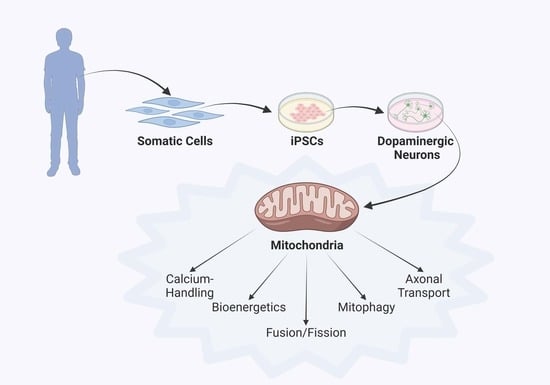
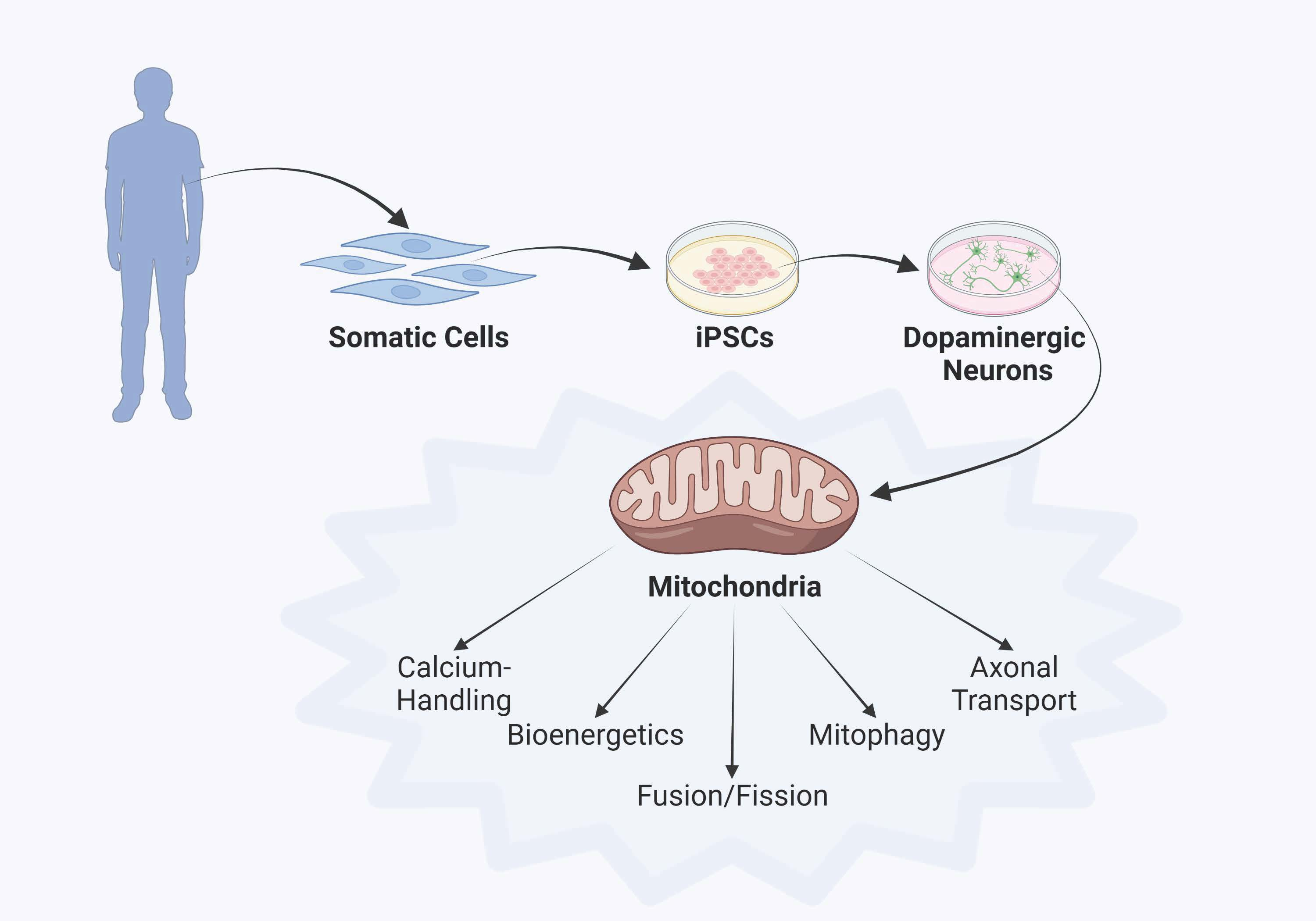
3. Parkinson’s Disease Modeling Using Human iPSC Technology

Like other neurodegenerative disorders, PD remains a sizeable clinical challenge with no cure and few, symptomatic treatment options. Disease models are indispensable tools for uncovering pathological mechanisms of neurodegeneration and development of novel therapeutics. The implementation of increasingly sophisticated animal models has led to greater understanding of the molecular and cellular mechanisms driving disease processes and has provided in vivo systems for assessment of innovative intervention strategies. Unfortunately, many therapeutic efforts developed in animals fall short in clinical trials. To overcome these persistent translational challenges, there is a critical need to generate disease models that more faithfully recapitulate human pathophysiology.
With the inaccessibility of primary neurons from human patients, iPSC technology provides an unprecedented tool to study the most affected cell populations from individuals with a verified pathogenic genetic background [27,28]. iPSCs can self-renew and differentiate into various cell lineages, offering a potentially unlimited platform to examine diverse cellular phenotypes and interactions. iPSC-derived platforms employed as in vitro disease models have added invaluable insight into PD pathogenesis by manifesting characteristics commonly found in human PD pathology [29,30]. This foundation allows for the investigation of phenotype-correcting effects of novel compounds on patient-specific iPSC-derived model systems, providing a valuable pipeline for the identification and preclinical development of therapeutic agents [31]. Since the advent of iPSC technology, enormous strides have been made in stem cell biology and regenerative medicine. Moreover, differentiation protocols and quality control criteria are regularly updated and improved, making iPSCs a rapidly evolving technology for physiologically-relevant in vitro modeling of human disease. These advances have generated protocols which routinely yield neuronal cultures comprised of 50–70% midbrain dopaminergic neurons, supporting the investigation of the physiology and function of this unique neuronal subset [32,33,34]. Given the numerous advantages of iPSCs over alternative models—including their human origin, the ability to be differentiated into many cell types, particularly those with limited accessibility, and the therapeutic potential of patient-specific iPSCs in personalized medicine—iPSC-based platforms present a unique opportunity for exploring disease pathogenesis and identifying novel drug targets for debilitating neurodegenerative diseases. In recent years, a number of research groups have successfully generated iPSCs from patients with sporadic or familial PD, and mounting evidence has revealed mitochondrial disturbances as a significant, and potentially shared, contributing factor to pathology, highlighting the importance of this organelle for neuronal health and disease.
Despite the many advantages, there are also limitations to the use of iPSC-derived neurons as a model system. To investigate pathogenic mechanisms relevant to PD, it is critical to establish mature dopaminergic neuronal cultures. While some indicators of dopaminergic lineage are present by 35 days in culture including tyrosine hydroxylase (TH) expression and dopamine synthesis and release, reports have shown that functional properties of iPSC-derived dopaminergic neurons change dynamically between 6 and 10 weeks in culture. In fact, spontaneous calcium oscillations and autonomous pacemaking activity are not fully established until roughly 70 days in culture [35]. In support of this, one group reported greater expression of immature, and lesser expression of mature, dopamine neuron transcripts at 50 days in culture compared to their primary human midbrain counterparts [36]. Thus, it is critical to allow sufficient time in culture for these model neurons to reach maturity and enable more accurate comparisons to the adult human brain. iPSC-derived neurons have found widespread use in early stage drug discovery pipelines, have been successfully used to test the impact of compounds with known modes of action, and have opened new avenues for evaluating drug repurposing strategies [37]. However, the sole use of two-dimensional monolayer cultures is unlikely to recapitulate the complexity and function of three-dimensional (3D) in vivo neural circuits, and do not take into account the unique challenge of the blood-brain barrier or the whole organism [38]. In contrast to single cell-type cultures, organoids consist of multiple cell types that self-organize spatially and can display enhanced cellular maturation and functionality, possibly due to the more physiological 3D niche environment [39]. Moreover, co-culture systems of neurons and glia in bioengineered neural circuit model systems are needed to fully understand non-cell autonomous determinants of PD disease progression. Astrocytes were previously considered merely metabolic support for neurons, but recent reports have revealed their active participation in regulating neuronal activity, the formation of neural networks, recycling of neurotransmitters, providing neurotrophic factors and detoxification [40,41,42].Another pitfall in iPSC modeling is the lack of standardization among differentiation protocols leading to variability in culture quality, maturation status, and experimental outcomes and data interpretation. The importance of this was shown in a study from Chung and coworkers which demonstrated the recapitulation of disease-related phenotypes in PD patient iPSC-derived midbrain dopamine neurons generated using a floor-plate-based protocol, but a lack of observable phenotypes using a neural-rosette-based directed differentiation strategy [43]. Another major confound is donor-to-donor variability, particularly among patients carrying identical genetic risk variants. To minimize this challenge, new technologies such as clustered regularly interspaced short palindromic repeats (CRISPR) genome editing can be combined with human iPSC model systems to generate isogenic mutant and control cell lines, facilitating the delineation of pathogenicity of variants of uncertain significance [44].
While iPSCs hold great promise for PD, there are still scientific and clinical challenges that must be addressed to improve the translational potential for development of novel therapeutics. Nevertheless, iPSC technology has significantly changed the field of neurodegenerative research and will be instrumental in improving our understanding of mechanistic pathways in the pathology of PD.

4. Discoveries of Mitochondria-Specific Phenotypes in iPSC Models of PD

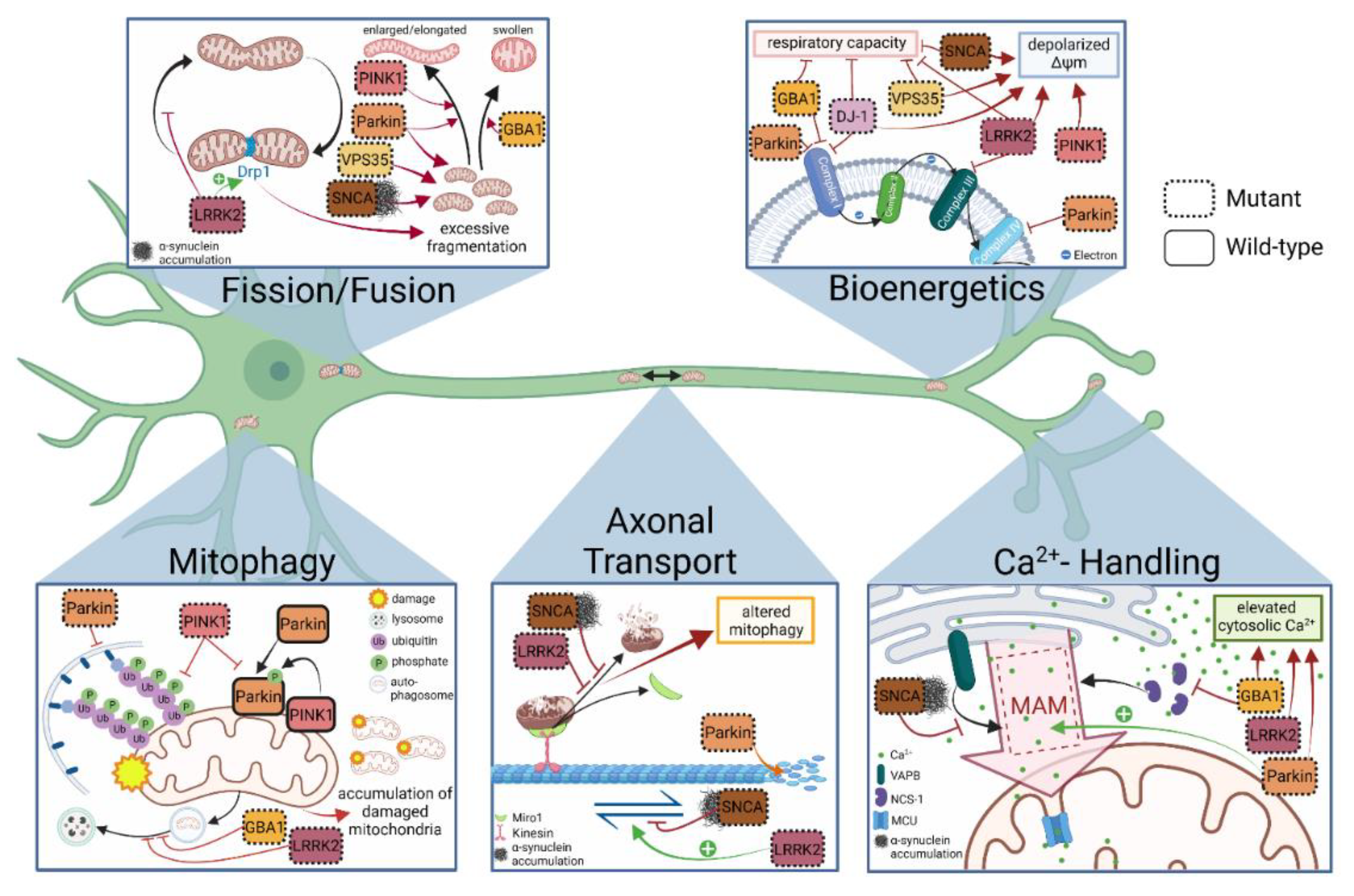
4.1. Mitochondrial Respiration and Membrane Integrity

Mitochondrial ATP synthesis is accomplished through the combined activity of the tricarboxylic acid cycle (TCA) (also known as the Krebs or citric acid cycle). This series of chemical reactions occurs in the mitochondrial matrix, feeding high energy NADH and FADH2 into the electron transport chain (ETC) in the inner mitochondrial membrane (IMM) to facilitate OXPHOS, the primary source of energy production in neurons. The flow of electrons from NADH or FADH2 to O2 through the ETC protein complexes I-V facilitates the pumping of protons out of the mitochondrial matrix. The resultant uneven distribution of protons generates a pH gradient and a transmembrane potential (ΔΨm) that creates a proton force and drives ATP synthesis through the activity of complex V, ATP synthase [45,46]. OXPHOS also generates potentially toxic byproducts in the form of ROS, which, if not tightly regulated by antioxidant defenses, can disrupt mitochondrial physiology, damage DNA, modify protein functions, cause lipid peroxidation, and negatively influence signaling pathways including transcriptional regulation [47,48]. Hence, all mitochondrial processes can be impacted by unbalanced ROS regulation and may thus represent a common pathway/potential starting point for neurodegenerative diseases [49,50,51]. Neurons are characterized by a high metabolic demand and require reliable ATP synthesis for diverse neuro-communication functions including maintenance of resting membrane potential, neurotransmitter synthesis and release [52], thus, disruption of this process is particularly detrimental to neuron health and survival. Consequently, determination of mitochondrial respiration defects, namely expression and function of ETC protein complexes, OXPHOS dynamics, changes to ΔΨm, and ATP synthesis capacity have become a major focus of PD research.
Several lines of evidence have emerged linking deficient ETC complex activity and dopaminergic degeneration. Early examinations of human post-mortem SNpc tissue demonstrated decreased complex I activity [53,54], and investigations in rodent neurons yielded insight into possible mechanism by showing that interactions of pathological α-syn oligomers, but not monomers, disrupted complex I and complex V of the ETC resulting in impaired respiration [55]. The connection between dysfunction of complex I and PD emerged in the mid-1980s, when young adults developed severe and irreversible parkinsonism shortly after injecting a new designer street drug contaminated with the compound MPTP (1-methyl-4-phenyl-1,2,3,6-tetrahydropyridine), a potent neurotoxin targeting mitochondria in dopaminergic neurons within the SNpc [56,57]. MPTP’s active metabolite MPP+ (N-methyl-phenylpyridinium ion) is a known inhibitor of complex I of the ETC [58,59], causing decreased production of ATP [57] and elevated generation of ROS [60]. Interestingly, deficient complex I activity has been confirmed in animal and non-neuronal models of genetic PD, i.e., Parkin [61], PINK1 [62], and DJ-1 [63,64,65,66], with the latter being shown to be able to directly bind to subunits of complex I [63]. In iPSC-derived neuronal models to date, impaired ETC complex I activity has been described in Parkin mutant [67] and GBA1 mutant neurons [68], while LRRK2 G2019S mutant neurons displayed reduced complex III expression [69] (Figure 1).
However, this finding has been debated for Parkin mutant neurons, as a recent proteomic analysis reported unchanged complex I levels but rather specific suppression of complex IV proteins [70]. This did not correlate with impairments in mitochondrial respiration, but rather impaired glycolysis and lactate metabolism which was associated with worsened neuronal viability. This finding may be noteworthy as astrocyte-derived lactate has proven neuroprotective potential in several pathological conditions [71,72]. Interestingly, the mitochondrial protein stomatin-like protein 2 (SLP-2) interacts with both Parkin and cardiolipin and promotes the assembly of respiratory chain proteins, and overexpression of this protein in Parkin mutant models rescued complex I function in iPSC-derived neurons [67].
While different PD genotypes appear to impact distinct components of the respiratory machinery, the resulting mitochondrial respiratory defects show many similarities. As determined by live-cell assessment of oxygen consumption rates (OCR), basal, maximal, and spare mitochondrial respiratory capacity is reduced in both VPS35 [73] and GBA1 mutant iPSC-derived dopaminergic neurons [68]. In a direct comparison of LRRK2 G2019S PD patient iPSC-derived dopaminergic, glutamatergic, and peripheral sensory neurons [69], only the central neuronal subtypes displayed impaired ATP-linked, maximal, and spare respiratory capacity, with intact basal mitochondrial respiration. However, deficits in basal respiration have been reported in LRRK2 G2019S and LRRK2 R1441C patient dopaminergic neurons [74]. Overall, this suggests that neurons of the central, but not the peripheral, nervous system are more susceptible to mitochondrial deficits. It was further demonstrated that only dopaminergic neurons had reduced ATP and ADP pools, which may have been associated with weakened PGC1α-dependent mitochondrial biogenesis [69], providing another potential mechanism contributing to the vulnerability of dopamine neurons. Interestingly, some of these impairments were also reported in LRRK2 G2019S neural stem cells (NSCs), indicating some pathological phenotypes may be neurodevelopmental [75]. In SNCA triplication PD patient iPSC-derived neural progenitor cells (NPCs), as well as CRISPR-induced SNCA mutant (A30P and A53T) neuroepithelial-like stem cells (NESCs) and patient-derived dopaminergic neurons increased α-syn oligomer burden was correlated with suppressed basal, ATP-linked, and spare respiratory capacity [76,77,78], as well as decreased ATP production which could be restored with SNCA knockdown [77]. Studies using mature patient-derived iPSC dopamine neurons from PD patients carrying either the A53T α-syn mutation or a triplication of the SNCA locus reported reduced maximal and spare mitochondrial respiration capacity and ATP production, with conflicting results on impaired basal respiration [79,80]. These investigations further demonstrate that despite a spectrum of mutation-specific impact on cellular bioenergetics, up-to-date investigations of PD neurons share the common hallmark of repressed mitochondrial respiratory function at baseline and/or in response to metabolic stress.
Proper functioning of the ETC generates and maintains the ΔΨm to facilitate ATP synthase activity and maintain organellar fitness. Sustained loss of membrane integrity can lead to impaired OXPHOS, reduced ATP pools, release of cytochrome c into the cytosol, and triggering of cell death pathways. In iPSC-based neuronal models of PD, NPCs [77], cortical neurons [55], and dopaminergic neurons [81] from SNCA mutant or triplication PD patients, as well as NESCs from LRRK2 G2019S PD patients [75] and dopaminergic neurons from VPS35 [73], displayed reduced ΔΨm. In iPSC-derived neurons from PD patients with PINK1 mutations, ΔΨm and ATP content was lowered [82,83], consistent with an earlier report of Morais and coworkers which demonstrated in animal models of diminished PINK1 function that impaired complex I function resulted in mitochondrial depolarization, reduced ATP synthesis, and increased sensitivity to apoptotic stress [62]. Interestingly, iPSC-derived dopaminergic neurons from PD patients with heterozygous GBA1 mutations (N370S, L444P, and RecNcil) showed no alterations to ΔΨm, despite substantial respiratory defects and elevated mitochondrial ROS generation [68]. Dysfunction of the ETC, compromised ΔΨm, and superoxide and ROS by-products of OXPHOS are strongly connected with the generation of mitochondrial oxidant stress in both DJ-1 knockout animals [25] and iPSC-derived dopaminergic neurons from PD patients with a loss-of-protein mutation in DJ-1 [34], as was demonstrated using a redox-sensitive variant of green fluorescent protein (roGFP) with a mitochondrial matrix targeting sequence. Interestingly, the latter study found a time-dependent toxic cascade starting with mitochondrial oxidant stress and reduced basal respiration leading to accumulation of α-syn and lysosomal dysfunction in DJ-1 mutant PD neurons and identified oxidized dopamine derivates as the mediators of this pathogenic sequence.
Taken together, the use of iPSC-derived patient neurons has revealed that while mitochondrial respiration may represent a shared pathology across sporadic and genetic forms of PD, the manner and extent to which these interconnected processes are influenced by accumulated α-syn and diverse genetic backgrounds varies and will therefore require more complex and personalized targeting for therapeutic development in future.

4.2. Mitochondrial Fusion, Fission and Morphology

Mitochondria are highly dynamic organelles that are able to adapt to different cellular states by changing their morphology [84]. To maintain network integrity, mitochondria utilize both fission and fusion to sustain fitness levels. These balanced transitions preserve organellar function and allow for versatile responses to cellular needs by adapting the network to nutrient availability or optimal metabolic state of the cell [85,86,87].
Mitochondrial fission is the division of a single organelle into two or more independent structures. This process is mediated by two proteins: dynamin-related protein 1 (Drp1) as part of the GTPase family and human fission 1 (hFis1) located in the outer mitochondrial membrane (OMM) which coordinate membrane scission [88], while the IMM constriction is independently regulated by calcium influx [86,89]. The two-step process of fusion results in the union of two mitochondria and both their inner and outer membranes. First, trans-dimerization between homologous proteins mitofusion 1 and 2 (MFN1/2) leads to formation of a curved complex which tethers both membranes together, followed by the GTPase activity which allows fusion of the OMM by initiating conformational change [90]. Subsequent fusion of the IMM is dependent on membrane potential and the GTPase optic atrophy protein 1 (OPA1) [91]. The regulation of mitochondrial size and length is very important for their subcellular localization and overall number. As neurons are post-mitotic and do not undergo further cell division, fusion and fission enable the exchange of mitochondrial contents and equal distribution of metabolites [92,93,94]. This ability to continuously adjust to the energy demands in distinct cellular compartments is particularly important for neurons which possess a highly polarized structure with areas of unique metabolic requirements and different mitochondrial distributions. Furthermore, fission is a critical prerequisite for isolating depolarized mitochondria for the selective autophagic process termed mitophagy and lysosomal-based degradation [95].
Abnormalities in mitochondrial fission or fusion have been suggested to occur as early events during the pathogenesis of many neurological disease states [93]. However, the exact mechanism of altered mitochondrial dynamics and morphology associated with mutations in various PD-associated genes is controversial. As an example, while Parkin mutations in Drosophila lead to abnormal mitochondrial morphology [96], mitochondria in Parkin knockout mice display no gross morphological abnormalities [97]. Interestingly, different Parkin patient models also differ in their representation of mitochondrial alterations, i.e., iPSC-derived neurons, but not fibroblasts or iPSCs, from patients with Parkin mutations present with abnormal or enlarged mitochondria [98]. This was independently confirmed in Parkin patient iPSC-derived dopamine neurons, with similar phenotypes observed in PINK1 patient neurons as well [43]. However, no altered mitochondrial morphology was observed in homozygous PINK1 knockout human dopaminergic neurons [99]. An increase in the numbers of elongated mitochondria in Parkin patient neurons was also reported by Bogetofte and colleagues [70]. Interestingly, their mitochondria-specific proteomic analysis revealed a large number of dysregulated mitochondrial proteins in Parkin knockout iPSC-derived dopaminergic neurons compared to isogenic controls. Conversely, Parkin patient-derived NPCs showed a subtle, but significant elevation in mitochondrial fragmentation that dose-dependently increased in severity with copper exposure, a known environmental risk factor for PD [100]. A recent study using Parkin KO iPSC-derived neurons elegantly showed that smaller and less functional mitochondria were present in dopaminergic, but absent in non-dopaminergic neurons using a tyrosine hydroxylase reporter cassette and correlative light electron microscopy [101]. This suggests that dopaminergic neurons possess intrinsic physiological differences, making them particularly vulnerable compared to other types of neurons. Moreover, iPSC-derived NESCs [75] and dopaminergic neurons [102] from LRRK2 G2019S patients with hyperactive LRRK2 displayed excessive mitochondrial fission and more fragmented organelles. Mechanistically, this may be linked with LRRK2-mediated increase in Drp1 activity, as pharmacological or genetic modification of Drp1 altered its translocation to the OMM and restored mitochondrial fission, thereby providing neuroprotective effects [102]. Finally, smaller and more fragmented mitochondria were observed in mice with dopamine neuron-specific deletion of VPS35, which was correlated with downregulated MFN2 expression [103], and in iPSC-derived dopaminergic neurons from PD patients with the VPS35 D620N mutation, which also demonstrated decreased network connectivity [73].
Direct association between mitochondrial membranes and α-syn lead to mitochondrial fission and disorganized cristae independent of Drp1, as demonstrated in α-syn-overexpressing HeLa cells and murine midbrain neurons [104]. While patient-derived NPCs from α-syn triplication patients did not show differences in mitochondrial shape [77], the phenotype of fragmented mitochondria associated with altered α-syn was confirmed in iPSC-derived dopaminergic neurons from patients with SNCA mutation (A53T) [105]. Interestingly, cardiolipin translocation to the OMM and the subsequent loss of mitochondrial surface charge preceded this mitochondrial fragmentation pathology in SNCA mutant neurons, revealing a potential novel mechanism. Altered cristae morphology and increased mitochondrial diameter was also observed in iPSC-derived dopaminergic neurons from PD patients carrying heterozygous GBA1 mutations (N370S, L444P, and RecNcil), and were concomitant with dysregulated levels of mitochondrial shaping proteins Drp1, OPA1, and Mfn1 [68].
The regulation of mitochondrial morphology is a complex balance between fusion- and fission-related proteins and situation-dependent distribution events. The exact link between altered mitochondrial morphology and the development of neurodegenerative diseases like PD is not yet clear, and will require more studies on disease-relevant cell types to shed light on the link between the actual structure of mitochondria and pathological processes in neurons.

4.3. Axonal Transport of Mitochondria

Mitochondrial transport allows cells to respond to regional changes in metabolism and protect mitochondrial integrity. In neurons, the location of mitochondria is particularly important due to the unique architecture of highly branched dendrites and long, complex axons which can extend up to one meter, as is the case for some human motor neurons. Mitochondrial proteins are primarily synthesized in the soma and therefore must be transported to distal sites to provide the energy required to maintain cellular function. Axonal transport of mitochondria happens bidirectionally and is almost entirely microtubule-dependent. Anterograde transport supplies distal axons with organelles, proteins and lipids required for maintaining presynaptic activity, while retrograde transport removes aged components that are primarily degraded and recycled back in the soma. Mitochondrial cargo is transported via the motor protein kinesin-1 [106] that binds to the membrane anchors Miro1/2, small Rho GTPases, and their motor adaptors Trak1+2 (Milton) to control proper anterograde trafficking to the synaptic compartment [107,108,109]. Retrograde transport of mitochondria uses the major motor protein dynein with its activator dynactin, a highly conserved multiprotein complex essential for normal neuronal function [110]. Perturbations in either form of axonal transport can be detrimental to neuronal health, causing metabolic dyshomeostasis, oxidative stress, and even cell death.
While various model systems have already contributed valuable insight into axonal transport of mitochondria, human iPSC-derived platforms offer the unique opportunity to disentangle this process in a human, potentially patient-derived system over prolonged periods of time in culture. Indeed, one report using wild-type iPSC-derived dopaminergic neurons “aged” in culture revealed a time-dependent increase in immobile mitochondria and reduction in anterograde transport from 40 to 100 days [111]. This may point to a general decline in mitochondrial trafficking during “aging” of human neurons with eventual consequences on neuronal viability.
Disturbed mitochondrial motility as a major factor in the development of PD is a longstanding debate. Animal studies have established that early signs of the disease begin in synaptic terminals, suggesting dysregulation of axonal transport of proteins and organelles, including ATP-generating mitochondria, towards the synapse as an early event in PD pathogenesis [112,113]. In recent years, data from human and mouse models suggest that dysfunction of the microtubule network contributes to the pathogenesis of PD, however, the extent of altered microtubules in PD patients is not yet clear. Patient fibroblast studies revealed microtubule destabilization in the context of PD pathogenesis [114], and initial studies in iPSC-derived neuronal cells from PD patients carrying mutations in LRRK2 confirmed altered mitochondrial mobility in the axon [69,74]. Fragmentation of microtubules also preceded mitochondrial transport defects in iPSC-derived dopamine neurons from PD patients with Parkin mutations, as well as in a knockout mouse model [115].
Further studies address the effects of PD-associated mutations on mitochondrial motility by modifying proteins that regulate mitochondrial trafficking in disease-relevant models. Miro1, the primary regulator of mitochondrial transport in both axons and dendrites, is particularly relevant to PD. In a Miro1 knockout mouse model [116], loss of protein function slowed mitochondrial trafficking along microtubules. In line with this, PD patient iPSC-derived neurons with mutant Miro1 (Miro-R272Q) exhibited decreased mitochondrial velocity when compared with control neurons [117]. Further, α-syn oligomerization in different iPSC-derived neuronal models of aberrant α-syn expression (duplication, mutant E46K, oligomer-prone E57K a-syn mutant) lead to a reduction of Miro1 and kinesin light chain 1 (KCL1) proteins in neurites resulting in disrupted anterograde axonal transport of mitochondria [118]. As an OMM protein, Miro1 anchors mitochondria to microtubule motors. To eliminate damaged mitochondria, Miro1 is removed from mitochondria, a procedure that arrests organellar transport and facilitates mitochondrial clearance. Interestingly, a study using human fibroblasts and iPSC-derived dopaminergic neurons demonstrated that LRRK2 forms a complex with Miro1 as a precondition for its removal from damaged mitochondria, a process that is disrupted in LRRK2 G2019S PD patient models [119]. In this study, parameters of mitochondrial transport were not changed under basal conditions comparing LRRK2 G2019S mutant neurons with controls. However, treatment with the complex III inhibitor antimycin A, known to trigger mitochondrial depolarization and clearance, reduced mitochondrial motility and induced mitochondrial clearance in wild-type neurons, a process that was heavily delayed in LRRK2 mutant neurons. Similarly, in a following study from the same group, baseline mitochondrial motility was unchanged between A53T α-syn mutant PD patient and control neurons, while antimycin A treatment evoked cessation of mitochondrial motility and subsequent mitophagy in wild-type neurons, both of which were delayed in SNCA mutant [120] and sporadic PD neurons [120,121]. Interestingly, pharmacological correction of Miro1 dynamics in both A53T α-syn mutant and sporadic PD iPSC neurons promoted clearance of damaged mitochondria and proved neuroprotective against mitochondrial stressors [121]. Dysregulation of Miro1 removal kinetics on mitochondria has emerged as a potential common mechanism shared by both sporadic and familial PD pathology, and may therefore represent a novel therapeutic target [122].

4.4. Mitophagy

Due to their post-mitotic state and high bioenergetic demand, neurons are particularly susceptible to mitochondrial dysfunction. Thus, mitochondrial quality control is essential for neuronal survival as defective mitochondria release ROS, eventually leading to neurodegeneration and cell death [123]. Dysfunctional mitochondria are removed via mitophagy, a selective form of autophagy.
In mammals, there are several pathways and proteins involved in mitophagy such as PINK1, Parkin, BNIP3L/NIX and FUNDC1 [124]. However, PINK1/Parkin-dependent mitophagy is the best characterized pathway for mitochondrial quality control [125,126,127,128]. Importantly, mutations in both PINK1 and Parkin are known to cause early-onset forms of PD [129,130]. In this pathway, PINK1 acts as a mitochondrial stress sensor. Under physiological conditions, PINK1 is imported into mitochondria via the translocase of the outer membrane (TOM) and the translocase of the inner membrane (TIM), dependent on the mitochondrial membrane potential [131]. Once PINK1 reaches the IMM, it is immediately cleaved and degraded [132]. In defective mitochondria, however, the compromised membrane potential impairs mitochondrial import of PINK1. Consequently, full-length PINK1 accumulates on the OMM serving as an identifier for damaged mitochondria. PINK1 then phosphorylates ubiquitin molecules at serine 65 bound to OMM proteins [133,134,135] which, in turn, results in recruitment of the cytosolic E3 ubiquitin ligase Parkin to damaged mitochondria and its partial activation. Full activation of Parkin is achieved by phosphorylation at serine 65 via PINK1 [136,137]. Activated Parkin then preferentially uses phospho-ubiquitin to modify several proteins on the mitochondrial surface [138]. In this way, phosphorylated ubiquitin leads to further recruitment and activation of Parkin, creating a positive feedback loop [139,140]. Eventually, the damaged mitochondria are covered with phosphorylated ubiquitin chains, which then interact with autophagy receptors such as optineurin and nuclear dot protein 52 (NDP52). This leads to recruitment of autophagosomes and the delivery of damaged mitochondria to the lysosomes for degradation [141].
Most studies investigating PINK1/Parkin-mediated mitophagy in PD research have been performed in immortalized cell lines overexpressing PINK1 and/or Parkin. However, the existence and physiological relevance of this pathway in neurons has been a matter of debate [142]. Early investigations did not observe mitochondrial recruitment of overexpressed Parkin upon mitochondrial depolarization in primary mouse neurons [143], suggesting that bioenergetic differences between primary neurons and cultured cell lines may be responsible for the differences in Parkin translocation following mitochondrial depolarization. Subsequent studies, however, confirmed that primary rodent neurons are definitively able to undergo PINK1/Parkin-mediated mitophagy [144,145,146,147].
After having established that PINK1/Parkin-dependent mitophagy occurs in non-neuronal cells and primary rodent neurons, it remains to be elucidated whether this pathway is also relevant in human neurons. One report in human iPSC-derived dopaminergic neurons demonstrated that endogenous levels of PINK1 and Parkin are not sufficient to induce loss of all mitochondrial proteins upon loss of mitochondrial membrane potential [148]. However, this study did not directly investigate autophagosomal uptake or lysosomal degradation capacity, which may equally explain the lack of mitochondrial protein loss in iPSC neurons. Other investigations using the same cell type have shown that both endogenous [149] and overexpressed Parkin [150] are translocated to mitochondria following valinomycin-induced mitochondrial depolarization [150]. In this study, endogenous levels of PINK11 and Parkin were sufficient to lower the levels of mtDNA in response to valinomycin, consistent with a role for PINK1/Parkin in mitophagy in dopaminergic neurons [150]. Similarly, stabilization of endogenous full-length PINK1 and increased ubiquitin phosphorylation was observed [149,151]. Finally, absence of PINK1 in human iPSC-derived dopaminergic neurons leads to inhibition of ionophore-induced mitophagy, providing further evidence for the role of PINK1 in this form of mitophagy [99].
Mitophagy is impaired in many neurodegenerative diseases, leading to accumulation of defective mitochondria. Hence, human iPSC-derived neuronal models have greatly contributed to the study of mitophagic clearance in the pathogenesis of PD [152]. Several studies, outlined below, demonstrate impaired mitophagic flux in human iPSC-derived dopaminergic neurons carrying PD-related PINK1 and Parkin mutations. Parkin mutant iPSC-derived dopaminergic neurons displayed impaired mitophagy using the mt-mKeima system that allows visualization of mitochondria fused with lysosomes [153]. Interestingly, Schwartzentruber and colleagues demonstrated that mitophagy levels are dependent on the cellular energetic status in Parkin mutant iPSC-derived neurons [154]. While Parkin mutant dopaminergic neurons depend on glycolysis early in differentiation and show increased mitophagy levels, neurons dependent on OXPHOS in the end stages of dopaminergic differentiation were clearly defective in basal and induced mitophagy.
In addition, iPSC studies using PD patient-derived neurons with PINK1 mutation confirmed an impaired recruitment of overexpressed Parkin to mitochondria upon depolarization, which could be corrected via wild-type PINK1 lentiviral expression [150]. Moreover, the kinase activity and levels of phosphorylated ubiquitin (Ser65) were significantly reduced in PINK1 mutant iPSC-derived neurons [155,156]. However, PD-related SNCA and LRRK2 mutations have also been shown to disrupt the PINK1/Parkin-mediated mitophagy pathway. Human iPSC-derived dopaminergic neurons containing either the A53T α-syn or the LRRK2 G2019S mutation displayed a stabilization of Miro1, thereby delaying the arrest of damaged mitochondria on microtubules and consequently their clearance via mitophagy [119,120]. Another study using LRRK2 G2019S iPSC-derived neurons reported higher mitochondrial motility and elevated bidirectional movement interfering with proper mitochondrial degradation processes [74]. This suggests that LRRK2 mutations can actively prevent mitochondria from undergoing mitophagy. Furthermore, in iPSC-derived LRRK2 G2019S knock-in neurons, the pathogenic increase in LRRK2 kinase activity was elegantly correlated with malfunctions in autophagosome transport, potentially revealing a mechanism for impaired mitophagy in cells with fully functional PINK1 and Parkin [157]. Lastly, PD-related mutations in GBA1 have also been associated with impaired mitophagy. Using GBA1 mutant iPSC-derived dopaminergic neurons reduced expression of the mitophagy adaptor protein BNIP3L/NIX and diminished mitochondrial-lysosomal colocalization was reported [68]. Interestingly, a study using non-neuronal cells and fly models reported a physiological regulatory mechanism of PINK1/Parkin-mediated mitophagy by BNIP3 [158]. However, whether and how the interaction of PINK1/Parkin-dependent mitophagy pathway coordinates with other mitophagy pathways remains unclear. Additional investigations may help to understand how other mitophagy receptors including BNIP3L/NIX and FUNDC1 may possibly also compensate for defects in the PINK1/Parkin pathway.

4.5. Mitochondrial Calcium Handling

Another important mitochondrial function is the regulation of intracellular Ca2+ needed for a multitude of cellular processes [159]. In neurons, the uptake of cytosolic or endoplasmic reticulum (ER)-stored Ca2+ into mitochondria regulates cellular Ca2+ homeostasis, prevents excitotoxicity, and facilitates neurotransmission [160,161]. Under physiological conditions, small Ca2+ oscillations are constitutively transferred from ER to mitochondria at contact sites termed mitochondria-associated ER membranes (MAMs), or present near voltage-gated calcium channels (VGCCs) in the synaptic terminal. Ca2+ enters the mitochondrial matrix through the mitochondrial Ca2+ uniporter (MCU) [162,163] where it is needed to maintain enzymatic function of the TCA cycle and subsequently ATP production [5]. This conversion of Ca2+ transients into ATP is especially crucial in the synaptic terminal, where mitochondria generate the fuel necessary for many aspects of neurotransmission. Mitochondria maintain their homeostasis through efflux of excess matrix Ca2+ via the mitochondrial permeability transition pore (mPTP), however, large Ca2+ transfers can lead to overload and subsequent opening of the mPTP, collapse of membrane potential, diminished OXPHOS and ATP production, elevated ROS formation, release of cytochrome c, and eventual cell death [164,165,166,167]. The ability of mitochondria to absorb, store, and release Ca2+ has been extensively reviewed elsewhere [168,169], therefore, the following section will focus on findings of mitochondrial Ca2+ dynamics in iPSC-derived neuronal models of PD.
Given the importance of mitochondrial function in highly-energetic neurons, deregulated mitochondrial Ca2+ dynamics and neuronal distress are closely intertwined. In PD, the highly vulnerable dopaminergic neurons of the SNpc exhibit autonomous pacemaking activity accompanied by increased oscillations resulting from high presynaptic density of the L-type voltage-gated calcium channel Cav1.3 [170,171]. This causes continuous influx and increased cytosolic concentrations of Ca2+ which, when combined with the limited Ca2+-buffering capacity [172], has been proposed to contribute to their unique vulnerability in PD [172,173]. Importantly, selectively antagonizing L-type Ca2+ channels using isradipine has been proposed to be neuroprotective in iPSC-derived dopaminergic neurons from PD patients carrying heterozygous and homozygous mutations in DJ-1, a protein known to have a role in oxidative defense mechanisms [34]. In this study, the isradipine-mediated reduction of Ca2+ influx was sufficient to abolish the accumulation of neurotoxic oxidized dopamine, a precursor to neuromelanin build-up. In contrast, one group demonstrated that isradipine was not protective in iPSC-derived dopaminergic neurons from PD patients with Parkin mutations, showing instead that elevated expression and activity of T-type Ca2+ channels are more deleterious for this pathogenic genotype [174]. Parkin mutant iPSC-derived dopaminergic neurons present with increased association between ER and mitochondria, and subsequently increased mitochondrial uptake of ER-released Ca2+, as well as decreased cytosolic Ca2+ transients [175]. Conversely, iPSC-derived dopaminergic neurons from patients carrying GBA1 mutations (N370S, L444P, and RecNcil) showed increased basal and caffeine-induced cytosolic Ca2+ levels, indicating impaired mitochondrial Ca2+ buffering and ER-mitochondrial Ca2+ transfer Additionally, LRRK2 G2019S PD patient neurons showed elevated intracellular Ca2+, possibly due to LRRK2 hyperactivity upregulating translation of Ca2+ signaling proteins [176]. Interestingly, these neurons also benefited from isradipine application, indicating that L-type Ca2+ channels are also dysregulated by LRRK2 overactivity.
Regulation of Ca2+ transfer from ER to mitochondria at MAM sites is critically important and is mediated by both ER- and mitochondrial-associated protein partners. One such ER-associated protein, vesicle-associated membrane protein-associated protein B (VAPB), was recently shown to bind α-syn [177]. Intriguingly, dopaminergic neurons from PD patients with SNCA triplication show high interference of α-syn with VAPB binding its partner on the OMM, resulting in disrupted MAM tethering, reduced Ca2+ transfer, and weakened mitochondrial ATP synthesis [177]. Interestingly, iPSC-derived dopaminergic neurons from GBA1 PD patients (N370S mutation) exhibited reduced levels of neuronal calcium sensor-1 (NCS-1) [24], a Ca2+-binding protein which facilitates MAM formation and Ca2+ transfer into mitochondria [178]. This protein demonstrated neuroprotection against neurodegeneration in a toxin-induced mouse model of PD, which was associated with regulation of nigral dopaminergic Ca2+ dynamics [24]. This suggests that insufficient NCS-1 activity may impair mitochondrial Ca2+ handling and contribute to the unique SNpc vulnerability events in human neurons. Altogether, mishandled Ca2+ appears to represent a consistent pathological phenotype in PD iPSC-derived dopaminergic neurons, with different genetic backgrounds causing distinct mitochondrial alterations in Ca2+ regulation which contribute to their unique predisposed risk to neurodegeneration.
Taken together, these findings demonstrate the value of patient-specific iPSCs for the delineation of disease- and cell type-specific mitochondrial Ca2+ handling pathologies including deregulated ER-associated channels and tethering machinery, synaptic terminal voltage-gated and Ca2+-permeable receptors, regulatory proteins and macromolecular complexes, and organellar membrane dynamics, all in a sophisticated model system with high translational relevance.

5. Conclusions

One of the greatest breakthroughs of regenerative medicine in this century was the discovery of iPSC technology in 2006 by Shinya Yamanaka and Kazutoshi Takahashi. Currently, human iPSC model systems are routinely used for disease modeling, deciphering cellular disease mechanisms, drug testing and screening, genetic engineering, and other valuable applications. In PD, patient-specific iPSC disease modeling has accelerated our understanding of the molecular mechanisms underlying dopamine neuron vulnerability and degeneration. The current lack of animal models capable of recapitulating human PD pathology in its entirety highlights the significant challenges in accurately defining the mechanisms underlying PD pathophysiology. PD-derived iPSC models are therefore a very powerful tool for examining disease mechanisms, defining novel targets, and developing effective therapeutic treatments in the future. Furthermore, advances in generating and differentiating iPSCs with greater consistency and reproducibility make iPSCs a promising cell source for tissue regeneration therapy. However, before iPSCs can be routinely used in clinical practice, their efficacy and safety need to be rigorously tested.
Mitochondrial dysfunction has been associated with neurodegeneration in PD for decades, but the precise role of mitochondrial perturbations in the vulnerability of dopaminergic neurons in PD remains undetermined. iPSC technology has broadened the understanding of mitochondria in health and disease, and therapies targeting mitochondrial dysfunction represent a promising potential tool to mitigate the damage to dopamine neurons in PD. Despite much progress in determining cell autonomous, disease-relevant phenotypes in human neurons (Table 1), more complex model systems with the inclusion of non-cell autonomous determinants of pathophysiology are needed to comprehensively define the molecular mechanisms underlying dopaminergic neuronal vulnerability.
In summary, iPSC disease modeling is an invaluable research tool to further understanding of disease progression and accelerate identification of relevant targets for drug development, which has immense potential to benefit large cohorts of patients affected by neurodegenerative diseases.

Author Contributions

Conceptualization, L.F.B.; writing—original draft preparation, L.M.H., R.M.W., J.T.H., A.B.H., L.F.B.; writing—review and editing, L.M.H., R.M.W., J.T.H., A.B.H., L.F.B. All authors have read and agreed to the published version of the manuscript.

Funding

L.F.B. is supported by the European Research Council (ERC) under the European Union’s Horizon 2020 research and innovation programme (grant agreement No. [948027]). A.B.H is supported by the Max Planck Society and the Deutsche Forschungsgemeinschaft (DFG, German Research Foundation, HA7728/2-1). L.F.B. and A.B.H. are supported by the DFG under Germany’s Excellence Strategy within the framework of the Munich Cluster of Systems Neurology (EXC 2145 SyNergy—ID 390857198).

Institutional Review Board Statement

Not applicable.

Informed Consent Statement

Not applicable.

Data Availability Statement

Not applicable.

Acknowledgments

Figures created with BioRender.com.

Conflicts of Interest

The authors declare no conflict of interest.

References

  1. Rolfe, D.F.; Brown, G.C. Cellular energy utilization and molecular origin of standard metabolic rate in mammals. Physiol. Rev. 1997, 77, 731–758. [Google Scholar] [CrossRef] [PubMed] [Green Version]
  2. Nicholls, D.G.; Budd, S.L. Mitochondria and neuronal survival. Physiol. Rev. 2000, 80, 315–360. [Google Scholar] [CrossRef] [PubMed] [Green Version]
  3. Engl, E.; Attwell, D. Non-signalling energy use in the brain. J. Physiol. 2015, 593, 3417–3429. [Google Scholar] [CrossRef] [Green Version]
  4. Balaban, R.S.; Nemoto, S.; Finkel, T. Mitochondria, Oxidants, and Aging. Cell 2005, 120, 483–495. [Google Scholar] [CrossRef] [Green Version]
  5. Rizzuto, R.; De Stefani, D.; Raffaello, A.; Mammucari, C. Mitochondria as sensors and regulators of calcium signalling. Nature Rev. Mol. Cell Biol. 2012, 13, 566–578. [Google Scholar] [CrossRef]
  6. Payne, B.A.I.; Chinnery, P.F. Mitochondrial dysfunction in aging: Much progress but many unresolved questions. Biochim. Et Biophys. Acta 2015, 1847, 1347–1353. [Google Scholar] [CrossRef] [PubMed] [Green Version]
  7. Falkenburger, B.H.; Saridaki, T.; Dinter, E. Cellular models for Parkinson’s disease. J. Neurochem. 2016, 139 (Suppl. S1), 121–130. [Google Scholar] [CrossRef]
  8. Jagmag, S.A.; Tripathi, N.; Shukla, S.D.; Maiti, S.; Khurana, S. Evaluation of Models of Parkinson’s Disease. Front. Neurosci. 2016, 9, 503. [Google Scholar] [CrossRef] [PubMed] [Green Version]
  9. Kin, K.; Yasuhara, T.; Kameda, M.; Date, I. Animal Models for Parkinson’s Disease Research: Trends in the 2000s. Int. J. Mol. Sci. 2019, 20, 5402. [Google Scholar] [CrossRef] [Green Version]
  10. Nussbaum, R.L.; Ellis, C.E. Alzheimer’s disease and Parkinson’s disease. New Engl. J. Med. 2003, 348, 1356–1364. [Google Scholar] [CrossRef] [Green Version]
  11. Schrag, A.; Horsfall, L.; Walters, K.; Noyce, A.; Petersen, I. Prediagnostic presentations of Parkinson’s disease in primary care: A case-control study. Lancet Neurol. 2015, 14, 57–64. [Google Scholar] [CrossRef] [Green Version]
  12. Poewe, W.; Seppi, K.; Tanner, C.M.; Halliday, G.M.; Brundin, P.; Volkmann, J.; Schrag, A.E.; Lang, A.E. Parkinson disease. Nat. Rev. Dis. Primers. 2017, 3, 17013. [Google Scholar] [CrossRef] [PubMed]
  13. Healy, D.G.; Falchi, M.; O’Sullivan, S.S.; Bonifati, V.; Durr, A.; Bressman, S.; Brice, A.; Aasly, J.; Zabetian, C.P.; Goldwurm, S.; et al. Phenotype, genotype, and worldwide genetic penetrance of LRRK2-associated Parkinson’s disease: A case-control study. Lancet Neurol. 2008, 7, 583–590. [Google Scholar] [CrossRef] [Green Version]
  14. Sidransky, E.; Nalls, M.A.; Aasly, J.O.; Aharon-Peretz, J.; Annesi, G.; Barbosa, E.R.; Bar-Shira, A.; Berg, D.; Bras, J.; Brice, A.; et al. Multicenter analysis of glucocerebrosidase mutations in Parkinson’s disease. New Engl. J. Med. 2009, 361, 1651–1661. [Google Scholar] [CrossRef] [PubMed] [Green Version]
  15. Chartier-Harlin, M.C.; Kachergus, J.; Roumier, C.; Mouroux, V.; Douay, X.; Lincoln, S.; Levecque, C.; Larvor, L.; Andrieux, J.; Hulihan, M.; et al. Alpha-synuclein locus duplication as a cause of familial Parkinson’s disease. Lancet 2004, 364, 1167–1169. [Google Scholar] [CrossRef]
  16. Singleton, A.B.; Farrer, M.; Johnson, J.; Singleton, A.; Hague, S.; Kachergus, J.; Hulihan, M.; Peuralinna, T.; Dutra, A.; Nussbaum, R.; et al. alpha-Synuclein locus triplication causes Parkinson’s disease. Science 2003, 302, 841. [Google Scholar] [CrossRef] [PubMed] [Green Version]
  17. Spillantini, M.G.; Schmidt, M.L.; Lee, V.M.Y.; Trojanowski, J.Q.; Jakes, R.; Goedert, M. α-Synuclein in Lewy bodies. Nature 1997, 388, 839–840. [Google Scholar] [CrossRef] [PubMed]
  18. Mahul-Mellier, A.L.; Burtscher, J.; Maharjan, N.; Weerens, L.; Croisier, M.; Kuttler, F.; Leleu, M.; Knott, G.W.; Lashuel, H.A. The process of Lewy body formation, rather than simply α-synuclein fibrillization, is one of the major drivers of neurodegeneration. Proc. Natl. Acad. Sci. USA 2020, 117, 4971–4982. [Google Scholar] [CrossRef] [PubMed] [Green Version]
  19. Postuma, R.B.; Aarsland, D.; Barone, P.; Burn, D.J.; Hawkes, C.H.; Oertel, W.; Ziemssen, T. Identifying prodromal Parkinson’s disease: Pre-motor disorders in Parkinson’s disease. Mov. Disord. 2012, 27, 617–626. [Google Scholar] [CrossRef]
  20. Alberico, S.L.; Cassell, M.D.; Narayanan, N.S. The Vulnerable Ventral Tegmental Area in Parkinson’s Disease. Basal Ganglia 2015, 5, 51–55. [Google Scholar] [CrossRef] [PubMed] [Green Version]
  21. Braak, H.; Del Tredici, K. Poor and protracted myelination as a contributory factor to neurodegenerative disorders. Neurobiol Aging 2004, 25, 19–23. [Google Scholar] [CrossRef] [PubMed]
  22. Matsuda, W.; Furuta, T.; Nakamura, K.C.; Hioki, H.; Fujiyama, F.; Arai, R.; Kaneko, T. Single nigrostriatal dopaminergic neurons form widely spread and highly dense axonal arborizations in the neostriatum. J. Neurosci. 2009, 29, 444–453. [Google Scholar] [CrossRef] [PubMed] [Green Version]
  23. Pacelli, C.; Giguere, N.; Bourque, M.J.; Levesque, M.; Slack, R.S.; Trudeau, L.E. Elevated Mitochondrial Bioenergetics and Axonal Arborization Size Are Key Contributors to the Vulnerability of Dopamine Neurons. Curr. Biol. 2015, 25, 2349–2360. [Google Scholar] [CrossRef] [PubMed] [Green Version]
  24. Benkert, J.; Hess, S.; Roy, S.; Beccano-Kelly, D.; Wiederspohn, N.; Duda, J.; Simons, C.; Patil, K.; Gaifullina, A.; Mannal, N.; et al. Cav2.3 channels contribute to dopaminergic neuron loss in a model of Parkinson’s disease. Nat. Commun. 2019, 10, 5094. [Google Scholar] [CrossRef]
  25. Guzman, J.N.; Sanchez-Padilla, J.; Wokosin, D.; Kondapalli, J.; Ilijic, E.; Schumacker, P.T.; Surmeier, D.J. Oxidant stress evoked by pacemaking in dopaminergic neurons is attenuated by DJ-1. Nature 2010, 468, 696–700. [Google Scholar] [CrossRef] [Green Version]
  26. Guzman, J.N.; Sanchez-Padilla, J.; Chan, C.S.; Surmeier, D.J. Robust pacemaking in substantia nigra dopaminergic neurons. J. Neurosci. 2009, 29, 11011–11019. [Google Scholar] [CrossRef] [PubMed]
  27. Takahashi, K.; Tanabe, K.; Ohnuki, M.; Narita, M.; Ichisaka, T.; Tomoda, K.; Yamanaka, S. Induction of pluripotent stem cells from adult human fibroblasts by defined factors. Cell 2007, 131, 861–872. [Google Scholar] [CrossRef] [Green Version]
  28. Unternaehrer, J.J.; Daley, G.Q. Induced pluripotent stem cells for modelling human diseases. Philos. Trans. R Soc. Lond. B Biol. Sci. 2011, 366, 2274–2285. [Google Scholar] [CrossRef] [PubMed]
  29. Kampmann, M. CRISPR-based functional genomics for neurological disease. Nat. Rev. Neurol. 2020, 16, 465–480. [Google Scholar] [CrossRef]
  30. Soldner, F.; Jaenisch, R. Medicine. iPSC disease modeling. Science 2012, 338, 1155–1156. [Google Scholar] [CrossRef] [PubMed]
  31. Robinton, D.A.; Daley, G.Q. The promise of induced pluripotent stem cells in research and therapy. Nature 2012, 481, 295–305. [Google Scholar] [CrossRef]
  32. Schöndorf, D.C.; Aureli, M.; McAllister, F.E.; Hindley, C.J.; Mayer, F.; Schmid, B.; Sardi, S.P.; Valsecchi, M.; Hoffmann, S.; Schwarz, L.K.; et al. iPSC-derived neurons from GBA1-associated Parkinson’s disease patients show autophagic defects and impaired calcium homeostasis. Nat. Commun. 2014, 5, 4028. [Google Scholar] [CrossRef] [PubMed] [Green Version]
  33. Mazzulli, J.R.; Zunke, F.; Tsunemi, T.; Toker, N.J.; Jeon, S.; Burbulla, L.F.; Patnaik, S.; Sidransky, E.; Marugan, J.J.; Sue, C.M.; et al. Activation of β-Glucocerebrosidase Reduces Pathological α-Synuclein and Restores Lysosomal Function in Parkinson’s Patient Midbrain Neurons. J. Neurosci. 2016, 36, 7693–7706. [Google Scholar] [CrossRef] [PubMed]
  34. Burbulla, L.F.; Song, P.; Mazzulli, J.R.; Zampese, E.; Wong, Y.C.; Jeon, S.; Santos, D.P.; Blanz, J.; Obermaier, C.D.; Strojny, C.; et al. Dopamine oxidation mediates mitochondrial and lysosomal dysfunction in Parkinson’s disease. Science 2017, 357, 1255–1261. [Google Scholar] [CrossRef] [Green Version]
  35. Hartfield, E.M.; Yamasaki-Mann, M.; Ribeiro Fernandes, H.J.; Vowles, J.; James, W.S.; Cowley, S.A.; Wade-Martins, R. Physiological characterisation of human iPS-derived dopaminergic neurons. PLoS ONE 2014, 9, e87388. [Google Scholar] [CrossRef]
  36. Xia, N.; Zhang, P.; Fang, F.; Wang, Z.; Rothstein, M.; Angulo, B.; Chiang, R.; Taylor, J.; Reijo Pera, R.A. Transcriptional comparison of human induced and primary midbrain dopaminergic neurons. Sci. Rep. 2016, 6, 20270. [Google Scholar] [CrossRef] [PubMed] [Green Version]
  37. Little, D.; Ketteler, R.; Gissen, P.; Devine, M.J. Using stem cell-derived neurons in drug screening for neurological diseases. Neurobiol Aging 2019, 78, 130–141. [Google Scholar] [CrossRef] [PubMed]
  38. Sasai, Y. Next-generation regenerative medicine: Organogenesis from stem cells in 3D culture. Cell Stem Cell 2013, 12, 520–530. [Google Scholar] [CrossRef] [PubMed] [Green Version]
  39. Kelava, I.; Lancaster, M.A. Dishing out mini-brains: Current progress and future prospects in brain organoid research. Dev. Biol. 2016, 420, 199–209. [Google Scholar] [CrossRef] [Green Version]
  40. Nedergaard, M.; Ransom, B.; Goldman, S.A. New roles for astrocytes: Redefining the functional architecture of the brain. Trends Neurosci. 2003, 26, 523–530. [Google Scholar] [CrossRef] [PubMed]
  41. Ricci, G.; Volpi, L.; Pasquali, L.; Petrozzi, L.; Siciliano, G. Astrocyte-neuron interactions in neurological disorders. J. Biol Phys. 2009, 35, 317–336. [Google Scholar] [CrossRef] [Green Version]
  42. Krencik, R.; Ullian, E.M. A cellular star atlas: Using astrocytes from human pluripotent stem cells for disease studies. Front. Cell. Neurosci. 2013, 7, 25. [Google Scholar] [CrossRef] [PubMed] [Green Version]
  43. Chung, S.Y.; Kishinevsky, S.; Mazzulli, J.R.; Graziotto, J.; Mrejeru, A.; Mosharov, E.V.; Puspita, L.; Valiulahi, P.; Sulzer, D.; Milner, T.A.; et al. Parkin and PINK1 Patient iPSC-Derived Midbrain Dopamine Neurons Exhibit Mitochondrial Dysfunction and alpha-Synuclein Accumulation. Stem Cell Rep. 2016, 7, 664–677. [Google Scholar] [CrossRef] [Green Version]
  44. Mali, P.; Yang, L.; Esvelt, K.M.; Aach, J.; Guell, M.; DiCarlo, J.E.; Norville, J.E.; Church, G.M. RNA-guided human genome engineering via Cas9. Science 2013, 339, 823–826. [Google Scholar] [CrossRef] [PubMed] [Green Version]
  45. Vogel, F.; Bornhovd, C.; Neupert, W.; Reichert, A.S. Dynamic subcompartmentalization of the mitochondrial inner membrane. J. Cell Biol. 2006, 175, 237–247. [Google Scholar] [CrossRef]
  46. Frey, T.G.; Mannella, C.A. The internal structure of mitochondria. Trends Biochem. Sci. 2000, 25, 319–324. [Google Scholar] [CrossRef]
  47. Murphy, M.P. How mitochondria produce reactive oxygen species. Biochem. J. 2009, 417, 1–13. [Google Scholar] [CrossRef] [Green Version]
  48. Zorov, D.B.; Juhaszova, M.; Sollott, S.J. Mitochondrial reactive oxygen species (ROS) and ROS-induced ROS release. Physiol. Rev. 2014, 94, 909–950. [Google Scholar] [CrossRef] [Green Version]
  49. Angelova, P.R.; Abramov, A.Y. Role of mitochondrial ROS in the brain: From physiology to neurodegeneration. FEBS Lett. 2018, 592, 692–702. [Google Scholar] [CrossRef]
  50. Kolodkin, N.A.; Sharma, R.P.; Colangelo, A.M.; Ignatenko, A.; Martorana, F.; Jennen, D.; Briedé, J.J.; Brady, N.; Barberis, M.; Mondeel, T.D.G.A.; et al. ROS networks: Designs, aging, Parkinson’s disease and precision therapies. NPJ Syst. Biol. Appl. 2020, 6, 34. [Google Scholar] [CrossRef] [PubMed]
  51. Lin, M.T.; Beal, M.F. Mitochondrial dysfunction and oxidative stress in neurodegenerative diseases. Nature 2006, 443, 787–795. [Google Scholar] [CrossRef]
  52. Kann, O.; Kovács, R. Mitochondria and neuronal activity. Am. J. Physiol. Cell Physiol. 2007, 292, C641–C657. [Google Scholar] [CrossRef]
  53. Schapira, A.H.; Cooper, J.M.; Dexter, D.; Clark, J.B.; Jenner, P.; Marsden, C.D. Mitochondrial complex I deficiency in Parkinson’s disease. J. Neurochem. 1990, 54, 823–827. [Google Scholar] [CrossRef]
  54. Parker, W.D., Jr.; Boyson, S.J.; Parks, J.K. Abnormalities of the electron transport chain in idiopathic Parkinson’s disease. Ann. Neurol. 1989, 26, 719–723. [Google Scholar] [CrossRef]
  55. Ludtmann, M.H.R.; Angelova, P.R.; Horrocks, M.H.; Choi, M.L.; Rodrigues, M.; Baev, A.Y.; Berezhnov, A.V.; Yao, Z.; Little, D.; Banushi, B.; et al. alpha-synuclein oligomers interact with ATP synthase and open the permeability transition pore in Parkinson’s disease. Nat. Commun. 2018, 9, 2293. [Google Scholar] [CrossRef] [Green Version]
  56. Ramsay, R.R.; Dadgar, J.; Trevor, A.; Singer, T.P. Energy-driven uptake of N-methyl-4-phenylpyridine by brain mitochondria mediates the neurotoxicity of MPTP. Life Sci. 1986, 39, 581–588. [Google Scholar] [CrossRef]
  57. Langston, J.W.; Ballard, P.; Tetrud, J.W.; Irwin, I. Chronic Parkinsonism in humans due to a product of meperidine-analog synthesis. Science 1983, 219, 979–980. [Google Scholar] [CrossRef] [PubMed] [Green Version]
  58. Mizuno, Y.; Sone, N.; Suzuki, K.; Saitoh, T. Studies on the toxicity of 1-methyl-4-phenylpyridinium ion (MPP+) against mitochondria of mouse brain. J. Neurol. Sci. 1988, 86, 97–110. [Google Scholar] [CrossRef]
  59. Nicklas, W.J.; Vyas, I.; Heikkila, R.E. Inhibition of NADH-linked oxidation in brain mitochondria by 1-methyl-4-phenyl-pyridine, a metabolite of the neurotoxin, 1-methyl-4-phenyl-1,2,5,6-tetrahydropyridine. Life Sci. 1985, 36, 2503–2508. [Google Scholar] [CrossRef]
  60. Salama, M.; Arias-Carrión, O. Natural toxins implicated in the development of Parkinson’s disease. Ther. Adv. Neurol. Disord. 2011, 4, 361–373. [Google Scholar] [CrossRef]
  61. Ming, F.; Tan, J.; Qin, L.; Zhang, H.; Tang, J.; Tan, X.; Wang, C. The PARK2 Mutation Associated with Parkinson’s Disease Enhances the Vulnerability of Peripheral Blood Lymphocytes to Paraquat. BioMed. Res. Int. 2020, 2020, 4658109. [Google Scholar] [CrossRef]
  62. Morais, V.A.; Verstreken, P.; Roethig, A.; Smet, J.; Snellinx, A.; Vanbrabant, M.; Haddad, D.; Frezza, C.; Mandemakers, W.; Vogt-Weisenhorn, D.; et al. Parkinson’s disease mutations in PINK1 result in decreased Complex I activity and deficient synaptic function. EMBO Mol. Med. 2009, 1, 99–111. [Google Scholar] [CrossRef]
  63. Hayashi, T.; Ishimori, C.; Takahashi-Niki, K.; Taira, T.; Kim, Y.-c.; Maita, H.; Maita, C.; Ariga, H.; Iguchi-Ariga, S.M.M. DJ-1 binds to mitochondrial complex I and maintains its activity. Biochem. Biophys. Res. Commun. 2009, 390, 667–672. [Google Scholar] [CrossRef] [Green Version]
  64. Krebiehl, G.; Ruckerbauer, S.; Burbulla, L.F.; Kieper, N.; Maurer, B.; Waak, J.; Wolburg, H.; Gizatullina, Z.; Gellerich, F.N.; Woitalla, D.; et al. Reduced basal autophagy and impaired mitochondrial dynamics due to loss of Parkinson’s disease-associated protein DJ-1. PLoS ONE 2010, 5, e9367. [Google Scholar] [CrossRef] [Green Version]
  65. Kim, R.H.; Smith, P.D.; Aleyasin, H.; Hayley, S.; Mount, M.P.; Pownall, S.; Wakeham, A.; You-Ten, A.J.; Kalia, S.K.; Horne, P.; et al. Hypersensitivity of DJ-1-deficient mice to 1-methyl-4-phenyl-1,2,3,6-tetrahydropyrindine (MPTP) and oxidative stress. Proc. Natl. Acad. Sci. USA 2005, 102, 5215–5220. [Google Scholar] [CrossRef] [Green Version]
  66. Meulener, M.; Whitworth, A.J.; Armstrong-Gold, C.E.; Rizzu, P.; Heutink, P.; Wes, P.D.; Pallanck, L.J.; Bonini, N.M. Drosophila DJ-1 mutants are selectively sensitive to environmental toxins associated with Parkinson’s disease. Curr. Biol. 2005, 15, 1572–1577. [Google Scholar] [CrossRef] [Green Version]
  67. Zanon, A.; Kalvakuri, S.; Rakovic, A.; Foco, L.; Guida, M.; Schwienbacher, C.; Serafin, A.; Rudolph, F.; Trilck, M.; Grünewald, A.; et al. SLP-2 interacts with Parkin in mitochondria and prevents mitochondrial dysfunction in Parkin-deficient human iPSC-derived neurons and Drosophila. Hum. Mol. Genet. 2017, 26, 2412–2425. [Google Scholar] [CrossRef] [Green Version]
  68. Schondorf, D.C.; Ivanyuk, D.; Baden, P.; Sanchez-Martinez, A.; De Cicco, S.; Yu, C.; Giunta, I.; Schwarz, L.K.; Di Napoli, G.; Panagiotakopoulou, V.; et al. The NAD+ Precursor Nicotinamide Riboside Rescues Mitochondrial Defects and Neuronal Loss in iPSC and Fly Models of Parkinson’s Disease. Cell Rep. 2018, 23, 2976–2988. [Google Scholar] [CrossRef]
  69. Schwab, A.J.; Sison, S.L.; Meade, M.R.; Broniowska, K.A.; Corbett, J.A.; Ebert, A.D. Decreased Sirtuin Deacetylase Activity in LRRK2 G2019S iPSC-Derived Dopaminergic Neurons. Stem Cell Rep. 2017, 9, 1839–1852. [Google Scholar] [CrossRef] [PubMed] [Green Version]
  70. Bogetofte, H.; Jensen, P.; Ryding, M.; Schmidt, S.I.; Okarmus, J.; Ritter, L.; Worm, C.S.; Hohnholt, M.C.; Azevedo, C.; Roybon, L.; et al. PARK2 Mutation Causes Metabolic Disturbances and Impaired Survival of Human iPSC-Derived Neurons. Front. Cell Neurosci. 2019, 13, 297. [Google Scholar] [CrossRef] [Green Version]
  71. Wyss, M.T.; Jolivet, R.; Buck, A.; Magistretti, P.J.; Weber, B. In vivo evidence for lactate as a neuronal energy source. J. Neurosci. 2011, 31, 7477–7485. [Google Scholar] [CrossRef] [PubMed] [Green Version]
  72. Jourdain, P.; Allaman, I.; Rothenfusser, K.; Fiumelli, H.; Marquet, P.; Magistretti, P.J. L-Lactate protects neurons against excitotoxicity: Implication of an ATP-mediated signaling cascade. Sci. Rep. 2016, 6, 21250. [Google Scholar] [CrossRef] [Green Version]
  73. Hanss, Z.; Larsen, S.B.; Antony, P.; Mencke, P.; Massart, F.; Jarazo, J.; Schwamborn, J.C.; Barbuti, P.A.; Mellick, G.D.; Krüger, R. Mitochondrial and Clearance Impairment in p.D620N VPS35 Patient-Derived Neurons. Mov. Disord. 2021, 36, 704–715. [Google Scholar] [CrossRef] [PubMed]
  74. Cooper, O.; Seo, H.; Andrabi, S.; Guardia-Laguarta, C.; Graziotto, J.; Sundberg, M.; McLean, J.R.; Carrillo-Reid, L.; Xie, Z.; Osborn, T.; et al. Pharmacological rescue of mitochondrial deficits in iPSC-derived neural cells from patients with familial Parkinson’s disease. Sci. Transl. Med. 2012, 4, 141–190. [Google Scholar] [CrossRef] [Green Version]
  75. Walter, J.; Bolognin, S.; Antony, P.M.A.; Nickels, S.L.; Poovathingal, S.K.; Salamanca, L.; Magni, S.; Perfeito, R.; Hoel, F.; Qing, X.; et al. Neural Stem Cells of Parkinson’s Disease Patients Exhibit Aberrant Mitochondrial Morphology and Functionality. Stem Cell Rep. 2019, 12, 878–889. [Google Scholar] [CrossRef] [Green Version]
  76. Arias-Fuenzalida, J.; Jarazo, J.; Qing, X.; Walter, J.; Gomez-Giro, G.; Nickels, S.L.; Zaehres, H.; Scholer, H.R.; Schwamborn, J.C. FACS-Assisted CRISPR-Cas9 Genome Editing Facilitates Parkinson’s Disease Modeling. Stem Cell Rep. 2017, 9, 1423–1431. [Google Scholar] [CrossRef] [Green Version]
  77. Flierl, A.; Oliveira, L.M.; Falomir-Lockhart, L.J.; Mak, S.K.; Hesley, J.; Soldner, F.; Arndt-Jovin, D.J.; Jaenisch, R.; Langston, J.W.; Jovin, T.M.; et al. Higher vulnerability and stress sensitivity of neuronal precursor cells carrying an alpha-synuclein gene triplication. PLoS ONE 2014, 9, e112413. [Google Scholar] [CrossRef] [Green Version]
  78. Ryan, S.D.; Dolatabadi, N.; Chan, S.F.; Zhang, X.; Akhtar, M.W.; Parker, J.; Soldner, F.; Sunico, C.R.; Nagar, S.; Talantova, M.; et al. Isogenic human iPSC Parkinson’s model shows nitrosative stress-induced dysfunction in MEF2-PGC1alpha transcription. Cell 2013, 155, 1351–1364. [Google Scholar] [CrossRef] [Green Version]
  79. Zambon, F.; Cherubini, M.; Fernandes, H.J.R.; Lang, C.; Ryan, B.J.; Volpato, V.; Bengoa-Vergniory, N.; Vingill, S.; Attar, M.; Booth, H.D.E.; et al. Cellular alpha-synuclein pathology is associated with bioenergetic dysfunction in Parkinson’s iPSC-derived dopamine neurons. Hum. Mol. Genet. 2019, 28, 2001–2013. [Google Scholar] [CrossRef]
  80. Ryan, B.J.; Lourenco-Venda, L.L.; Crabtree, M.J.; Hale, A.B.; Channon, K.M.; Wade-Martins, R. alpha-Synuclein and mitochondrial bioenergetics regulate tetrahydrobiopterin levels in a human dopaminergic model of Parkinson disease. Free Radic. Biol. Med. 2014, 67, 58–68. [Google Scholar] [CrossRef] [Green Version]
  81. Little, D.; Luft, C.; Mosaku, O.; Lorvellec, M.; Yao, Z.; Paillusson, S.; Kriston-Vizi, J.; Gandhi, S.; Abramov, A.Y.; Ketteler, R.; et al. A single cell high content assay detects mitochondrial dysfunction in iPSC-derived neurons with mutations in SNCA. Sci. Rep. 2018, 8, 9033. [Google Scholar] [CrossRef] [Green Version]
  82. Vos, M.; Geens, A.; Böhm, C.; Deaulmerie, L.; Swerts, J.; Rossi, M.; Craessaerts, K.; Leites, E.P.; Seibler, P.; Rakovic, A.; et al. Cardiolipin promotes electron transport between ubiquinone and complex I to rescue PINK1 deficiency. J. Cell Biol. 2017, 216, 695–708. [Google Scholar] [CrossRef] [PubMed]
  83. Morais, V.A.; Haddad, D.; Craessaerts, K.; De Bock, P.-J.; Swerts, J.; Vilain, S.; Aerts, L.; Overbergh, L.; Grünewald, A.; Seibler, P.; et al. PINK1 Loss-of-Function Mutations Affect Mitochondrial Complex I Activity via NdufA10 Ubiquinone Uncoupling. Science 2014, 344, 203–207. [Google Scholar] [CrossRef]
  84. Safiulina, D.; Kaasik, A. Energetic and Dynamic: How Mitochondria Meet Neuronal Energy Demands. PLoS Biol. 2014, 11, e1001755. [Google Scholar] [CrossRef] [PubMed]
  85. Wai, T.; Langer, T. Mitochondrial Dynamics and Metabolic Regulation. Trends Endocrinol. Metab. 2016, 27, 105–117. [Google Scholar] [CrossRef]
  86. Tilokani, L.; Nagashima, S.; Paupe, V.; Prudent, J. Mitochondrial dynamics: Overview of molecular mechanisms. Essays Biochem. 2018, 62, 341–360. [Google Scholar] [CrossRef] [Green Version]
  87. Liu, Y.J.; McIntyre, R.L.; Janssens, G.E.; Houtkooper, R.H. Mitochondrial fission and fusion: A dynamic role in aging and potential target for age-related disease. Mech. Ageing Dev. 2020, 186, 111212. [Google Scholar] [CrossRef] [PubMed]
  88. Knott, A.B.; Bossy-Wetzel, E. Impairing the mitochondrial fission and fusion balance: A new mechanism of neurodegeneration. Ann. N. Y. Acad. Sci. 2008, 1147, 283–292. [Google Scholar] [CrossRef] [Green Version]
  89. MacAskill, A.F.; Kittler, J.T. Control of mitochondrial transport and localization in neurons. Trends Cell Biol. 2010, 20, 102–112. [Google Scholar] [CrossRef] [PubMed]
  90. Koshiba, T.; Detmer, S.A.; Kaiser, J.T.; Chen, H.; McCaffery, J.M.; Chan, D.C. Structural basis of mitochondrial tethering by mitofusin complexes. Science 2004, 305, 858–862. [Google Scholar] [CrossRef] [Green Version]
  91. Meeusen, S.; McCaffery, J.M.; Nunnari, J. Mitochondrial fusion intermediates revealed in vitro. Science 2004, 305, 1747–1752. [Google Scholar] [CrossRef] [PubMed] [Green Version]
  92. Sheng, Z.-H.; Cai, Q. Mitochondrial transport in neurons: Impact on synaptic homeostasis and neurodegeneration. Nat. Rev. Neurosci. 2012, 13, 77–93. [Google Scholar] [CrossRef] [PubMed] [Green Version]
  93. Itoh, K.; Nakamura, K.; Iijima, M.; Sesaki, H. Mitochondrial dynamics in neurodegeneration. Trends Cell Biol. 2013, 23, 64–71. [Google Scholar] [CrossRef] [PubMed] [Green Version]
  94. Youle, R.J.; van der Bliek, A.M. Mitochondrial fission, fusion, and stress. Science 2012, 337, 1062–1065. [Google Scholar] [CrossRef] [PubMed] [Green Version]
  95. Twig, G.; Elorza, A.; Molina, A.J.; Mohamed, H.; Wikstrom, J.D.; Walzer, G.; Stiles, L.; Haigh, S.E.; Katz, S.; Las, G.; et al. Fission and selective fusion govern mitochondrial segregation and elimination by autophagy. Embo. J. 2008, 27, 433–446. [Google Scholar] [CrossRef] [PubMed] [Green Version]
  96. Greene, J.C.; Whitworth, A.J.; Kuo, I.; Andrews, L.A.; Feany, M.B.; Pallanck, L.J. Mitochondrial pathology and apoptotic muscle degeneration in Drosophila parkin mutants. Proc. Natl. Acad. Sci. USA 2003, 100, 4078–4083. [Google Scholar] [CrossRef] [PubMed] [Green Version]
  97. Palacino, J.J.; Sagi, D.; Goldberg, M.S.; Krauss, S.; Motz, C.; Wacker, M.; Klose, J.; Shen, J. Mitochondrial dysfunction and oxidative damage in parkin-deficient mice. J. Biol. Chem. 2004, 279, 18614–18622. [Google Scholar] [CrossRef] [Green Version]
  98. Imaizumi, Y.; Okada, Y.; Akamatsu, W.; Koike, M.; Kuzumaki, N.; Hayakawa, H.; Nihira, T.; Kobayashi, T.; Ohyama, M.; Sato, S.; et al. Mitochondrial dysfunction associated with increased oxidative stress and alpha-synuclein accumulation in PARK2 iPSC-derived neurons and postmortem brain tissue. Mol. Brain 2012, 5, 35. [Google Scholar] [CrossRef] [PubMed] [Green Version]
  99. Bus, C.; Zizmare, L.; Feldkaemper, M.; Geisler, S.; Zarani, M.; Schaedler, A.; Klose, F.; Admard, J.; Mageean, C.J.; Arena, G.; et al. Human Dopaminergic Neurons Lacking PINK1 Exhibit Disrupted Dopamine Metabolism Related to Vitamin B6 Co-Factors. iScience 2020, 23, 101797. [Google Scholar] [CrossRef] [PubMed]
  100. Aboud, A.A.; Tidball, A.M.; Kumar, K.K.; Neely, M.D.; Han, B.; Ess, K.C.; Hong, C.C.; Erikson, K.M.; Hedera, P.; Bowman, A.B. PARK2 patient neuroprogenitors show increased mitochondrial sensitivity to copper. Neurobiol. Dis. 2015, 73, 204–212. [Google Scholar] [CrossRef] [Green Version]
  101. Yokota, M.; Kakuta, S.; Shiga, T.; Ishikawa, K.-i.; Okano, H.; Hattori, N.; Akamatsu, W.; Koike, M. Establishment of an in vitro model for analyzing mitochondrial ultrastructure in PRKN-mutated patient iPSC-derived dopaminergic neurons. Mol. Brain 2021, 14, 58. [Google Scholar] [CrossRef] [PubMed]
  102. Su, Y.C.; Qi, X. Inhibition of excessive mitochondrial fission reduced aberrant autophagy and neuronal damage caused by LRRK2 G2019S mutation. Hum. Mol. Genet. 2013, 22, 4545–4561. [Google Scholar] [CrossRef] [Green Version]
  103. Tang, F.L.; Liu, W.; Hu, J.X.; Erion, J.R.; Ye, J.; Mei, L.; Xiong, W.C. VPS35 Deficiency or Mutation Causes Dopaminergic Neuronal Loss by Impairing Mitochondrial Fusion and Function. Cell Rep. 2015, 12, 1631–1643. [Google Scholar] [CrossRef] [PubMed] [Green Version]
  104. Nakamura, K.; Nemani, V.M.; Azarbal, F.; Skibinski, G.; Levy, J.M.; Egami, K.; Munishkina, L.; Zhang, J.; Gardner, B.; Wakabayashi, J.; et al. Direct membrane association drives mitochondrial fission by the Parkinson disease-associated protein alpha-synuclein. J. Biol. Chem. 2011, 286, 20710–20726. [Google Scholar] [CrossRef] [PubMed] [Green Version]
  105. Ryan, T.; Bamm, V.V.; Stykel, M.G.; Coackley, C.L.; Humphries, K.M.; Jamieson-Williams, R.; Ambasudhan, R.; Mosser, D.D.; Lipton, S.A.; Harauz, G.; et al. Cardiolipin exposure on the outer mitochondrial membrane modulates alpha-synuclein. Nat. Commun. 2018, 9, 817. [Google Scholar] [CrossRef] [PubMed] [Green Version]
  106. Morris, R.L.; Hollenbeck, P.J. Axonal transport of mitochondria along microtubules and F-actin in living vertebrate neurons. J. Cell Biol. 1995, 131, 1315–1326. [Google Scholar] [CrossRef] [PubMed] [Green Version]
  107. Loss, O.; Stephenson, F.A. Developmental changes in trak-mediated mitochondrial transport in neurons. Mol. Cell Neurosci. 2017, 80, 134–147. [Google Scholar] [CrossRef] [PubMed]
  108. Glater, E.E.; Megeath, L.J.; Stowers, R.S.; Schwarz, T.L. Axonal transport of mitochondria requires milton to recruit kinesin heavy chain and is light chain independent. J. Cell Biol. 2006, 173, 545–557. [Google Scholar] [CrossRef] [PubMed] [Green Version]
  109. Stowers, R.S.; Megeath, L.J.; Górska-Andrzejak, J.; Meinertzhagen, I.A.; Schwarz, T.L. Axonal transport of mitochondria to synapses depends on milton, a novel Drosophila protein. Neuron 2002, 36, 1063–1077. [Google Scholar] [CrossRef] [Green Version]
  110. LaMonte, B.H.; Wallace, K.E.; Holloway, B.A.; Shelly, S.S.; Ascaño, J.; Tokito, M.; Van Winkle, T.; Howland, D.S.; Holzbaur, E.L. Disruption of dynein/dynactin inhibits axonal transport in motor neurons causing late-onset progressive degeneration. Neuron 2002, 34, 715–727. [Google Scholar] [CrossRef] [Green Version]
  111. Burbulla, L.F.; Beaumont, K.G.; Mrksich, M.; Krainc, D. Micropatterning Facilitates the Long-Term Growth and Analysis of iPSC-Derived Individual Human Neurons and Neuronal Networks. Adv. Healthc. Mater. 2016, 5, 1894–1903. [Google Scholar] [CrossRef] [PubMed]
  112. Janezic, S.; Threlfell, S.; Dodson, P.D.; Dowie, M.J.; Taylor, T.N.; Potgieter, D.; Parkkinen, L.; Senior, S.L.; Anwar, S.; Ryan, B.; et al. Deficits in dopaminergic transmission precede neuron loss and dysfunction in a new Parkinson model. Proc. Natl. Acad. Sci. USA 2013, 110, E4016–E4025. [Google Scholar] [CrossRef] [PubMed] [Green Version]
  113. Schirinzi, T.; Madeo, G.; Martella, G.; Maltese, M.; Picconi, B.; Calabresi, P.; Pisani, A. Early synaptic dysfunction in Parkinson’s disease: Insights from animal models. Mov. Disord. 2016, 31, 802–813. [Google Scholar] [CrossRef] [PubMed]
  114. Cartelli, D.; Goldwurm, S.; Casagrande, F.; Pezzoli, G.; Cappelletti, G. Microtubule Destabilization Is Shared by Genetic and Idiopathic Parkinson’s Disease Patient Fibroblasts. PLoS ONE 2012, 7, e37467. [Google Scholar] [CrossRef]
  115. Cartelli, D.; Amadeo, A.; Calogero, A.M.; Casagrande, F.V.M.; De Gregorio, C.; Gioria, M.; Kuzumaki, N.; Costa, I.; Sassone, J.; Ciammola, A.; et al. Parkin absence accelerates microtubule aging in dopaminergic neurons. Neurobiol. Aging 2018, 61, 66–74. [Google Scholar] [CrossRef] [PubMed]
  116. López-Doménech, G.; Higgs, N.F.; Vaccaro, V.; Roš, H.; Arancibia-Cárcamo, I.L.; MacAskill, A.F.; Kittler, J.T. Loss of Dendritic Complexity Precedes Neurodegeneration in a Mouse Model with Disrupted Mitochondrial Distribution in Mature Dendrites. Cell Rep. 2016, 17, 317–327. [Google Scholar] [CrossRef] [PubMed] [Green Version]
  117. Berenguer-Escuder, C.; Grossmann, D.; Antony, P.; Arena, G.; Wasner, K.; Massart, F.; Jarazo, J.; Walter, J.; Schwamborn, J.C.; Grünewald, A.; et al. Impaired mitochondrial-endoplasmic reticulum interaction and mitophagy in Miro1-mutant neurons in Parkinson’s disease. Hum. Mol. Genet. 2020, 29, 1353–1364. [Google Scholar] [CrossRef]
  118. Prots, I.; Grosch, J.; Brazdis, R.M.; Simmnacher, K.; Veber, V.; Havlicek, S.; Hannappel, C.; Krach, F.; Krumbiegel, M.; Schutz, O.; et al. alpha-Synuclein oligomers induce early axonal dysfunction in human iPSC-based models of synucleinopathies. Proc. Natl. Acad. Sci. USA 2018, 115, 7813–7818. [Google Scholar] [CrossRef] [PubMed] [Green Version]
  119. Hsieh, C.H.; Shaltouki, A.; Gonzalez, A.E.; Bettencourt da Cruz, A.; Burbulla, L.F.; St Lawrence, E.; Schüle, B.; Krainc, D.; Palmer, T.D.; Wang, X. Functional Impairment in Miro Degradation and Mitophagy Is a Shared Feature in Familial and Sporadic Parkinson’s Disease. Cell Stem Cell 2016, 19, 709–724. [Google Scholar] [CrossRef] [Green Version]
  120. Shaltouki, A.; Hsieh, C.H.; Kim, M.J.; Wang, X. Alpha-synuclein delays mitophagy and targeting Miro rescues neuron loss in Parkinson’s models. Acta Neuropathol. 2018, 136, 607–620. [Google Scholar] [CrossRef]
  121. Hsieh, C.H.; Li, L.; Vanhauwaert, R.; Nguyen, K.T.; Davis, M.D.; Bu, G.; Wszolek, Z.K.; Wang, X. Miro1 Marks Parkinson’s Disease Subset and Miro1 Reducer Rescues Neuron Loss in Parkinson’s Models. Cell Metab. 2019, 30, 1131–1140 e1137. [Google Scholar] [CrossRef] [PubMed]
  122. Bharat, V.; Wang, X. Precision Neurology for Parkinson’s Disease: Coupling Miro1-Based Diagnosis with Drug Discovery. Mov. Disord. 2020, 35, 1502–1508. [Google Scholar] [CrossRef] [PubMed]
  123. Guo, C.; Sun, L.; Chen, X.; Zhang, D. Oxidative stress, mitochondrial damage and neurodegenerative diseases. Neural. Regen Res. 2013, 8, 2003–2014. [Google Scholar] [CrossRef] [PubMed]
  124. Ashrafi, G.; Schwarz, T.L. The pathways of mitophagy for quality control and clearance of mitochondria. Cell Death Differ. 2013, 20, 31–42. [Google Scholar] [CrossRef] [Green Version]
  125. Matsuda, N.; Sato, S.; Shiba, K.; Okatsu, K.; Saisho, K.; Gautier, C.A.; Sou, Y.S.; Saiki, S.; Kawajiri, S.; Sato, F.; et al. PINK1 stabilized by mitochondrial depolarization recruits Parkin to damaged mitochondria and activates latent Parkin for mitophagy. J. Cell Biol. 2010, 189, 211–221. [Google Scholar] [CrossRef]
  126. Narendra, D.; Tanaka, A.; Suen, D.-F.; Youle, R.J. Parkin is recruited selectively to impaired mitochondria and promotes their autophagy. J. Cell Biol. 2008, 183, 795–803. [Google Scholar] [CrossRef] [Green Version]
  127. Narendra, D.P.; Jin, S.M.; Tanaka, A.; Suen, D.F.; Gautier, C.A.; Shen, J.; Cookson, M.R.; Youle, R.J. PINK1 is selectively stabilized on impaired mitochondria to activate Parkin. PLoS Biol. 2010, 8, e1000298. [Google Scholar] [CrossRef] [PubMed] [Green Version]
  128. Vives-Bauza, C.; Zhou, C.; Huang, Y.; Cui, M.; de Vries, R.L.; Kim, J.; May, J.; Tocilescu, M.A.; Liu, W.; Ko, H.S.; et al. PINK1-dependent recruitment of Parkin to mitochondria in mitophagy. Proc. Natl. Acad. Sci. USA 2010, 107, 378–383. [Google Scholar] [CrossRef] [PubMed] [Green Version]
  129. Kitada, T.; Asakawa, S.; Hattori, N.; Matsumine, H.; Yamamura, Y.; Minoshima, S.; Yokochi, M.; Mizuno, Y.; Shimizu, N. Mutations in the parkin gene cause autosomal recessive juvenile parkinsonism. Nature 1998, 392, 605–608. [Google Scholar] [CrossRef] [PubMed]
  130. Valente, E.M.; Abou-Sleiman, P.M.; Caputo, V.; Muqit, M.M.; Harvey, K.; Gispert, S.; Ali, Z.; Del Turco, D.; Bentivoglio, A.R.; Healy, D.G.; et al. Hereditary early-onset Parkinson’s disease caused by mutations in PINK1. Science 2004, 304, 1158–1160. [Google Scholar] [CrossRef] [Green Version]
  131. Silvestri, L.; Caputo, V.; Bellacchio, E.; Atorino, L.; Dallapiccola, B.; Valente, E.M.; Casari, G. Mitochondrial import and enzymatic activity of PINK1 mutants associated to recessive parkinsonism. Hum. Mol. Genet. 2005, 14, 3477–3492. [Google Scholar] [CrossRef] [PubMed] [Green Version]
  132. Yamano, K.; Youle, R.J. PINK1 is degraded through the N-end rule pathway. Autophagy 2013, 9, 1758–1769. [Google Scholar] [CrossRef] [PubMed] [Green Version]
  133. Kane, L.A.; Lazarou, M.; Fogel, A.I.; Li, Y.; Yamano, K.; Sarraf, S.A.; Banerjee, S.; Youle, R.J. PINK1 phosphorylates ubiquitin to activate Parkin E3 ubiquitin ligase activity. J. Cell Biol. 2014, 205, 143–153. [Google Scholar] [CrossRef] [PubMed]
  134. Kazlauskaite, A.; Kondapalli, C.; Gourlay, R.; Campbell, D.G.; Ritorto, M.S.; Hofmann, K.; Alessi, D.R.; Knebel, A.; Trost, M.; Muqit, M.M. Parkin is activated by PINK1-dependent phosphorylation of ubiquitin at Ser65. Biochem. J. 2014, 460, 127–139. [Google Scholar] [CrossRef] [PubMed] [Green Version]
  135. Koyano, F.; Okatsu, K.; Kosako, H.; Tamura, Y.; Go, E.; Kimura, M.; Kimura, Y.; Tsuchiya, H.; Yoshihara, H.; Hirokawa, T.; et al. Ubiquitin is phosphorylated by PINK1 to activate parkin. Nature 2014, 510, 162–166. [Google Scholar] [CrossRef] [PubMed]
  136. Kondapalli, C.; Kazlauskaite, A.; Zhang, N.; Woodroof, H.I.; Campbell, D.G.; Gourlay, R.; Burchell, L.; Walden, H.; Macartney, T.J.; Deak, M.; et al. PINK1 is activated by mitochondrial membrane potential depolarization and stimulates Parkin E3 ligase activity by phosphorylating Serine 65. Open Biol. 2012, 2, 120080. [Google Scholar] [CrossRef] [Green Version]
  137. Shiba-Fukushima, K.; Imai, Y.; Yoshida, S.; Ishihama, Y.; Kanao, T.; Sato, S.; Hattori, N. PINK1-mediated phosphorylation of the Parkin ubiquitin-like domain primes mitochondrial translocation of Parkin and regulates mitophagy. Sci. Rep. 2012, 2, 1002. [Google Scholar] [CrossRef]
  138. Wauer, T.; Simicek, M.; Schubert, A.; Komander, D. Mechanism of phospho-ubiquitin-induced PARKIN activation. Nature 2015, 524, 370–374. [Google Scholar] [CrossRef] [PubMed] [Green Version]
  139. Okatsu, K.; Koyano, F.; Kimura, M.; Kosako, H.; Saeki, Y.; Tanaka, K.; Matsuda, N. Phosphorylated ubiquitin chain is the genuine Parkin receptor. J. Cell Biol. 2015, 209, 111–128. [Google Scholar] [CrossRef] [PubMed]
  140. Ordureau, A.; Sarraf, S.A.; Duda, D.M.; Heo, J.M.; Jedrychowski, M.P.; Sviderskiy, V.O.; Olszewski, J.L.; Koerber, J.T.; Xie, T.; Beausoleil, S.A.; et al. Quantitative proteomics reveal a feedforward mechanism for mitochondrial PARKIN translocation and ubiquitin chain synthesis. Mol. Cell 2014, 56, 360–375. [Google Scholar] [CrossRef] [Green Version]
  141. Lazarou, M.; Sliter, D.A.; Kane, L.A.; Sarraf, S.A.; Wang, C.; Burman, J.L.; Sideris, D.P.; Fogel, A.I.; Youle, R.J. The ubiquitin kinase PINK1 recruits autophagy receptors to induce mitophagy. Nature 2015, 524, 309–314. [Google Scholar] [CrossRef] [Green Version]
  142. Grenier, K.; McLelland, G.L.; Fon, E.A. Parkin- and PINK1-Dependent Mitophagy in Neurons: Will the Real Pathway Please Stand Up? Front. Neurol. 2013, 4, 100. [Google Scholar] [CrossRef] [Green Version]
  143. Van Laar, V.S.; Arnold, B.; Cassady, S.J.; Chu, C.T.; Burton, E.A.; Berman, S.B. Bioenergetics of neurons inhibit the translocation response of Parkin following rapid mitochondrial depolarization. Hum. Mol. Genet. 2011, 20, 927–940. [Google Scholar] [CrossRef] [PubMed] [Green Version]
  144. Ashrafi, G.; Schlehe, J.S.; LaVoie, M.J.; Schwarz, T.L. Mitophagy of damaged mitochondria occurs locally in distal neuronal axons and requires PINK1 and Parkin. J. Cell Biol. 2014, 206, 655–670. [Google Scholar] [CrossRef]
  145. Cai, Q.; Zakaria, H.M.; Simone, A.; Sheng, Z.H. Spatial parkin translocation and degradation of damaged mitochondria via mitophagy in live cortical neurons. Curr. Biol. 2012, 22, 545–552. [Google Scholar] [CrossRef] [Green Version]
  146. Koyano, F.; Okatsu, K.; Ishigaki, S.; Fujioka, Y.; Kimura, M.; Sobue, G.; Tanaka, K.; Matsuda, N. The principal PINK1 and Parkin cellular events triggered in response to dissipation of mitochondrial membrane potential occur in primary neurons. Genes Cells 2013, 18, 672–681. [Google Scholar] [CrossRef] [PubMed] [Green Version]
  147. Van Laar, V.S.; Roy, N.; Liu, A.; Rajprohat, S.; Arnold, B.; Dukes, A.A.; Holbein, C.D.; Berman, S.B. Glutamate excitotoxicity in neurons triggers mitochondrial and endoplasmic reticulum accumulation of Parkin, and, in the presence of N-acetyl cysteine, mitophagy. Neurobiol. Dis. 2015, 74, 180–193. [Google Scholar] [CrossRef] [PubMed] [Green Version]
  148. Rakovic, A.; Shurkewitsch, K.; Seibler, P.; Grünewald, A.; Zanon, A.; Hagenah, J.; Krainc, D.; Klein, C. Phosphatase and tensin homolog (PTEN)-induced putative kinase 1 (PINK1)-dependent ubiquitination of endogenous Parkin attenuates mitophagy: Study in human primary fibroblasts and induced pluripotent stem cell-derived neurons. J. Biol. Chem. 2013, 288, 2223–2237. [Google Scholar] [CrossRef] [PubMed] [Green Version]
  149. Oh, C.-K.; Sultan, A.; Platzer, J.; Dolatabadi, N.; Soldner, F.; McClatchy, D.B.; Diedrich, J.K.; Yates, J.R., 3rd; Ambasudhan, R.; Nakamura, T.; et al. S-Nitrosylation of PINK1 Attenuates PINK1/Parkin-Dependent Mitophagy in hiPSC-Based Parkinson’s Disease Models. Cell Rep. 2017, 21, 2171–2182. [Google Scholar] [CrossRef] [PubMed] [Green Version]
  150. Seibler, P.; Graziotto, J.; Jeong, H.; Simunovic, F.; Klein, C.; Krainc, D. Mitochondrial Parkin recruitment is impaired in neurons derived from mutant PINK1 induced pluripotent stem cells. J. Neurosci. 2011, 31, 5970–5976. [Google Scholar] [CrossRef] [PubMed]
  151. Soutar, M.P.M.; Kempthorne, L.; Miyakawa, S.; Annuario, E.; Melandri, D.; Harley, J.; O’Sullivan, G.A.; Wray, S.; Hancock, D.C.; Cookson, M.R.; et al. AKT signalling selectively regulates PINK1 mitophagy in SHSY5Y cells and human iPSC-derived neurons. Sci. Rep. 2018, 8, 8855. [Google Scholar] [CrossRef] [Green Version]
  152. Trombetta-Lima, M.; Sabogal-Guáqueta, A.M.; Dolga, A.M. Mitochondrial dysfunction in neurodegenerative diseases: A focus on iPSC-derived neuronal models. Cell Calcium. 2021, 94, 102362. [Google Scholar] [CrossRef]
  153. Suzuki, S.; Akamatsu, W.; Kisa, F.; Sone, T.; Ishikawa, K.I.; Kuzumaki, N.; Katayama, H.; Miyawaki, A.; Hattori, N.; Okano, H. Efficient induction of dopaminergic neuron differentiation from induced pluripotent stem cells reveals impaired mitophagy in PARK2 neurons. Biochem. Biophys. Res. Commun. 2017, 483, 88–93. [Google Scholar] [CrossRef]
  154. Schwartzentruber, A.; Boschian, C.; Lopes, F.M.; Myszczynska, M.A.; New, E.J.; Beyrath, J.; Smeitink, J.; Ferraiuolo, L.; Mortiboys, H. Oxidative switch drives mitophagy defects in dopaminergic parkin mutant patient neurons. Sci. Rep. 2020, 10, 15485. [Google Scholar] [CrossRef]
  155. Puschmann, A.; Fiesel, F.C.; Caulfield, T.R.; Hudec, R.; Ando, M.; Truban, D.; Hou, X.; Ogaki, K.; Heckman, M.G.; James, E.D.; et al. Heterozygous PINK1 p.G411S increases risk of Parkinson’s disease via a dominant-negative mechanism. Brain 2017, 140, 98–117. [Google Scholar] [CrossRef]
  156. Shiba-Fukushima, K.; Ishikawa, K.I.; Inoshita, T.; Izawa, N.; Takanashi, M.; Sato, S.; Onodera, O.; Akamatsu, W.; Okano, H.; Imai, Y.; et al. Evidence that phosphorylated ubiquitin signaling is involved in the etiology of Parkinson’s disease. Hum. Mol. Genet. 2017, 26, 3172–3185. [Google Scholar] [CrossRef]
  157. Boecker, C.A.; Goldsmith, J.; Dou, D.; Cajka, G.G.; Holzbaur, E.L.F. Increased LRRK2 kinase activity alters neuronal autophagy by disrupting the axonal transport of autophagosomes. Curr Biol 2021, 31, 2140–2154.e2146. [Google Scholar] [CrossRef]
  158. Zhang, T.; Xue, L.; Li, L.; Tang, C.; Wan, Z.; Wang, R.; Tan, J.; Tan, Y.; Han, H.; Tian, R.; et al. BNIP3 Protein Suppresses PINK1 Kinase Proteolytic Cleavage to Promote Mitophagy. J. Biol. Chem. 2016, 291, 21616–21629. [Google Scholar] [CrossRef] [Green Version]
  159. Friedman, J.R.; Nunnari, J. Mitochondrial form and function. Nature 2014, 505, 335–343. [Google Scholar] [CrossRef] [Green Version]
  160. Berridge, M.J. Neuronal calcium signaling. Neuron 1998, 21, 13–26. [Google Scholar] [CrossRef] [Green Version]
  161. Pivovarova, N.B.; Andrews, S.B. Calcium-dependent mitochondrial function and dysfunction in neurons. FEBS J. 2010, 277, 3622–3636. [Google Scholar] [CrossRef] [Green Version]
  162. Baughman, J.M.; Perocchi, F.; Girgis, H.S.; Plovanich, M.; Belcher-Timme, C.A.; Sancak, Y.; Bao, X.R.; Strittmatter, L.; Goldberger, O.; Bogorad, R.L.; et al. Integrative genomics identifies MCU as an essential component of the mitochondrial calcium uniporter. Nature 2011, 476, 341–345. [Google Scholar] [CrossRef] [Green Version]
  163. De Stefani, D.; Raffaello, A.; Teardo, E.; Szabo, I.; Rizzuto, R. A forty-kilodalton protein of the inner membrane is the mitochondrial calcium uniporter. Nature 2011, 476, 336–340. [Google Scholar] [CrossRef] [PubMed]
  164. Briston, T.; Roberts, M.; Lewis, S.; Powney, B.; Staddon, J.M.; Szabadkai, G.; Duchen, M.R. Mitochondrial permeability transition pore: Sensitivity to opening and mechanistic dependence on substrate availability. Sci. Rep. 2017, 7, 10492. [Google Scholar] [CrossRef] [PubMed]
  165. Haworth, R.A.; Hunter, D.R. The Ca2+-induced membrane transition in mitochondria. II. Nature of the Ca2+ trigger site. Arch. Biochem. Biophys. 1979, 195, 460–467. [Google Scholar] [CrossRef]
  166. Ichas, F.; Jouaville, L.S.; Mazat, J.P. Mitochondria are excitable organelles capable of generating and conveying electrical and calcium signals. Cell 1997, 89, 1145–1153. [Google Scholar] [CrossRef] [Green Version]
  167. Ichas, F.; Mazat, J.P. From calcium signaling to cell death: Two conformations for the mitochondrial permeability transition pore. Switching from low- to high-conductance state. Biochim. Biophys. Acta 1998, 1366, 33–50. [Google Scholar] [CrossRef] [Green Version]
  168. Giorgi, C.; Marchi, S.; Pinton, P. The machineries, regulation and cellular functions of mitochondrial calcium. Nat. Rev. Mol. Cell Biol. 2018, 19, 713–730. [Google Scholar] [CrossRef]
  169. Rossi, A.; Pizzo, P.; Filadi, R. Calcium, mitochondria and cell metabolism: A functional triangle in bioenergetics. Biochim Biophys Acta Mol. Cell Res. 2019, 1866, 1068–1078. [Google Scholar] [CrossRef]
  170. Chan, C.S.; Guzman, J.N.; Ilijic, E.; Mercer, J.N.; Rick, C.; Tkatch, T.; Meredith, G.E.; Surmeier, D.J. ‘Rejuvenation’ protects neurons in mouse models of Parkinson’s disease. Nature 2007, 447, 1081–1086. [Google Scholar] [CrossRef]
  171. Khaliq, Z.M.; Bean, B.P. Pacemaking in dopaminergic ventral tegmental area neurons: Depolarizing drive from background and voltage-dependent sodium conductances. J. Neurosci. 2010, 30, 7401–7413. [Google Scholar] [CrossRef] [PubMed]
  172. Foehring, R.C.; Zhang, X.F.; Lee, J.C.; Callaway, J.C. Endogenous calcium buffering capacity of substantia nigral dopamine neurons. J. Neurophysiol. 2009, 102, 2326–2333. [Google Scholar] [CrossRef] [PubMed] [Green Version]
  173. Surmeier, D.J.; Schumacker, P.T. Calcium, bioenergetics, and neuronal vulnerability in Parkinson’s disease. J. Biol Chem 2013, 288, 10736–10741. [Google Scholar] [CrossRef] [Green Version]
  174. Tabata, Y.; Imaizumi, Y.; Sugawara, M.; Andoh-Noda, T.; Banno, S.; Chai, M.; Sone, T.; Yamazaki, K.; Ito, M.; Tsukahara, K.; et al. T-type Calcium Channels Determine the Vulnerability of Dopaminergic Neurons to Mitochondrial Stress in Familial Parkinson Disease. Stem Cell Rep. 2018, 11, 1171–1184. [Google Scholar] [CrossRef]
  175. Gautier, C.A.; Erpapazoglou, Z.; Mouton-Liger, F.; Muriel, M.P.; Cormier, F.; Bigou, S.; Duffaure, S.; Girard, M.; Foret, B.; Iannielli, A.; et al. The endoplasmic reticulum-mitochondria interface is perturbed in PARK2 knockout mice and patients with PARK2 mutations. Hum. Mol. Genet. 2016, 25, 2972–2984. [Google Scholar] [CrossRef] [Green Version]
  176. Kim, J.W.; Yin, X.; Jhaldiyal, A.; Khan, M.R.; Martin, I.; Xie, Z.; Perez-Rosello, T.; Kumar, M.; Abalde-Atristain, L.; Xu, J.; et al. Defects in mRNA Translation in LRRK2-Mutant hiPSC-Derived Dopaminergic Neurons Lead to Dysregulated Calcium Homeostasis. Cell Stem Cell 2020, 27, 633–645.e637. [Google Scholar] [CrossRef]
  177. Paillusson, S.; Gomez-Suaga, P.; Stoica, R.; Little, D.; Gissen, P.; Devine, M.J.; Noble, W.; Hanger, D.P.; Miller, C.C.J. alpha-Synuclein binds to the ER-mitochondria tethering protein VAPB to disrupt Ca(2+) homeostasis and mitochondrial ATP production. Acta Neuropathol. 2017, 134, 129–149. [Google Scholar] [CrossRef] [Green Version]
  178. Angebault, C.; Fauconnier, J.; Patergnani, S.; Rieusset, J.; Danese, A.; Affortit, C.A.; Jagodzinska, J.; Mégy, C.; Quiles, M.; Cazevieille, C.; et al. ER-mitochondria cross-talk is regulated by the Ca(2+) sensor NCS1 and is impaired in Wolfram syndrome. Sci. Signal. 2018, 11, eaaq1380. [Google Scholar] [CrossRef] [Green Version]
Figure 1. Bioenergetics:GBA1 and Parkin deficiency, with mutant Parkin also being involved in suppression of complex IV proteins. Furthermore, a major bioenergetic burden has been reported in genetic PD neurons with mutations in DJ-1, GBA1, LRRK2, VPS35 and SNCA to result in reduced mitochondrial respiration capacity, or in neurons with mutant LRRK2, VPS35, SNCA and PINK1 to suffer from reduced mitochondrial membrane potential (ΔΨm). Fission/Fusion: The balance of fission and fusion has been shown be disrupted in several iPSC-derived models of genetic PD with mutant PINK1 and Parkin leading to enlarged/elongated organelles, and mutant GBA1 to rather swollen mitochondrial structures. Excessive mitochondrial fragmentation has been demonstrated in mutant Parkin and VPS35 neurons, as well as in neurons from SNCA and LRRK2 patients, a phenotype that is suggested to be associated with increased Drp1 activity in the latter. Axonal transport: Aberrant α-syn expression was shown to interfere with anterograde axonal transport, while mutant LRRK2 leads to enhanced mitochondrial motility in human neurons. Alterations of both proteins have also been shown to be involved in Miro1 kinetics by delaying Miro1’s removal from mitochondria, thereby interfering with proper mitochondrial clearance (mitophagy) under conditions of mitochondrial depolarization. Additionally, destruction of microtubules themselves has been reported in Parkin patient neurons. Mitophagy: PINK1/Parkin-dependent mitophagy has been well characterized, hence, not surprisingly, mutant PINK1 and Parkin patient neurons demonstrate impaired mitophagic flux, partially based on studies reporting reduced levels of phosphorylated ubiquitin (Ser65) and impaired recruitment of Parkin to mitochondria upon mitochondrial depolarization. Both LRRK2 and GBA1 mutations have been shown to interfere with autophagosome (AP) to lysosome transport or mitochondrial-lysosomal colocalization, respectively. Ca2+ handling: Disrupted association between ER and mitochondria for regulation of Ca2+ transfer at MAM sites is a shared phenotype among GBA1, Parkin and SNCA PD mutant neurons, reported to show altered Ca2+ handling. While Parkin mutant neurons show an increased ER-mitochondria association resulting in excessive uptake of Ca2+ into mitochondria, both SNCA triplication neurons and GBA1 mutant neurons suffer from reduced Ca2+ transfer into mitochondria due to (a) interference of α-syn with the ER-associated protein VAPB (SNCA triplication), and (b) reduced levels of neuronal calcium sensor-1 (NCS-1) (GBA1 mutant neurons) that facilitates MAM formation under normal conditions.
Cells 10 03436 g001
Table 1. Literature on mitochondrial phenotypes described in iPSC-derived neuronal models of Parkinson’s disease.
Mitochondrial PhenotypePD-Associated GeneMitochondrial Dysfunction in PDReferences
Respiration and membrane integrityParkinDeficits of ETC complex I activity[67]
PINK1[83]
LRRK[74]
GBA1[68]
LRRK2Deficits of ETC complex III activity[69]
ParkinSuppression of ETC complex IV proteins [70]
SNCAReduced respiratory capacity[77,78,79]
DJ-1[34]
LRRK2[69]
VPS35[73]
GBA1[68]
SNCAReduced membrane potential[77,81]
PINK1[82,83]
LRRK2[75]
VPS35 [73]
DJ-1Mitochondrial oxidant stress[34]
Fusion, fission and morphologySNCAAbnormal mitochondrial morphology[105]
Parkin[43,70,98,100,101]
PINK1[43]
LRRK2[75,102]
VPS35[73]
GBA1[68]
Axonal mitochondrial transportSNCAAltered mitochondrial motility[118,120]
LRRK2[69,74,119]
ParkinMicrotubule fragmentation[115]
MitophagyParkinImpaired mitophagy/mitophagic flux[153,154]
PINK1[99]
PINK1Reduction in kinase activity[155,156]
LRRK2Malfunction in autophagosome transport[157]
SNCADelayed mitophagy through altered Miro1 kinetics[120]
LRRK2[119]
PINK1Impaired Parkin recruitment[150]
GBA1Diminished mitochondrial-lysosomal colocalization [68]
Calcium handlingLRRK2Increased cytosolic Ca2+ level[32]
GBA1[176]
ParkinIncreased association between ER and mitochondria[175]
SNCADisrupted MAM tethering [177]
GBA1Reduced neuronal calcium sensor-1 (NCS-1) level[178]
Publisher’s Note: MDPI stays neutral with regard to jurisdictional claims in published maps and institutional affiliations.

