Review

Targeting Mitochondrial Metabolism as a Strategy to Treat Senescence

1
Division of Life Sciences, College of Life Sciences and Bioengineering, Incheon National University, Incheon 22012, Korea
2
Department of Medical Sciences, Catholic Kwandong University College of Medicine, Incheon 22711, Korea
3
The Future Life & Society Research Center, Chonnam National University, Gwangju 61186, Korea
*
Authors to whom correspondence should be addressed.
Cells 2021, 10(11), 3003; https://doi.org/10.3390/cells10113003
Submission received: 5 October 2021 / Revised: 2 November 2021 / Accepted: 2 November 2021 / Published: 3 November 2021
(This article belongs to the Section Mitochondria)

Abstract

:
Mitochondria are one of organelles that undergo significant changes associated with senescence. An increase in mitochondrial size is observed in senescent cells, and this increase is ascribed to the accumulation of dysfunctional mitochondria that generate excessive reactive oxygen species (ROS). Such dysfunctional mitochondria are prime targets for ROS-induced damage, which leads to the deterioration of oxidative phosphorylation and increased dependence on glycolysis as an energy source. Based on findings indicating that senescent cells exhibit mitochondrial metabolic alterations, a strategy to induce mitochondrial metabolic reprogramming has been proposed to treat aging and age-related diseases. In this review, we discuss senescence-related mitochondrial changes and consequent mitochondrial metabolic alterations. We assess the significance of mitochondrial metabolic reprogramming for senescence regulation and propose the appropriate control of mitochondrial metabolism to ameliorate senescence. Learning how to regulate mitochondrial metabolism will provide knowledge for the control of aging and age-related pathologies. Further research focusing on mitochondrial metabolic reprogramming will be an important guide for the development of anti-aging therapies, and will provide novel strategies for anti-aging interventions.

1. Introduction

Senescence is characterized by a condition in which somatic cells lose their capacity to proliferate after a limited number of mitotic divisions [1]. Moreover, senescence is defined as changes in the shape and function of organelles, the most prominent of which occur in mitochondria. Specifically, mitochondria undergo structural changes associated with significant increases in size and volume [2]. Such increases are due to the accumulation of mitochondria that produce reactive oxygen species (ROS) as a by-product of inefficient electron transport in the electron transport complex (ETC) [3]. The energy transferred by flowing electrons transports protons across the inner mitochondrial membrane (IMM), which in turn forms an electrochemical proton gradient that is used to make adenosine triphosphate (ATP) during oxidative phosphorylation (OXPHOS). Dysfunctional mitochondria are not only a major producer of excessive ROS, but also a major target of ROS-induced ETC damage, which impairs OXPHOS efficiency [4]. Thus, changes in mitochondrial metabolism occur during senescence, demonstrating that senescent fibroblasts rely less on OXPHOS but more on glycolysis as a source of energy [5]. This finding is supported by observations that alterations in mitochondrial metabolism contribute to premature reductions in organ function [6]; however, the underlying mechanisms for reprogramming altered mitochondrial metabolism remain elusive. Therefore, basic knowledge of mitochondrial metabolic reprogramming and mechanism-based strategies for regulating mitochondrial metabolism are needed.
This review aims to discuss changes in mitochondrial metabolism during senescence, and suggest a role for mitochondrial metabolic reprogramming in the regulation of senescence. An extensive literature search was performed on PubMed (a search engine that accesses the MEDLINE database) using search terms such as senescence-related mitochondrial dysfunction, senescence-related metabolic alteration, and mitochondrial metabolic reprogramming. A systematic review strategy, which is the gold standard in medical review writing, was employed to analyze the retrieved results [7]. Based on previous and recent findings, we herein provide new insights into the crosstalk between mitochondrial metabolism and senescence, and propose mitochondrial metabolic reprogramming as a therapeutic target for aging and age-related diseases.

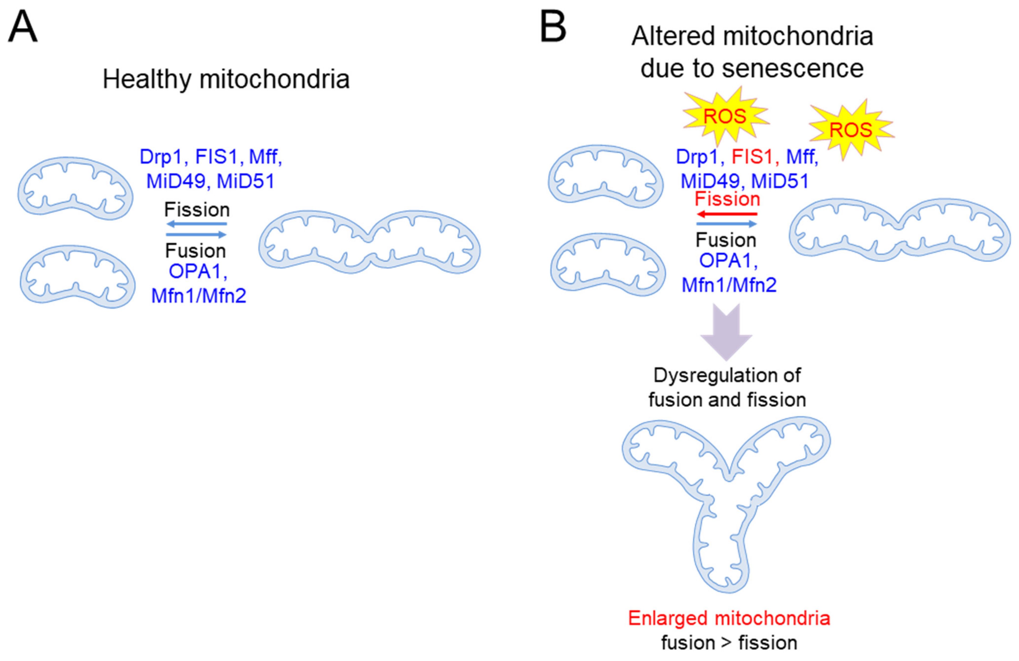
2. Mitochondrial Alterations Associated with Senescence

Mitochondria are organelles that undergo a continuous cycle of fusion and division, called “mitochondrial dynamics”, to maintain proper function [8]. Mitochondrial fusion consists of two steps: the outer mitochondrial membrane (OMM) is fused by mitofusin 1 (Mfn1) and mitofusin 2 (Mfn2), and then the IMM is fused by mitochondrial dynamin-like GTPase (OPA1). Mitochondrial fusion mixes the contents of partially damaged and healthy mitochondria, contributing to a relatively homogeneous network (Figure 1A). This process helps in quality control by maintaining mitochondrial integrity and homeostasis, especially under environmental and metabolic stress. Mitochondrial fission is initiated by membrane contraction by the endoplasmic reticulum (ER), wherein receptors on OMM, including fission 1 protein (FIS1), mitochondrial fission factor (Mff), and mitochondrial dynamics proteins of 49 kDa and 51 kDa (MiD49 and MiD51, respectively), recruit the fission mediator, dynamin-related protein 1 (Drp1), to the mitochondrial surface [9,10,11,12]. Drp1 assembles around the mitochondrial surface to form higher-order oligomers, which divide the mitochondrion into two mitochondria [13]. Thus, mitochondrial fission produces new mitochondria, providing a sufficient number of them for cell growth and division (Figure 1A). Mff–Drp1 interaction is important for mitochondrial fission, as evidenced by the discovery that inhibitors of the Mff–Drp1 interaction interfere with fission, leading to mitochondrial dysfunction [14]. However, the Mff–Drp1 interaction does not prevent Drp1 from interacting with other receptors [14]. A recent study demonstrated a novel role for FIS1 in mitochondrial fission by showing that FIS1 stimulates mitochondrial fission by preventing mitochondrial fusion through the inhibition of GTPase activity in Mfn1, Mfn2 and OPA1 [15]. Mitochondrial fission is also a form of quality control, in which defective mitochondria are removed through autophagy, preventing the accumulation of defective mitochondria [16]. Thus, a fine-tuned balance between fusion and fission allows for the maintenance of mitochondrial quality [17].
Alteration in mitochondrial morphology is a pathogenic hallmark of senescence (Table 1). Defects in lysosomal function due to senescence prevent lysosomal enzymes from targeting autophagosomes, leading to defects in the removal of dysfunctional mitochondria [18,19]. Thus, dysfunctional mitochondria are not eliminated efficiently, but accumulate [18,19]. Dysfunctional mitochondria are vulnerable to further oxidative damage and constitute a major source of excessive ROS. Increased oxidative damage depletes the mitochondrial fission regulator, FIS1, and disrupts the mitochondrial fusion/fission balance, resulting in the formation of giant mitochondria featuring highly interconnected networks [20] (Figure 1B). The enlarged mitochondria limit the effectiveness of quality control, in which damaged mitochondria are efficiently eliminated through autophagy [20,21]. Extending the relevance of these findings, a significant increase in the proportion of giant mitochondria was observed in the livers of aged mice, with an average increase in mitochondrial size of 60% [22]. Increases in mitochondrial size are also induced by a feedback mechanism to compensate for reduced mitochondrial function due to ROS damage, as excessive ROS deteriorates ETC function and subsequently dissipates mitochondrial membrane potential (ΔΨm) [23]. This phenomenon is reinforced by findings showing that large aggregates of mitochondria with low ΔΨm and impaired ATP production are frequently observed in senescent endothelial cells (ECs) [24].
The accumulation of mitochondria with various defects correlates strongly with the pathogenesis of aging and age-related diseases. For example, the excessive ROS generated by dysfunctional mitochondria activates the p53 and pRb pathways, resulting in permanent cell cycle arrest and the exacerbation of cellular senescence [41,42,43,44]. Excessive ROS also activates polyADP-ribose polymerase 1, a NAD+ consuming enzyme [45]. This leads to a significant reduction in the NAD+ levels and NAD+/NADH ratio in parallel with increased oxidative stress and decreased antioxidant capacity, consequently exacerbating senescence [46]. Furthermore, ROS can damage the proteins involved in mitochondrial proteostasis, which play an important role in maintaining and regulating protein quality within mitochondria [47]. Defects in mitochondrial proteostasis impair protein folding and the processing of misfolded or aggregated proteins, leading to aging and age-related diseases through accelerated proteostatic collapse [47,48]. The causal relationship between senescence and dysfunctional mitochondria is supported by findings that the accumulation of dysfunctional mitochondria leads to detrimental effects, including altered cellular homeostasis and degenerative changes in tissues [49,50,51,52]. Support for the causal relationship is evident in the finding that extensive targeted mitochondrial depletion inhibits ROS generation and the secretion of key senescence-associated secretory phenotype (SASP) factors, such as IL-6 and IL-8 [53]. Mitochondria are required for the development of the pro-oxidant and pro-inflammatory features of senescence, suggesting that mitochondria are candidate therapeutic targets for reducing the deleterious effects of senescence [53]. Taken together, these results suggest that dysfunctional mitochondria are not only a concomitant phenomenon of senescence, but also a cause of senescence, implying the existence of a vicious cycle involving multiple feedback loops rather than a linear causal relationship [54].
Mitochondria also become targets of toxin-induced ROS, which damage mitochondria and impair mitochondrial function (Table 1). For example, rotenone is a mitochondrial complex I inhibitor that interrupts electron transfer from the iron–sulfur clusters of complex I to coenzyme Q10 (CoQ) [55]. Once inside the mitochondria, rotenone acquires electrons from complex I for the redox cycle, forming excess mitochondrial ROS. The impairment of complex I by rotenone induces senescence-related Parkinson’s disease [56]. Another example is antimycin A, a mitochondrial complex III inhibitor that inhibits electron transfer from cytochrome b to cytochrome c1 in ETC [57]. In this process, antimycin A leaks a single electron to O2 to generate mitochondrial ROS through a non-enzymatic reaction. The oxidative stress induced by antimycin A deteriorates mitochondrial function and induces premature senescence [25].

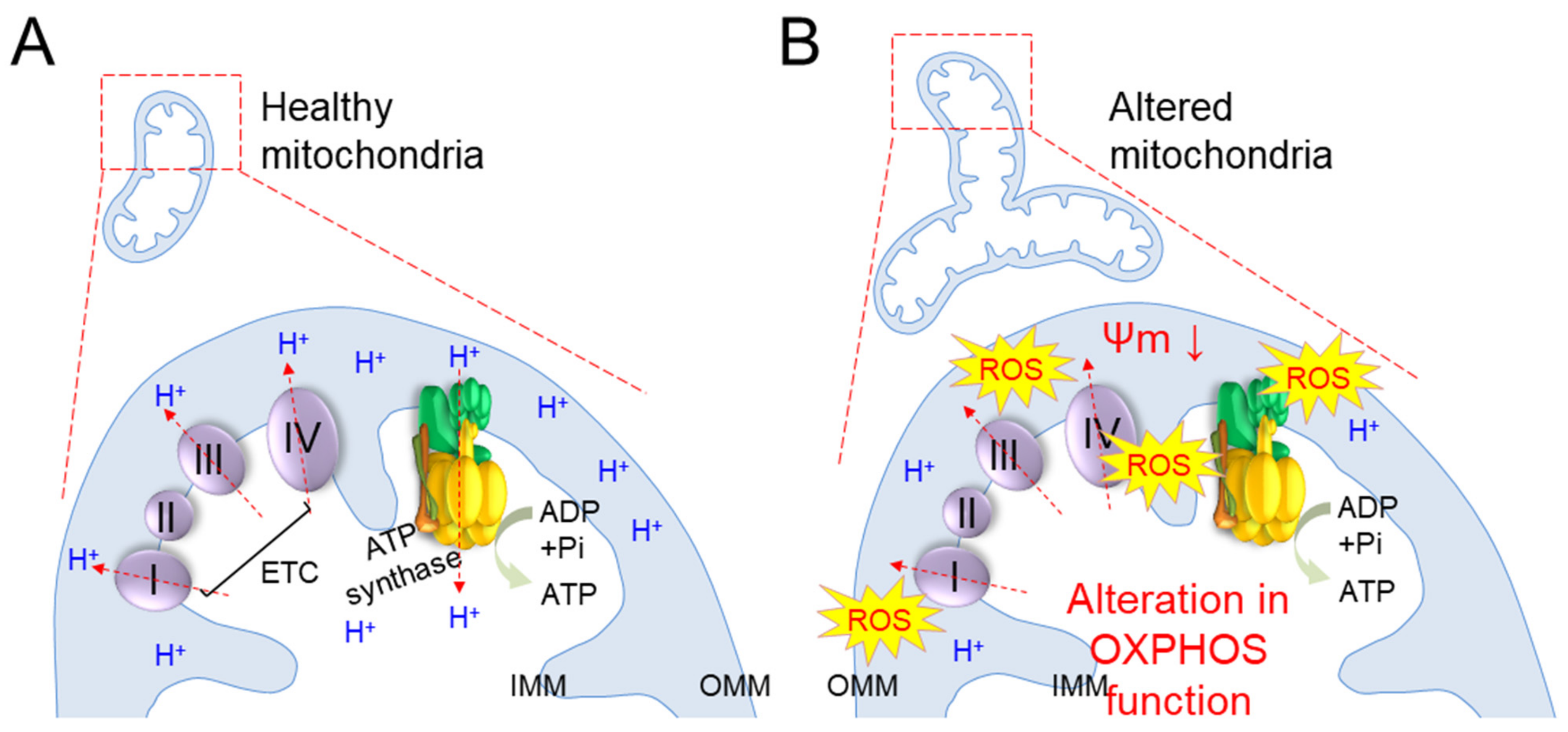
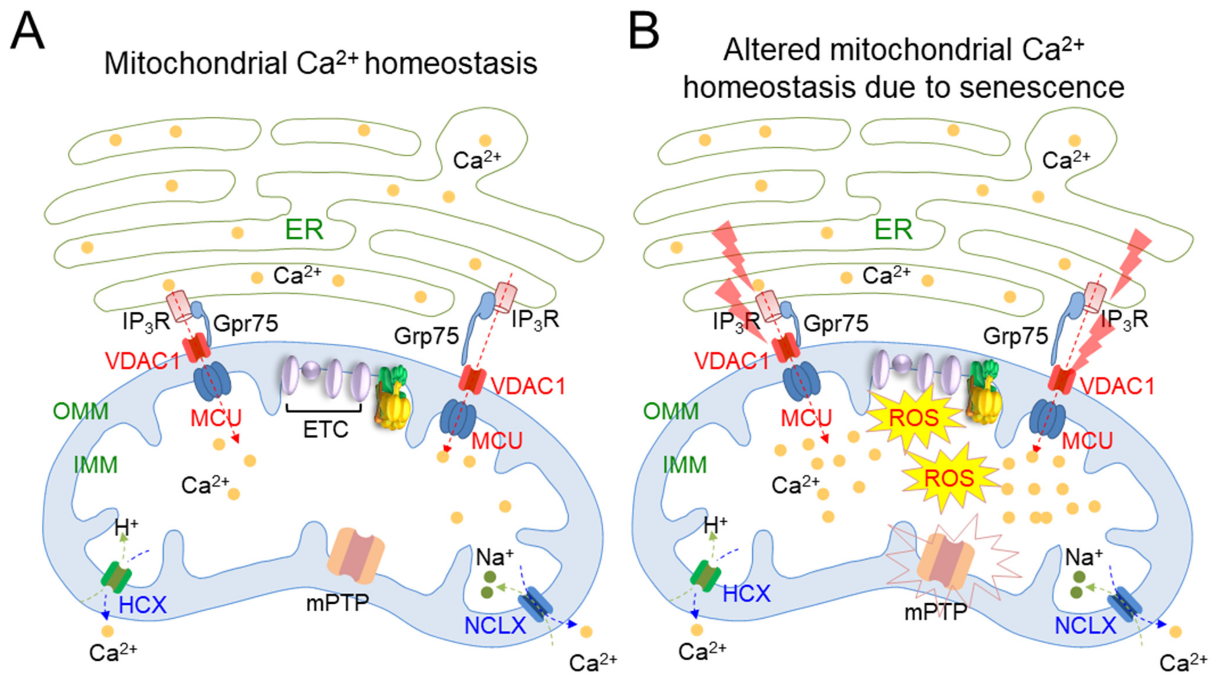
3. Alterations in Mitochondrial Metabolism Associated with Senescence

Mitochondria generate the chemical energy needed to power biochemical reactions in the form of ATP, and the amount of ATP accounts for about 90% of ATP production in the cell [58]. ATP production in mitochondria is accomplished by a sequential reaction called OXPHOS, which involves four ETC complexes (complexes I through IV) and ATP synthase [59]. During OXPHOS, the redox process of the ETC produces a hydrogen ion (H+) concentration gradient, leading to the movement of H+ from ATP synthase to the matrix to generate ATP (Figure 2A).
Alterations in OXPHOS function are observed in various models of senescence [30,31] (Table 1). As senescence progresses, dysfunctional mitochondria produce excess ROS, causing the unwanted oxidation of proteins involved in OXPHOS and impairment of their function [60] (Figure 2B). Thus, electron transport in the ETC is disturbed and electrons leak out of the ETC. Inefficient electron transport concurrently impairs proton transport through the IMM and dissipates ΔΨm, thereby reducing the efficiency of OXPHOS and accompanying a lack of ATP production (Figure 2B). The leaked electrons also react with O2, generating excessive mitochondrial ROS [61]. The deterioration of OXPHOS function with senescence is evidenced by findings that senescence induces the deterioration of the ETC complexes in liver, brain, and muscle tissues, leading to a decrease in mitochondrial respiratory function [26,27,28].
Alterations in OXPHOS lead to the development of aging and age-related diseases, which suggests a causal relationship between senescence and OXPHOS deterioration (Table 1). For example, a mouse model of senescence produced by mev-1 (ortholog of the complex II) mutation exhibits deterioration of OXPHOS accompanying precocious age-dependent corneal physiological changes [29]. Support for this phenomenon is evident from observations that iron chelation with deferoxamine reduces complex II activity through the translational inhibition of iron–sulfur clusters in complex II [30,31]. Decreases in complex II activity sustain the disruption of ΔΨm with significantly reduced intracellular ATP levels prior to the acquisition of the senescence phenotype. Similarly, the inhibition of complex IV activity by transforming growth factor β1 (TGF-β1) induces mitochondrial ROS generation and the persistent disruption of ΔΨm. Thus, the TGF-β1-mediated inhibition of complex IV directly triggers senescence arrest in mink lung epithelial cells through prolonged mitochondrial ROS generation and decreased ATP generation, confirming the causal relationship between senescence and OXPHOS deterioration [32,33,34].
Alterations in mitochondrial metabolism with decreasing dependence on OXPHOS but increasing dependence on glycolysis constitute one of the characteristic changes observed with senescence [62,63] (Table 1). The higher reliance on glycolysis during replicative senescence is attributed to the less energetic state reflected by the marked decrease in ATP levels in senescent cells [37]. Specifically, glycolysis is upregulated to generate additional ATP to compensate for the loss of energy production in dysfunctional mitochondria [35]. Furthermore, the analysis of energy metabolism in senescent cells reveals an increase in glucose consumption and lactic acid production, indicating that glycolysis is actively proceeding [36]. This phenomenon is further assessed by metabolic profiling, which reveals age-dependent changes in mitochondrial metabolism, marked by significant transitions to more glycolytic states [37].
Mitochondrial Ca2+ homeostasis plays an important role in the regulation of mitochondrial metabolism [23,64] (Table 1). This homeostasis is regulated by protein channels localized in the IMM and OMM, and also by crosstalk with the ER [65] (Figure 3A). Mitochondrial Ca2+ influx occurs through porin-like proteins called voltage-dependent anion channels (VDAC) in the OMM. Then, Ca2+ enters the mitochondrial matrix through the mitochondrial calcium uniporter (MCU) in the IMM. The mitochondrial Ca2+ efflux from the mitochondrial matrix is driven by two channels, a H+/Ca2+ exchanger (HCX) and a Na+/Ca2+ exchanger (NCLX), present in the IMM. Intracellular Ca2+ buffering from the ER to mitochondria is achieved by inositol 1,4,5-trisphosphate receptor (IP3R)–Grp75–VDAC interaction [66,67,68] (Figure 3A). Grp75, linking IP3R in the ER with VDAC in the mitochondria, creates a tight juxtaposition between the ER and mitochondria to regulate intracellular Ca2+ buffering [66,67,68]. During senescence, IP3R in the ER and VDAC1/MUC in the mitochondria act as senescence regulators by controlling the concentration of mitochondrial Ca2+. In particular, senescence triggers IP3R to release Ca2+ from the ER and causes VDAC/MCU channels to initiate the inward flow of Ca2+, leading to mitochondrial Ca2+ overload [38] (Figure 3B). Mitochondria overloading with Ca2+ causes the collapse of the electron transport in the ETC, resulting in increased electron leak and consequent mitochondrial ROS generation [38]. An increase in mitochondrial ROS due to mitochondrial Ca2+ overload induces the sustained opening of the mitochondrial transition pore (mPTP) [39]. Then, mPTP opening causes a rapid collapse in ΔΨm and swelling of the mitochondria, resulting in the loss of cytochrome c, a component of the ETC that transports electrons to complex IV. Inefficient electron transport by leaked electrons results in a deficiency in the generation of electrochemical proton gradient, leading to decreased OXPHOS efficiency [40] (Figure 3B). Furthermore, the frequency and duration of mPTP opening increases with the progression of senescence, and increased mPTP activity is associated with several neurodegenerative diseases [23]. Mitochondrial Ca2+ overload appears to be the initiator of alteration in mitochondrial metabolism, which contributes to the deficits observed during senescence and neurodegeneration [23]. Thus, the maintenance of an appropriate level of mitochondrial Ca2+ concentration to regulate mitochondrial metabolism might be a novel strategy for the treatment of senescence.

4. Targeting Mitochondrial Metabolism as a Strategy to Treat Senescence

As mentioned in Section 3, senescence triggers mitochondrial metabolic alteration from OXPHOS to glycolysis, and senescent cells exhibit greater dependence on glycolysis as an energy source [69]. The close relationship between mitochondrial metabolism and senescence is demonstrated by the discovery that alterations in mitochondrial metabolism provoke premature reductions in tissue and organ function [6], whereas improvements in OXPHOS efficiency extend the lifespan of cells and organisms [70]. These interconnections suggest that regulatory mechanisms of mitochondrial metabolism are essential for adequately controlling senescence [71,72]. Therefore, herein, we systematically characterize and propose a potential therapeutic strategy targeting mitochondrial metabolism to induce mitochondrial metabolic reprogramming for the treatment of senescence.
Activation of OXPHOS coupled with increased ATP production supports the importance of mitochondrial metabolic reprogramming in regulating senescence. Specifically, senescent cells exhibit a deficiency in CoQ, which accepts electrons from complex I/II and transfers them to complex III in the ETC (Figure 4A; green CoQ indicates a deficiency in CoQ) [73]. Thus, electron transport in ETC is perturbed in cells with CoQ deficiency, resulting in electron leakage with loss of ΔΨm [73]. Electrons that leak from the ETC prematurely react with O2, causing excessive mitochondrial ROS production [61] (Figure 4A). In agreement with this finding, CoQ activates a proton-motive Q cycle, allowing complex III to pump protons from the mitochondrial matrix into the intermembrane space, generating a proton motive force ΔΨm [74]. A deficiency of CoQ reduces the efficiency of OXPHOS based on ΔΨm in flowing protons back to the mitochondrial matrix via ATP synthase, resulting in decreased ATP production [74]. Given that CoQ plays an important role in mitochondrial OXPHOS and ATP production, CoQ-deficient fibroblasts were treated with CoQ [75]. CoQ treatment improves mitochondrial metabolism, which manifests as a significant increase in ATP production and a significant decrease in mitochondrial ROS generation [75] (Figure 4B; pink CoQ indicates a higher level of CoQ in the IMM). The beneficial effects of CoQ supplementation on OXPHOS are further supported by animal model experiments using senescence-accelerated mice [76]. CoQ supplementation improves OXPHOS efficiency by increasing complex I/IV activity and subsequently decreasing mitochondrial ROS generation. Additionally, CoQ supplementation slows the progression of aging-related symptoms and prevents aging, suggesting that strategies to activate OXPHOS efficiency may be effective in treating senescence [76].
There are other strategies that modulate OXPHOS efficiency to induce mitochondrial metabolic reprogramming for senescence amelioration. Long-term caloric restriction (CR) increases complex IV levels, resulting in the partial compensation of electron leakage, thereby decreasing ROS generation [77]. A CR mimetic, epigallocatechin 3-gallate (EGCG), rescues the catalytic activity of complex I/ATP synthetase and restores OXPHOS efficiency [78]. In addition, EGCG acts as an activator of sirtuin 1 (SIRT1), a protein deacetylase, and reduces the acetylation of PGC-1α. Thereby, EGCG activates PGC-1α, which regulates mitochondrial biosynthesis and function [78]. Consistent with this finding, resveratrol (RSV) induces SIRT1-dependent PGC-1α deacetylation [79]. RSV significantly increases OXPHOS efficiency, as evidenced by the induction of OXPHOS genes, including the ETC complexes, ATP synthase, and respiratory apparatus [80]. Furthermore, RSV prolongs lifespan by enabling the long-term supply of ATP through the restoration of metabolic homeostasis [80]. In line with this finding, dietary supplementation containing essential and branched-chain amino acids (called PD-0E7) deacetylates and activates PGC-1α to amplify mitochondrial responses. Accordingly, PD-0E7 upregulates the activity of complexes I, II, and IV, and improves muscular and cognitive performance in the senescence-accelerated mouse prone 8 model [81]. Finally, boosting NAD+ levels with nicotinamide riboside (NR) constitutes an efficient way to increase OXPHOS efficiency. NR supplementation increases NAD+ levels and the NAD+/NADH ratio, which are known to be significantly reduced in senescent cells [82]. Increased NAD+ levels by NR stimulate mitophagy to remove damaged mitochondria and enhance OXPHOS efficiency by upregulating basal/maximal ATP-linked oxygen consumption rates [83]. Concurrently, improved mitochondrial function by NR prevents senescence and SASP [83].
Targeting a higher glycolytic state of senescent cells is an alternative strategy that highlights the importance of mitochondrial metabolic reprogramming in regulating senescence. PFKFB3 is a gene that encodes 6-phosphofructo-2-kinase/fructose-2,6-biphosphatase 3, which functions as a pivotal activator of glycolysis by activating phosphofructokinase 1 (PFK1), converting fructose-6-phosphate to fructose-1,6-bisphophate [84]. The pharmacological inhibition of the glycolytic activator, PFKFB3, inactivates glycolysis and alleviates age-related cerebral ischema/reperfusion injury in mice [85] (Figure 4B). The effectiveness of the strategy to limit glycolysis upon senescence amelioration is further supported by experiments using D-glucosamine (GlcN), which inhibits the activity of glyceraldehyde-3-phosphate-dehydrogenase (GAPDH) in the glycolytic pathway. GlcN increases mitochondrial respiration by promoting the dependence of energy metabolism on OXPHOS, while impairing glycolysis, thus prolonging lifespan in many species, including mammals [86] (Figure 4B). While inhibiting the specific enzymes required for glycolysis may be a strategy to control senescence, there is growing evidence that inhibiting the entire glycolysis process may serve as a platform to regulate senescence. For example, the impaired glucose metabolism, induced by glucose restriction, improves mitochondrial OXPHOS and consequently prolongs the lifespan of Caenorhabditis elegans [87].
Mitochondria play an important role in regulating intracellular metabolism, serving as a platform for receiving signals from key elements of cells and tissues [72]. Several cellular signaling pathways are directly or indirectly linked with mitochondrial metabolism [88]. Targeting such pathways might be an alternative strategy to induce mitochondrial metabolic reprogramming. These pathways involve ataxia telangiectasia mutated (ATM), rho-associated protein kinase (ROCK), and serine/threonine protein kinase B-Raf (BRAF) (Figure 4C). Targeting these pathways has been evaluated to be effective in regulating senescence through the modulation of the mitochondrial metabolism.
(i)
Targeting ATM signal pathway.
ATM controls lysosomal pH by regulating the assembly/disassembly of the V1 and V0 domains in the V-ATPase proton pumps present in lysosomal membranes [62]. Notably, the inhibition of the ATM signaling pathway promotes V1-V0 assembly, leading to the re-acidification of lysosomes. In turn, this leads to a functional restoration of the lysosome, thus enhancing the clearance of dysfunctional mitochondria, a key mechanism that maintains mitochondrial function [89]. The restoration of mitochondrial function by ATM inhibition is accompanied by mitochondrial metabolic reprogramming from glycolysis to OXPHOS, resulting in increased ATP production and the restoration of senescence-related phenotypes [62,90]. The effect of targeting the ATM signaling pathway, indirectly linked to mitochondrial metabolism, highlights the importance of mitochondrial metabolic reprogramming in the regulation of senescence, and may have clinical applications in controlling aging and age-related diseases.
(ii)
Targeting ROCK signal pathway.
ROCK controls mitochondrial ROS generation while regulating the interaction between Rac1b and cytochrome c [63]. The inhibition of the ROCK signaling pathway reduces mitochondrial ROS production and simultaneously interrupts electron transfer from cytochrome c, preventing the partial reduction of O2 [63]. Reducing oxidative damage by modulating ROCK activity enhances complex IV activity in the ETC, leading to improved mitochondrial function along with metabolic reprogramming [63,91]. The importance of metabolic reprogramming via the regulation of ROCK activity is elucidated by experiments using metabolic reprogrammers that artificially shift the metabolism from OXPHOS to glycolysis. Artificial metabolic reprogramming to glycolysis interferes with ROCK inhibition-mediated senescence improvement, suggesting that metabolic reprogramming by modulating ROCK activity plays a prerequisite role in senescence regulation [63,91]. The effect of targeting the ROCK signaling pathway, directly linked to mitochondrial metabolism, also supports the significance of mitochondrial metabolic reprogramming as a therapeutic strategy for treating senescence.
(iii)
Targeting BRAF signal pathway.
BRAF regulates MAP kinase/ERK signaling pathways and modulates the activity of substrates in response to various signals to maintain cellular homeostasis [92]. The inhibition of the BRAF signaling pathway increases mitophagy, which regulates mitochondrial quality by eliminating dysfunctional mitochondria [93]. Concurrently, ROS generation is decreased by modulating BRAF activity, which leads to mitochondrial function restoration with an increase in OXPHOS and a decrease in glycolysis. Furthermore, metabolic reprogramming induced by the inhibition of BRAF activity is a prerequisite for ameliorating senescence, as evidenced by the discovery that artificial metabolic reprogrammers block senescence amelioration induced by BRAF inhibition.
The maintenance of mitochondrial metabolism through mitochondrial Ca2+ homeostasis represents a therapeutic strategy to induce mitochondrial metabolic reprogramming for senescence amelioration. Adequate levels of mitochondrial Ca2+ activate the enzymatic activity of the ETC and stimulate the entire OXPHOS machinery [94], whereas mitochondrial Ca2+ overload generates excessive ROS and induces metabolic derangement [23]. As the fine-tuning of mitochondrial Ca2+ concentration initiates mitochondrial metabolic reprogramming [95], maintaining adequate levels of mitochondrial Ca2+ can be a novel treatment for senescence. For example, myocardial reperfusion is an age-related disease that manifests as decreased resistance to myocardial reperfusion injury in the elderly [96]. Myocardial reperfusion is also associated with alterations in mitochondrial Ca2+ homeostasis and mitochondrial metabolism [97]. As the transport of Ca2+ from the cytoplasm to the mitochondria is facilitated by MCU in the IMM, mitochondria from the reperfused hearts are treated with the cell-permeable MCU inhibitor, Ruthenium 360 (Ru360) [98,99] (Figure 4D). The inhibition of MCU by Ru360 improves OXPHOS efficiency by maintaining mitochondrial Ca2+ at basal levels and reducing the proportion of mitochondria exhibiting Ca2+ overload. Improving mitochondrial metabolism is accompanied by the restoration of the pathophysiological symptoms of the reperfused heart to a physiological state [99]. The importance of maintaining adequate levels of mitochondrial Ca2+ homeostasis is further supported by the discovery that the inhibition of MCU by microRNA-mediated silencing protects cardiomyocytes from oxidative damage and restores mitochondrial function [100] (Figure 4D).

5. Conclusions and Perspectives

The role of mitochondrial metabolic reprogramming in senescence has been analyzed and reviewed by several researchers. One review specifically presented senescence-related mitochondrial metabolic changes and their effects on the modulation of the immune response underlying senescence [101]. Although the importance of mitochondrial metabolism in senescence has been discussed, it has been limited to its role in ECs. Cell-specific limitations were addressed in another review dealing with the regulation of mitochondrial metabolism in several aging models [102]. This review focused on the mechanistic involvement of metabolic regulators in several aging models, and has proposed metabolic switches as modulators of senescence. The causes and consequences of mitochondrial metabolic changes have been detailed, but no specific strategies to induce metabolic reprogramming have been proposed. Recently, a review paper highlighted cellular metabolism as one of the regulatory factors controlling various senescence-related phenotypes [103]. This review focused on the associations between metabolism and senescence-related phenotypes. A putative mechanism that can target metabolic differences between young and senescent cells has been proposed as a strategy to eliminate the deleterious effects of senescent cells. However, considering that many cellular pathways are damaged by senescence, it is not clear whether therapeutic approaches that restore only mitochondrial metabolism will be effective in the treatment of senescence.
In this review, we discussed and summarized senescence-related mitochondrial dysfunction and consequent mitochondrial metabolic alterations. We further assessed the causal relationship between mitochondrial metabolism and senescence, and suggested several therapeutic strategies targeting mitochondrial metabolism for the treatment of senescence. Changes in mitochondrial metabolism occur during senescence, and restoring mitochondrial metabolism to a normal state by several approaches restores senescence and senescence-associated phenotypes. Furthermore, mitochondrial metabolic reprogramming based on multiple approaches serves as a prerequisite for aging treatment. Thus, the appropriate regulation of mitochondrial metabolism, which does not rely on one approach, may open up the possibility of addressing the pathological symptoms of senescence, by which many cellular pathways are damaged. Here, we propose mitochondrial metabolic reprogramming as a promising therapeutic target for the treatment of senescence. Further studies of the molecular mechanisms that support the role of mitochondrial metabolic reprogramming in the initiation and progression of senescence will provide novel therapeutic strategies for aging and age-related diseases.

Author Contributions

Writing—original draft preparation, Y.H.L., J.Y.P., H.W.K., J.T.P., and S.C.P.; writing—review and editing, Y.H.L., J.Y.P., H.W.K., J.T.P., and S.C.P.; writing—data analysis, H.L., E.S.S., M.U.K., J.J., and S.O. All authors have read and agreed to the published version of the manuscript.

Funding

This research was supported by the Basic Science Research Program through the National Research Foundation of Korea (NRF), funded by the Ministry of Science, ICT & Future Planning (NRF-2021R1A2C1004298). This research was also supported by the priority Research Centers Program through the National Research Foundation of Korea (NRF), funded by the Ministry of Education (2020R1A6A1A0304195411).

Institutional Review Board Statement

Not applicable.

Informed Consent Statement

Not applicable.

Data Availability Statement

Not applicable.

Conflicts of Interest

The authors declare no conflict of interest. The funders had no role in the collection, analyses, or interpretation of data; in the writing of the manuscript; or in the decision to publish the paper.

References

  1. Bolden, J.E.; Lowe, S.W. 15—Cellular senescence. In The Molecular Basis of Cancer, 4th ed.; Mendelsohn, J., Gray, J.W., Howley, P.M., Israel, M.A., Thompson, C.B., Eds.; Elsevier: Amsterdam, The Netherlands, 2015; pp. 229–238.e222. [Google Scholar]
  2. Boffoli, D.; Scacco, S.C.; Vergari, R.; Solarino, G.; Santacroce, G.; Papa, S. Decline with age of the respiratory chain activity in human skeletal muscle. Biochim. Biophys. Acta 1994, 1226, 73–82. [Google Scholar] [CrossRef]
  3. Hwang, E.; Yoon, G.; Kang, H. A comparative analysis of the cell biology of senescence and aging. Cell. Mol. Life Sci. 2009, 66, 2503–2524. [Google Scholar] [CrossRef] [PubMed]
  4. Zorov, D.B.; Juhaszova, M.; Sollott, S.J. Mitochondrial reactive oxygen species (ROS) and ROS-induced ROS release. Physiol. Rev. 2014, 94, 909–950. [Google Scholar] [CrossRef] [PubMed] [Green Version]
  5. Rivera-Torres, J.; Acín-Perez, R.; Cabezas-Sánchez, P.; Osorio, F.G.; Gonzalez-Gómez, C.; Megias, D.; Cámara, C.; López-Otín, C.; Enríquez, J.A.; Luque-García, J.L.; et al. Identification of mitochondrial dysfunction in Hutchinson–Gilford progeria syndrome through use of stable isotope labeling with amino acids in cell culture. J. Proteom. 2013, 91, 466–477. [Google Scholar] [CrossRef]
  6. Din, S.; Konstandin, M.H.; Johnson, B.; Emathinger, J.; Völkers, M.; Toko, H.; Collins, B.; Ormachea, L.; Samse, K.; Kubli, D.A.; et al. Metabolic dysfunction consistent with premature aging results from deletion of Pim kinases. Circ. Res. 2014, 115, 376–387. [Google Scholar] [CrossRef] [Green Version]
  7. Liberati, A.; Altman, D.G.; Tetzlaff, J.; Mulrow, C.; Gøtzsche, P.C.; Ioannidis, J.P.A.; Clarke, M.; Devereaux, P.J.; Kleijnen, J.; Moher, D. The PRISMA statement for reporting systematic reviews and meta-analyses of studies that evaluate healthcare interventions: Explanation and elaboration. BMJ 2009, 339, b2700. [Google Scholar] [CrossRef] [Green Version]
  8. Henze, K.; Martin, W. Evolutionary biology: Essence of mitochondria. Nature 2003, 426, 127–128. [Google Scholar] [CrossRef]
  9. Otera, H.; Wang, C.; Cleland, M.M.; Setoguchi, K.; Yokota, S.; Youle, R.J.; Mihara, K. Mff is an essential factor for mitochondrial recruitment of Drp1 during mitochondrial fission in mammalian cells. J. Cell Biol. 2010, 191, 1141–1158. [Google Scholar] [CrossRef] [Green Version]
  10. Palmer, C.S.; Osellame, L.D.; Laine, D.; Koutsopoulos, O.S.; Frazier, A.E.; Ryan, M.T. MiD49 and MiD51, new components of the mitochondrial fission machinery. EMBO Rep. 2011, 12, 565–573. [Google Scholar] [CrossRef]
  11. Zhao, J.; Liu, T.; Jin, S.; Wang, X.; Qu, M.; Uhlén, P.; Tomilin, N.; Shupliakov, O.; Lendahl, U.; Nistér, M. Human MIEF1 recruits Drp1 to mitochondrial outer membranes and promotes mitochondrial fusion rather than fission. EMBO J. 2011, 30, 2762–2778. [Google Scholar] [CrossRef]
  12. Losón, O.C.; Song, Z.; Chen, H.; Chan, D.C. Fis1, Mff, MiD49, and MiD51 mediate Drp1 recruitment in mitochondrial fission. Mol. Biol. Cell 2013, 24, 659–667. [Google Scholar] [CrossRef]
  13. Strack, S.; Cribbs, J.T. Allosteric modulation of Drp1 mechanoenzyme assembly and mitochondrial fission by the variable domain. J. Biol. Chem. 2012, 287, 10990–11001. [Google Scholar] [CrossRef] [Green Version]
  14. Kornfeld, O.S.; Qvit, N.; Haileselassie, B.; Shamloo, M.; Bernardi, P.; Mochly-Rosen, D. Interaction of mitochondrial fission factor with dynamin related protein 1 governs physiological mitochondrial function in vivo. Sci. Rep. 2018, 8, 14034. [Google Scholar] [CrossRef]
  15. Yu, R.; Jin, S.B.; Lendahl, U.; Nistér, M.; Zhao, J. Human Fis1 regulates mitochondrial dynamics through inhibition of the fusion machinery. EMBO J. 2019, 38, e99748. [Google Scholar] [CrossRef]
  16. Okamoto, K.; Kondo-Okamoto, N. Mitochondria and autophagy: Critical interplay between the two homeostats. Bba Gen. Subj. 2012, 1820, 595–600. [Google Scholar] [CrossRef]
  17. Picca, A.; Guerra, F.; Calvani, R.; Bucci, C.; Lo Monaco, M.R.; Bentivoglio, A.R.; Coelho-Júnior, H.J.; Landi, F.; Bernabei, R.; Marzetti, E. Mitochondrial dysfunction and aging: Insights from the analysis of extracellular vesicles. Int. J. Mol. Sci. 2019, 20, 805. [Google Scholar] [CrossRef] [Green Version]
  18. Kissová, I.; Deffieu, M.; Manon, S.; Camougrand, N. Uth1p is involved in the autophagic degradation of mitochondria. J. Biol. Chem. 2004, 279, 39068–39074. [Google Scholar] [CrossRef] [Green Version]
  19. Ding, W.X.; Yin, X.M. Mitophagy: Mechanisms, pathophysiological roles, and analysis. Biol. Chem. 2012, 393, 547–564. [Google Scholar] [CrossRef] [Green Version]
  20. Lee, H.C.; Yin, P.H.; Chi, C.W.; Wei, Y.H. Increase in mitochondrial mass in human fibroblasts under oxidative stress and during replicative cell senescence. J. Biomed. Sci. 2002, 9, 517–526. [Google Scholar] [CrossRef]
  21. Passos, J.F.; Saretzki, G.; Ahmed, S.; Nelson, G.; Richter, T.; Peters, H.; Wappler, I.; Birket, M.J.; Harold, G.; Schaeuble, K.; et al. Mitochondrial dysfunction accounts for the stochastic heterogeneity in telomere-dependent senescence. PLoS Biol. 2007, 5, e110. [Google Scholar] [CrossRef]
  22. Wilson, P.D.; Franks, L.M. The effect of age on mitochondrial ultrastructure. Gerontologia 1975, 21, 81–94. [Google Scholar] [CrossRef]
  23. Müller, M.; Ahumada-Castro, U.; Sanhueza, M.; Gonzalez-Billault, C.; Court, F.A.; Cárdenas, C. Mitochondria and calcium regulation as basis of neurodegeneration associated with aging. Front. Neurosci. 2018, 12, 470. [Google Scholar] [CrossRef]
  24. Jendrach, M.; Pohl, S.; Vöth, M.; Kowald, A.; Hammerstein, P.; Bereiter-Hahn, J. Morpho-dynamic changes of mitochondria during ageing of human endothelial cells. Mech. Ageing Dev. 2005, 126, 813–821. [Google Scholar] [CrossRef] [Green Version]
  25. Stöckl, P.; Hütter, E.; Zwerschke, W.; Jansen-Dürr, P. Sustained inhibition of oxidative phosphorylation impairs cell proliferation and induces premature senescence in human fibroblasts. Exp. Gerontol. 2006, 41, 674–682. [Google Scholar] [CrossRef]
  26. Benzi, G.; Pastoris, O.; Marzatico, F.; Villa, R.F.; Dagani, F.; Curti, D. The mitochondrial electron transfer alteration as a factor involved in the brain aging. Neurobiol. Aging 1992, 13, 361–368. [Google Scholar] [CrossRef]
  27. Lenaz, G.; Bovina, C.; Castelluccio, C.; Fato, R.; Formiggini, G.; Genova, M.L.; Marchetti, M.; Pich, M.M.; Pallotti, F.; Parenti Castelli, G.; et al. Mitochondrial complex I defects in aging. Mol. Cell. Biochem. 1997, 174, 329–333. [Google Scholar] [CrossRef]
  28. Manczak, M.; Jung, Y.; Park, B.S.; Partovi, D.; Reddy, P.H. Time-course of mitochondrial gene expressions in mice brains: Implications for mitochondrial dysfunction, oxidative damage, and cytochrome c in aging. J. Neurochem. 2005, 92, 494–504. [Google Scholar] [CrossRef]
  29. Ishii, T.; Miyazawa, M.; Onouchi, H.; Yasuda, K.; Hartman, P.S.; Ishii, N. Model animals for the study of oxidative stress from complex II. Biochim. Biophys. Acta 2013, 1827, 588–597. [Google Scholar] [CrossRef] [Green Version]
  30. Yoon, Y.S.; Byun, H.O.; Cho, H.; Kim, B.K.; Yoon, G. Complex II defect via down-regulation of iron-sulfur subunit induces mitochondrial dysfunction and cell cycle delay in iron chelation-induced senescence-associated growth arrest. J. Biol. Chem. 2003, 278, 51577–51586. [Google Scholar] [CrossRef] [Green Version]
  31. Yoon, G.; Kim, H.J.; Yoon, Y.S.; Cho, H.; Lim, I.K.; Lee, J.H. Iron chelation-induced senescence-like growth arrest in hepatocyte cell lines: Association of transforming growth factor beta1 (TGF-beta1)-mediated p27Kip1 expression. Biochem. J. 2002, 366, 613–621. [Google Scholar] [CrossRef] [Green Version]
  32. Byun, H.O.; Jung, H.J.; Kim, M.J.; Yoon, G. PKCδ phosphorylation is an upstream event of GSK3 inactivation-mediated ROS generation in TGF-β1-induced senescence. Free Radic. Res. 2014, 48, 1100–1108. [Google Scholar] [CrossRef] [PubMed]
  33. Byun, H.O.; Jung, H.J.; Seo, Y.H.; Lee, Y.K.; Hwang, S.C.; Hwang, E.S.; Yoon, G. GSK3 inactivation is involved in mitochondrial complex IV defect in transforming growth factor (TGF) β1-induced senescence. Exp. Cell Res. 2012, 318, 1808–1819. [Google Scholar] [CrossRef] [PubMed]
  34. Yoon, Y.S.; Lee, J.H.; Hwang, S.C.; Choi, K.S.; Yoon, G. TGF beta1 induces prolonged mitochondrial ROS generation through decreased complex IV activity with senescent arrest in Mv1Lu cells. Oncogene 2005, 24, 1895–1903. [Google Scholar] [CrossRef] [PubMed] [Green Version]
  35. Yang, M.; Chadwick, A.; Dart, C.; Kamishima, T.; Quayle, J. Bioenergetic profile of human coronary artery smooth muscle cells and effect of metabolic intervention. PLoS ONE 2017, 12, e0177951. [Google Scholar] [CrossRef] [Green Version]
  36. Bittles, A.H.; Harper, N. Increased glycolysis in ageing cultured human diploid fibroblasts. Biosci. Rep. 1984, 4, 751–756. [Google Scholar] [CrossRef]
  37. Zwerschke, W.; Mazurek, S.; Stöckl, P.; Hütter, E.; Eigenbrodt, E.; Jansen-Dürr, P. Metabolic analysis of senescent human fibroblasts reveals a role for AMP in cellular senescence. Biochem. J. 2003, 376, 403–411. [Google Scholar] [CrossRef] [Green Version]
  38. Wiel, C.; Lallet-Daher, H.; Gitenay, D.; Gras, B.; Le Calvé, B.; Augert, A.; Ferrand, M.; Prevarskaya, N.; Simonnet, H.; Vindrieux, D.; et al. Endoplasmic reticulum calcium release through ITPR2 channels leads to mitochondrial calcium accumulation and senescence. Nat. Commun. 2014, 5, 3792. [Google Scholar] [CrossRef] [Green Version]
  39. Goodell, S.; Cortopassi, G. Analysis of oxygen consumption and mitochondrial permeability with age in mice. Mech. Ageing Dev. 1998, 101, 245–256. [Google Scholar] [CrossRef]
  40. Hou, Y.; Ghosh, P.; Wan, R.; Ouyang, X.; Cheng, H.; Mattson, M.P.; Cheng, A. Permeability transition pore-mediated mitochondrial superoxide flashes mediate an early inhibitory effect of amyloid beta1-42 on neural progenitor cell proliferation. Neurobiol. Aging 2014, 35, 975–989. [Google Scholar] [CrossRef] [Green Version]
  41. Macip, S.; Igarashi, M.; Fang, L.; Chen, A.; Pan, Z.Q.; Lee, S.W.; Aaronson, S.A. Inhibition of p21-mediated ROS accumulation can rescue p21-induced senescence. EMBO J. 2002, 21, 2180–2188. [Google Scholar] [CrossRef]
  42. Passos, J.F.; Nelson, G.; Wang, C.; Richter, T.; Simillion, C.; Proctor, C.J.; Miwa, S.; Olijslagers, S.; Hallinan, J.; Wipat, A.; et al. Feedback between p21 and reactive oxygen production is necessary for cell senescence. Mol. Syst. Biol. 2010, 6, 347. [Google Scholar] [CrossRef]
  43. Luo, Y.; Zou, P.; Zou, J.; Wang, J.; Zhou, D.; Liu, L. Autophagy regulates ROS-induced cellular senescence via p21 in a p38 MAPKα dependent manner. Exp. Gerontol. 2011, 46, 860–867. [Google Scholar] [CrossRef] [Green Version]
  44. Takahashi, A.; Ohtani, N.; Yamakoshi, K.; Iida, S.; Tahara, H.; Nakayama, K.; Nakayama, K.I.; Ide, T.; Saya, H.; Hara, E. Mitogenic signalling and the p16INK4a-Rb pathway cooperate to enforce irreversible cellular senescence. Nat. Cell Biol. 2006, 8, 1291–1297. [Google Scholar] [CrossRef]
  45. Xie, N.; Zhang, L.; Gao, W.; Huang, C.; Huber, P.E.; Zhou, X.; Li, C.; Shen, G.; Zou, B. NAD+ metabolism: Pathophysiologic mechanisms and therapeutic potential. Signal Transduct. Target. Ther. 2020, 5, 227. [Google Scholar] [CrossRef]
  46. Braidy, N.; Guillemin, G.J.; Mansour, H.; Chan-Ling, T.; Poljak, A.; Grant, R. Age related changes in NAD+ metabolism oxidative stress and Sirt1 activity in wistar rats. PLoS ONE 2011, 6, e19194. [Google Scholar] [CrossRef]
  47. Jensen, M.B.; Jasper, H. Mitochondrial proteostasis in the control of aging and longevity. Cell Metab. 2014, 20, 214–225. [Google Scholar] [CrossRef] [Green Version]
  48. Zimmermann, A.; Madreiter-Sokolowski, C.; Stryeck, S.; Abdellatif, M. Targeting the mitochondria-proteostasis axis to delay aging. Front. Cell Dev. Biol. 2021, 9, 656201. [Google Scholar] [CrossRef]
  49. Nojiri, H.; Shimizu, T.; Funakoshi, M.; Yamaguchi, O.; Zhou, H.; Kawakami, S.; Ohta, Y.; Sami, M.; Tachibana, T.; Ishikawa, H.; et al. Oxidative stress causes heart failure with impaired mitochondrial respiration. J. Biol. Chem. 2006, 281, 33789–33801. [Google Scholar] [CrossRef] [Green Version]
  50. Manczak, M.; Anekonda, T.S.; Henson, E.; Park, B.S.; Quinn, J.; Reddy, P.H. Mitochondria are a direct site of A beta accumulation in Alzheimer’s disease neurons: Implications for free radical generation and oxidative damage in disease progression. Hum. Mol. Genet. 2006, 15, 1437–1449. [Google Scholar] [CrossRef]
  51. Blake, R.; Trounce, I.A. Mitochondrial dysfunction and complications associated with diabetes. Biochim. Biophys. Acta 2014, 1840, 1404–1412. [Google Scholar] [CrossRef]
  52. Lin, M.T.; Beal, M.F. Mitochondrial dysfunction and oxidative stress in neurodegenerative diseases. Nature 2006, 443, 787–795. [Google Scholar] [CrossRef]
  53. Correia-Melo, C.; Marques, F.D.; Anderson, R.; Hewitt, G.; Hewitt, R.; Cole, J.; Carroll, B.M.; Miwa, S.; Birch, J.; Merz, A.; et al. Mitochondria are required for pro-ageing features of the senescent phenotype. EMBO J. 2016, 35, 724–742. [Google Scholar] [CrossRef]
  54. Correia-Melo, C.; Passos, J.F. Mitochondria: Are they causal players in cellular senescence? Biochim. Biophys. Acta 2015, 1847, 1373–1379. [Google Scholar] [CrossRef] [Green Version]
  55. Brandt, U. Energy converting NADH: Quinone oxidoreductase (complex I). Annu. Rev. Biochem. 2006, 75, 69–92. [Google Scholar] [CrossRef]
  56. Greenamyre, J.T.; Sherer, T.B.; Betarbet, R.; Panov, A.V. Complex I and Parkinson’s disease. IUBMB Life 2001, 52, 135–141. [Google Scholar] [CrossRef]
  57. Quinlan, C.L.; Treberg, J.R.; Brand, M.D. Chapter 3—Mechanisms of mitochondrial free radical production and their relationship to the aging process. In Handbook of the Biology of Aging, 7th ed.; Masoro, E.J., Austad, S.N., Eds.; Academic Press: San Diego, CA, USA, 2011; pp. 47–61. [Google Scholar]
  58. Song, J.; Pfanner, N.; Becker, T. Assembling the mitochondrial ATP synthase. Proc. Natl. Acad. Sci. USA 2018, 115, 2850–2852. [Google Scholar] [CrossRef] [Green Version]
  59. Zhao, R.Z.; Jiang, S.; Zhang, L.; Yu, Z.B. Mitochondrial electron transport chain, ROS generation and uncoupling (Review). Int. J. Mol. Med. 2019, 44, 3–15. [Google Scholar] [CrossRef] [Green Version]
  60. Kühlbrandt, W. Structure and function of mitochondrial membrane protein complexes. BMC Biol. 2015, 13, 89. [Google Scholar] [CrossRef] [Green Version]
  61. De la Mata, M.; Cotán, D.; Oropesa-Ávila, M.; Garrido-Maraver, J.; Cordero, M.D.; Villanueva Paz, M.; Delgado Pavón, A.; Alcocer-Gómez, E.; de Lavera, I.; Ybot-González, P.; et al. Pharmacological chaperones and coenzyme Q10 treatment improves mutant β-glucocerebrosidase activity and mitochondrial function in neuronopathic forms of gaucher disease. Sci. Rep. 2015, 5, 10903. [Google Scholar] [CrossRef] [Green Version]
  62. Kang, H.T.; Park, J.T.; Choi, K.; Kim, Y.; Choi, H.J.C.; Jung, C.W.; Lee, Y.S.; Park, S.C. Chemical screening identifies ATM as a target for alleviating senescence. Nat. Chem. Biol. 2017, 13, 616–623. [Google Scholar] [CrossRef]
  63. Kang, H.T.; Park, J.T.; Choi, K.; Choi, H.J.C.; Jung, C.W.; Kim, G.R.; Lee, Y.S.; Park, S.C. Chemical screening identifies ROCK as a target for recovering mitochondrial function in Hutchinson-Gilford progeria syndrome. Aging Cell 2017, 16, 541–550. [Google Scholar] [CrossRef] [PubMed]
  64. Bravo-Sagua, R.; Parra, V.; López-Crisosto, C.; Díaz, P.; Quest, A.F.; Lavandero, S. Calcium Transport and signaling in mitochondria. Compr. Physiol. 2017, 7, 623–634. [Google Scholar] [CrossRef] [PubMed]
  65. Rowland, A.A.; Voeltz, G.K. Endoplasmic reticulum-mitochondria contacts: Function of the junction. Nat. Rev. Mol. Cell Biol. 2012, 13, 607–625. [Google Scholar] [CrossRef] [PubMed] [Green Version]
  66. Rizzuto, R.; Marchi, S.; Bonora, M.; Aguiari, P.; Bononi, A.; De Stefani, D.; Giorgi, C.; Leo, S.; Rimessi, A.; Siviero, R.; et al. Ca(2+) transfer from the ER to mitochondria: When, how and why. Biochim. Biophys. Acta 2009, 1787, 1342–1351. [Google Scholar] [CrossRef] [Green Version]
  67. Xu, H.; Guan, N.; Ren, Y.L.; Wei, Q.J.; Tao, Y.H.; Yang, G.S.; Liu, X.Y.; Bu, D.F.; Zhang, Y.; Zhu, S.N. IP(3)R-Grp75-VDAC1-MCU calcium regulation axis antagonists protect podocytes from apoptosis and decrease proteinuria in an Adriamycin nephropathy rat model. BMC Nephrol. 2018, 19, 140. [Google Scholar] [CrossRef]
  68. Szabadkai, G.; Bianchi, K.; Várnai, P.; De Stefani, D.; Wieckowski, M.R.; Cavagna, D.; Nagy, A.I.; Balla, T.; Rizzuto, R. Chaperone-mediated coupling of endoplasmic reticulum and mitochondrial Ca2+ channels. J. Cell Biol. 2006, 175, 901–911. [Google Scholar] [CrossRef] [Green Version]
  69. James, E.L.; Michalek, R.D.; Pitiyage, G.N.; de Castro, A.M.; Vignola, K.S.; Jones, J.; Mohney, R.P.; Karoly, E.D.; Prime, S.S.; Parkinson, E.K. Senescent human fibroblasts show increased glycolysis and redox homeostasis with extracellular metabolomes that overlap with those of irreparable DNA damage, aging, and disease. J. Proteome Res. 2015, 14, 1854–1871. [Google Scholar] [CrossRef]
  70. Barros, M.H.; Bandy, B.; Tahara, E.B.; Kowaltowski, A.J. Higher respiratory activity decreases mitochondrial reactive oxygen release and increases life span in Saccharomyces cerevisiae. J. Biol. Chem. 2004, 279, 49883–49888. [Google Scholar] [CrossRef] [Green Version]
  71. Park, J.T.; Lee, Y.-S.; Cho, K.A.; Park, S.C. Adjustment of the lysosomal-mitochondrial axis for control of cellular senescence. Ageing Res. Rev. 2018, 47, 176–182. [Google Scholar] [CrossRef]
  72. Ohya, Y.; Umemoto, N.; Tanida, I.; Ohta, A.; Iida, H.; Anraku, Y. Calcium-sensitive cls mutants of Saccharomyces cerevisiae showing a Pet- phenotype are ascribable to defects of vacuolar membrane H(+)-ATPase activity. J. Biol. Chem. 1991, 266, 13971–13977. [Google Scholar] [CrossRef]
  73. Barcelos, I.P.d.; Haas, R.H. CoQ10 and aging. Biology 2019, 8, 28. [Google Scholar] [CrossRef] [Green Version]
  74. Alcázar-Fabra, M.; Navas, P.; Brea-Calvo, G. Coenzyme Q biosynthesis and its role in the respiratory chain structure. Biochim. Biophys. Acta 2016, 1857, 1073–1078. [Google Scholar] [CrossRef]
  75. Quinzii, C.M.; Hirano, M. Coenzyme Q and mitochondrial disease. Dev. Disabil. Res. Rev. 2010, 16, 183–188. [Google Scholar] [CrossRef]
  76. Tian, G.; Sawashita, J.; Kubo, H.; Nishio, S.Y.; Hashimoto, S.; Suzuki, N.; Yoshimura, H.; Tsuruoka, M.; Wang, Y.; Liu, Y.; et al. Ubiquinol-10 supplementation activates mitochondria functions to decelerate senescence in senescence-accelerated mice. Antioxid. Redox Signal. 2014, 20, 2606–2620. [Google Scholar] [CrossRef]
  77. Olgun, A.; Akman, S.; Serdar, M.A.; Kutluay, T. Oxidative phosphorylation enzyme complexes in caloric restriction. Exp. Gerontol. 2002, 37, 639–645. [Google Scholar] [CrossRef]
  78. Valenti, D.; De Rasmo, D.; Signorile, A.; Rossi, L.; de Bari, L.; Scala, I.; Granese, B.; Papa, S.; Vacca, R.A. Epigallocatechin-3-gallate prevents oxidative phosphorylation deficit and promotes mitochondrial biogenesis in human cells from subjects with Down’s syndrome. Biochim. Biophys. Acta 2013, 1832, 542–552. [Google Scholar] [CrossRef] [Green Version]
  79. Higashida, K.; Kim, S.H.; Jung, S.R.; Asaka, M.; Holloszy, J.O.; Han, D.-H. Effects of resveratrol and SIRT1 on PGC-1α activity and mitochondrial biogenesis: A reevaluation. PLoS Biol. 2013, 11, e1001603. [Google Scholar] [CrossRef] [Green Version]
  80. Lagouge, M.; Argmann, C.; Gerhart-Hines, Z.; Meziane, H.; Lerin, C.; Daussin, F.; Messadeq, N.; Milne, J.; Lambert, P.; Elliott, P.; et al. Resveratrol improves mitochondrial function and protects against metabolic disease by activating SIRT1 and PGC-1α. Cell 2006, 127, 1109–1122. [Google Scholar] [CrossRef]
  81. Brunetti, D.; Bottani, E.; Segala, A.; Marchet, S.; Rossi, F.; Orlando, F.; Malavolta, M.; Carruba, M.O.; Lamperti, C.; Provinciali, M.; et al. Targeting multiple mitochondrial processes by a metabolic modulator prevents sarcopenia and cognitive decline in SAMP8 mice. Front. Pharmacol. 2020, 11, 1171. [Google Scholar] [CrossRef]
  82. Mehmel, M.; Jovanović, N.; Spitz, U. Nicotinamide riboside-the current state of research and therapeutic uses. Nutrients 2020, 12, 1616. [Google Scholar] [CrossRef]
  83. Yang, B.; Dan, X.; Hou, Y.; Lee, J.-H.; Wechter, N.; Krishnamurthy, S.; Kimura, R.; Babbar, M.; Demarest, T.; McDevitt, R.; et al. NAD+ supplementation prevents STING-induced senescence in ataxia telangiectasia by improving mitophagy. Aging Cell 2021, 20, e13329. [Google Scholar] [CrossRef]
  84. Yi, M.; Ban, Y.; Tan, Y.; Xiong, W.; Li, G.; Xiang, B. 6-Phosphofructo-2-kinase/fructose-2,6-biphosphatase 3 and 4: A pair of valves for fine-tuning of glucose metabolism in human cancer. Mol. Metab. 2019, 20, 1–13. [Google Scholar] [CrossRef] [PubMed]
  85. Burmistrova, O.; Olias-Arjona, A.; Lapresa, R.; Jimenez-Blasco, D.; Eremeeva, T.; Shishov, D.; Romanov, S.; Zakurdaeva, K.; Almeida, A.; Fedichev, P.O.; et al. Targeting PFKFB3 alleviates cerebral ischemia-reperfusion injury in mice. Sci. Rep. 2019, 9, 11670. [Google Scholar] [CrossRef] [Green Version]
  86. Weimer, S.; Priebs, J.; Kuhlow, D.; Groth, M.; Priebe, S.; Mansfeld, J.; Merry, T.L.; Dubuis, S.; Laube, B.; Pfeiffer, A.F.; et al. D-Glucosamine supplementation extends life span of nematodes and of ageing mice. Nat. Commun. 2014, 5, 3563. [Google Scholar] [CrossRef] [Green Version]
  87. Schulz, T.J.; Zarse, K.; Voigt, A.; Urban, N.; Birringer, M.; Ristow, M. Glucose restriction extends Caenorhabditis elegans life span by inducing mitochondrial respiration and increasing oxidative stress. Cell Metab. 2007, 6, 280–293. [Google Scholar] [CrossRef] [Green Version]
  88. Tait, S.W.G.; Green, D.R. Mitochondria and cell signalling. J. Cell Sci. 2012, 125, 807–815. [Google Scholar] [CrossRef] [Green Version]
  89. Cimolai, M.C.; Alvarez, S.; Bode, C.; Bugger, H. Mitochondrial mechanisms in septic cardiomyopathy. Int. J. Mol. Sci. 2015, 16, 17763–17778. [Google Scholar] [CrossRef]
  90. Kuk, M.U.; Kim, J.W.; Lee, Y.S.; Cho, K.A.; Park, J.T.; Park, S.C. Alleviation of senescence via ATM Inhibition in accelerated aging models. Mol. Cells 2019, 42, 210. [Google Scholar] [CrossRef]
  91. Park, J.T.; Kang, H.T.; Park, C.H.; Lee, Y.S.; Cho, K.A.; Park, S.C. A crucial role of ROCK for alleviation of senescence-associated phenotype. Exp. Gerontol. 2018, 106, 8–15. [Google Scholar] [CrossRef]
  92. Hussain, M.R.M.; Baig, M.; Mohamoud, H.S.A.; Ulhaq, Z.; Hoessli, D.C.; Khogeer, G.S.; Al-Sayed, R.R.; Al-Aama, J.Y. BRAF gene: From human cancers to developmental syndromes. Saudi J. Biol. Sci. 2015, 22, 359–373. [Google Scholar] [CrossRef] [PubMed] [Green Version]
  93. Kim, J.W.; Kuk, M.U.; Choy, H.E.; Park, S.C.; Park, J.T. Mitochondrial metabolic reprograming via BRAF inhibition ameliorates senescence. Exp. Gerontol. 2019, 126, 110691. [Google Scholar] [CrossRef] [PubMed]
  94. Marcu, R.; Wiczer, B.M.; Neeley, C.K.; Hawkins, B.J. Mitochondrial matrix Ca2+ accumulation regulates cytosolic NAD+/NADH metabolism, protein acetylation, and sirtuin expression. Mol. Cell Biol. 2014, 34, 2890–2902. [Google Scholar] [CrossRef] [PubMed] [Green Version]
  95. Gherardi, G.; Monticelli, H.; Rizzuto, R.; Mammucari, C. The Mitochondrial Ca(2+) Uptake and the fine-tuning of aerobic metabolism. Front. Physiol. 2020, 11, 554904. [Google Scholar] [CrossRef]
  96. Jahangir, A.; Sagar, S.; Terzic, A. Aging and cardioprotection. J. Appl. Physiol. 2007, 103, 2120–2128. [Google Scholar] [CrossRef]
  97. Kuznetsov, A.V.; Javadov, S.; Margreiter, R.; Grimm, M.; Hagenbuchner, J.; Ausserlechner, M.J. The role of mitochondria in the mechanisms of cardiac ischemia-reperfusion injury. Antioxidants 2019, 8, 454. [Google Scholar] [CrossRef] [Green Version]
  98. Woods, J.J.; Nemani, N.; Shanmughapriya, S.; Kumar, A.; Zhang, M.; Nathan, S.R.; Thomas, M.; Carvalho, E.; Ramachandran, K.; Srikantan, S.; et al. A selective and cell-permeable mitochondrial calcium uniporter (MCU) inhibitor preserves mitochondrial bioenergetics after hypoxia/reoxygenation injury. ACS Cent. Sci. 2019, 5, 153–166. [Google Scholar] [CrossRef]
  99. De Jesús García-Rivas, G.; Guerrero-Hernández, A.; Guerrero-Serna, G.; Rodríguez-Zavala, J.S.; Zazueta, C. Inhibition of the mitochondrial calcium uniporter by the oxo-bridged dinuclear ruthenium amine complex (Ru360) prevents from irreversible injury in postischemic rat heart. FEBS J. 2005, 272, 3477–3488. [Google Scholar] [CrossRef]
  100. Pan, L.; Huang, B.J.; Ma, X.E.; Wang, S.Y.; Feng, J.; Lv, F.; Liu, Y.; Liu, Y.; Li, C.M.; Liang, D.D.; et al. MiR-25 protects cardiomyocytes against oxidative damage by targeting the mitochondrial calcium uniporter. Int. J. Mol. Sci. 2015, 16, 5420–5433. [Google Scholar] [CrossRef] [Green Version]
  101. Sabbatinelli, J.; Prattichizzo, F.; Olivieri, F.; Procopio, A.D.; Rippo, M.R.; Giuliani, A. Where metabolism meets senescence: Focus on endothelial cells. Front. Physiol. 2019, 10, 1523. [Google Scholar] [CrossRef] [Green Version]
  102. Kwon, S.M.; Hong, S.M.; Lee, Y.-K.; Min, S.; Yoon, G. Metabolic features and regulation in cell senescence. BMB Rep. 2019, 52, 5–12. [Google Scholar] [CrossRef] [Green Version]
  103. Wiley, C.D.; Campisi, J. From ancient pathways to aging cells—Connecting metabolism and cellular senescence. Cell Metab. 2016, 23, 1013–1021. [Google Scholar] [CrossRef] [PubMed] [Green Version]
Figure 1. Schematic representation of basic mechanisms of senescence-induced mitochondrial damage. (A) Mitochondria are organelles that undergo a continuous cycle of fusion and division. The proteins involved in mitochondrial fusion include mitofusin 1 (Mfn1), mitofusin 2 (Mfn2), and mitochondrial dynamin-like GTPase (OPA1). Proteins involved in mitochondrial fission include fission 1 protein (FIS1), mitochondrial fission factor (Mff), mitochondrial dynamics proteins of 49 kDa and 51 kDa (MiD49 and MiD51, respectively), and dynamin-related protein 1 (Drp1). (B) As senescence progresses, dysfunctional mitochondria are not efficiently eliminated and constitute a major cause of excessive ROS production. Increased oxidative damage by senescence depletes FIS1 and disrupts the mitochondrial fusion/fission balance, resulting in the formation of enlarged mitochondria. ROS: reactive oxygen species.
Cells 10 03003 g001
Figure 2. Schematic representation of basic mechanisms of senescence-induced mitochondrial metabolic changes. (A) Adenosine triphosphate (ATP) production is accomplished by a sequential reaction called oxidative phosphorylation (OXPHOS), which involves four electron transport complexes (ETC; complexes I through IV) and ATP synthase. During OXPHOS, the redox process of the ETC produces a hydrogen ion (H+) concentration gradient, leading to the movement of H+ from ATP synthase to the matrix to generate ATP. IMM: inner mitochondrial membrane; OMM: outer mitochondrial membrane. (B) During senescence, dysfunctional mitochondria produce excess reactive oxygen species (ROS), causing the unwanted oxidation of proteins involved in OXPHOS and impairment of their function. Thus, electron transport in the ETC is disturbed and electrons leak out of the ETC. Inefficient electron transport concurrently impairs proton transport through the IMM and dissipates ΔΨm, thereby reducing the efficiency of OXPHOS and accompanying a lack of ATP production. The leaked electrons also react with O2, generating excessive mitochondrial ROS.
Cells 10 03003 g002
Figure 3. Schematic representation of the basic mechanisms of mitochondrial Ca2+ homeostasis. (A) Mitochondrial Ca2+ homeostasis is regulated by protein channels localized in the mitochondrial inner membrane (IMM) and mitochondrial outer membrane (OMM), and also by crosstalk with the ER. ER: endoplasmic reticulum; IP3R: inositol 1,4,5-trisphosphate receptor; VDAC: voltage-dependent anion channels; MCU: mitochondrial calcium uniporter; HCX: H+/Ca2+ exchanger; NCLX: Na+/Ca2+ exchanger; mPTP: mitochondrial transition pore. (B) Senescence triggers IP3R to release Ca2+ from the ER and causes VDAC/MCU channels to initiate the inward flow of Ca2+, leading to mitochondrial Ca2+ overload. Mitochondria overloaded with Ca2+ cause the collapse of the electron transfer in the ETC, resulting in increased electron leak and consequent mitochondrial ROS generation. An increase in mitochondrial ROS due to mitochondrial Ca2+ overload induces the sustained opening of the mitochondrial transition pore (mPTP). The lightning bolt indicates stress caused by senescence.
Cells 10 03003 g003
Figure 4. Targeting mitochondrial metabolism as a strategy to treat senescence. (A) Senescent cells exhibit a deficiency in coenzyme Q10 (CoQ), which accepts electrons from complex I/II and transfers them to complex III in the ETC (green CoQ indicates a deficiency in CoQ). Electrons that leaked from the ETC prematurely react with O2, causing excessive mitochondrial ROS production. A deficiency of CoQ reduces the efficiency of OXPHOS based on ΔΨm in flowing protons back to the mitochondrial matrix via ATP synthase, resulting in decreased ATP production. CoQ supplementation improves OXPHOS efficiency, which manifests as a significant increase in ATP production and a significant decrease in mitochondrial ROS generation (pink CoQ indicates a higher level of CoQ in the IMM). (B) PFKFB3 is a gene that encodes 6-phosphofructo-2-kinase/fructose-2,6-biphosphatase 3, which activates phosphofructokinase 1 (PFK1), converting fructose-6-phosphate to fructose-1,6-bisphophate. Pharmacological inhibition of the glycolytic activator, PFKFB3, inhibits glycolysis and prevents the senescence-associated secretory phenotype (SASP)-mediated spread of senescence in endothelial cells (ECs). D-glucosamine (GlcN) inhibits the activity of glyceraldehyde-3-phosphate-dehydrogenase (GAPDH) in the glycolytic pathway. GlcN increases mitochondrial respiration by promoting the dependence of energy metabolism on OXPHOS while impairing glycolysis. (C) The strategy of targeting pathways directly or indirectly linked to mitochondrial metabolism. ATM: ataxia telangiectasia mutated; ROCK: rho-associated protein kinase; BRAF: serine/threonine protein kinase B-Raf. (D) The strategy of maintaining mitochondrial metabolism through mitochondrial Ca2+ homeostasis. The cell-permeable MCU inhibitor, Ruthenium 360 (Ru360), maintains mitochondrial Ca2+ at basal levels and improves OXPHOS efficiency. The inhibition of MCU by microRNA-mediated silencing also protects cardiomyocytes from oxidative damage and restores mitochondrial function restoration.
Cells 10 03003 g004
Table 1. A summary of mitochondrial alterations associated with senescence.
Mitochondrial AlterationOutcome(s)Experimental Model and References
Alteration in mitochondrial morphologyFormation of giant mitochondria featuring highly interconnected networksHuman fibroblasts [20]
A significant increase in the proportion of giant mitochondria 30-month-old C57/BL mice [22]
Alteration in mitochondrial functionLarge aggregates of mitochondria with low ΔΨm and impaired ATP productionSenescent endothelial cells [24]
The oxidative stress induced by rotenone and antimycin A deteriorates mitochondrial functionHuman fibroblasts [25]
Alteration in OXPHOS functionDeterioration of the ETC complexes in liver, brain and muscle tissues
Decrease in mitochondrial respiratory function
20-, 60-, or 100-week-old Wistar rat [26]
Tissues from aged rats [27]
2-, 12-, 18-, or 24-month-old C57BL6 mice [28]
A mouse model of senescence produced by mev-1 (ortholog of the complex II) mutation exhibits deterioration of OXPHOS accompanying precocious age-dependent corneal physiological changesTet-mev-1 conditional transgenic mice [29]
Decrease in complex II activity sustains the disruption of ΔΨm with significantly reduced intracellular ATP levels prior to the acquisition of the senescence phenotypeChang cells [30]
Hepatocyte cell lines [31]
TGF-β1-mediated inhibition of complex IV directly triggers the senescence arrest in mink lung epithelial cells Mink lung epithelial cells [32,33,34]
Decreasing dependence on OXPHOS but increasing dependence on glycolysisGlycolysis is upregulated to generate additional ATP to compensate for the loss of energy production in dysfunctional mitochondriaHuman coronary artery smooth muscle cells [35]
The increase in glucose consumption and lactic acid productionHuman fibroblasts [36]
Significant transitions to more glycolytic statesHuman fibroblasts [37]
Alteration in mitochondrial Ca2+ homeostasisSenescence triggers IP3R to release Ca2+ from the ER and causes VDAC/MCU channels to initiate inward flow of Ca2+ Human endothelial cells and human fibroblasts [38]
Mitochondria overloaded with Ca2+ causes the collapse of electron transport in the ETCHuman endothelial cells and human fibroblasts [38]
Sustained opening of the mitochondrial transition pore (mPTP)36-month-old C57BL/6J mice [39]
Then, mPTP opening causes a rapid collapse in ΔΨm and swelling of mitochondriaNeural progenitor cells [40]
Publisher’s Note: MDPI stays neutral with regard to jurisdictional claims in published maps and institutional affiliations.

