Article

Influence of LED Light Spectra on Morphogenesis, Secondary Metabolite Production and Antioxidant Potential in Eucomis autumnalis Cultured In Vitro

by
Monika Cioć
1,*,
Agnieszka Szopa
2,
Barbara Prokopiuk
1,
Bożena Pawłowska
1 and
Łukasz Łopusiewicz
3,4,*
1
Department of Ornamental Plants and Garden Art, Faculty of Biotechnology and Horticulture, University of Agriculture in Krakow, al. 29 Listopada 54, 31-425 Kraków, Poland
2
Department of Medicinal Plant and Mushroom Biotechnology, Faculty of Pharmacy, Jagiellonian University Medical College, Medyczna 9, 30-688 Kraków, Poland
3
School of Medical & Health Sciences, Vizja University, Okopowa 59, 01-043 Warsaw, Poland
4
Department Pharmaceutical Biology, Institute of Pharmacy, Greifswald Univeristy, Friedrich-Ludwig-Jahn-Str 17, 17489 Greifswald, Germany
*
Authors to whom correspondence should be addressed.
Agronomy 2025, 15(9), 2197; https://doi.org/10.3390/agronomy15092197
Submission received: 14 August 2025 / Revised: 2 September 2025 / Accepted: 13 September 2025 / Published: 15 September 2025
(This article belongs to the Special Issue Light Environment Regulation of Crop Growth)

Abstract

Eucomis autumnalis is a bulbous ornamental species with ethnobotanical relevance. In vitro cultures offer a sustainable tool for biomass propagation and metabolite production. This study investigates the effects of nine LED light spectra: red (R), blue (B), red–blue (RB), RB with green (RBG), yellow (RBY), far-red (RBfR), ultraviolet (RBUV), white (WLED), and fluorescent light (Fl, control), on the morphogenesis, polyphenol production, and antioxidant potential of E. autumnalis shoot cultures. Cultures were maintained on MS medium with 5 µM BA and 0.5 µM NAA. HPLC-DAD analysis identified 11 phenolic acids and 4 flavonoids, including eucomic acid, characteristic of the genus. Light quality impacted compound-specific accumulation and antioxidant activity, with responses varying among compounds and treatments. R and B light increased catechin, gentisic acid and hesperidin (289, 195, 245 mg/100 g DW), while UV suppressed flavonoids by ca. 2-fold for catechin and flavanones compared to other lights. RBG and RBfR induced the highest eucomic acid accumulation (424 mg/100 g DW), ferulic acid and epicatechin, correlating strongly with ABTS•+ activity (18–19% higher than other lights; r > 0.6–0.8). These findings highlight LED spectral modulation as a tool to enhance the phytochemical quality of E. autumnalis in vitro and provide a foundation for future biotechnological applications.

1. Introduction

Eucomis autumnalis (Mill.) Chitt. (Asparagaceae, formerly Hyacinthaceae), also known as pineapple lily, is a bulbous perennial plant native to southern Africa [1]. It typically grows in damp grasslands, woodland margins, wet rocky terrain, and montane meadows [2,3]. Characterized by a basal rosette of wavy-edged, strap-shaped leaves and an erect inflorescence spike bearing greenish-white to creamy flowers tipped with a tuft of bracts [4]. The genus Eucomis comprises approximately 9–12 species, adapted to various habitats across southern Africa [3]. E. autumnalis is distinctive among them for its autumnal flowering habit (blooms mid-summer to autumn) and prominent terminal ‘pineapple-like’ bracts [3,4]. In temperate climates, including those in Poland and much of Europe, E. autumnalis does not reliably overwinter outdoors. Nevertheless, it is highly valued for its ornamental appeal and thus cultivated seasonally in pots or garden beds. In containers on balconies or terraces, it requires lifting and overwintering under frost-free conditions or indoor cultivation with a rest period [4,5]. As a cut flower, inflorescences maintain ornamental value for approximately 3–4 weeks [4].
Although not officially listed in European pharmacopoeias, E. autumnalis holds traditional medicinal value in southern Africa. Indigenous communities use bulb decoctions to alleviate back pain, heal wounds and fractures, treat infections, fever, gastrointestinal disorders, and reduce swelling [2,3,6]. Phytochemical studies have revealed the presence of phenolic acids, flavonoids, terpenoids, saponins, and homoisoflavones, which exhibit anti-inflammatory, antioxidant, antibacterial, and cytotoxic activities [7,8]. Intensive harvesting for medicinal use has led to regional decline and conservation concern [2,3,8].
In vitro culture techniques, such as micropropagation, offer routes for both conservation and sustainable metabolite production. These controlled systems enable large-scale propagation without depleting wild populations, and can also serve as biotechnological platforms to study and enhance biosynthesis of pharmaceutically relevant compounds [8,9,10], which is the case with many plants, including ornamental for example Rehmannia glutinosa [11], Streptocarpus × hybridus [12], Leucojum aestivum [13], Dendrobium or Aster [14].
A key regulatory factor in plant secondary metabolite production is light quality [2,8]. LED lighting offers precise control over spectral composition, allowing experimental dissection of metabolic responses [15,16,17]. Different light wavelengths—particularly in red (R) and blue (B) regions—can modulate gene expression in phenylpropanoid pathways, enhance enzyme activities (e.g., phenylalanine ammonia-lyase), and shift the accumulation profile of antioxidants including polyphenols and flavonoids [18,19,20]. In contrast to fluorescent lamps, which emit a broad, fixed spectrum with limited experimental flexibility, LED-driven in vitro systems provide narrow, adjustable wavebands. This enables researchers to isolate the effect of specific wavelengths and directly link them with metabolic outcomes, making LEDs particularly suitable for optimizing bioactive metabolite production [16,21].
The aim of this study was to comprehensively evaluate the impact of nine distinct LED light spectra, including monochromatic red, blue, mixed red–blue, and combinations enriched with green, yellow, far-red, ultraviolet, and white light, on the morphogenesis, photosynthetic pigment accumulation, and biosynthesis of bioactive polyphenolic compounds in E. autumnalis cultured in vitro. The study was designed to assess both biometric parameters and biochemical responses, including antioxidant activity measured by 2,2-diphenyl-1-picrylhydrazyl radical scavenging (DPPH), 2,2′-azino-bis(3-ethylbenzothiazoline-6-sulfonic acid) radical scavenging (ABTS•+) and ferric reducing antioxidant power (FRAP) complementary assays. Particular emphasis was placed on identifying compound-specific effects of spectral light quality on the accumulation of phenolic acids and flavonoids using HPLC-DAD profiling. Additionally, correlation analyses were performed to explore relationships between polyphenol content and antioxidant potential. This research provides novel insights into the photobiological regulation of secondary metabolism in E. autumnalis, a bulbous plant of ethnopharmacological relevance, and contributes to the development of LED-based biotechnological strategies for metabolite enhancement in vitro.

2. Materials and Methods

2.1. Plant Material and In Vitro Culture Conditions

The initial plant material of Eucomis autumnalis used for the experiment was derived from established in vitro cultures, originally initiated from seedlings obtained from seeds produced in experimental plots under greenhouse conditions at the West Pomeranian University of Technology in Szczecin, Poland [4]. Leaf segments approximately 1 cm in length were excised and used as explants for the experiment. Each leaf fragment was transversely divided into two equal parts and placed on growth medium in Petri dishes, five fragments per dish (ten halves in total).
Explants were cultured on solid Murashige and Skoog (MS) [22] medium supplemented with 30 g·dm−3 sucrose and growth regulators: 5 µM of 6-benzylaminopurine (BA), and 0.5 µM 1-naphthaleneacetic acid (NAA). The pH was adjusted to 5.7. The photoperiod was set to 16 h of light and 8 h of darkness. The temperature was regulated at 23 ± 1 °C during the day and 21 ± 1 °C at night, while the relative humidity was maintained at 80%. The LED (light-emitting diodes) light of different quality was used as a light source, while fluorescent lamps served as the control. The photosynthetic photon flux density (PPFD) was maintained at 40 µmol·m−2·s−1.
The applied light treatments included, in total with control, 9 different spectra: 100% of blue LED light (430 nm) (B), 100% of red LED light (670 nm) (R), and mixed red + blue LED light in a 7:3 ratio (RB). Additional variants of mixed RB light enriched with other spectral ranges were also used, including 50% of RB supplemented with 50% of green (528 nm; RBG) or yellow light (600 nm; RBY). White LED light (Wled) with a color temperature of 2700 K (warm), 4500 K (neutral), and 5700 K (cool) in 1:1:1 ratio was also tested. For the analyses of polyphenol content and antioxidant activity, additional treatments included 50% RB light enriched with 50% of far-red (720 nm; RBfR) and ultraviolet (400 nm; RBUV) were used. Fluorescent lamp light (Fl) (Philips TL-D36W/54) served as the control (Figure 1).
Light conditions were set using specially designed LED panels and the light parameters were set using an LI-250A light meter with a Q 50,604 sensor (Li-COR, Lincoln, NE, USA) and BTS256 spectrometer (Gigahertz-Optik, Türkenfeld, Germany) [23].

2.2. Experiment Design, Biometric Parameters and Photosynthetic Pigments Content

The experiment was conducted using eight different LED light treatments and the fluorescent lamp light as a control. Each light treatment was replicated five times (five culture vessels per treatment), with an additional set of three vessels per treatment used for biochemical response analyses, resulting in a total of 620 explants. Cultures and biometric observations were established and collected in November and December 2023. The plant material obtained and stored for experimental work was analyzed in September and October 2024 (the collected data have not been previously published).
Biometric parameters were observed to evaluate phenotype plant responses to light treatments. Fresh weight (FW) of the multiplied plant material was measured using a precision laboratory scale (Chyo Balance Corp., Kyoto, Japan). The number and length of shoots, the number of leaves per shoot, and the number and length of roots were determined. Additionally, to assess dry weight (DW), 1 g of shoot was collected from each treatment (in four replicates for analysis per treatment), dried in a laboratory air sterilizer at 65 °C (Sanyo Electric Co., MOV-112S, Tokyo, Japan) until a constant weight was reached.
Photosynthetic pigment content was also quantified according to the method. For this purpose, 200 mg of shoot tissue samples were extracted in 80% (v/v) acetone. Extracts were filtered until transparent and their absorbance was measured with a UV/VIS Helios Alpha spectrophotometer (Unicam Ltd., Cambridge, UK). The pigment content was determined based on specific absorbance peaks: chlorophyll a at 663.2 nm, chlorophyll b at 646.8 nm, and total carotenoids at 470 nm. The following equations were used to calculate pigment concentrations: chlorophyll a (µg·mL−1) = 12.25 × A663.2 − 279 × A646.8 (Chl a); chlorophyll b (µg·mL−1) = 21.50 × A646.8 − 5.10 × A663.2 (Chl b); total carotenoids (µg·mL−1) = (1000 × A470 − 1.82 × Ca − 85.02 × Cb)/198 (Car) (where Ca and Cb represent the concentrations of Chl a and Chl b (µg·mL−1) calculated from the preceding equations) [24].

2.3. Analyses of Polyphenol Content

Dried and finely powdered plant material of E. autumnalis (0.3 g) was extracted twice with 2 mL of analytical-grade methanol using an ultrasonic bath (Polsonic II, Warsaw, Poland), each cycle lasting 30 min. The combined extracts were centrifuged at 5000× g for 10 min using a MPW-351R centrifuge (MPW Med. Instruments, Warsaw, Poland). The supernatants were filtered through 0.22 µm nylon syringe filters (Merck, Darmstadt, Germany), evaporated to dryness under reduced pressure, and reconstituted in methanol prior to chromatographic analysis, following the procedure described by Szopa et al. [25,26].
The qualitative and quantitative profiling of phenolic compounds was performed using a high-performance liquid chromatography system coupled with a diode-array detector (HPLC-DAD; Merck-Hitachi, Darmstadt, Germany), equipped with an autosampler (L-2200), quaternary pump, column oven, and diode array detector. Separation was carried out on a Purospher® RP-18e reversed-phase analytical column (250 mm × 4 mm, 5 μm; Merck, Darmstadt, Germany), thermostated at 25 °C. The mobile phase consisted of solvent A (methanol:0.5% acetic acid, 1:4, v/v) and solvent B (methanol), with a gradient elution program as follows: 0–20 min, 0% B; 20–35 min, 0–20% B; 35–45 min, 20–30% B; 45–55 min, 30–40% B; 55–60 min, 40–50% B; 60–65 min, 50–75% B; 65–70 min, 75–100% B, followed by a 15-min hold. The flow rate was 1.0 mL·min−1, the injection volume was 10 μL, and detection was carried out at 254 nm.
Identification and quantification of phenolic compounds were based on retention times and UV spectra compared with analytical standards (Sigma-Aldrich, Saint Louis, MO, USA). The analysis included benzoic acid derivatives such as 3,4-dihydroxyphenylacetic acid, gallic acid, gentisic acid, p-hydroxybenzoic acid, protocatechuic acid, salicylic acid, syringic acid, and vanillic acid; cinnamic acid derivatives including caffeic acid, o-coumaric acid, m-coumaric acid, p-coumaric acid, ferulic acid, hydrocaffeic acid, isoferulic acid, and sinapic acid; depsides such as chlorogenic acid, neochlorogenic acid, and rosmarinic acid; as well as the precursor cinnamic acid. In addition, eucomic acid, a compound characteristic of the Eucomis genus, was included in the analysis. Flavonoid profiling encompassed both aglycones (kaempferol, luteolin, quercetin, myricetin) and glycosides (apigetrin, cynaroside, hyperoside, quercitrin, rutoside, trifolin, and vitexin). Quantification was performed using calibration curves constructed for each compound.

2.4. Antioxidant Potential Assay

The antioxidant activity of E. autumnalis methanolic extracts was determined using three in vitro assays: DPPH (2,2-diphenyl-1-picrylhydrazyl), ABTS•+ (2,2′-azino-bis(3-ethylbenzothiazoline-6-sulfonic acid)) and FRAP (ferric reducing antioxidant power).
Methanolic extracts were prepared by mixing 0.3 g of lyophilized and powdered plant material with 4 mL of a methanol. Extraction was carried out in an ultrasonic bath (Elmasonic S30H, Elma Schmidbauer GmbH, Singen, Germany) for 30 min. The extracts were centrifuged at 5000× g for 5 min (Centrifuge 5418, Eppendorf, Warsaw, Poland), filtered through 0.22 µm nylon membrane filters (Merck, Darmstadt, Germany), and stored at −20 °C until analysis. All extractions were performed in triplicate.
The radical scavenging capacity against the DPPH radical was measured according to the method described by Łopusiewicz et al. [27]. A total of 0.5 mL of 0.01 mM DPPH methanolic solution was mixed with 0.5 mL of extract in a 1:1 ratio and incubated in the dark at 22 ± 2 °C for 30 min. After incubation, the absorbance of the analyzed samples was measured at 517 nm using a BMG Labtech Clariostar Plus microplate reader (Ortenberg, Germany). Antioxidant activity was expressed as Trolox equivalents per gram of dry weight (µg TE/g DW).
For the ABTS•+ assay (also Łopusiewicz et al. [27] method), 1.5 mL of pre-generated ABTS•+ solution (produced by mixing 7 mM ABTS with 2.45 mM potassium persulfate, 16 h before assays) was mixed with 25 µL of extract and incubated for 10 min in the dark at room temperature. Absorbance was recorded at 734 nm. Antioxidant activity was expressed as Trolox equivalents per gram of dry weight (µg TE/g DW).
The FRAP assay was carried out according to the method described by Pietrak et al. [28]. The FRAP reagent was freshly prepared by mixing 2.5 mL of 10 mM TPTZ in 40 mM HCl, 2.5 mL of 20 mM FeCl3, and 25 mL of 300 mM acetate buffer (pH 3.6). In a 96-well plate, 20 µL of the extract was added to 280 µL of FRAP reagent and gently mixed. Absorbance was measured at 595 nm after 10 min of incubation. Results were expressed as mg ascorbic acid equivalent per g dry weight (μg AAE/g DW).
For the DPPH and ABTS tests, calibration curves were prepared using Trolox in the concentration range of 0–800 μg/mL. For the FRAP assay, the calibration curve was prepared using ascorbic acid in the concentration range of 0–200 μg/mL.

2.5. Statistical Analysis

The results collected during the experiment underwent statistical analysis using one-way ANOVA analysis of variance to determine the effect of different light quality on the studied parameters. The Duncan’s post hoc multiple range test was used to separate significantly different means and provide homogeneous groups for the measures (at p ≤ 0.05). Additionally, each value of polyphenol content obtained under a specific LED light treatment was individually compared with the control (fluorescent light, FL) using Student’s t-test. The effects of the treatments were estimated at two levels of significance: p ≤ 0.05 and p ≤ 0.01. Also, a correlation analysis was calculated to determine the strength of relationships between the identified polyphenols and their antioxidant properties regarding the different methods used. A map of the correlations was created and principal component analysis (PCA) was performed on mean values to visualize relationships among variables. Statistica 13.3 software was used to carry out the aforementioned analyses (StatSoft, TIBCO Software Inc., Palo Alto, CA, USA).

3. Results

3.1. Biometric and Photosynthetic Content Response

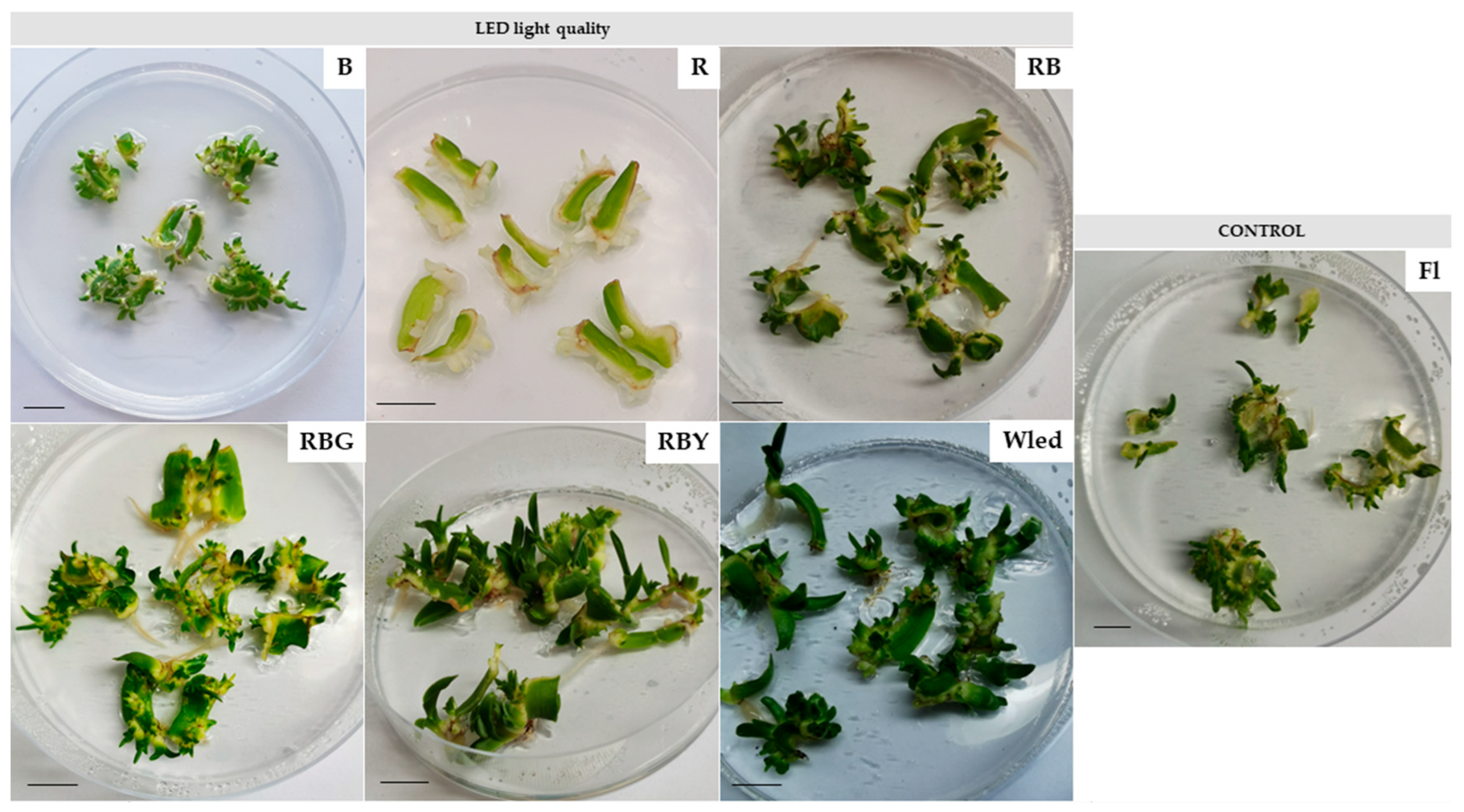
In our study all explants cultured on the medium regenerated successfully and continued their growth and development under the applied light conditions, resulting in a regeneration rate of 100%. The RBY LED light combination exhibited a stimulatory effect on biometric parameters of the regenerated plants, whereas monochromatic red (R) or blue (B) LED light acted as limiting factors, significantly reducing growth and development. Plants grown under RBY light conditions achieved the highest fresh weight (average 0.77 g) and height (average 0.57 cm), and also formed the greatest number of shoots and leaves (averaging 15.48 and 1.72, respectively). However, plants exposed to RBY LED light (as well as to the R LED light) exhibited the lowest dry matter content (DM), averaging 6.15%. In contrast, the highest DM content was recorded in plants grown under RBG and B LED light treatments, reaching 8.16%. The development of the root system, including root number and length, was negatively affected by monochromatic red or blue LED light, indicating a limiting effect of these spectra on rhizogenesis (Figure 2, Table 1). High variability in root number and length was observed among individual shoots, with some replicates producing no roots and others up to 5–7 roots, resulting in elevated standard deviations.
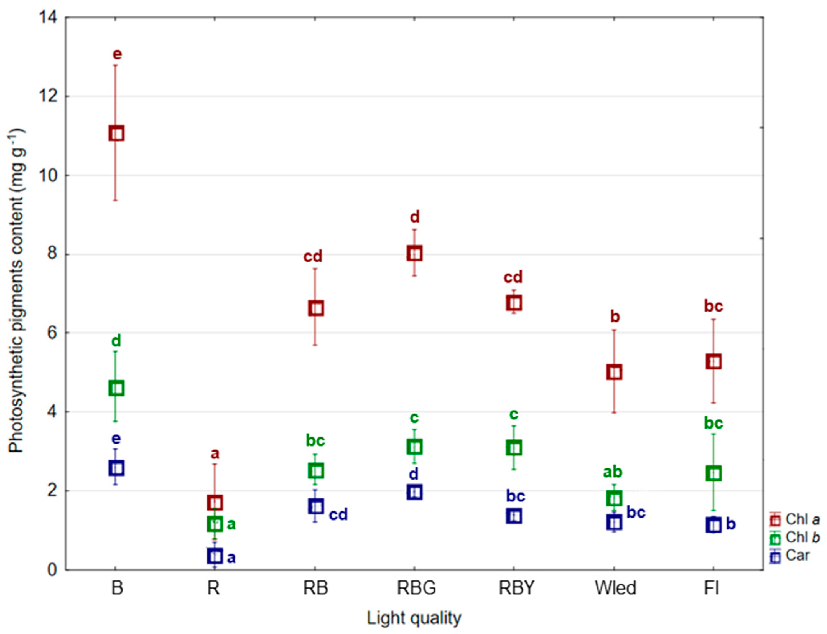
The application of B and RBG LED lights significantly influenced the accumulation of photosynthetic pigments in the tested plant material. Both these LED light treatments stimulated the synthesis of chlorophyll a and b, as well as carotenoids, with mean total pigment contents reaching 13.14 mg·g−1 and 18.34 mg·g−1, respectively. In contrast, exposure to red (R) LED light resulted in a statistically significant reduction in pigment content, which was approximately four times lower compared to the blue light treatment (Figure 3, Table S1).

3.2. Analyses of Polyphenol Content

Qualitative and quantitative analysis using HPLC-DAD, supported by reference standards, enabled the identification of 11 phenolic acids: eucomic, protocatechuic, gentisic, chlorogenic, p-hydroxybenzoic, vanillic, caffeic, syringic, ferulic, o-coumaric, and cinnamic acid, as well as 4 flavonoids which included 2 flavonols: catechin and epicatechin, and 2 flavanones: hesperetin and hesperidin. The applied light conditions significantly affected the accumulation of secondary metabolites, although the response was highly compound-specific.
Among the phenolic compounds, eucomic acid, a compound characteristic of the Eucomis genus, was the most abundant. Its accumulation was most strongly stimulated by RGB LED light, reaching 429 mg·100 g−1 DW. The lowest statistically confirmed content of this compound in our study was observed under red LED light (R), yet the amount was still relatively high compared to other phenolics—approximately 1.31 times lower than the level under RGB light.
Interestingly, R LED light promoted the highest accumulation of catechin, reaching 306 mg·100 g−1 DW, which was nearly two times higher than the amount detected in plant tissues E. autumnalis grown under RBUV LED light (146 mg·100 g−1 DW), where the lowest statistically significant catechin content was observed. Moreover, RBUV LED light had limiting influence on the accumulation of other flavonols and flavanones. In contrast, the white fluorescent light (Fl) used as a control proved most effective in stimulating the accumulation of epicatechin, hesperetin, and hesperidin. The second most abundant phenolic acid detected in the tissues was gentisic acid, with concentrations ranging from 129 to 229 mg·100 g−1 DW, and its accumulation was also most significantly enhanced under fluorescent light.
Compared to the Fl control light, all tested LED light conditions promoted higher levels of chlorogenic and p-hydroxybenzoic acids. A similar stimulatory effect was observed for protocatechuic, syringic, and cinnamic acids, particularly in plants grown under RB and RGB LED light (and additionally B LED in the case of protocatechuic acid). Notably, the additional RBfR and RBUV LED treatments used in the experiment design also stimulated the accumulation of syringic and cinnamic acids, highlighting their compound-specific effects (Table 2 and Table S2).

3.3. Antioxidant Activity

The antioxidant activity of E. autumnalis extracts obtained from plants multiplied under different LED light quality in in vitro culture was evaluated using three analytical methods: the DPPH and ABTS•+ radical scavenging assays, and the ferric reducing antioxidant power (FRAP) assay. The results obtained by the different methods were not entirely consistent, indicating that the antioxidant potential of the plant material depended not only on the applied light spectrum but also by the analytical method employed (Table 3).
According to the DPPH assay, the lowest antioxidant potential was observed in samples derived from plants grown under RBUV light. The highest antioxidant activity, as indicated by both the ABTS•+ and FRAP assays, was recorded in plants cultivated under RBfR light. The ABTS•+ assay also highlighted a high antioxidant potential in samples from plants grown under RBG light, whereas the FRAP assay showed similarly elevated values for plants grown under RBY and monochromatic blue (B) LED light. Interestingly, in the ABTS•+ assay, blue light (B) was associated with relatively low activity, in contrast to the FRAP results. Both the ABTS•+ and FRAP assays consistently indicated the lowest antioxidant activity in samples from plants grown under 100% red (R) LED light. Overall, these findings suggest that certain LED light spectra are more effective than fluorescent lighting in stimulating antioxidant potential in E. autumnalis. The fluorescent light control generally produced intermediate values, neither strongly enhancing nor markedly suppressing antioxidant activity compared to the LED treatments.
The correlation analysis revealed a significant positive association between antioxidant activity and the increased content of eucomic acid, ferulic acid, and epicatechin; however, this relationship was observed exclusively in the ABTS•+ assay (Table 4, Figure S1). Notably, eucomic acid exhibited the highest concentrations among all identified phenolic compounds in E. autumnalis extracts, with the greatest accumulation observed under RBfR and RBG light treatments. Similarly, ferulic acid and epicatechin reached their highest levels under the same LED light conditions, compared to the other treatments (Table 2). For other identified compounds, antioxidant activity generally exhibited weak to moderate correlations with their content, or no significant associations were detected. In the FRAP assay, an opposite trend was observed for certain compounds, where increasing content was accompanied by a decrease in antioxidant activity. Nevertheless, these negative correlations were of low to moderate magnitude, as exemplified by vanillic acid.

4. Discussion

4.1. Biometric and Photosynthetic Content Response

Several studies highlight the experimental advantage of LEDs over traditional fluorescent lamps due to their narrow-band and tunable output. For example, Beuel et al. [29] noted that fluorescent tubes lack a defined spectrum and degrade over time, whereas LEDs offer precise control over light quality, facilitating detailed correlation between specific wavelengths and metabolic responses. These characteristics make LEDs particularly suitable for investigating plant physiological and metabolic responses under controlled spectral conditions. Currently, the impact of various LED light qualities on plant in vitro cultures is receiving considerable attention in plant biotechnology research. While the effects of red and blue light on in vitro growth and development have been extensively studied and are relatively well understood, recent studies increasingly focus on other spectral regions and specific wavelengths. This shift aims to deepen our understanding of how different light qualities influence photomorphogenesis and related physiological responses in cultured plants. Studies on various ornamental and bulbous species consistently show that mixed red and blue LED light (often in ratios such as 7:3 or 8:2) promotes superior growth, shoot multiplication, and overall plantlet quality compared to monochromatic red or blue light alone. For example, in Dendrobium nobile, mixed red/blue LEDs enhanced root number, length, fresh and dry weight, and chlorophyll content, outperforming single-color treatments [30]. Similarly, in Gerbera jamesonii, mixed red/blue LEDs (especially 70% red, 30% blue) resulted in the highest shoot multiplication rates and pigment concentrations, while monochromatic red or blue light limited leaf area and pigment accumulation [23,31]. In Streptocarpus × hybridus, RBY (red, blue, yellow) LEDs yielded the best morphological quality, paralleling our findings in E. autumnalis [12]. Importantly, this research provides the first experimental evidence on how various LED light spectra influence the development, but also secondary metabolism of E. autumnalis cultured in vitro. Nevertheless, it is important to recognize that plant responses to varying light spectra are highly genotype-specific, and may differ even among cultivars of the same species, like for example in the case of Pyrus communis [21]. In this study, shoot and leaf growth were more influenced by certain LED spectra than by fluorescent lamps, but the effect was cultivar-dependent. For some cultivars, the highest shoot and node numbers were observed under specific LEDs, while for others, the control (fluorescent) light was superior. Notably, photosynthetic pigment content was significantly decreased under LEDs compared to fluorescent lamps, which contrasts with our findings for E. autumnalis. Also, in a multi-species study (Chrysanthemum, Gerbera, Heuchera, Ficus, Lamprocapnos), the highest propagation ratios were often observed under red- and far-red-abundant LEDs, but the effect was highly species-dependent. For some species, propagation ratios under LEDs were similar to or lower than under fluorescent light. Additionally, lighting conditions did not affect dry matter or rooting in most species, except Gerbera [32].
Blue and mixed red/blue LED treatments generally stimulate chlorophyll a, b, and carotenoid synthesis, while monochromatic red light often reduces pigment content. This pattern is observed in Gerbera [23,31], Dendrobium [30], and Doritaenopsis [33], where mixed or blue LEDs led to higher pigment levels and improved photosynthetic efficiency, as well as in results in there experiment in E. autumnalis. These effects can be explained by photoreceptor-mediated molecular mechanisms: phytochromes, cryptochromes, and phototropins perceive specific light wavelengths and trigger signal transduction cascades, which modulate gene expression involved in pigment biosynthesis, photosynthetic enzyme activity, and cell elongation. Such pathways influence chlorophyll and carotenoid accumulation, ultimately affecting growth and morphogenesis in plant in vitro cultures. Additionally, light quality can regulate hormone signaling (auxins, cytokinins, gibberellins) and oxidative stress responses, further modulating growth and secondary metabolism [34,35,36,37].

4.2. Analyses of Polyphenol Content

The literature strongly indicates that light quality influences and directs plant in vitro growth, photosynthetic pigment accumulation, and secondary metabolite production. These effects are mediated by light-specific photoreceptors influencing hormone signaling and gene expression. Compound-specific responses in polyphenol accumulation are common, and in vitro conditions can enhance or alter secondary metabolite profiles compared to ex vitro growth [38,39]. In Myrtus communis, red LEDs increased myricetin flavonoid, while blue and mixed LEDs enhanced fresh weight and other phenolics [40]. In Dracocephalum forrestii, blue and red/blue LEDs maximized polyphenol content and antioxidant potential [41]. In Cichorium intybus, blue LEDs increased both polyphenol and chlorophyll content, while red LEDs shifted the polyphenol profile toward kaempferol derivatives [42]. In Artemisia argyi, blue light increased total phenols and flavonoids, while red and RB light promoted specific phenolic acids like chlorogenic acid and gallic acid [38]. These findings are consistent with our results, which demonstrated that specific LED spectra selectively stimulated the accumulation of individual phenolic compounds in E. autumnalis. For example, red LED light (R) significantly increased the content of catechin (306 mg/100 g DW) and gentisic acid (214 mg/100 g DW), while blue light (B) was most effective in promoting hesperidin accumulation (251 mg/100 g DW). In contrast, ultraviolet-enriched light (RBUV) limited the accumulation of several flavonoids, including catechin and hesperetin. Interestingly, the fluorescent light (Fl), used as a control, stimulated the accumulation of epicatechin and syringic acid, suggesting that broad-spectrum light may favor the biosynthesis of certain phenolics. For instance, in Lilium candidum, all identified phenolic acids decreased under fluorescent and red light, while RB LEDs led to the highest total phenolic content [39]. However, in our study on E. autumnalis, RB light did not induce the highest levels of total phenolics, highlighting interspecies variability in light response. Moreover, in L. candidum, bulbs grown ex vitro accumulated two to three times more chlorogenic acid than those cultured in vitro, emphasizing the influence of cultivation system on metabolite profiles. Furthermore, for L. candidum bulbs grown in soil (ex vitro) accumulated two to three times more chlorogenic acid than those formed in vitro. In Cichorium intybus, red LEDs induced a distinct polyphenol profile with a predominance of kaempferol 3-O-glucuronide, while blue and white LEDs favored quercetin derivatives [42]. Such cultivar-dependent responses further support the need for species-specific optimization of light conditions in in vitro systems.
The observed effects of LED spectra on phenolic compound accumulation are mediated by photoreceptors—phytochromes (red/far-red), cryptochromes, and phototropins (blue)—which regulate gene expression related to growth, hormone balance, and secondary metabolism. These photoreceptors initiate signaling cascades that modulate enzyme activity and metabolic fluxes in biosynthetic pathways, including the phenylpropanoid pathway [43,44]. Transcriptomic and proteomic studies reveal that red and blue light modulate genes involved in photosynthesis, hormone signaling (auxins, gibberellins, cytokinins), and stress responses, leading to changes in morphogenesis and metabolite accumulation [44,45]. Balanced LED spectra such as RBY or RBfR can simultaneously activate multiple photoreceptor pathways, enhancing both growth parameters (fresh weight, shoot/leaf number) and the accumulation of phenolic compounds, whereas monochromatic red or blue light may act as a limiting factor for these processes [38,41]. Blue light, in particular, upregulates genes responsible for pigment biosynthesis and antioxidant enzyme activity [40,44,46], while red light can selectively favor certain compounds such as catechin but limit others [42]. Mixed spectra also induce mild oxidative stress, triggering reactive oxygen species (ROS) signaling that further promotes antioxidant secondary metabolite production [47]. As the literature indicates, blue light is particularly effective at stimulating the synthesis of phenolic acids and flavonoids. For example, in Schisandra chinensis, blue light increased the total content of phenolic acids (including chlorogenic and protocatechuic acids) by up to 2.72 times compared to white light, likely by upregulating key biosynthetic buenzymes. Red and far-red can also influence secondary metabolism, but their effects are often species- and compound-specific. In some cases, red light may promote or inhibit certain phenolic pathways depending on the plant and developmental stage. Ultraviolet (UV) light acts as a potent abiotic elicitor, enhancing the accumulation of bioactive compounds, including polyphenols, by inducing stress responses and activating defense-related pathways. This leads to increased synthesis of antioxidant compounds, as observed in our study under RBUV treatment [48,49]. Mixed LED spectra often produce synergistic effects by simultaneously activating multiple photoreceptor pathways, thereby enhancing both growth and secondary metabolite production [47,50,51]. The overall mechanism involves light perception by photoreceptors, signal transduction, and transcriptional regulation of genes encoding enzymes such as phenylalanine ammonia-lyase (PAL), a key entry point to the phenylpropanoid pathway, which leads to the biosynthesis of most polyphenols [52].
These findings highlight the potential of spectral light manipulation as a precise and effective tool for modulating the biosynthesis of specific phenolic compounds in vitro. By tailoring light quality, it is possible to enhance the accumulation of target metabolites in E. autumnalis, thereby improving the phytochemical value of cultured biomass for potential pharmaceutical or nutraceutical applications. We acknowledge that comparative data on phenolic acids, flavonols, and flavanones in field- or greenhouse-grown Eucomis species are limited. Aremu et al. [8] reported that certain compounds, such as eucomic acid, are present in both in vitro and acclimatized plants, but most phenolic acids and flavonoids were higher in in vitro regenerants. After acclimatization, caffeic and p-coumaric acids were generally <1 μg/g DW, and ferulic acid ranged 0.17–0.39 μg/g DW in leaves and 0.78–1.89 μg/g DW in underground parts. In comparison, in our LED-grown E. autumnalis plantlets, caffeic acid and ferulic acid averaged 3.08 mg/100 g DW and 5.13 mg/100 g DW, representing an approximately 300- to 650-fold increase. These findings emphasize the strong stimulatory effect of controlled LED light on phenolic acid accumulation in vitro and the potential of spectral light manipulation to enhance phytochemical content in Eucomis plantlets.

4.3. Antioxidant Activity

Polyphenols, including both hydroxybenzoic acids (e.g., protocatechuic, gentisic, p-hydroxybenzoic, vanillic, syringic) and hydroxycinnamic acids (e.g., chlorogenic, caffeic, ferulic, o-coumaric, cinnamic), as well as flavonoids (catechin, epicatechin, hesperetin, hesperidin), are well-documented for their diverse pharmacological activities. Most polyphenols are potent antioxidants, neutralizing free radicals and protecting against oxidative stress-related diseases such as cancer, diabetes, and cardiovascular disorders. Many phenolic acids and flavonoids also exhibit antibacterial and antifungal properties, contributing to plant defense and potential therapeutics [52,53,54]. Compounds like protocatechuic acid, caffeic acid, and ferulic acid have demonstrated anti-inflammatory effects in various models. Also, some polyphenols show anticancer, neuroprotective, cardioprotective, antidiabetic, and hepatoprotective effects [53,54,55].
Antioxidant activity in plant extracts is commonly evaluated using multiple assays, such as DPPH, ABTS•+, and FRAP, which differ in mechanism and sensitivity [56,57]. DPPH measures hydrogen-donating capacity [58,59], ABTS•+ evaluates electron transfer to quench the ABTS•+ radical cation [56,60], and FRAP reflects reducing power of iron ions conversion [57,61]. Using multiple methods is essential, as results may differ due to the distinct chemical reactions each assay captures [58,62].
In this study, the ABTS•+ assay was particularly informative, showing the highest antioxidant activity in E. autumnalis extracts from plants grown under RBfR and RBG light. Activity positively correlated with eucomic acid, ferulic acid, and epicatechin, with eucomic acid being the most abundant phenolic compound, characteristic for this species. FRAP and DPPH results generally supported these trends but showed some discrepancies, highlighting method-specific differences in detecting antioxidant potential. These differences among antioxidant assays reflect the compound-specific responses to light quality: certain phenolics may contribute more to electron transfer measured in ABTS•+ than to hydrogen-donating capacity in DPPH, or reducing power in FRAP, explaining why correlations were strongest for ABTS•+. Additionally, ABTS•+ is sensitive to a broader range of hydrophilic and lipophilic antioxidants, thus better reflecting the cumulative effect of compound accumulation [60,63,64]. In contrast, DPPH primarily reacts in organic media and favors specific hydrogen-donating phenolics, while FRAP measures only the reducing power toward Fe3+, which is not equally relevant for all compounds identified in this study [60,63,65,66]. Furthermore, the chemical structure, solubility, and localization of specific phenolic compounds in the tissue can influence their reactivity in each assay, leading to assay-dependent differences in measured antioxidant activity. Therefore, while ABTS•+ captured the activity of dominant compounds such as eucomic acid, ferulic acid, and epicatechin, DPPH and FRAP were less sensitive to these specific constituents.
The elevated eucomic acid content under RBfR and RBG light may be linked to the effects of green and far-red light on phenylpropanoid metabolism in in vitro cultures, promoting the accumulation of bioactive compounds [48,67,68]. Also, light quality can induce mild oxidative stress, leading to increased production of reactive oxygen species (ROS). Plants respond by upregulating antioxidant secondary metabolites (e.g., phenolics, flavonoids) as a defense mechanism [69,70]. In the research of Chen at al. [68] for Prunella vulgaris, the far-red light spectrum significantly increased total phenolic content, rutin, and rosmarinic acid, and enhanced DPPH scavenging activity and reducing power, indicating improved antioxidant capacity. This effect was strongly correlated with phenolic content, mirroring the positive correlation between phenolics and antioxidant activity described in the excerpt. For callus cultures, green light induced the highest callogenic response and, along with red and blue light, enhanced DPPH-free radical scavenging activity, suggesting that green light can also stimulate antioxidant secondary metabolite production [71]. As previously noted, the effects of blue and red light on plant metabolism are the most extensively studied in the literature, whereas the specific influence of green and far-red light is less commonly investigated. Consequently, numerous reports indicate that blue and red light frequently enhance phenolic and flavonoid accumulation, as well as antioxidant activity, in various species, including Eclipta alba, Operculina turpethum, Rhodiola imbricata, Ocimum basilicum, and Stevia rebaudiana [62,71,72,73,74,75,76,77,78].
These findings indicate that LED spectral quality can effectively modulate antioxidant potential in E. autumnalis, with ABTS•+ assay providing the most sensitive measure of these changes.

5. Conclusions

This study provides the first comprehensive evidence that spectral quality of LED light significantly modulates both morphogenesis and secondary metabolism in Eucomis autumnalis cultured in vitro. Among the tested treatments, RBY light (50% red + 30% blue + 20% yellow) most effectively stimulated shoot proliferation and fresh biomass accumulation (up to 0.77 g per explant), while blue (B) and RBG light enhanced photosynthetic pigment content (up to 18.34 mg·g−1 DW of carotenoids). HPLC-DAD analysis revealed that light quality had a compound-specific effect on polyphenol biosynthesis. Red light (R) induced the highest accumulation of catechin (306 mg·100 g−1 DW) and gentisic acid (214 mg·100 g−1 DW), while blue light (B) promoted hesperidin (251 mg·100 g−1 DW). In contrast, ultraviolet-enriched light (RBUV) suppressed the accumulation of several flavonoids. Antioxidant activity, assessed by DPPH, ABTS•+, and FRAP assays, further confirmed that light spectrum plays a pivotal role in modulating the bioactive potential of E. autumnalis. RBfR and RBG light treatments were particularly effective, coinciding with the highest accumulation of eucomic acid, ferulic acid, and epicatechin, and yielding superior ABTS•+ assay results. These findings underscore that targeted LED spectral manipulation can simultaneously stimulate the biosynthesis of key polyphenols and enhance antioxidant performance, demonstrating its effectiveness as a powerful and precise tool for optimizing in vitro culture conditions for the production of high-value phytochemicals. Such an approach may also serve as a model for enhancing light-driven biosynthesis of bioactive compounds in other bulbous or ethnobotanically important species.

Supplementary Materials

The following supporting information can be downloaded at https://www.mdpi.com/article/10.3390/agronomy15092197/s1, Table S1: Mean content of photosynthetic pigments in the tissues of Eucomis autumnalis in vitro under different light quality (mg g−1); Table S2: Content of identified polyphenols in tissues of E. autumnalis cultured in vitro under different LED light quality compared to the control white light of a fluorescent lamp; Figure S1: Principal component analysis (PCA) biplot showing the projection of active variables (phenolic and flavonoid compounds and antioxidant activity assays) on the first two principal components (PC1 and PC2). Arrows indicate the contribution and direction of individual variables, while points represent mean values for plants grown under different light treatments. The plot illustrates the relationships and correlations between the studied compounds and antioxidant activity.

Author Contributions

Conceptualization, M.C. and B.P. (Bożena Pawłowska); methodology, M.C., A.S., B.P. (Bożena Pawłowska) and Ł.Ł.; software, M.C., A.S. and Ł.Ł.; validation, M.C., A.S. and Ł.Ł.; formal analysis, M.C., A.S. and Ł.Ł.; investigation, M.C., A.S., B.P. (Barbara Prokopiuk) and Ł.Ł.; resources, M.C., A.S. and Ł.Ł.; data curation, M.C., A.S. and Ł.Ł.; writing—original draft preparation, M.C.; writing—review and editing, M.C., A.S. and Ł.Ł.; visualization, M.C.; supervision, B.P. (Bożena Pawłowska); project administration, M.C.; funding acquisition, M.C. and Ł.Ł. All authors have read and agreed to the published version of the manuscript.

Funding

This research was supported by the Ministry of Science and Higher Education of the Republic of Poland. Part of this research was funded by the Scientific Activity Activation Fund of the University of Agriculture in accordance with Rector’s Order 107/2022 of 18 November 2022 (registration number A557).

Data Availability Statement

Data are contained within the article.

Acknowledgments

The author gratefully acknowledges Piotr Salachna, ZUT, from the Department of Horticulture, Faculty of Environmental Management and Agriculture, West Pomeranian University of Technology in Szczecin, for kindly providing the initial plant material introduced into in vitro culture. The author also wishes to thank for experimental assistance provided by Joanna Kaczmarczyk as part of her MSc thesis.

Conflicts of Interest

The authors declare no conflicts of interest.

Abbreviations

The following abbreviations are used in this manuscript:
ABTS•+2,2′-azino-bis(3-ethylbenzothiazoline-6-sulfonic acid) radical scavenging assay
B100% blue LED light
BA6-benzylaminopurine
Carcarotenoids
Chl achlorophyll a
Chl bchlorophyll b
DPPH2,2-diphenyl-1-picrylhydrazyl radical scavenging assay
DWdry weight
FRAPferric reducing antioxidant power assay
FWfresh weight
LEDlight-emitting diodes
MSMurashige and Skoog
NAA1-naphthaleneacetic acid
PPFDphotosynthetic photon flux density
RB70% red + 30% blue LED light
RBfR50% RB + 50% far-red LED light
RBG50% RB + 50% green LED light
RBY50% RB + 50% yellow LED light
RBUV50% RB + 50% ultraviolet LED light
R100% red LED light
WLedwhite LED light

References

  1. Salachna, P.; Zawadzińska, A. Foliar Application of Depolymerised Chitosan Enhances the growth and Content of Polyphenols and L-Ascorbic Acid in Eucomis autumnalis, an Ornamental and Medicinal Plant. Prog. Chem. Appl. Chitin Deriv. 2024, 29, 254–262. [Google Scholar] [CrossRef]
  2. Ndhlala, A.R.; Mulaudzi, R.B.; Kulkarni, M.G.; Van Staden, J. Effect of Environmental Factors on Seedling Growth, Bulb Development and Pharmacological Properties of Medicinal Eucomis autumnalis. S. Afr. J. Bot. 2012, 79, 1–8. [Google Scholar] [CrossRef]
  3. Alaribe, F.N.; Maepa, M.J.; Mkhumbeni, N.; Motaung, S.C.K.M. Possible Roles of Eucomis autumnalis in Bone and Cartilage Regeneration: A Review. Trop. J. Pharm Res. 2018, 17, 741–749. [Google Scholar] [CrossRef]
  4. Salachna, P.; Zawadzińska, A. Effect of Daminozide and Flurprimidol on Growth, Flowering and Bulb Yield of Eucomis autumnalis (Mill.) Chitt. Folia Hortic. 2017, 29, 33–38. [Google Scholar] [CrossRef]
  5. Salachna, P.; Zawadzińska, A.; Wierzbiński, Ł.; Senderek, W. Enhancing Growth in Eucomis autumnalis (Mill.) Chitt. Seedlings with Exogenous Application of Nitric Oxide. J. Hortic. Res. 2016, 24, 13–17. [Google Scholar] [CrossRef]
  6. Mkhumbeni, N.; Pillay, M.; Mtunzi, F.; Motaung, K.S.C. Effect of Eucomis autumnalis on the Osteogenic Differentiation of Adipose-Derived Stem Cells. Tissue Eng. Part A 2022, 28, 136–149. [Google Scholar] [CrossRef]
  7. Aremu, A.O.; Masondo, N.A.; Rengasamy, K.R.R.; Amoo, S.O.; Gruz, J.; Bíba, O.; Šubrtová, M.; Pěnčík, A.; Novák, O.; Doležal, K.; et al. Physiological Role of Phenolic Biostimulants Isolated from Brown Seaweed Ecklonia maxima on Plant Growth and Development. Planta 2015, 241, 1313–1324. [Google Scholar] [CrossRef]
  8. Aremu, A.O.; Masondo, N.A.; Gruz, J.; Doležal, K.; Van Staden, J. Potential of Smoke-Water and One of Its Active Compounds (Karrikinolide, KAR1) on the Phytochemical and Antioxidant Activity of Eucomis autumnalis. Antioxidants 2019, 8, 611. [Google Scholar] [CrossRef] [PubMed]
  9. Masondo, N.A.; Aremu, A.O.; Finnie, J.F.; Van Staden, J. Growth and Phytochemical Levels in Micropropagated Eucomis autumnalis Subspecies Autumnalis Using Different Gelling Agents, Explant Source, and Plant Growth Regulators. Vitr. Cell. Dev. Biol.-Plant 2015, 51, 102–110. [Google Scholar] [CrossRef]
  10. Masondo, N.A.; Aremu, A.O.; Finnie, J.F.; Van Staden, J. Plant Growth Regulator Induced Phytochemical and Antioxidant Variations in Micropropagated and Acclimatized Eucomis autumnalis Subspecies Autumnalis (Asparagaceae). Acta Physiol. Plant. 2014, 36, 2467–2479. [Google Scholar] [CrossRef]
  11. Manivannan, A.; Soundararajan, P.; Halimah, N.; Ko, C.H.; Jeong, B.R. Blue LED Light Enhances Growth, Phytochemical Contents, and Antioxidant Enzyme Activities of Rehmannia glutinosa Cultured in Vitro. Hortic. Environ. Biotechnol. 2015, 56, 105–113. [Google Scholar] [CrossRef]
  12. Cioć, M.; Łopusiewicz, Ł.; Pietrak, A.; Firszt, R.; Sienkiewicz, M. Organogenesis and Antioxidant Capacity of Streptocarpus × hybridus In Vitro under Different LED Light Spectrum Composition. Agronomy 2023, 13, 3009. [Google Scholar] [CrossRef]
  13. Morańska, E.; Simlat, M.; Warchoł, M.; Skrzypek, E.; Waligórski, P.; Laurain-Mattar, D.; Spina, R.; Ptak, A. Phenolic Acids and Amaryllidaceae Alkaloids Profiles in Leucojum aestivum L. In Vitro Plants Grown under Different Light Conditions. Molecules 2023, 28, 1525. [Google Scholar] [CrossRef]
  14. Niazian, M.; Sabbatini, P. Traditional in vitro strategies for sustainable production of bioactive compounds and manipulation of metabolomic profile in medicinal, aromatic and ornamental plants. Planta 2021, 254, 111. [Google Scholar] [CrossRef]
  15. Alrifai, O.; Hao, X.; Marcone, M.F.; Tsao, R. Current Review of the Modulatory Effects of LED Lights on Photosynthesis of Secondary Metabolites and Future Perspectives of Microgreen Vegetables. J. Agric. Food Chem. 2019, 67, 6075–6090. [Google Scholar] [CrossRef]
  16. Wan Mahari, W.A.; Wan Razali, W.A.; Waiho, K.; Wong, K.Y.; Foo, S.S.; Kamaruzzan, A.S.; Derek, C.J.C.; Ma, N.L.; Chang, J.S.; Dong, C.D.; et al. Light-Emitting Diodes (LEDs) for Culturing Microalgae and Cyanobacteria. Chem. Eng. J. 2024, 485, 149619. [Google Scholar] [CrossRef]
  17. Dutta Gupta, S.; Agarwal, A. Influence of LED Lighting on In Vitro Plant Regeneration and Associated Cellular Redox Balance. In Light Emitting Diodes for Agriculture: Smart Lighting; Dutta Gupta, S., Ed.; Springer: Singapore, 2017; pp. 273–303. [Google Scholar]
  18. Gam, D.T.; Khoi, P.H.; Ngoc, P.B.; Linh, L.K.; Hung, N.K.; Anh, P.T.L.; Thu, N.T.; Hien, N.T.T.; Khanh, T.D.; Ha, C.H. Led Lights Promote Growth and Flavonoid Accumulation of Anoectochilus roxburghii and Are Linked to the Enhanced Expression of Several Related Genes. Plants 2020, 9, 1344. [Google Scholar] [CrossRef] [PubMed]
  19. Xie, D.; Chen, L.; Zhou, C.; Tarin, M.W.K.; Yang, D.; Ren, K.; He, T.; Rong, J.; Zheng, Y. Transcriptomic and Metabolomic Profiling Reveals the Effect of LED Light Quality on Morphological Traits, and Phenylpropanoid-Derived Compounds Accumulation in Sarcandra glabra Seedlings. BMC Plant Biol. 2020, 20, 476. [Google Scholar] [CrossRef]
  20. Dutta Gupta, S.; Pradhan, S. Regulation of Gene Expression by LED Lighting. In Light Emitting Diodes for Agriculture: Smart Lighting; Dutta Gupta, S., Ed.; Springer: Singapore, 2017; pp. 237–258. [Google Scholar]
  21. Lupo, M.; Bashir, M.A.; Silvestri, C.; Brunori, E.; Pica, A.L.; Cristofori, V. LED Lighting Effects on Plant Growth and Quality of Pyrus communis L. Propagated In Vitro. Agronomy 2022, 12, 2531. [Google Scholar] [CrossRef]
  22. Murasnige, T.; Skoog, F. A Revised Medium for Rapid Growth and Bio Assays with Tobacco Tissue Cultures. Physiol. Plant 1962, 15, 473–497. [Google Scholar] [CrossRef]
  23. Pawłowska, B.; Żupnik, M.; Szewczyk-Taranek, B.; Cioć, M. Impact of LED Light Sources on Morphogenesis and Levels of Photosynthetic Pigments in Gerbera jamesonii Grown in Vitro. Hortic. Environ. Biotechnol. 2018, 59, 115–123. [Google Scholar] [CrossRef]
  24. Sumanta, N.; Imranul Haque, C.; Nishika, J.; Suprakash, R. Spectrophotometric Analysis of Chlorophylls and Carotenoids from Commonly Grown Fern Species by Using Various Extracting Solvents. Res. J. Chem. Sci. 2014, 4, 63–69. [Google Scholar]
  25. Szopa, A.; Kubica, P.; Ekiert, H. Agitated Shoot Cultures of Aronia Arbutifolia and Aronia × prunifolia: Biotechnological Studies on the Accumulation of Phenolic Compounds and Biotransformation Capability. Plant Cell Tissue Organ Cult. 2018, 134, 467–479. [Google Scholar] [CrossRef]
  26. Szopa, A.; Kokotkiewicz, A.; Kubica, P.; Banaszczak, P.; Wojtanowska-Krośniak, A.; Krośniak, M.; Marzec-Wróblewska, U.; Badura, A.; Zagrodzki, P.; Bucinski, A.; et al. Comparative Analysis of Different Groups of Phenolic Compounds in Fruit and Leaf Extracts of Aronia Sp.: A. melanocarpa, A. arbutifolia, and A. × prunifolia and Their Antioxidant Activities. Eur. Food Res. Technol. 2017, 243, 1645–1657. [Google Scholar] [CrossRef]
  27. Łopusiewicz, Ł.; Drozłowska, E.; Siedlecka, P.; Mężyńska, M.; Bartkowiak, A.; Sienkiewicz, M.; Zielińska-Bliźniewska, H.; Kwiatkowski, P. Development, Characterization, and Bioactivity of Non-Dairy Kefir-like Fermented Beverage Based on Flaxseed Oil Cake. Foods 2019, 8, 544. [Google Scholar] [CrossRef]
  28. Pietrak, A.; Łopusiewicz, Ł.; Salachna, P. Growth, Leaf Pigment Content, and Antioxidant Potential of Ferns Grown in Peat Substrate Amended with Camelina Press Cake. Agronomy 2022, 12, 3100. [Google Scholar] [CrossRef]
  29. Beuel, A.K.; Jablonka, N.; Heesel, J.; Severin, K.; Spiegel, H.; Rasche, S. LEDitSHAKE: A lighting system to optimize the secondary metabolite content of plant cell suspension cultures. Sci. Rep. 2021, 11, 23353. [Google Scholar] [CrossRef]
  30. Guo, Y.; Zhong, Y.; Mo, L.; Zhang, W.; Chen, Y.; Wang, Y.C.; Chen, H.; Wang, Z.; Song, X.; Meng, X. Different Combinations of Red and Blue LED Light Affect the Growth, Physiology and Photosynthesis of in Vitro-Cultured Dendrobium nobile ‘Zixia’. Hortic. Environ. Biotechnol. 2023, 64, 393–407. [Google Scholar] [CrossRef]
  31. Cioć, M.; Tokarz, K.; Dziurka, M.; Pawłowska, B. Energy-Saving Led Light Affects the Efficiency of the Photosynthetic Apparatus and Carbohydrate Content in Gerbera Jamesonii Bolus Ex Hook. F. Axillary Shoots Multiplied in Vitro. Biology 2021, 10, 1035. [Google Scholar] [CrossRef]
  32. Miler, N.; Kulus, D.; Woźny, A.; Rymarz, D.; Hajzer, M.; Wierzbowski, K.; Nelke, R.; Szeffs, L. Application of Wide-Spectrum Light-Emitting Diodes in Micropropagation of Popular Ornamental Plant Species: A Study on Plant Quality and Cost Reduction. Vitr. Cell. Dev. Biol.-Plant 2019, 55, 99–108. [Google Scholar] [CrossRef]
  33. Shin, K.S.; Murthy, H.N.; Heo, J.W.; Hahn, E.J.; Paek, K.Y. The Effect of Light Quality on the Growth and Development of In Vitro Cultured Doritaenopsis Plants. Acta Physiol. Plant 2008, 30, 339–343. [Google Scholar] [CrossRef]
  34. Mawphlang, O.; Kharshiing, E. Photoreceptor Mediated Plant Growth Responses: Implications for Photoreceptor Engineering toward Improved Performance in Crops. Front. Plant Sci. 2017, 8, 1181. [Google Scholar] [CrossRef]
  35. Pech, R.; Volná, A.; Hunt, L.; Bartas, M.; Červeň, J.; Pečinka, P.; Špunda, V.; Nezval, J. Regulation of Phenolic Compound Production by Light Varying in Spectral Quality and Total Irradiance. Int. J. Mol. Sci. 2022, 23, 6533. [Google Scholar] [CrossRef]
  36. Weremczuk-Jeżyna, I.; Hnatuszko-Konka, K.; Lebelt, L.; Grzegorczyk-Karolak, I. The Protective Function and Modification of Secondary Metabolite Accumulation in Response to Light Stress in Dracocephalum forrestii Shoots. Int. J. Mol. Sci. 2021, 22, 7965. [Google Scholar] [CrossRef]
  37. Appolloni, E.; Pennisi, G.; Zauli, I.; Carotti, L.; Paucek, I.; Quaini, S.; Orsini, F.; Gianquinto, G. Beyond vegetables: Effects of indoor LED light on specialized metabolite biosynthesis in medicinal and aromatic plants, edible flowers, and microgreens. J. Sci. Food Agric. 2021, 102, 472–487. [Google Scholar] [CrossRef]
  38. Su, P.; Ding, S.; Wang, D.; Kan, W.; Yuan, M.; Chen, X.; Tang, C.; Hou, J.; Wu, L. Plant Morphology, Secondary Metabolites and Chlorophyll Fluorescence of Artemisia argyi under Different LED. Photosynth. Res. 2024, 159, 153–164. [Google Scholar] [CrossRef]
  39. Pałka, P.; Muszyńska, B.; Szewczyk, A.; Pawłowska, B. Elicitation and Enhancement of Phenolics Synthesis with Zinc Oxide Nanoparticles and LED Light in Lilium candidum L. Cultures In Vitro. Agronomy 2023, 13, 1437. [Google Scholar] [CrossRef]
  40. Cioć, M.; Szewczyk, A.; Żupnik, M.; Kalisz, A.; Pawłowska, B. LED Lighting Affects Plant Growth, Morphogenesis and Phytochemical Contents of Myrtus communis L. in Vitro. Plant Cell Tissue Organ Cult. 2018, 132, 433–447. [Google Scholar] [CrossRef]
  41. Weremczuk-Jeżyna, I.; Kuźma, Ł.; Grzegorczyk-Karolak, I. The Effect of Different Light Treatments on Morphogenesis, Phenolic Compound Accumulation and Antioxidant Potential of Dracocephalum forrestii Transformed Shoots Cultured in Vitro. J. Photochem. Photobiol. B 2021, 224, 112329. [Google Scholar] [CrossRef] [PubMed]
  42. Menicucci, F.; Marino, G.; Sillo, F.; Carli, A.; dos Santos Nascimento, L.B.; Detti, C.; Centritto, M.; Brunetti, C.; Balestrini, R.M. Blue and Red LEDs Modulate Polyphenol Production in Precoce and Tardiva Cultivars of Cichorium intybus L. Front. Plant Sci. 2025, 16, 1529804. [Google Scholar] [CrossRef]
  43. Kołton, A.; Długosz-Grochowska, O.; Wojciechowska, R.; Czaja, M. Biosynthesis Regulation of Folates and Phenols in Plants. Sci. Hortic. 2022, 291, 110561. [Google Scholar] [CrossRef]
  44. Chen, L.l.; Wang, H.Y.; Gong, X.C.; Zeng, Z.H.; Xue, X.Z.; Hu, Y.G. Transcriptome Analysis Reveals Effects of Red and Blue Light-Emitting Diodes (LEDs) on the Growth, Chlorophyll Fluorescence and Endogenous Plant Hormones of Potato (Solanum tuberosum L.) Plantlets Cultured in Vitro. J. Integr. Agric. 2021, 20, 2914–2931. [Google Scholar] [CrossRef]
  45. Oliveira, T.d.R.; Aragão, V.P.M.; Moharana, K.C.; Fedosejevs, E.; do Amaral, F.P.; Sousa, K.R.; Thelen, J.J.; Venâncio, T.M.; Silveira, V.; Santa-Catarina, C. Light Spectra Affect the in Vitro Shoot Development of Cedrela fissilis Vell. (Meliaceae) by Changing the Protein Profile and Polyamine Contents. Biochim. Biophys. Acta Proteins Proteom. 2020, 1868, 140529. [Google Scholar] [CrossRef] [PubMed]
  46. Manivannan, A.; Soundararajan, P.; Park, Y.G.; Jeong, B.R. Physiological and Proteomic Insights Into Red and Blue Light-Mediated Enhancement of in Vitro Growth in Scrophularia kakudensis—A Potential Medicinal Plant. Front. Plant Sci. 2021, 11, 607007. [Google Scholar] [CrossRef]
  47. Rai, N.; Kumari, S.; Singh, S.; Saha, P.; Pandey, A.K.; Pandey-Rai, S. Modulation of Morpho-Physiological Attributes and in Situ Analysis of Secondary Metabolites Using Raman Spectroscopy in Response to Red and Blue Light Exposure in Artemisia annua. Environ. Exp. Bot. 2024, 217, 105563. [Google Scholar] [CrossRef]
  48. Murthy, H.N.; Joseph, K.S.; Paek, K.Y.; Park, S.Y. Light as an Elicitor for Enhanced of Secondary Metabolites in Plant Cell, Tissue, and Organ Cultures. Plant Growth Regul. 2024, 104, 31–49. [Google Scholar] [CrossRef]
  49. Landi, M.; Zivcak, M.; Sytar, O.; Brestic, M.; Allakhverdiev, S.I. Plasticity of Photosynthetic Processes and the Accumulation of Secondary Metabolites in Plants in Response to Monochromatic Light Environments: A Review. Biochim. Biophys. Acta–Bioenerg. 2020, 1861, 148131. [Google Scholar] [CrossRef]
  50. Ouzounis, T.; Rosenqvist, E.; Ottosen, C.-O. Spectral Effects of Artificial Light on Plant Physiology and Secondary Metabolism: A Review. Hortscience 2015, 50, 1128–1135. [Google Scholar] [CrossRef]
  51. Li, Y.; Xu, J.; Zhang, F.; Gu, Y.; Tian, W.; Tian, W.; Tong, Y.; Li, J. The Combination of Red and Blue Light Increases the Biomass and Steroidal Saponin Contents of Paris Polyphylla Var. Yunnanensis. Ind. Crops Prod. 2023, 194, 116311. [Google Scholar] [CrossRef]
  52. Da Silva, A.P.G.; Sganzerla, W.G.; John, O.D.; Marchiosi, R. A Comprehensive Review of the Classification, Sources, Biosynthesis, and Biological Properties of Hydroxybenzoic and hydroxycinnamic Acids. Phytochem. Rev. 2023, 24, 94. [Google Scholar] [CrossRef]
  53. Kumar, N.; Goel, N. Phenolic Acids: Natural Versatile Molecules with Promising Therapeutic Applications. Biotechnol. Rep. 2019, 24, e00370. [Google Scholar] [CrossRef]
  54. Sun, W.; Shahrajabian, M.H. Therapeutic Potential of Phenolic Compounds in Medicinal Plants—Natural Health Products for Human Health. Molecules 2023, 28, 1845. [Google Scholar] [CrossRef]
  55. Song, J.; He, Y.; Luo, C.; Feng, B.; Ran, F.; Xu, H.; Ci, Z.; Xu, R.; Han, L.; Zhang, D. New Progress in the Pharmacology of Protocatechuic Acid: A Compound Ingested in Daily Foods and Herbs Frequently and Heavily. Pharmacol. Res. 2020, 161, 105109. [Google Scholar] [CrossRef] [PubMed]
  56. Platzer, M.; Kiese, S.; Herfellner, T.; Schweiggert-Weisz, U.; Miesbauer, O.; Eisner, P. Common Trends and Differences in Antioxidant Activity Analysis of Phenolic Substances Using Single Electron Transfer Based Assays. Molecules 2021, 26, 1244. [Google Scholar] [CrossRef] [PubMed]
  57. Chaves, N.; Santiago, A.; Alías, J.C. Quantification of the Antioxidant Activity of Plant Extracts: Analysis of Sensitivity and Hierarchization Based on the Method Used. Antioxidants 2020, 9, 76. [Google Scholar] [CrossRef] [PubMed]
  58. Munteanu, I.G.; Apetrei, C. Analytical Methods Used in Determining Antioxidant Activity: A Review. Int. J. Mol. Sci. 2021, 22, 3380. [Google Scholar] [CrossRef]
  59. Abramovič, H.; Grobin, B.; Ulrih, N.P.; Cigić, B. Relevance and Standardization of in Vitro Antioxidant Assays: ABTS, DPPH, and Folin–Ciocalteu. J. Chem. 2018, 2018, 1–9. [Google Scholar] [CrossRef]
  60. Ilyasov, I.R.; Beloborodov, V.L.; Selivanova, I.A.; Terekhov, R.P. ABTS/PP Decolorization Assay of Antioxidant Capacity Reaction Pathways. Int. J. Mol. Sci. 2020, 21, 1131. [Google Scholar] [CrossRef]
  61. Shah, P.; Modi, H.A. Comparative Study of DPPH, ABTS and FRAP Assays for Determination of Antioxidant Activity. Int. J. Res. Appl. Sci. Eng. Technol. 2015, 3, 636–641. [Google Scholar]
  62. Nazir, M.; Ullah, M.A.; Younas, M.; Siddiquah, A.; Shah, M.; Giglioli-Guivarc’h, N.; Hano, C.; Abbasi, B.H. Light-Mediated Biosynthesis of Phenylpropanoid Metabolites and Antioxidant Potential in Callus Cultures of Purple Basil (Ocimum basilicum L. Var Purpurascens). Plant Cell Tissue Organ Cult. 2020, 142, 107–120. [Google Scholar] [CrossRef]
  63. Wołosiak, R.; Drużyńska, B.; Derewiaka, D.; Piecyk, M.; Majewska, E.; Ciecierska, M.; Worobiej, E.; Pakosz, P. Verification of the Conditions for Determination of Antioxidant Activity by ABTS and DPPH Assays—A Practical Approach. Molecules 2021, 27, 50. [Google Scholar] [CrossRef]
  64. Ozgen, M.; Reese, R.; Tulio, A.; Scheerens, J.; Miller, A. Modified 2,2-azino-bis-3-ethylbenzothiazoline-6-sulfonic acid (abts) method to measure antioxidant capacity of Selected small fruits and comparison to ferric reducing antioxidant power (FRAP) and 2,2′-diphenyl-1-picrylhydrazyl (DPPH) methods. J. Agric. Food Chem. 2006, 54, 1151–1157. [Google Scholar] [CrossRef]
  65. Yamauchi, M.; Kitamura, Y.; Nagano, H.; Kawatsu, J.; Gotoh, H. DPPH Measurements and Structure—Activity Relationship Studies on the Antioxidant Capacity of Phenols. Antioxidants 2024, 13, 309. [Google Scholar] [CrossRef]
  66. Minarti, M.; Ariani, N.; Megawati, M.; Hidayat, A.; Hendra, M.; Primahana, G.; Darmawan, A. Potential Antioxidant Activity Methods DPPH, ABTS, FRAP, Total Phenol and Total Flavonoid Levels of Macaranga hypoleuca (Reichb. f. & Zoll.) Leaves Extract and Fractions. E3S Web Conf. 2024, 503, 07005. [Google Scholar] [CrossRef]
  67. Zhang, S.; Zhang, L.; Zou, H.; Qiu, L.; Zheng, Y.; Yang, D.; Wang, Y. Effects of Light on Secondary Metabolite Biosynthesis in Medicinal Plants. Front. Plant Sci. 2021, 12, 781236. [Google Scholar] [CrossRef]
  68. Chen, C.L.; Yang, J.P.; Huang, W.D.; Chen, C.C. The Effect of Far-Red Light and Nutrient Level on the Growth and Secondary Metabolites of the In Vitro Culture of Prunella vulgaris. Agronomy 2023, 13, 2250. [Google Scholar] [CrossRef]
  69. Hashim, M.; Ahmad, B.; Drouet, S.; Hano, C.; Abbasi, B.H.; Anjum, S. Comparative Effects of Different Light Sources on the Production of Key Secondary Metabolites in Plants in Vitro Cultures. Plants 2021, 10, 1521. [Google Scholar] [CrossRef] [PubMed]
  70. Jung, W.S.; Chung, I.M.; Hwang, M.H.; Kim, S.H.; Yu, C.Y.; Ghimire, B.K. Application of Light-Emitting Diodes for Improving the Nutritional Quality and Bioactive Compound Levels of Some Crops and Medicinal Plants. Molecules 2021, 26, 1477. [Google Scholar] [CrossRef] [PubMed]
  71. Fazal, H.; Abbasi, B.H.; Ahmad, N.; Ali, S.S.; Akbar, F.; Kanwal, F. Correlation of Different Spectral Lights with Biomass Accumulation and Production of Antioxidant Secondary Metabolites in Callus Cultures of Medicinally Important Prunella vulgaris L. J. Photochem. Photobiol. B 2016, 159, 1–7. [Google Scholar] [CrossRef] [PubMed]
  72. Khurshid, R.; Ullah, M.A.; Tungmunnithum, D.; Drouet, S.; Shah, M.; Zaeem, A.; Hameed, S.; Hano, C.; Abbasi, B.H. Lights Triggered Differential Accumulation of Antioxidant and Antidiabetic Secondary Metabolites in Callus Culture of Eclipta alba L. PLoS ONE 2020, 15, e0233963. [Google Scholar] [CrossRef]
  73. Biswal, B.; Jena, B.; Giri, A.K.; Acharya, L. Monochromatic Light Elicited Biomass Accumulation, Antioxidant Activity, and Secondary Metabolite Production in Callus Culture of Operculina turpethum (L.). Plant Cell Tissue Organ Cult. 2022, 149, 123–134. [Google Scholar] [CrossRef]
  74. Kapoor, S.; Raghuvanshi, R.; Bhardwaj, P.; Sood, H.; Saxena, S.; Chaurasia, O.P. Influence of Light Quality on Growth, Secondary Metabolites Production and Antioxidant Activity in Callus Culture of Rhodiola imbricata Edgew. J. Photochem. Photobiol. B 2018, 183, 258–265. [Google Scholar] [CrossRef]
  75. Ahmad, N.; Ahmad, N.; Rab, A. Light-Induced Biochemical Variations in Secondary Metabolite Production and Antioxidant Activity in Callus Cultures of Stevia rebaudiana (Bert). J. Photochem. Photobiol. B 2016, 154, 51–56. [Google Scholar] [CrossRef]
  76. Biswajit, J.; Bhagyashree, B.; Kumar, G.A.; Laxmikanta, A. Influence of Monochromatic Light on Growth, Secondary Metabolites and Antioxidant Activity in in Vitro Culture of Eryngium foetidum L. Res. J. Biotechnol. 2024, 19, 75–84. [Google Scholar] [CrossRef]
  77. Santos-Tierno, R.; Garcia, R.; Fonseca, E.; Faleiro, F.; Moreira, D.; Pacheco, G.; Mansur, E. Light Quality and Explant Type Modulate Growth, Antioxidant Properties and Bioactive Compounds Production of Calluses of Passiflora setacea Cv BRS Pérola Do Cerrado. Plant Cell Tissue Organ Cult. 2021, 147, 635–646. [Google Scholar] [CrossRef]
  78. Lai, C.C.; Pan, H.; Zhang, J.; Wang, Q.; Que, Q.X.; Pan, R.; Lai, Z.X.; Lai, G.T. Light Quality Modulates Growth, Triggers Differential Accumulation of Phenolic Compounds, and Changes the Total Antioxidant Capacity in the Red Callus of Vitis davidii. J. Agric. Food Chem. 2022, 70, 13264–13278. [Google Scholar] [CrossRef] [PubMed]
Figure 1. Spectral quality variants of LED light and the control white light of a fluorescent lamp used in the study.
Agronomy 15 02197 g001
Figure 2. Eucomis autumnalis multiplicated in vitro on MS medium with 5 µM BA and 0.5 µM NAA under different LED light conditions: B—100% blue, R—100% red, RB—red and blue in 7:3 proportion, RBG—50% RB + 50% green, RBY—50% RB + 50% yellow, Wled—white and Fl—control (white fluorescent lamp light); bar—1 cm.
Agronomy 15 02197 g002
Figure 3. Effect of different light quality on photosynthetic pigments concent in E. autumnalis in vitro. LED light treatments: B—100% blue; R—100% red, RB—red and blue in 7:3 proportion, RBG—50% RB + 50% green, RBY—50% RB + 50% yellow, Wled—white and Fl—control (white fluorescent lamp light). Whiskers in the box plot represent standard deviations. Means and standard deviations followed by the same letter are not significantly different according to Duncan’s multiple range test at p ≤ 0.05 (different letters indicate significantly different means).
Agronomy 15 02197 g003
Table 1. Biometric parameters response of E. autumnalis cultured in vitro under different light quality.
Light QualityNo. of ShootsPlants
Height (cm)
No. of
Leaves
No. of RootsRoots
Lenght (cm)
FW (g)DW (%)
B 18.92 ± 3.60 ab 20.29 ± 0.09 a0.96 ± 0.12 a 1.08 ± 1.04 b0.26 ± 0.25 a 0.27 ± 0.06 a 8.49 ± 0.24 d
R6.72 ± 2.79 a0.36 ± 0.23 ab1.07 ± 0.11 a0.12 ± 0.33 a0.19 ± 0.64 a0.28 ± 0.10 a5.80 ± 0.32 a
RB9.64 ± 2.84 b0.37 ± 0.14 ab2.52 ± 2.14 b0.59 ± 0.49 cd0.59 ± 0.49 b0.40 ± 0.08 ab7.33 ± 0.35 bc
RBG12.36 ± 3.47 c0.36 ± 0.12 ab1.30 ± 0.16 b2.84 ± 2.08 d0.66 ± 0.38 b0.51 ± 0.15 ab 7.83 ± 0.43 cd
RBY15.48 ± 4.69 d0.57 ± 0.23 c 1.72 ± 0.43 c 1.68 ± 1.31 bc0.67 ± 0.61 b0.77 ± 0.30 c6.45 ± 0.68 ab
Wled12.32 ± 4.36 c0.39 ± 0.14 ab1.30 ± 0.13 b2.12 ± 1.99 cd0.43 ± 0.42 ab0.48 ± 0.14 ab7.30 ± 0.78 bc
Fl12.08 ± 5.47 c0.36 ± 0.07 ab1.34 ± 0.19 b1.76 ± 2.10 bc0.44 ± 0.43 ab0.46 ± 0.16 bc7.40 ± 0.95 c
1 B—100% blue, R—100% red, RB—red and blue in 7:3 proportion, RBG—50% RB + 50% green, RBY—50% RB + 50% yellow, Wled—white and Fl—control (white fluorescent lamp light); bar—1 cm. 2 Means ± standard deviations within a column followed by the same letter are not significantly different according to Duncan’s multiple range test at p ≤ 0.05 (different letters indicate significantly different means).
Table 2. Mean content of identified polyphenols compounds in the tissues of E. autumnalis in vitro under different light quality (mg/100 g DW).
Light QualityPolyphenol Content (mg/100 g DW)
Phenolic AcidsFlavonolsFlavanones
EucomicProtocatechuicGentisicChlorogenicp-HydroxybenzoicVanillicCaffeicSyringicFerulico-CoumaricCinnamicCatechinEpicatechinHesperetinHesperidin
B 1358.7 ± 2.0 bc 25.6 ± 0.1 e175.9 ± 1.2 d14.3 ± 1.0 g1.0 ± 0.2 de3.5 ± 0.5 bc0.8 ± 0.1 a3.3 ± 0.2 a4.3 ± 0.4 a8.7 ± 0.3 bc6.5 ± 0.1 e273.2 ± 2.2 e162.6 ± 7.6 b52.7 ± 0.4 cd250.7 ± 2.3 e
R329.1 ± 3.4 a4.9 ± 0.1 bc214.3 ± 10.3 e9.4 ± 0.1 b0.8 ± 0.1 b4.9 ± 0.1 f2.5 ± 0.1 b5.3 ± 0.1 b5.1 ± 0.5 b8.9 ± 0.1 cd5.4 ± 0.1 b305.6 ± 1.4 f145.6 ± 1.5 a51.8 ± 0.7 cd238.3 ± 1.6 e
RB373.4 ± 3.9 d5.3 ± 0.1 de160.8 ± 1.3 c12.8 ± 0.3 d–f0.8 ± 0.1 cd4.0 ± 0.1 de2.1 ± 0.1 b5.8 ± 0.1 cd5.3 ± 0.1 b8.8 ± 0.1 cd5.7 ± 0.1 c235.7 ± 2.4 cd165.1 ± 1.7 b50.4 ± 0.9 c229.8 ± 2.2 d
RBG429.5 ± 4.2 g5.2 ± 0.4 c–e162.4 ± 7.9 c13.3 ± 0.3 e–g0.9 ± 0.3 cd3.7 ± 0.3 cd2.2 ± 0.2 b6.1 ± 0.1 d5.5 ± 0.1 bc8.9 ± 0.3 cd5.9 ± 0.1 d242.3 ± 13.2 d182.0 ± 14.7 c34.6 ± 0.4 a228.7 ± 3.4 d
RBY353.1 ± 1.0 b2.8 ± 0.3 a129.1 ± 0.9 a12.6 ± 0.2 de1.1 ± 0.1 e3.2 ± 0.1 ab5.3 ± 0.2 d5.0 ± 0.2 b4.9 ± 0.4 b7.0 ± 0.1 a5.5 ± 0.6 bc228.4 ± 12.4 c156.2 ± 0.4 ab43.2 ± 5.1 b201.4 ± 2.1 c
RBfR418.9 ± 5.7 f5.1 ± 0.2 b–d210.5 ± 5.1 e13.8 ± 1.4 fg0.8 ± 0.2 c4.1 ± 0.2 de2.1 ± 0.2 b5.9 ± 0.3 cd5.5 ± 0.5 bc8.4 ± 0.5 b6.0 ± 0.2 d262.4 ± 7.7 e181.3 ± 8.8 c53.0 ± 2.5 cd245.4 ± 5.9
RBUV364.1 ± 6.9 c4.8 ± 0.1 b140.6 ± 0.8 b12.0 ± 0.2 cd1.1 ± 0.1 e3.1 ± 0.2 a3.6 ± 0.6 c6.1 ± 0.6 d5.1 ± 0.3 b7.4 ± 0.1 a5.6 ± 0.1 bc146.0 ± 0.5 a165.0 ± 5.5 b31.3 ± 0.5 a142.0 ± 4.7 a
Wled406.7 ± 5.0 e4.9 ± 0.1 bc182.3 ± 1.9 d11.1 ± 0.6 c0.9 ± 0.1 cd4.2 ± 0.1 e6.1 ± 0.2 e5.4 ± 0.1 bc5.3 ± 0.1 b9.3 ± 0.2 d5.2 ± 0.1 a192.1 ± 3.0 b179.9 ± 2.2 c34.1 ± 0.5 a160.1 ± 0.6 b
Fl409.7 ± 4.4 e5.1 ± 0.1 b–d229.2 ± 3.2 f8.1 ± 0.1 a0.7 ± 0.1 a5.0 ± 0.1 f9.6 ± 0.1 f5.7 ± 0.2 cd6.1 ± 0.2 c10.8 ± 0.2 e5.5 ± 0.1 b272.3 ± 3.6 e181.2 ± 2.0 c55.3 ± 1.5 d287.9 ± 3.1 f
1 Used LED light quality: B—100% blue, R—100% red, RB—red and blue in 7:3 proportion, RBG—50% RB + 50% green, RBY—50% RB + 50% yellow, RBfR—50% RB + 50% far-red, RBUV—50% RB + 50% ultraviolet, Wled—white and Fl—control (white fluorescent lamp light). 2 Means ± standard deviations within a column followed by the same letter are not significantly different according to Duncan’s multiple range test at p ≤ 0.05 (different letters indicate significantly different means).
Table 3. Antioxidant activity analyses of extracts obtained from E. autumnalis plants multiplied in vitro under the influence of different light quality.
Light QualityDPPH
(µg TE/g DW)
ABTS•+
(µg TE/g DW)
FRAP
(µg AAE/g DW)
B 1882.50 ± 76.60 ab 2864.58 ± 41.25 a227.25 ± 0.87 de
R905.42 ± 79.55 ab847.92 ± 6.14 a 175.80 ± 0.34 a
RB959.58 ± 97.23 ab968.75 ± 23.57 bc184.49 ± 1.02 b
RBG999.17 ± 100.17 b1112.50 ± 8.84 d221.45 ± 2.05 d
RBY1036.67 ± 76.60 b972.92 ± 35.36 bc224.83 ± 3.42 de
RBfR1059.58 ± 85.44 b1127.08 ± 11.79 d231.84 ± 8.54 e
RBUV799.17 ± 70.71 a931.25 ± 5.89 b205.75 ± 3.76 c
Wled922.08 ± 32.41 ab1002.08 ± 53.03 c200.19 ± 0.68 c
Fl1009.58 ± 61.87 b1022.92 ± 11.79 c205.99 ± 2.73 c
1 Used LED light quality: B—100% blue, R—100% red, RB—red and blue in 7:3 proportion, RBG—50% RB + 50% green, RBY—50% RB + 50% yellow, RBfR—50% RB + 50% far-red, RBUV – 50% RB + 50% ultraviolet, Wled—white and Fl—control (fluorescent lamp light). 2 Means ± standard deviations within a column followed by the same letter are not significantly different according to Duncan’s multiple range test at p ≤ 0.05 (different letters indicate significantly different means).
Table 4. Correlation between the identified substances in E. autumnalis cultured in vitro under different LED light quality and their antioxidant activity depending on the method used.
Identified SubstancesAntioxidant Activity Analyses
DPPHABTS•+FRAP
Phenolic acids
eucomic0.4933950.908473 10.421057
protocatechuic−0.3083700.028803−0.183731
gentisic0.2402830.106341−0.208743
chlorogenic0.0622580.2012670.603172
p-hydroxybenzoic−0.402752−0.2744330.362846
vanilic0.2574480.039558−0.515750
caffeic0.2356960.178663−0.090315
syryngic0.1910070.591515−0.244962
ferulic0.4629640.671658−0.138310
o-coumaric0.1847650.163838−0.282185
cinnamic0.0281670.0468700.613751
Flavonols
catechin0.433175-0.104443-0.064575
epicatechin0.3834710.8358990.427261
Flavanones
hesperetin0.382889−0.158628−0.054075
hesperidin0.5339250.1224070.095530
r≥−1−0.8−0.6−0.4−0.200.20.40.60.81
1 Colors show the strength of correlation. Red font indicates the presence of correlation; p ≤ 0.05.
Disclaimer/Publisher’s Note: The statements, opinions and data contained in all publications are solely those of the individual author(s) and contributor(s) and not of MDPI and/or the editor(s). MDPI and/or the editor(s) disclaim responsibility for any injury to people or property resulting from any ideas, methods, instructions or products referred to in the content.